specificity tyrosine phosphorylation 4 · pdf filekinase 4 (dyrk4) differ in their subcellular...
TRANSCRIPT

1
SPLICE VARIANTS OF THE DUAL-SPECIFICITY TYROSINE PHOSPHORYLATION-REGULATED KINASE 4 (DYRK4) DIFFER IN THEIR SUBCELLULAR LOCALIZATION AND CATALYTIC ACTIVITY
Chrisovalantis Papadopoulos1,2, Krisztina Arato1,3, Eva Lilienthal2, Johannes Zerweck5, Mike Schutkowski5,6, Nicolas Chatain4, Gerhard Müller-Newen4, Walter Becker2 and Susana de la
Luna1,3,7* 1Centre for Genomic Regulation (CRG), UPF, Barcelona, Spain. 2Institute of Pharmacology and
Toxicology, RWTH Aachen University, Aachen, Germany. 3Centro de Investigación Biomédica en Red de Enfermedades Raras (CIBERER), Barcelona, Spain. 4Department of Biochemistry, RWTH Aachen
University, Aachen, Germany. 5JPT Peptide Technologies GmbH, Berlin, Germany. 6University of Halle-Wittenberg, Institute of Biochemistry & Biotechnology, Halle (Saale), Germany. 7Institució Catalana de
Recerca i Estudis Avançats (ICREA), Barcelona, Spain. Running head: DYRK4 regulation by splicing
Address correspondence to: Susana de la Luna, Centre de Regulació Genòmica-CRG, Dr. Aiguader 88, 08003 Barcelona, Spain. Ph: +34 93 316 0144; Fax: +34 93 316 0088; Email: [email protected]
Dual-specificity tyrosine phosphorylation-regulated kinases, DYRKs, are a family of conserved protein kinases that play key roles in the regulation of cell differentiation, proliferation and survival. Of the five mammalian DYRKs, DYRK4 is the least studied family member. Here we show that several splice variants of DYRK4 are expressed in tissue-specific patterns and that these variants have distinct functional capacities. One of these variants contains a nuclear localization signal in its extended N-terminus that mediates its interaction with importin α3 and α5, and that is capable of targeting a heterologous protein to the nucleus. Consequently, the nucleocytoplasmic mobility of this variant differs from that of a shorter isoform in live cell imaging experiments. Other splicing events affect the catalytic domain, including a three amino acid deletion within subdomain XI that markedly reduces the enzymatic activity of DYRK4. We also show that autophosphorylation of a tyrosine residue within the activation loop is necessary for full DYRK4 kinase activity, a defining feature of the DYRK family. Finally, by comparing the phosphorylation of an array of 720 peptides we show that DYRK1A, DYRK2 and DYRK4 differ in their target recognition sequence and that preference for an arginine residue at position P-3 is a feature of DYRK1A but not of DYRK2 and DYRK4. Therefore, we highlight the use of subcellular localization as an important regulatory mechanism for DYRK proteins and we propose that substrate
specificity could be a source of functional diversity among DYRK kinases.
Dual-specificity tyrosine phosphorylation-regulated kinases (DYRKs) are members of an evolutionarily conserved family of protein kinases that play key roles in the regulation of cell differentiation, proliferation and survival (1,2). DYRKs share a conserved kinase domain and adjacent N-terminal DYRK homology (DH)-box, although they differ in their N- and C-terminal extensions (3). From a phylogenetic point of view, DYRKs are divided into two subclasses that can be identified through the presence of specific protein motifs (2). Class I DYRKs harbor a functional, bipartite nuclear localization signal (NLS) N-terminal to the DH-box, and a C-terminal PEST or GAS region. This class includes the Drosophila minibrain kinase (mnb), Caenorhabditis elegans MBK-1, and mammalian DYRK1A and DYRK1B (4-6). Class II DYRKs do not present any known protein domain within the N- and C-terminal extensions, except for the NAPA domain N-terminal to the DH-box (7), and they include mammalian DYRK2, DYRK3 and DYRK4, Drosophila dDYRK2 (smi35), C. elegans MBK-2 and Schizosaccharomyces pombe Pom1p (4,5,8,9).
DYRK kinases contain a conserved Tyr-X-Tyr motif in the activation loop, and the phosphorylation of the second tyrosine residue is essential for full catalytic activity of all the DYRKs tested to date (2,10). Using dDYRK2 as a model, it has been shown that this critical tyrosine residue is autophosphorylated during translation (11), although notably, mature DYRKs only
http://www.jbc.org/cgi/doi/10.1074/jbc.M110.157909The latest version is at JBC Papers in Press. Published on December 2, 2010 as Manuscript M110.157909
Copyright 2010 by The American Society for Biochemistry and Molecular Biology, Inc.
by guest on May 6, 2018
http://ww
w.jbc.org/
Dow
nloaded from
2
phosphorylate substrates on serine or threonine residues. The definition of a consensus phosphorylation sequence for DYRK1A, (RPX(S/T)P (12), has stimulated the identification of numerous phosphorylation sites in DYRK1A substrates (2). Some DYRK1A substrates, such as eukaryotic initiation factor 2Bε, the microtubule-associated protein tau or glycogen synthase are also phosphorylated by DYRK2 in vitro (13,14), whereas histone H2B is only phosphorylated by DYRK2 and DYRK3 but not by DYRK1A (4). A comparative analysis of peptide substrates has also revealed both similarities and differences in the substrate specificity of DYRK1A and DYRK2 or DYRK3 (15). However, the substrate specificities of different members of the DYRK family have not yet been systematically compared.
Since no activating kinase appears to be required, other control mechanisms may regulate the biological activity of DYRK kinases. Indeed, the subcellular localization of DYRKs has emerged as one such mechanism. Yeast Yak1p translocates to the nucleus in response to glucose availability (16) and changes in MBK-2 localization from the cortex to the cytoplasm have been described during C. elegans zygote maturation (17). DYRK1B accumulates in the cytosol of cells from rhabdomyosarcoma tumors, whereas it is predominantly found in the nucleus of undifferentiated NIH-3T3 cells (18), and DYRK2 has been shown to translocate to the nucleus upon genotoxic stress (19).
Among the mammalian class II DYRKs, DYRK2 and DYRK3 are most closely related and they are encoded by paralogous genes that originated by gene duplication (20). Despite this close relationship, they have acquired very different functions. DYRK2 is involved in the response to DNA damage through p53 phosphorylation (19), whereas DYRK3 regulates erythropoiesis through as yet unknown molecular pathways (21,22). In contrast to DYRK2 and DYRK3, very little is known about the function of the other mammalian class II DYRK, DYRK4, and no substrate has been identified for this kinase. Rat and murine DYRK4 were reported to be testis-specific kinases expressed only in stage VIII post-meiotic spermatids (4,23). However, Dyrk4 deficient mice are fertile (23), which could possibly reflect some redundancy in function of the class II DYRKs, all of which are strongly
expressed in the testis. Conversely, it remains unclear whether DYRK4 can phosphorylate the same substrates as other DYRKs and thus, substitute for a loss of DYRK3 or DYRK2.
In the present study, we aimed to comprehensively characterize the expression of DYRK4 in human tissues, as well as its subcellular localization, catalytic properties and substrate specificity. We found that, in contrast to the rat and mouse orthologues, human DYRK4 is widely expressed. Alternative splicing generates several DYRK4 isoforms: some of the splicing events affect the subcellular localization, whereas others result in distinct kinase activities. In addition, we show that autophosphorylation of the second tyrosine residue within the activation loop is necessary for full activity of DYRK4. Finally, we show that DYRK1A, DYRK2 and DYRK4 differ in their substrate specificities and that class II DYRKs (DYRK2 and DYRK4) are less biased than class I DYRK1A towards peptides with an arginine at position P-3.
Experimental Procedures Plasmids- Details on the generation of all the plasmids used in this study, as well as the sequences of the oligonucleotides used, are provided in the Supplemental Experimental Procedures. Fluorescence microscopy and shuttling analysis- The conditions for transfection and cell growth of the cells on coverslips are provided in Supplemental Experimental Procedures. To detect green fluorescent protein (GFP)-tagged proteins in the transfected cells, they were first fixed in 4% paraformaldehyde in phosphate buffered saline (PBS) for 15 min at room temperature. The coverslips were then mounted in Vectashield (Vector Laboratories) containing 500 ng/ml 4',6-diamidino-2-phenylindole (DAPI), they were analyzed under a Zeiss Observer.Z1 and images were acquired with an AxioCam MRm camera.
Confocal imaging and the fluorescence loss in photobleaching (FLIP) assays were performed using a Zeiss LSM 510 Meta confocal microscope equipped with an argon laser. Cells were examined with a 63x 1.2 NA Zeiss water immersion objective. GFP was excited at λ = 488 nm and detected using a 505-530 bandpass filter. Yellow fluorescent protein (YFP) was excited at λ = 514
by guest on May 6, 2018
http://ww
w.jbc.org/
Dow
nloaded from

3
nm and using a 530-600 bandpass filter. In the FLIP assay, bleaching was performed with 14 pulses each consisting of 50 iterations in a cytoplasmic region of interest (ROI) until most of the cytoplasmic fluorescent protein was bleached. Data were recorded after each bleach pulse and subsequently every 30 s. Four ROIs were used to measure the fluorescence intensities. The first ROI was taken from the bleached region (ROI) and a second ROI from the nucleus (Nuc) of the bleached cell. A third ROI was taken from a non-bleached neighboring cell to measure the loss of fluorescence during image acquisition (Con), while the fourth ROI monitored the background fluorescence (BG). Normalized fluorescence intensities were calculated from five representative FLIP measurements as follows: 1) Background subtraction: ROI (t) – BG (t), Nuc
(t) – BG (t), Con (t) – BG (t) 2) Correction: (ROI (t) – BG (t))/ (Con (t) – BG
(t)), (Nuc (t) – BG (t))/ (Con (t) – BG (t)) 3) Normalization: ((ROI (t) – BG (t))/ (Con (t) –
BG (t))) * ((ROI (0) – BG (0))/ (Con (0) – BG (0))).
Immunoprecipitation and pull-down- Transfected cells were washed twice in cold PBS and lysed in buffer A (50 mM Hepes pH 7.4, 150 mM NaCl, 0.1 mM EGTA, 1% Igepal, supplemented with 1 mM NaVO4, 1 mM phenylmethylsulphonyl fluoride, 10 µg/ml aprotinin, 10 µg/ml pepstatin and 10 µg/ml leupeptin). After centrifugation the supernatant was obtained and incubated with 1 µg/sample of rabbit anti-GFP (Rockland) or mouse monoclonal anti-HA (Covance) for 1 h at 4°C. Equilibrated EZview Protein G Affinity Gel (Sigma) was added to each sample and incubated for 2 h at 4ºC, and the gel was then washed twice with washing buffer (50 mM Tris-HCl pH 7.5, 150 mM NaCl, 2 mM EDTA and 0.1% Igepal) and twice with washing buffer without Igepal. The samples recovered were analyzed in Western blots (as described in Supplemental Experimental procedures) or in in vitro kinase assays (as described below). Glutathione S-transferase (GST) pull-downs were performed as described previously (24). In vitro kinase assays- The in vitro kinase assays (IVK) were performed with either immunocomplexes from transfected cells or with bacterially expressed recombinant GST-fusion proteins. Bacterially expressed GST-DYRK1A,
GST-DYRK1AΔC or GST-DYRK2 were prepared as described previously (4,12,24). GST-DYRK4 proteins were expressed by inducing transformed Escherichia coli BL21(DE3)pLysS with 0.1 mM isopropyl-β-D-1-thiogalactopyranoside for 4-6 h at 20ºC. Recombinant human GST-DYRK4 expressed in insect cells (Invitrogen) was used for the peptide assay shown in Fig. 6 and 7B. One unit of DYRK activity was the amount that catalyzed the phosphorylation of 1 nmol of the synthetic peptide DYRKtide (at 100 µM: RRRFRPASPLRGPPK; ref. 12) in 1 min at 30°C (25). For IVK assays from mammalian cells, immunocomplexes were incubated for 30 min at 30ºC in 20 µl of kinase buffer (25 mM Hepes pH 7.4, 5 mM MgCl2, 0.5 mM DTT), with a final concentration of 10 µM ATP and [γ33P]ATP (100–150 mCi/pmol) and with 100 µM of peptide where necessary. Incorporation of 33P into DYRKtide and SAPtide (RRARKLTATPTPLGG) was determined in triplicate by dotting aliquots of the reaction mixture onto Whatman P-81 ion exchange paper, washing in 5% phosphoric acid and counting by liquid scintillation. Incorporation of 33P into Pep285 (Biotin-Ttds-VGLLKLASPELER, JPT Peptide Technologies) and Pep3 (Biotin-Ttds-TPGSRSRTPSLPT, JPT Peptide Technologies) was determined by dotting aliquots of the reaction onto a SAM² biotin capture membrane (Promega), washing twice in 2 M NaCl for 2 min, twice in 2 M NaCl in phosphoric acid for 3 min, twice in H2O for 30 s, once in ethanol for 30 s and subsequently counting by liquid scintillation. To ensure equal expression of the fusion proteins and autophosphorylation, the kinases were eluted from the beads and analyzed in Western blots by autoradiography, and the absolute counts per minute (cpm) were normalized to the intensity of the signal in the immunoblots. For GST-fusion proteins, eluted proteins (50 ng) were incubated for 20 min at 30ºC in 50 µl of kinase buffer with 50 µM ATP and [γ32P]-ATP (1x10⁻³ µCi/pmol). Reactions were stopped by adding Laemmli sample buffer, and the samples were resolved by SDS-PAGE, the gels were stained with Coomassie blue, dried and 32P incorporation was detected by autoradiography. Phosphosite array screening- Peptide microarrays displaying phosphosite-derived peptides were prepared as described previously (26). Briefly, 720
by guest on May 6, 2018
http://ww
w.jbc.org/
Dow
nloaded from

4
13-mer peptides derived from known phosphorylation sites in human proteins were printed in triplicate onto aldehyde-modified glass slides. Two peptide chips were incubated with the reaction mix face to face, with parafilm spacers placed at both ends. The reaction was allowed to proceed for 2 h at 30°C in a total volume of 0.3-ml in kinase buffer with 1 µM [γ-33P] ATP (150 µCi), 0.5 mg/ml bovine serum albumin and 0.5-1 unit of the recombinant kinases. The slides were washed 15-20 times in 5% phosphoric acid, and then with water and ethanol. The phosphorylated peptides were detected using a Storm 820 phosphorimager (Molecular Dynamics) at a resolution of 50 µm. The data were corrected for the background using local ring algorithm implemented in the software package ArrayPro Analyzer 4.0 (Media Cybernetics). False positive signals due to irregularities such as air bubbles or spots were eliminated by comparison of the individual subarrays within the microarrays and by visual inspection. Only peptides exceeding a threshold intensity of 20% of the strongest signal on the array were considered positive. None of the peptides positive for any of the three kinases was phosphorylated by a kinase-negative mutant of GST-DYRK1AK188R (data not shown). Reverse Transcriptase and PCR analysis- Total RNA from cell lines was isolated using the NucleoSpin RNA II Kit (Macherey-Nagel) and subjected to reverse transcription using an oligo(dT) 12-18 primer and Superscript II Reverse Transcriptase (Invitrogen). A panel of human cDNAs normalized to the expression of housekeeping genes was purchased from Clontech (Human MTC Panel I). The sequences of the oligonucleotides used for the specific detection of DYRK4 splice variants are provided in Supplemental Table S1. PCR reactions were performed with the GoTaq DNA polymerase (Promega) and the identity of the PCR products was verified by DNA sequencing.
RESULTS
Human and murine DYRK4 are expressed as multiple alternative splicing variants. Database mining indicates that at least two different murine Dyrk4 transcripts exist (Fig. 1A and Supplemental Fig. S1A). One of them encodes a protein product of 594 amino acids (mDYRK4594, corresponding
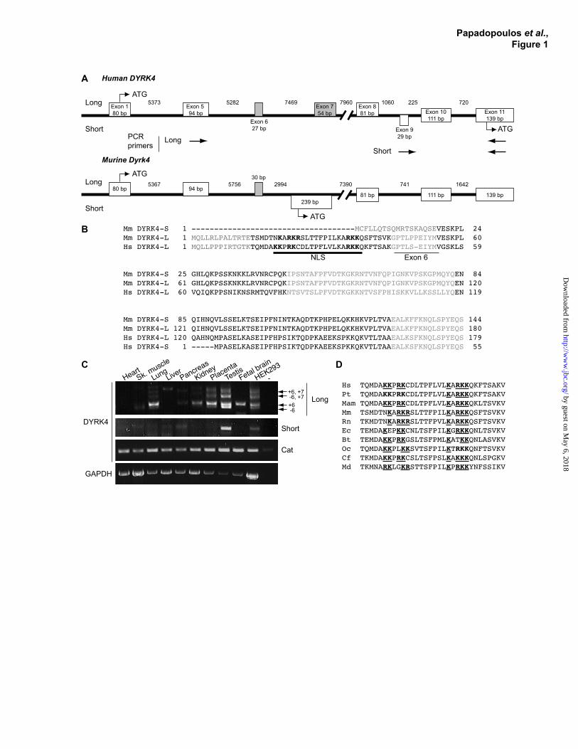
to Accession Number [Acc. Nº] NM_207210), while the other (Acc. Nº AK077117) has a different 5’-terminal sequence and encodes a protein of 632 amino acids. Similarly, two different human DYRK4 protein products can be predicted: a 520 amino acid protein described previously (hDYRK4520, translated from Acc. Nº NM_003845; ref. 23), and a 635 amino acid protein (hDYRK4635, translated from Acc. Nº AK308260: Supplemental Fig. S1B). The existence of human DYRK4 transcripts with different 5’-ends was confirmed by RT-PCR and sequencing of the PCR products (Fig. 1C). This analysis identified an alternative splicing event that involved the inclusion of an extra exon (exon 6 in humans; exon 3 in mice), which leads to a 9/10 amino acid inclusion in the human/murine DYRK4 proteins (Fig. 1B and C), and would give rise to a protein isoform of 644/642 amino acids (hDYRK4644 and mDYRK4642).
The N-terminal region of the long murine and human DYRK4 isoforms is encoded by orthologous exons (Fig. 1A and B, and Supplemental Fig. S1 and 2). In contrast, the short isoform of mouse Dyrk4 starts with a 5’-exon that is not conserved in human DYRK4 (Fig. 1A). We suggest that the longer isoform could be considered as the reference sequence for DYRK4, because the additional N-terminal sequence is conserved in mammals (Fig. 1D). By contrast, the corresponding first exon of the short variants appears to be species-specific, at least in human and mouse.
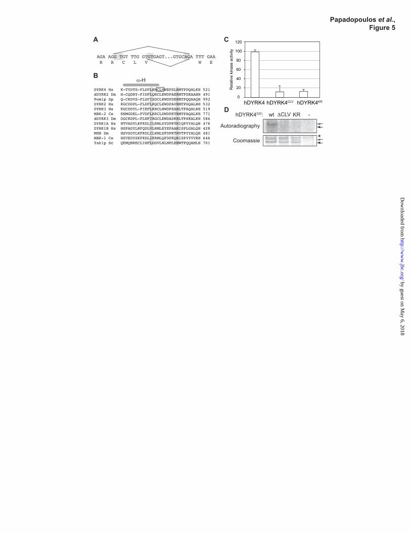
The analysis of several other human DYRK4 transcripts obtained by RT-PCR, by sequencing of IMAGE clones or collected from databases, revealed further alternative splicing events. In the first place, exclusion of exon 18 leads to a frame-shift and the generation of a protein with a truncated kinase domain, which would be predicted to lack activity (Supplemental Fig. S3B and S4C-E). Indeed, although the number of clones examined was small, exclusion of exon 18 appears to be more often represented in libraries from tumor tissue (Supplemental Table S2). In addition, the use of an alternative splice donor site in exon 19 leads to the inclusion/exclusion of three amino acids (CLV) in the kinase subdomain XI (Fig. 5A, Supplemental Fig. S2 and Table S2), an event also observed in murine Dyrk4 (present in AK077117, absent in NM_207210). Finally, an
by guest on May 6, 2018
http://ww
w.jbc.org/
Dow
nloaded from

5
alternative acceptor site in exon 21 is responsible for the inclusion/exclusion of an alanine residue in the C-terminal region (Supplemental Fig. S2 and Table S2), probably representing an example of wobble splicing (27). In summary, both human and murine DYRK4 genes express several transcripts as a result of alternative splicing, with the two main protein products differing in their amino-terminal domain.
Differential expression of human DYRK4 variants in human tissues. When we analyzed the expression of DYRK4 in different human tissues by RT-PCR using oligonucleotides matching exons within the catalytic domain, we detected the expression of DYRK4 mRNA in all the tissues analyzed (Fig. 1C). By contrast, Dyrk4 is predominately expressed in the testis of the mouse and rat (4,23). The broader tissue distribution of human DYRK4 is also evident through the sources of the ESTs listed in the Unigene database (http://www.ncbi.nlm.nih.gov/unigene), where 9 of the 10 murine ESTs but only 30 of 150 human ESTs are from testis. Moreover, DYRK4 mRNA is detected in human cell lines not derived from testis, such as A549 or HEK-293 (Supplemental Fig. S4A-B), NTERA-2 (28), SH-SY5Y (29) or fibroblasts (30).
PCR reactions with primer pairs specific for the transcripts encoding the long or the short N-terminal protein variants highlighted the differentially expression of the corresponding transcripts, with the long isoform present in several tissues and the short isoform predominantly expressed in testis (Fig. 1C). In addition to the bands resulting from the inclusion/exclusion of exon 6, the primers also produced longer PCR products in several tissues. Sequencing of these products showed that they are the result of the inclusion of exon 7 (Fig. 1C), which would lead to an in-frame premature stop codon (Supplemental Fig. S3A). Notably, the ratio between exon 6+/- or 7+/- isoforms was different when distinct tissues were compared (Fig. 1C), suggesting that these splicing events are tissue specific and subjected to regulation.
Alternative splicing alters the subcellular localization of DYRK4. Human DYRK4 (the 520 amino acid isoform) is located in the cytosol of COS-7 cells when expressed exogenously (23). We confirmed this distribution in HeLa cells using wild type human DYRK4520 and two kinase-
deficient mutants (the KR mutant where Lys133 in the ATP binding site is mutated to Arg, and the YF mutant where Tyr264 in the activation loop is mutated to Phe: Supplemental Fig. S5A).
The PSORT II algorithm (31) predicts the presence of a putative bipartite NLS within the extended N-terminal region, which is conserved in the human and murine long protein variants of DYRK4 (Fig. 1B). Therefore, we wondered whether the distinct protein isoforms differed in their subcellular distribution. To test this hypothesis, we analyzed the ability of the extended N-terminal region of human and mouse DYRK4 (amino acids 1-100 from hDYRK4644; amino acids 1-64 from mDYRK4642) to translocate a cytosolic protein to the nucleus. We thus generated fusion proteins of the small DYRK4 protein fragments with a chimeric GST-GFP protein, whose size would not allow the proteins to freely diffuse to the nucleus. Whereas GST-GFP was preferentially located in the cytoplasm, the extended DYRK4 N-termini drove the accumulation of the chimeric protein in the nucleus, suggesting that active transport is occurring (Fig. 2A).
Classic NLSs mediate the nuclear import of proteins through binding to a complex formed by importin α and β1. Importin α is the element within the complex that recognizes the NLSs, both monopartite or bipartite, in the cargo protein (32). To further test the ability of the N-terminus of DYRK4 to act as an NLS, we analyzed the binding of this region of the protein to importin α. Thus, lysates of HEK-293T cells expressing the N-terminus of hDYRK4644 or mDYRK4642 fused to GFP were incubated with different bacterially expressed importin α proteins fused to GST, and the protein complexes were recovered with glutathione-Sepharose beads. An interaction with importin α3 and α5 was detected, indicating indeed the presence of a sequence in the DYRK4 N-termini able to bind importins (Fig. 2B). The lack of binding to importin α1 might reflect the cargo specificity of different importin α isoforms (32). Thus, the localization assay and the importin α pull-down experiments indicated that the first 100/64 amino acids of hDYRK4644/mDYRK4642 contain a bona fide NLS. In fact, the corresponding full-length human and mouse DYRK4 proteins fused to GFP, which include the putative NLS, were localized to the nucleus of SH-
by guest on May 6, 2018
http://ww
w.jbc.org/
Dow
nloaded from

6
SY5Y cells to a significant extent, whereas GFP-hDYRK4520 or GFP-mDYRK4594, which do not have the N-terminal extension, were mainly detected in the cytosol (Fig. 2C). However, both isoforms (long and short, in human and mouse) showed similar behavior when overexpressed in HeLa cells (Fig. 2D). Together, these results confirm that different DYRK4 isoforms display distinct subcellular localizations, although this might be cell type dependent. Moreover, these data further suggest that the nuclear accumulation of full-length DYRK4 driven by the NLS is partially prevented by a mechanism that remains to be identified.
For many proteins, their distribution between the cytoplasm and the nucleus is based on a dynamic equilibrium of nuclear import and nuclear export, with contributions from protein retention in any of these subcellular compartments. We reasoned that the preferential cytoplasmic localization of DYRK4 isoforms could result from a strong nuclear export signal (NES)-dependent and CRM1 (exportin-1)-mediated nuclear export that might mask the effect of the NLS in the N-terminal extensions of the long DYRK4 isoforms. To determine the contribution of CRM1-mediated nuclear export to the cytosolic accumulation of DYRK4, we exposed cells to the inhibitor of CRM1, leptomycin B. No nuclear accumulation of either isoform was evident after 4 h in the presence of this inhibitor (Fig. 3A).
The DYRK4 isoforms showed a preferential cytoplasmic localization; however, low amounts of the proteins were also detectable in the nucleus by confocal microscopy (clearly visible after the cytoplasm was bleached; see Fig. 3B and Supplemental Fig. S5C). To answer the question whether the observed subcellular distribution is based on a dynamic equilibrium of constant nuclear export and nuclear import, we analyzed the dynamics of DYRK4 shuttling in single living cells using the fluorescence loss in photobleaching (FLIP) technique (33). A defined region of interest (ROI) in the cytoplasm was subjected to repeated photobleaching to wipe out the cytoplasmic GFP-fluorescence (Fig. 3B, time point 0). Subsequent reduction in GFP-fluorescence in the nucleus reflects the nucleocytoplasmic shuttling of bleached protein into the nucleus and non-bleached protein out of the nucleus. The diffusible character of unfused GFP was reflected in its fast
nucleocytoplasmic shuttling, which reached equilibrium after 7 min (Fig. 3B and C and Supplemental Fig. S5C-D). The two isoforms of mDYRK4 displayed distinct nucleocytoplasmic shuttling, with the nucleocytoplasmic exchange of the shorter isoform (mDYRK4594) converging to equilibrium and shuttling more slowly (see graph in Fig. 3C, middle panel, and Supplemental Video 1 and 2). Treatment with leptomycin B had no effect on mDYRK4594 nucleocytoplasmic shuttling, further supporting that nuclear export was independent of CRM1 (Supplemental Fig. S5B). By contrast, cytoplasmic bleaching of the longer isoform (mDYRK4642) did not significantly decrease GFP-fluorescence in the nucleus, indicating that this isoform did not exit the nucleus. Similar results were obtained when the corresponding human isoforms (hDYRK4520 and hDYRK4644) were assayed (Supplemental Fig. S5C-D). The static nature of the long DYRK4 isoform could indicate that it is retained in the nucleus by strong interactions with nuclear proteins or chromatin. Taken together, these results indicate that the alternatively spliced N-terminal region, common to human and mouse, confers distinct cellular properties to DYRK4.
Autophosphorylation of DYRK4 in the activation loop is required for kinase activity. All members of the DYRK family studied so far are autophosphorylated at a conserved tyrosine motif in the activation loop (YXY), within the catalytic domain (10). Indeed, phosphorylation of the second tyrosine residue is required for the complete activation of DYRK protein kinases (11,34). To test whether this is also the case for DYRK4, we generated a mutant version of human DYRK4520 in which the second tyrosine of the YXY motif (Tyr264) was mutated to phenylalanine (DYRK4YF). While immunoblotting of DYRK4 overexpressed in HEK-293 cells revealed that DYRK4 is indeed tyrosine phosphorylated (Fig. 4A), in neither the kinase-deficient mutant DYRK4KR nor the DYRK4YF mutant were phosphotyrosines detected with a phosphotyrosine-specific antibody (Fig. 4A). Mass spectrometry analysis confirmed that Tyr264 in the activation loop was phosphorylated (Fig. 4B), yet no differences in tyrosine phosphorylation were evident when the long and the short human DYRK4 isoforms were compared (Fig. 4C). Moreover, the DYRK4YF mutant was neither
by guest on May 6, 2018
http://ww
w.jbc.org/
Dow
nloaded from

7
autophosphorylated nor displayed kinase activity on a synthetic substrate, the exogenous peptide substrate DYRKtide (designed on the basis of the phosphorylation consensus sequence for DYRK1A (ref. 12: Fig. 4D and E). Together, these results show that DYRK4 is autophosphorylated on tyrosine residues and that the phosphorylation of the second tyrosine in the YXY motif is necessary for its kinase activity.
DYRK4 expressed in mammalian cells migrates as a doublet in SDS gels (Fig. 4A). The appearance of the low electrophoretic mobility band does not depend on autophosphorylation, because the band is also present in the two kinase deficient mutants (Fig. 4A, D and F). Nevertheless, treatment of DYRK4 proteins with phosphatase led to the disappearance of the slower migrating band (Fig. 4F). Mass spectrometry analysis of hDYRK4520 expressed in mammalian cells, both the wild-type and a kinase inactive version, identified several peptides with phosphorylated residues (Supplemental Fig. S6), indicating that DYRK4 is phosphorylated by cellular kinases.
The use of an alternative splicing donor site in exon 19 alters DYRK4 activity. The alternative use of different 5’-exons does not affect the catalytic domain of DYRK4, and accordingly both the long and short isoforms are catalytically active protein kinases (Supplemental Fig. S7A). In contrast, we found differences in the kinase activity of variants derived from another splicing event affecting both human and mouse DYRK4 that involved the use of an alternative splicing donor site in exon 19 (Fig. 5A). This event leads to the deletion/inclusion of three amino acids, CLV, at the end of the α-helix H, immediately prior to the conserved arginine residue in subdomain XI of the kinase domain (Fig. 5B). To assess the impact of this small deletion on DYRK4 enzymatic activity, we compared the in vitro behavior of hDYRK4520 wild-type and the corresponding mutant hDYRK4∆CLV in kinase assays. The autophosphorylation activity of the CLV-deleted protein was greatly impaired, as was its ability to phosphorylate an exogenous peptide (Fig. 5C and D). The central leucine residue within the CLV triplet corresponds to one of the α-helix H hydrophobic residues involved in anchoring to α-helix F (35). The interaction between these two helices is thought to play an important role in
forming the substrate-binding structure (35) and altering this interaction may induce changes in catalytic activity. Therefore, our results indicate that an alternative splicing event influences the catalytic properties of the resulting DYRK4 protein isoform.
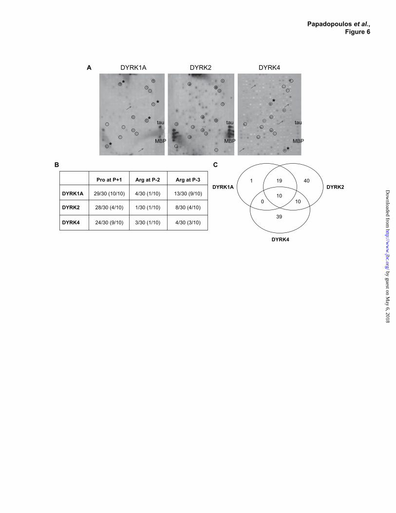
Substrate specificity of DYRK4. When characterizing the catalytic activity of DYRK4 we found that the phosphorylation of DYRKtide by DYRK4 was much less efficient than that by DYRK1A (Supplemental Fig. S7B-D). Hence, DYRKtide may not be an optimal substrate for DYRK4 and thus, we examined whether DYRK4 exhibited different substrate specificity than other members of the DYRK family. Accordingly, the phosphorylation specificity of DYRK4, or representative mammalian class I and class II DYRKs (DYRK1A and DYRK2, respectively), was studied in phosphosite arrays containing 720 peptides derived from annotated phosphorylation sites in human proteins (Fig. 6A). Reassuringly, two known phosphorylation sites of DYRK1A, Thr212 in tau and Ser640 in glycogen synthase (13,14) were considered to be among the best peptide substrates of DYRK1A in these arrays (Supplemental Table S4). In a test for reproducibility, classification of 27 out of 720 peptides differed between two independent experiments with different batches of arrays, whereas only 9 of 720 peptides were differently classified in parallel reactions with arrays from the same batch (Supplemental Fig. S8A and B). The experiment presented in Fig. 6 was done with arrays from the same batch.
An analysis of the top 30 peptide substrates for DYRK4 revealed that 24 contained a serine or threonine residue followed by a proline (Fig. 6B and Supplemental Table S4), indicating that DYRK4, like DYRK1A, can be classified as a proline-directed kinase (12). However, the requirement for the proline appeared to be less stringent for DYRK4 than for DYRK1A. An arginine residue at position P-3 or P-2 was previously shown to be important for optimal substrate recognition by DYRK1A and DYRK2 (12,15). Arginines at P-2 or P-3 were present in 17 of the 30 best substrates of DYRK1A, but were much less represented in the DYRK4 and DYRK2 datasets (7 or 9 out of 30; Fig. 6B and Supplemental Table S4). Interestingly, only one third of the peptides phosphorylated by DYRK4
by guest on May 6, 2018
http://ww
w.jbc.org/
Dow
nloaded from
8
were also substrates of DYRK1A or DYRK2 (50% of the top 30: Fig. 6C). Moreover, 10 peptides were phosphorylated by both DYRK4 and DYRK2 but not by DYRK1A, whereas no substrate was recognized by DYRK4 and DYRK1A but not by DYRK2 (Fig. 6C).
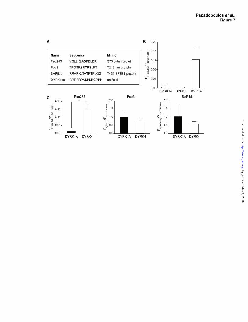
To confirm that the substrate specificity of DYRK4 differed from that of DYRK1A, we selected a peptide specifically recognized by DYRK4 for further analysis. Peptide 285 mimics Ser73 in c-Jun (Fig. 7A) and it was chosen as the top scoring DYRK4 substrate whose phosphorylation by DYRK1A or DYRK2 was sub-threshold (Supplemental Table S4). For comparison, DYRKtide was used as non-specific peptide substrate for all kinases of the DYRK family (12). Phosphorylation assays confirmed that Pep285 was phosphorylated by DYRK4 but not by DYRK1A and DYRK2 (Fig. 7B), supporting the proposal that DYRK4 can phosphorylate target sequences not recognized by DYRK1A or DYRK2. These experiments were performed with recombinant kinases expressed in E. coli (DYRK1A, DYRK2) or in insect cells (DYRK4) and thus, we also assayed the activity of DYRK4 and DYRK1A isolated from HeLa cells on Pep285 and two other DYRK substrates, Pep3 and SAPtide (Fig. 7A). These assays showed that Pep285 was the only peptide phosphorylated distinctly by DYRK1A and DYRK4 (Fig. 7C and Supplemental Fig. S8C).
DISCUSSION
The DYRK family of protein kinases is comprised of 5 members in mammals, of which DYRK1A, DYRK1B, DYRK2 and DYRK3 have been studied extensively due to their roles in cell differentiation and survival, and their critical participation in processes such as neurogenesis or cancer (2). By contrast, very little is known about the fifth member of the family, DYRK4. Our characterization of mammalian DYRK4 highlights several aspects that are common to DYRK kinases, such as the mode of activation or the subcellular distribution as a regulatory mechanism. However, other features such as substrate specificity were also characterized that could be important for establishing functional differences between DYRK family members.
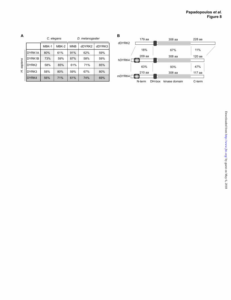
DYRK4 gene expression and regulation by alternative splicing. Several distinct transcripts of human and mouse DYRK4 are expressed, each encoding distinct protein products, due to alternative promoter use and first exon choice, as evident for other mammalian DYRK genes (2). By contrast to the restricted expression in the testis of rodents (4,23), human DYRK4 shows a broad pattern of expression in various tissues that is suggestive of a functional divergence between mouse and humans, and which severely limits the value of mouse models for the functional analysis of human DYRK4. The proposal that DYRK4 has acquired different functions during evolution is also supported by the unusually weak sequence conservation between human and mouse DYRK4 when compared with other orthologous DYRK pairs (Fig. 8).
The existence of distinct promoters probably underlies the differential expression pattern of the two DYRK4 5’ variants in human tissues. Surprisingly, the transcript for the short N-terminal DYRK4 isoform appears to be limited to the testis in humans, although the corresponding 5`-exon is not conserved in the mouse. Further variants are generated by distinct alternative splicing events, some of which lead to small in-frame deletions/insertions while others give rise to aberrant protein products. In particular, the exclusion of exon 18 is a frequent event, documented in about one third of the ESTs in the database (Supplemental Table S2), and it would give rise to a truncated protein that lacks the C-terminal end of the kinase domain and that is therefore predicted to be catalytically inactive. This situation resembles the splicing of DYRK1B that affects a region between subdomains X and XI, and that renders a kinase-deficient isoform (36). Kinase independent activities of several DYRK proteins have already been reported, suggestive of a scaffolding role for these proteins rather than a catalytic one (37-40). Hence, we cannot rule out that DYRK4 proteins lacking catalytic activity, such as the truncated protein resulting from exon 18 skipping or the small CLV deletion affecting α-helix H, may be biologically active.
The subcellular localization of DYRK4 could represent a regulatory mechanism. The subcellular localization of proteins larger than 45 kDa (the exclusion size of the nuclear pore), such as
by guest on May 6, 2018
http://ww
w.jbc.org/
Dow
nloaded from
9
DYRK4, depends on several mechanisms including active nuclear import, active nuclear export, and nuclear or cytosolic retention. Our results show that all these mechanisms could act on DYRK4 and they might indeed contribute to the regulation of the biological activity of DYRK4 by altering the possibility of finding substrates or modulators in particular subcellular compartments.
The short isoform of human DYRK4 was characterized and GFP-DYRK4520 was localized in the cytosol of COS-7 cells (23). We demonstrate that the newly identified long DYRK4 isoform (both in human and mouse) has a different subcellular localization and that the extended N-terminal region, which is highly conserved, accounts for this difference (Fig. 8B). The N-terminal extension harbors a classic NLS based on the positive binding to importin α, and on the nuclear import of a heterologous protein when fused to it. In addition, the extended N-terminal region mediates the nuclear retention of the long isoform (hDYRK4644 or mDYRK4642), as suggested by the lack of nucleocytoplasmic shuttling in FLIP experiments. Given that detectable amounts of hDYRK4520 and mDYRK4594, which lack the conserved N-terminal NLS, could be detected in the nucleus, it is possible that a second NLS could exist in DYRK4, such as that described between the kinase subdomains X and XI in DYRK1A (41).
DYRK4 long isoform was only partially segregated in the nucleus suggesting that full nuclear accumulation was prevented by mechanisms such as strong, CRM1-independent nuclear export, NLS masking or an interaction with cytosolic proteins. Nuclear translocation in response to extracellular cues has been demonstrated for DYRK2 or yeast Yak1p (16,19), which could be a common feature of this family of kinases. Further research should help uncover the signals that modulate the subcellular localization of DYRK4. Finally, and given that NLSs have been identified in DYRK4 (this study) and DYRK2 (19), and that DYRK3 is also at least partially localized in the nucleus (22,42), the distinction of “cytosolic” class II DYRKs from the “nuclear” class I DYRKs must be regarded as an oversimplification of the complex behavior of these kinases.
DYRK4 catalytic activity and substrate specificity. Like other DYRK kinases (9,43-45),
our mutational analysis indicates that phosphorylation of the second tyrosine residue within the activation loop (the YXY motif) appears to be necessary for the full activity of DYRK4. The fact that bacterially expressed GST-DYRK4 was catalytically active and that the MS analysis found that wild type, but not a kinase-deficient mutant of DYRK4, was phosphorylated in the second tyrosine of the activation loop is an indication that DYRK4 autophosphorylates the activation loop tyrosine. These findings fit with the current model for the activation of DYRK kinases (11), although it remains to be established whether DYRK4 behaves as a bona fide class II DYRK in terms of the contribution of the NAPA-domain to the activation process (7). The inhibitory effect of manganese cations on the activity of class II Drosophila dDYRK2 (the fly orthologue of DYRK4; Fig. 8A) is noteworthy (9), as also observed in human DYRK4 (Supplemental Fig. S7D), suggesting that the evolutionary relationship is translated into functional features.
We identify phosphorylated residues outside the catalytic domain in both wild type and a kinase-inactive DYRK4, which indicates that DYRK4 is phosphorylated by upstream kinases. Hence, DYRK4 could be modulated by phosphorylation, although we still do not know the processes that are affected by such regulation. Regulatory phosphorylation of other DYRK kinases has been reported recently, as in the case of MBK-2 by CKI or DYRK2 by MAP3K2 and ATM (17,46,47).
The substrate specificity of DYRK kinases has been little explored, although DYRK1A, DYRK2 and DYRK3 have previously been reported to differ in their specificities towards protein and peptide substrate (4,15). However, only a few substrates were compared in these studies since the experiments were directed towards systematically analyzing the importance of specific residues in a given peptide on substrate recognition (4,15). The experimental set-up employed here offers a broader view by using an array with peptides that reflect known human phosphorylation sites. The validity of the assay was not only supported by the identification of two known DYRK1A phosphorylation sites but also, by the fact that the majority of the peptides phosphorylated by DYRK1A match its consensus target sequence, with a proline at P+1 and an
by guest on May 6, 2018
http://ww
w.jbc.org/
Dow
nloaded from

10
arginine at P-3 (12). However, the relatively high number of DYRK substrates that do not contain an arginine residue at P-3 or P-2 contrasts with the almost absolute requirement for the arginine observed elsewhere (15), where DYRKs were considered as arginine-directed protein kinases. This difference is most likely due to the fact that in the previous study, the importance of the arginine was only tested in the context of one specific DYRK target site (Ser539 in eIF2Bε), whereas the phosphosite array contains a large selection of diverse peptides. One example of an arginine-independent target site of DYRK1A is Thr434 in SF3B1 (48), which is mimicked by the peptide SAPtide, a peptide that is phosphorylated with a similar efficiency as DYRKtide by both DYRK1A and DYRK4.
We found that DYRK1A, DYRK2 and DYRK4 phosphorylate distinct, though overlapping sets of the 720 different peptides in the phosphosite array. These results lead to the conclusion that DYRK2 and DYRK4 are much less biased towards peptides with an arginine in the P-2 or P-3 position
than DYRK1A, which is consistent with the identification of DYRK2 as a p53 kinase targeting Ser46 within the DDLMLSPDDI motif (19). The fact that DYRK4 shares several peptide substrates with DYRK2 that are not phosphorylated by DYRK1A may reflect the closer evolutionary relationship between these class II DYRK kinases. Moreover, and based on the results of the phosphosite array, we have identified a peptide (Pep285) that could be used as a specific substrate for DYRK4 in kinase assays.
As a final consideration, and given that human DYRKs appear to be co-expressed in different tissues, and that they could be localized in the same subcellular compartments, substrate specificity may represent a critical factor that governs biological specificity among this family of protein kinases. Further research will be necessary to positively identify the structural determinants that distinguish target site recognition by DYRK4 from that of the other members of the DYRK family.
REFERENCES
1. Park, J., Song, W. J., and Chung, K. C. (2009) Cell. Mol. Life Sci. 66, 3235-3240 2. Aranda, S., Laguna, A., and de la Luna, S. (2010) Faseb J., doi: 10.1096/fj.1010-165837 3. Becker, W., and Joost, H. G. (1999) Pro.g Nucleic Acid. Res. Mol. Biol. 62, 1-17 4. Becker, W., Weber, Y., Wetzel, K., Eirmbter, K., Tejedor, F. J., and Joost, H. G. (1998) J. Biol.
Chem. 273, 25893-25902 5. Raich, W. B., Moorman, C., Lacefield, C. O., Lehrer, J., Bartsch, D., Plasterk, R. H., Kandel, E.
R., and Hobert, O. (2003) Genetics 163, 571-580 6. Tejedor, F., Zhu, X. R., Kaltenbach, E., Ackermann, A., Baumann, A., Canal, I., Heisenberg, M.,
Fischbach, K. F., and Pongs, O. (1995) Neuron 14, 287-301 7. Kinstrie, R., Luebbering, N., Miranda-Saavedra, D., Sibbet, G., Han, J., Lochhead, P. A., and
Cleghon, V. (2010) Sci. Signal 3, ra16 8. Bahler, J., and Pringle, J. R. (1998) Genes Dev. 12, 1356-1370. 9. Lochhead, P. A., Sibbet, G., Kinstrie, R., Cleghon, T., Rylatt, M., Morrison, D. K., and Cleghon,
V. (2003) Biochem. J. 374, 381-391 10. Becker, W., and Sippl, W. (2010) FEBS J., doi: 10.1111/j.1742-4658.2010.07956.x 11. Lochhead, P. A., Sibbet, G., Morrice, N., and Cleghon, V. (2005) Cell 121, 925-936 12. Himpel, S., Tegge, W., Frank, R., Leder, S., Joost, H. G., and Becker, W. (2000) J. Biol. Chem.
275, 2431-2438 13. Skurat, A. V., and Dietrich, A. D. (2004) J. Biol. Chem. 279, 2490-2498 14. Woods, Y. L., Cohen, P., Becker, W., Jakes, R., Goedert, M., Wang, X., and Proud, C. G. (2001)
Biochem. J. 355, 609-615 15. Campbell, L. E., and Proud, C. G. (2002) FEBS Lett. 510, 31-36 16. Moriya, H., Shimizu-Yoshida, Y., Omori, A., Iwashita, S., Katoh, M., and Sakai, A. (2001)
Genes Dev. 15, 1217-1228 17. Cheng, K. C., Klancer, R., Singson, A., and Seydoux, G. (2009) Cell 139, 560-572
by guest on May 6, 2018
http://ww
w.jbc.org/
Dow
nloaded from
11
18. Mercer, S. E., Ewton, D. Z., Shah, S., Naqvi, A., and Friedman, E. (2006) Cancer Res. 66, 5143-5150
19. Taira, N., Nihira, K., Yamaguchi, T., Miki, Y., and Yoshida, K. (2007) Mol. Cell 25, 725-738 20. Zhang, D., Li, K., Erickson-Miller, C. L., Weiss, M., and Wojchowski, D. M. (2005) Genomics
85, 117-130 21. Geiger, J. N., Knudsen, G. T., Panek, L., Pandit, A. K., Yoder, M. D., Lord, K. A., Creasy, C. L.,
Burns, B. M., Gaines, P., Dillon, S. B., and Wojchowski, D. M. (2001) Blood 97, 901-910 22. Lord, K. A., Creasy, C. L., King, A. G., King, C., Burns, B. M., Lee, J. C., and Dillon, S. B.
(2000) Blood 95, 2838-2846. 23. Sacher, F., Moller, C., Bone, W., Gottwald, U., and Fritsch, M. (2007) Mol Cell Endocrinol. 267,
80-88 24. Alvarez, M., Altafaj, X., Aranda, S., and de la Luna, S. (2007) Mol. Biol. Cell 18, 1167-1178 25. de Graaf, K., Hekerman, P., Spelten, O., Herrmann, A., Packman, L. C., Bussow, K., Muller-
Newen, G., and Becker, W. (2004) J. Biol. Chem. 279, 4612-4624 26. Schutkowski, M., Reimer, U., Panse, S., Dong, L., Lizcano, J. M., Alessi, D. R., and Schneider-
Mergener, J. (2004) Angew Chem. Int. Ed. Engl. 43, 2671-2674 27. Hiller, M., and Platzer, M. (2008) Trends Genet. 24, 246-255 28. Leypoldt, F., Lewerenz, J., and Methner, A. (2001) J. Neurochem. 76, 806-814 29. Reimertz, C., Kogel, D., Rami, A., Chittenden, T., and Prehn, J. H. (2003) J. Cell Biol. 162, 587-
597 30. Kyng, K. J., May, A., Kolvraa, S., and Bohr, V. A. (2003) Proc. Natl. Acad. Sci. U S A 100,
12259-12264 31. Nakai, K., and Horton, P. (2007) Methods Mol. Biol. 390, 429-466 32. Lange, A., Mills, R. E., Lange, C. J., Stewart, M., Devine, S. E., and Corbett, A. H. (2007) J.
Biol. Chem. 282, 5101-5105 33. Lippincott-Schwartz, J., Snapp, E., and Kenworthy, A. (2001) Nat. Rev. Mol. Cell. Biol. 2, 444-
456 34. Himpel, S., Panzer, P., Eirmbter, K., Czajkowska, H., Sayed, M., Packman, L. C., Blundell, T.,
Kentrup, H., Grotzinger, J., Joost, H. G., and Becker, W. (2001) Biochem. J. 359, 497-505 35. Kornev, A. P., Taylor, S. S., and Ten Eyck, L. F. (2008) Proc. Natl. Acad. Sci. U S A 105, 14377-
14382 36. Leder, S., Czajkowska, H., Maenz, B., De Graaf, K., Barthel, A., Joost, H. G., and Becker, W.
(2003) Biochem. J. 372, 881-888 37. von Groote-Bidlingmaier, F., Schmoll, D., Orth, H. M., Joost, H. G., Becker, W., and Barthel, A.
(2003) Biochem. Biophys. Res. Commun. 300, 764-769 38. Sitz, J. H., Tigges, M., Baumgartel, K., Khaspekov, L. G., and Lutz, B. (2004) Mol. Cell. Biol.
24, 5821-5834 39. Kelly, P. A., and Rahmani, Z. (2005) Mol. Biol. Cell 16, 3562-3573 40. Maddika, S., and Chen, J. (2009) Nat. Cell Biol. 11, 409-419 41. Alvarez, M., Estivill, X., and de la Luna, S. (2003) J. Cell Sci. 116, 3099-3107 42. Guo, X., Williams, J. G., Schug, T. T., and Li, X. (2010) J. Biol. Chem. 285, 13223-13232 43. Kentrup, H., Becker, W., Heukelbach, J., Wilmes, A., Schurmann, A., Huppertz, C., Kainulainen,
H., and Joost, H. G. (1996) J. Biol. Chem. 271, 3488-3495 44. Kassis, S., Melhuish, T., Annan, R. S., Chen, S. L., Lee, J. C., Livi, G. P., and Creasy, C. L.
(2000) Biochem. J. 348, 263-272 45. Lee, K., Deng, X., and Friedman, E. (2000) Cancer Res. 60, 3631-3637 46. Varjosalo, M., Bjorklund, M., Cheng, F., Syvanen, H., Kivioja, T., Kilpinen, S., Sun, Z.,
Kallioniemi, O., Stunnenberg, H. G., He, W. W., Ojala, P., and Taipale, J. (2008) Cell 133, 537-548
47. Taira, N., Yamamoto, H., Yamaguchi, T., Miki, Y., and Yoshida, K. (2010) J. Biol. Chem. 285, 4909-4919
by guest on May 6, 2018
http://ww
w.jbc.org/
Dow
nloaded from
12
48. de Graaf, K., Czajkowska, H., Rottmann, S., Packman, L. C., Lilischkis, R., Luscher, B., and Becker, W. (2006) BMC Biochem. 7, 7
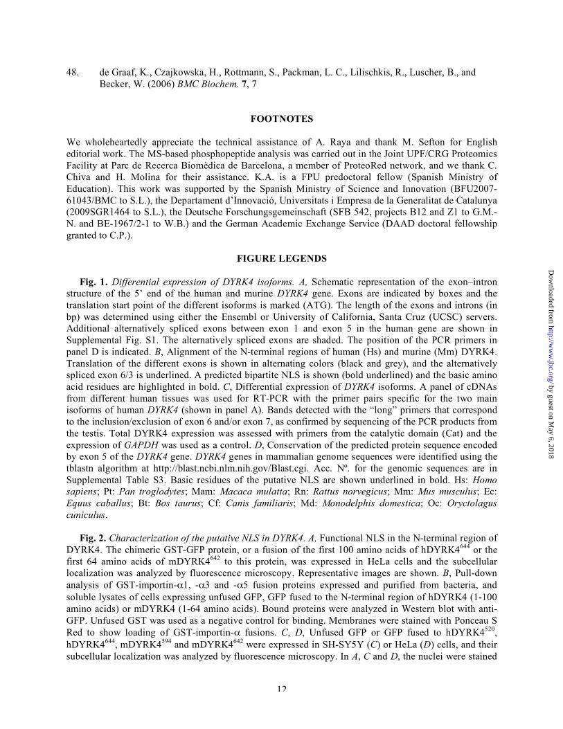
FOOTNOTES We wholeheartedly appreciate the technical assistance of A. Raya and thank M. Sefton for English editorial work. The MS-based phosphopeptide analysis was carried out in the Joint UPF/CRG Proteomics Facility at Parc de Recerca Biomèdica de Barcelona, a member of ProteoRed network, and we thank C. Chiva and H. Molina for their assistance. K.A. is a FPU predoctoral fellow (Spanish Ministry of Education). This work was supported by the Spanish Ministry of Science and Innovation (BFU2007-61043/BMC to S.L.), the Departament d’Innovació, Universitats i Empresa de la Generalitat de Catalunya (2009SGR1464 to S.L.), the Deutsche Forschungsgemeinschaft (SFB 542, projects B12 and Z1 to G.M.-N. and BE-1967/2-1 to W.B.) and the German Academic Exchange Service (DAAD doctoral fellowship granted to C.P.).
FIGURE LEGENDS
Fig. 1. Differential expression of DYRK4 isoforms. A, Schematic representation of the exon–intron structure of the 5’ end of the human and murine DYRK4 gene. Exons are indicated by boxes and the translation start point of the different isoforms is marked (ATG). The length of the exons and introns (in bp) was determined using either the Ensembl or University of California, Santa Cruz (UCSC) servers. Additional alternatively spliced exons between exon 1 and exon 5 in the human gene are shown in Supplemental Fig. S1. The alternatively spliced exons are shaded. The position of the PCR primers in panel D is indicated. B, Alignment of the N-terminal regions of human (Hs) and murine (Mm) DYRK4. Translation of the different exons is shown in alternating colors (black and grey), and the alternatively spliced exon 6/3 is underlined. A predicted bipartite NLS is shown (bold underlined) and the basic amino acid residues are highlighted in bold. C, Differential expression of DYRK4 isoforms. A panel of cDNAs from different human tissues was used for RT-PCR with the primer pairs specific for the two main isoforms of human DYRK4 (shown in panel A). Bands detected with the “long” primers that correspond to the inclusion/exclusion of exon 6 and/or exon 7, as confirmed by sequencing of the PCR products from the testis. Total DYRK4 expression was assessed with primers from the catalytic domain (Cat) and the expression of GAPDH was used as a control. D, Conservation of the predicted protein sequence encoded by exon 5 of the DYRK4 gene. DYRK4 genes in mammalian genome sequences were identified using the tblastn algorithm at http://blast.ncbi.nlm.nih.gov/Blast.cgi. Acc. Nº. for the genomic sequences are in Supplemental Table S3. Basic residues of the putative NLS are shown underlined in bold. Hs: Homo sapiens; Pt: Pan troglodytes; Mam: Macaca mulatta; Rn: Rattus norvegicus; Mm: Mus musculus; Ec: Equus caballus; Bt: Bos taurus; Cf: Canis familiaris; Md: Monodelphis domestica; Oc: Oryctolagus cuniculus.
Fig. 2. Characterization of the putative NLS in DYRK4. A, Functional NLS in the N-terminal region of DYRK4. The chimeric GST-GFP protein, or a fusion of the first 100 amino acids of hDYRK4644 or the first 64 amino acids of mDYRK4642 to this protein, was expressed in HeLa cells and the subcellular localization was analyzed by fluorescence microscopy. Representative images are shown. B, Pull-down analysis of GST-importin-α1, -α3 and -α5 fusion proteins expressed and purified from bacteria, and soluble lysates of cells expressing unfused GFP, GFP fused to the N-terminal region of hDYRK4 (1-100 amino acids) or mDYRK4 (1-64 amino acids). Bound proteins were analyzed in Western blot with anti-GFP. Unfused GST was used as a negative control for binding. Membranes were stained with Ponceau S Red to show loading of GST-importin-α fusions. C, D, Unfused GFP or GFP fused to hDYRK4520, hDYRK4644, mDYRK4594 and mDYRK4642 were expressed in SH-SY5Y (C) or HeLa (D) cells, and their subcellular localization was analyzed by fluorescence microscopy. In A, C and D, the nuclei were stained
by guest on May 6, 2018
http://ww
w.jbc.org/
Dow
nloaded from
13
with DAPI. Scale bars, 10 µm.
Fig. 3. Nucleocytoplasmic distribution of DYRK4. A, CRM1-independent distribution of GFP-mDYRK4. The subcellular localization of GFP-mDYRK4594 and GFP-mDYRK4642 expressed in COS-7 cells was analyzed before (untreated) and 4 h after addition of leptomycin B. STAT5A fused to YFP was used as a positive control responding to CRM1 inhibition. B, Differences in nucleocytoplasmic shuttling of GFP-DYRK4 isoforms. Cytoplasmic FLIP analysis was performed on GFP, GFP-mDYRK4594 or GFP-mDYRK4642 expressed in COS-7 cells. The panels show representative images in which the bleached area is indicated with a white circle and the nucleus with an arrow. C, Fluorescence was measured in the cytoplasmic and nuclear regions of interest along the time period represented. The diagrams show the averaged normalized fluorescence intensities of n=4 or 5 experiments as indicated. Scale bars, 10 µm.
Fig. 4. Phosphorylation of the YXY motif is required for DYRK4 kinase activity. A, DYRK4 is tyrosine phosphorylated. Extracts of HEK-293 cells expressing HA-hDYRK4520 wt, KR or YF mutants were immunoprecipitated with an anti-HA antibody, and analyzed in Western blot with anti-HA and anti-phosphotyrosine antibodies as indicated. B, MS/MS spectrum of the phosphopeptide VYTY(P)IQSR from wild type HA-hDYRK4520. C, Human DYRK4520 and DYRK4644 fused to GFP were analyzed by Western blot with anti-phosphotyrosine and anti-GFP antibodies. D, E, Phosphorylation of the YXY motif is required for DYRK4 kinase activity. Cell extracts expressing HA-hDYRK4520 wt or the mutant HA-hDYRK4KR or HA-hDYRK4YF versions were immunoprecipitated with anti-HA and the immunocomplexes were subjected to an IVK assay using DYRKtide as the exogenous substrate (E). DYRK4 autophosphorylation was assessed by autoradiography of dried gels (D). The bands corresponding to DYRK4 are indicated with arrows. The graph in E represents the means ± SD of two independent experiments performed in triplicate. Equal protein loading was confirmed by anti-HA probing of Western blots. F, DYRK4 is phosphorylated by cellular kinases. Immunoprecipitates of HA-hDYRK4520 or its kinase deficient derivatives were split in several aliquots that were incubated in the absence or presence of calf intestinal phosphatase (CIP). The hyper- and hypophosphorylated forms are indicated.
Fig. 5. Alternative splicing affects the catalytic properties of DYRK4. A, Alternative splicing affecting exon 19. Acceptor and donor sites are highlighted in grey. B, Amino acid comparison of the kinase subdomain XI of several DYRK family members, including alpha helix α-H. The Acc. Nº are provided in the Supplemental Table S3. The invariant arginine residue in this subdomain and the highly conserved leucine are highlighted in grey. The CLV deletion in DYRK4 is indicated. C, D, Cell extracts expressing HA-hDYRK4520 or the corresponding mutants HA-hDYRK4KR or HA-hDYRK4∆CLV were immunoprecipitated with anti-HA and the immunocomplexes were subjected to an IVK assay using DYRKtide (C). The graph represents the means ± SD of three independent experiments performed in triplicate (relative activity as percentage of DYRK4 wild type). DYRK4 autophosphorylation was assessed by autoradiography of dried gels (D). Equal amount of protein in the immunoprecipitated samples was confirmed by Coomassie staining of the gels and the bands corresponding to DYRK4 are indicated with arrows. The star shows a non-specific band.
Fig. 6. Identification of specific substrates for DYRK4. A, Specificity of DYRK4 (hDYRK4520), DYRK1AΔC and DYRK2 was determined using a phosphorylation of peptide arrays as indicated in the Experimental Procedures. Only a section of the array is shown. Peptides phosphorylated by all three kinases are circled. Peptides positive for either DYRK1A or DYRK4 (asterisks) but not for the other (arrows) are marked. Only peptides exceeding a threshold intensity of 20% of the strongest signal on the array were considered positive. Four spots contained control proteins (including tau and MBP) to define the position of the array on the autoradiograph. B, Congruence of the peptide substrates identified on the phosphosite array with the consensus target sequence for DYRK1A. The occurrence of the amino acids matching the DYRK1A consensus target site (Rx1-2S/TP) in the top 30 and the top 10 substrates (in
by guest on May 6, 2018
http://ww
w.jbc.org/
Dow
nloaded from

14
brackets) of DYRK1A, DYRK2 and DYRK4 is listed. Several peptides harbor more than one possible phosphorylation site, and one peptide (Pep269) contains arginines at both P-2 and P-3 (see Supplemental Table S4). C, Distribution of the peptides phosphorylated exclusively by only one DYRK, or by two or all three DYRKs are shown in the Venn diagram.
Fig. 7. Differential DYRK4 peptide phosphorylation. A, Amino acid sequence of the peptides used in this study. B, Pep285 is a specific substrate for DYRK4. Peptide phosphorylation by bacterially expressed GST-DYRK1AΔC or GST-DYRK2, and commercial GST-DYRK4 (hDYRK4520) expressed in Sf9 cells, was measured as the extent of phosphate incorporated into DYRKtide and Pep285. The histogram represents the ratio of Pep285 phosphorylation to DYRKtide phosphorylation by the indicated kinases after 30 min of the IVK reaction (means + SD from three experiments). C, Peptide selectivity of DYRK4. Anti-GFP immunocomplexes from HeLa cells expressing GFP-DYRK1A or GFP-hDYRK4520 were assessed in an IVK assay with different peptides as the substrates. Incorporation of 33P into the indicated peptides was normalized to the phosphate incorporation into DYRKtide. The histogram represents the means + SD from three independent experiments (*, p=0.0232; paired t test). Raw data corresponding to these experiments is shown in Supplemental Fig. S8C.
Fig. 8. Homology within the DYRK family members. A, Comparison of the members of the DYRK family from different species. The amino acid identity calculated by BLAST (blastp) is given as percentage. B, Detailed comparison of the non-catalytic N-terminal (N-term) and C-terminal (C-term), and kinase domain (including the DH-box) in human DYRK4 with those of Drosophila dDYRK2 and murine DYRK4 as the closest homologues. NLS, putative nuclear localization sequence. Sequence sources are listed in the supplemental Table S3.
by guest on May 6, 2018
http://ww
w.jbc.org/
Dow
nloaded from
Papadopoulos et al.,Figure 1
Hs TQMDAKKPRKCDLTPFLVLKARKKQKFTSAKVPt TQMDAKKPRKCDLTPFLVLKARKKQKFTSAKVMam TQMDAKKPRKCDLTPFLVLKARKKQKLTSVKVMm TSMDTNKARKRSLTTFPILKARKKQSFTSVKVRn TKMDTNKARKRSLTTFPVLKARKKQSFTSVKVEc TEMDAKEPKKCNLTSFPILKGRKKQNLTSVKVBt TEMDAKKPRKGSLTSFPMLKATKKQNLASVKVOc TQMDAKKPLKKSVTSFPILKTRKKQNFTSVKVCf TKMDAKKPRKCSLTSFPSLKAKKKQNLSPGKVMd TKMNARKLGKRSTTSFPILKPRKKYNFSSIKV
DCHeart
Sk. muscle
LungLiver
Pancreas
KidneyPlacenta
TestisFetal brain
HEK293-
Cat
Short
+6-6
Long+6, +7-6, +7
GAPDH
DYRK4
Mm DYRK4-S 1 ------------------------------------MCFLLQTSQMRTSKAQSEVESKPL 24Mm DYRK4-L 1 MQLLRLPALTRTETSMDTNKARKRSLTTFPILKARKKQSFTSVKGPTLPPEIYMVESKPL 60Hs DYRK4-L 1 MQLLPPPIRTGTKTQMDAKKPRKCDLTPFLVLKARKKQKFTSAKGPTLS-EIYMVGSKLS 59
Mm DYRK4-S 25 GHLQKPSSKNKKLRVNRCPQKIPSNTAFPFVDTKGKRNTVNFQPIGNKVPSKGPMQYQEN 84Mm DYRK4-L 61 GHLQKPSSKNKKLRVNRCPQKIPSNTAFPFVDTKGKRNTVNFQPIGNKVPSKGPMQYQEN 120Hs DYRK4-L 60 VQIQKPPSNIKNSRMTQVFHKNTSVTSLPFVDTKGKKNTVSFPHISKKVLLKSSLLYQEN 119
Mm DYRK4-S 85 QIHNQVLSSELKTSEIPFNINTKAQDTKPHPELQKKHKVPLTVAEALKFFKNQLSPYEQS 144Mm DYRK4-L 121 QIHNQVLSSELKTSEIPFNINTKAQDTKPHPELQKKHKVPLTVAEALKFFKNQLSPYEQS 180Hs DYRK4-L 120 QAHNQMPASELKASEIPFHPSIKTQDPKAEEKSPKKQKVTLTAAEALKSFKNQLSPYEQS 179Hs DYRK4-S 1 -----MPASELKASEIPFHPSIKTQDPKAEEKSPKKQKVTLTAAEALKSFKNQLSPYEQS 55
NLS
B
Exon 6
5373Exon 5 94 bp
5282
Exon 627 bp
7960Exon 881 bp
Exon 929 bp
1060 225Exon 10111 bp
720Exon 11139 bp
Long
Short
ATG
ATG
80 bp5367
94 bp5756
30 bp
239 bp
2994 7390
81 bp
Long
Short
ATG
ATG
Human DYRK4
Murine Dyrk4
111 bp 139 bp
741 1642
A
LongShort
PCRprimers
Exon 180 bp
Exon 754 bp
7469
by guest on May 6, 2018
http://ww
w.jbc.org/
Dow
nloaded from

DAPIGFPD
GFP
mD
YR
K459
4m
DY
RK
4642
hDY
RK
4520
hDY
RK
4644
HeLaCDAPIGFP
GFP
mD
YR
K459
4m
DY
RK
4642
hDY
RK
4520
hDY
RK
4644
SH-SY5Y
B
GFP
GFP-mD41-64
inputGST
GST-Impα1
GST-Impα3
GST-Impα5
GST
GST-Impα
WB
: α-G
FPP
once
au
95
72
55
43
34
26
GFP-hD41-100
DAPIGFPAGST-GFP
GST-GFP-hD41-100
GST-GFP-mD41-64
Papadopoulos et al.,Figure 2
by guest on May 6, 2018
http://ww
w.jbc.org/
Dow
nloaded from

Cyt. GFP-DYRK4 Nuc.GFP-DYRK4
fluor
esce
nce
time (min)0 1 2 3 4 5 6 7 8 9 10 11 12 13 14 15 16 17
0.0
0.2
0.4
0.6
0.8
1.0
1.2
n = 5FLIP GFP-mDYRK4642
FLIP GFP-mDYRK4594 n = 5
0.0
0.2
0.4
0.6
0.8
1.0
1.2
0 1 2 3 4 5 6 7 8 9 10 11 12 13 14 15 16 17time (min)
fluor
esce
nce
Cyt. GFP-DYRK4 Nuc.GFP-DYRK4
FLIP GFP
0.0
0.2
0.4
0.6
0.8
1.0
1.2
0 1 2 3 4 5 6 7 8 9 10 11 12 13 14 15 16 17time (min)
fluor
esce
nce
Cyt. GFP Nuc. GFP
n = 4
CB
GFP
-mD
4YR
K64
2G
FP-m
DY
RK
4594
GFP
pre-bleach post-bleach (0 min) post-bleach (12 min)
Papadopoulos et al.,Figure 3
A
lept
omyc
inB
STAT5A-YFP GFP-mDYRK4642GFP-mDYRK4594
untre
ated
by guest on May 6, 2018
http://ww
w.jbc.org/
Dow
nloaded from

HA-hDYRK4520:
WB: α-HA
wt KR YF wt KR YF
+CIP-CIPF
Hyper-P
Hypo-P
Dwt KR YF
HA-hDYRK4520
IP: α-HAWB: α-HA
IP: α-HAIVK
E
Rel
ativ
e ki
nase
act
ivity
(cpm
)
0
1.000
2.000
3.000
4.000
5.000
6.000
hDYRK4YFhDYRK4KRhDYRK4-
C
IP: α-GFPWB.α-pTyr
IP: α-GFPWB:α-GFP
hDYRK4520
hDYRK4644
-
520644
B
300 400 500 600 700 800 900 m/z0123456789
101112
847.5y6
720.3b5
607.2b4
935.4y6
390.2y3364.2
b3
263.1b2
235.2a2
262.1y2
848.4b6
746. 4y5
y6 y5 y2y3
a2 b3 b4 b7b6b5
- V Y T Y I Q S R -
Rel
ativ
e A
bund
ance
A
IP: α-HAWB. α-pTyr
wt KR YF
Papadopoulos et al.,Figure 4
IP: α-HAWB: α-HA
HA-hDYRK4520
by guest on May 6, 2018
http://ww
w.jbc.org/
Dow
nloaded from
Papadopoulos et al.,Figure 5
wt -KRΔCLV
Coomassie
Autoradiography
*
hDYRK4520:D
C
Rel
ativ
e ki
nase
act
ivity
0
20
40
60
80
100
120
hDYRK4 hDYRK4CLV hDYRK4KR
DYRK4 Hs K-TYDTS-FLDFLRRCLVWEPSLRMTPDQALKH 521dDYRK2 Dm H-CQDRY-FIDFLQRCLEWDPAERMTPDEAAHH 491Pom1p Sp Q-CKDVS-FLSFISDCLKWDPDERMTPQQAAQH 992DYRK2 Hs KGCDDPL-FLDFLKQCLEWDPAVRMTPGQALRH 532DYRK3 Hs KGCDDYL-FIEFLKRCLHWDPSARLTPAQALRH 519MBK-2 Ce KNMGDEL-FVDFLKRCLDWDPETRMTPAQALKH 771dDYRK3 Dm DGCKDPL-FLNFIRGCLEWDADKRLTPSEALKH 586DYRK1A Hs HTVADYLKFKDLILRMLDYDPKTRIQPYYALQH 476DYRK1B Hs HSPADYLRFQDLVLRMLEYEPAARISPLGALQH 428MNB Dm HSVSDYLKFKDLILRMLDFDPKTRVTPYYALQH 481MBK-1 Ce HSVEDYSKFKDLIKRMLQFDPKQRISPYYVVRH 646Yak1p Sc QEMQNRECLIHFLGGVLNLNPLERWTPQQAMLH 701
α-HB
AGA AGG TGT TTG GTGTGAGT...GTGCAGA TTT GAA R R C L V W E
A
by guest on May 6, 2018
http://ww
w.jbc.org/
Dow
nloaded from
Papadopoulos et al.,Figure 6
DYRK1A1
1010
19 40
0
39
DYRK4
DYRK2
CB
Pro at P+1 Arg at P-2 Arg at P-3
DYRK1A 29/30 (10/10) 4/30 (1/10) 13/30 (9/10)
DYRK2 28/30 (4/10) 1/30 (1/10) 8/30 (4/10)
DYRK4 24/30 (9/10) 3/30 (1/10) 4/30 (3/10)
DYRK1A
tau
MBP
*
*
*
*
*
*
tau tau
MBP MBP
A DYRK2 DYRK4
by guest on May 6, 2018
http://ww
w.jbc.org/
Dow
nloaded from
Papadopoulos et al.,Figure 7
C
0.00
0.05
0.10
0.15
0.20
0.0
0.5
1.0
1.5
2.0
0.0
0.5
1.0
1.5
2.0
DYRK1A DYRK1A DYRK1A
Pi(P
ep28
5)/P
i(DY
RK
tide)
Pi(P
ep3)
/Pi(D
YR
Ktid
e)
Pi(S
AP
tide)
/Pi(D
YR
Ktid
e)
DYRK4 DYRK4DYRK4
*Pep285 Pep3 SAPtide
0.00
0.04
0.08
0.12
0.16
0.20
Pi(P
ep28
5)/P
i(DY
RK
tide)
DYRK4DYRK2DYRK1A
Name
Pep285
Pep3
SAPtide
DYRKtide
Sequence
VGLLKLASPELER
TPGSRSRTPSLPT
RRARKLTATPTPLGG
RRRFRPASPLRGPPK
Mimic
S73 c-Jun protein
T212 tau protein
T434 SF3B1 protein
artificial
A B
by guest on May 6, 2018
http://ww
w.jbc.org/
Dow
nloaded from
H. s
apie
ns
C. elegans D. melanogaster
MBK-1 MBK-2 MNB dDYRK2 dDYRK3
DYRK3
DYRK4
DYRK2
DYRK1B
DYRK1A 80% 61% 91% 62% 59%
73% 59% 87% 58% 59%
58%
58%
56%
80% 80%
61%
61%71%
67%
69%
71%
59%
85% 85%
74%
A
dDYRK2
mDYRK4
hDYRK4
179 aa
209 aa
210 aa
228 aa
120 aa
117 aa
308 aa
308 aa
308 aa
kinase domain C-termN-term DH-box
18%
63%
11%67%
93% 47%
NLS
B
NLS
Papadopoulos et al.Figure 8
by guest on May 6, 2018
http://ww
w.jbc.org/
Dow
nloaded from

LunaSchutkowski, Nicolas Chatain, Gerhard Müller-Newen, Walter Becker and Susana de la Chrisovalantis Papadopoulos, Krisztina Arato, Eva Lilienthal, Johannes Zerweck, Mike
(DYRK4) differ in their subcellular localization and catalytic activitySplice variants of the dual-specificity tyrosine phosphorylation-regulated kinase 4
published online December 2, 2010J. Biol. Chem.
10.1074/jbc.M110.157909Access the most updated version of this article at doi:
Alerts:
When a correction for this article is posted•
When this article is cited•
to choose from all of JBC's e-mail alertsClick here
Supplemental material:
http://www.jbc.org/content/suppl/2010/12/02/M110.157909.DC1
by guest on May 6, 2018
http://ww
w.jbc.org/
Dow
nloaded from