specific fracture managementfac.ksu.edu.sa/sites/default/files/fractures.pdf · specific fracture...
TRANSCRIPT
Specific fracture management 211
Types of fracture 212
Immobilisation times 216
Pathological fractures 220
Trauma in pregnancy 230
Pediatric trauma 232
Fractures in the elderly 254
Falls from a height 258
Falls on the hand 260
Orthopaedic splints 262
Plaster of Paris 266
Skelecasts 270
Complications of trauma 278
Chapter 4
Specific fracturemanagement
212
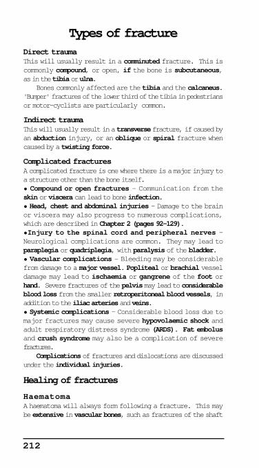
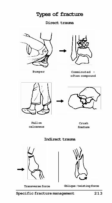
Types of fractureDirect traumaThis will usually result in a comminuted fracture. This iscommonly compound, or open, if the bone is subcutaneous,as in the tibia or ulna.
Bones commonly affected are the tibia and the calcaneus.'Bumper' fractures of the lower third of the tibia in pedestriansor motor-cyclists are particularly common.
Indirect traumaThis will usually result in a transverse fracture, if caused byan abduction injury, or an oblique or spiral fracture whencaused by a twisting force.
Complicated fracturesA complicated fracture is one where there is a major injury toa structure other than the bone itself.• Compound or open fractures — Communication from theskin or viscera can lead to bone infection.• Head, chest and abdominal injuries — Damage to the brainor viscera may also progress to numerous complications,which are described in Chapter 2 (pages 92-129).•Injury to the spinal cord and peripheral nerves —Neurological complications are common. They may lead toparaplegia or quadriplegia, with paralysis of the bladder.• Vascular complications — Bleeding may be considerablefrom damage to a major vessel. Popliteal or brachial vesseldamage may lead to ischaemia or gangrene of the foot orhand. Severe fractures of the pelvis may lead to considerableblood loss from the smaller retroperitoneal blood vessels, inaddition to the iliac arteries and veins.• Systemic complications — Considerable blood loss due tomajor fractures may cause severe hypovolaemic shock andadult respiratory distress syndrome (ARDS). Fat embolusand crush syndrome may also be a complication of severefractures.
Complications of fractures and dislocations are discussedunder the individual injuries.
Healing of fractures
HaematomaA haematoma will always form following a fracture. This maybe extensive in vascular bones, such as fractures of the shaft
Specific fracture management 213
Fall oncalcaneus
Types of fracture
Direct trauma
Bumper Comminuted —often compound
Crushfracture
Transverse force
Indirect trauma
Oblique: twisting force
Huckstep 1999©
Huckstep 1999©
Huckstep 1999©
Huckstep 1999©
!!!!!
!!!!!
!!!!!
214
of the femur and in major fractures of the pelvis. Thehaematoma is not normally visible on X-ray.
Granulation tissueGranulation tissue forms by organisation of the haematomawith ingrowth of osteoblasts and osteoclasts. Granulationtissue again is not usually seen on X-ray, except as a soft tissueshadow.
Callus formationCallus formation is due to calcification within the granulationtissue with laying down of cartilage across the bone ends. Thiswill show as a shadow on X-ray.
Bony consolidationBony consolidation with the formation of woven bone occursdue to ossification in the cartilage. This can be seen on X-ray.
RemodellingRemodelling of the fracture site occurs due to the stresses onthe fracture site by muscle pull and weight-bearing. This isdue to activity of the osteoblasts which lay down bone andosteoclasts which remove unstressed new bone. The wovenbone is then replaced with definitive bone.
Bone union in childrenBone union in children takes approximately half the time ofthat seen in adults. In babies and young children union ismuch more rapid.
Bone union and type of fractureUnion usually occurs earlier in oblique fractures than intransverse fractures. It is also more rapid in fractures with agood blood supply, such as the metacarpals, metatarsals andphalanges, than in the shaft of major long bone fractures, dueto less vascular supply to the fractured bone ends.
Delayed unionExcessive movement, or alternatively too rigid internalfixation can delay fracture union. Infection or pathologicalbone due to secondary neoplastic malignant deposits inbone, or diseased bone in Paget’s disease will also delay unionor lead to established non-union.
Specific fracture management 215
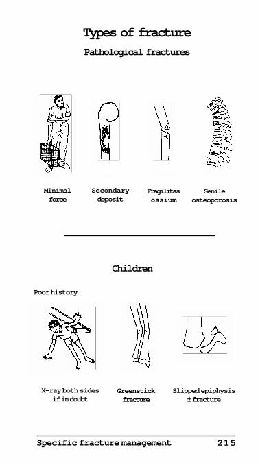
Children
Poor history
Types of fracture
Pathological fractures
Minimalforce
Secondarydeposit
Fragilitasossium
Senileosteoporosis
X-ray both sidesif in doubt
Greenstickfracture
Slipped epiphysis± fracture
Huckstep 1999©
Huckstep 1999©
Huckstep 1999©
Huckstep 1999©
Huckstep 1999©
Huckstep 1999©
Huckstep 1999©
216
Immobilisation timesUpper limbThe union or immobilisation times are illustrated. These arevery approximate and there are many exceptions. Somefractures, such as the shafts of metacarpals, require almostno immobilisation. Other fractures, such as the waist of thescaphoid or the shaft of the radius and ulna, require rigidsplinting or internal fixation.• Internal fixation — This is now the usual treatment of mostdisplaced fractures of the shaft of the radius and ulna and ofthe olecranon. Limited mobilisation of the limb is usuallypermitted after a few days.
Many pathological fractures of long bones, due tosecondary deposits, are now also treated by early internalfixation followed by radiotherapy, with or withoutchemotherapy or hormones.
Spine and pelvisIsolated minor fractures of the pelvis, sacrum and coccyxwithout complications require little or no immobilisation.Major fractures and dislocations of the cervical, thoracic andlumbar spine, often with instability, need protection for 2-3months.
The same applies to severe central dislocation of the hip,and unstable fractures and disruptions of the pelvis, whichmay require internal fixation.
Lower limbFractures requiring minimal immobilisation include isolatedfractures around the hip which do not affect stability, suchas isolated fractures of the greater and lesser trochanters, andfractures of the fibula, metatarsals and toes.
Major fractures of the hip, shaft and lower end of thefemur, and major fractures of the tibia, neck of the talus andthe calcaneus involving the subtalar joint, take 3 months toheal. Except for the calcaneus they often need internalfixation to obtain early mobility. Internal fixation is especiallyindicated in elderly patients with major fractures of the hip,avoiding the many complications associated with prolongedbed rest.
Specific fracture management 217
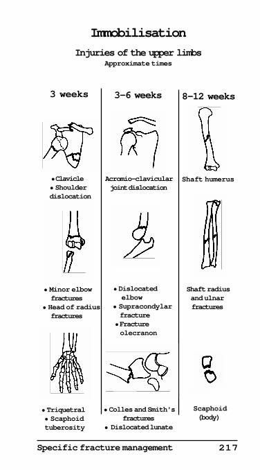
Immobilisation
Injuries of the upper limbsApproximate times
3 weeks
• Clavicle • Shoulder dislocation
• Colles and Smith'sfractures
• Dislocated lunate
3-6 weeks 8-12 weeks
• Minor elbowfractures
• Head of radiusfractures
Acromio-clavicularjoint dislocation
Shaft humerus
Shaft radiusand ulnarfractures
• Dislocatedelbow
• Supracondylar fracture• Fracture
olecranon
• Triquetral • Scaphoid tuberosity
Scaphoid(body)
Huckstep 1999©
Huckstep 1999©
Huckstep 1999©
218
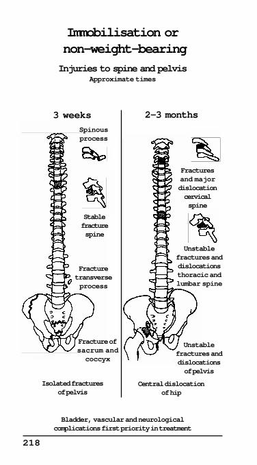
2-3 months
Immobilisation or non-weight-bearing
Injuries to spine and pelvisApproximate times
Fracture ofsacrum andcoccyx
Isolated fracturesof pelvis
Bladder, vascular and neurologicalcomplications first priority in treatment
Unstablefractures anddislocationsof pelvis
Central dislocationof hip
3 weeks
Unstablefractures anddislocationsthoracic andlumbar spine
Fracturesand majordislocationcervicalspine
Fracturetransverseprocess
Stablefracturespine
Spinousprocess
Huckstep 1999©
Huckstep 1999©
Specific fracture management 219
3 weeks or less
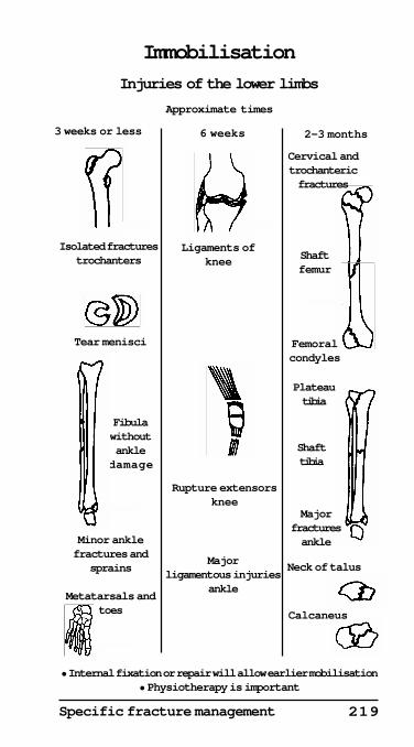
Immobilisation
Injuries of the lower limbs
Approximate times
Ligaments ofknee
Rupture extensorsknee
Majorligamentous injuries
ankle
6 weeks 2-3 months
Isolated fracturestrochanters Shaft
femur
Neck of talus
Calcaneus
Tear menisci
Fibulawithoutankle
damage
Minor anklefractures and
sprains
Metatarsals andtoes
• Internal fixation or repair will allow earlier mobilisation• Physiotherapy is important
Majorfracturesankle
Plateautibia
Shafttibia
Cervical andtrochantericfractures
Femoralcondyles
Huckstep 1999© Huckstep 1999©
Huckstep 1999©
Huckstep 1999©
Huckstep 1999©
220
Pathological fractures
Pathological fractures occur in bones that have been weakenedfor any reason.
Causes• Senile osteoporosis — This is the most common cause.• Secondary tumour deposits — These also commonly causepathological fractures, particularly from carcinoma of thebreast, lung, thyroid, kidney, prostate, cervix, bowel, andfrom multiple myeloma. The treatment of pathologicalfractures resulting from secondary deposits usually involvesinternal fixation plus radiotherapy. Hormones orchemotherapy may also be necessary.• Other causes — These vary from congenital bone cysts andfibrous dysplasia, through to Paget’s disease. They alsoinclude osteopenic bone in paralysis, such as in poliomyelitisand paraplegia.
HistoryThe history of a previous primary tumour, particularly of thebreast, lungs, kidneys and prostate, associated with one ormore fractures or potential fractures occurring with minimaltrauma, would make one strongly suspect a secondarydeposit.
In elderly patients, senile osteoporosis may lead tofractures of the spine, hip and wrist following fairly minorinjuries.
ExaminationExamination of the patient with a suspected pathologicalfracture must include a complete physical examination,including a rectal examination. Bones which may beparticularly susceptible to secondary deposits are the spine,ribs, pelvis, humeri and femora and, unless fractured, maycause little or no pain or tenderness. Secondary depositsbelow the knee or elbow are much less common.
Investigations
Blood investigationsA full haematological and biochemical pre-operativeassessment is essential, as anaemia is common and anuncorrected pre-operative hypercalcaemia a potentiallylethal post-operative complication.
Specific fracture management 221
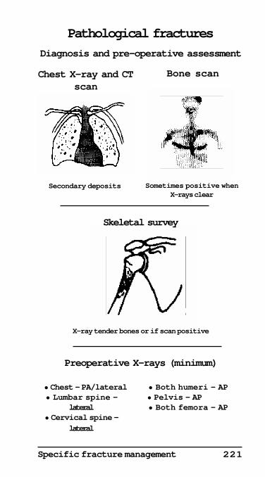
Pathological fractures
Diagnosis and pre-operative assessment
Bone scan
Sometimes positive whenX-rays clear
Skeletal survey
X-ray tender bones or if scan positive
Preoperative X-rays (minimum)
Chest X-ray and CTscan
Secondary deposits
• Chest — PA/lateral• Lumbar spine —
lateral• Cervical spine —
lateral
• Both humeri — AP• Pelvis — AP
• Both femora — AP
Huckstep 1999©
Huckstep 1999©
Huckstep 1999©
222
Imaging techniquesThe appearance on X-ray of an ill-defined, osteolyticdestructive lesion, which is often multiple and usuallyproximal to the knee and elbow, is strongly suggestive of asecondary deposit. Carcinoma of the prostate, however, isusually osteoblastic. Occasionally other secondaries such asfrom carcinoma of the breast, may also give osteoblasticsecondaries.
The minimum X-rays necessary when an operation isplanned for a secondary deposit, are:• PA and lateral view of the chest — This may also showsecondary deposits, both in the lungs and in the ribs.• AP of pelvis.• AP of both femora and humeri — These bones are ofteninvolved in secondary deposits. They may also have apotential pathological fracture which could be stabilised atthe time of initial operation.• Lateral view of the cervical spine — A secondary depositmay be asymptomatic. There is a risk of fracture ordislocation during anaesthetic intubation, which may causea paraplegia or quadriplegia.• Lateral view of lumbar and thoracic spine — The lateralview of the chest may adequately show the thoracic spine.• Lateral skull view — If multiple myeloma is suspected.• Other views — AP and lateral views of any bones whichare tender and which may require internal fixation.• Nuclear bone scan — A bone scan may be a usefuladditional investigation and may show secondary depositswhen the X-ray is negative. Conversely, in multiple myelomaand secondary deposits from the thyroid and kidney, thebone scan is sometimes ‘cold’ due to a large haematoma atthe site of the lesion, while the X-ray may demonstrate thelesion.• Thallium scan — Occasionally a thallium scan is indicated.This is more specific than technetium 99 for rapidly dividingcells in malignant tumours.• CT scan — In most cases of secondary deposits, these arenot necessary. A CT scan may be useful, however, indiagnosing secondaries in the chest and showing the extentof tumour spread up the medullary cavity of a bone.• MRI scan — This is a more accurate, but a much moreexpensive investigation for routine investigations and for mostsecondary tumours. It may be invaluable, however, inassessing the extent of primary tumour spread. It is notnecessary for most secondary deposits.
Specific fracture management 223
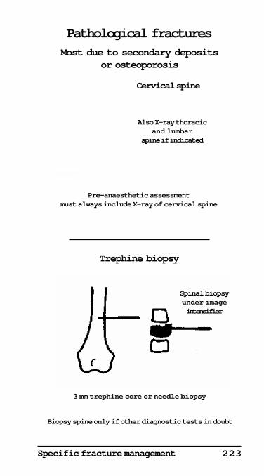
Pathological fracturesMost due to secondary deposits
or osteoporosis
Cervical spine
Pre-anaesthetic assessmentmust always include X-ray of cervical spine
Trephine biopsy
Biopsy spine only if other diagnostic tests in doubt
3 mm trephine core or needle biopsy
Spinal biopsyunder imageintensifier
Also X-ray thoracicand lumbar
spine if indicated
Huckstep 1999©
Huckstep 1999©
224
Trephine biopsyA trephine biopsy is sometimes necessary where the diagnosisis uncertain. A simple needle biopsy may sometimes besufficient for soft tissue. A trephine will produce a bony coreof about 10 - 30 mm in length and 2 - 3 mm in diameter, suitablefor a histological diagnosis. Only occasionally will an openbiopsy be necessary. The biopsy specimen may, however, takeseveral days to decalcify and a definitive diagnosis canseldom be made by frozen section alone.
Pathological fractures — upper limb
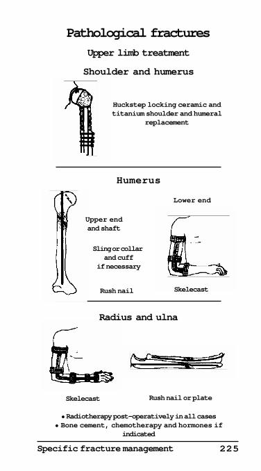
Treatment• Forearm and hand — Pathological fractures due tosecondary deposits below the elbow are uncommon. Theyshould be internally fixed if possible, or treated with askelecast, followed by radiotherapy. Chemotherapy orhormones should also be given if indicated.• Humerus — Fractures, or potential fractures of the shaft ofthe humerus, should be treated by internal fixation by thesimplest method possible, such as by a Rush nail.
Fractures of the lower end of the humerus are best treatedwith a simple skelecast, a plaster back slab or plastic support,followed by radiotherapy.
Fractures of the head and neck of the humerus, due tosecondary deposits, can be treated by a simple triangular slingplus radiotherapy. A prosthetic shoulder and upper humeralreplacement may sometimes be indicated, and possiblychemotherapy or hormones.
Pathological fractures of the spine andpelvis
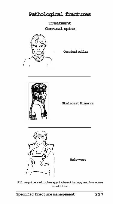
Treatment• Cervical spine — Fractures of the cervical spine should betreated either with a neck collar or, if unstable, by a halo jacketsupport, a skelecast or other type of neck splint. Sometimesinternal stabilisation by wire fixation is required.Radiotherapy should always be given, plus chemotherapyor hormones, if indicated.• Cervical collar — In stable cervical spines with secondarydeposits and no neurological signs, a simple cervical collar,as illustrated, is usually sufficient during the day, while a softcervical collar is worn at night. Radiotherapy, with or withouthormones and chemotherapy, must always be given in
Specific fracture management 225
Pathological fractures
Upper limb treatment
Skelecast Rush nail or plate
• Radiotherapy post-operatively in all cases• Bone cement, chemotherapy and hormones if
indicated
Rush nail
Upper endand shaft
Sling or collar and cuff
if necessary
Lower end
Skelecast
Radius and ulna
Shoulder and humerus
Huckstep locking ceramic andtitanium shoulder and humeral
replacement
Humerus
Huckstep 1999©
Huckstep 1999©
Huckstep 1999©
Huckstep 1999©
Huckstep 1999©
226
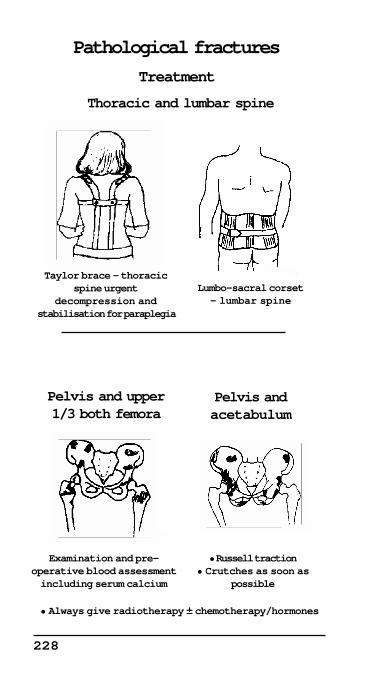
addition. In more severe cases a SOMI (suboccipital mentalimmobilising collar) will prevent rotation as well as flexion.• Minerva support — A skelecast or plaster support may benecessary for unstable fractures or potential fractures.• Halo-vest — A halo-vest support, as illustrated, incorporatesa halo attached to the outer table of the skull and is supportedin turn on to a lightweight vest. This will give additionalsupport for unstable fractures and will also allowradiotherapy. It will provide additional stability followingoperative stabilisation.• Thoracic and lumbar spine — Most fractures withoutneurological signs require either a Taylor brace for thethoracic spine, or a lumbo-sacral corset for the lumbar spine.
Fractures with neurological signs require emergencydecompression and stabilisation.• Pelvis and acetabulum — These are usually treatedconservatively with skin traction, non-weight-bearing andradiotherapy, plus hormones and chemotherapy if indicated.
Combined fractures of the neck of the femur and theacetabulum may require a cemented total hip replacement,if the patient is fit and has a life expectancy of at least 3-6months. It is also essential that any hypercalcaemia iscorrected pre-operatively, as this is a possible lethalcomplication following operations for secondarycarcinomatosis.
Pathological fractures of the lower limb
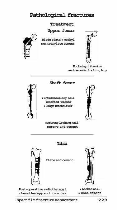
Treatment• Upper femur — Fractures of the neck or trochanteric regionof the femur require a blade plate strengthened with methylmethacrylate cement. Destruction of the head of the femurwill usually either require a cemented hemiarthroplasty, ortotal hip replacement if the patient is young with a reasonablelife span.• Shaft of femur — Fractures, or potential fractures of theshaft of the femur, should be treated by internal nailing witha locked nail, if possible. They may also require extrastabilisation with methyl methacrylate cement.• Prophylactic internal fixation — Potential pathologicalfractures should be internally stabilised.• Pathological fractures of the tibia — These are uncommonand usually will require internal fixation by nails or plates,plus methyl methacrylate cement.
It is essential that radiotherapy is given post-operativelyin all cases and, if indicated, chemotherapy and/or hormones.
Specific fracture management 227
Skelecast Minerva
Cervical collar
Pathological fractures
TreatmentCervical spine
All require radiotherapy ± chemotherapy and hormonesin addition
Halo-vest
Huckstep 1999©
Huckstep 1999©
Huckstep 1999©
228
Examination and pre-operative blood assessmentincluding serum calcium
• Russell traction• Crutches as soon as
possible
Pelvis andacetabulum
Lumbo-sacral corset— lumbar spine
Pelvis and upper1/3 both femora
• Always give radiotherapy ± chemotherapy/hormones
Taylor brace — thoracicspine urgent
decompression and stabilisation for paraplegia
Pathological fractures
Treatment
Thoracic and lumbar spine
Huckstep 1999©
Huckstep 1999©
Huckstep 1999©
Huckstep 1999©
Specific fracture management 229
Upper femur
Pathological fractures
Treatment
• Intramedullary nail inserted 'closed'• Image intensifier
Blade plate + methylmethacrylate cement
• Locked nail• Bone cement
Huckstep titaniumand ceramic locking hip
Shaft femur
Tibia
Post-operative radiotherapy ±chemotherapy and hormones
Plate and cement
Huckstep locking nail,screws and cement
Huckstep 1999©
Huckstep 1999©
Huckstep 1999©
Huckstep 1999©
Huckstep 1999©
Huckstep 1999©
230
Trauma in pregnancy
Overall treatment
Treatment will require modification, especially as fetalsurvival is dependent on maternal survival. An obstetricianshould be informed. The intravascular volume is increased,and gastric emptying is delayed in pregnancy. There may alsobe a respiratory alkalosis, and an increase in tidal volume.
Other changes include the risk of eclampsia.
Management of mother
This includes the ABCDE of trauma, with the exception thatonly the leg compartments of MAST suit should be inflatedfor severe lower limb trauma. X-rays of the abdomen andpelvis should be minimal and ultrasound used wherepossible. Peritoneal lavage, if indicated, should be performedby the supra-umbilical route. The risk of increasedretroperitoneal haemorrhage in pelvic fractures due to thedilated pelvic veins should be borne in mind.
Various other physiological changes occur in pregnancy.These include an increase of pulse rate of up to 90 per minute,and decrease in blood pressure of about 10 mmHg.
Fetus
The fetus is well protected by the thick uterine wall in thefirst trimester of pregnancy, and by the amniotic fluid in thesecond trimester. In the third trimester the uterus is thinwalled and placental disruption may occur, particularly withshearing forces.
Clinical assessment
Assessment will include the date of the last menstrual period,and assessment of the uterus for contractions, tenderness andheight. The fetal heart should be auscultated and fetalmovements felt for. Finally a catheter should be passed andthe urine assessed, and a vaginal examination carried out forbleeding or amniotic fluid. The normal fetal heart rate is 120-160 beats per minute and bradycardia is a sign of fetal distress.There can be delay in the onset of fetal distress, soobservations should be repeated, if necessary, over severaldays following the initial trauma.
Specific fracture management 231
Investigations
Ultrasound is a useful diagnostic test in later pregnancy toassess the volume of amniotic fluid, the presence of intra-amniotic haemorrhage, and also to assess the position of theplacenta. Doppler ultrasonography can be used to assess thefetal heart rate from about the third month of pregnancy.Later in pregnancy cardiotocography will compare fetal heartrate with uterine contractions. The Kleihauer test can be usedto assess feto-maternal haemorrhage and anaemia in thefetus. If this occurs in a Rhesus negative mother prophylacticanti-D is indicated to protect the fetus against Rhesussensitisation.
Signs of placental separation
Fetal distress may be the only sign of this complication. Othersigns are maternal hypovolaemic shock, abdominaltenderness, increasing height of the fundus, increasedirritability of the uterus, vaginal bleeding and amniotic fluidloss. In major separation there is a risk also of disseminatedintravascular coagulation.
Penetrating trauma
This is another cause of fetal death although the mother islikely to survive.
Burns in pregnancy
In burns affecting over 50%-60% of the body after the 5thmonth of pregnancy the fetus should be deliveredimmediately. This is due to the otherwise poor prognosis forboth mother and foetus.
Indications for an emergency Caesarian
In severe injuries there is a need for an immediate Caesariansection for a viable fetus in the event of maternal death fromwhatever cause.
232
Pediatric trauma
Emergency treatmentThe emergency treatment of the child with multiple or severeinjuries may be difficult for the inexperienced. The emergencyresuscitation of the injured child is summarised on pages 57-59.
In adults with multiple severe injuries adequateemergency treatment in the first, or ‘golden’ hour may makethe difference in survival.
With children this is only half an hour, the so called‘platinum’ half hour.
Drug dosages and fluid replacement in children shouldbe based on the weight of the child. In the severely injuredchild this is best achieved by measuring the length of the childand assessing approximate weight and drug dosages frompediatric charts, which should be available in all accidentcentres.
AirwayThe anatomy of the child is also different. Children have arelatively large head and small oropharynx, with the glottisat the level of C3 or C4 instead of C6, as in the adult. Thetrachea is also shorter and this may cause problems inintubation. A conscious child also has a well developed gagreflex. As a result an oral airway may cause vomiting andshould be avoided if possible.
DiagnosisChildren are not small adults, and their fractures anddislocations may be difficult to treat and also to diagnose. Thisis because much of the epiphysis in young children may bemade of cartilage which is radiolucent. Damage to theepiphysis may not be fully appreciated, especially in a crushinjury (Grade V Salter-Harris), and the X-ray may appear tobe virtually normal. If in doubt the opposite joint should beX-rayed in the same position or an ultrasound or arthrogramused to make a diagnosis.
Types of fracturesSpecific fractures usually seen only in children includefractures in fragilitas ossium (a history usually of multipleprevious fractures, and X-ray with clinical evidence of these),fractures through bone cysts, and fractures due to child abuse.Child abuse may account for many of the fractures under the
Specific fracture management 233
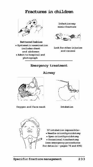
Fractures in children
If intubation impossible— • Needle cricothyroidotomy• Open cricothyroidotomy• Occasional tracheotomy(see emergency procedures
for details — pages 76 and 496)
Infection maymimic fracture
Emergency treatment
Airway
Oxygen and face mask Intubation
Battered babies
Look for other injuriesand causes
• Systematic examination includes chest and abdomen
• Admit to hospital andphotograph
Huckstep 1999©
Huckstep 1999©
Huckstep 1999©
Huckstep 1999©
Huckstep 1999©
234
age of 1 in some countries, and must always be considered inall fractures in babies, particularly of the long bones.
Investigations
Imaging techniquesA CT scan by itself or combined with an arthrogram may alsobe necessary to define cartilage which is not visible on a plainX-ray.
Other investigations which may be indicated are a nuclearbone scan or magnetic resonance imaging. The latter has theadvantage of not exposing the child to unnecessary irradiation,but is an expensive investigation which may be frighteningto small children and require general anaesthesia. The needfor anaesthesia should always be a consideration, whenprescribing any investigation for children.
Another useful investigation in children is ultrasoundimaging for soft tissue swellings, and for cartilage injuriesparticularly of the elbow, provided it is performed by a skilledoperator. It can also be very useful for injuries of the abdomen.Ultrasound is safe, painless and inexpensive, but is not asdiagnostic for bone injuries as X-rays.
Haematology and biochemistryRoutine blood, urine and other investigations will help todifferentiate infection from a fracture. The white blood countmay sometimes not be raised in infections in children, andtherefore may be of limited diagnostic help.
Children also have relatively little subcutaneous tissue.This may result in shivering, which in turn leads tobiochemical disturbances such as metabolic acidosis.
Epiphyseal and physeal injurieIn children the growth plate is the weakest part of the bone.Injuries which would cause ligamentous rupture or a fracturein an adult will often result in a growth plate fractureseparation in the child.
DiagnosisDiagnosis of an injury to the growth plate is essential. Followup of a child with growth plate damage for at least 1-2 yearsafter the injury is important.
InvestigationsThe routine X-ray which will show a definite fracture in anadult, may show very little, or just a flake of bone, in a child.
Specific fracture management 235
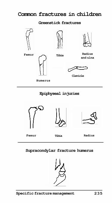
Common fractures in children
Greenstick fractures
Humerus
Clavicle
TibiaFemur Radiusand ulna
Epiphyseal injuries
Femur Tibia Radius
Supracondylar fracture humerus
Huckstep 1999©
Huckstep 1999©
Huckstep 1999©
236
This is particularly so in elbow injuries in children. Anapparent small epicondylar fracture may actually be a fractureof the entire lateral condyle.An epiphyseal separation mayalso have spontaneously reduced itself and then would onlybe apparent on a stress X-ray.
If in doubt, take X-rays of the opposite joint in exactly thesame position, for comparison. In young children anarthrogram may also be necessary to show the outline of theradiotranslucent cartilage. An ultrasound or magneticresonance imaging (MRI) can also help in the diagnosis.
Classification and treatment• Epiphyseal injuries — These may be subdivided intoshearing, avulsion, splitting and crush types. Such injuriesmay cause considerable disturbance of growth.
Salter-Harris classification
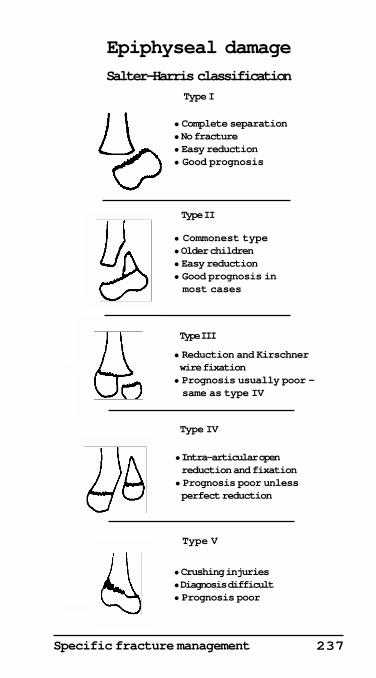
Epiphyseal injuriesThe Salter-Harris classification describes damage to theepiphyseal plate with or without a fracture.• Type I — No fracture. There is separation of the epiphysisat the level of the growth plate.• Type II — This involves separation of part of the epiphysisfrom the metaphysis through the epiphyseal plate, plus ametaphyseal fracture.• Types I and II Salter-Harris epiphyseal fractures, ifadequately reduced by closed reduction, usually have a goodprognosis, even if less than perfectly reduced. Salter Type IIinjuries, however, may lead to growth disturbance in 5% ofchildren.• Type III — The fracture line extends from the joint space tothe growth plate. It then extends laterally to the edge of theplate separating the fractured epiphysis from the metaphysis.It must be reduced perfectly and may require open operationand wire fixation. The prognosis is usually poor.• Type IV — These fractures extend from the joint spacethrough the growth plate and across the metaphysis. Thesecommonly occur in the lateral condyle of the humerus andalmost always require open reduction and smooth wirefixation. The prognosis is poor. In the elbow this may resultin a valgus deformity with a tardy or late ulnar nerve palsydue to stretching of the ulnar nerve. The valgus deformityincreases due to the cessation of growth of the damaged lateralpart of the epiphysis, and the continued medial growth.
Specific fracture management 237
Epiphyseal damage
Salter-Harris classification
Type I
Type II
Type III
Type IV
Type V
• Crushing injuries• Diagnosis difficult• Prognosis poor
• Intra-articular open reduction and fixation• Prognosis poor unless perfect reduction
• Reduction and Kirschner wire fixation• Prognosis usually poor — same as type IV
• Commonest type• Older children• Easy reduction• Good prognosis in most cases
• Complete separation• No fracture• Easy reduction• Good prognosis
Huckstep 1999©
Huckstep 1999©
Huckstep 1999©
Huckstep 1999©
Huckstep 1999©
238
• Type V — This is a severe crush injury of the growth plateitself. Displacement is unusual and injury may be unnoticed.There is almost always a poor prognosis with cessation ofgrowth of the epiphyseal growth plate.
It is essential to be very gentle with reduction of children’sinjuries and not damage the epiphysis with sharp instrumentsat the time of operation.
It is also important to warn relatives about the risks inthese injuries and to follow the child up with X-rays for atleast 2 years from the time of injury.
ComplicationsDamage to the epiphyseal plate, especially in Salter-HarrisType III, IV or V injuries (see illustration), may lead topremature fusion of part of the epiphysis. This in turn maylead to continued growth of the remaining germinal layer witha resulting varus or valgus deformity. If the whole epiphysisis damaged in this growing period the leg or arm will be short.Conversely, after a fracture of the shaft of a long bone, growthstimulation may actually occur with up to 2 cm of overgrowth.This is mainly due to the hyperaemia following injury.
Growth arrest• Treatment — If this is less than 50% of the epiphysis, thefused area of the epiphyseal plate should be excised andreplaced with a fat graft. If more than 50% of the plate isinvolved the fused epiphysis is usually left without operation,and any later deformity corrected by an osteotomy when thechild is skeletally mature.
ShorteningShortening of a limb can be treated by lengthening withIlizarov wire external fixateurs. The alternative is to carry outan epiphyseal arrest on the opposite leg at an appropriateage to equalise the leg lengths by the time growth has ceased.This is done either by temporary epiphyseal stapling, or bypermanent epiphyseodesis, which entails excision of theepiphyseal plate.
Operative treatmentIn children, operative internal stabilisation should be avoidedif possible, as this may interfere with growth at the epiphyses.• Smooth wire fixation — Even when this extends acrossepiphyseal plates, it seldom causes major problems providedthe wires are removed within 4-6 weeks. They are particularly
Specific fracture management 239
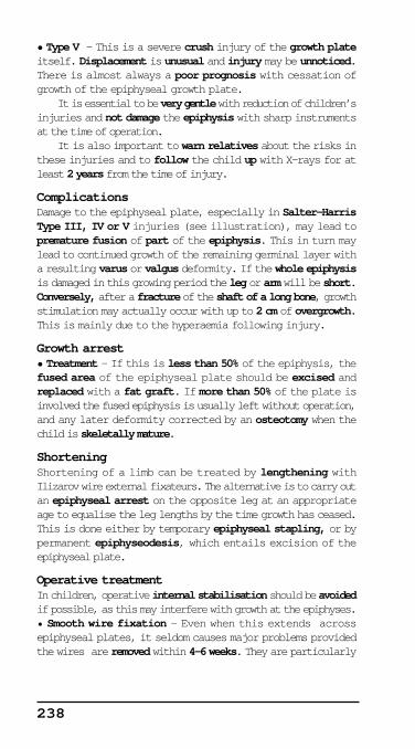
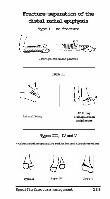
Fracture-separation of thedistal radial epiphysis
Type I — no fracture
• Manipulation and plaster
Type II
Lateral X-rayAP X-ray
• Manipulationand plaster
Types III, IV and V
• Often require operative reduction and Kirschner wires
Type III Type IV Type V
Huckstep 1999©
Huckstep 1999©
Huckstep 1999©
Huckstep 1999©
Huckstep 1999©

240
indicated in unstable fractures such as a supracondylarfracture of the humerus.• Older child — In the older child, screws, plates and evenan intramedullary nail may be sometimes used, provided theydo not damage the epiphyseal cartilage.
Compound fracturesIn compound fractures, as with the adult, external fixateurs,after adequate debridement, may be indicated. As withadults, however, all compound fractures, even with minimalskin damage, should be opened, explored, and left open. Inmost cases delayed primary closure is the treatment of choice.
HealingFractures in children heal much more quickly than in adults.• Femoral shaft fractures — These will usually be united in 1week in an infant, 1 month in a 1 year old, and 2 months in a10 year old, compared to 3 months in an adult.
RemodellingRemodelling of the growth plate may also occur, with up to20°-30° of correction possible. An angulation of the bone,which may not be acceptable in an adult, will often beacceptable in the child, especially in an infant, when 45°-60°of correction can occur in bones such as the upper humerus.This will only occur if the deformity is in plane of movementof the nearest joint.
Upper limb
Lower radius
Fracture-separation of the distal epiphysisThis is the pediatric equivalent of a Colles’ fracture.• Diagnosis — The distal radial fragment is both dorsallydisplaced and rotated. There is also radial displacement andimpaction of the fragment. There may be an associatedmetaphyseal fracture of the radius.• Treatment —The methods of reduction and the prognosishave already been discussed under the Salter-Harrisclassification of epiphyseal injuries.In summary:Type I — Separation of the epiphysis with no fracture requires a closed reduction and has a good prognosis.Type II — This involves separation of the epiphysis, plus ametaphyseal fracture, as illustrated. Again closed reductionwith a good prognosis except in 5% of cases.
Specific fracture management 241
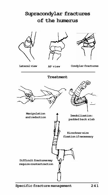
Kirschner wire fixation if necessary
Supracondylar fracturesof the humerus
Treatment
Lateral view AP view Condylar fractures
Manipulationand reduction Immobilisation:
padded back slab
Difficult fractures mayrequire countertraction
Huckstep 1999©
Huckstep 1999©
Huckstep 1999©
Huckstep 1999©
Huckstep 1999©
242
Types III and IV — These were described earlier in thischapter and are illustrated on page 237. They must beaccurately reduced and may require wire fixation. Theprognosis is usually poor in both Type III and Type IVinjuries.Type V — This causes a severe crush of the growth plate. Theprognosis is poor.
Forearm fracturesThese are very common fractures in children and account for55% of all children’s fractures. Fractures of the distal onethird of the radius account for 75% of forearm fractures.
Treatment• Simple fractures — Many of these fractures are greenstickfractures and, as a result, the bone may have to be ‘refractured’to reduce the fracture satisfactorily.• Young children — Nearly all diaphyseal fractures in youngchildren can be treated without an operation, and with anabove elbow plaster in either 45° of pronation or supination,after reduction if necessary.• Over the age of 8 years — In this age group, open reductionand plating or nailing may be necessary with displaced andangulated fractures if adequate closed reduction has failed.• Compound fractures — In compound or open fractures thewound should be explored and always left open with delayedprimary closure. External fixateurs may have to be used tostabilise these fractures.
Malunion and non-unionThe following is an approximate working rule in childrenprovided a good reduction cannot be easily obtained bymanipulation.• Under 8 years — In children some molding can occur and,under the age of 8, 20° of angulation and 20° of malrotationcan sometimes be accepted.• Over the age of 8 years — In this age group only 10° ofangulation may correct as the child grows.• Forearm fractures — Apposition of bone ends should be50% or more in all forearm fractures. If less than this is present,manipulation or operation will be required. Otherwisemalunion is likely, and occasionally non-union may occur.
Elbow injuriesElbow injuries in children are common and can cause vascularand neurological complications.
Specific fracture management 243
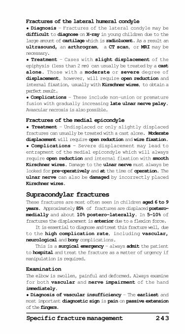
Fractures of the lateral humeral condyle• Diagnosis — Fractures of the lateral condyle may bedifficult to diagnose on X-ray in young children due to thelarge amount of cartilage which is radiolucent. As a result anultrasound, an arthrogram, a CT scan, or MRI may benecessary.• Treatment — Cases with slight displacement of theepiphysis (less than 2 mm) can usually be treated by a castalone. Those with a moderate or severe degree ofdisplacement, however, will require open reduction andinternal fixation, usually with Kirschner wires, to obtain aperfect result.• Complications — These include non-union or prematurefusion with gradually increasing late ulnar nerve palsy.Avascular necrosis is also possible.
Fractures of the medial epicondyle• Treatment — Undisplaced or only slightly displacedfractures can usually be treated with a cast alone. Moderatedisplacement will require open reduction and wire fixation.• Complications — Severe displacement may lead toentrapment of the medial epicondyle which will alwaysrequire open reduction and internal fixation with smoothKirschner wires. Damage to the ulnar nerve must always belooked for pre-operatively and at the time of operation. Theulnar nerve can also be damaged by incorrectly placedKirschner wires.
Supracondylar fracturesThese fractures are most often seen in children aged 6 to 9years. Approximately 85% of fractures are displaced postero-medially and about 10% postero-laterally. In 5-10% offractures the displacement is anterior due to a flexion force.
It is essential to diagnose and treat this fracture well, dueto the high complication rate, including vascular,neurological and bony complications.
This is a surgical emergency — always admit the patientto hospital and treat the fracture as a matter of urgency ifmanipulation is required.
ExaminationThe elbow is swollen, painful and deformed. Always examinefor both vascular and nerve impairment of the handimmediately.• Diagnosis of vascular insufficiency — The earliest andmost important diagnostic sign is pain on passive extensionof the fingers.
244
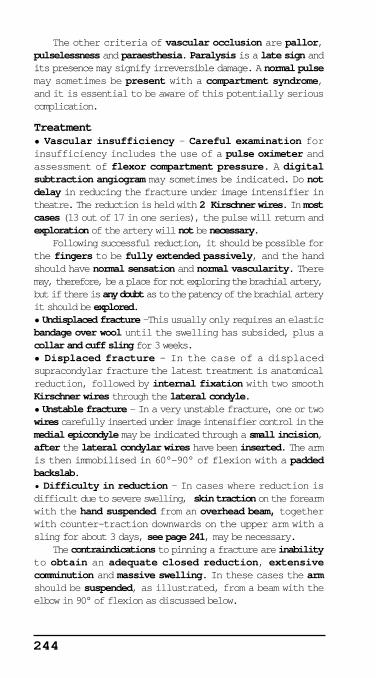
The other criteria of vascular occlusion are pallor,pulselessness and paraesthesia. Paralysis is a late sign andits presence may signify irreversible damage. A normal pulsemay sometimes be present with a compartment syndrome,and it is essential to be aware of this potentially seriouscomplication.
Treatment• Vascular insufficiency — Careful examination forinsufficiency includes the use of a pulse oximeter andassessment of flexor compartment pressure. A digitalsubtraction angiogram may sometimes be indicated. Do notdelay in reducing the fracture under image intensifier intheatre. The reduction is held with 2 Kirschner wires. In mostcases (13 out of 17 in one series), the pulse will return andexploration of the artery will not be necessary.
Following successful reduction, it should be possible forthe fingers to be fully extended passively, and the handshould have normal sensation and normal vascularity. Theremay, therefore, be a place for not exploring the brachial artery,but if there is any doubt as to the patency of the brachial arteryit should be explored.• Undisplaced fracture —This usually only requires an elasticbandage over wool until the swelling has subsided, plus acollar and cuff sling for 3 weeks.• Displaced fracture — In the case of a displacedsupracondylar fracture the latest treatment is anatomicalreduction, followed by internal fixation with two smoothKirschner wires through the lateral condyle.• Unstable fracture — In a very unstable fracture, one or twowires carefully inserted under image intensifier control in themedial epicondyle may be indicated through a small incision,after the lateral condylar wires have been inserted. The armis then immobilised in 60°-90° of flexion with a paddedbackslab.• Difficulty in reduction — In cases where reduction isdifficult due to severe swelling, skin traction on the forearmwith the hand suspended from an overhead beam, togetherwith counter-traction downwards on the upper arm with asling for about 3 days, see page 241, may be necessary.
The contraindications to pinning a fracture are inabilityto obtain an adequate closed reduction, extensivecomminution and massive swelling. In these cases the armshould be suspended, as illustrated, from a beam with theelbow in 90° of flexion as discussed below.
Specific fracture management 245
• Open fractures — In the case of open fractures, an adequatedebridement should be carried out before the Kirschner wiresare inserted.
Post-reduction careThe limb should be elevated and careful observation madefor symptoms and signs of vascular impairment. Thisincludes:• Terminal finger perfusion — This is the most impotantobservation and will include the use of a pulse oximeter on afinger.• Distal radial pulse — This should be examined every hourfor 48 hours following reduction, but is less diagnostic thanadequate finger perfusion and the ability to extend the fingers.• Fingers — These should be examined for warmth, sensationand inability to extend the fingers, associated with severeforearm tenderness.• Manipulation — A supracondylar fracture should neverbe manipulated more than 3 times, however poor the position.Immobilise for about 3 weeks in total.• Immobilisation in extension — Occasionallyimmobilisation in extension is required.
Early complications• Vascular complications — Forearm ischaemia — Thisrequires urgent treatment. The plaster should be removedimmediately, the elbow extended, and the fracture reducedwith Kirschner wires. Do not waste time on sympatheticblocks or vasodilators. Operate immediately if the circulationdoes not return with closed reduction and Kirschner wires.• Operative exposure — Extend elbow, incise the lower thirdof the upper arm and most of the forearm overlying thebrachial artery. Split the fascia and lacertus fibrosus.• Brachial artery — Identify the artery medial to the bicepstendon. Open the artery if in spasm, or if there is any doubt,and look for an intimal tear. Consider inserting a reversedvein graft. Intimal damage is common in these cases and maynot be apparent on external exposure of the artery.• Wound closure — Never suture fascia, and close part ofthe skin only if this can be done easily. Leave the wound openand perform a secondary closure.• Compartment syndrome — This is due to swelling due toischaemia, and to a lesser extent to bleeding into the flexorcompartment of the forearm. This may occur with or withoutmajor damage to the brachial artery. A fasciotomy of the entireflexor compartment should be carried out. Delayed primaryclosure of the wound is a necessity.
246
Late complications• Vascular impairment — Volkmann’s ischaemic contractureor gangrene of the arm.• Neurological complications — The nerves which arecommonly damaged in a supracondylar fracture are the radialand the median nerves. The anterior interosseous and ulnarnerves may also be damaged. The continuity and function ofthe flexor pollicis longus, and the flexor digitorum longusto the index finger must always be tested. A cubitus valgusdeformity may also produce a late ulnar nerve palsy.• Bony complications — Bony complications of asupracondylar fracture of the humerus include a cubitus varusor cubitus valgus deformity. This is usually due to anincomplete reduction, or to epiphyseal damage, and will resultin poor appearance and function.
Humeral shaft fractures
CausesBirth trauma, child abuse and benign bone cysts must alwaysbe considered in the diagnosis.
Common sites• Metaphysis — This occurs commonly in the metaphysealregion from 4 -12 years.• Diaphysis — The diaphysis is most often affected underthe age of 3 and over the age of 12.
Treatment• Conservative treatment — Most cases can be treated merelyin a collar and cuff sling or hanging cast.• Operative treatment — The only indication for explorationin closed fractures in children is in cases where radial nerveparalysis is increasing, or where there is obvious muscleinterposition between the bone ends which cannot becorrected by manipulation alone. Open fractures will requireexploration and adequate debridement and delayed primaryclosure, if necessary.• Angulation of up to 45°— In the newborn this cansometimes be accepted.• Overgrowth — This may be due to increased vascularity,.
ComplicationsRadial nerve injury is less common than in adults. Theprognosis is excellent and most cases recover completely inclosed injuries without operation.
Specific fracture management 247
Proximal humeral fractures80% of the growth of the humerus occurs in its proximalsegment. It has three ossification centres but only one growthplate, and so the diagnosis of a fracture is more difficult inchildren.
Fracture displacement• Children under 5 years — Considerable remodelling ispossible up to the age of 5 years. In these young children 40°of angulation, and total displacement of the fractures cansometimes be accepted provided there is some bonyapposition.• Children 5 - 12 years — Between the ages of 5 and 12 yearsup to 40° of angulation can often be accepted.• Children over 12 years — Over the age of 12 up to 40° ofangulation with more 50% apposition of the bone fragmentscan sometimes be accepted, provided there is at least 2 yearsof growth remaining.
Treatment• Conservative treatment — The patient can often be treatedmerely by a collar and cuff sling. In the case of severedisplacement, a shoulder spica or an abduction brace maybe required for up to 6 weeks.• Operative treatment — Only occasionally is open reductionand K wire fixation required. This is in patients wheredisplacement cannot be controlled by conservative measures.Open fractures, again, will require adequate debridement anddelayed primary closure, if necessary.
Lower limb
Hip and upper femur
CausesIn some countries in children under the age of 1 year, 70% offemoral shaft fractures have been reported as being due tochild abuse. Overall, 30% of fractures in children of all agesare also said to be due to child abuse. This diagnosis shouldbe especially suspected if there is a history of previous childabuse, if there is delay in attending hospital, or if there isevidence of other acute fractures or of multiple past fractures.This cause must be particularly considered if fractures of longbones are transverse, diaphyseal, or short oblique.• Pathological fractures — About 12.5% of fractures inchildren are pathological and include osteogenesis imperfecta,
248
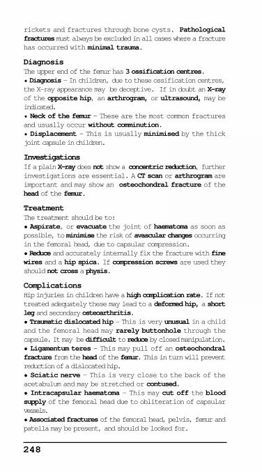
rickets and fractures through bone cysts. Pathologicalfractures must always be excluded in all cases where a fracturehas occurred with minimal trauma.
DiagnosisThe upper end of the femur has 3 ossification centres.• Diagnosis — In children, due to these ossification centres,the X-ray appearance may be deceptive. If in doubt an X-rayof the opposite hip, an arthrogram, or ultrasound, may beindicated.• Neck of the femur — These are the most common fracturesand usually occur without comminution.• Displacement — This is usually minimised by the thickjoint capsule in children.
InvestigationsIf a plain X-ray does not show a concentric reduction, furtherinvestigations are essential. A CT scan or arthrogram areimportant and may show an osteochondral fracture of thehead of the femur.
TreatmentThe treatment should be to:• Aspirate, or evacuate the joint of haematoma as soon aspossible, to minimise the risk of avascular changes occurringin the femoral head, due to capsular compression.• Reduce and accurately internally fix the fracture with finewires and a hip spica. If compression screws are used theyshould not cross a physis.
ComplicationsHip injuries in children have a high complication rate. If nottreated adequately these may lead to a deformed hip, a shortleg and secondary osteoarthritis.• Traumatic dislocated hip — This is very unusual in a childand the femoral head may rarely buttonhole through thecapsule. It may be difficult to reduce by closed manipulation.• Ligamentum teres — This may pull off an osteochondralfracture from the head of the femur. This in turn will preventreduction of a dislocated hip.• Sciatic nerve — This is very close to the back of theacetabulum and may be stretched or contused.• Intracapsular haematoma — This may cut off the bloodsupply of the femoral head due to obliteration of capsularvessels.• Associated fractures of the femoral head, pelvis, femur andpatella may be present, and should be looked for.
Specific fracture management 249
�����������������������������������������������������������������������������������������������
���������������������������������������������������
������������������������������������������������
������������������������������������������������
������������������������
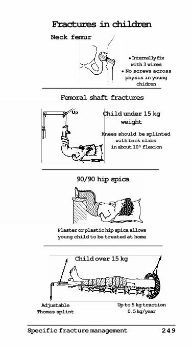
Plaster or plastic hip spica allowsyoung child to be treated at home
Fractures in childrenNeck femur
90/90 hip spica
Femoral shaft fractures
Knees should be splintedwith back slabs
in about 10° flexion
Child under 15 kgweight
Up to 5 kg traction0.5 kg/year
AdjustableThomas splint
Child over 15 kg
• Internally fixwith 3 wires
• No screws acrossphysis in young
chidren
Huckstep 1999©
Huckstep 1999©
Huckstep 1999©
Huckstep 1999©
250
Femoral shaft fractures
Treatment under 6 years• Gallows traction — In children under the age of 3 yearsgallows traction for 3-4 weeks with both knees flexed about10° in simple padded plaster of Paris back splints.• Hip spica — In the child under the age of 6 years, with lessthan 2.5 cm of shortening, a hip spica in the sitting position(90° flexion of hip and 90° flexion of knee) is often the bestmethod of management, once the fracture is stable.• Special cases — Compound fractures may require eitherskin or skeletal traction. The fractured leg should be in slightvalgus as the femur tends to displace into varus.
Treatment 6-12 yearsSkin or skeletal traction in a Thomas splint for 3 weeks,followed by a spica, is indicated. Shortening should not beover corrected as 0.5-1.5 cm of shortening will allow for futurebone overgrowth which commonly occurs following femoralshaft fractures in children, and may be up to 2 cm due toincreased vascularity.
Treatment over 12 yearsOver the age of 12, and in patients with head injuries, eitherEnders nails or locked intramedullary nails may be indicatedif closed reduction and traction in a Thomas splint isunsuccessful.
Complications• Avascular necrosis — The rate is very high in children andoccurs in the head of the femur in about 40% of cases. It hasbeen reported present in 80% of cases with a displacedepiphysis, 35% with displaced fractures of the cervical regionof the femur, and 25% in those with displaced fractures of thebase of the neck of the femur.
Tibial fractures
TreatmentTibial shaft fractures in children should usually be treatedconservatively by a padded above-knee plaster with the kneeflexed to about 10°.
Specific fracture management 251
Fractures in children
Complications
Correct angulation Epiphysiodesisopposite side
Overgrowth of 1-2cmcommon
Avoid overdistractingfractures
Malunion Shortening
Overgrowth
Late osteoarthritis
Avascular necrosisGrowth disturbances
Epiphyseal damage causesvarus, valgus or shortening
Huckstep 1999©
Huckstep 1999©
Huckstep 1999©
Huckstep 1999©
Huckstep 1999©
252
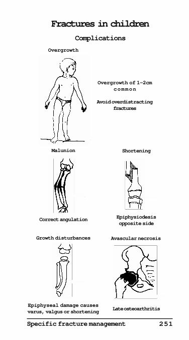
ComplicationsThe most common complication of proximal metaphysealfractures of the tibia in children is a valgus deformity. Thisdeformity often corrects spontaneously. Operativecorrection, if necessary via a tibial osteotomy, should bedelayed until growth has ceased.
Occult fractures in children
Occult or hidden fractures are common in children. This isbecause there is a large amount of cartilage in immature bones,particularly about the epiphyses, which have not yet ossified,and are therefore radiolucent.
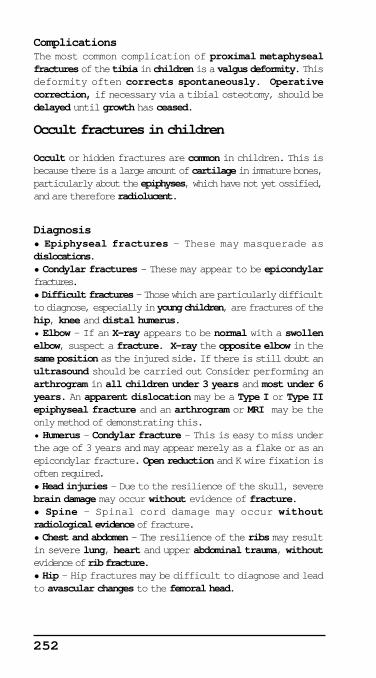
Diagnosis• Epiphyseal fractures — These may masquerade asdislocations.• Condylar fractures — These may appear to be epicondylarfractures.• Difficult fractures — Those which are particularly difficultto diagnose, especially in young children, are fractures of thehip, knee and distal humerus.• Elbow — If an X-ray appears to be normal with a swollenelbow, suspect a fracture. X-ray the opposite elbow in thesame position as the injured side. If there is still doubt anultrasound should be carried out Consider performing anarthrogram in all children under 3 years and most under 6years. An apparent dislocation may be a Type I or Type IIepiphyseal fracture and an arthrogram or MRI may be theonly method of demonstrating this.• Humerus — Condylar fracture — This is easy to miss underthe age of 3 years and may appear merely as a flake or as anepicondylar fracture. Open reduction and K wire fixation isoften required.• Head injuries — Due to the resilience of the skull, severebrain damage may occur without evidence of fracture.• Spine — Spinal cord damage may occur withoutradiological evidence of fracture.• Chest and abdomen — The resilience of the ribs may resultin severe lung, heart and upper abdominal trauma, withoutevidence of rib fracture.• Hip — Hip fractures may be difficult to diagnose and leadto avascular changes to the femoral head.
Specific fracture management 253
• Femoral shaft and tibia — Crush injuries and minordisplacements of the epiphyseal growth plate may be difficultto diagnose and may result in epiphyseal growth arrest ordeformity, particularly in the lower femur and upper andlower tibia. In addition, greenstick fractures may be missedand cause overgrowth of the affected limb.
Specific complications of fractures inchildren
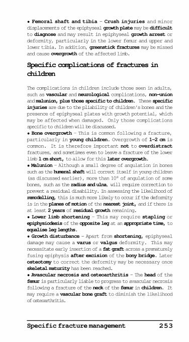
The complications in children include those seen in adults,such as vascular and neurological complications, non-unionand malunion, plus those specific to children. These specificinjuries are due to the pliability of children's bones and thepresence of epiphyseal plates with growth potential, whichmay be affected when damaged. Only those complicationsspecific to children will be discussed.• Bone overgrowth — This is common following a fracture,particularly in young children. Overgrowth of 1-2 cm iscommon. It is therefore important not to overdistractfractures, and sometimes even to leave a fracture of the lowerlimb l cm short, to allow for this later overgrowth.• Malunion — Although a small degree of angulation in bonessuch as the humeral shaft will correct itself in young children(as discussed earlier), more than 10° of angulation of somebones, such as the radius and ulna, will require correction toprevent a residual disability. In assessing the likelihood ofremodelling, this is much more likely to occur if the deformityis in the planes of motion of the nearest joint, and if there isat least 2 years of residual growth remaining.• Lower limb shortening — This may require stapling orepiphysiodesis of the opposite leg at an appropriate time, toequalise leg lengths.• Growth disturbance — Apart from shortening, epiphysealdamage may cause a varus or valgus deformity. This maynecessitate early insertion of a fat graft across a prematurelyfusing epiphysis after excision of the bony bridge. Laterosteotomy to correct the deformity may be necessary onceskeletal maturity has been reached.• Avascular necrosis and osteoarthritis — The head of thefemur is particularly liable to progress to avascular necrosisfollowing a fracture of the neck of the femur in children. Itmay require a vascular bone graft to diminish the likelihoodof osteoarthritis.
254
Fractures in the elderly
Causes
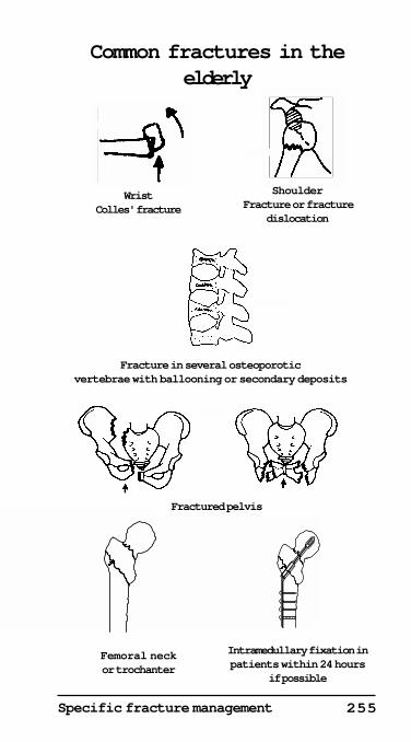
MetabolicThe elderly patient is more likely to slip and fall, and also,having fallen, to sustain a fracture.• Osteoporosis — This is a relatively common finding in theelderly and is an important factor in fracture aetiology.• Vertebrae — These may sustain crush fractures followinglittle or no trauma. This is partly due to prolapse of theintervertebral disc into the soft osteoporotic body of thevertebrae. Multiple vertebrae, particularly in the thoracicregion, commonly show stable crush fractures and cause thesmooth thoracic kyphosis seen in the elderly.• Hip — This also is osteoporotic in the elderly, and mostlikely to fracture following a fall.
Pathological fractures• Secondary tumour — Deposits in bone from carcinomaelsewhere, particularly from the breast and lung in females,and prostate and lung in males, may lead to fracturesfollowing little or no trauma.• Paget's disease — Pathological fractures may also occur inPaget’s disease.
MedicationMedication for conditions such as Parkinson’s disease mayalso cause osteoporosis. Long term use of oralglucocorticosteroids for conditions such as asthma, chronicairway limitations and various connective tissue disorders,such as rheumatoid arthritis, will often result in bonydemineralisation, thus making the patients more prone topathological fractures.
Common fracture sitesThe following are the most common fractures in the elderly.The detailed treatment of all these fractures is discussed inthe relevant section of this book.
Colles’ fractureThis is a very common injury, particularly in elderly females.It is often secondary to a fall on the outstretched hand.
255Specific fracture management
Common fractures in theelderly
Shoulder Fracture or fracture
dislocation
WristColles' fracture
Femoral neckor trochanter
Intramedullary fixation inpatients within 24 hours
if possible
Fracture in several osteoporoticvertebrae with ballooning or secondary deposits
Fractured pelvis
Huckstep 1999©
Huckstep 1999©
Huckstep 1999©
Huckstep 1999©
Huckstep 1999©
256
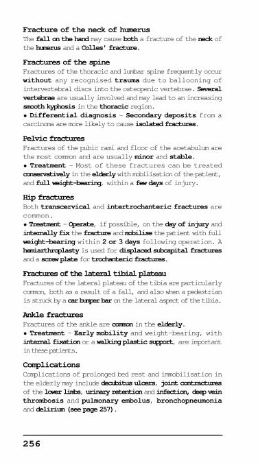
Fracture of the neck of humerusThe fall on the hand may cause both a fracture of the neck ofthe humerus and a Colles' fracture.
Fractures of the spineFractures of the thoracic and lumbar spine frequently occurwithout any recognised trauma due to ballooning ofintervertebral discs into the osteopenic vertebrae. Severalvertebrae are usually involved and may lead to an increasingsmooth kyphosis in the thoracic region.• Differential diagnosis — Secondary deposits from acarcinoma are more likely to cause isolated fractures.
Pelvic fracturesFractures of the pubic rami and floor of the acetabulum arethe most common and are usually minor and stable.• Treatment — Most of these fractures can be treatedconservatively in the elderly with mobilisation of the patient,and full weight-bearing, within a few days of injury.
Hip fracturesBoth transcervical and intertrochanteric fractures arecommon.• Treatment — Operate, if possible, on the day of injury andinternally fix the fracture and mobilise the patient with fullweight-bearing within 2 or 3 days following operation. Ahemiarthroplasty is used for displaced subcapital fracturesand a screw plate for trochanteric fractures.
Fractures of the lateral tibial plateauFractures of the lateral plateau of the tibia are particularlycommon, both as a result of a fall, and also when a pedestrianis struck by a car bumper bar on the lateral aspect of the tibia.
Ankle fracturesFractures of the ankle are common in the elderly.• Treatment — Early mobility and weight-bearing, withinternal fixation or a walking plastic support, are importantin these patients.
ComplicationsComplications of prolonged bed rest and immobilisation inthe elderly may include decubitus ulcers, joint contracturesof the lower limbs, urinary retention and infection, deep veinthrombosis and pulmonary embolus, bronchopneumoniaand delirium (see page 257).
257Specific fracture management
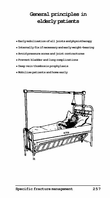
General principles inelderly patients
• Early mobilisation of all joints and physiotherapy
• Internally fix if necessary and early weight-bearing
• Avoid pressure sores and joint contractures
• Prevent bladder and lung complications
• Deep vein thombosis prophylaxis
• Mobilise patients and home early
Huckstep 1999©
258
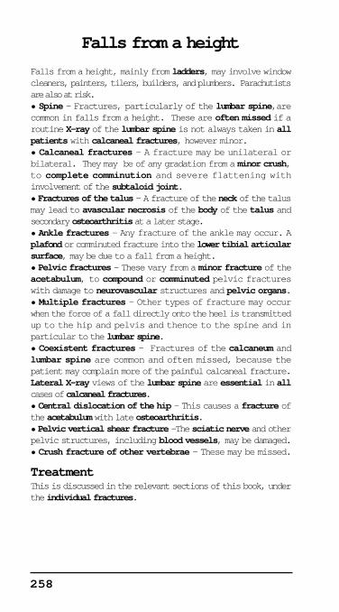
Falls from a height
Falls from a height, mainly from ladders, may involve windowcleaners, painters, tilers, builders, and plumbers. Parachutistsare also at risk.• Spine — Fractures, particularly of the lumbar spine,arecommon in falls from a height. These are often missed if aroutine X-ray of the lumbar spine is not always taken in allpatients with calcaneal fractures, however minor.• Calcaneal fractures — A fracture may be unilateral orbilateral. They may be of any gradation from a minor crush,to complete comminution and severe flattening withinvolvement of the subtaloid joint.• Fractures of the talus — A fracture of the neck of the talusmay lead to avascular necrosis of the body of the talus andsecondary osteoarthritis at a later stage.• Ankle fractures — Any fracture of the ankle may occur. Aplafond or comminuted fracture into the lower tibial articularsurface, may be due to a fall from a height.• Pelvic fractures — These vary from a minor fracture of theacetabulum, to compound or comminuted pelvic fractureswith damage to neurovascular structures and pelvic organs.• Multiple fractures — Other types of fracture may occurwhen the force of a fall directly onto the heel is transmittedup to the hip and pelvis and thence to the spine and inparticular to the lumbar spine.• Coexistent fractures — Fractures of the calcaneum andlumbar spine are common and often missed, because thepatient may complain more of the painful calcaneal fracture.Lateral X-ray views of the lumbar spine are essential in allcases of calcaneal fractures.• Central dislocation of the hip — This causes a fracture ofthe acetabulum with late osteoarthritis.• Pelvic vertical shear fracture —The sciatic nerve and otherpelvic structures, including blood vessels, may be damaged.• Crush fracture of other vertebrae — These may be missed.
TreatmentThis is discussed in the relevant sections of this book, underthe individual fractures.
259Specific fracture management
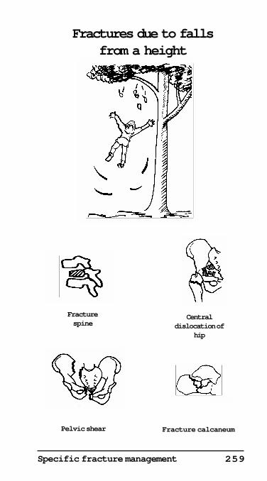
Fractures due to fallsfrom a height
Fracture calcaneumPelvic shear
Fracturespine
Centraldislocation of
hip
Huckstep 1999©
Huckstep 1999©
Huckstep 1999©
Huckstep 1999©
Huckstep 1999©
260
Falls on the hand
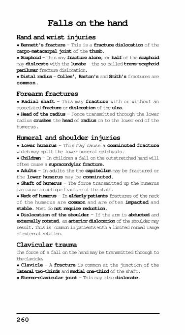
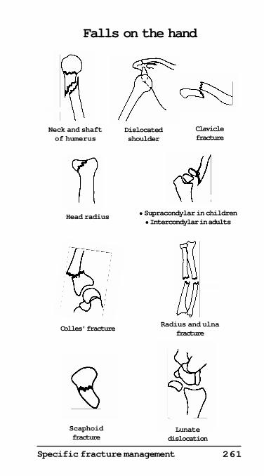
Hand and wrist injuries• Bennett's fracture — This is a fracture dislocation of thecarpo-metacarpal joint of the thumb.• Scaphoid — This may fracture alone, or half of the scaphoidmay dislocate with the lunate — the so called trans-scaphoidperilunar fracture dislocation.• Distal radius — Colles’, Barton's and Smith's fractures arecommon.
Forearm fractures• Radial shaft — This may fracture with or without anassociated fracture or dislocation of the ulna.• Head of the radius — Force transmitted through the lowerradius crushes the head of radius on to the lower end of thehumerus.
Humeral and shoulder injuries• Lower humerus — This may cause a comminuted fracturewhich may split the lower humeral epiphysis.• Children — In children a fall on the outstretched hand willoften cause a supracondylar fracture.• Adults — In adults the the capitellum may be fractured orthe lower humerus may be comminuted.• Shaft of humerus — The force transmitted up the humeruscan cause an oblique fracture of the shaft.• Neck of humerus — In elderly patients fractures of the neckof the humerus are common and are often impacted andstable. Most do not require reduction.• Dislocation of the shoulder — If the arm is abducted andexternally rotated, an anterior dislocation of the shoulder mayresult. This is common in patients with a limited normal rangeof external rotation.
Clavicular traumaThe force of a fall on the hand may be transmitted through tothe clavicle.• Clavicle — A fracture is common at the junction of thelateral two-thirds and medial one-third of the shaft.• Sterno-clavicular joint — This may also dislocate.
261Specific fracture management
Falls on the hand
Scaphoidfracture
Lunatedislocation
Head radius
Neck and shaftof humerus
Claviclefracture
• Supracondylar in children• Intercondylar in adults
Dislocatedshoulder
Radius and ulnafracture
Colles' fracture
Huckstep 1999©
Huckstep 1999©
Huckstep 1999©
Huckstep 1999©
Huckstep 1999©
Huckstep 1999©
Huckstep 1999©
Huckstep 1999©
Huckstep 1999©
262
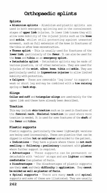
Orthopaedic splintsSplints• Aluminium splints — Aluminium and plastic splints areused in both emergency splinting and in the convalescentstages of upper limb injuries. In lower limb trauma they willallow some mobility of the injured joints such as the kneeand ankle, whilst still protecting against unwantedmovement, such as full extension of the knee in fractures ofthe tibia or after knee reconstruction.• Thomas splint — This is usually used for fractures of thelower limb, particularly of the femur. It can also be used forthe knee and tibia as an emergency splint.• Detachable splint — Detachable splints may be made ofvarious plastics, or of other materials. They are used forinjuries of the wrist, elbow, knee, tibia and ankle. They areparticularly useful in ligamentous injuries to allow limitedmobility with protection.• Calipers — These are removable ‘leg irons’ to support aweak or short leg and may be combined with a toe raisingspring or back stop.
SlingsCollar and cuff and triangular slings are used mainly for theupper limb and these have already been described.
TractionThis may include skin traction such as is used in fractures ofthe femoral neck. Skeletal traction is used where moretraction is needed. It is used for some fractures of the shaft ofthe femur and tibia.
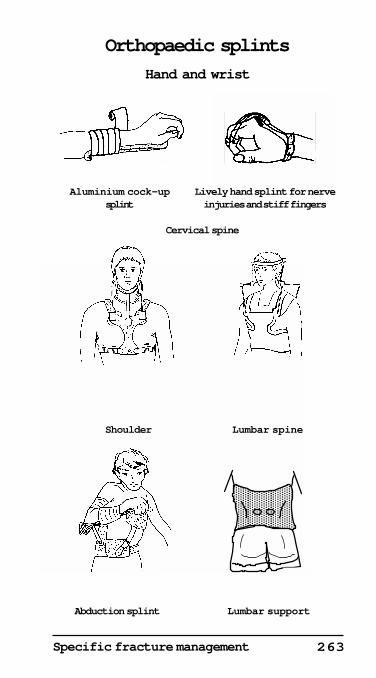
Plastic supportsPlastic supports, particularly the newer lightweight versionsare being used increasingly. These are plastics that can bedipped in either hot or cold water and molded to the patient.• Indications — They have a place where there is not muchswelling or following a preliminary treatment with plasterwhere further support is required.• Advantages — These supports can be permanent ordetachable with 'velcro' straps and are lighter and morecomfortable than plaster of Paris.• Disadvantages — The disadvantages of plastic supportsare their cost and their difficulty in application. They cannotbe molded as well as plaster of Paris.• Spinal supports — There are many neck and spinalsupports and some of these are illustrated. They are usually
263Specific fracture management
��������������������������������������������������������������������������������������������������������
Orthopaedic splints
Hand and wrist
Aluminium cock-upsplint
Lively hand splint for nerveinjuries and stiff fingers
Abduction splint Lumbar support
Lumbar spineShoulder
Cervical spine
Huckstep 1999©
Huckstep 1999©
Huckstep 1999©
Huckstep 1999©
Huckstep 1999©
Huckstep 1999©
264
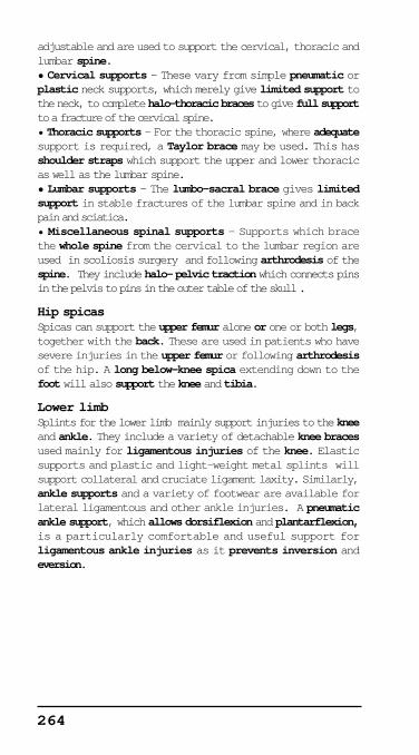
adjustable and are used to support the cervical, thoracic andlumbar spine.• Cervical supports — These vary from simple pneumatic orplastic neck supports, which merely give limited support tothe neck, to complete halo-thoracic braces to give full supportto a fracture of the cervical spine.• Thoracic supports — For the thoracic spine, where adequatesupport is required, a Taylor brace may be used. This hasshoulder straps which support the upper and lower thoracicas well as the lumbar spine.• Lumbar supports — The lumbo-sacral brace gives limitedsupport in stable fractures of the lumbar spine and in backpain and sciatica.• Miscellaneous spinal supports — Supports which bracethe whole spine from the cervical to the lumbar region areused in scoliosis surgery and following arthrodesis of thespine. They include halo- pelvic traction which connects pinsin the pelvis to pins in the outer table of the skull .
Hip spicasSpicas can support the upper femur alone or one or both legs,together with the back. These are used in patients who havesevere injuries in the upper femur or following arthrodesisof the hip. A long below-knee spica extending down to thefoot will also support the knee and tibia.
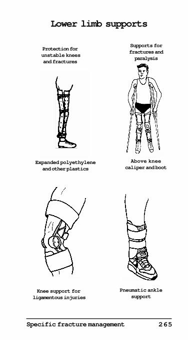
Lower limbSplints for the lower limb mainly support injuries to the kneeand ankle. They include a variety of detachable knee bracesused mainly for ligamentous injuries of the knee. Elasticsupports and plastic and light-weight metal splints willsupport collateral and cruciate ligament laxity. Similarly,ankle supports and a variety of footwear are available forlateral ligamentous and other ankle injuries. A pneumaticankle support, which allows dorsiflexion and plantarflexion,is a particularly comfortable and useful support forligamentous ankle injuries as it prevents inversion andeversion.
265Specific fracture management
Lower limb supports
Knee support for ligamentous injuries
Pneumatic anklesupport
Protection for unstable kneesand fractures
Supports forfractures andparalysis
Expanded polyethyleneand other plastics
Above kneecaliper and boot
Huckstep 1999©
Huckstep 1999©
Huckstep 1999©
Huckstep 1999©
266
Plaster of Paris (POP)
• Plaster of Paris — This has the advantage of ease ofapplication. It is used in most acute fractures requiring eitheremergency splinting or immobilisation after manipulation.• Padded backslab — This is applied to splint acute fracturesand is completed when the swelling has subsided.• Full plaster — In the acute stage it should be split along itsentire length so that it can be opened if severe swelling occurs.• Lower limb — A completed plaster may be either weight-bearing or non-weight-bearing.
Types of plaster bandagePlaster bandages come in the following sizes.• Hands and arms — 5, 7.5, 10 and 15 cm.• Legs, hip spicas and plaster jackets — 10, 15 and 20cm.Plaster hardens completely in 24 to 48 hours, but is fairlystrong in 1-2 hours and firm in about 5 minutes, dependingon the type of plaster. Weight-bearing should be delayed forat least 48 hours to allow the plaster to harden properly.
Preparation of plaster applicationIt is important that plaster bandages are applied quickly andevenly. Padding with plaster wool and foam is important ifpressure sores and other complications are to be avoided,especially in acute fractures where oedema is common.
Immersion of the plaster bandage• Cold water — This is usually used. Warm water can be usedif more rapid setting is required.• Dip the plaster — This is done in the water until the bubblesstop appearing.• Removing the bandage — The bandage is lifted out of thewater and the surplus water drips back into the bucket (notonto the floor!). Squeeze gently to extract water and not plaster.
Application of plaster bandage• Acute fractures — Those needing manipulation will usuallyrequire a well padded plaster or backslab. A thin layer ofplaster wool should be used, plus a stockinette under theplaster. Additional padding with wool or foam should be usedover pressure areas.• Apply the plaster evenly and make sure that the joints arein the correct position. Use a backslab to strengthen a plasterwhere possible.• Pad pressure areas carefully with plaster wool or foamplastic. Toes and fingers must be free to move.
267Specific fracture management
Usecarefully
Plaster of Paris
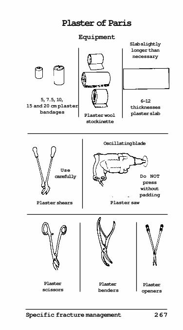
EquipmentSlab slightlylonger thannecessary
6-12thicknessesplaster slab
5, 7.5, 10,15 and 20 cm plaster
bandagesPlaster woolstockinette
Plaster shears
Oscillating blade
Plaster saw
Do NOTpresswithoutpadding
Plasterscissors
Plaster benders
Plasteropeners
Huckstep 1999©
268
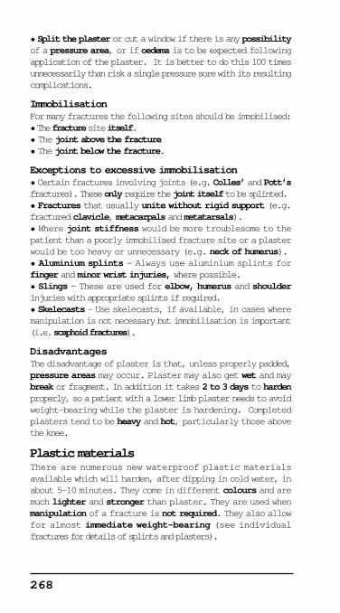
• Split the plaster or cut a window if there is any possibilityof a pressure area, or if oedema is to be expected followingapplication of the plaster. It is better to do this 100 timesunnecessarily than risk a single pressure sore with its resultingcomplications.
ImmobilisationFor many fractures the following sites should be immobilised:• The fracture site itself.• The joint above the fracture• The joint below the fracture.
Exceptions to excessive immobilisation• Certain fractures involving joints (e.g. Colles’ and Pott’sfractures). These only require the joint itself to be splinted.• Fractures that usually unite without rigid support (e.g.fractured clavicle, metacarpals and metatarsals).• Where joint stiffness would be more troublesome to thepatient than a poorly immobilised fracture site or a plasterwould be too heavy or unnecessary (e.g. neck of humerus).• Aluminium splints — Always use aluminium splints forfinger and minor wrist injuries, where possible.• Slings — These are used for elbow, humerus and shoulderinjuries with appropriate splints if required.• Skelecasts — Use skelecasts, if available, in cases wheremanipulation is not necessary but immobilisation is important(i.e. scaphoid fractures).
DisadvantagesThe disadvantage of plaster is that, unless properly padded,pressure areas may occur. Plaster may also get wet and maybreak or fragment. In addition it takes 2 to 3 days to hardenproperly, so a patient with a lower limb plaster needs to avoidweight-bearing while the plaster is hardening. Completedplasters tend to be heavy and hot, particularly those abovethe knee.
Plastic materialsThere are numerous new waterproof plastic materialsavailable which will harden, after dipping in cold water, inabout 5-10 minutes. They come in different colours and aremuch lighter and stronger than plaster. They are used whenmanipulation of a fracture is not required. They also allowfor almost immediate weight-bearing (see individualfractures for details of splints and plasters).
269Specific fracture management
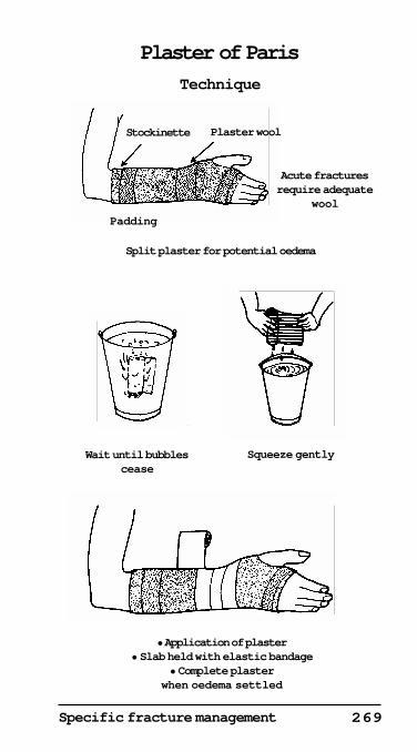
Padding
Plaster of Paris
Technique
Stockinette Plaster wool
Acute fracturesrequire adequate
wool
Split plaster for potential oedema
Wait until bubblescease
Squeeze gently
Dipping plaster
• Application of plaster• Slab held with elastic bandage
• Complete plasterwhen oedema settled
Huckstep 1999©
Huckstep 1999©
Huckstep 1999©
Huckstep 1999©
270
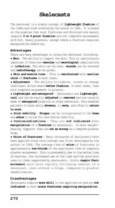
Skelecasts
The skelecast is a simple concept of lightweight fixation ofthe limbs and trunk invented by the author in 1966. It is basedon the premise that most fractures and dislocations merelyrequire 3 or 4 point fixation and not complete encasementwith hot, heavy plasters, except where a fracture requiresmanipulative reduction.
AdvantagesThere are many advantages in using the skelecast including:• Skin — The ability to inspect the skin. This is particularlyimportant if there are vascular and neurological complicationsor infection. The skin can be seen, wounds can be dressedand radiotherapy can be given.• Skin and muscle tone — This is maintained with earlierunion of fractures in most cases.• Adjustment — The ability to tighten, loosen or changeindividual struts means better fixation, in most cases, thanwith complete encasement in plaster.• Lightweight and waterproof — Skelecasts are lightweight,cool and can be easily adjusted or removed and are usuallymade of waterproof plastics or other materials. This enablespatients to have daily showers, to swim, and often to returnto work.• Joint mobility — Hinges can be incorporated in the kneeand elbow to allow for even better mobility.• Contraindications — They are not indicated whenmanipulation of a fracture is necessary. In most weight-bearing supports they are not as strong as a complete plasterwrap.• Union of fractures — Many thousands of skelecasts havebeen applied since this concept was first developed by theauthor in 1966. The average time of union of fractures isapproximately two-thirds of the equivalent time of completeplaster encasement. This is presumably due to the better toneof muscles, the increased use of the limb and the good skincare in limbs supported by skelecasts. Joints regain theirmovement much more rapidly following removal of askelecast, even without a hinge, compared to plasterimmobilisation.
DisadvantagesSkelecasts require more skill in the application and are notindicated in most acute fractures requiring manipulation.
271Specific fracture management
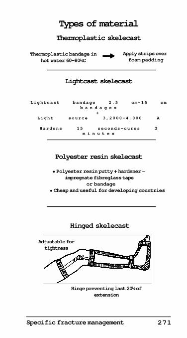
Lightcast skelecast
Polyester resin skelecast
Lightcast bandage 2.5 cm-15 cmb a n d a g e s
+Light source 3,2000-4,000 A
Hardens 15 seconds-cures 3m i n u t e s
Adjustable fortightness
Hinged skelecast
• Polyester resin putty + hardener — impregnate fibreglass tape
or bandage • Cheap and useful for developing countries
Thermoplastic bandage inhot water 60-80½C
Apply strips over foam padding
Types of material
Thermoplastic skelecast
Hinge preventing last 20½ ofextension
!!!!!
272
They are also not as strong as a completed plaster or completeplastic splint for weight-bearing.
Removal and X-raysThe supports can be removed easily by simply cutting throughthe struts with ‘tin snips’, and in most cases, X-rays can betaken through the gaps in the skelecast without removing theskelecast, as is often necessary with plaster. Deep X-raytherapy can also be given for pathological fractures.
Acute fractures• Oedema — In acute fractures oedema is controlled initiallyby merely putting a little wool and an elastic compressionbandage around the limb and the struts until the swelling hassettled.• Contraindications — Skelecasts are not usually indicatedwhere a complete wrap is necessary after a manipulation offracture. The skelecast can be applied, however, as soon asthe fracture is sufficiently stable, to enable the support to bechanged without the danger of the fracture slipping.
MaterialThe skelecast can be made out of a variety of materials, asillustrated. Many of the newer plastics can be used. Theseinclude thermoplastics which are softened in hot water at 65-80°C. They are applied to the patient as struts directly overwaterproof lining. Some cold water plastics can also be used.They are dipped into cold water and applied directly to thepatient in the form of struts. Ordinary hardware shoppolyester resin, or a light sensitive polyester resin, can alsobe used. Newer materials are constantly being developed. Theone most suited, and available, should be used.• Longitudinal struts — These are usually thin aluminiumstruts covered by one or two layers of the plastic or othermaterial used in the manufacture of the support. The exacttype of plastic or metal used is unimportant, provided thatgood rigid fixation is obtained. The support should also bewaterproof and strong, and the points of contact between thestruts are properly molded and strengthened if necessary.
Indications
Upper limb• Specific indications —The scaphoid skelecast is ideal. Othersuitable indications for this type of support include above-elbow skelecasts for the radius and ulna following internalfixation of fractures. The Colles’ type of skelecast is also
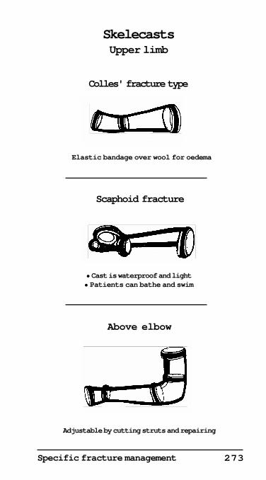
273Specific fracture management
SkelecastsUpper limb
Above elbow
Scaphoid fracture
Colles' fracture type
Elastic bandage over wool for oedema
• Cast is waterproof and light• Patients can bathe and swim
Adjustable by cutting struts and repairing
Huckstep 1999©
Huckstep 1999©
Huckstep 1999©
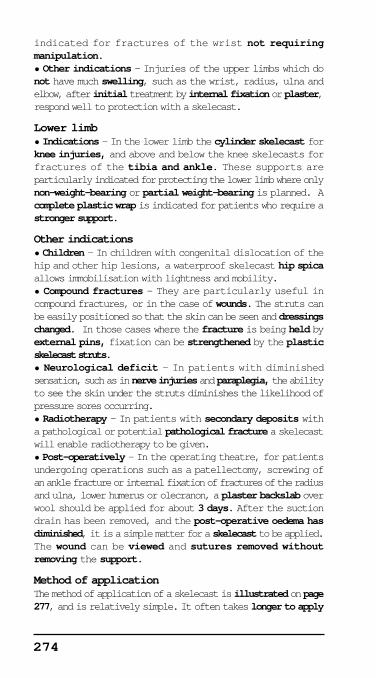
274
indicated for fractures of the wrist not requiringmanipulation.• Other indications — Injuries of the upper limbs which donot have much swelling, such as the wrist, radius, ulna andelbow, after initial treatment by internal fixation or plaster,respond well to protection with a skelecast.
Lower limb• Indications — In the lower limb the cylinder skelecast forknee injuries, and above and below the knee skelecasts forfractures of the tibia and ankle. These supports areparticularly indicated for protecting the lower limb where onlynon-weight-bearing or partial weight-bearing is planned. Acomplete plastic wrap is indicated for patients who require astronger support.
Other indications• Children — In children with congenital dislocation of thehip and other hip lesions, a waterproof skelecast hip spicaallows immobilisation with lightness and mobility.• Compound fractures — They are particularly useful incompound fractures, or in the case of wounds. The struts canbe easily positioned so that the skin can be seen and dressingschanged. In those cases where the fracture is being held byexternal pins, fixation can be strengthened by the plasticskelecast struts.• Neurological deficit — In patients with diminishedsensation, such as in nerve injuries and paraplegia, the abilityto see the skin under the struts diminishes the likelihood ofpressure sores occurring.• Radiotherapy — In patients with secondary deposits witha pathological or potential pathological fracture a skelecastwill enable radiotherapy to be given.• Post-operatively — In the operating theatre, for patientsundergoing operations such as a patellectomy, screwing ofan ankle fracture or internal fixation of fractures of the radiusand ulna, lower humerus or olecranon, a plaster backslab overwool should be applied for about 3 days. After the suctiondrain has been removed, and the post-operative oedema hasdiminished, it is a simple matter for a skelecast to be applied.The wound can be viewed and sutures removed withoutremoving the support.
Method of applicationThe method of application of a skelecast is illustrated on page277, and is relatively simple. It often takes longer to apply
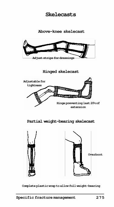
275Specific fracture management
Skelecasts
Adjustable fortightness
Hinge preventing last 20½ ofextension
Overboot
Complete plastic wrap to allow full weight-bearing
Partial weight-bearing skelecast
Hinged skelecast
Adjust strips for dressings
Above-knee skelecast
Huckstep 1999©
Huckstep 1999©
Huckstep 1999©
Huckstep 1999©
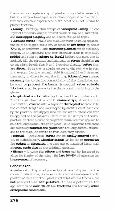
276
than a simple complete wrap of plaster or synthetic material,but its many advantages more than compensate for this.Patients who have experienced a skelecast will not return toplaster fixation.• Lining — Firstly, thin strips of waterproof lining, in onelayer of thickness, are put around the arm or leg, as illustrated,and overlapped slightly and held with strips of tape.• Circular struts — While one circular strut is being applied,the next is dipped for a few seconds in hot water at about70°C by an assistant. Some cold water plastics can be similarlydipped. lt is important that each circular strut be carefullymolded and made to adhere to itself before the next one isapplied. All the circular and longitudinal struts should be cutto the right length from 5 or 7.5 cm wide plastic, before theyare dipped. It is then a simple matter to dip each one in turnin the water, lay it on a towel, fold it on itself 2 or 3 times andthen apply it directly over the lining. Rubber gloves are notnecessary due to the low conductivity of the plastic but canbe worn to protect the hands. A special water solublelubricant supplied prevents the thermoplastic sticking to thegloves.• Longitudinal struts — After application of the circular strut,2 or 3 longitudinal struts of aluminium strips, about 1-1.5 cmin diameter, covered with a layer of thermoplastic and cut tothe correct length and overlapped by about l cm at each endby the plastic, are dipped into the hot water. These can thenbe applied to the patient. Extra circular strips of thermo-plastic, or other plastic or polyester resin, are then applied tohold the longitudinal struts in place. It is important that theseare carefully molded at the joints with the longitudinal struts,and to the circular struts to make sure they adhere.• Removal — Individual struts can be easily removed for X-ray or radiotherapy. Circular struts can be cut or opened outfor oedema or closed in. The area can be repaired later withan epoxy resin glue or the original material.• Hinges — A hinge for elbows and knees can be inserted toallow for flexion of the joint. The last 20°-30° of extension canbe prevented if necessary.
ConclusionA skelecast, if applied properly and carefully and for thecorrect indications, is superior to complete encasement withplaster-of-Paris or other plastic material where a fracture hasnot needed to be manipulated. It has a potential forapplication of over 50% of all fractures and for many otherorthopaedic conditions.
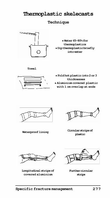
277Specific fracture management
Thermoplastic skelecasts
Technique
Waterproof lining
Longitudinal strips ofcovered aluminium
Further circularstrips
• Water 65-80½ forthermoplastics
• Dip thermoplastic brieflyinto water
Towel
• Fold hot plastic into 2 or 3thicknesses
• Aluminium covered plasticwith 1 cm overlap at ends
Circular strips ofplastic
Huckstep 1999©
Huckstep 1999©
Huckstep 1999©
Huckstep 1999©
Huckstep 1999©
Huckstep 1999©
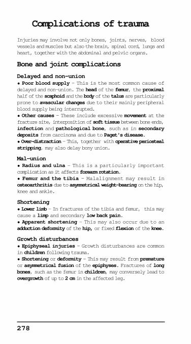
278
Complications of trauma
Injuries may involve not only bones, joints, nerves, bloodvessels and muscles but also the brain, spinal cord, lungs andheart, together with the abdominal and pelvic organs.
Bone and joint complications
Delayed and non-union• Poor blood supply — This is the most common cause ofdelayed and non-union. The head of the femur, the proximalhalf of the scaphoid and the body of the talus are particularlyprone to avascular changes due to their mainly peripheralblood supply being interrupted.• Other causes — These include excessive movement at thefracture site, interposition of soft tissue between bone ends,infection and pathological bone, such as in secondarydeposits from carcinoma and due to Paget's disease.• Over-distraction — This, together with operative periostealstripping, may also delay bony union.
Mal-union• Radius and ulna — This is a particularly importantcomplication as it affects forearm rotation.• Femur and the tibia — Malalignment may result inosteoarthritis due to asymmetrical weight-bearing on the hip,knee and ankle.
Shortening• Lower limb — In fractures of the tibia and femur, this maycause a limp and secondary low back pain.• Apparent shortening — This may also occur due to anadduction deformity of the hip, or fixed flexion of the knee.
Growth disturbances• Epiphyseal injuries — Growth disturbances are commonin children following trauma.• Shortening or deformity — This may result from prematureor asymmetrical fusion of the epiphyses. Fractures of longbones, such as the femur in children, may conversely lead toovergrowth of up to 2 cm in the affected leg.
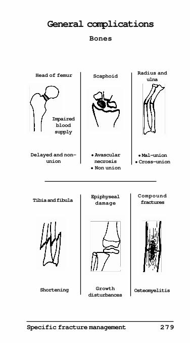
279Specific fracture management
General complications
Bones
Impairedbloodsupply
Head of femur Scaphoid
Delayed and non-union
• Avascularnecrosis
• Non union
• Mal-union• Cross-union
Radius andulna
Epiphysealdamage
Compound fractures
Shortening Growthdisturbances
Osteomyelitis
Tibia and fibula
Huckstep 1999©
Huckstep 1999©
Huckstep 1999©
Huckstep 1999©
Huckstep 1999©
Huckstep 1999©
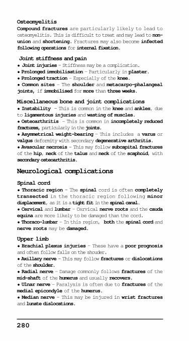
280
OsteomyelitisCompound fractures are particularly likely to lead toosteomyelitis. This is difficult to treat and may lead to non-union and shortening. Fractures may also become infectedfollowing operations for internal fixation.
Joint stiffness and pain• Joint injuries — Stiffness may be a complication.• Prolonged immobilisation — Particularly in plaster.• Prolonged traction — Especially of the knee.• Common sites — The shoulder and metacarpo-phalangealjoints, if immobilised for more than three weeks.
Miscellaneous bone and joint complications• Instability — This is common in the knee and ankles, dueto ligamentous injuries and wasting of muscles.• Osteoarthritis — This is common in incompletely reducedfractures, particularly in the joints.• Asymmetrical weight-bearing — This includes a varus orvalgus deformity with secondary degenerative arthritis.• Avascular necrosis — This may follow subcapital fracturesof the hip, neck of the talus and neck of the scaphoid, withsecondary osteoarthritis.
Neurological complications
Spinal cord• Thoracic region — The spinal cord is often completelytransected in the thoracic region following minordisplacement, as it is a tight fit in the spinal canal.• Cervical and lumbar — Cervical nerve roots and the caudaequina are more likely to be damaged than the cord.• Thoraco-lumbar — In this region, both the spinal cord andnerve roots may be damaged.
Upper limb• Brachial plexus injuries — These have a poor prognosisand often follow falls on the shouder.• Axillary nerve — This may follow fractures or dislocationsof the shoulder.• Radial nerve — Damage commonly follows fractures of themid-shaft of the humerus and usually recovers.• Ulnar nerve — Paralysis is often due to fractures of themedial epicondyle of the humerus.• Median nerve — This may be injured in wrist fracturesand lunate dislocations.
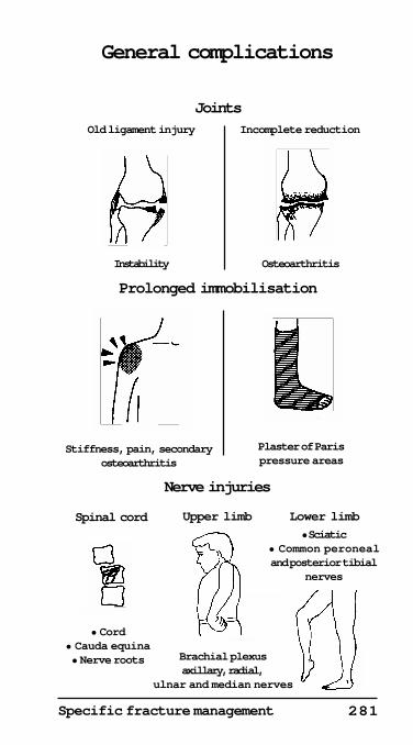
281Specific fracture management
General complications
• Sciatic• Common peronealand posterior tibial
nerves
Brachial plexusaxillary, radial,
ulnar and median nerves
JointsOld ligament injury Incomplete reduction
Instability Osteoarthritis
Prolonged immobilisation
Plaster of Parispressure areas
Upper limbSpinal cord Lower limb
Nerve injuries
• Cord• Cauda equina• Nerve roots
Stiffness, pain, secondaryosteoarthritis
Huckstep 1999©
Huckstep 1999©
Huckstep 1999©
Huckstep 1999©
Huckstep 1999©
Huckstep 1999©
Huckstep 1999
©
Huckstep 1999©
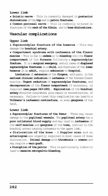
282
Lower limb• Sciatic nerve — This is commonly damaged in posteriordislocations of the hip and in pelvic fractures.• Common peroneal nerve — This is commonly injured infractures of the neck of the fibula, and in knee dislocations.
Vascular complications
Upper limb• Supracondylar fracture of the humerus — This maydamage the brachial artery.• Compartment syndrome with ischaemia of the flexormuscles — This is due to oedema or to bleeding into the flexorcompartment of the forearm following a supracondylarfracture. It is a surgical emergency, and all cases of displacedsupracondylar fractures in a child, and fractures of the lowerhumerus in an adult, require admission to hospital.
Limitation of extension of the fingers, with pain, is theearliest clinical indication of ischaemia of the forearm flexormuscles. Urgent reduction of supracondylar fractures, anddecompression of the flexor compartment if necessary, isrequired (see pages 243-245). Exploration of the brachialartery should be considered, plus repair or reconstruction, ifnecessary. Failure to treat this complication can lead to aVolkmann's ischaemic contracture, or even gangrene of thehand.
Lower limb• Supracondylar fractures of the femur — These may causedamage to the popliteal vessels. The popliteal artery has apoor collateral blood supply and may lead to ischaemia ofthe calf muscles and gangrene of the toes, similar to thebrachial artery causing ischaemia to the upper limb.• Dislocation of the knee — A Doppler scan and anarteriogram are urgently required in most cases of kneedislocation. Intimal damage with thrombosis is common andmay require a vein graft.• Disruption of the pelvis — This is particularly liable tolead to massive retropelvic bleeding.
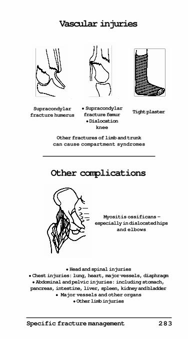
283Specific fracture management
Vascular injuries
Supracondylarfracture humerus
• Supracondylarfracture femur• Dislocation
knee
Tight plaster
Myositis ossificans —especially in dislocated hips
and elbows
• Head and spinal injuries• Chest injuries: lung, heart, major vessels, diaphragm• Abdominal and pelvic injuries: including stomach,pancreas, intestine, liver, spleen, kidney and bladder
• Major vessels and other organs• Other limb injuries
Other complications
Other fractures of limb and trunkcan cause compartment syndromes
Huckstep 1999©
Huckstep 1999©
Huckstep 1999©
Huckstep 1999©
284
Miscellaneous complications• Plaster of Paris — An unpadded or poorly fitting plastermay cause a vascular compromise and pressure sores.• Local complications — These include traumatic ossificationor myositis ossificans due to calcification, followed byossification in a haematoma following a fracture, jointdislocation or periosteal damage. This is particularly commonif there is associated neurological damage.• General complications — These include respiratoryobstruction in head, jaw and chest injuries. Othercomplications are shock, fat embolus, and the crushsyndrome following abdominal, pelvic and limb injuries.
Details of the complications associated with head, chest,abdomen, spine and limb injuries and their treatment arediscussed in the individual sections of this book.