specific features of occupational medicine in nuclear ...€¦ · sciences division for mox – and...
TRANSCRIPT

CLEFS CEA - No. 48 - SUMMER 200368
Risk assessment at low exposure levels
The first harmful effects of ionising radiation– “ray burns” (radiodermatitis) – were observed
almost immediately after its physical properties werediscovered. Years later between the two world wars
delayed effects were demonstrated – leukemia amongradiologists and researchers such as Marie Curie.Thus when the first CEA laboratories were set up inthe late forties, measures to prevent exposure ofpersonnel were implemented and research intoradioprotection undertaken.At the same time, France voted in legislation to providefor preventive occupational medicine to monitorworkers’health. Occupational medical services adaptedto the specific hazards of exposure to ionisingradiation were then set up. This medical monitoringhas since kept pace with advances in research and inthe industrial applications of ionising radiation, inparticular the development of nuclear-fuelledelectricity generation and the fuel cycle industry.
Several specific features
This branch of occupational medicine, which comple-ments technical radiation safety provisions, is specificin several ways. The initial emphasis on possiblehæmatological, skin or ocular effects resulted in thesetting up of a thorough medical monitoringprovision: six-monthly clinical examinations, bloodtests, and specialist consultation (ophthalmology,for example).Alongside exposure to ionising radiation in the strictsense, working conditions in hostile environmentsrequiring personal protection equipment to be worn(e.g., breathing equipment for the protection ofairways or special garments) require allowance to bemade for the physical and psychological conditionsassociated with certain workplaces. Also, in bothresearch laboratories and industrial plants, exposureto ionising radiation is often accompanied byexposure to chemical agents (see Beryllium, anexample of a non-radioactive nuclear toxic element)and sometimes biological substances.
Specific investigation methods
The monitoring of internal exposure has requiredthe development of specific investigation methods
Measures to prevent the exposure of personnel to ionising radiation were taken as soon as the first nuclear laboratories were set up. This branch of occupational preventivemedicine has since kept pace with advances in research and in the industrial applications of nuclear energy.
Specific features of occupational medicine in nuclear research and industrial applications
Radiographic examination at the occupational health service at the CEA Saclay Centre. A thorough system of medical monitoring was set up long ago in nuclear research and the nuclear industry.
Fran
cis
Vigo
urou
x/C
EA
CLEFS CEA - No. 48 - SUMMER 2003 69
Fran
cis
Vigo
urou
x/C
EA
Cog
ema
to measure, either in vivo or in vitro, the activityincorporated by individuals.In vivo measurement uses whole-body gammaradiation measurement, which determines theradiation emitted by the body, making allowance ofcourse for the natural background level. Thisexamination is used to detect radioactive iodine inthe thyroid, and thoracic deposits of fission productsor actinides in the lungs.In vitro measurements, which are complementary,determine activity in excreta, namely urine or stools,after suitable preparation. In addition, measure-ment of activity on disposable nasal wipes is use-ful for the screening of exposure by inhalation.The results of these examinations, carried outexclusively by specialised biological and medicalanalysis laboratories, reflect the processes ofmetabolism of the radioelements. To evaluate thedoses for internal exposure, which will add to thedoses measured by external exposure measurementdevices, it is therefore necessary to make calculationswith reference to models established by the scientificcommunity.
MOX, not just uranium plus plutonium
Accurate knowledge of modes of exposure and ofthe physicochemical forms of radioelements is alsomost important. For example, uranium goes throughdifferent successive states during the fuel enrichmentprocess: from solid to gas, oxide, fluoride, etc., whichinfluence how it is transferred inside the body.Likewise, the behaviour of MOX fuel is not the simplesum of “uranium” and “plutonium” models, amongother things because the mixture is never strictlyhomogeneous, and the particle sizes vary accordingto the manufacturing stage.These aspects therefore require special comple-mentary training of occupational health professionalsand laboratory biologists, careful study of workplaces,reference radiobiology research – through CEA’s LifeSciences Division for MOX – and analysis of the veryhighest quality.
Special care
In the event of a body contamination incident,medical personnel are required to dispense specialcare. For skin contamination, this includes cleaningprocedures, mostly by washing with non-aggressiveagents, to avert transcutaneous penetration.In the case of internal contamination, special therapyis implemented, either to accelerate the naturalelimination of the radioelement, or to trap it beforeit binds to target organs, using chelating agentssuch as, for example, DTPA for plutonium.In the case of a contaminated wound, the occupationhealth physician may have to prescribe surgery withdosimetric monitoring.Lastly, the medical monitoring of workers exposedto ionising radiation has long been governed bynumerous regulatory prescriptions (Box G, Theregulatory dose limits, p. 72) that have often setstandards for rules concerning other occupational
hazards. For example they provide for individualmedical files that group, among other data, theresults of clinical and complementary examinationsand a description of the exposure. This takes theform of work station or successive activity recordsbased on workplace hazard assessment, and fulldosimetric results. These files have to be kept forfifty years after the end of the exposure.In the future, medical monitoring will have to adaptto new situations due to other occupational hazardsarising from further scientific and technical advance,in particular from the operation of fourth-generation or thermonuclear fusion reactors.
> Dr Jean-Michel GiraudMedical adviser
Human Resources and Social Relations DivisionCEA Headquarters (Paris)
> Dr Benoît QuesneMedical co-ordination
Cogema (Vélizy, Yvelines)
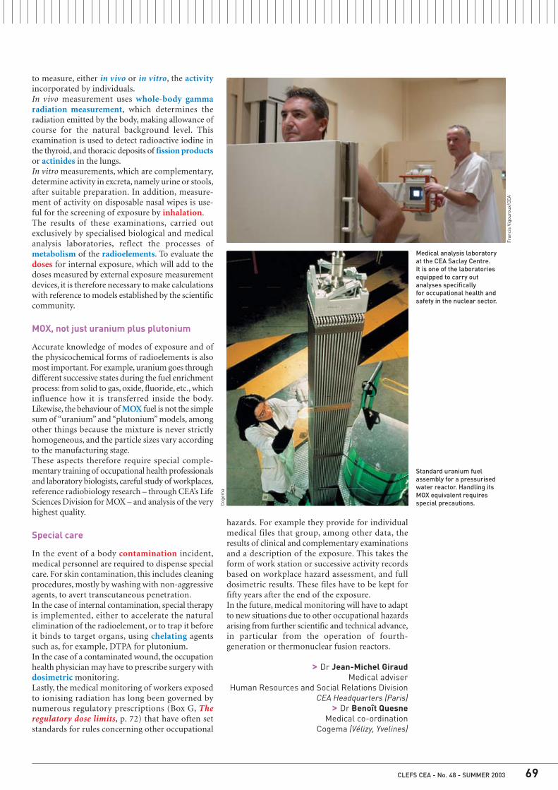
Medical analysis laboratory at the CEA Saclay Centre. It is one of the laboratoriesequipped to carry outanalyses specifically for occupational health andsafety in the nuclear sector.
Standard uranium fuelassembly for a pressurisedwater reactor. Handling itsMOX equivalent requiresspecial precautions.

Everything on the earth’s surface hasalways been exposed to the action
of ionising radiation from natural sources.Natural radiation, which accounts for85.5% of total radioactivity (natural plusartificial), is made up of 71% telluric radi-ation and about 14.5% cosmic radiation.The radionuclides formed by theinteraction of cosmic rays arriving fromstars, and especially the Sun, withthe nuclei of elements present in theatmosphere (oxygen and nitrogen) are,in decreasing order of dose (Box F, Fromrays to dose) received by the population,carbon-14, beryllium-7, sodium-22 andtritium (hydrogen-3). The last two areresponsible for only very low doses.Carbon-14, with a half life of 5,730 years,is found in the human body. Its activityper unit mass of carbon has varied overtime: it has diminished as carbon dioxideemissions from the combustion of fossilfuels have risen, then was increased byatmospheric nuclear weapon tests.Beryllium-7, with a half life of 53.6 days,falls onto the leaf surfaces of plantsand enters the body by ingestion (Box B,Human exposure routes). About 50 Bq(becquerels) per person per year ofberyllium-7 are ingested.The main or “primordial” radionuclidesare potassium-40, uranium-238 andthorium-232. Along with their radioactivedecay products, these elements arepresent in rocks and soil and aretherefore found in many buildingmaterials. Their concentrations aregenerally very low, but vary according tothe nature of the mineral. The gammaradiation emitted by these radionuclidesforms the telluric radiation, which isresponsible for the external exposureof the body. The primordial radionuclidesand many of their long-lived descendants
Natural and artificial radioactivityA
are also found in trace amounts indrinking water and plants: this resultsin an internal exposure by ingestion, plusan additional low exposure by inhalationof airborne suspended dust particles.Potassium-40 is a beta and gammaemitter with a half life of 1.2 thousandmillion years, and has no radioactivedescendants. This radioactive isotopemakes up 0.0118% of all naturalpotassium, and enters the body byingestion. The mass of natural potassiumin the human body is independent of thequantity ingested.Uranium-238 is an alpha emitter witha half life of 4.47 thousand millionyears. It has thirteen main alpha-,beta- and gamma-emitting radioactivedescendants, including radon-222(3.82 days) and uranium-234 (0.246 mil-lion years). Uranium-238 and its twodescendants thorium-234 (24.1 days)and protactinium-234m(1) (1.18 min),and uranium-234 are essentially incor-porated by ingestion and are mainlyconcentrated in the bones and kidneys.Thorium-230, derived from uranium-234, is an alpha emitter with a periodof 80,000 years. It is an osteotrope,but enters the body mainly by the pul-monary route (inhalation). Radium-226,a descendant of thorium-230, is analpha emitter with a half life of1,600 years. It is also an osteotrope andenters the body mainly via food. Anotherosteotrope, lead-210 (22.3 years), isincorporated by inhalation thoughmostly by ingestion.Thorium-232 is an alpha emitter witha half life of 14.1 thousand million
years. It possesses ten main alpha-,beta- and gamma-emitting radioactivedescendants including radon-220 (55 s).Thorium-232 enters the body mainlyby inhalation. Radium-228, a directdescendant of thorium-232, is a beta-emitter with a half life of 5.75 years. Itenters the body mainly in food.Radon, a gaseous radioactive descen-dant of uranium-238 and thorium-232,emanates from the soil and buildingmaterials, and along with its short-livedalpha-emitting descendants constitutesa source of internal exposure throughinhalation. Radon is the most abundantsource of natural radiation (about 40%of total radioactivity).The human body contains nearly 4,500Bqof potassium-40, 3,700 Bq of carbon-14and 13 Bq of radium-226 essentiallyimported in food.Natural radiation is supplemented by ananthropic component, resulting from themedical applications of ionising radia-tion and to a lesser extent from thenuclear industry. It accounts for about14.5% of the total radioactivity world-wide, but much more in the developedcountries. In the medical field (more than1 mSv/year on average in France), irra-diation by external sources predo-minates: radiodiagnosis (X-rays) andradiotherapy, long based on cæsium-137 and cobalt-60 sources, but now moreand more often using linear accelera-tors. Irradiation by internal routes (curie-therapy with iridium-192) has morespecialised indications (cervical cancer,for example). The metabolic and physico-chemical properties of some twentyradionuclides are put to use for medicalactivities and in biological research. Themedical applications comprise radio-diagnostics (scintigraphy and radio-
(1) m for metastable. A nuclide is saidmetastable when a transition delay existsbetween the excited state of the atom and the stable one.

A
immunology), and treatment, includingthyroid disorders using iodine-131,radioimmunotherapy in certain blooddiseases (phosphorus-32) and thetreatment of bone metastasis with stron-tium-89 or radiolabelled phosphonatesalongside other uses of radiopharma-ceuticals. Among the most widely usedradionuclides are: technetium-99m(half life 6.02 hours) and thallium-201(half life 3.04 days) (scintigraphy), iodine-131 (half life 8.04 days) (treatment ofhyperthyroidism), iodine-125 (half life60.14 days) (radioimmunology), cobalt-60 (half life 5.27 years) (radiotherapy),and iridium-192 (half life 73.82 days)(curietherapy). The average contributionof radiological examinations to totalradioactivity amounts to 14.2%.The early atmospheric nuclear weapontests scattered fallout over the wholeof the earth’s surface and causedthe exposure of populations and thecontamination of the food chain bya certain number of radionuclides,most of which, given their shortradioactive half lives, have now vanished.There remain cæsium-137 (30 years),strontium-90 (29.12 years), somekrypton-85 (10.4 years) and tritium(12.35 years), and the isotopes ofplutonium (half lives 87.7 years to24,100 years). Currently, the dosescorresponding to the fallout fromthese tests are essentially attributableto fission products (cæsium-137) andto carbon-14, rather than activationproducts and plutonium.In the Chernobyl accident (Ukraine),which occurred in 1986, the totalradioactivity dispersed into the atmos-phere was of the order of 12 milliardmilliard (1018) becquerels over a periodof 10 days. Three categories of radionu-
clides were disseminated. The first con-sisted of volatile fission products suchas iodine-131, iodine-133 (20.8 hours),cæsium-134 (2.06 years), cæsium-137,tellurium-132 (3.26 days). The secondwas composed of solid fission productsand actinides released in much smalleramounts, in particular the strontiumisotopes 89Sr (half life 50.5 days) and90Sr, the ruthenium isotopes 103Ru(half life 39.3 days) and 106Ru (halflife 368.2 days), and plutonium-239(24,100 years). The third category wasrare gases which although they repre-sented most of the activity released,were rapidly diluted in the atmosphere.They were mainly xenon-133 (5.24 days)and krypton-85.The contributions of the early atmos-pheric nuclear weapon tests and theChernobyl accident to the total radio-activity are roughly 0.2% (0.005 mSv) and0.07% (0.002 mSv) respectively.The whole of the nuclear-poweredelectricity production cycle representsonly about 0.007% of total radioactivity.Almost all the radionuclides remainconfined inside the nuclear reactorsand the fuel cycle plants. In a nuclearreactor, the reactions that take placeinside the fuel yield transuranics.Uranium-238, which is non-fissile,can capture neutrons to give in particu-lar plutonium isotopes 239Pu, 240Pu(half life 6,560 years) and 241Pu (halflife 14.4 years), and americium-241(432.7 years). The main fission productsgenerated by the fission of uranium-235(704 million years) and plutonium-239are iodine-131, cæsium-134, cæsium-137, strontium-90 and selenium-79(1.1 million years).The main radionuclides present inreleases, which are performed in a
very strict regulatory framework are,in liquid release, tritium, cobalt-58(70.8 days), cobalt-60, iodine-131,cæsium-134, cæsium-137 and silver-110m (249.9 days). In gaseous releasescarbon-14 is the most abundantradionuclide, emitted most often ascarbon dioxide. In all the reactors in theworld, the total production of radiocarbondioxide amounts to one tenth of theannual production formed naturally bycosmic radiation.In addition, certain radionuclidesrelated to the nuclear industry exhibitchemical toxicity (Box D, Radiologicaland chemical toxicity).
Classical scintigraphy performed at theFrédéric-Joliot Hospital Service (SHFJ). Thegamma-ray camera is used for functionalimaging of an organ after administration,usually by the intravenous route, of aradioactive drug (radiopharmaceutical) tothe patient. The radionuclides used arespecific to the organ being studied: forexample, technetium-99m for the kidneysand bones, thallium-201 for the myocardium.The injected radiopharmaceutical emitsgamma photons, which are captured by two planar detectors placed at 180° or 45°according to the examination.
Laur
ence
Méd
ard/
CEA
(next)

Human exposure, i.e., the effect onthe body of a chemical, physical
or radiological agent (irrespective ofwhether there is actual contact), canbe external or internal. In the case ofionising radiation, exposure results inan energy input to all or part of the body.There can be direct external irradiationwhen the subject is in the path ofradiation emitted by a radioactivesource located outside the body. Theperson can be irradiated directly or afterreflection off nearby surfaces.The irradiation can be acute or chronic.The term contamination is used todesignate the deposition of matter (hereradioactive) on structures, surfaces,objects or, as here, a living organism.Radiological contamination, attributableto the presence of radionuclides, canoccur by the external route from the
receptor medium (air, water) andvector media (soils, sediments, plantcover, materials) by contact with skinand hair (cutaneous contamination),or by the internal route when theradionuclides are intaken, by inhalation(gas, particles) from the atmosphere,by ingestion, mainly from foods andbeverages (water, milk), or by pen-etration (injury, burns or diffusionthrough the skin). The term intoxicationis used when the toxicity in question isessentially chemical.In the case of internal contamination,the dose delivered to the body over time(called the committed dose) is calcu-lated for 50 years in adults, and untilage 70 years in children. The para-meters taken into account for the cal-culation are: the nature and the intakenquantity of the radionuclide (RN), its
chemical form, its effective half life(1)
in the body (combination of physical andbiological half lives), the type of radi-ation, the mode of exposure (inhalation,ingestion, injury, transcutaneous), thedistribution in the body (deposition intarget organs or even distribution), theradiosensitivity of the tissues and theage of the contaminated subject.Lastly, the radiotoxicity is the toxicitydue to the ionising radiation emitted bythe inhaled or ingested radionuclide.The misleading variable called potentialradiotoxicity is a radiotoxic inventorythat is difficult to evaluate and madeimprecise by many uncertainties.
(1) The effective half life (Te) is calculated from the physical half life (Tp)and the biological half life (Tb) by 1 / Te = 1 / Tp + 1 / Tb.
Human exposure routesB

From rays to doseF
Radioactivity is a process by whichcertain naturally-occurring or
artificial nuclides (in particular thosecreated by fission, the splitting of aheavy nucleus into two smaller ones)undergo spontaneous decay, with arelease of energy, generally resultingin the formation of new nuclides.Termed radionuclides for this reason,they are unstable owing to the numberof nucleons they contain (protons andneutrons) or their energy state. Thisdecay process is accompanied bythe emission of one or more types ofradiation, ionising or non-ionising,and (or) particles. Ionising radiationis electromagnetic or corpuscularradiation that has sufficient energy toionise certain atoms of the matter inits path by stripping electrons fromthem. This process can be direct (thecase with alpha particles) or indirect(gamma rays and neutrons).Alpha radiation, consisting of helium-4nuclei (two protons and two neutrons),has low penetrating power and isstopped by a sheet of paper or theoutermost layers of the skin. Its pathin biological tissues is no longer thana few tens of micrometres. Thisradiation is therefore strongly ionising,i.e., it easily strips electrons from theatoms in the matter it travels through,because the particles shed all theirenergy over a short distance. Forthis reason, the hazard due to
radionuclides that are alpha emittersis internal exposure.Beta radiation, made up of electrons(beta minus radioactivity) or positrons(beta plus radioactivity), has moderatepenetrating power. The particlesemitted by beta emitters are stoppedby a few metres of air, aluminium foil,or a few millimetres of biologicaltissue. They can therefore penetratethe outer layers of the skin. Gamma radiation composed of highenergy photons, which are weaklyionising but have high penetratingpower (more than the X-ray photonsused in radiodiagnosis), can travelthrough hundreds of meters of air.Thick shielding of concrete or lead isnecessary to protect persons. The interaction of neutron radiationis random, and so it is stopped only bya considerable thickness of concrete,water or paraffin wax. As it is electri-cally neutral, a neutron is stopped inair by the nuclei of light elements, themass of which is close to that of theneutron.• The quantity of energy delivered byradiation is the dose, which is evalu-ated in different ways, according towhether it takes into account the quan-tity of energy absorbed, its rate of deliv-ery, or its biological effects. • The absorbed dose is the quantityof energy absorbed at a point perunit mass of matter (inert or living),
according to the definition of theInternational Commission on RadiationUnits and Measurements (ICRU). It isexpressed in grays (Gy): 1 gray is equalto an absorbed energy of 1 joule per kilo-gramme of matter. The organ absorbeddose is obtained by averaging the dosesabsorbed at different points accordingto the definition of the InternationalCommission on Radiological Protection(ICRP).• The dose rate, dose divided by time,measures the intensity of the irradiation(energy absorbed by the matter per unitmass and per unit time). The legal unitis the gray per second (Gy/s), but thegray per minute (Gy/min) is commonlyused. Also, radiation has a higherrelative biological effectiveness (RBE)if the effects produced by the samedose are greater or when the dosenecessary to produce a given effectis lower. • The dose equivalent is equal to thedose absorbed in a tissue or organmultiplied by a weighting factor, whichdiffers according to the nature of theradiation energy, and which ranges from1 to 20. Alpha radiation is consideredto be 20 times more harmful thangamma radiation in terms of itsbiological efficiency in producing random(or stochastic) effects. The equivalentdose is expressed in sieverts (Sv). • The effective dose is a quantityintroduced to try to evaluate harm

Technicians operating remote handling equipment on a line at the Atalante facility at CEA Marcoule. The shielding of the lines stops radiation.The operators wear personal dosimeters to monitor the efficacy of the protection.
Foul
on/C
EA
in terms of whole-body stochasticeffects. It is the sum of equivalent dosesreceived by the different organs andtissues of an individual, weighted bya factor specific to each of them(weighting factors) according to itsspecific sensitivity. It makes it possibleto sum doses from different sources,and both external and internalradiation. For internal exposuresituations (inhalation, ingestion), theeffective dose is calculated on thebasis of the number of becquerels
incorporated of a given radionuclide(DPUI, dose per unit intake). It isexpressed in sieverts (Sv). • The committed dose, as a result ofinternal exposure, is the cumulateddose received in fifty years (for workersand adults) or until age 70 (for thoseaged below 20) after the year ofincorporation of the radionuclide,unless it has disappeared by physicalshedding or biological elimination.• The collective dose is the dosereceived by a population, defined
as the product of the number ofindividuals (e.g., those working in anuclear plant, where it is a usefulparameter in the optimisation andapplication of the ALARA system) andthe average equivalent or effectivedose received by that population, or asthe sum of the individual effectivedoses received. It is expressed in man-sieverts (man.Sv). It should be usedonly for groups that are relativelyhomogeneous as regards the natureof their exposure.
F (next)

Radiological and chemical toxicity
The chemical toxics linked to thenuclear industry include uranium
(U), cobalt (Co), boron (B), used for itsneutron-absorbing properties in theheat-exchange fluids of nuclear powerplants, beryllium (Be), used to slowneutrons, and cadmium (Cd), used tocapture them. Boron is essential for thegrowth of plants. Cadmium, like lead(Pb), produces toxic effects on the centralnervous system. When the toxicity of anelement can be both radiological andchemical, for example that of plutonium(Pu), uranium, neptunium, technetiumor cobalt, it is necessary wheneverpossible to determine what toxiceffects are radiological, what arechemical, and what can be eitherradiological or chemical (see Limits ofthe comparison between radiological andchemical hazards).For radioactive elements with longphysical half lives, the chemical tox-icity is a much greater hazard than theradiological toxicity, as exemplified byrubidium (Rb) and natural uranium.
Thus the chemical toxicity of uranium,which is more important than its radio-logical toxicity, has led the French regu-lators to set the ingested and inhaledmass limits for uranium in chemicalcompounds at 150 mg and 2.5 mg perday respectively, regardless of the iso-topic composition of the element.
Certain metals or metalloids that arenon-toxic at low concentrations canbecome toxic at high concentrations orin their radioactive form. This is thecase for cobalt, which can be genotoxic,selenium (Se) (naturally incorporatedin proteins or RNA), technetium (Tc)and iodine (I).
D
Two-dimensional gel electrophoresis image analysis carried out in the course of nucleartoxicology work at CEA Marcoule Centre in the Rhone Valley.
Cyr
ille
Dup
ont/
CEA

The regulatory dose limitsG
Individual protection against thedangers of ionising radiation is based
on two principles: (i) making surea given radiation source irradiatesexposed persons as little as possible(principle of optimisation), and (ii)making sure the exposure of a givenindividual remains below a certain levelirrespective of the radiation source(principle of the dose limit). These two principles are set outin the ICRP 60 recommendationpublished in November 1990(1) by the International Commission onRadiological Protection, the interna-tionally recognised reference in thedomain, and taken up in the Euratom96/29 European directive of May 131996. The provisions of this directivewere transposed into French law bythe order of March 28 2001, the decreeof March 8 2001 (modifying that of June20 1966) and the decree of March 312003, which modify the public healthand work codes accordingly. Expressed in sieverts (Sv), the limitsare of two sorts, global and local. Globallimits are expressed in values ofeffective dose (Box F). It represents theacceptable risk level concerning thecarcinogenic effect of ionising radiation.It is 20 mSv in 12 months for workers(2)
in the nuclear field (in the broad sense)and 1 mSv per year for the generalpublic. For a certain number of tissuesand organs (skin, hands and feet, eyelens), a local limit is set with referenceto deterministic risks of ionisingradiation, namely radiodermatitis andcataract. This dose equivalent is thusset at 500 mSv for the skin and for thehands and feet, and 150 mSv for theeye lens. These values are ten timeslower for the general public. These
limits are for exposure resulting fromhuman activities other than medicalexposure(3).The effective dose takes into accountboth external exposure and internalexposure. For internal exposure, there are tablessetting limits for each radionuclide,mode of exposure (inhalation-ingestion)and age, taking into account theirranging “transferability” in biologicalmedia, and the dose per unit intake(DPUI) coefficients expressed in sievertsper becquerel (Sv / Bq), the becquerelbeing the unit of activity.They indicate the internal dose that is“committed” for 50 years in adults andup to age 70 for children, taking intoaccount the effective half life of theradionuclide in question. Because ofchildren’s greater susceptibility toradiation and the possibility of longerexposure for radionuclides with longeffective half lives, the most restrictiveannual intake limits are for infants
aged up to one year, and the leastrestrictive for adults from age 17 asprescribed in the ICRP 67 publicationof 1993.The “inhalation” and “ingestion” DPUIvalues take respectively into accountthe new values of digestive absorptionand the latest lung model(4) of theICRP. From these regulatory limits, radio-protection experts can calculate“derived” limits of levels in air or onsurfaces, for example, for internalexposure hazards.
Gon
in/C
EA
(1) Superseding ICRP 26 published in 1977.
(2) Persons directly assigned to work withionising radiation in industry, research andmedicine.
(3) The treatment of hyperthyroidism byirradiation, for example, involves an organdelivered dose of 70,000 mSv!
(4) Publication ICRP 66 of 1994 on themodelling of the human respiratory tractfor radiological protection, whichsupersedes the lung model of ICRP 30.
Passing through a detector frame for individual contamination at the exit from a controlledarea – here the Osiris reactor at CEA Saclay Centre – is a regulatory obligation.
Dosicard dosimeter for real-timedosimetric monitoring.
Gon
in/C
EA