special article for four sixes -...
TRANSCRIPT

418
Orthodontic cases involving the extractionof permanent first molars are thought to be technicallymore difficult to treat and that even a good result is insome way a compromise. Many cases that would bene-fit from this approach because of the doubtful long-termprognosis of the molars are treated with the extraction ofhealthy premolars (Fig 1). The avoidance of first molarcases may be due to a number of factors including thefollowing: operator comfort with premolar extractioncases, lack of experience in handling molar extractioncases, and the inter-dependence for patients betweenendodontists, orthodontists, and crown and bridge spe-cialists. This last factor is rarely an issue in the UnitedKingdom but may increase in the future as SpecialistRegistration has in 1998 become a reality.
The extraction of first permanent molar teeth accountsfor a considerable proportion of cases treated within theNational Health Service. An analysis of patients referredto consultant orthodontists found that nearly 12% of allextraction cases involved first permanent molars.1
The aim of this article is to discuss a rationale forextraction of first molars and to highlight some of theproblems and pitfalls that surround provision of appli-ance therapy for these cases.
LITERATURE REVIEW
Approaches to first molar extraction cases rangebetween unbridled enthusiasm claiming that the extrac-tion of all 4 first permanent molar teeth at the age of 101⁄2years prevented not only malocclusion but also dentaldecay and pyorrhea2 to total skepticism suggesting thatsignificant occlusal disturbance, an increase in caries,and detrimental change in facial contour and appearancewere all inevitable as a result of this intervention.3
“First permanent molar extractions doubling thetreatment time and halving the prognosis” was thephrase coined by Mills.4 This statement may have beencorrect when orthodontics involved removable appli-ances but now has little relevance. On the other hand,Daugaard-Jensen5 suggested that first molar cases areno more time consuming than 4 premolar cases and inmany cases offer distinct advantages in terms ofanchorage management. Houston et al6 suggested thatchildren undergoing first permanent molar extractionsoften had a deprived social background and that theyshowed a reduced interest in their dental care. Thisoften presented difficulties in providing anything otherthan the most basic of orthodontic treatment thusinevitably leading to an orthodontic compromise.
Space closure after the extraction of the first perma-nent molar teeth has been studied in some detail and hasled to conclusions that satisfactory closure of spaces wasbest achieved on children and young adults.7 Adultsshowed less bone apposition when moving second molarteeth into the narrowed space, and poor maintenance ofthe closed space and, in some cases, resorption of thesecond molar roots was also noted. Other authors8 con-cluded that significant if not always complete closurecould be achieved with the roots of the second molarteeth moving almost twice as far as the crowns. Theyagreed that most cases showed a crestal bone loss mesialto the second molars posttreatment but suggested thatroot resorption of the second molars was only minimal.
Extraction of unopposed maxillary first permanentmolars after the removal of the mandibular counterpartwas thought to prevent the likely over eruption of themaxillary tooth.9 Compensating extractions were notautomatically advised after the loss of the maxillaryfirst permanent molar because space closure in themandibular arch was more problematic.
The effect of various extraction patterns on provisionof space both anteriorly and posteriorly within the archeswas discussed in some detail by Williams and Hosila.10
They highlighted the fact that first molar extraction casesare likely to have less effect on the profile than premolar
SPECIAL ARTICLE
For four sixes
Paul Jonathan Sandler, BDS(Hons), MSc, FDSRCPS, DOrth, MOrth,a Robert Atkinson, BDS(Hons),LDSRCS, FDSRCS,b and Alison Margaret Murray, BDS, MSc, FDSRCPS, DOrth, MOrthc
Chesterfield, UK
This article reviews the literature on orthodontic treatment involving extraction of first molars and highlightsmany of the clinical considerations when treating such cases. Case reports illustrate the potential problemsand indicate some solutions. (Am J Orthod Dentofacial Orthop 2000;117:418-34)
aConsultant Orthodontist, Royal Hospital Chesterfield.bSenior House Officer, Royal Hospital Chesterfield.cConsultant Orthodontist, Derbyshire Royal Infirmary.Reprint requests to: Paul Jonathan Sandler, BDS (Hons), MSc, FDSRCPS,DOrth, MOrth, Consultant Orthodontist, Royal Hospital Chesterfield, Calow,Chesterfield, S44 5BL United Kingdom; e-mail, [email protected] © 1999 by the American Association of Orthodontists.0889-5406/99/$8.00 + 0 8/1/97617doi.10.1067/mod.2000.97617

American Journal of Orthodontics and Dentofacial Orthopedics Sandler, Atkinson, and Murray 419Volume 117, Number 4
extraction cases. Also in their cases involving first molarextractions there was about a 90% chance of successfulthird molar eruption compared with approximately 55%chance with cases involving premolar extractions.
CLINICAL INDICATIONS
There are many clinical situations in which extrac-tion of first permanent molars should at least be con-sidered and these can be summarized as follows:
Fig 1. Premolars extracted despite limited prognosis offirst molars.
Fig 2. Gentle forces essential regardless of the methodof space closure.
Fig 3. Nance button palatal arch will maximize intraoralanchorage.
Fig 4. Upper second molars almost completely replacefirst molars regardless of timing of extraction.
Fig 5. Timing of lower first molar extraction is importantif spontaneous space closure is desired.

420 Sandler, Atkinson, and Murray American Journal of Orthodontics and Dentofacial OrthopedicsApril 2000
• Extensively carious first molars• Hypoplastic first molars• Heavily filled first molars where premolars are per-
fectly healthy• Apical pathoses or root treated first molars• Crowding at the distal part of the arches and wisdom
teeth reasonably positioned• High maxillary/mandibular planes angle• Anterior open bite cases
Whether first molars are extracted is dependent onmany factors, including the patient’s attitude to fixedappliance therapy, the standard of oral hygiene, theamount and site of crowding, and the presence orabsence of other permanent teeth. The patients suit-ability for this invariably lengthy course of fixed appli-ance therapy must be considered to ensure that thebenefits of treatment far outweigh the potential risks.
POTENTIAL PROBLEMSLower Arch Space Closure
Space closure is perhaps the most challenging aspect offirst molar extraction cases. When the lower second molarsare subjected to a mesially directed force, there is always atendency for them to both tilt mesially and roll lingually.This tendency can be reduced by the use of full size (19/25)
stainless steel arch wires. Active space closure should rarelybe attempted before the patient has full alignment of all thelower teeth and is in this full-sized working arch wire.
The new McLaughlin, Bennett, Trevisi, (3MUnitek; Monravia, Calif) prescription should help themolar position during space closure in that they arespecifically designed to reduce molar lingual roll con-siderably. The second molar prescription has 10° lin-gual crown torque, which is some 20° to 25° lesstorque than other popular prescriptions.
When using either traction ligatures or nickel tita-nium coil springs, space closing forces should always
Fig 6. Class 2 elastics to lingual of lower molars tocounter lingual roll.
Fig 7. Stainless steel tubing prevents arch wire distor-tion and movement.
Fig 8. Distal twister aids turning in of arch wires

American Journal of Orthodontics and Dentofacial Orthopedics Sandler, Atkinson, and Murray 421Volume 117, Number 4
be gentle, which should once again minimize undesir-able side effects (Fig 2). In many first molar cases thetotal treatment time is determined by the time taken tobring the lower second molars in a good relationshipwith the lower second premolars.
Upper Arch Space Closure
Closure of the upper first molar extraction spaces israrely time consuming. Indeed, because space closureoccurs so readily, the extraction of upper first molars
Fig 9. Pretreatment records of case 1.

422 Sandler, Atkinson, and Murray American Journal of Orthodontics and Dentofacial OrthopedicsApril 2000
often does not provide much more than a few millimeterseither side of the arch to relieve anterior crowding or tocorrect an increased overjet. If a reasonable amount of theextraction space is required, consideration should begiven to provision of a palatal arch with Nance button onthe second molars (Fig 3). This should provide severalextra millimeters of space on either side of the arch com-pared with cases where anchorage was not reinforced.
If all the first molar space is required either forrelief of crowding or overjet reduction, headgearmust be provided to the upper second molars to pre-vent them moving mesially. In these cases, thepatient’s compliance with headgear will determinethe ultimate success or failure of treatment. Thismust be borne in mind when carrying out the formalspace analysis; if much more than 8 mm of space isrequired on either side, some other approach to treat-ment may need to be considered.
Timing of Extractions
If the upper second molars are unerupted at the timeof extraction of the upper first molars, they will almostcompletely replace them, thus contributing little spacefor correction of the malocclusion (Fig 4). It has beensuggested that effective distalization of upper premo-lars with a removable appliance can be achieved if thefirst molars are extracted early.11 This method, how-ever, depends totally on superb patient cooperationwith the patient wearing the headgear for at least 12 to
14 hours per day. Anything short of this and most if notall the space provided by the extractions will be lost.
If there is a space requirement in the upper archtherefore, extraction of the first molars must be delayeduntil the second molars have erupted sufficiently to
Fig 9. Con’d
Fig 10. Power chain to move the canine occlusally.
American Journal of Orthodontics and Dentofacial Orthopedics Sandler, Atkinson, and Murray 423Volume 117, Number 4
allow a palatal arch with Nance button or headgear tobe placed. With modern medicaments and pulp man-agement techniques, it should be possible to delayextractions and keep first molars in almost every casethat will direct the second molars into their normalposition. This will subsequently provide useful spaceafter extraction of the first molars.
If space requirement in the upper arch is minimal,earlier extraction will allow “nature” to assist withmuch of the space closure. The second molars are oftenquite high and only need to alter their eruption pathwayslightly more mesially to allow them to erupt almostinto the first molar sockets.
In the lower arch, timing of extractions is also crit-ical. It is unlikely that the lower second molars willcompletely replace the lower first molars after theirextraction as they have a much more vertical path oferuption. If little or no space is required in the lowerarch for correction of the malocclusion, it is oftenadvisable to extract lower first molars early, when thebifurcation dentine on second molars is calcifying andthe roots are about half formed. This will maximizespontaneous space closure in the lower arch thus min-imizing retraction of the lower labial segment, whichis an undesirable side effect often seen during spaceclosure (Fig 5).
If, after space analysis, it is calculated that all thelower first molar space is required for relief of crowd-ing, a lingual arch may be necessary until the premo-lars and canines have been retracted sufficiently toallow incisor alignment.
Root Paralleling
Toward the end of treatment, if there is any doubtwhether root positions exist, an Orthopantomogramcan be taken to assess whether the long axis secondpremolars and second molars are reasonably parallel.If 8 to 9 mm of space has been closed between thesecond premolar and second molar, there is always aslight tendency for divergence between the two rootsand gentle tip back bends may be placed if necessaryin the final rectangular wires to fully correct the rootpositions. Once space is closed and the crowns arecorrectly positioned, a dead ligature should be placedacross the extraction spaces to hold them closed for afew months. This keeps these teeth in position whilethe gingival fibers reorganize and the bone around theteeth matures in the hope that this will minimize anytendency for the extraction spaces to reopen.
Class II Elastics
If Class II elastics are being used in first molarextraction cases, there is an increased tendency forlingual roll of the lower second permanent molars.Class II elastics should not be used until the patientis in a full-sized (19/25) stainless steel arch wire and,if necessary, buccal crown torque is applied to thelower molars. Another possibility is to run the ClassII elastics from a lingual cleat on the lower molarbands, thus providing some lift to the lingual surfaceof the lower molars thus reducing any tendency tolingual roll (Fig 6).
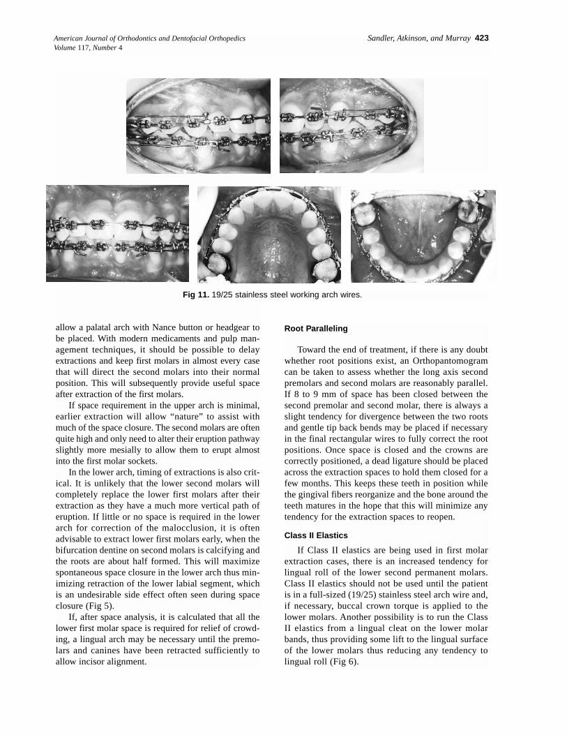
Fig 11. 19/25 stainless steel working arch wires.

424 Sandler, Atkinson, and Murray American Journal of Orthodontics and Dentofacial OrthopedicsApril 2000
Arch Wire Problems
Fig 12. Final records of case 1.
American Journal of Orthodontics and Dentofacial Orthopedics Sandler, Atkinson, and Murray 425Volume 117, Number 4
Arch Wire ProblemsThe long span of arch wire between the second pre-
molar and the second molar can lead to problems suchas trauma to the soft tissues as well as deflection of thearch wire during mastication. This arch wire distortioncan, on occasion, cause movement leading to the wirecoming forward out of one tube and moving distallyand piercing the cheek on the opposite side. Introduc-tion of deflections and asymmetries into the arch will atthe very least delay progression of treatment. Theabove consequences can all be avoided by placing 0.9
mm internal diameter stainless steel tubing over thearch wire in the extraction space. The tube should beonly 1 to 2 mm short of the interbracket span for max-imum rigidity of the arch wire section but also allow-ing some alignment and movement of the teeth (Fig 7).
In addition, the arch wires up to but excluding the full-sized rectangular stainless steel should be annealed andturned down or in at the end. An instrument called a “dis-tal twister” (Instrument number 001-505,American Ortho-dontics, Sheboygan, Wis), which hooks over the annealedend of the wire, allows this to be done with ease (Fig 8).
Fig 12. Con’d
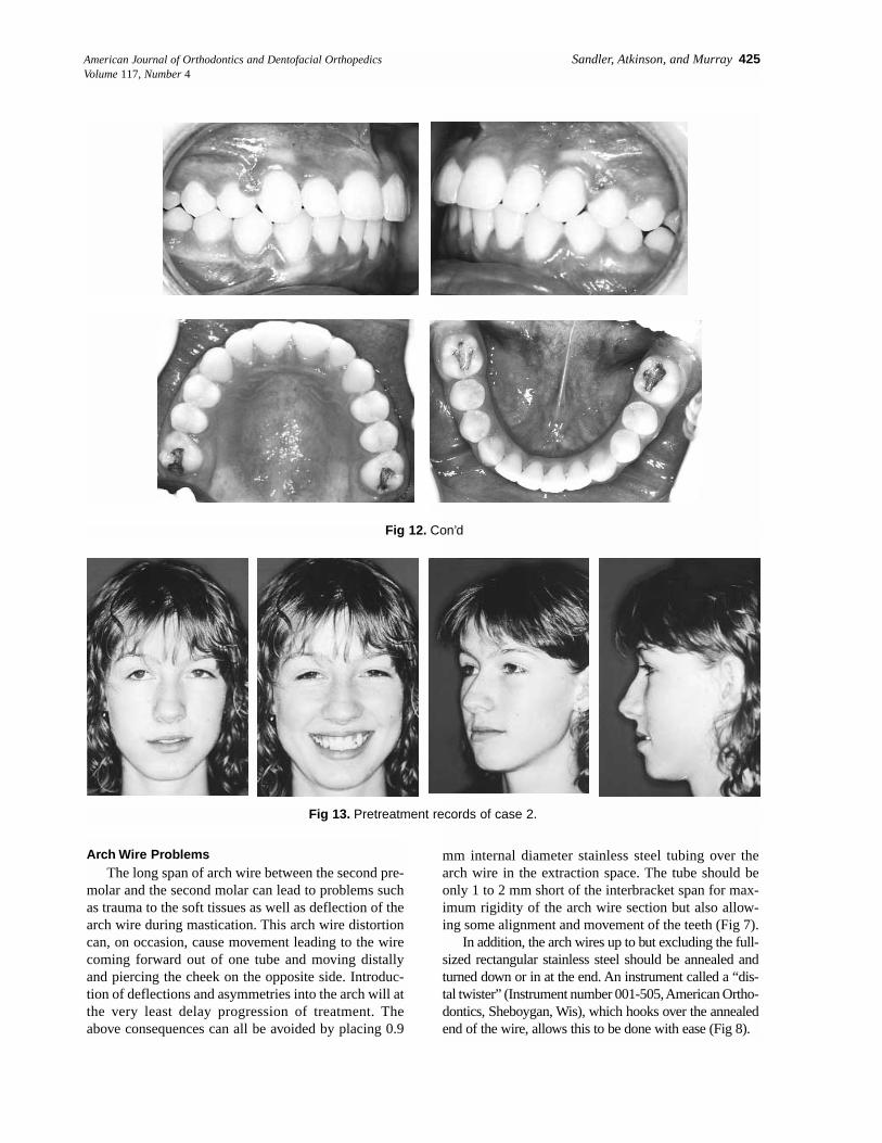
Fig 13. Pretreatment records of case 2.

426 Sandler, Atkinson, and Murray American Journal of Orthodontics and Dentofacial OrthopedicsApril 2000
Fig 13. Con’d

American Journal of Orthodontics and Dentofacial Orthopedics Sandler, Atkinson, and Murray 427Volume 117, Number 4
CASE REPORTS
These cases are used to illustrate the kind of resultthat can be expected with careful case managementafter extraction of 4 first molars.
Case Report 1
The patient presented at 12 years of age11 with aClass II Division II incisor relationship on a skeletal Ibase. There was severe crowding in the upper labialsegment with almost complete exclusion of the upperright canine and partial exclusion of the upper leftcanine. The lower buccal segments were moderatelycrowded. Dental examination revealed caries in 3 of the4 first molar teeth and an OPT radiograph showed thepresence of third molars (Fig 9).
As the first molars were carious we decided toextract them. Careful anchorage management shouldallow sufficient space for relief of crowding, align-ment of the labial segment teeth, and full correction ofthe malocclusion.
We placed fixed appliances in January 1995 and pro-gressed from 0.016 nickel titanium through to 18/25nickel titanium to 19/25 steel. Full-sized stainless steelarch wires were in place by the middle of 1995 at whichstage push coil was used to reopen space for the upperright canine. We did not attempt space closure on the archwires before the 19/25 stainless steel arch wires to mini-mize dumping or arch distortion in the extraction areas.Lengths of 0.9 mm internal diameter stainless steel tub-ing were used in the extraction sites on both of the align-ing arch wires to make the arch wire more rigid thus pre-venting distortion during mastication. To aid correctionof the upper right canine, a crimpable hook was placedon the arch wire in an inverted position and elastic chainattached to it from the bracket on the canine (Fig 10).
Once the anterior teeth had been fully aligned ontothe 19/25 arch wire, space closure was commenced.Traction ligatures were used in all 4 quadrants andrenewed every 6 weeks until full space closure wasachieved (Fig 11). We removed the stainless steel arch
Fig 14. Stainless steel tubing to protect the arch wire
Fig 15. 19/25 stainless steel working arch wires, spaces almost completely closed, Class II elastics for lateral open bite.
428 Sandler, Atkinson, and Murray American Journal of Orthodontics and Dentofacial OrthopedicsApril 2000
wires every second visit to check arch wire coordina-tion and to place gentle reverse curve in the lower archand increased curve in the upper arch. We removed theappliances in August 1996, therefore, the total durationof active treatment to fully correct this malocclusionwas 19 months (Fig 12).
Cephalometric analysis revealed a small favorablechange in the sagittal relationship of the jaws as a resultof the 1° decrease in SNA and 1° increase in SNB. Therewas a small decrease in the maxillary-mandibular planesangle during treatment and superimposition demonstratessome forward mandibular growth that is most welcome.There was marked improvement in the inclinations of theincisors with a concomitant improvement in the interin-cisal angle. The OPT radiograph taken 12 months aftertreatment showed a marked improvement in the positionof the third molars that are expected to erupt into a morethan acceptable position over the next few years.
Case Report 2
The patient presented at 14 years of age in full perma-nent dentition with a Class III incisor relationship on aClass I skeletal base. There was moderate crowding of theupper labial segment and mild to moderate crowding ofthe lower labial segment. The lower first molars had beenrestored, and the OPT revealed the presence and goodmorphologic characteristics of third molars (Fig 13).
We decided that extraction of first molars wouldprovide sufficient room for correction of the malocclu-sion and would also leave the patient with a completelyhealthy dentition. Flexible nickel titanium arch wireswere used to provide initial alignment of the anteriorteeth with the exception of the upper left lateral incisor.
Stainless steel tubing protected the wire in the extrac-tion spaces during this alignment phase (Fig 14). Weprogressed to an upper 19/25 stainless steel arch wire,then a nickel titanium pushcoil was used to reopenspace for the upper lateral incisors.
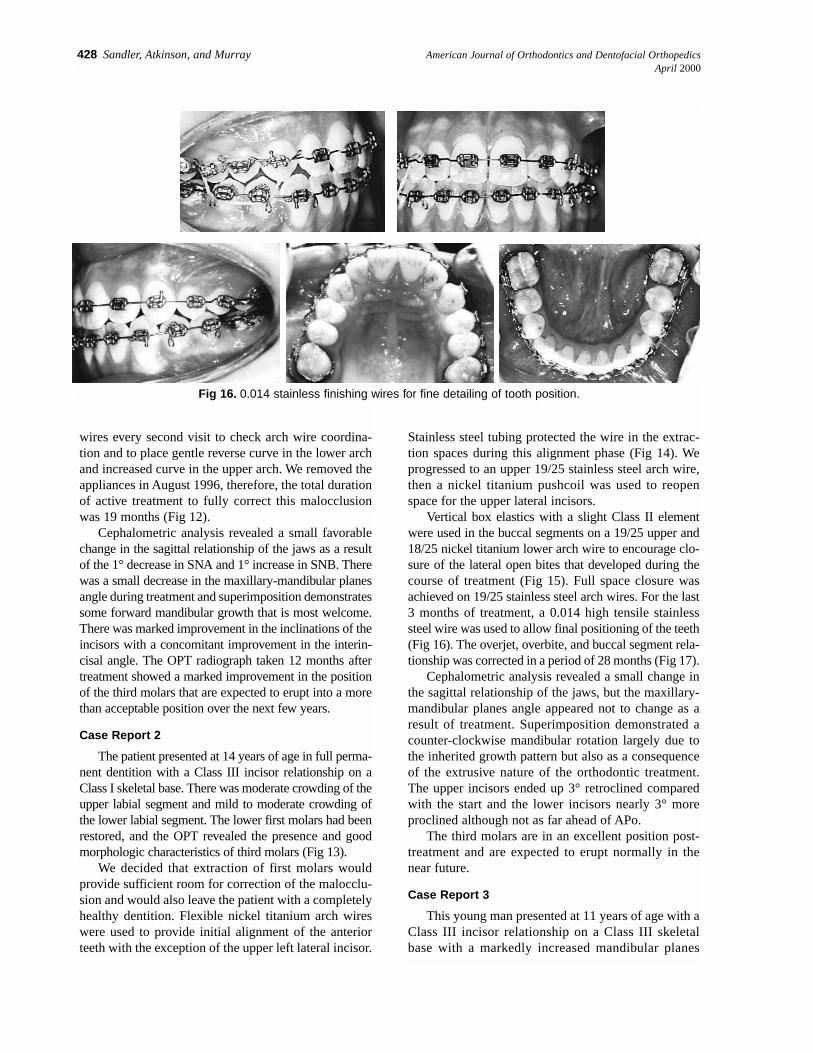
Vertical box elastics with a slight Class II elementwere used in the buccal segments on a 19/25 upper and18/25 nickel titanium lower arch wire to encourage clo-sure of the lateral open bites that developed during thecourse of treatment (Fig 15). Full space closure wasachieved on 19/25 stainless steel arch wires. For the last3 months of treatment, a 0.014 high tensile stainlesssteel wire was used to allow final positioning of the teeth(Fig 16). The overjet, overbite, and buccal segment rela-tionship was corrected in a period of 28 months (Fig 17).
Cephalometric analysis revealed a small change inthe sagittal relationship of the jaws, but the maxillary-mandibular planes angle appeared not to change as aresult of treatment. Superimposition demonstrated acounter-clockwise mandibular rotation largely due tothe inherited growth pattern but also as a consequenceof the extrusive nature of the orthodontic treatment.The upper incisors ended up 3° retroclined comparedwith the start and the lower incisors nearly 3° moreproclined although not as far ahead of APo.
The third molars are in an excellent position post-treatment and are expected to erupt normally in thenear future.
Case Report 3
This young man presented at 11 years of age with aClass III incisor relationship on a Class III skeletalbase with a markedly increased mandibular planes
Fig 16. 0.014 stainless finishing wires for fine detailing of tooth position.

American Journal of Orthodontics and Dentofacial Orthopedics Sandler, Atkinson, and Murray 429Volume 117, Number 4
Fig 17. Final records of case 2.
430 Sandler, Atkinson, and Murray American Journal of Orthodontics and Dentofacial OrthopedicsApril 2000
Fig 17. Con’d
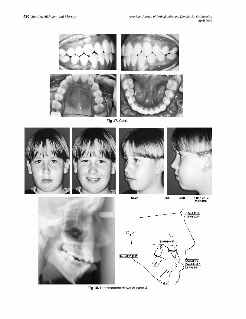
Fig 18. Pretreatment views of case 3.

American Journal of Orthodontics and Dentofacial Orthopedics Sandler, Atkinson, and Murray 431Volume 117, Number 4
angle of 44°. He was in the permanent dentition with anormally inclined upper labial segment, a proclinedlower labial segment, and an anterior open bite of 3mm. The upper and lower right first molars were veryheavily filled, and the lower left first molar was veryheavily decayed (Fig 18).
We appreciated that this young man would be a dif-ficult case to treat and that the prognosis for successfulclosure of the anterior open bite was limited withorthodontics alone. However, the patient was not inter-
ested in considering any form of orthognathic surgery.A decision was made to attempt to get as much correc-tion as possible with orthodontic therapy alone.
We felt that posterior extractions may help withclosure of the anterior open bite and that in this highangle case space closure was unlikely to be a majorproblem. We decided that the patient would benefitfrom the removal of all 4 first molars to relieve thecrowding in the upper labial segment and to improvethe interarch relationships.
Fig 18. Con’d.
Fig 19. 0.016 nickel titanium for initial alignment, tubing to protect arch wire.

432 Sandler, Atkinson, and Murray American Journal of Orthodontics and Dentofacial OrthopedicsApril 2000
We placed the appliances in December 1994 (Fig19). We made very slow progress through flexible(0.016) and 18/25 nickel titanium arch wires (Fig 20)as cooperation with treatment was not really forthcom-ing, and the patient had 17 breakages repaired duringhis course of treatment. We finally progressed up to19/25 stainless steel. At each appointment chlorhexi-dine varnish was applied to the upper incisor bracketsin the hope that this would reduce enamel damage dur-ing treatment (Fig 21). Elastic traction ligatures wereregularly replaced and space closure eventuallyachieved. After 1 visit in 0.014 stainless steel finishingwires to finalize tooth positions (Fig 22), we finallyremoved the appliances in July 1997. Active treatmenttime for correction of the malocclusion was 21⁄ 2 years.We felt that this was acceptable in view of the numberof breakages and the many failed appointments.
Final intraoral photographs show reasonable buccalsegment interdigitation, correction of the labial segmentsand correction of the anterior open bite (Fig 23). Essixupper and lower retainers were used, and the overbite cor-rection will be monitored over the next few years.
Near the end of treatment, cephalometric analysisshows slight retroclination of the upper labial segmentand retroclination of the previously proclined lower labialsegment that was necessary to close the anterior open bite.The mandibular planes angle did not increase during treat-ment although the lower anterior facial height proportionincreased slightly. Superimposition shows closure of theanterior open bite and a moderate change in the jaw posi-tions, which is largely in a vertical direction.
An OPT radiograph taken 2 months before debond-ing shows good root paralleling and some slight rootresorption on the distal root of both lower secondmolars. The treatment had been unnecessarily prolongedas a result of the poor patient cooperation, and this willhave contributed to the root resorption. The upper sec-ond molars were in a reasonable position to the uppersecond premolars and there should now be sufficientroom for the third molars to erupt into the mouth.
SUMMARY
Even with careful planning and execution of treatmentany orthodontic case involving the extraction of first
Fig 20. Second stage alignment with 18/25 nickel titanium.
Fig 21. 19/25 stainless steel working arch wires to achieve space closure.

American Journal of Orthodontics and Dentofacial Orthopedics Sandler, Atkinson, and Murray 433Volume 117, Number 4
Fig 22. Finishing on 0.014 stainless steel, near end oftreatment radiographs.

434 Sandler, Atkinson, and Murray American Journal of Orthodontics and Dentofacial OrthopedicsApril 2000
molars will almost certainly take between 6 and 9months longer than an equivalent case in which 4 pre-molars are extracted. Careful case assessment must beundertaken before treatment to ensure that the bene-fits of treatment will outweigh any potential disad-vantages of this extended treatment. A knowledge ofthe potential problems with first molar cases willallow the necessary action to be taken before many ofthese problems arise.
REFERENCES
1. Bradbury AJ. A current view on patterns of extraction therapy in British health serviceorthodontics. Br Dent J 1985;159:47-50.
2. Wilkinson AA. The first permanent molar again. Br Dent J 1940;8:269-84.3. Salzmann JA. A study of orthodontic and facial changes and effects on dentition attend-
ing the loss of first molars in five hundred adults. J Am Dent Assoc 1938;25:892-905.4. Mills JRE. Principles and practice of orthodontics, 2nd edition, New York: Churchill
Livingstone; 1987. p. 123-5.5. Daugaard-Jensen I. Extraction of first molars in discrepancy cases. Am J Orthod
1973;64:115-36.6. Houston WJB, Stephens CD, Tulley WJ. A textbook of orthodontics, 2nd edition,
Oxford: John Wright; 1992. p. 196-9.7. Stepovich ML. A clinical study on closing edentulous spaces in the mandible. Angle
Orthod 1979;49:227-33.8. Hom BM, Turley PK. The effects of space closure of the mandibular first molar area
in adults. Am J Orthod 1984;4:457-69.9. Mitchell L. An introduction to orthodontics, Oxford, England: Oxford University
Press; 1996. p. 26-8.10. Williams R, Hosila L. The effect of different extraction sites upon incisor retraction.
Am J Orthod 1976;69:388-410.11. Orton H, Carter NE. Initial management of first molar extraction cases. J Clin Orthod
1988;22:230-8.
Fig 23. Final photos of case 3.