sox2 cooperates with chd7 to regulate genes that are mutated in human syndromes
TRANSCRIPT
©20
11 N
atu
re A
mer
ica,
Inc.
All
rig
hts
res
erve
d.
Nature GeNetics ADVANCE ONLINE PUBLICATION �
l e t t e r s
The HMG-box transcription factor Sox2 plays a role throughout neurogenesis� and also acts at other stages of development2, as illustrated by the multiple organs affected in the anophthalmia syndrome caused by SOX2 mutations3–5. Here we combined proteomic and genomic approaches to characterize gene regulation by Sox2 in neural stem cells. Chd7, a chromatin remodeling ATPase associated with CHARGE syndrome6,7, was identified as a Sox2 transcriptional cofactor. Sox2 and Chd7 physically interact, have overlapping genome-wide binding sites and regulate a set of common target genes including Jag1, Gli3 and Mycn, genes mutated in Alagille, Pallister-Hall and Feingold syndromes, which show malformations also associated with SOX2 anophthalmia syndrome or CHARGE syndrome8–�0. Regulation of disease-associated genes by a Sox2-Chd7 complex provides a plausible explanation for several malformations associated with SOX2 anophthalmia syndrome or CHARGE syndrome. Indeed, we found that Chd7-haploinsufficient embryos showed severely reduced expression of Jag1 in the developing inner ear.
As a first step to gain more insight into the transcriptional network in which Sox2 operates, we identified Sox2-interacting proteins in neural stem cells (NSCs). Sox2 is essential for the in vivo maintenance of mouse embryonic and adult NSCs and subsequent neurogenesis1. NSCs are therefore an appropriate cell type in which to study gene regulation by Sox2. NSCs that stably express FLAG-Sox2 (ref. 11) (F-Sox2) have a normal morphology and expressed NSC markers such as Nestin, RC2 (ref. 11) and Pax6 (Supplementary Fig. 1a). To identify interaction partners, we purified F-Sox2 from NSC nuclear extract by a FLAG-affinity–based protocol12, separated proteins by polyacrylamide gel (Supplementary Fig. 1b) and analyzed them using mass spectrometry. We identified 50 Sox2-interacting factors that were specifically present in two F-Sox2 purifications (Table 1 and
Sox2 cooperates with Chd7 to regulate genes that are mutated in human syndromesErik Engelen1,10, Umut Akinci1,10, Jan Christian Bryne2, Jun Hou1, Cristina Gontan3,9, Maaike Moen1, Dorota Szumska4, Christel Kockx5, Wilfred van IJcken5, Dick H W Dekkers6, Jeroen Demmers6, Erik-Jan Rijkers7, Shoumo Bhattacharya4, Sjaak Philipsen1, Larysa H Pevny8, Frank G Grosveld1, Robbert J Rottier3, Boris Lenhard2 & Raymond A Poot1
1Department of Cell Biology, Erasmus Medical Center (MC), Rotterdam, The Netherlands. 2Department of Biology, University of Bergen, Bergen, Norway. 3Department of Pediatric Surgery, Erasmus MC, Rotterdam, The Netherlands. 4Wellcome Trust Centre for Human Genetics, University of Oxford, Oxford, UK. 5Center for Biomics, Erasmus MC, Rotterdam, The Netherlands. 6Proteomics Center, Erasmus MC, Rotterdam, The Netherlands. 7Department of Biochemistry, Erasmus MC, Rotterdam, The Netherlands. 8Department of Genetics, University of North Carolina Neuroscience Center, University of North Carolina at Chapel Hill, Chapel Hill, North Carolina, USA. 9Present address: Department of Reproduction and Development, Erasmus MC, Rotterdam, The Netherlands. 10These authors contributed equally to this work Correspondence should be addressed to R.A.P. ([email protected]).
Received 31 January; accepted 8 April; published online 1 May 2011; doi:10.1038/ng.825
table 1 Interaction partners of sox2 in neural stem cellsProtein Average mascota Average peptidesa
sox2 479 5
spalt (3) 888 12
NurD complex (9) 727 11
trrap complex (3) 421 6
sMrt/Ncor complex (5) 307 6
sWI/sNF complex 271 4
transcription factorsChd7 1,253 20
Cutl1 1,192 17
Dbc1 679 10
Zeb1 312 4
Ctbp1 242 3
Supt16h 187 3
Rfx3 167 2
Sox8 167 3
Hoxa5 166 2
Mef2 159 3
Nfi-β 139 3
Ctbp2 109 2
Nac1 104 2
Tead1 87 1
Snf2h 87 2
Sox5 69 1
Twist1 63 1
Tcf3 46 1
Zfp191 41 1
OtherExp4 1,667 24
Skiv2l2 179 3
Dock7 135 2Dnaja2 93 1aAverages are from three experiments (supplementary table 1). Parentheses indicate the number of identified subunits.
©20
11 N
atu
re A
mer
ica,
Inc.
All
rig
hts
res
erve
d.
2 ADVANCE ONLINE PUBLICATION Nature GeNetics
l e t t e r s
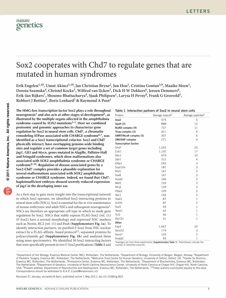
Supplementary Table 1). We validated many of these interactions using an independent method: immunoprecipitation of endogenous Sox2 (Supplementary Table 1). We confirmed the NuRD complex subunits Mi2-β and MTA2 to interact with Sox2 by immunoprecipi-tation protein blot (Fig. 1a,b). Notably, many of the identified Sox2-interacting factors, such as the transcription factors Nfi-β and Twist1 and the chromatin modifying complexes SWI-SNF and SMRT, are involved in neural development (Supplementary Table 2). We con-clude that Sox2 interacts with multiple factors that have importance in neurogenesis. Prominent on the list of Sox2 interactors with many peptides identified (Table 1 and Supplementary Table 1) is Chd7, a member of the family of CHD chromatin remodeling ATPases. Chd7−/− mouse embryos have neural and other defects and die at embryonic day (E) 10.5 (refs. 13,14). In humans, CHD7 haploinsuf-ficiency causes CHARGE syndrome6,7 (incidence ~1 in 10,000), a clustering of coloboma, heart malformation, atresia of the choanae, retarded growth and development, genital anomalies and ear anoma-lies and/or deafness. Chd7 and Sox2 have a similar expression pattern in E14.5 mouse embryos, with high expression in the ventricular zones of the brain, the pituitary gland, the olfactory bulbs, the eyes and the inner ears (Genepaint15; Supplementary Fig. 2 and the references therein). CHARGE syndrome overlaps in several features reported for the anophthalmia syndrome caused by SOX2 mutations, such as malformations of the esophagus and trachea, genital abnormalities and pituitary defects3–5,7, which occasionally leads to a misdiagnosis3. We confirmed that Sox2 interacts with Chd7 by Sox2 immunoprecipi-tation protein blot, Chd7 immunoprecipitation protein blot and GST pull down (Fig. 1b–e). The Sox2-Chd7 interaction was insensitive to ethidium bromide (Fig. 1b,c) and the nuclease benzonase (Fig. 1d) and is therefore unlikely to be mediated by DNA. We subsequently immunoprecipitated Chd7 and analyzed the spectrum of Chd7 bind-ing partners by mass spectrometry. Although the Chd7 immunopre-cipitations were efficient and depleted Chd7 from the nuclear extract, we consistently identified only three transcription factors: Sox2, Olig1 and Zbtb20 (Table 2 and Supplementary Table 3). This suggests that Chd7 is a specialized cofactor for Sox2 and a limited number of other transcription factors in NSCs.
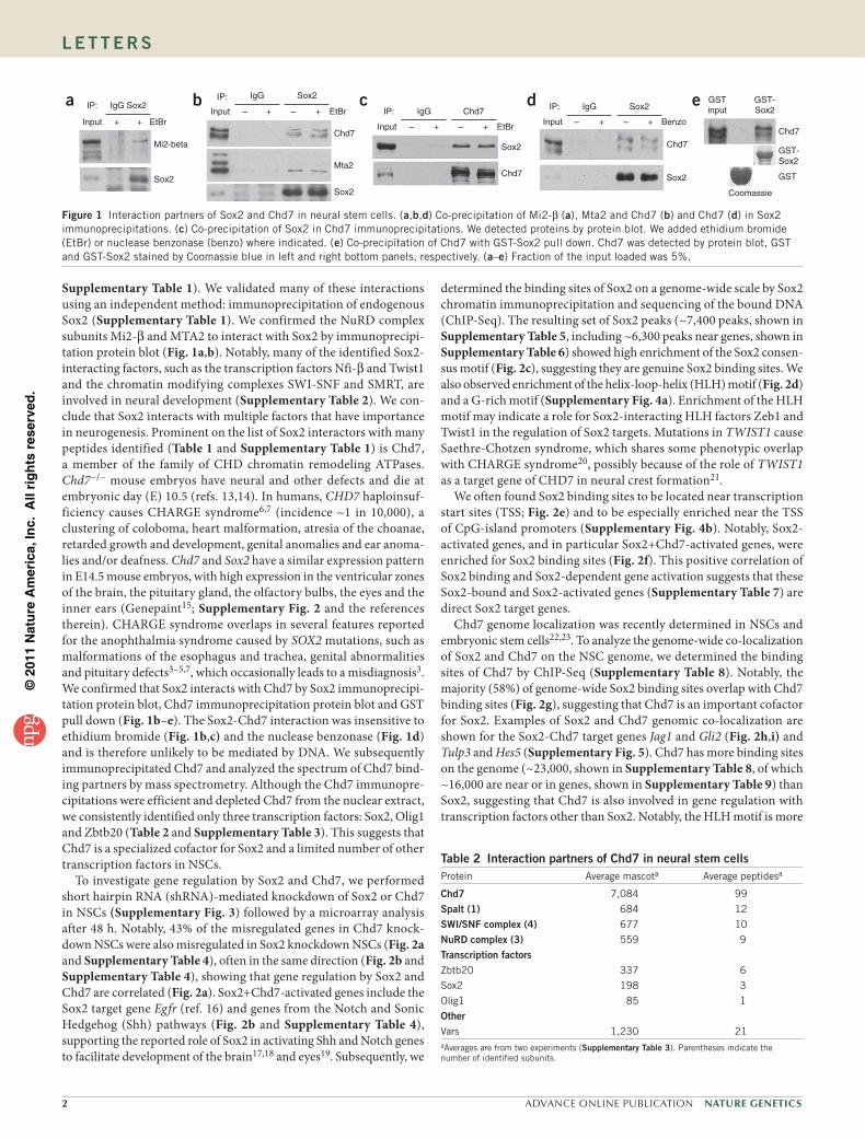
To investigate gene regulation by Sox2 and Chd7, we performed short hairpin RNA (shRNA)-mediated knockdown of Sox2 or Chd7 in NSCs (Supplementary Fig. 3) followed by a microarray analysis after 48 h. Notably, 43% of the misregulated genes in Chd7 knock-down NSCs were also misregulated in Sox2 knockdown NSCs (Fig. 2a and Supplementary Table 4), often in the same direction (Fig. 2b and Supplementary Table 4), showing that gene regulation by Sox2 and Chd7 are correlated (Fig. 2a). Sox2+Chd7-activated genes include the Sox2 target gene Egfr (ref. 16) and genes from the Notch and Sonic Hedgehog (Shh) pathways (Fig. 2b and Supplementary Table 4), supporting the reported role of Sox2 in activating Shh and Notch genes to facilitate development of the brain17,18 and eyes19. Subsequently, we
determined the binding sites of Sox2 on a genome-wide scale by Sox2 chromatin immunoprecipitation and sequencing of the bound DNA (ChIP-Seq). The resulting set of Sox2 peaks (~7,400 peaks, shown in Supplementary Table 5, including ~6,300 peaks near genes, shown in Supplementary Table 6) showed high enrichment of the Sox2 consen-sus motif (Fig. 2c), suggesting they are genuine Sox2 binding sites. We also observed enrichment of the helix-loop-helix (HLH) motif (Fig. 2d) and a G-rich motif (Supplementary Fig. 4a). Enrichment of the HLH motif may indicate a role for Sox2-interacting HLH factors Zeb1 and Twist1 in the regulation of Sox2 targets. Mutations in TWIST1 cause Saethre-Chotzen syndrome, which shares some phenotypic overlap with CHARGE syndrome20, possibly because of the role of TWIST1 as a target gene of CHD7 in neural crest formation21.
We often found Sox2 binding sites to be located near transcription start sites (TSS; Fig. 2e) and to be especially enriched near the TSS of CpG-island promoters (Supplementary Fig. 4b). Notably, Sox2-activated genes, and in particular Sox2+Chd7-activated genes, were enriched for Sox2 binding sites (Fig. 2f). This positive correlation of Sox2 binding and Sox2-dependent gene activation suggests that these Sox2-bound and Sox2-activated genes (Supplementary Table 7) are direct Sox2 target genes.
Chd7 genome localization was recently determined in NSCs and embryonic stem cells22,23. To analyze the genome-wide co-localization of Sox2 and Chd7 on the NSC genome, we determined the binding sites of Chd7 by ChIP-Seq (Supplementary Table 8). Notably, the majority (58%) of genome-wide Sox2 binding sites overlap with Chd7 binding sites (Fig. 2g), suggesting that Chd7 is an important cofactor for Sox2. Examples of Sox2 and Chd7 genomic co-localization are shown for the Sox2-Chd7 target genes Jag1 and Gli2 (Fig. 2h,i) and Tulp3 and Hes5 (Supplementary Fig. 5). Chd7 has more binding sites on the genome (~23,000, shown in Supplementary Table 8, of which ~16,000 are near or in genes, shown in Supplementary Table 9) than Sox2, suggesting that Chd7 is also involved in gene regulation with transcription factors other than Sox2. Notably, the HLH motif is more
table 2 Interaction partners of Chd7 in neural stem cellsProtein Average mascota Average peptidesa
Chd7 7,084 99
spalt (1) 684 12
sWI/sNF complex (4) 677 10
NurD complex (3) 559 9
transcription factorsZbtb20 337 6
Sox2 198 3
Olig1 85 1
OtherVars 1,230 21aAverages are from two experiments (supplementary table 3). Parentheses indicate the number of identified subunits.
GSTinput
Coomassie
GST
GST-Sox2
GST-Sox2
Chd7
ea IP:
Input EtBr
Mi2-beta
Sox2
+ +
IgG Sox2 d IP:
Input – + +–
IgG Sox2
Chd7
Sox2
Benzo
cIP:
Input EtBr– + +–
IgG Chd7
Chd7
Sox2
b IP:
Input EtBr
Chd7
Mta2
Sox2
– + – +
IgG Sox2
Figure 1 Interaction partners of Sox2 and Chd7 in neural stem cells. (a,b,d) Co-precipitation of Mi2-β (a), Mta2 and Chd7 (b) and Chd7 (d) in Sox2 immunoprecipitations. (c) Co-precipitation of Sox2 in Chd7 immunoprecipitations. We detected proteins by protein blot. We added ethidium bromide (EtBr) or nuclease benzonase (benzo) where indicated. (e) Co-precipitation of Chd7 with GST-Sox2 pull down. Chd7 was detected by protein blot, GST and GST-Sox2 stained by Coomassie blue in left and right bottom panels, respectively. (a–e) Fraction of the input loaded was 5%.
©20
11 N
atu
re A
mer
ica,
Inc.
All
rig
hts
res
erve
d.
Nature GeNetics ADVANCE ONLINE PUBLICATION 3
l e t t e r s
enriched in Sox2 peaks that overlap with Chd7 peaks (Supplementary Fig. 6a), suggesting that HLH factors may be involved in the regula-tion of Sox2 targets, especially in the context of Chd7.
The physical interaction of Sox2 and Chd7 and the overlap in regu-lated genes and genomic localization indicated that Sox2 and Chd7 may act synergistically in gene activation. To investigate this further,
a
e
f
g
h
b c
d
Downstream(to +10 kb)
IntergenicInternal
Promoter(–1 kb to +1 kb)
Upstream(–1 to –10 kb)
Sox2 regulated
Sox2+Chd7-regulated
(P < 3 × 10–31)
Chd7 regulated
2,062 299 400
10
8
6
4
2
0
–4 –2Distance to peak (kb)
Hit
coun
ts
0 2 4
6
4
2
0
Hit
coun
ts
–4 –2Distance to peak (kb)
0 2 4
Sox2 ChlP
100 kb15
050
0
Gli2
Chd7 ChlP
Control ChlP (Sox2)
Control ChlP (Chd7)
iSox2 ChlP
20 kb15
050
0
Jag1
Chd7 ChlP
Control ChlP (Sox2)
Control ChlP (Chd7)
% Sox2-bound genes
All genes
Sox2 repressed
Sox2 activated (P < 4.0 × 10–31)
Sox2+Chd7 activated (P < 3.5 × 10⁻⁹)
0 10 20 30 40 50
% genome-wide overlap Sox2 peakswith Chd7 peaks
0 20 40 60 80 100
Aph1b
Sox2 knockdown Chd7 knockdownEgfr
Hey1Jag1
Tulp3Mycn
Gli2
Gli3
RbpjHes5
Notch3Figure 2 Target genes of Sox2 and Chd7 in neural stem cells. (a) Venn diagram of the number of genes misregulated in Sox2 knockdown NSCs and/or Chd7 knockdown NSCs as determined by microarray. The Fisher’s exact test P value for the correlation of gene misregulation by Sox2 or Chd7 is indicated. (b) Heatmap of genes regulated by Sox2 and Chd7. Shades of green and red indicate downregulation and upregulation, respectively. Note that genes are often misregulated in the same direction in Sox2 knockdown NSCs and Chd7 knockdown NSCs. Blue and red labeling indicates members of the Notch pathway or Sonic Hedgehog pathway, respectively. (c) Enrichment of Sox2 consensus DNA motif in Sox2 ChIP-Seq peaks. (d) Enrichment of HLH transcription factor DNA motif in Sox2 peaks. (e) Pie chart of the distribution of Sox2 peaks. (f) Percentage of genes with Sox2 peaks within the different indicated categories of genes on the microarray. Fisher’s exact test P values for correlation with Sox2-bound genes are indicated. (g) Genome-wide overlap of Sox2 peaks with Chd7 peaks. The percentage of the genome-wide Sox2 peaks (supplementary table 6) that overlaps with Chd7 peaks (supplementary table 8) is indicated. (h,i) Localization of Sox2 and Chd7 on Sox2-Chd7 target genes. We plotted sequence reads from the indicated ChIP experiments relative to chromosomal position. Sox2 ChIP, Chd7 ChIP and their corresponding control (IgG) ChIP experiments are shown. Genome locations of the Gli2 locus (h) and Jag1 locus (i) are shown. Arrowheads indicate direction of the locus.
a c
d
b
Rel
ativ
e m
RN
A e
xpre
ssio
n (%
)
Per
cent
age
of in
put
Per
cent
age
of in
put
f
g
Per
cent
age
of in
put
Per
cent
age
of in
put
Rel
ativ
e m
RN
Aex
pres
sion
(%
)
Rel
ativ
e m
RN
A e
xpre
ssio
n (%
)
100
Ctrl RNAi
75
50
25
0
Sox2 ChlP
Ctrl RNAi, Sox2 ChlP
IgG ChlPChd7 ChlP
NecdinTSS
NecdinTSS
NECDINTSS
Jag1+6.4 kb
JAG1+6.4 kb
Jag1+6.4 kb
RbpjTSS
Hes5–0.1 kb
Hes5–0.1 kb
Gli2+3.5 kb
GLI2+4.2 kb
Gli3+0.6 kb
GLI3+0.3 kb
Tulp3+6 kb
Tulp3+6 kb
Mycn–0.7 kb
MYCN–0.7 kb
NECDINTSS
JAG1+6.4 kb
JAG1
GLI2+4.2 kb
GLI2
GLI3+0.3 kb
GLI3
MYCN–0.7 kb
MYCN
Mycn–0.7 kb
100
75
50
25
0
Sox2 RNAiChd7 RNAi
WTSox2+/–
Chd7+/–
Jag1 Rbpj Hes5 Gli2 Gli3 Mycn Tulp3 Jag1 Rbpj Hes5 Gli2 Gli3 Mycn Tulp3
0.4
1.0
1.1
0.3
0.2
0.1
0
0.8
1.0
1.2
1.4
0.6
0.4
0.2
0.3
0.16
100
0
Sox2 RNAi, Sox2 ChlPCtrl RNAi, IgG ChlPSox2 RNAi, IgG ChlP
IgG ChlPSOX2 ChlP
IgG ChlPCHD7 ChlP
Ctrl RNAi
e
Per
cent
age
of in
put
Ctrl RNAi, Chd7 ChlP
NecdinTSS
Jag1+6.4 kb
Hes5–0.1 kb
Tulp3+6 kb
Mycn–0.7 kb
0.6
0.5
0.4
0.3
0.2
0.1
0
Sox2 RNAi, Chd7 ChlPCtrl RNAi, IgG ChlPSox2 RNAi, IgG ChlP
0.2
0.1
0
0.12
0.08
0.04
0
75
50
25
0
SOX2 RNAiCHD7 RNAi
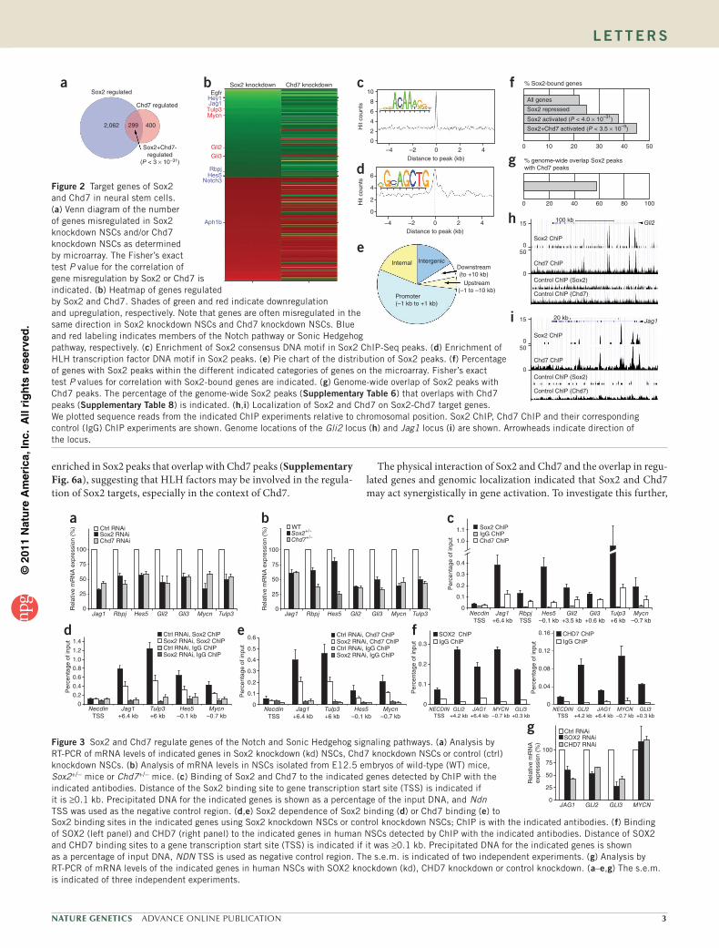
Sox2 binding sites in the indicated genes using Sox2 knockdown NSCs or control knockdown NSCs; ChIP is with the indicated antibodies. (f) Binding of SOX2 (left panel) and CHD7 (right panel) to the indicated genes in human NSCs detected by ChIP with the indicated antibodies. Distance of SOX2 and CHD7 binding sites to a gene transcription start site (TSS) is indicated if it was ≥0.1 kb. Precipitated DNA for the indicated genes is shown as a percentage of input DNA, NDN TSS is used as negative control region. The s.e.m. is indicated of two independent experiments. (g) Analysis by RT-PCR of mRNA levels of the indicated genes in human NSCs with SOX2 knockdown (kd), CHD7 knockdown or control knockdown. (a–e,g) The s.e.m. is indicated of three independent experiments.
Figure 3 Sox2 and Chd7 regulate genes of the Notch and Sonic Hedgehog signaling pathways. (a) Analysis by RT-PCR of mRNA levels of indicated genes in Sox2 knockdown (kd) NSCs, Chd7 knockdown NSCs or control (ctrl) knockdown NSCs. (b) Analysis of mRNA levels in NSCs isolated from E12.5 embryos of wild-type (WT) mice, Sox2+/− mice or Chd7+/− mice. (c) Binding of Sox2 and Chd7 to the indicated genes detected by ChIP with the indicated antibodies. Distance of the Sox2 binding site to gene transcription start site (TSS) is indicated if it is ≥0.1 kb. Precipitated DNA for the indicated genes is shown as a percentage of the input DNA, and Ndn TSS was used as the negative control region. (d,e) Sox2 dependence of Sox2 binding (d) or Chd7 binding (e) to
©20
11 N
atu
re A
mer
ica,
Inc.
All
rig
hts
res
erve
d.
� ADVANCE ONLINE PUBLICATION Nature GeNetics
l e t t e r s
we focused on 7 genes out of the set of 46 identified Sox2-Chd7 target genes (Supplementary Table 10). These genes are part of the Notch pathway (Jag1, Rbpj and Hes5) or the Shh pathway (Gli2, Gli3, Mycn and Tulp3). Mutations in human GLI2, GLI3 and MYCN cause pitui-tary hypoplesia10,24 and esophageal atresia9, respectively. We confirmed downregulation of the selected genes upon Sox2 or Chd7 knockdown (Fig. 3a). We subsequently analyzed the expression of these genes in NSCs from E12.5 embryos of Sox2+/− mice and Chd7+/− mice13,25. We again found all selected genes to be downregulated, except for Hes5 in Sox2+/− NSCs (Fig. 3b). Thus, the expression of these genes was also affected in the context of Sox2 or Chd7 haploinsufficiency. We con-firmed by ChIP analysis that Chd7 binds at Sox2 binding sites in all tested genes (Fig. 3c). Knockdown of Sox2 not only reduced Sox2 bind-ing, as expected (Fig. 3d), but also reduced binding of Chd7 (Fig. 3e), suggesting that Sox2 facilitates the recruitment of Chd7. Chd7 knock-down did not affect Sox2 binding (Supplementary Fig. 6b). Sox2 or Chd7 knockdown also did not affect the H3K4me3 histone mark at the TSS of their target genes (Supplementary Fig. 6c). We conclude that Sox2 and Chd7 cooperate to activate a set of common target genes with potential relevance for SOX2 anophthalmia syndrome and CHARGE syndrome. Analysis of human NSCs that were positive for markers of SOX2 and NESTIN (Supplementary Fig. 7a–c) using ChIP and knock-down (Supplementary Fig. 7d,e) showed that SOX2 and CHD7 bind the disease-relevant genes JAG1, GLI2, GLI3 and MYCN (Fig. 3f) and activate the expression of JAG1, GLI2 and GLI3 (Fig. 3g), indicating that the regulation of these genes by SOX2 and CHD7 has a high level of conservation in human cells.
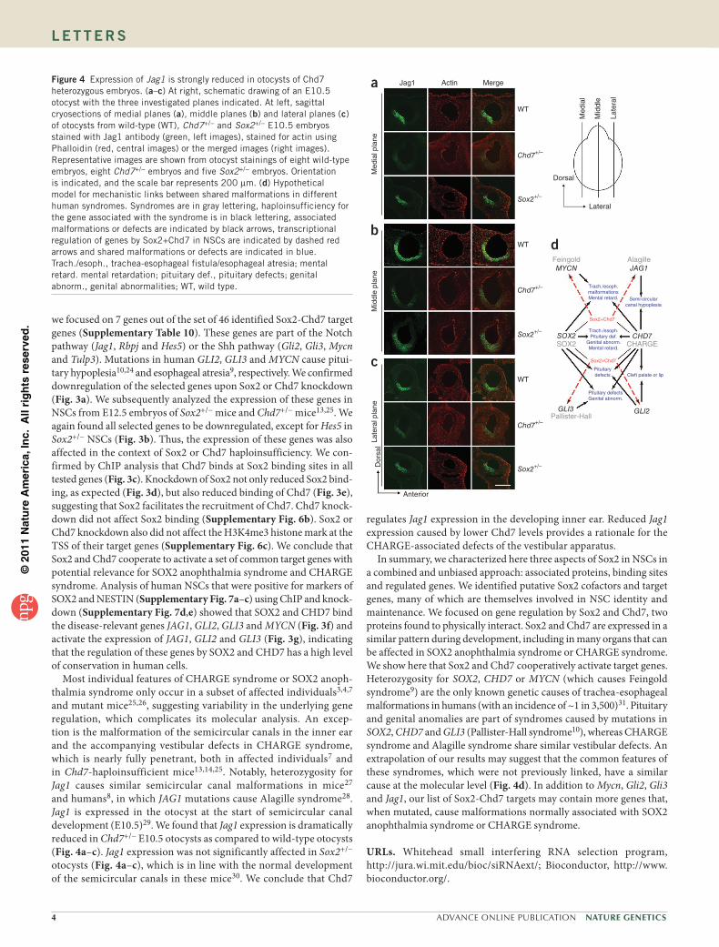
Most individual features of CHARGE syndrome or SOX2 anoph-thalmia syndrome only occur in a subset of affected individuals3,4,7 and mutant mice25,26, suggesting variability in the underlying gene regulation, which complicates its molecular analysis. An excep-tion is the malformation of the semicircular canals in the inner ear and the accompanying vestibular defects in CHARGE syndrome, which is nearly fully penetrant, both in affected individuals7 and in Chd7-haploinsufficient mice13,14,25. Notably, heterozygosity for Jag1 causes similar semicircular canal malformations in mice27 and humans8, in which JAG1 mutations cause Alagille syndrome28. Jag1 is expressed in the otocyst at the start of semicircular canal development (E10.5)29. We found that Jag1 expression is dramatically reduced in Chd7+/− E10.5 otocysts as compared to wild-type otocysts (Fig. 4a–c). Jag1 expression was not significantly affected in Sox2+/− otocysts (Fig. 4a–c), which is in line with the normal development of the semicircular canals in these mice30. We conclude that Chd7
regulates Jag1 expression in the developing inner ear. Reduced Jag1 expression caused by lower Chd7 levels provides a rationale for the CHARGE-associated defects of the vestibular apparatus.
In summary, we characterized here three aspects of Sox2 in NSCs in a combined and unbiased approach: associated proteins, binding sites and regulated genes. We identified putative Sox2 cofactors and target genes, many of which are themselves involved in NSC identity and maintenance. We focused on gene regulation by Sox2 and Chd7, two proteins found to physically interact. Sox2 and Chd7 are expressed in a similar pattern during development, including in many organs that can be affected in SOX2 anophthalmia syndrome or CHARGE syndrome. We show here that Sox2 and Chd7 cooperatively activate target genes. Heterozygosity for SOX2, CHD7 or MYCN (which causes Feingold syndrome9) are the only known genetic causes of trachea-esophageal malformations in humans (with an incidence of ~1 in 3,500)31. Pituitary and genital anomalies are part of syndromes caused by mutations in SOX2, CHD7 and GLI3 (Pallister-Hall syndrome10), whereas CHARGE syndrome and Alagille syndrome share similar vestibular defects. An extrapolation of our results may suggest that the common features of these syndromes, which were not previously linked, have a similar cause at the molecular level (Fig. 4d). In addition to Mycn, Gli2, Gli3 and Jag1, our list of Sox2-Chd7 targets may contain more genes that, when mutated, cause malformations normally associated with SOX2 anophthalmia syndrome or CHARGE syndrome.
URLs. Whitehead small interfering RNA selection program, http://jura.wi.mit.edu/bioc/siRNAext/; Bioconductor, http://www.bioconductor.org/.
a
b
c
Jag1
Anterior
Actin Merge
WT
WT
WT
Chd7+/–
Chd7+/–
Chd7+/–
d
Dorsal
Lateral
Med
ial
Mid
dle
Late
ral
Sox2+/–
Sox2+/–
Sox2+/–
Med
ial p
lane
Mid
dle
plan
eLa
tera
l pla
neD
orsa
l
Feingold Alagille
CHD7
Sox2+Chd7
Sox2+Chd7
Trach./esoph.Pituitary def.
Genital abnorm.Mental retard.
Pituitarydefects
Pituitary defectsGenital abnorm.
Cleft palate or lip
SOX2SOX2 CHARGE
GLI2GLI3Pallister-Hall
Trach./esoph.malformationsMental retard. Semi-circular
canal hypoplesia
JAG1MYCN
Figure 4 Expression of Jag1 is strongly reduced in otocysts of Chd7 heterozygous embryos. (a–c) At right, schematic drawing of an E10.5 otocyst with the three investigated planes indicated. At left, sagittal cryosections of medial planes (a), middle planes (b) and lateral planes (c) of otocysts from wild-type (WT), Chd7+/− and Sox2+/− E10.5 embryos stained with Jag1 antibody (green, left images), stained for actin using Phalloidin (red, central images) or the merged images (right images). Representative images are shown from otocyst stainings of eight wild-type embryos, eight Chd7+/− embryos and five Sox2+/− embryos. Orientation is indicated, and the scale bar represents 200 µm. (d) Hypothetical model for mechanistic links between shared malformations in different human syndromes. Syndromes are in gray lettering, haploinsufficiency for the gene associated with the syndrome is in black lettering, associated malformations or defects are indicated by black arrows, transcriptional regulation of genes by Sox2+Chd7 in NSCs are indicated by dashed red arrows and shared malformations or defects are indicated in blue. Trach./esoph., trachea-esophageal fistula/esophageal atresia; mental retard. mental retardation; pituitary def., pituitary defects; genital abnorm., genital abnormalities; WT, wild type.

©20
11 N
atu
re A
mer
ica,
Inc.
All
rig
hts
res
erve
d.
Nature GeNetics ADVANCE ONLINE PUBLICATION 5
l e t t e r s
METHOdSMethods and any associated references are available in the online version of the paper at http://www.nature.com/naturegenetics/.
Accession codes. ChIP sequencing data are available through the Sequence Read Archive with the accession code ERP000239. Microarray data are available through the EBI ArrayExpress database with the accession code E-MEXP-2743.
Note: Supplementary information is available on the Nature Genetics website.
ACKnoWLEDGMEntSWe thank G. Abelo for advice on the otocyst stainings, A. Smith for 46C ES cells, S. Pollard for advice on deriving neural stem cells, Z. Ozgür for micro-array hybridizations, M. van den Hout-van Vroonhoven for Illumina GAP analyses and P. Wade for Mi2-β antibody. R.A.P., E.E. and U.A. were supported by a Vidi grant, ALW-open program grant and a Chemical Sciences ECHO grant, respectively, all from the Netherlands Organisation for Scientific Research (NWO). J.C.B. was supported by EuTRACC, B.L. was supported by grants from the Norwegian Research Council (YFF) and the Bergen Research Foundation. C.G. and R.J.R. were supported in part by the Sophia Foundation for Medical Research. S.B. was supported by a British Heart Foundation Chair Award (CH/09/003) and Project Grant award (PG/08/045/25069).
AUtHoR ContRIBUtIonSE.E. and U.A. performed nearly all experiments and analyzed the data. J.C.B. and B.L. normalized the ChIP-Seq data and performed all bioinformatic analyses. J.H. and S.P. normalized and formatted the microarray gene expression data. C.G., R.A.P. and R.J.R. created the F-Sox2 embryonic stem cells. D.S. and S.B. assisted in the mouse work, M.M. performed the GST pull down experiment, C.K. and W.v.IJ. performed the microarray analyses and Illumina sequencing of the ChIP material. D.H.W.D. and J.D. performed the mass spectrometry analyses. E.-J.R. provided bioinformatic assistance in the early stages of this work. L.H.P. provided Sox2 COND mice. F.G.G. set up the ChIP sequencing facility and the bioinformatics infrastructure. R.J.R. created Sox2+/− mice from Sox2 COND mice. R.A.P. designed the study, analyzed the data and wrote the manuscript with support from coauthors.
CoMPEtInG FInAnCIAL IntEREStSThe authors declare no competing financial interests.
Published online at http://www.nature.com/naturegenetics/. Reprints and permissions information is available online at http://npg.nature.com/reprintsandpermissions/.
1. Pevny, L.H. & Nicolis, S.K. Sox2 roles in neural stem cells. Int. J. Biochem. Cell Biol. 42, 421–424 (2010).
2. Guth, S.I. & Wegner, M. Having it both ways: Sox protein function between conservation and innovation. Cell. Mol. Life Sci. 65, 3000–3018 (2008).
3. Williamson, K.A. et al. Mutations in SOX2 cause anophthalmia-esophageal-genital (AEG) syndrome. Hum. Mol. Genet. 15, 1413–1422 (2006).
4. Fantes, J. et al. Mutations in SOX2 cause anophthalmia. Nat. Genet. 33, 461–463 (2003).
5. Kelberman, D. et al. Mutations within Sox2/SOX2 are associated with abnormalities in the hypothalamo-pituitary-gonadal axis in mice and humans. J. Clin. Invest. 116, 2442–2455 (2006).
6. Vissers, L.E. et al. Mutations in a new member of the chromodomain gene family cause CHARGE syndrome. Nat. Genet. 36, 955–957 (2004).
7. Zentner, G.E., Layman, W.S., Martin, D.M. & Scacheri, P.C. Molecular and phenotypic aspects of CHD7 mutation in CHARGE syndrome. Am. J. Med. Genet. A. 152A, 674–686 (2010).
8. Okuno, T., Takahashi, H., Shibahara, Y., Hashida, Y. & Sando, I. Temporal bone histopathologic findings in Alagille’s syndrome. Arch. Otolaryngol. Head Neck Surg. 116, 217–220 (1990).
9. van Bokhoven, H. et al. MYCN haploinsufficiency is associated with reduced brain size and intestinal atresias in Feingold syndrome. Nat. Genet. 37, 465–467 (2005).
10. Kang, S., Graham, J.M. Jr., Olney, A.H. & Biesecker, L.G. GLI3 frameshift mutations cause autosomal dominant Pallister-Hall syndrome. Nat. Genet. 15, 266–268 (1997).
11. Gontan, C. et al. Exportin 4 mediates a novel nuclear import pathway for Sox family transcription factors. J. Cell Biol. 185, 27–34 (2009).
12. van den Berg, D.L. et al. An Oct4-centered protein interaction network in embryonic stem cells. Cell Stem Cell 6, 369–381 (2010).
13. Hurd, E.A. et al. Loss of Chd7 function in gene-trapped reporter mice is embryonic lethal and associated with severe defects in multiple developing tissues. Mamm. Genome 18, 94–104 (2007).
14. Alavizadeh, A. et al. The Wheels mutation in the mouse causes vascular, hindbrain, and inner ear defects. Dev. Biol. 234, 244–260 (2001).
15. Visel, A., Thaller, C. & Eichele, G. GenePaint.org: an atlas of gene expression patterns in the mouse embryo. Nucleic Acids Res. 32, D552–D556 (2004).
16. Hu, Q. et al. The EGF receptor-sox2-EGF receptor feedback loop positively regulates the self-renewal of neural precursor cells. Stem Cells 28, 279–286 (2010).
17. Bani-Yaghoub, M. et al. Role of Sox2 in the development of the mouse neocortex. Dev. Biol. 295, 52–66 (2006).
18. Favaro, R. et al. Hippocampal development and neural stem cell maintenance require Sox2-dependent regulation of Shh. Nat. Neurosci. 12, 1248–1256 (2009).
19. Taranova, O.V. et al. SOX2 is a dose-dependent regulator of retinal neural progenitor competence. Genes Dev. 20, 1187–1202 (2006).
20. Howard, T.D. et al. Mutations in TWIST, a basic helix-loop-helix transcription factor, in Saethre-Chotzen syndrome. Nat. Genet. 15, 36–41 (1997).
21. Bajpai, R. et al. CHD7 cooperates with PBAF to control multipotent neural crest formation. Nature 463, 958–962 (2010).
22. Schnetz, M.P. et al. Genomic distribution of CHD7 on chromatin tracks H3K4 methylation patterns. Genome Res. 19, 590–601 (2009).
23. Schnetz, M.P. et al. CHD7 targets active gene enhancer elements to modulate ES cell-specific gene expression. PLoS Genet. 6, e1001023 (2010).
24. Roessler, E. et al. Loss-of-function mutations in the human GLI2 gene are associated with pituitary anomalies and holoprosencephaly-like features. Proc. Natl. Acad. Sci. USA 100, 13424–13429 (2003).
25. Bosman, E.A. et al. Multiple mutations in mouse Chd7 provide models for CHARGE syndrome. Hum. Mol. Genet. 14, 3463–3476 (2005).
26. Avilion, A.A. et al. Multipotent cell lineages in early mouse development depend on SOX2 function. Genes Dev. 17, 126–140 (2003).
27. Kiernan, A.E. et al. The Notch ligand Jagged1 is required for inner ear sensory development. Proc. Natl. Acad. Sci. USA 98, 3873–3878 (2001).
28. Li, L. et al. Alagille syndrome is caused by mutations in human Jagged1, which encodes a ligand for Notch1. Nat. Genet. 16, 243–251 (1997).
29. Brooker, R., Hozumi, K. & Lewis, J. Notch ligands with contrasting functions: Jagged1 and Delta1 in the mouse inner ear. Development 133, 1277–1286 (2006).
30. Kiernan, A.E. et al. Sox2 is required for sensory organ development in the mammalian inner ear. Nature 434, 1031–1035 (2005).
31. Shaw-Smith, C. Oesophageal atresia, tracheo-oesophageal fistula, and the VACTERL association: review of genetics and epidemiology. J. Med. Genet. 43, 545–554 (2006).
©20
11 N
atu
re A
mer
ica,
Inc.
All
rig
hts
res
erve
d.
Nature GeNetics doi:10.1038/ng.825
ONLINE METHOdSNeural stem cell culturing and derivation. F-Sox2 NSCs11 and wild-type NSCs used for biochemical and RNAi studies were cultured as described32 using N2B27 medium (Stem Cell Sciences) supplemented with epidermal growth factor (EGF) and fibroblast growth factor (FGF) (both from Peprotech). Sox2+/−, Chd7+/− and wild-type littermate NSCs used for gene expression stud-ies were derived from forebrains of E12.5 mouse embryos, filtered through a 70 µm cell strainer (Falcon) and cultured in N2B27 with EGF and FGF as previously described32. Human NSCs (embryonic stem cell–derived) were pur-chased from Invitrogen (N7800-100) and cultured as described33 on laminin (Roche) coated dishes in Euromed-N (Euroclone) supplemented with N2/B27 (Invitrogen), EGF and FGF (Peprotech) and L-glutamine (Invitrogen).
Identification of interacting proteins of Sox2 and Chd7. FLAG-Sox2 was purified from F-Sox2 NSC11 nuclear extract prepared from 2 × 10⁸ NSCs using a FLAG-affinity protocol as previously described12. Control purifica-tions were from nuclear extract from wild-type NSCs. Immunoprecipitation of Sox2 with a Sox2 antibody (Y-17, sc-17320) or immunoprecipitation of Chd7 with a Chd7 antibody (ab-31824, Abcam) from NSC nuclear extract was as described12, and EtBr (25 µg/ml) or benzonase (150 U/ml, Novagen) was added, where indicated. GST pull downs were as described12. Identification of interacting proteins by mass spectrometry was as described12 and proteins were included if specifically identified in both purifications of F-Sox2 or both immunoprecipitations of Chd7.
Transfection with shRNA constructs. shRNA sequence for mouse Sox2 was as previously described34. shRNA sequences for mouse Chd7 and human SOX2 and CHD7 (Supplementary Table 11) were designed with help of Whitehead small interfering RNA selection program (see URLs) and cloned into pSuper-puro (Oligoengine). pSuper-control-shRNA (Dharmacon) was used as a control. We transfected 3 × 10⁶ NSCs with pSuper constructs by electroporation using an Amaxa Nucleofector and Nucleofector Kit V (Lonza). Puromycin (1 µg/ml) was added after 24 h, and NSCs were harvested 48 h after electroporation.
Expression analyses. For expression analysis by microarray, total RNA was isolated in experimental triplicates from NSCs electroporated with the differ-ent shRNA constructs and converted into biotin-labeled single stranded DNA as previously described35 and hybridized to GeneChIP Mouse Genome 430 2.0 arrays (Affymetrix) according to the manufacturer’s recommendations. Array data quality control, normalization and statistical analysis were as described36. Quantitative real-time PCR analyses on complementary DNA transcribed from total RNA with Superscript II Reverse Transcriptase was performed on a DNA Engine Opticon2/CFX96 (Biorad) and normalized for CalR or Hprt expression. Primer sequences are listed in Supplementary Table 12.
Mice. Chd7+/edy mice25,37, here called Chd7+/− mice, were obtained from the European Mouse Mutant Archive (EMMA) consortium and maintained in an FVB background. Sox2+/floxKO mice, here called Sox2+/− mice, were generated by crossing Sox2+/COND mice19 with mice expressing CRE recombinase from a CAG promoter38 and maintained on a C57Bl/6 background. All animal studies were conducted under the guidelines for animal experimentation approved by the Erasmus University Animal Welfare Committee.
Immunostainings. For immunostaining of the otocysts, sagittal cryostat sec-tions of 10 µm thickness of heads of E10.5 embryos were fixed in PBS 2% paraformaldehyde (PFA) for 15 min at room temperature, permeabilized with PBS 0.1% Triton (PBS-0.1T) for 2 × 10 min and incubated in blocking buffer (PBS-0.5% BSA, 0.15% glycin) for 15 min. Slides were incubated overnight at 4 °C in blocking buffer with anti-Jag1 (1:100; Santa Cruz Biotechnology; H-114). After washing in PBS 0.1T, slides were incubated for 1 h in blocking buffer with Alexa 488 goat anti-rabbit (1:200; Invitrogen; A11008) and Alexa
594-conjugated phalloidin (1:100; Invitrogen; A12381), washed and mounted in Vectashield Mounting Medium with DAPI (Vector laboratories).
Mouse NSCs were grown on poly-D-lysine–coated cover slips, fixed in PBS 4% PFA, permeabilized with PBS 0.4T in PBS, blocked in PBS 10% FCS, incu-bated in anti-Pax6 (1:10 dilution, Developmental Studies Hybridoma Bank) for 2 h at room temperature, washed, incubated in Alexa 594 goat anti-mouse IgG (1:200; Invitrogen; A11032), washed and mounted in Vectashield Mounting Medium with DAPI (Vector laboratories). Human NSCs were grown on lam-inin-coated coverslips and stained with antibodies against SOX2 (AF2018, R&D systems) and NESTIN (mAb1259, R&D systems) as described above. Digital images were captured on a Zeiss microscope Axio Imager Z1.
Chromatin immunoprecipitations (ChIP) and high-throughput sequencing. For large-scale chromatin preparation, 108 NSCs were crosslinked with 2 mM disuccinimidyl glutarate (Thermo Fisher Scientific) and 1% formaldehyde as previously described39, nuclei were isolated in 50 mM Tris-Cl pH 8.0, 1 mM EDTA, 1% SDS and lysed in pre-immunoprecipitation buffer (10 mM Tris, 10 mM NaCl, 3 mM MgCl2 and 1 mM CaCl2). Chromatin was prepared and ChIP was performed according to the Millipore online protocol using 15 µg of antibodies against Sox2 (Y17, sc-17320) and Chd7 (ab-31824) or goat IgG (sc-2028) as a control. ChIP DNA library preparation, ChIP sequencing on an Illumina Genome Analyzer or HiSequation (2000) and processing of the raw data and mapping the peaks to the mouse genome (NCBI build 37.1) were as previously described35. ChIP sequencing data have been submitted to the Sequence Read Archive, accession number pending. For small scale ChIP, 107 NSCs electroporated with the different shRNA constructs were directly lysed in pre-immunoprecipitation buffer and ChIP was performed as above using 5 µg of antibodies. Small scale ChIP in human NSCs was performed as described above using antibodies against SOX2 (AF2018, R&D systems) and CHD7 (Bethyl laboratories A301-223A). Primers for amplification of genomic regions by quantitative PCR are listed in Supplementary Table 13.
Bioinformatic analyses. Significance estimations of Sox2 ChIP peaks and Chd7 peaks were calculated using the Poisson distribution as previously described40 followed by multiple testing correction by controlling the false dis-covery rate. A threshold false discovery rate of 2 × 10⁻¹⁰ was applied for Sox2 peaks and 1 × 10⁻¹³ for Chd7 peaks. Derivation of motifs was performed using MEME41 on genomic sequences of 400 bp centered on Sox2 peaks. Mapping Sox2 peaks and Chd7 peaks to different regions of genes was performed with R/Bioconductor (see URLs).
32. Conti, L. et al. Niche-independent symmetrical self-renewal of a mammalian tissue stem cell. PLoS Biol. 3, e283 (2005).
33. Sun, Y. et al. Long-term tripotent differentiation capacity of human neural stem (NS) cells in adherent culture. Mol. Cell. Neurosci. 38, 245–258 (2008).
34. Ivanova, N. et al. Dissecting self-renewal in stem cells with RNA interference. Nature 442, 533–538 (2006).
35. Soler, E. et al. The genome-wide dynamics of the binding of Ldb1 complexes during erythroid differentiation. Genes Dev. 24, 277–289 (2010).
36. Hou, J. et al. Gene expression-based classification of non-small cell lung carcinomas and survival prediction. PLoS ONE 5, e10312 (2010).
37. Kiernan, A.E. et al. ENU mutagenesis reveals a highly mutable locus on mouse chromosome 4 that affects ear morphogenesis. Mamm. Genome 13, 142–148 (2002).
38. Sakai, K. & Miyazaki, J. A transgenic mouse line that retains Cre recombinase activity in mature oocytes irrespective of the cre transgene transmission. Biochem. Biophys. Res. Commun. 237, 318–324 (1997).
39. Nowak, D.E., Tian, B. & Brasier, A.R. Two-step cross-linking method for identification of NF-κB gene network by chromatin immunoprecipitation. Biotechniques 39, 715–725 (2005).
40. Jothi, R., Cuddapah, S., Barski, A., Cui, K. & Zhao, K. Genome-wide identification of in vivo protein-DNA binding sites from ChIP-Seq data. Nucleic Acids Res. 36, 5221–5231 (2008).
41. Bailey, T.L. & Elkan, C. The value of prior knowledge in discovering motifs with MEME. Proc. Int. Conf. Intell. Syst. Mol. Biol. 3, 21–29 (1995).