sources of variation in the microbiome of pre-weaned dairy ......play an integral role in calf...
TRANSCRIPT

Sources of Variation in the Microbiome of Pre-Weaned Dairy Calves
Haley Garrett Huffard
Thesis submitted to the faculty of the Virginia Polytechnic Institute and State University
in partial fulfillment of the requirements for the degree of
Master of Science in Life Sciences
In
Dairy Science
Katharine F. Knowlton, Chair
Robert E. James
Rebecca R. Cockrum
June 13, 2019
Blacksburg, VA
Keywords: dairy calves, microbiome, microbiota, gut health
Sources of Variation in the Microbiome of Pre-Weaned Dairy Calves
Haley Garrett Huffard
ABSTRACT
Death loss of pre-weaned dairy heifers is a major challenge confronting U.S. dairy
producers, creating economic loses of at least $100 million each year. Neonatal calf
diarrhea, the leading cause of neonatal calf mortality, is controlled through colostrum
feeding to ensure the passive transfer of immunity. However, these methods are not
always effective in preventing calf morbidity and mortality. Therefore, other factors must
play an integral role in calf health, morbidity, and mortality; the gut microbiome of the
calf is likely significant. The dam’s vaginal, fecal, and oral microbiomes are believed to
be the primary sources of the newborn calf’s gut microbiome, but the long-term health
effects of the colonization by these microbes are yet to be determined. The objective of
this study was to characterize the dam’s fecal, vaginal, and oral microbiomes and the calf
fecal microbiome. Our hypothesis stated that the dam’s vaginal, fecal, and oral
microbiomes will influence the early microbiome of the calf.
Multiparous Holstein cows (n = 6) were sampled and observed during the final
weeks of pregnancy. Vaginal swab samples were collected from cows ~24 h prior to
parturition. Oral, rectal, and vaginal swab samples were collected from cows immediately
following parturition. Oral and rectal swabs were collected from calves (n = 6) at birth (h
0), h 24, d 7, d 42, and during the post-weaning period (d 60). Metagenomic 16S rDNA
data was converted to inferred amplicon sequence variants (ASVs) and clustered into
operational taxonomic units (OTUs) to assess microbial diversity and relative abundance
within fecal, vaginal, and oral microbiome samples. One-way ANOVA followed by
Tukey’s procedure for pair-wise comparisons were used to determine sample type
differences. Significance was declared at P ≤ 0.05. Using phylum count data, a negative
binomial regression model was used to determine the effects of the dam’s vaginal, fecal,
and oral microbiomes on the calf fecal microbiome.
Metagenomic analysis revealed a diverse microbiome within the meconium of
newborn calves, suggesting that microbial colonization of the neonatal calf GIT begins in
utero. We identified 10 phyla associated with the calf meconium and fecal microbiome:
Bacteroidetes, Firmicutes, Proteobacteria, Actinobacteria, Cyanobacteria,
Verrucomicrobia, Spirochaetes, Tenericutes, Fibrobacteres, and Lentisphaerae. Principle-
coordinate analysis revealed a strong relationship between the vaginal microbiome of the
dam and the meconium microbiome of the calf. The dam vaginal microbiome and the calf
meconium microbiome were highly similar, displaying significant differences (P = 0.05)
in only two phyla, Bacteroidetes and Fusobacteria. The abundances of three phyla,
Bacteroidetes, Proteobacteria, and Verrucomicrobia, differed significantly (P = 0.05)
between the dam fecal microbiome and the meconium microbiome. The dam fecal
microbiome was highly similar to the calf fecal microbiome post-weaning (d 60), making
the fecal microbiome of the dam a strong indicator of the microbial composition of post-
weaning calf feces. Overall, the calf meconium and fecal microbiome is influenced by a
combination of the maternal vagina, oral, and fecal microbiomes.
Further studies will be needed to identify the transference mechanisms of
maternal microbes to offspring and the associated host-microbial interactions.

Sources of Variation in the Microbiome of Pre-Weaned Dairy Calves
Haley Garrett Huffard
GENERAL AUDIENCE ABSTRACT
This study investigated the microbial colonization and maternal influences on the
neonatal calf gut microbiome. Microbiome samples were collected from dams (n = 6) and
calves (n = 6) using sterile flocked swabs. The vaginal, oral, and fecal bacterial
communities were examined from the dam and the fecal community of calves was
examined from birth to 60 d of age. Microbial communities varied by anatomical location
and age of the calf. Metagenomic analysis 16s ribosomal DNA revealed ten phyla
associated with microbiomes of the dam and the same ten phyla associated with calf feces
at various time points: Bacteroidetes, Firmicutes, Proteobacteria, Actinobacteria,
Cyanobacteria, Verrucomicrobia, Spirochaetes, Tenericutes, Fibrobacteres, and
Lentisphaerae. Overall, the calf meconium and fecal microbiome is influenced by a
combination of the maternal vagina, oral, and fecal microbiomes. Further studies will be
needed to identify the transference mechanisms of maternal microbes to offspring and the
associated host-microbial interactions.
v
ACKNOWLEDGEMENTS
I would like to thank my advisor, Dr. Katharine Knowlton, for all of your
encouragement and the opportunities you have given me throughout my time at Virginia
Tech. You have shaped me into becoming a patient, independent student, while pushing
me far beyond my own expectations. I will be forever grateful for the potential you saw
in me when we first met in 2014 and all of the times you have gone out of the way to
offer guidance. I am thankful for your support, mentorship, and the opportunities you
have allowed me to pursue throughout working with you.
To my committee members, Dr. Bob James and Dr. Rebecca Cockrum, your
advice, encouragement, and support throughout my Master’s has been invaluable. I am
honored to have had the opportunity to work closely with you both. The advice you both
have offered to me throughout this process has been beyond appreciated. A special thank
you to Dr. James for the immense efforts you have made on my behalf throughout my
career search. I owe my future success to you!
I am proud to have been a part of the Virginia Tech Dairy Science family for the
past two years. This department has allowed me to form valuable relationships with
faculty and industry professionals, travel, and build lifelong friendships. Thank you to my
fellow graduate students all of your encouragement and showing me that graduate school
can be fun too. Chrissy Teets, I have truly enjoyed working closely with you for the past
five years. I really couldn’t have made it through my studies without you! Thank you so
much for all of your help and always lending an ear.
Most importantly, thank you to my incredible parents and family. To my mom
Lorri, and my dad John, there are not words to thank you both for helping me achieve

vi
anything I thought possible. The both of you are extraordinary role models and teachers
for showing me importance of honesty, hard-work, and strength. I won the parent lottery
with you two!

vii
TABLE OF CONTENTS
ABSTRACT ....................................................................................................................... vi
GENERAL AUDIENCE ABSTRACT............................................................................ viii
TABLE OF CONTENTS .............................................................................................. vii
LIST OF TABLES ............................................................................................................. ix
LIST OF FIGURES ............................................................................................................ x
CHAPTER 1 ....................................................................................................................... 1
1. INTRODUCTION ....................................................................................................... 1
2. THE MATERNAL MICROBIOME ........................................................................... 2
Core microbiota of the human vagina ......................................................................... 3
The influence of microbes on vaginal health............................................................... 4
Microbial changes during pregnancy .......................................................................... 6
Microbiome colonization in the infant ...................................................................... 11
Environmental influences on host microbiota ........................................................... 16
3. THE MATERNAL MICROBIOME OF THE DAIRY COW .................................. 18
Gut microbiota in ruminants ...................................................................................... 18
The vaginal microbiota of dairy cows ....................................................................... 19
Microbial colonization and maternal influences of the newborn calf ....................... 20
4. EARLY IMMUNE DEVELOPMENT AND HOST HEALTH ............................... 21
Microbiota-induced maturation of epithelial barrier function ................................... 21
Effect of antimicrobials on gut microbiota composition ........................................... 23
5. THE ROLE OF METAGENOMICS IN ADVANCING MICROBIOME
UNDERSTANDING ..................................................................................................... 27
Metagenomics: methods and applications ................................................................. 27
16S-Based Metagenomics ......................................................................................... 28
Whole-genome Shotgun sequencing ......................................................................... 29
Classification of bacterial sequences ......................................................................... 29
6. SUMMARY .............................................................................................................. 30
REFERENCES ................................................................................................................. 31
CHAPTER 2 ..................................................................................................................... 42
ABSTRACT ...................................................................................................................... 42
MATERIALS AND METHODS ...................................................................................... 44

viii
Ethical Approval ........................................................................................................ 44
Animals and Observation Units ................................................................................. 44
Newborn Processing .................................................................................................. 44
Housing and Feeding ................................................................................................. 45
Calf Health ................................................................................................................. 46
Sample Collection...................................................................................................... 46
DNA Extraction and Quality Testing ........................................................................ 47
Library Preparation and Sequencing ......................................................................... 48
Statistical Analysis .................................................................................................... 48
RESULTS ......................................................................................................................... 49
Animal characteristics ............................................................................................... 49
Dam associated microbiota ........................................................................................ 50
Calf associated microbiota ......................................................................................... 50
Relationship between dam microbiota and calf microbiome .................................... 51
DISCUSSION ................................................................................................................... 53
Maternal influence on the calf microbiome ............................................................... 53
Development of calf fecal microbiota throughout weaning ...................................... 57
Influence of host genetics on cow and calf microbiota ............................................. 58
REFERNCES ................................................................................................................ 70
CHAPTER 3 ..................................................................................................................... 75
CONCLUSION AND IMPLICATIONS ...................................................................... 75
Summary and Future Studies ..................................................................................... 75
REFERNCES ................................................................................................................ 77

ix
LIST OF TABLES
Table 1. Body weight and ADG of Holstein calves (n = 6) ............................................. 61
Table 2. Relative abundance of bacterial phyla in microbiomes of cows and calves ...... 62
Table 3. Dam microbiome samples as indicators of the calf fecal microbiome ............... 63
x
LIST OF FIGURES
Figure 1. Average relative abundance of bacterial phylum in each sample type. Stacked
bar plots showing: dam vaginal pre-birth (n = 4), dam oral at birth (n = 6), dam fecal at
birth (n = 6), calf fecal at birth (n = 6), calf fecal at 24 h (n = 6), calf fecal at 7 d (n = 6),
calf fecal at 42 d (n = 6), calf fecal at 0 d (n = 6). ............................................................ 64
Figure 2. Individual variation within the cow vaginal microbiome. Stacked bar plot
showing relative abundance of bacterial phylum within dam vaginal samples (n = 4). ... 65
Figure 3. Bacterial composition of the dam vaginal and corresponding calf fecal
microbiome at phylum level. ............................................................................................ 66
Figure 4. Relative abundance of bacterial genera present in cow and calf microbiomes.
Heatmap color (dark red to dark blue) displays the relative abundance of each taxon
across samples. Each sample is identified at the bottom of the heatmap by the sample
name. Colors on top of each heatmap represent the time point to which each sample
belongs. ............................................................................................................................. 67
Figure 5. Similarity of cow and calf samples. Principal component analyses (PCA) of
Beta diversity at the phylum level. Variable contributions are labeled by color (A), where
arrow direction indicates quality of representation on the factor map. Arrow length
indicates variable contribution to the principal components (A). Points represent
individual samples from the different sample locations and were colored by sample type.
........................................................................................................................................... 68
Figure 6. Hierarchical relationship between individual dam and calf samples. Sample
relationships shown as a cluster dendrogram at phylum level of dam and calf microbiota
at different anatomical locations and time points. The length of the scale bar represents a
distance of 0.10. ................................................................................................................ 69

1
CHAPTER 1
REVIEW OF LITERATURE
1. INTRODUCTION
Pre-weaned dairy heifer death loss is a major challenge confronting U.S. dairy
producers, with neonatal calf diarrhea accounting for more than 50% of deaths and
creating substantial economic losses. Current disease prevention protocols consist of
colostrum feeding practices to ensure the passive transfer of immunity. However, this
method is not always effective in alleviating calf morbidity and mortality (Meganck et
al., 2015). The persisting issue of pre-weaned heifer death loss and the public’s growing
concern of antibiotic usage in production livestock has presented a major need for an
alternate method of disease prevention and treatment. Optimal microbial composition of
the gastrointestinal tract (GIT) is of vital importance to the long-term health and
performance of livestock. However, the gut microbiota of pre-weaned dairy calves is not
well understood. This presents the need for an enhanced understanding of the calf gut
microbiota for developing new approaches to improve calf gut and overall health
(Malmuthuge, 2017). The GIT microbiome is responsible for a number of physiological
and functional processes, including nutrient digestion and absorption, host metabolism,
mucus layer development, barrier function, and mucosal immune responses (Kogut and
Arsenault, 2016). Inoculation of the neonatal calf’s GIT is initiated at birth and is
facilitated by the dam’s vaginal, fecal, oral, and colostral microbiota. The structure and
function of maternal microbial communities and the impact of these communities on
maternal and neonatal health has been studied extensively in human and murine models,
but these investigations are still in early phases for dairy cattle. Existing findings from

2
human and murine investigations have highlighted the importance of GIT microbial
establishment and colonization to host development and health status (Malmuthuge et al.,
2015b, Charbonneau et al., 2016). Identification of the ideal colonization processes and
microbial communities of the GIT could allow for targeted microbial therapies, vital to
addressing the economic and ethical concerns associated with calf mortality
(Malmuthuge et al., 2015b).
2. THE MATERNAL MICROBIOME
The community of microorganisms that live in and on the human body, consisting
of upwards of 100 trillion cells, is known as the human microbiome. The cells of the
human microbiome outnumber host cells by a factor of ten and contains 27 times more
genes than in the human genome. Different sites on and within the human body such as
the skin, mouth, gut, and reproductive tract are home to discrete populations of microbes.
Advancements in sequencing technologies have allowed for findings that present clear
associations between microbiome composition and host health (Dunlop et al., 2015).
Although most research has focused on the microbiome of the gastrointestinal tract, there
is increasing interest in the female reproductive microbiome and the associated health of
the offspring. The vaginal microbiome is key in successful fertilization and a healthy
pregnancy. The microbiome also plays an essential role in producing healthy and
reproductively fit offspring (Mueller et al., 2015). Microbial communities have been
identified in niches that were traditionally considered sterile, such as the uterus, placenta,
and fallopian tubes. The combination of female reproductive microbiome, postnatal
environment, and infant colonization patterns heavily influence health throughout a
lifetime (Younes et al., 2018).

3
Core microbiota of the human vagina
Research involving the vaginal microbiome in women has advanced in the last 15
years due to studies the development of high-throughput sequencing. Early molecular
studies used general 16S ribosomal RNA (rRNA) sequencing techniques that expanded
the understanding of the vaginal microbiota. These studies led to the characterization of
the vaginal microbiome. The vaginal microbiome is composed of microorganisms
including Lactobacillus sp., Atopobium vaginae, Sneathia, Leptotrichia, Megasphaera,
Dialister, and Eggerthela (Martin and Marrazzo, 2016). Ravel et al (2011) characterized
the “core microbiome” of the healthy, adult vagina through pyrosequencing 16S rRNA
genes. The results of this study indicate that although the vaginal microbiome of healthy,
reproductive-age women is dominated by Lactobacillus iners and Lactobacillus
crispatus, there is no single core microbiome. Instead, there are a variety of core
microbiomes that can be identified by community groups. These groups can be
categorized by two criteria: (1) the constituent communities are dominated by
Lactobacillus, and (2) the distinct species of Lactobacillus that are present (Ravel et al.,
2011). Although there is not a core microbiome of the vagina, there are core functions,
such as lactic acid production that remain among communities (Ravel et al., 2011).
Disease, sexual activity, and menstruation are events known to cause variation in
the vaginal microbiome. Intervals of dysbiosis within the microbiome lead to increased
susceptibility to disease (Gajer et al., 2012). Identifying factors that disrupt the stability
of the vaginal microbiome could provide useful insight into predicting disease risk and
women’s health.

4
The influence of microbes on vaginal health
Microbial communities of the vagina are composed of mutualistic bacteria that
are the first line of defense against urogenital disease and infection (Kaewsrichan et al.,
2006, Ravel et al., 2011). These preventable diseases include bacterial vaginosis, yeast
infections, sexually transmitted infections, urinary tract infections, and human
immunodeficiency virus type 1 (HIV-1) infection. Vaginal disease prevention by the
microbiome is largely attributed to lactic acid-producing bacteria that commonly inhabit
the vagina. Lactobacilli are found dominant in vaginal fluid of healthy women at
concentrations of 107-108 colony-forming units (CFU)g-1 (Sobel and Chaim, 1996).
Lactobacillus species are thought to provide disease protection by lowering the pH of the
vagina, producing bacteriostatic and bactericidal compounds, and through competitive
exclusion (Ravel et al., 2011). Some lactobacilli, such as Lactobacillus crispatus and L.
gasseri, are able to control the overgrowth and infection processes of pathogens, as well
as modulate systemic inflammation, cell proliferation, and apoptosis of the female (Wang
et al., 2018).
A healthy vaginal environment has a pH of 4 ± 0.5 and maintenance of pH is
essential to reproductive and vaginal health. The acidic environment reduces the
infectivity for a variety of sexually transmitted disease pathogens, including HIV-1 (Tevi-
Bénissan et al., 1997, Boskey et al., 2001). Also, elevated pH and resulting dysbiosis of
the vaginal microbiome is linked to Bacterial vaginosis (BV), a prevalent condition
characterized by overgrowth of certain anaerobic bacteria and reduced abundance of
Lactobacillus. Bacterial vaginosis is associated with significant adverse health effects
including abnormal pregnancy (Martin and Marrazzo, 2016), premature birth (Sagawa et

5
al., 1995), increased risk of HIV infection (Taha et al., 1998), and pelvic inflammatory
disease (Hillier et al., 1996) (Boskey et al., 2001).
The acidic environment of the vagina is produced by lactic acid-producing
bacteria. Glycogen is deposited in the vaginal epithelium during times of high estrogen,
and it is metabolized into lactic acid supporting the desired pH (Boskey et al., 2001).
Metabolism of glycogen was originally thought to be by the vaginal epithelium, but this
theory was refuted after exploration of vaginal lactate structures. This investigation
revealed that the majority of vaginal lactic acid was of the D-isoform, which cannot be
produced by human metabolism (Prince et al., 2015). Vaginal lactobacilli have been
found to be capable of producing acid at sufficient rates to account for the rate at which
the vagina acidifies after neutralization in an in vitro setting (Boskey et al., 1999).
Although this finding is consistent with the theory of vaginal acidity being primarily of
bacterial origin, evidence is lacking to determine if this is the only mechanism
responsible for acid production in the vagina (Boskey et al., 2001).
Lactobacilli are effective in restricting the growth of pathogens through the
production of bacteriostatic and bactericidal compounds. Hydrogen peroxide (H2O2) is
produced by lactobacilli present in a healthy vaginal environment. Klebanoff and
colleagues (1991) found that H2O2-dependent activity is bactericidal to Gardnerella
vaginalis in an in vitro setting (Klebanoff et al., 1991) (Kaewsrichan et al., 2006). An
overgrowth of Gardnerella vaginalis in the vagina is one of the primary origins of
bacterial vaginosis. Lactic acid bacteria, which include lactobacilli, streptococci, and
pneumococci, form and release the H2O2 required for the peroxidase-mediated
antimicrobial system; a toxic event to a variety of microorganisms and mammalian cells.

6
When peroxidases are combined with H2O2 and a halide, the halide is oxidized by
peroxidase and H2O2 to form a potent oxidant. This created oxidant (corresponding
hypohalous acid or halogen) attacks the target cell at its oxidizable sites, resulting in cell
destruction and the prevention of pathogenic infection (Klebanoff et al., 1991).
Microbial changes during pregnancy
Pregnancy causes a myriad of physiological changes to support the growth and
development of the fetus. These changes include shifts in hormones, metabolism, and
immunity; all are major factors inducing dramatic changes in the microbiome during
pregnancy (Nuriel-Ohayon et al., 2016). Hormonal changes include the dramatic rise of
progesterone and estrogen. A successful pregnancy is largely dependent on progesterone
for creating a suitable endometrial environment for the growing fetus and preventing
preterm labor (Kumar and Magon, 2012). Metabolic changes that occur during pregnancy
include weight gain, elevated fasting blood glucose levels, insulin resistance, glucose
intolerance, and changes in metabolic hormone concentration (Nuriel-Ohayon et al.,
2016). Also, the female body must maintain a level of immune suppression to accept the
fetus and its own developing immune system, while enabling immunity to external
infectious agents. (Mor and Cardenas, 2010, Nuriel-Ohayon et al., 2016). The changes
the female human body undergoes during pregnancy initiate major shifts in the
microbiota at distinct sites.
The vaginal microbiome during pregnancy
The human vaginal microbiome is the primary protection against disease to
offspring. A Lactobacillus-dominated microbiota stands as a strong biomarker for a
healthy vaginal environment (Petrova et al., 2015). Lactobacillus crispatus, Lactobacillus

7
gasseri, Lactobacillus iners, and Lactobacillus jensenii are the most frequently isolated
species. The depletion of these species is associated with several adverse consequences,
including increased risk and transmission of sexually transmitted infections, infertility,
preterm birth, and pelvic inflammatory disease. A Lactobacillus-dominated microbiota
establishes anti-pathogenic mechanisms that promote health and maintain reproductive
fitness of the host. Lactobacilli produce biochemically active compounds that directly kill
or inhibit the stabilization of pathogens (Petrova et al., 2015).
The microbial community of the vagina varies in association with normal
pregnancy, creating a “signature” taxonomy related to a state of pregnancy. These
changes include a significant decrease in microbial diversity and an enrichment with
species of Lactobacilli (Aagaard et al., 2012, Nuriel-Ohayon et al., 2016). Aagaard et al
(2012) demonstrated significant differences between the vaginal microbiomes of
pregnant and non-pregnant women (Aagaard et al., 2012) (Martin and Marrazzo, 2016).
The signature vaginal taxonomy of pregnant women consists of an increased abundance
of Lactobacillus spp, along with an overall decreased richness and diversity. The
microbial community of the vagina during pregnancy varied by gestational age and
proximity to the cervix (Aagaard et al., 2012). Prince et al (2015) discovered an
enrichment of the orders Lactobacillus, Clostridiales, Bacteriodales, and
Actinomycetales. At the species level, the vaginal microbiome during pregnancy was
enriched in L. iners, L. crispatus, L. jensenii, and L. johnsonii. Enrichment in lactobacilli
could be a result of the increase of estrogen induced by pregnancy (Prince et al., 2015).
Reconstruction of the vaginal microbiome during the postpartum period is
induced by a sudden halt in estrogen production, which significantly reduces the
8
proportion of lactobacilli and enriches bacteria associated with vaginosis. Further studies
are necessary for understanding the effects of sex hormones on the vaginal microbiome
during and post-pregnancy (MacIntyre et al., 2015).
The gut microbiome during pregnancy
The microbial environment of the intestine undergoes considerable changes
during pregnancy. The combination of stool samples, diet information, and clinical data
from 91 pregnant women revealed shifts in the intestinal microbiome throughout
pregnancy and that the gut microbiota is profoundly altered by pregnancy (Koren et al.,
2012). The transition from the first to third trimester is characterized by a decrease in
species diversity and an increase in Proteobacteria, these third trimester stool samples
also exhibited markers of inflammation and energy loss (Koren et al., 2012). Microbial
profiles associated with the third trimester induce physiological changes in germ-free
(sterile) mice. Fecal transplants of first and third trimester stool into germ-free mice were
performed to investigate if the changes in the microbiota are cause or consequence of an
increased state of inflammation. After third trimester gut-microbiota were transferred to
germ-free mice, the mice gained a significant amount of weight, developed insulin
resistance, and showed an increased inflammatory response. The results indicate that
third trimester microbiota are associated with metabolic changes in the host, suggesting a
link between pregnancy-induced microbial shifts and changes in host metabolism and
immunology. These microbial shifts are postulated to be necessary for proper fetal
development and a healthy pregnancy (Koren et al., 2012, Nuriel-Ohayon et al., 2016).

9
The oral microbiome during pregnancy
The oral microbiome is also susceptible to composition changes with pregnancy.
The oral cavity is home to over 600 diverse species including Streptococci, Lactobacilli,
Staphylococci, and Corynebacteria, each within their own microenvironment (e.g.
tongue, teeth, palates, gums, cheeks, lips). When compared to non-pregnant women,
pregnant women had higher total viable microbial counts of the oral microbiome. This
finding stands true in all stages of pregnancy (Fujiwara et al., 2017). Efforts have been
made to elucidate the mechanisms responsible for the oral microbiome changes by
pregnancy, but they remain unclear. Hormonal changes during pregnancy, such as the
increase in estrogen and progesterone, may be responsible (Nuriel-Ohayon et al., 2016).
Human milk microbiome
Breast milk is considered to be the optimal source of the infant’s nutritional
needs. This includes proteins, lipids, and 200 different types of unique oligosaccharides.
Breast milk has traditionally been considered sterile, but recent studies have shown
otherwise. Breast milk is a completely adapted nutritional source for a newborn with an
impressive array of immune-active molecules, acting as the leading source for
establishing an infant’s gut microbiome (Nuriel-Ohayon et al., 2016). Breastfeeding
during the first few months of life can influence development of the immune system and
modify susceptibility to disease (Vieira Borba et al., 2018).
Human milk is one of the main sources of bacteria to the breastfed infant gut. An
infant consuming 800 mL of milk per day will ingest between 1 x 10⁵ and 1 x 10⁷
bacteria daily. Staphylococcus, Streptococcus, Lactobacillus, and Bifidobacterium spp.
have been isolated from human milk using culture-dependent techniques (Gomez-

10
Gallego et al., 2016). An in-depth analysis of breast milk using high throughput
sequencing techniques revealed that breast milk has an extremely diverse microbiome
that changes throughout lactation (Hunt et al., 2011). Weisella, Leuconostoc,
Staphylococcus, Streptococcus, and Lactococcus were found to be the dominant species
in colostrum. Compared to breast milk during early lactation, samples collected 1-6
months post-partum contained significantly higher amounts of bacteria that are typical
inhabitants of the infant oral cavity, Veillonella, Leptotrichia, and Prevotella (Cabrera-
Rubio et al., 2012).
The presence of the microbiota in human milk was originally believed to be a
result of contamination with bacteria from the mother’s skin or infant’s oral cavity. While
suckling does create retrograde flow back into the mammary ducts, breast milk is a
source of bacteria to the infant’s mouth. Ecological niches in the human microbiome are
not isolated environments, but rather a network of interrelated communities. Therefore, it
is likely that the milk microbiome is influenced by other maternal microbial populations
(Fernandez et al., 2013). Although the pathway and mechanisms have not yet been
elucidated, the maternal gut is thought to be the origin of the live bacteria found in breast
milk. The mechanism though which bacteria travel from the gut to the mammary glands
may be modulated by maternal dendritic cells (DCs), macrophages, and hormonal
changes during pregnancy. Dendritic cells penetrate the gut epithelium to take up non-
pathogenic bacteria directly from the gut lumen and open tight junctions to send dendrites
outside the epithelium. Progesterone is thought to increase gut permeability, which could
aid DCs in penetrating the gut epithelium. Dendritic cells also retain small number of live
commensal bacteria for several days in the mesenteric lymph nodes, allowing the

11
commensal bacteria to spread to other locations (Fernandez et al., 2013, Nuriel-Ohayon
et al., 2016).
Microbiome colonization in the infant
Infant colonization of the maternal microbiome
The microbial communities within of segments of the reproductive tract have
significant influence on reproductive success (Younes et al., 2018). The placenta
functions not just as a method of nutrient exchange between mother and fetus; it also
harbors a metabolically rich microbiome (Aagaard et al., 2014). Microbes in the uterus,
placenta, and amniotic fluid make contact with a developing fetus, suggesting that the
development of an organism's microbiome begins in utero.
The microbial colonization of the fetus appears to begin before birth (Jiménez et
al., 2008). This was demonstrated using orally inoculated pregnant mice with a
genetically labeled Enterococcus faecium strain. This strain was isolated from the
meconium taken from healthy murine neonates delivered by C-section. Bacteria from the
maternal microbiome are able to enter the GI tract of the fetus, clearly suggesting that the
pre-birth environment of the neonate is not sterile (Jiménez et al., 2008, Nuriel-Ohayon et
al., 2016).
Another study in 2012 also alludes to the ability of maternal bacteria to enter the
GI tract of the fetus. In this human study, pregnant mothers received probiotics and were
compared to placebo controls. In the treatment group, bacterial composition changes
were detected in the infant meconium and placenta. These results clearly indicate the
presence of early microbial colonization of the fetus and interactions between developing
host and microbiota (Rautava et al., 2012, Nuriel-Ohayon et al., 2016).

12
Analogous to the fetus and placenta, meconium was also previously believed to
be sterile. However, studies using a variety of methods show the presence of
Enterococcus, Escherichia, Leuconostoc, Lactococcus, and Streptococcus (Nuriel-
Ohayon et al., 2016). In comparison to adult fecal samples infant meconium shows lower
species diversity, higher between-subject variation, and an enrichment of Proteobacteria
and reduced numbers of Bacteroidetes (Hu et al., 2013, Nuriel-Ohayon et al., 2016).
Mode of delivery
The intestinal (Koren et al., 2012) and vaginal (Romero et al., 2014) microbiomes
see the greatest shifts during pregnancy. These are particularly important because these
sites are responsible for vertical microbial transmission from mother to infant (Mueller et
al., 2015). While there is exposure to microbes in utero, the majority of colonization of
an infant occurs at birth. An infant born vaginally is exposed to microbiota of the vagina,
feces, and skin (Palmer et al., 2007). These microbes play an essential role in the
postnatal development of the immune and metabolic system of the infant (Torrazza and
Neu, 2011), so if this colonization is disrupted, the infant can suffer from lifelong
consequences.
The greatest disruption to this vital assembly of microbes to the infant is the
Caesarian-section (C-section) delivery. Epidemiological evidence shows that diseases
such as asthma (Negele et al., 2004), type 1 diabetes (Bonifacio et al., 2011), obesity
(Mueller et al., 2014), and food allergies (Eggesbø et al., 2003) are more common in
infants delivered via cesarean section compared to those born vaginally (Torrazza and
Neu, 2011). The contact between that infant and the maternal vaginal and intestinal flora
is a major source of colonization that can only occur during vaginal delivery. During a
13
cesarean delivery, the absence of this maternal contact allows environmental bacteria to
influence infants’ intestinal colonization (Neu and Rushing, 2011).
Mode of delivery plays a key role in the development of an infant’s intestinal
microbiota, striking microbiological differences have been observed between C-section
and vaginally delivered infants. When comparing the gut microbiome of infants born via
C-section or vaginally, culture-independent techniques reveal the C-section delivered
infants are deprived of strict and facultative anaerobes such as Clostridium, and have a
decreased abundance of Bifidobacterium and B. fragilis (Penders et al., 2006, Prince et
al., 2015). Unlike infants born vaginally, the gut microbiota of infants delivered via C-
section is slow to gain diversity. The consequences of C-section delivery last well beyond
infancy, with observations of microbial differences between C-section and vaginally
delivered children seen after one month, two years, and seven years of life (Salminen et
al., 2004, Mueller et al., 2015).
Breast feeding
Human milk contains a wide range of beneficial constituents, including
carbohydrates, human milk oligosaccharides (HMOs), nucleotides, fatty acids,
immunoglobulins, cytokines, lysozymes, lactoferrin, and other immunomodulatory
factors, all of which influence the infant gut microbiome. Human milk is a unique source
of nutrients and energy essential to an infant’s immune maturation, metabolic and
cognitive development, gut maturation, and optimal gut colonization. The composition
and quality of human milk varies among individuals during lactation with material
lifestyle, nutritional and immunological status, dietary habits, and lactation time as
contributing factors (Castanys-Muñoz et al., 2016).

14
The interactions that occur between human milk, the developing intestinal tract,
and the gut microbiota are vital to infant health. Maturation of the infant’s
underdeveloped intestinal epithelium is triggered by microbial colonization and linked to
diet and microbiota-derived metabolites (Castanys-Muñoz et al., 2016). The rich
bioactive factors in breast milk promote bacterial colonization of the infant with high
amounts of Bifidobacteria and Lactobacillus species (Vieira Borba et al., 2018). Breast
feeding is associated with a reduced risk of necrotizing enterocolitis (NEC), type 1
diabetes, type 2 diabetes, multiple sclerosis, inflammatory bowel disease, and asthma
(Castanys-Muñoz et al., 2016, Vieira Borba et al., 2018).
Profound differences have been found in the gut microbiota of formula-fed vs.
breast-fed infants (Mueller et al., 2015). Bifidobacterium are the dominant species in the
gut of breast-fed infants, while the gut microbiota of formula-fed infants is dominated by
Enterococci and Clostridia (Balmer and Wharton, 1989, Nuriel-Ohayon et al., 2016). An
increased bacterial diversity, increased prevalence of C. difficile, Bacteroides fragilis, and
E. coli, and decreased prevalence of Bifidobacteria is associated with formula feeding.
The differences in the gut microbiota of formula-fed and breast-fed infants is
largely due to human milk oligosaccharides (HMOs), structurally diverse unconjugated
glycans that are the third largest component of human milk (Bode, 2012). HMOs are
bioactive compounds that are indigestible to infants due to their complex structure.
HMOs travel intact through the intestinal tract to the colon, where they are liable to
digestion by hydrolytic enzymes of the colonic microbiota. Bifidobacterium,
Lactobacillus, and Bacteroides have the ability to degrade oligosaccharides into smaller
sugars that the infant absorbs and uses for energy. The prebiotic effects of HMOs

15
selectively predominate in the large intestine of breast-fed infants. This increased
proportion of Bifidobacteria in the microbiome of breastfed infants is associated with
good health (LoCascio et al., 2010, Albenberg and Wu, 2014).
Reduced Bifidobacteria in the infant microbiota is associated with defects in the
mucosal immune system which include, reduces production of mucosal Immunoglobulin
A (IgA), reduced capacity for inflammatory responses, and decreased intestinal surface
area (Kalliomäki et al., 2001). The presence of Bifidobacteria, specifically Bifidobacteria
longum, also aids in disease prevention through competitive exclusion against infectious
pathogens. Bifidobacterial strains isolated from infant feces have proven inhibitory
against the colonization of E. coli O157:H7. B. infantis and B. longum excrete broad
spectrum antimicrobial peptides associated with the bacterium’s antagonistic properties
that prohibit pathogenic infection (Gagnon et al., 2004).
The oral microbiome of the infant is influenced by the skin-to-skin contact
involved with breast feeding. The oral cavity of breast-fed infants houses lactobacilli with
antimicrobial properties that were not found in infants that were formula fed (Holgerson
et al., 2013). After an infant has transitioned to solid food, the gut microbiota closely
resembles the microbiota of an adult, becoming enriched with species including
Bacteroidetes, Firmicutes, Clostridium, and Faecalibacterium. These changes in the gut
microbiota are dependent on diet and feeding mode and enhance the infant’s ability to
utilize carbohydrates and synthesize vitamins (Bäckhed et al., 2015, Nuriel-Ohayon et al.,
2016).

16
Environmental influences on host microbiota
Diet, antibiotic treatment, and maternal environment are the primary exogenous
(environmental) factors influencing an infant’s intestinal microbiota (Spor et al., 2011).
Changes in bacterial taxa over the first two and a half years are associated with life events
such as illness, dietary changes, and antibiotic treatment (Albenberg and Wu, 2014).
Improved understanding of the exogenous effects on the maternal microbiome and the
consequences faced by the offspring are critical for the improvement to overall health of
the offspring (Nuriel-Ohayon et al., 2016).
Dietary influence of the gut microbiota
Nutrient composition affects the structure of the microbiome and provides
substrates for microbial metabolism. Microbial differences observed among different
societal and age groups are at least, in part, a result of different dietary practices. For
example, differences are observed in adults who consume plant-based diets vs diets high
in meat and fat (Albenberg and Wu, 2014). Similarly, diet is the primary factor shaping
microbiome diversity during pregnancy (Nuriel-Ohayon et al., 2016). Pregnant mice fed a
high-fat diet were found to have high amounts of microbes involved in pathways
contributing to fatty acid, ketone, vitamin, and bile synthesis and significant differences
exist in the abundance of genes favoring lipid metabolism, glycolysis, and gluconeogenic
metabolic pathways (Gohir et al., 2015, Nuriel-Ohayon et al., 2016).
Of all the life events that influence an infant’s intestinal microbiota, weaning and
the introduction of solid foods induces the greatest change. This begins a microbial shift
toward an adult-like microbiota, with the intestinal microbiota resembling that of an adult
by three years of age (Yatsunenko et al., 2012, Albenberg and Wu, 2014).
17
Influence of antibiotic exposure on gut microbiota
Antibiotic treatment of mothers during pregnancy influences intestinal
colonization and microbiome composition of their infants; this has been well established
using murine models. The administration of antibiotics during pregnancy significantly
reduces bacterial diversity and encourages weight gain (Khan et al., 2016).
Administration of category B antibiotics (antibiotics that do not cause pregnancy
complications or birth defects), during pregnancy increases the relative abundance of
Proteobacteria and Enterobacter, while reducing amounts of Firmicutes and
Lactobacillus in feces (Gonzalez-Perez et al., 2016). Maternal antibiotic treatment during
pregnancy and lactation also reduced adaptive antiviral immune responses in the infants,
suggesting a broad immune effect on the offspring (Gonzalez-Perez et al., 2016, Nuriel-
Ohayon et al., 2016)
Antibiotics are the most commonly prescribed drug for children, but use of these
drugs can disrupt the delicate ecosystem of the neonatal microbiome (Tamburini et al.,
2016). Epidemiologic evidence suggests that frequent courses of antibiotics during
infancy and childhood alter the intestinal microbiota and could serve as a significant risk
factor in the development of allergic and autoimmune diseases (Vangay et al., 2015,
Gonzalez-Perez et al., 2016). The effects of antibiotic exposure on the microbiome
depends on body site, dose, and type of antibiotic used, but these are not well understood,
leaving a gap in our knowledge during a critical developmental period (Tamburini et al.,
2016).
The response of the gut microbiome to a course of antibiotics transitions through
four stages: pre-treatment, mid-treatment, recovery, and long-term stasis. Antibiotic

18
treatment results in dysbiosis, provoking loss of keystone taxa and short-term metabolic
shifts. The gut microbiome begins a process of recovery immediately after the course of
antibiotics, but permanent metabolic consequences can persist after the gut microbiome
has reached a new stasis, including a loss of biodiversity, bloom of pathobionts, and
increased risk of infectious disease (Vangay et al., 2015). Mouse models have
demonstrated significant long term disruptions of the gut microbiome, including a
reduction in Lactobacillus, Allobaculum, and segmented filamentous bacteria, creating a
T helper 17 cell response in the colon (Ivanov et al., 2009, Cox et al., 2014).
3. THE MATERNAL MICROBIOME OF THE DAIRY COW
Gut microbiota in ruminants
The gut microbiome, mainly of the rumen, provides 70% of a ruminants’ daily
energy requirements through the fermentation of indigestible dietary substrates. The
microbiome of the rumen is composed of bacteria, archaea, protozoa, and fungi that are
all involved in the fermentation process. The microbial composition of the rumen is
affected by the species of ruminant, diet, age, season, and geographic region
(Malmuthuge et al., 2015b).
The bacterial composition of the gastrointestinal tract of preweaned calves
influences early development as well as performance and health after weaning. The GIT
of preweaned calves is dominated by with Firmicutes, Bacteroidetes, and Proteobacteria.
The rumen and large intestinal regions are primarily composed of Bacteroidetes and
Firmicutes, while the small intestine contains a bacterial composition of more than 95%
Firmicutes. The small intestine mucosa is home to primarily Bacteroidetes, Firmicutes,

19
Proteobacteria, and 17 other genera of bacteria that are specific to this region of the GIT
(Malmuthuge et al., 2014).
Distinct mucosa-attached bacterial phylotypes are established by three weeks of
life. The mucosa-associated bacterial communities have a higher concentration of
bacteria than the content-associated community. Analysis of these mucosa-specific
bacteria may provide a better understanding of early gut function and development
(Malmuthuge et al., 2014, Malmuthuge et al., 2015b). There is more variation in
microbial composition in young ruminants in comparison to adults (Jami et al., 2013).
This suggests that the gut microbiome is more susceptible to change during early life and
could explain why prebiotics and probiotics have a much greater effect in young than in
mature animals. Improved understanding of early gut colonization and microbiome
establishment could provide strategies to improve the health and performance of
ruminants (Malmuthuge et al., 2015b).
The vaginal microbiota of dairy cows
Data characterizing the microbial community in the bovine vaginal tract is limited
(Nesengani et al., 2017). Bacterial composition and diversity of the vaginal microbiome
are breed-dependent. In attempt to evaluate and compare microbial diversity among
breeds, Nesengani et al. (2017) characterized the vaginal microbiota of Holstein and
Fleckvieh cows. Holstein cattle have less abundant and diverse bacteria than the
Fleckvieh cattle, while Fusobacteria were more abundant in Holsteins (Nesengani et al.,
2017). Characterization of the vaginal microbiome in Criollo Limonero cows revealed a
microbiome composed of Gram-positive aerobic and anaerobic bacteria. Staphylococcus
aureus, Staphylococcus epidermis, Arcanobacterium pyogenes, Erysipelothrix
20
rhusiopathiae and Escherichia coli were the primary aerobic species isolated from
vaginal swab samples. Isolated anaerobic species included S. aureus, Staphylococcus
intermedius, A. pyogenes, Peptostreptococcus spp., and Bacteroides spp (Zambrano-Nava
et al., 2011). Laguardia-Nascimento et al. (2015) found that the vaginal microbiota of
Nellore cattle is dominated by Firmicutes (40-50%), followed by Bacteroidetes (15-25%)
and Proteobacteria (5-25%) (Laguardia-Nascimento et al., 2015b). Swartz et al. (2014)
reported that the vaginal microbiota of crossbred beef cows is predominated by
Bacteroidetes, followed by Fusobacteria and Proteobacteria (Swartz et al., 2014). A low
abundance of Fusobacteria is associated with healthy cattle, while a high abundance is
associated with uterine diseases and infections, such as postpartum fever and metritis
(Bicalho et al., 2017). Characterization of the vaginal microbiome could predict
susceptibility and reduce the occurrence of reproductive-associated infections (Nesengani
et al., 2017).
Microbial colonization and maternal influences of the newborn calf
The fetal calf develops in the absence of maternal antigens leaving both the
systemic and mucosal immune systems premature at birth. There is, however, a
progressive development of various immune defense mechanisms in utero. The thymus,
spleen, and lymph nodes begin to develop during the first nine weeks of gestation, while
the mucosa-associated lymphoid tissue (MALT) develops during late gestation with the
mesenteric lymph nodes, diffuse lymphoid tissues, and Peyer’s patches fully formed at
birth. As the fetal calf develops, the abundance and functional capacity of leukocytes
increases (Taschuk and Griebel, 2012). Fetal neutrophils and macrophages developed in
utero and are released into the bloodstream after 130 days of gestation, however these

21
phagocytes have a reduced functionality and are unable to defend the host against
pathogens at birth (Sherwin and Down, 2018). The adaptive immune system also
develops throughout gestation, with naïve T-cell and B-cell populations present at birth.
Newborn calves are born with an immune repertoire that is quickly educated by the
microbial bombardment that begins at birth (Taschuk and Griebel, 2012).
Colonization of the newborn calf’s GI tract is mediated by successional and host-
mediated factors. During birth, the calf is first exposed to Firmicutes and Bacteroidetes,
the dominant phyla in the bovine birth canal (Zambrano-Nava et al., 2011). As discussed
earlier, human and murine studies have identified the importance of the neonate’s first
exposure to maternal flora. Vaginally-delivered infants acquire a gut microbiota closely
resembling populations of the maternal birth canal, while infants delivered by Caesarian-
section obtain a GIT microbiota that closely resembles the maternal epidermal flora. This
is accompanied by lifelong health consequences. Vaginal birth provides pioneering
bacteria necessary for the development of a superior-functioning mucosal immune system
(Dominguez-Bello, Blaser, Ley, & Knight, 2011; Taschuk & Griebel, 2012).
4. EARLY IMMUNE DEVELOPMENT AND HOST HEALTH
Microbiota-induced maturation of epithelial barrier function
Colonization of the ruminant gastrointestinal tract begins at birth with the birth
canal, along with feces, colostrum, and saliva from the dam providing the inaugural
microbes. This early period of colonization, within the immediate hours after birth, is
vital to the development of the neonate’s immune system by providing fundamental
microbes and favorable substrates for bacterial growth (Jost et al., 2012).
22
The intestinal mucus barrier allows the calf to tolerate mutualistic microorganisms
that reside in the intestinal lumen while concurrently restricting the growth of pathogens
(Sommer and Backhed, 2013). Thickness of the intestinal mucus barrier layer is
stimulated and highly influenced by intestinal microbes, as germ-free animals that retain
a thinner mucus layer than conventional animals (Deplancke and Gaskins, 2001). The use
of gnotobiotic and conventionally raised mice has revealed that microorganisms act as a
major influence on mucus composition and thickness. In comparison to conventionally
raised mice, gnotobiotic animals have fewer goblet cells, a higher percentage of neutral
mucins, and a thinner mucus layer (Sharma et al., 1995). Petersson et al (2011) found that
when microbe-derived lipopolysaccharides and peptidoglycans where administered to
germ-free mice, production of the colonic mucosal surface was stimulated and restored
thickness to that of conventional mice, demonstrating that the gut microbiota is essential
for the secretion of intestinal mucus (Petersson et al., 2011).
The gut microbiota plays an important role in shaping the development of
lymphoid structures including Peyer’s patches, mesenteric lymph nodes, and isolated
lymphoid follicles (Sommer and Backhed, 2013). These secondary lymphoid structures
allow lymphocytes to effectively encounter antigen-presenting cells that bring antigens
from tissue (van de Pavert and Mebius, 2010). The development of intestinal immune
cells is subservient to host-specific microbiota. Chung et al (2012) found low amounts of
CD4+ and CD8+ T cells, proliferating T cells, and dendritic cells when gnotobiotic mice
were inoculated with human microbiota in comparison to mice inoculated with mouse
microbiota. Interestingly, no changes were observed in the microbiota of germfree mice
colonized with the human microbiota (Chung et al., 2012). These observations suggest

23
that microbial interactions in the developing gut must be studied within relevant host
species (Malmuthuge et al., 2015b).
Effect of antimicrobials on gut microbiota composition
Antimicrobial use in the dairy industry has traditionally focused on promoting
growth while lowering the risk of enteric infections; however, there is limited
understanding of the effects of antimicrobials on the neonatal gut microbiota in pre-
weaned heifers (Malmuthuge, 2017). Oultram et al (2015) observed microbial alterations
in feces of calves treated with antibiotics (oxytetracycline, tulathromycin, or florfenicol).
All antibiotic treatments resulted in a decreased abundance of Lactobacillus, with
oxytetracycline creating the greatest reduction in abundance (Oultram et al., 2015,
Malmuthuge, 2017). Also, feeding calves with waste milk containing residual antibiotics
decreases the relative abundance of Clostridium and Streptococcus in the feces (Van
Vleck Pereira et al., 2016). Waste milk containing traces of β-lactam antibiotics increased
the presence of β-lactamase resistance genes in the E. coli population of preweaned
calves (Maynou et al., 2016). Early exposure to antibiotics influences microbial
composition, which could lead to an increase of enteric infection in preweaned calves
(Malmuthuge, 2017).
Colostrum feeding and the bacterial colonization process
The timely feeding of three to four liters of clean, high-quality colostrum (IgG >
50 mg/mL of colostrum) is a principal practice for limiting instances of diarrhea in
neonatal calves (Weaver et al., 2000). Maternal colostrum is a vital source of
Immunoglobulin G (IgG), carbohydrates, fat, and protein, essential to passive transfer of
immunity and metabolic fuel to the newborn calf, preventing enteric disease (Morrill et

24
al., 2012). While the importance of colostrum management for the passive transfer of
immunity is well known, the effects of colostrum on establishing a microbial community
in the gut is not clearly understood (Malmuthuge et al., 2015b).
Colostrum acts as a natural probiotic by promoting the establishment of
commensal bacteria in the gut, leading to the suppression of enteropathogens and reduced
prevalence of enteric infection (Barrington and Parish, 2001, Malmuthuge, 2017).
Malmuthuge et al (2012) found that feeding colostrum within 1 hour after birth
established bacterial colonization of the small intestine within 12 hours postpartum.
Calves fed high quality colostrum within one hour of birth retained bacterial counts
similar to older calves, while calves deprived of colostrum acquired significantly fewer
bacteria (Malmuthuge et al., 2012). A single feeding of heat-treated colostrum soon after
birth promotes the colonization of Bifidobacteria and reduces the colonization of E. coli
in the small intestine, while calves deprived of colostrum showed a significantly lower
abundance of Bifidobacterium and a higher abundance of E. coli in intestinal samples
(Malmuthuge et al., 2015a).
Enteric infection can be prevented by the presence of Bifidobacteria in the
intestinal microbiota through pathogen inhibition. The ability of Bifidobacteria to protect
its host is largely related to how easily these bacteria adhere to the intestinal epithelium.
Strains including B. bifidum RBL 71, B. bifidum RBL 460, and B. pseudolongum ATCC
25526 are efficient in reducing the adhesion of E. coli O157:H7 on Caco-2 (colonic) cells
by over 50% in a dose-dependent manner (Gagnon et al., 2004). Multiple studies have
highlighted the effectiveness of certain strains of Bifidobacteria in reducing the adhesion
of pathogens including, L. monocytogenes, E. coli, C. difficile, Ent. sakazakii, and S.

25
enterica serovar Typhimurium. Although Bifidobacteria have been shown effective in
pathogen displacement, the exact mechanisms of this phenomena are not known. Another
possible mechanism through which Bifidobacteria longum protects a host from an
infectious pathogen, E. coli O157:H7, is by the production of acetate. Acetate, a
byproduct of B. longum metabolism, promotes the defense functions of host epithelial
cells by inducing the presence of anti-inflammatory genes in Caco-2 cells. The presence
of acetate prohibits the Stx2 toxin, produced by E. coli O157, from reaching the intestinal
lumen. The presence of B. longum preserved the life of murine hosts by preventing the
lethal dose of Stx2 from entering the bloodstream, suggesting that short-fatty acids
produced by Bifidobacteria are effective in preventing pathogen-induced death to the host
(Fukuda et al., 2011).
Supplementation of direct-fed microbials (probiotics)
Growing concern of antimicrobial-resistance by the public as contributed to
pressure for limiting antibiotic uses in livestock (Sneeringer et al., 2017). Therefore,
interest in the effects of feeding probiotics as an alternate method for disease prevention
and health in livestock has increased in recent years. The feeding of freeze dried
microbes has been widely studied as a strategy to improve health and production in
livestock. The term probiotic has been defined as “a live microbial feed supplement,
which beneficially affects the host animal by improving its intestinal microbial balance”
(Fuller, 1989, Krehbiel et al., 2003). Probiotics are also referred to as direct-fed
microbials (DFMs) and the terms are used interchangeably to describe viable microbial
cultures, culture extracts, enzyme preparations, and various combinations of the above
(Quigley, 2011). Live microbial cultures have been shown to reduce mortality and
26
morbidity through preventing pathogen colonization (Krehbiel et al., 2003). DFMs
reduce the colonization of enteric pathogens through the production of antimicrobials and
competing with pathogens for nutrients and space in the gut (Malmuthuge et al., 2015b).
Direct-fed microbials can increase daily gain and feed efficiency in beef cattle,
enhance milk production in dairy cows, and decrease incidence of scours in neonatal
calves (Krehbiel et al., 2003). Direct fed Lactobacillus acidophilus significantly lowered
scour index scores in calves (Abu-Tarboush et al., 1996). Supplementation of L.
acidophilus BT1386 in the diets of pre- and post-weaned calves, impacted bacterial
community structure of the ileum in the calves. L. acidophilus supplementation
significantly reduced potentially pathogenic bacteria genera Streptococcus and
Tyzzerella_4, while increasing Fibrobacter, a beneficial bacterium (Fomenky et al.,
2018). There are no reported calf performance benefits attributed to the supplementation
of Lactobacillus, but in many cases improved health and reduction in severity and
incidence of neonatal diarrhea is a sufficient response to microbial supplementation in
pre-ruminants (Krehbiel et al., 2003).
Instances of neonatal diarrhea are followed by dysbiosis, microbial imbalances
associated with intestinal disorders (Carding et al., 2015) (Oikonomou et al., 2013).
Inflammatory bowel disease in humans is accompanied by a reduced prevalence of
Bifidobacterium and Faecalibacterium, suggesting that these bacterial groups play key
roles in the prevention of infection (Malmuthuge et al., 2015b). Faecalibacterium
prausnitzii, a bacterium negatively associated with neonatal diarrhea (Oikonomou et al.,
2013, Foditsch et al., 2016, Malmuthuge, 2017), administered during the first week of
life, effectively reduced the incidence of diarrhea and death during the first seven weeks

27
of life in calves. Supplementation of F. prausnitzii may be effective in decreasing the
susceptibility of preweaned calves to enteric infection (Foditsch et al., 2016,
Malmuthuge, 2017). F. prausnitzii secretes anti-inflammatory metabolites that could
explain the negative association between F. prausnitzii supplementation and diarrhea
(Oikonomou et al., 2013). Although the responses to direct-fed microbials have been
positive, the underlying mechanisms are not clearly understood, presenting the need for
further studies to better define the mechanisms related to these effects (Fomenky et al.,
2018). Clearly understanding the variation in the abundance of bacterial species of the gut
microbiota between healthy and diseased individuals presents the opportunity to use
targeted microbials as a method of treatment. Expanding our understanding on the mode
of action of feeding of live microbials would allow for individual supplementation
protocols, relating to a specific animal or pathogen (Krehbiel et al., 2003).
5. THE ROLE OF METAGENOMICS IN ADVANCING MICROBIOME
UNDERSTANDING
Metagenomics: methods and applications
Metagenomics is a fast growing and diverse field that is applied to obtain
knowledge on genomes of microbial communities. Metagenomics is the study of genetic
material found within environmental samples, which provides insight into the metabolic
potential of uncultivated microbes and has led to the discovery of novel genes and
metabolic pathways. Two methodologies of metagenomics are recognized: function-
based and sequence-based. Function-based metagenomics relies on cloning
environmental DNA into expression vectors and propagating them in appropriate hosts.
Once the clone is determined, the gene of interest is further analyzed for its
28
biotechnological potential. Sequence-based metagenomics is applied using prior
knowledge on proteins, allowing for a screening of genes that are predicted to encode
proteins indicative of their functionality (Chistoserdova, 2010). As a result of the
availability of ever-expanding gene databases, the experimental component of searching
for specific proteins has been eliminated. Instead, searches for genes, proteins, or
metabolic pathways can be conducted electronically. In recent years, the area of
metagenomics has been revolutionized by the application of whole genome shotgun
(WGS) sequencing. Advances in next-generation sequencing led to a significant price
drop of DNA sequencing, allowing for sequencing to be performed on a significantly
larger scale than with traditional technologies. These developments have introduced the
ability to address new, previously unattainable questions and have a potential to
significantly accelerate genome based discoveries (Chistoserdova, 2010).
16S-Based Metagenomics
Amplicon analysis of the 16S ribosomal DNA (rDNA) gene has played a pivotal
role in the accurate identification of bacterial isolates and the discovery of novel bacteria
(Woo et al., 2008). This is the most common sequencing method used to analyze the
microbiome, which was used to compile most of the data collected by the Human
Microbiome Project (HMP) (Group, 2012). The 16S rDNA, which codes for the subunit
of ribosomal RNA, is present in all prokaryotic cells. The 16S subunit is the most widely
used informational macromolecule for bacterial systematic studies at the family, genus,
species, and subspecies levels. The 16S subunit contains sequences that are used to infer
relationships between distantly related species and variable regions that can be used to
separate closely related ones. 16S rDNA sequencing is particularly useful when
29
identifying bacteria that are rare, slow-growing, uncultivable, from culture-negative
infections, and display unusual phenotypic traits (Woo et al., 2008). Sequencing 16S
rDNA makes it possible to isolate and sequence the complete or selected regions of DNA
from unknown isolates, compare them with the 16s rDNA sequences in a database, and
identify most similar species (Cocconcelli et al., 1997, Chen et al., 2000). This provides a
functional identification technique for bacteria that are unculturable or difficult to culture
(Amann et al., 1995) (Chen et al., 2000).
Whole-genome Shotgun sequencing
Whole-genome Shotgun sequencing is known as the most powerful method to
identify genomic diversity among closely related strains or isolates (Fraser et al., 2002).
A major advantage of WGS sequencing is the ability to sequence broad regions of the
genome, while 16S-based methods only sequence a single region of the bacterial genome
(Ranjan et al., 2016). Sequencing large regions of the bacterial genome allows for a broad
and in-depth coverage of the genome, even in bacteria of rare abundance (Ranjan et al.,
2016). When preforming WGS sequencing, an entire bacterial genome is isolated and
broken into small fragments. Portions of these small fragments are then assembled into
larger pieces by identifying overlaps between fragments. The complete genome is
established by filling in gaps between the larger pieces (Eisen, 2007).
Classification of bacterial sequences
Precise classification of bacterial sequences remains to be a challenge for both
16S and shotgun libraries. Classification limitations observed by either clustering
phylotypes or grouping bacteria into operational taxonomic units (OTUs), can be
overcome by using a hybrid method that combines both approaches (Jovel et al., 2016).

30
Comprehensive reference databases have been compiled for the classification of bacterial
metagenomes. The Greengenes database, the Ribosomal Database Project (RDP), and
SILVA are used for the annotation of 16S sequenced genes. These databases are publicly
available and provide extensive catalogs of 16S rDNA and rRNA sequences with
bioinformatics tools for analysis (Jovel et al., 2016).
6. SUMMARY
There is growing evidence that abnormal bacterial communities in early life can
alter immune development and lead to disease (Tamburini et al., 2016). The
establishment of a desired gut microbiome in early life plays a vital role in the long term
health of the host by stimulating mucosal epithelium development, maintaining the
integrity of the intestinal barrier, and promoting overall immune efficacy (Malmuthuge,
2017). Specific species of commensal bacteria can contribute to proper immune system
development, pathogen evasion, and other health promoting functions (Ruiz et al., 2016).
However, our understanding of the microbiome and the macromolecular mechanisms that
influence immune development in dairy cattle is limited. Understanding the microbiome
and the related mechanisms allows for the identification of targeted probiotic and
supplementation strategies to better prevent pathogen-associated disease (Ruiz et al.,
2016). Microbial interventions could be used as an effective strategy to prevent or reduce
the severity of diseases associated with altered microbial colonization during early life
(Tamburini et al., 2016).

31
REFERENCES
Aagaard, K., J. Ma, K. M. Antony, R. Ganu, J. Petrosino, and J. Versalovic. 2014. The
Placenta Harbors a Unique Microbiome. Science Translational Medicine
6(237):237ra265-237ra265.
Aagaard, K., K. Riehle, J. Ma, N. Segata, T.-A. Mistretta, C. Coarfa, S. Raza, S.
Rosenbaum, I. Van den Veyver, A. Milosavljevic, D. Gevers, C. Huttenhower, J.
Petrosino, and J. Versalovic. 2012. A Metagenomic Approach to Characterization of the
Vaginal Microbiome Signature in Pregnancy. PLOS ONE 7(6):e36466.
Abu-Tarboush, H. M., M. Y. Al-Saiady, and A. H. Keir El-Din. 1996. Evaluation of diet
containing Lactobacilli on performance, Fecal Coliform, and Lactobacilli of young dairy
calves. Animal Feed Science and Technology 57(1):39-49.
Albenberg, L. G. and G. D. Wu. 2014. Diet and the intestinal microbiome: associations,
functions, and implications for health and disease. Gastroenterology 146(6):1564-1572.
Amann, R. I., W. Ludwig, and K.-H. Schleifer. 1995. Phylogenetic identification and in
situ detection of individual microbial cells without cultivation. Microbiol. Mol. Biol. Rev.
59(1):143-169.
Bäckhed, F., J. Roswall, Y. Peng, Q. Feng, H. Jia, P. Kovatcheva-Datchary, Y. Li, Y.
Xia, H. Xie, H. Zhong, Muhammad T. Khan, J. Zhang, J. Li, L. Xiao, J. Al-Aama, D.
Zhang, Ying S. Lee, D. Kotowska, C. Colding, V. Tremaroli, Y. Yin, S. Bergman, X. Xu,
L. Madsen, K. Kristiansen, J. Dahlgren, and J. Wang. 2015. Dynamics and Stabilization
of the Human Gut Microbiome during the First Year of Life. Cell Host & Microbe
17(5):690-703.
Balmer, S. and B. Wharton. 1989. Diet and faecal flora in the newborn: breast milk and
infant formula. Archives of Disease in Childhood 64(12):1672-1677.
Barrington, G. M. and S. M. Parish. 2001. Bovine neonatal immunology. Veterinary
Clinics of North America: Food Animal Practice 17(3):463-476.
Bicalho, M. L. S., T. Santin, M. X. Rodrigues, C. E. Marques, S. F. Lima, and R. C.
Bicalho. 2017. Dynamics of the microbiota found in the vaginas of dairy cows during the
transition period: Associations with uterine diseases and reproductive outcome. Journal
of Dairy Science 100(4):3043-3058.
Bonifacio, E., K. Warncke, C. Winkler, M. Wallner, and A.-G. Ziegler. 2011. Cesarean
Section and Interferon-Induced Helicase Gene Polymorphisms Combine to Increase
Childhood Type 1 Diabetes Risk. Diabetes 60(12):3300-3306.

32
Boskey, E. R., K. M. Telsch, K. J. Whaley, T. R. Moench, and R. A. Cone. 1999. Acid
Production by Vaginal Flora In Vitro Is Consistent with the Rate and Extent of Vaginal
Acidification. Infection and Immunity 67(10):5170.
Boskey, E. R., K. J. Whaley, R. A. Cone, and T. R. Moench. 2001. Origins of vaginal
acidity: high d/l lactate ratio is consistent with bacteria being the primary source. Human
Reproduction 16(9):1809-1813.
Cabrera-Rubio, R., M. C. Collado, K. Laitinen, S. Salminen, E. Isolauri, and A. Mira.
2012. The human milk microbiome changes over lactation and is shaped by maternal
weight and mode of delivery. The American Journal of Clinical Nutrition 96(3):544-551.
Carding, S., K. Verbeke, D. T. Vipond, B. M. Corfe, and L. J. Owen. 2015. Dysbiosis of
the gut microbiota in disease. Microbial ecology in health and disease 26:26191-26191.
Castanys-Muñoz, E., M. J. Martin, and E. Vazquez. 2016. Building a Beneficial
Microbiome from Birth. Advances in Nutrition 7(2):323-330.
Charbonneau, M. R., L. V. Blanton, D. B. DiGiulio, D. A. Relman, C. B. Lebrilla, D. A.
Mills, and J. I. Gordon. 2016. A microbial perspective of human developmental biology.
Nature 535(7610):48.
Chen, J., D. Banks, R. L. Jarret, C. J. Chang, and B. J. Smith. 2000. Use of 16S rDNA
sequences as signature characters to identify Xylella fastidiosa. Current microbiology
40(1):29-33.
Chistoserdova, L. 2010. Recent progress and new challenges in metagenomics for
biotechnology. Biotechnology letters 32(10):1351-1359.
Chung, H., Sünje J. Pamp, Jonathan A. Hill, Neeraj K. Surana, Sanna M. Edelman,
Erin B. Troy, Nicola C. Reading, Eduardo J. Villablanca, S. Wang, Jorge R. Mora, Y.
Umesaki, D. Mathis, C. Benoist, David A. Relman, and Dennis L. Kasper. 2012. Gut
Immune Maturation Depends on Colonization with a Host-Specific Microbiota. Cell
149(7):1578-1593.
Cocconcelli, P., M. Parisi, L. Senini, and V. Bottazzi. 1997. Use of RAPD and 16S rDNA
sequencing for the study of Lactobacillus population dynamics in natural whey culture.
Letters in Applied Microbiology 25(1):8-12.
Cox, Laura M., S. Yamanishi, J. Sohn, Alexander V. Alekseyenko, Jacqueline M. Leung,
I. Cho, Sungheon G. Kim, H. Li, Z. Gao, D. Mahana, Jorge G. Zárate Rodriguez, Arlin B.
Rogers, N. Robine, P. n. Loke, and Martin J. Blaser. 2014. Altering the Intestinal
Microbiota during a Critical Developmental Window Has Lasting Metabolic
Consequences. Cell 158(4):705-721.

33
Deplancke, B. and H. R. Gaskins. 2001. Microbial modulation of innate defense: goblet
cells and the intestinal mucus layer. The American Journal of Clinical Nutrition
73(6):1131S-1141S.
Dunlop, A. L., J. G. Mulle, E. P. Ferranti, S. Edwards, A. B. Dunn, and E. J. Corwin.
2015. Maternal Microbiome and Pregnancy Outcomes That Impact Infant Health: A
Review. Advances in neonatal care : official journal of the National Association of
Neonatal Nurses 15(6):377-385.
Eggesbø, M., G. Botten, H. Stigum, P. Nafstad, and P. Magnus. 2003. Is delivery by
cesarean section a risk factor for food allergy? Journal of Allergy and Clinical
Immunology 112(2):420-426.
Eisen, J. A. 2007. Environmental shotgun sequencing: its potential and challenges for
studying the hidden world of microbes. PLoS biology 5(3):e82
Fernandez, L., S. Langa, V. Martin, A. Maldonado, E. Jimenez, R. Martin, and J. M.
Rodriguez. 2013. The human milk microbiota: origin and potential roles in health and
disease. Pharmacol Res 69(1):1-10.
Foditsch, C., R. V. V. Pereira, E. K. Ganda, M. S. Gomez, E. C. Marques, T. Santin, and
R. C. Bicalho. 2016. Oral Administration of Faecalibacterium prausnitzii Decreased the
Incidence of Severe Diarrhea and Related Mortality Rate and Increased Weight Gain in
Preweaned Dairy Heifers. PLOS ONE 10(12):e0145485.
Fomenky, B. E., D. N. Do, G. Talbot, J. Chiquette, N. Bissonnette, Y. P. Chouinard, M.
Lessard, and E. M. Ibeagha-Awemu. 2018. Direct-fed microbial supplementation
influences the bacteria community composition of the gastrointestinal tract of pre-and
post-weaned calves. Scientific reports 8(1):14147.
Fraser, C. M., J. A. Eisen, K. E. Nelson, I. T. Paulsen, and S. L. Salzberg. 2002. The
Value of Complete Microbial Genome Sequencing (You Get What You Pay For). Journal
of Bacteriology 184(23):6403.
Fujiwara, N., K. Tsuruda, Y. Iwamoto, F. Kato, T. Odaki, N. Yamane, Y. Hori, Y.
Harashima, A. Sakoda, A. Tagaya, H. Komatsuzawa, M. Sugai, and M. Noguchi. 2017.
Significant increase of oral bacteria in the early pregnancy period in Japanese women.
Journal of Investigative and Clinical Dentistry 8(1):e12189.
Fukuda, S., H. Toh, K. Hase, K. Oshima, Y. Nakanishi, K. Yoshimura, T. Tobe, J. M.
Clarke, D. L. Topping, T. Suzuki, T. D. Taylor, K. Itoh, J. Kikuchi, H. Morita, M.
Hattori, and H. Ohno. 2011. Bifidobacteria can protect from enteropathogenic infection
through production of acetate. Nature 469:543.
Fuller, R. 1989. Probiotics in man and animals. The Journal of applied bacteriology
66(5):365-378.

34
Gagnon, M., E. E. Kheadr, G. Le Blay, and I. Fliss. 2004. In vitro inhibition of
Escherichia coli O157:H7 by bifidobacterial strains of human origin. International
Journal Of Food Microbiology 92(1):69-78.
Gajer, P., R. M. Brotman, G. Bai, J. Sakamoto, U. M. Schütte, X. Zhong, S. S. Koenig, L.
Fu, Z. S. Ma, and X. Zhou. 2012. Temporal dynamics of the human vaginal microbiota.
Science translational medicine 4(132):132ra152-132ra152.
Gohir, W., F. J. Whelan, M. G. Surette, C. Moore, J. D. Schertzer, and D. M. Sloboda.
2015. Pregnancy-related changes in the maternal gut microbiota are dependent upon the
mother's periconceptional diet. Gut Microbes 6(5):310-320.
Gomez-Gallego, C., I. Garcia-Mantrana, S. Salminen, and M. C. Collado. 2016. The
human milk microbiome and factors influencing its composition and activity. Seminars in
Fetal and Neonatal Medicine 21(6):400-405.
Gonzalez-Perez, G., A. L. Hicks, T. M. Tekieli, C. M. Radens, B. L. Williams, and E. S.
Lamousé-Smith. 2016. Maternal antibiotic treatment impacts development of the
neonatal intestinal microbiome and antiviral immunity. The Journal of Immunology
196(9):3768-3779.
Group, J. C. H. M. P. D. G. W. 2012. Evaluation of 16S rDNA-based community
profiling for human microbiome research. PloS one 7(6):e39315.
Gueimonde, M., A. Margolles, C. G. de los Reyes-Gavilán, and S. Salminen. 2007.
Competitive exclusion of enteropathogens from human intestinal mucus by
Bifidobacterium strains with acquired resistance to bile — A preliminary study.
International Journal of Food Microbiology 113(2):228-232.
Hillier, S. L., N. B. Kiviat, S. E. Hawes, M. B. Hasselquist, P. W. Hanssen, D. A.
Eschenbach, and K. K. Holmes. 1996. Role of bacterial vaginosis–associated
microorganisms in endometritis. American journal of obstetrics and gynecology
175(2):435-441.
Holgerson, P. L., N. R. Vestman, R. Claesson, C. Öhman, M. Domellöf, A. C. Tanner, O.
Hernell, and I. Johansson. 2013. Oral microbial profile discriminates breastfed from
formula-fed infants. Journal of pediatric gastroenterology and nutrition 56(2):127.
Hu, J., Y. Nomura, A. Bashir, H. Fernandez-Hernandez, S. Itzkowitz, Z. Pei, J. Stone, H.
Loudon, and I. Peter. 2013. Diversified Microbiota of Meconium Is Affected by Maternal
Diabetes Status. PLOS ONE 8(11):e78257.
Hunt, K. M., J. A. Foster, L. J. Forney, U. M. E. Schütte, D. L. Beck, Z. Abdo, L. K. Fox,
J. E. Williams, M. K. McGuire, and M. A. McGuire. 2011. Characterization of the
35
Diversity and Temporal Stability of Bacterial Communities in Human Milk. PLOS ONE
6(6):e21313.
Ivanov, I. I., K. Atarashi, N. Manel, E. L. Brodie, T. Shima, U. Karaoz, D. Wei, K. C.
Goldfarb, C. A. Santee, S. V. Lynch, T. Tanoue, A. Imaoka, K. Itoh, K. Takeda, Y.
Umesaki, K. Honda, and D. R. Littman. 2009. Induction of Intestinal Th17 Cells by
Segmented Filamentous Bacteria. Cell 139(3):485-498.
Jami, E., A. Israel, A. Kotser, and I. Mizrahi. 2013. Exploring the bovine rumen bacterial
community from birth to adulthood. The Isme Journal 7:1069.
Jiménez, E., M. L. Marín, R. Martín, J. M. Odriozola, M. Olivares, J. Xaus, L. Fernández,
and J. M. Rodríguez. 2008. Is meconium from healthy newborns actually sterile?
Research in Microbiology 159(3):187-193.
Jost, T., C. Lacroix, C. P. Braegger, and C. Chassard. 2012. New Insights in Gut
Microbiota Establishment in Healthy Breast Fed Neonates. PLOS ONE 7(8):e44595.
Jovel, J., J. Patterson, W. Wang, N. Hotte, S. O'Keefe, T. Mitchel, T. Perry, D. Kao, A. L.
Mason, K. L. Madsen, and G. K.-S. Wong. 2016. Characterization of the Gut
Microbiome Using 16S or Shotgun Metagenomics. Frontiers in Microbiology 7(459).
Kaewsrichan, J., K. Peeyananjarassri, and J. Kongprasertkit. 2006. Selection and
identification of anaerobic lactobacilli producing inhibitory compounds against vaginal
pathogens. FEMS Immunology & Medical Microbiology 48(1):75-83.
Kalliomäki, M., P. Kirjavainen, E. Eerola, P. Kero, S. Salminen, and E. Isolauri. 2001.
Distinct patterns of neonatal gut microflora in infants in whom atopy was and was not
developing. Journal of Allergy and Clinical Immunology 107(1):129-134.
Khan, I., E. I. Azhar, A. T. Abbas, T. Kumosani, E. K. Barbour, D. Raoult, and M. Yasir.
2016. Metagenomic Analysis of Antibiotic-Induced Changes in Gut Microbiota in a
Pregnant Rat Model. Frontiers in Pharmacology 7(104).
Klebanoff, S., S. Hillier, D. Eschenbach, and A. Waltersdorph. 1991. Control of the
microbial flora of the vagina by H202-generating lactobacilli. Journal of Infectious
Diseases 164(1):94-100.
Kogut, M. H. and R. J. Arsenault. 2016. Editorial: Gut Health: The New Paradigm in
Food Animal Production. Frontiers in Veterinary Science 3(71).
Koren, O., Julia K. Goodrich, Tyler C. Cullender, A. Spor, K. Laitinen, H. Kling
Bäckhed, A. Gonzalez, Jeffrey J. Werner, Largus T. Angenent, R. Knight, F. Bäckhed, E.
Isolauri, S. Salminen, and Ruth E. Ley. 2012. Host Remodeling of the Gut Microbiome
and Metabolic Changes during Pregnancy. Cell 150(3):470-480.

36
Krehbiel, C., S. Rust, G. Zhang, and S. Gilliland. 2003. Bacterial direct-fed microbials in
ruminant diets: Performance response and mode of action. Journal of Animal Science
81(14_suppl_2):E120-E132.
Kumar, P. and N. Magon. 2012. Hormones in pregnancy. Nigerian medical journal :
journal of the Nigeria Medical Association 53(4):179-183.
Laguardia-Nascimento, M., K. M. G. R. Branco, M. R. Gasparini, S. Giannattasio-Ferraz,
L. R. Leite, F. M. G. Araujo, A. C. d. M. Salim, J. R. Nicoli, G. C. de Oliveira, and E. F.
Barbosa-Stancioli. 2015. Vaginal Microbiome Characterization of Nellore Cattle Using
Metagenomic Analysis. PLOS ONE 10(11):e0143294.
LoCascio, R. G., P. Desai, D. A. Sela, B. Weimer, and D. A. Mills. 2010. Broad
Conservation of Milk Utilization Genes in Bifidobacterium longum subsp. infantis as
Revealed by Comparative Genomic Hybridization. Applied and Environmental
Microbiology 76(22):7373-7381.
MacIntyre, D. A., M. Chandiramani, Y. S. Lee, L. Kindinger, A. Smith, N.
Angelopoulos, B. Lehne, S. Arulkumaran, R. Brown, T. G. Teoh, E. Holmes, J. K.
Nicoholson, J. R. Marchesi, and P. R. Bennett. 2015. The vaginal microbiome during
pregnancy and the postpartum period in a European population. Scientific Reports
5:8988.
Malmuthuge, N. 2017. Understanding the gut microbiome of dairy calves: opportunities
to improve early-life gut health. Journal of dairy science 100(7):5996-6005.
Malmuthuge, N., Y. Chen, G. Liang, L. A. Goonewardene, and L. L. Guan. 2015a. Heat-
treated colostrum feeding promotes beneficial bacteria colonization in the small intestine
of neonatal calves. Journal of Dairy Science 98(11):8044-8053.
Malmuthuge, N., P. J. Griebel, and L. Guan le. 2015b. The Gut Microbiome and Its
Potential Role in the Development and Function of Newborn Calf Gastrointestinal Tract.
Front Vet Sci 2:36.
Malmuthuge, N., P. J. Griebel, and L. L. Guan. 2014. Taxonomic Identification of
Commensal Bacteria Associated with the Mucosa and Digesta throughout the
Gastrointestinal Tracts of Preweaned Calves. Applied and Environmental Microbiology
80(6):2021.
Malmuthuge, N., M. Li, Y. Chen, P. Fries, P. J. Griebel, B. Baurhoo, X. Zhao, and L. L.
Guan. 2012. Distinct commensal bacteria associated with ingesta and mucosal epithelium
in the gastrointestinal tracts of calves and chickens. FEMS Microbiol Ecol 79(2):337-
347.

37
Martin, D. H. and J. M. Marrazzo. 2016. The Vaginal Microbiome: Current
Understanding and Future Directions. The Journal of Infectious Diseases
214(suppl_1):S36-S41.
Maynou, G., L. Migura-Garcia, J. Subirats, H. Chester-Jones, D. Ziegler, A. Bach, and
M. Terre. 2016. Impact of milk-feeding programs on fecal bacteria population and
antimicrobial resistance genes in Escherichia coli isolated from feces in preweaned
calves. Journal of Animal Science 94:593-593.
Meganck, V., G. Hoflack, S. Piepers, and G. Opsomer. 2015. Evaluation of a protocol to
reduce the incidence of neonatal calf diarrhoea on dairy herds. Preventive Veterinary
Medicine 118(1):64-70.
Mor, G. and I. Cardenas. 2010. The immune system in pregnancy: a unique complexity.
American journal of reproductive immunology (New York, N.Y. : 1989) 63(6):425-433.
Morrill, K. M., E. Conrad, A. Lago, J. Campbell, J. Quigley, and H. Tyler. 2012.
Nationwide evaluation of quality and composition of colostrum on dairy farms in the
United States. Journal of Dairy Science 95(7):3997-4005.
Mueller, N. T., E. Bakacs, J. Combellick, Z. Grigoryan, and M. G. Dominguez-Bello.
2015. The infant microbiome development: mom matters. Trends in Molecular Medicine
21(2):109-117.
Mueller, N. T., R. Whyatt, L. Hoepner, S. Oberfield, M. G. Dominguez-Bello, E. M.
Widen, A. Hassoun, F. Perera, and A. Rundle. 2014. Prenatal exposure to antibiotics,
cesarean section and risk of childhood obesity. International Journal Of Obesity 39:665.
Negele, K., J. Heinrich, M. Borte, A. Berg, B. Schaaf, I. Lehmann, H. E. Wichmann, and
G. Bolte. 2004. Mode of delivery and development of atopic disease during the first 2
years of life. Pediatric Allergy and Immunology 15(1):48-54.
Nesengani, L. T., J. Wang, Y. Yang, L. Yang, and W. Lu. 2017. Unravelling vaginal
microbial genetic diversity and abundance between Holstein and Fleckvieh cattle. RSC
Advances 7(88):56137-56143.
Neu, J. and J. Rushing. 2011. Cesarean versus vaginal delivery: long-term infant
outcomes and the hygiene hypothesis. Clinics in perinatology 38(2):321-331.
Nuriel-Ohayon, M., H. Neuman, and O. Koren. 2016. Microbial Changes during
Pregnancy, Birth, and Infancy. Frontiers in microbiology 7:1031-1031.
Oikonomou, G., A. G. V. Teixeira, C. Foditsch, M. L. Bicalho, V. S. Machado, and R. C.
Bicalho. 2013. Fecal Microbial Diversity in Pre-Weaned Dairy Calves as Described by
Pyrosequencing of Metagenomic 16S rDNA. Associations of Faecalibacterium Species
with Health and Growth. PLOS ONE 8(4):e63157.

38
Oultram, J., E. Phipps, A. G. V. Teixeira, C. Foditsch, M. L. Bicalho, V. S. Machado, R.
C. Bicalho, and G. Oikonomou. 2015. Effects of antibiotics (oxytetracycline, florfenicol
or tulathromycin) on neonatal calves’ faecal microbial diversity. Veterinary Record
177(23):598.
Palmer, C., E. M. Bik, D. B. DiGiulio, D. A. Relman, and P. O. Brown. 2007.
Development of the Human Infant Intestinal Microbiota. PLOS Biology 5(7):e177.
Penders, J., C. Thijs, C. Vink, F. F. Stelma, B. Snijders, I. Kummeling, P. A. van den
Brandt, and E. E. Stobberingh. 2006. Factors Influencing the Composition of the
Intestinal Microbiota in Early Infancy. Pediatrics 118(2):511.
Petersson, J., O. Schreiber, G. C. Hansson, S. J. Gendler, A. Velcich, J. O. Lundberg, S.
Roos, L. Holm, and M. Phillipson. 2011. Importance and regulation of the colonic mucus
barrier in a mouse model of colitis. American journal of physiology. Gastrointestinal and
liver physiology 300(2):G327-G333.
Petrova, M. I., E. Lievens, S. Malik, N. Imholz, and S. Lebeer. 2015. Lactobacillus
species as biomarkers and agents that can promote various aspects of vaginal health.
Frontiers in Physiology 6(81).
Prince, A. L., D. M. Chu, M. D. Seferovic, K. M. Antony, J. Ma, and K. M. Aagaard.
2015. The perinatal microbiome and pregnancy: moving beyond the vaginal microbiome.
Cold Spring Harbor perspectives in medicine 5(6):a023051.
Quigley, J. 2011. Direct-fed microbials (probiotics) in calf diets.
http://www.calfnotes.com/pdffiles/CN157. pdf. Accessed March 1:2014.
Ranjan, R., A. Rani, A. Metwally, H. S. McGee, and D. L. Perkins. 2016. Analysis of the
microbiome: Advantages of whole genome shotgun versus 16S amplicon sequencing.
Biochemical and biophysical research communications 469(4):967-977.
Rautava, S., M. C. Collado, S. Salminen, and E. Isolauri. 2012. Probiotics modulate host-
microbe interaction in the placenta and fetal gut: a randomized, double-blind, placebo-
controlled trial. Neonatology 102(3):178-184.
Ravel, J., P. Gajer, Z. Abdo, G. M. Schneider, S. S. K. Koenig, S. L. McCulle, S.
Karlebach, R. Gorle, J. Russell, C. O. Tacket, R. M. Brotman, C. C. Davis, K. Ault, L.
Peralta, and L. J. Forney. 2011. Vaginal microbiome of reproductive-age women.
Proceedings of the National Academy of Sciences 108(Supplement 1):4680.
Romero, R., S. S. Hassan, P. Gajer, A. L. Tarca, D. W. Fadrosh, L. Nikita, M. Galuppi,
R. F. Lamont, P. Chaemsaithong, J. Miranda, T. Chaiworapongsa, and J. Ravel. 2014.
The composition and stability of the vaginal microbiota of normal pregnant women is
different from that of non-pregnant women. Microbiome 2(1):4.

39
Ruiz, L., S. Delgado, P. Ruas-Madiedo, A. Margolles, and B. Sánchez. 2016.
Proteinaceous Molecules Mediating Bifidobacterium-Host Interactions. Frontiers in
Microbiology 7(1193).
Sagawa, T., H. Negishi, T. Kishida, H. Yamada, and S. Fujimoto. 1995. Vaginal and
cervical pH in bacterial vaginosis and cervicitis during pregnancy. [Hokkaido igaku
zasshi] The Hokkaido journal of medical science 70(6):839-846.
Salminen, S., G. R. Gibson, A. L. McCartney, and E. Isolauri. 2004. Influence of mode of
delivery on gut microbiota composition in seven year old children. Gut 53(9):1388.
Sharma, R., U. Schumacher, V. Ronaasen, and M. Coates. 1995. Rat intestinal mucosal
responses to a microbial flora and different diets. Gut 36(2):209-214.
Sherwin, G. and P. Down. 2018. Calf immunology and the role of vaccinations in dairy
calves. In Practice 40(3):102-114.
Sneeringer, S., J. M. MacDonald, N. Key, W. D. McBride, and K. Mathews. 2017.
Economics of antibiotic use in US livestock production.
Sobel, J. D. and W. Chaim. 1996. Vaginal microbiology of women with acute recurrent
vulvovaginal candidiasis. Journal of Clinical Microbiology 34(10):2497.
Sommer, F. and F. Backhed. 2013. The gut microbiota--masters of host development and
physiology. Nat Rev Microbiol 11(4):227-238.
Spor, A., O. Koren, and R. Ley. 2011. Unravelling the effects of the environment and
host genotype on the gut microbiome. Nature Reviews Microbiology 9:279.
Swartz, J. D., M. Lachman, K. Westveer, T. O’Neill, T. Geary, R. W. Kott, J. G.
Berardinelli, P. G. Hatfield, J. M. Thomson, and A. Roberts. 2014. Characterization of
the vaginal microbiota of ewes and cows reveals a unique microbiota with low levels of
lactobacilli and near-neutral pH. Frontiers in veterinary science 1:19.
Taha, T. E., G. A. Dallabetta, D. R. Hoover, J. D. Chiphangwi, L. A. Mtimavalye, G. N.
Liomba, N. I. Kumwenda, and P. G. Miotti. 1998. Trends of HIV-1 and sexually
transmitted diseases among pregnant and postpartum women in urban Malawi. Aids
12(2):197-203.
Tamburini, S., N. Shen, H. C. Wu, and J. C. Clemente. 2016. The microbiome in early
life: implications for health outcomes. Nature Medicine 22:713.
Taschuk, R. and P. J. Griebel. 2012. Commensal microbiome effects on mucosal immune
system development in the ruminant gastrointestinal tract. Animal Health Research
Reviews 13(1):129-141.
40
Tevi-Bénissan, C., L. Bélec, M. Lévy, V. Schneider-Fauveau, A. Si Mohamed, M. C.
Hallouin, M. Matta, and G. Grésenguet. 1997. In vivo semen-associated pH
neutralization of cervicovaginal secretions. Clinical and Diagnostic Laboratory
Immunology 4(3):367.
Torrazza, R. M. and J. Neu. 2011. The developing intestinal microbiome and its
relationship to health and disease in the neonate. Journal Of Perinatology 31:S29.
Uetake, K. 2013. Newborn calf welfare: a review focusing on mortality rates. Anim Sci J
84(2):101-105.
van de Pavert, S. A. and R. E. Mebius. 2010. New insights into the development of
lymphoid tissues. Nature Reviews Immunology 10:664.
Van Vleck Pereira, R., S. Lima, J. D. Siler, C. Foditsch, L. D. Warnick, and R. C.
Bicalho. 2016. Ingestion of Milk Containing Very Low Concentration of Antimicrobials:
Longitudinal Effect on Fecal Microbiota Composition in Preweaned Calves. PLOS ONE
11(1):e0147525.
Vangay, P., T. Ward, Jeffrey S. Gerber, and D. Knights. 2015. Antibiotics, Pediatric
Dysbiosis, and Disease. Cell Host & Microbe 17(5):553-564.
Vieira Borba, V., K. Sharif, and Y. Shoenfeld. 2018. Breastfeeding and autoimmunity:
Programing health from the beginning. American Journal of Reproductive Immunology
79(1):e12778.
Wang, K.-D., D.-J. Xu, B.-Y. Wang, D.-H. Yan, Z. Lv, and J.-R. Su. 2018. Inhibitory
Effect of Vaginal Lactobacillus Supernatants on Cervical Cancer Cells. Probiotics and
Antimicrobial Proteins 10(2):236-242.
Weaver, D. M., J. W. Tyler, D. C. VanMetre, D. E. Hostetler, and G. M. Barrington.
2000. Passive transfer of colostral immunoglobulins in calves. Journal of Veterinary
Internal Medicine 14(6):569-577.
Willing, B. P., D. M. Pepin, C. S. Marcolla, A. J. Forgie, N. E. Diether, and B. C.
Bourrie. 2018. Bacterial resistance to antibiotic alternatives: a wolf in sheep’s clothing?
Animal Frontiers 8(2):39-47.
Woo, P. C. Y., S. K. P. Lau, J. L. L. Teng, H. Tse, and K. Y. Yuen. 2008. Then and now:
use of 16S rDNA gene sequencing for bacterial identification and discovery of novel
bacteria in clinical microbiology laboratories. Clinical Microbiology and Infection
14(10):908-934.
Yatsunenko, T., F. E. Rey, M. J. Manary, I. Trehan, M. G. Dominguez-Bello, M.
Contreras, M. Magris, G. Hidalgo, R. N. Baldassano, A. P. Anokhin, A. C. Heath, B.
Warner, J. Reeder, J. Kuczynski, J. G. Caporaso, C. A. Lozupone, C. Lauber, J. C.
41
Clemente, D. Knights, R. Knight, and J. I. Gordon. 2012. Human gut microbiome viewed
across age and geography. Nature 486:222.
Younes, J. A., E. Lievens, R. Hummelen, R. van der Westen, G. Reid, and M. I. Petrova.
2018. Women and Their Microbes: The Unexpected Friendship. Trends Microbiol
26(1):16-32.
Zambrano-Nava, S., J. Boscán-Ocando, and J. Nava. 2011. Normal bacterial flora from
vaginas of Criollo Limonero cows. Tropical Animal Health and Production 43(2):291-
294.
42
CHAPTER 2
SOURCES OF VARIATION IN THE MICROBIOME OF PRE-WEANED DAIRY
CALVES
ABSTRACT
Death loss of pre-weaned dairy heifers is a major challenge confronting U.S. dairy
producers, creating economic loses of at least $100 million each year. Neonatal calf
diarrhea, the leading cause of neonatal calf mortality, is controlled through colostrum
feeding to ensure the passive transfer of immunity. However, these methods are not
always effective in preventing calf morbidity and mortality. Therefore, other factors must
play an integral role in calf health, morbidity, and mortality; the gut microbiome of the
calf is likely significant. The dam’s vaginal, fecal, and oral microbiomes are believed to
be the primary sources of the newborn calf’s gut microbiome, but the long-term health
effects of the colonization by these microbes are yet to be determined. The objective of
this study was to characterize the dam’s fecal, vaginal, and oral microbiomes and the calf
fecal microbiome. Our hypothesis stated that the dam’s vaginal, fecal, and oral
microbiomes will influence the early microbiome of the calf.
Multiparous Holstein cows (n = 6) were sampled and observed during the final
weeks of pregnancy. Vaginal swab samples were collected from cows ~24 h prior to
parturition. Oral, rectal, and vaginal swab samples were collected from cows immediately
following parturition. Oral and rectal swabs were collected from calves (n = 6) at birth (h
0), h 24, d 7, d 42, and during the post-weaning period (d 60). Metagenomic 16S rDNA

43
data was converted to inferred amplicon sequence variants (ASVs) and clustered into
operational taxonomic units (OTUs) to assess microbial diversity and relative abundance
within fecal, vaginal, and oral microbiome samples. One-way ANOVA followed by
Tukey’s procedure for pair-wise comparisons were used to determine sample type
differences. Significance was declared at P ≤ 0.05. Using phylum count data, a negative
binomial regression model was used to determine the effects of the dam’s vaginal, fecal,
and oral microbiomes on the calf fecal microbiome.
Metagenomic analysis revealed a diverse microbiome within the meconium of
newborn calves, suggesting that microbial colonization of the neonatal calf GIT begins in
utero. We identified 10 phyla associated with the calf meconium and fecal microbiome.
The fecal community of the newborn calf is composed of Bacteroidetes, Firmicutes,
Proteobacteria, Actinobacteria, Cyanobacteria, Verrucomicrobia, Spirochaetes,
Tenericutes, Fibrobacteres, and Lentisphaerae. Principle-coordinate analysis revealed a
strong relationship between the vaginal microbiome of the dam and the meconium
microbiome of the calf. The dam vaginal microbiome and the calf meconium microbiome
were highly similar, displaying significant differences (P = 0.05) in only two phyla,
Bacteroidetes and Fusobacteria. The abundances of three phyla, Bacteroidetes,
Proteobacteria, and Verrucomicrobia, differed significantly (P = 0.05) between the dam
fecal microbiome and the meconium microbiome. The dam fecal microbiome was highly
similar to the calf fecal microbiome post-weaning (d 60), making the fecal microbiome of
the dam a strong indicator of the microbial composition of calf feces. Overall, the calf
meconium and fecal microbiome is influenced by a combination of the maternal vagina,
oral, and fecal microbiomes.
44
Further studies will be needed to identify the transference mechanisms of
maternal microbes to offspring and the associated host-microbial interactions.
MATERIALS AND METHODS
Ethical Approval
This experiment was conducted at the Virginia Tech Dairy Research Complex in
Blacksburg, VA, under the review and approval of the Virginia Polytechnic Institute and
State University Institutional Animal Care and Use Committee, 17-187-DASC.
Animals and Observation Units
Multiparous pregnant Holstein cows (n = 12) were selected from the Virginia
Tech Dairy Center (Blacksburg, VA) to observe microbial composition within target
body sites and in the calves they delivered during the study.
Newborn Processing
Calves (n = 12; bulls = 8, heifers = 4) were separated from their dams
immediately post-birth and calf navels were dipped in a 7% solution of iodine. Calves
were transferred into a large plastic tub to prevent contact with the environment.
Colostrum was collected using a stainless steel portable bucket milking machine and all
calves received one 4 L feeding of their dam’s colostrum within 1 h post-birth. All
milking equipment was cleaned using soap and hot water after each milking. Colostrum
was required to meet a quality requirement of ≥ 18 brix using a brix refractometer (VEE
GEE Scientific, Vernon Hills, IL). All colostrum met quality requirements, with
measurements ranging from 22 to 30 brix. Calves were assigned to a new bottle and
nipple at the first colostrum feeding, specific to each individual throughout the entirety of
the study. All calves were relocated from the maternity pen to an individual hutch after
45
the first colostrum feeding. Calves received one 4 L feeding at 12 h post-birth of their
dam’s transition milk, collected from the dam 12 h post-birth using a stainless steel
portable bucket milking machine. All calves were vaccinated with INFORCE™ 3
(Bovine Respiratory Syncytial Virus Vaccine; 2 ml intranasally; Zoetis Inc., Kalamazoo,
MI).
Housing and Feeding
Approximately three weeks prior to expected parturition, pregnant cows were
moved to group housing with access to a close-up dry cow TMR. Cows were fed twice
daily at 0900 h and 1900 h and were provided ad libitum access to water. At 12 to 14 d
prior to parturition, cows were moved from group maternity housing to individual box
stalls and closely monitored. Box stalls were bedded with sawdust and re-bedded after
each calving, to avoid contamination between dams. At 7 d prior to estimated parturition,
the Moocall calving alert system (Moocall Ltd., Dublin, Ireland) was placed on the dam’s
tail to monitor calving.
Calves were housed at the Virginia Tech Dairy Center in individual sawdust
bedded plastic hutches (outside dimensions: 219 cm L x 121 cm W x 139 cm H) with
metal hog panels providing a 2.5 m2 fenced area. Calves were placed in hutches after
their initial colostrum feeding and remained there until the end of the study.
Calves were fed 4 L of a 27.0% CP, 20.0% fat milk replacer (Cow’s Match®
ColdFront® Medicated (67 mg/kg lasalocid sodium), Land O’Lakes® Animal Milk
Products Co., Shoreview, MN) twice daily at 0600 h and 1800 h. Milk replacer was made
by mixing 567.0 g of milk replacer powder per 3.8 L of water heated to 43°C. Calves
were fed using bottles and nipples specifically assigned to the individual to avoid cross-
46
contamination. Calves were supervised during feeding and were allowed as much time as
needed to consume the milk replacer. Bottles and nipples were thoroughly cleaned
immediately after each feeding using soap and hot water. Calves were fed 22% CP starter
grain (Intensity 22% Textured Calf Starter Medicated, Cargill Animal Nutrition,
Minneapolis, MN) starting at 4 weeks of age and were allowed ad libitum access to water
beginning at 1 d of age. Starter and water refusals were recorded at each feeding (twice
daily). Weaning was initiated at 42 d of age and all calves were weaned by 60 d of age.
Calves were weighed once weekly approximately 1 h before evening feeding.
Calf Health
Calves were assigned a fecal score twice daily throughout the trial, using the
scoring system of the School of Veterinary Medicine at University of Wisconsin-
Madison (McGuirk and Peek, 2014). All calves remained healthy throughout the study
and no calves received antibiotic treatment.
Sample Collection
Sterile flocked swabs (Puritan; Guilford, ME) were used to collect fecal and oral
microbiome samples. Vaginal swab samples were collected from all pregnant cows ~24 h
prior to estimated parturition. Vaginal samples were collected from the posterior vaginal
wall using a clean glove and palpation sleeve for each collection. Vaginal samples were
placed in cyrotubes and snap frozen in -200°C liquid nitrogen immediately after
collection and stored at -80°C until analysis.
Fecal swab samples were collected rectally from cows immediately after
parturition, using a clean glove and palpation sleeve for each collection. Fecal samples

47
were placed in cyrotubes and snap frozen in -200°C liquid nitrogen immediately after
collection and stored until analysis.
Oral swab samples were collected from the left and right inner buccal wall from
cows immediately after parturition, using a clean glove and palpation sleeve for each
collection. Oral swab samples were placed in cyrotubes and snap frozen in -200°C liquid
nitrogen immediately after collection and stored at -80°C until analysis.
Sterile flocked swabs were used to collect microbiome samples from the oral and
rectal cavities from all calves at birth (0 h), 24 h, 7 d, 42 d, and 60 d. Swab samples were
placed in cyrotubes and snap frozen in -200°C liquid nitrogen immediately after
collection and stored at -80°C until analysis.
Ten ml of blood was collected from each calf via jugular venipuncture into
Monoject blood tubes with no additive approximately 24 h after birth, approximately 12 h
after the 2nd feeding (transition milk). Samples were refrigerated (4°C) for approximately
12 h, then centrifuged at 2200 x g for 20 min at 4°C to isolate serum.
DNA Extraction and Quality Testing
The QIAamp® BiOstic Bacteremia DNA Kit (QIAGEN, Germantown, MD) was
used to extract DNA from all vaginal, fecal, and oral swab samples. Samples were
extracted using the QIAamp® BiOstic Bacteremia DNA Kit manufacturer’s protocol
with one modification. In step 11, prior to centrifugation, 4 µL of RNase A (Illumina, San
Diego, CA) was added and incubated at room temperature for 3 minutes to rid the sample
of any RNA contamination. Quantity and quality of DNA were examined using the
Qubit® 2.0 Fluorometer (Life Technologies™, Grand Island, NY) with the Qubit®
dsDNA HS Assay Kit. The NanoPhotometer® (Implen, Inc., Westlake Villiage, CA) was
48
used before sequencing to confirm that the 260/280 value was approximately 1.8,
indicating pure DNA. Samples were run on an electrophoresis gel with 2.66 µL loading
dye (sybergreen, monensin blue, and glycerol) to 4 µL sample.
Library Preparation and Sequencing
Samples were submitted to the Virginia Bioinformatics Institute Genomics
Research Laboratory (Blacksburg, VA) for DNA Seq library purification and 16S
Illumina paired-end MiSeq, 300 cycle sequencing. 16S rDNA amplicons covering
variable regions V4 to V5 were generated using primers 515F – 806R (reverse barcoded:
FWD: GTGCCAGCMGCCGCGGTAA; REV: GGACTACHVGGGTWTCTAAT)
(Caporaso et al., 2012). Amplicons were pooled and purified using a Pippin Prep 1.5%
gel cassette (Sage Science, Inc., Beverly, MA). 16s rDNA sequence data was converted
to inferred amplicon sequence variants (ASVs) and clustered into operational taxonomic
units (OTUs) using the DADA2 (version 1.10) package in R version 3.4.4 (R Core Team,
2018). The DADA2 (1.10) package is used to remove substitutions and chimeric errors in
amplicon data. The DADA2 package infers sample sequences within a collection of
amplicon sequence reads. Reads were clustered and assigned OTUs using the Greengenes
(Greengenes, Berkeley, CA) reference genome.
Statistical Analysis
Statistical analysis of metagenomic data was with R version 3.6.1 (R Core Team,
2019). The phyloseq package was used for all graphics, microbial diversity, and relative
abundancy analyses using the following libraries: ggplot2, vegan, treemap, dplyr. One-
way ANOVA followed by Tukey’s procedure for pair-wise comparisons were used to
determine sample type differences. Significance was declared at P ≤ 0.05.
49
Using count data by phylum, the effect of the dam’s vaginal, fecal, and oral
microbiomes on the calf fecal microbiome were evaluated with R version 3.6.1 (R Core
Team, 2019) with the following negative binomial regression model:
ln µ = β0 + β1 x1 + β2 x2 + β3 x3 + ln t
Where
ln µ = Natural log of calf fecal bacterial count
β0 = Intercept
β1 = Expected change in ln µ if x1 changes by 1
x1 = Dam fecal bacterial count
β2 = Expected change in ln µ if x2 changes by 1
x2 = Dam oral bacterial count
β3 = Expected change in ln µ if x3 changes by 1
x3 = Dam vaginal bacterial count
ln t = Natural log of time.
Count data was collected at the phylum level. The dependent variable included
the count of each bacterium within a phylum in individual calf fecal samples. Predictors
were declared significant at P ≤ 0.05.
RESULTS
Animal characteristics
Beginning this study, we aimed to enroll 12 cow-calf pairs, but the mis-estimation
of calving events led to incomplete sample sets from eight of the cow-calf pairs. Six cow-
calf pairs were removed from the study because they were missing two or more key pre-
calving samples, leaving this study to include six cow-calf pairs, with three female and

50
three male calves. Two cow-calf pairs yielded nearly complete sample sets, only missing
pre-birth vaginal samples, while the four remaining cow-calf pairs yielded complete
sample sets.
Male calves (n = 3) had an average daily again (ADG) of 0.76 kg per day, while
female (n = 3) calves had an ADG of 0.65 kg per day (Table 1).
Dam associated microbiota
Nine phyla comprised the vaginal community of the dams’ (n = 4) 24 hours prior
to parturition (Figure 1): Firmicutes, Proteobacteria, Tenericutes, Bacteroidetes,
Verrucomicrobia, Actinobacteria, Cyanobacteria, Lentisphaerae, and Spirochaetes
(Figure 1). Mogibacterium and Streptococcus were the two most abundant genera in the
pre-birth vaginal microbiome (Figure 1).
No core vaginal microbiome of pregnant Holstein cows was obvious because high
individual variation was observed between the subjects. Two dams’ vaginal microbiomes
were dominated by Firmicutes, while the remaining two dams’ vaginal microbiomes were
dominated by Proteobacteria and Tenericutes (Figure 2).
The dams’ oral (n = 6) and fecal (n = 6) microbiomes at birth were comprised of
the same nine phyla as observed in the vagina. The oral community was dominated by
Proteobacteria, while the fecal community was dominated by Bacteroidetes and
Firmicutes (Figure 1).
Calf associated microbiota
The results of this study indicate that calves are born with a diverse bacterial
population within the GIT. The fecal microbiome of pre-weaned dairy calves (n = 6) was
dominated by 10 phyla. We identified Bacteroidetes, Firmicutes, Proteobacteria,
51
Actinobacteria, Cyanobacteria, Verrucomicrobia, Spirochaetes, Tenericutes,
Fibrobacteres, and Lentisphaerae in the meconium of newborn calves collected within
one hour after birth.
At h 24 of age, the fecal gut microbiota shifted from Bacteroidetes and
Proteobacteria dominated to being heavily dominated by Proteobacteria. The calf fecal
microbiome at 1 day of age (h 24), was comprised of only Proteobacteria and Firmicutes
(Figure 1). Little individual variation was observed in the calf fecal microbiome at 24
hours of age (Figure 1).
At d 7, the abundance of Proteobacteria significantly decreased (P = 0.05) (Table
2), while Firmicutes and Bacteroidetes increased. Firmicutes and Bacteroidetes continued
to increase throughout (d 42) and post-weaning (d 60; post-weaning). Proteobacteria,
Bacteroidetes, and Firmicutes were the main phyla detected in feces on days 42 and 60
(Figure 1).
As the calves aged, the fecal microbiome began to resemble the fecal microbiome
of an adult (Figure 1). High abundances of Bifidobacterium, Lactobacillus, and
Butyricicoccus were detected in the feces calves on d 7 (Figure 3). Roseburia, Sutterella,
Parabacteroides, Sharpea, Treponema, and Phascolarctobacterium were the most
abundant genera in the calf fecal microbiome post-weaning (d 60) (Figure 3).
Relationship between dam microbiota and calf microbiome
High individual variation was observed in the vaginal microbiomes of the four
dams observed in this study (Figure 2). A strong relationship was observed between the
vaginal microbiome of dam 1 and the fecal microbiome of her calf at birth (Figure 4). In
this case, both the vaginal microbiome of the dam and the fecal microbiome of the calf at

52
birth were dominated by Proteobacteria, Bacteroidetes, and Firmicutes (Figure 4). Direct
correlations between dam and offspring microbiomes were less obvious in the remaining
three cow-calf pairs.
A negative binomial regression model was used to identify dam microbiomes that
could be significant predictors of the bacterial composition of the calf fecal microbiome
at birth (Table 3). Of the four phyla that were above the limit of detection used for this
model, which includes Firmicutes, Bacteroidetes, Proteobacteria, and Actinobacteria,
none of the dam’s oral, fecal, or vaginal microbiomes were found to be significant
predictors at the phylum level (Table 3).
Principle-coordinate analysis revealed a strong relationship between the vaginal
microbiome of the dam and the meconium microbiome of the calf at birth (Figure 5).
Principle-coordinate analysis allows for the visualization of individual data points
described by multiple inter-correlated quantitative variables. This analysis identified
principal components along which the data variation is maximal, thus allowing us to see
important relationships that were not revealed by the regression model.
When comparing the dam vaginal microbiome and the calf meconium
microbiome, significant differences (P = 0.05) were detected in only two phyla,
Bacteroidetes and Fusobacteria (Table 2). The calf meconium microbiome more closely
resembled the dam’s vaginal microbiome than the dam’s fecal microbiome (Figure 6).
Abundances of all phyla in the calf meconium and dam feces were not significantly
different (P = 0.05), with the exceptions of 3 phyla, Bacteroidetes, Proteobacteria, and
Verrucomicrobia (Table 2). High similarities were detected between the fecal
microbiome of the dam and the fecal microbiome of the weaned calf (d 60) (Figure 6).

53
Comparison of these samples revealed a significant difference between only one phylum,
Verrucomicrobia, revealing that maternal feces is a strong indicator of the bacterial
composition of calf feces post-weaning (Table 2). Significant differences in the
abundances of Bacteroidetes and Proteobacteria were detected between the dams’ oral
microbiome and the calf meconium microbiome (Table 2). Overall, the calf meconium
and fecal microbiome was influenced by a combination of the maternal vagina, oral, and
fecal microbiomes (Figure 5).
DISCUSSION
The goal of this study was to characterize the oral, fecal, and vaginal microbiomes
from cows and the fecal microbiomes of their calves. The hypothesis stated that the
dam’s vaginal, fecal, and oral microbiomes will influence the early microbiome of the
calf and that this is related to health and growth outcomes of pre-weaned calves.
Maternal influence on the calf microbiome
The present study revealed a diverse microbiome present in the meconium and
feces of calves that was influenced by a combination of the maternal vaginal, fecal, and
oral microbiomes. The transference of maternal microbes to offspring during early labor
and birth has been well documented in humans and mice, but the timing of the
establishment of the intestinal microbiome is unknown, especially in dairy cattle
(Romano-Keeler and Weitkamp, 2015). The presence of a rich meconium microbiome in
newborn calves suggests that microbial contributions may be from a combination of
exposure in utero and during labor, such as from the placenta and allatonic fluid and/or
the vaginal secretions or feces from the dam (Guzman et al., 2015).
54
The human fecal microbiome of vaginally-delivered infants closely resembles the
maternal fecal microbiome (Dominguez-Bello et al., 2010, Bäckhed et al., 2015).
Although the maternal fecal microbiome closely resembles that of the post-weaned calf,
our study revealed the maternal fecal microbiome as less influential to the calf meconium
microbiome than the maternal vaginal microbiome (Figure 5). Research in humans
suggests that amniotic fluid is swallowed by the fetus during the second trimester of
pregnancy (Gilbert and Brace, 1993) and the ingestion of this fluid is thought to serve as
a source of microbes of the fetal gut (Guzman et al., 2015), possibly suggesting that
allatonic fluid could act as the source of the microbiome found in the meconium of
newborn calves. Proteobacteria, Bacteroidetes, and Firmicutes dominate the pregnant
cow endometrial microbiota, another possible in utero source of microbes to the neonatal
calf (Karstrup et al., 2017, Alipour et al., 2018).
This study revealed Proteobacteria, Firmicutes, and Bacteroidetes as the dominant
phyla in the Holstein cow vagina ~24 hours before parturition. The vaginal microbiome
of pregnant Nellore cattle has previously been characterized and was dominated by the
same three phyla (Laguardia-Nascimento et al., 2015a). High amounts of individual
variation were observed between the vaginal microbiomes of the dams observed in this
study (Figure 2) (Figure 3). High individual variation (more than 50%) has also been
observed in the vaginal microbiome of pregnant Nellore cows (Laguardia-Nascimento et
al., 2015a). Further studies involving a greater number of cows will be necessary to
potentially identify a core vaginal microbiome of pregnant cows of a specific breed.
The vaginal microbiome of the dam is influential to the pre-weaned calf GIT
microbiome. In alignment with the three dominant phyla previously identified in the
55
pregnant Nellore vaginal microbiome (Laguardia-Nascimento et al., 2015a), Firmicutes,
Bacteroidetes, and Proteobacteria have also been identified as the dominant phyla of the
pre-weaned calf GIT (Malmuthuge et al., 2014). Similarities between the vaginal
microbiome of the cow and fecal microbiome of the calf suggest that the maternal vaginal
microbiome plays a key role in influencing the microbial composition the calf GIT.
The bacterial community of the calf meconium was dominated by Proteobacteria,
Firmicutes, and Bacteroides. Characterization of the calf meconium microbiome revealed
Proteobacteria and Firmicutes, specifically Citrobacter spp., Lactococcus spp.,
Leuconostoc spp., and Lactobacillus spp., as the first main colonizers of the GIT (Mayer
et al., 2012). These primary colonizers of the calf GIT were also identified in the dam’s
fecal, vaginal, and oral communities. 16S rDNA data has indicated the presence of a
diverse microbiota, composed of Firmicutes, Proteobacteria, Actinobacteria and
Bacteroidetes in neonatal rectal meconium and mucosa (Alipour et al., 2018).
Maternal colostrum is likely to influence the community of microorganisms
within the neonatal calf gut microbiome. Although the colostrum microbiome was not
considered in the present study, it serves as a source of immunoglobulins, proteins,
growth factors, enzymes, and a rich microbiome that influences the gut microbial
establishment in young ruminants (Malmuthuge et al., 2015b). Previous characterization
of the bovine colostrum microbiome revealed Firmicutes, Bacteroietes, Proteobacteria,
Actinobacteria, Fusobacteria, and Tenerucutes as the dominant phyla (Lima et al., 2017).
The colostrum microbiome contains valuable probiotic bacteria, such as members of the
Bifidobacterium and Lactobacillus genera (Lindner et al., 2011). The colostrum
microbiome could be a contributing source of high amounts of Proteobacteria and

56
Firmicutes seen in the feces of calves at 24 hours of age and supply microorganisms that
begin to shift the neonatal fecal microbiome toward maturation. (These data from the
current study were outside of the scope of this thesis, therefore not presented here).
The core microbiome of the 3-week old calf gut is comprised of Firmicutes,
Bacteroidetes, Proteobacteria, Actinobacteria, Cyanobacteria, Elusimicrobia,
Fibrobacteres, Fusobacteria, Lentisphaerae, Spirochaetes, Synergistetes, Tenericutes, and
Verrucomicrobia (Malmuthuge et al., 2014). The phyla identified by Malmuthuge et al.,
align with the phyla identified in this study, except that we did not observe Fusobacteria,
Synergistetes, and Elusimicrobia. These differences in phyla could be explained by
differences in sampling method and location. Malmuthuge et al. (2014), identified
Fusobacteria, Synergistetes, and Elusimicrobia in the tissue of the cecum while our study
focused on the meconium and fecal microbiome. Although the dam microbiomes were
not identified as significant predictors of the calf fecal microbiome (Table 3), these data
do suggest a link between the oral, vaginal, fecal, and reproductive tract microbiota of the
dam and the calf meconium and fecal microbiome. Further examinations of the maternal
reproductive tract microbiome are necessary to fully investigate the link and exact
transference mechanisms of microbes to offspring, beginning at conception.
In-depth understanding of the maternal sources and transference mechanisms of
bacteria to the neonatal calf GIT will allow for adapted maternity and calf management
protocols to improve calf health throughout early life. The transference of maternal
microbiota to the neonatal GIT is vital for the development of intestinal epithelium,
mucosal layer (Sharma et al., 1995), and overall immune efficacy (Malmuthuge and
Guan, 2017), highlighting the importance early life gut inoculation by maternal microbes.
57
Development of calf fecal microbiota throughout weaning
There is a diverse microbiome present within calf feces prior to birth. Our
findings align with data from Dill McFarland et al. (2017), who identified fecal-
associated bacterial communities of Holstein cows from two weeks to two years of age.
These fecal-associated communities were dominated by Firmicutes and Bacteroidetes
with smaller contributions from Actinobacteria, Proteobacteria, Spirochaetes,
Tenericutes, and Fibrobacteres (Dill-McFarland et al., 2017).
Dramatic changes occur in the neonatal calf fecal microbiome between birth and
24 hours of age. In the current study, the fecal microbiota at 24 hours of age was
comprised of only Proteobacteria and Firmicutes, indicating a lower diversity than of
meconium collected at birth. This has been seen previously in a recent study investigating
the composition of the perinatal intestinal microbiome in Holstein and Ayrshire calves
(Alipour et al., 2018). Others have observed the neonatal gut microbiota as an unstable
community due to its rapid variation and colonization by facultative anaerobes,
specifically Proteobacteria (Shin et al., 2015). Proteobacteria play an important role in
preparing the neonatal gut microbiota for successive colonization by strict anaerobes by
consuming oxygen, altering pH, lowering redox potential, and producing carbon dioxide,
and nutrients (Wilson, 2005, Chow and Lee, 2006, Shin et al., 2015). Proteobacteria have
been observed as a dominant phylum in many environmental niches including, soil
(Lauber et al., 2009), plants (Redford and Fierer, 2009), freshwater (Pascault et al.,
2014), seawater (Teeling et al., 2012), and the atmosphere (Whon et al., 2012, Shin et al.,
2015), suggesting that the high prevalence of Proteobacteria in the fecal microbiome of
calves at 24 h of age could be a result of the calf’s first environmental exposure. This
58
suggests significant environmental effects on calf gut microbiome within a short period
of time.
The present study demonstrates that as the calf matures, the bacterial composition
of the fecal microbiome becomes more similar to the fecal microbiome of a mature cow.
Similarities between the dam fecal microbiome and weaned calf (60 d) fecal microbiome
are noted by high amounts of Spirochaetes, which is unique to these communities
(Figure 1).
The neonatal calf microbiome is likely influenced by the birth environment. The
neonatal calf is born with a diverse microbiome that is immediately subject to rapid
changes due to exposure to the environment (Alipour et al., 2018). A limitation of our
study are the unknown microbial communities within the birth environment (calving
pen). All cows calved in stalls bedded with clean shavings 3 to 4 days prior to birth, but
the degree of fecal, aerial or other bacterial contamination in that short pre-calving
window certainly varied. In a study characterizing the bacterial communities present in
the birth environment, calf body habitat microbiomes were different from maternal
microbiomes, but did not differ from the calving pen floor microbiome (Malmuthuge,
2016). These results suggest that the calf gut microbiome is influenced by bacterial
communities from both the dam and the calving environment.
Influence of host genetics on cow and calf microbiota
Host genetics play a role in determining the composition of the host’s
microbiome. Differences observed between the meconium microbiome of humans and
calves suggest species-specific microbial colonization at birth (Malmuthuge, 2016, Hall
et al., 2017) . Major differences in the gut microbiome are observed between different

59
species, while many similarities can be observed in animals that are phylogenetically
related. For instance, Lactobacillus, Bifidobacterium, Enterobacteriaceae, and
Enterococcaceae dominate the fecal microbiome of human infants during the first 24
hours of life (Dominguez-Bello et al., 2010) while Malmuthuge et al. (2016) reported
high abundances of Propionibacterium, Pseudomonas, Prevotella, and Ralstonia in the
calf small intestine at birth, and the calf fecal microbiota is dominated by Firmicutes
during the first weeks of life (Oikonomou et al., 2013, Malmuthuge, 2016).
Although hosts belonging to similar species are similar in their microbiomes,
variation in the microbiome is commonly seen among individuals within a species due to
genetic differences (The Human Microbiome Project et al., 2012, Goodrich et al., 2016).
High individual variation has been previously reported in the vaginal microbiome of
Nellore cattle (Laguardia-Nascimento et al., 2015a). Metagenomic analysis revealed
individual characteristics as a greater source of variation in the vaginal microbiome than
age, pregnancy status, or hormonal maturity (Laguardia-Nascimento et al., 2015a). Host
genetics influence the chemical and physical landscapes occupied by the microbiome,
possibly contributing to the high amounts of individual variation seen in the vaginal
microbiome between subjects (Laguardia-Nascimento et al., 2015a, Hall et al., 2017).
Several studies have revealed that high individual variation is common in the
human microbiome, even in individuals with similar functional profiles (Lozupone et al.,
2012, Bonder et al., 2016). Data from the Human Microbiome Project revealed that most
idividual variation in the human gut microbiome is not well explained by phenotypic
measures and can only be related to individual genetic charactarisitics. Relationships
among host properties including age, body mass index (BMI), and ethnicity, accounted

60
for little varation in the human microbiome, suggesting that this variation is due to host
genetics (The Human Microbiome Project et al., 2012).
Individual variation in within a host’s microbiome is related to an individual’s
predisposition to metabolic and immune disorders. Further genome-wide association
studies investigating the interrelationships between genetic variants and microbiome
composition will help to understand the specific gene variants that have an effect on the
composition of host microbiome in a manner that promotes disease (Hill et al., 2017). An
enhanced understanding of the relationships between microbiome and disease
predisposition will allow for specific health-related selection methods.
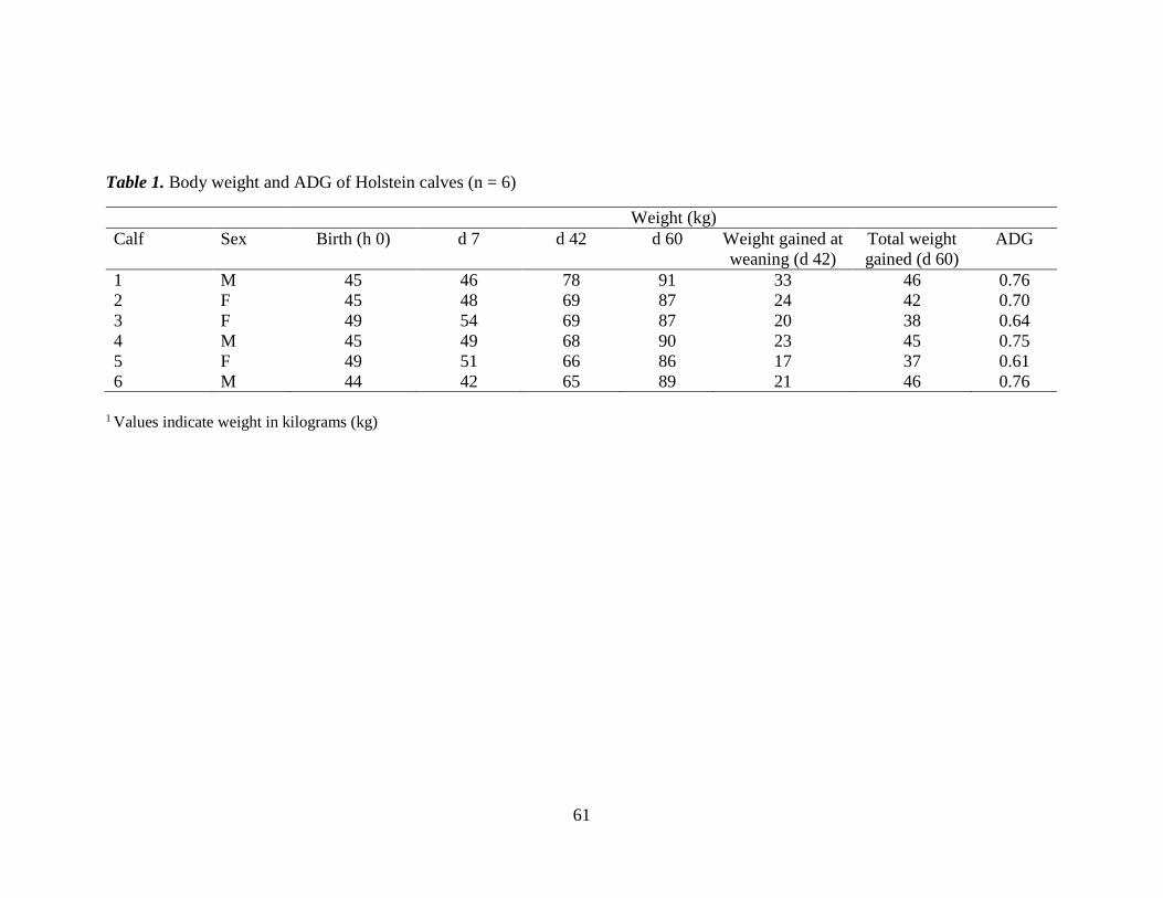
61
Table 1. Body weight and ADG of Holstein calves (n = 6)
Weight (kg)
Calf Sex Birth (h 0) d 7 d 42 d 60 Weight gained at
weaning (d 42)
Total weight
gained (d 60)
ADG
1 M 45 46 78 91 33 46 0.76
2 F 45 48 69 87 24 42 0.70
3 F 49 54 69 87 20 38 0.64
4 M 45 49 68 90 23 45 0.75
5 F 49 51 66 86 17 37 0.61
6 M 44 42 65 89 21 46 0.76
1 Values indicate weight in kilograms (kg)
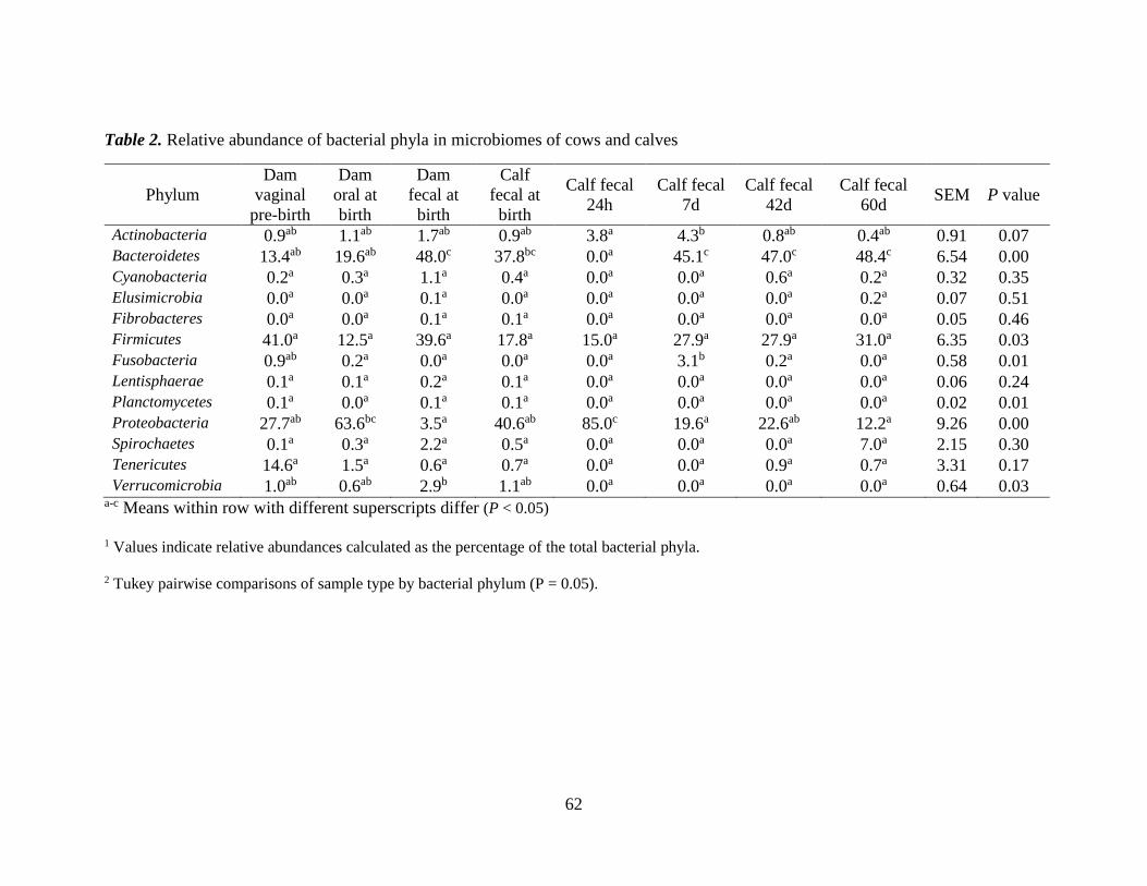
62
Table 2. Relative abundance of bacterial phyla in microbiomes of cows and calves
Phylum
Dam
vaginal
pre-birth
Dam
oral at
birth
Dam
fecal at
birth
Calf
fecal at
birth
Calf fecal
24h
Calf fecal
7d
Calf fecal
42d
Calf fecal
60d SEM P value
Actinobacteria 0.9ab 1.1ab 1.7ab 0.9ab 3.8a 4.3b 0.8ab 0.4ab 0.91 0.07 Bacteroidetes 13.4ab 19.6ab 48.0c 37.8bc 0.0a 45.1c 47.0c 48.4c 6.54 0.00 Cyanobacteria 0.2a 0.3a 1.1a 0.4a 0.0a 0.0a 0.6a 0.2a 0.32 0.35 Elusimicrobia 0.0a 0.0a 0.1a 0.0a 0.0a 0.0a 0.0a 0.2a 0.07 0.51 Fibrobacteres 0.0a 0.0a 0.1a 0.1a 0.0a 0.0a 0.0a 0.0a 0.05 0.46 Firmicutes 41.0a 12.5a 39.6a 17.8a 15.0a 27.9a 27.9a 31.0a 6.35 0.03 Fusobacteria 0.9ab 0.2a 0.0a 0.0a 0.0a 3.1b 0.2a 0.0a 0.58 0.01 Lentisphaerae 0.1a 0.1a 0.2a 0.1a 0.0a 0.0a 0.0a 0.0a 0.06 0.24 Planctomycetes 0.1a 0.0a 0.1a 0.1a 0.0a 0.0a 0.0a 0.0a 0.02 0.01 Proteobacteria 27.7ab 63.6bc 3.5a 40.6ab 85.0c 19.6a 22.6ab 12.2a 9.26 0.00 Spirochaetes 0.1a 0.3a 2.2a 0.5a 0.0a 0.0a 0.0a 7.0a 2.15 0.30 Tenericutes 14.6a 1.5a 0.6a 0.7a 0.0a 0.0a 0.9a 0.7a 3.31 0.17 Verrucomicrobia 1.0ab 0.6ab 2.9b 1.1ab 0.0a 0.0a 0.0a 0.0a 0.64 0.03
a-c Means within row with different superscripts differ (P < 0.05)
1 Values indicate relative abundances calculated as the percentage of the total bacterial phyla.
2 Tukey pairwise comparisons of sample type by bacterial phylum (P = 0.05).

63
Table 3. Dam microbiome samples as indicators of the calf fecal microbiome*
Phylum Sample type Estimate P value
Firmicutes
Dam fecal 1.46E-06 0.853
Dam oral -2.95E-06 0.715
Dam vaginal -1.37E-05 0.237
Bacteroidetes
Dam fecal 5.25E-07 0.984
Dam oral 3.10E-07 0.983
Dam vaginal -1.24E-05 0.964
Proteobacteria
Dam fecal -6.12E-06 0.886
Dam oral -5.13E-07 0.626
Dam vaginal 7.90E-08 0.912
Actinobacteria
Dam fecal -6.04E-05 0.787
Dam oral 3.60E-04 0.257
Dam vaginal 6.96E-04 0.553
*Parameter estimates for Firmicutes, Bacteroidetes, Proteobacteria, and Actinobacteria were fit according to a negative binomial
regression model. Convergence was not met for the four models.

64
Figure 1. Average relative abundance of bacterial phylum in each sample type. Stacked bar plots showing: dam vaginal pre-birth (n = 4), dam
oral at birth (n = 6), dam fecal at birth (n = 6), calf fecal at birth (n = 6), calf fecal at 24 h (n = 6), calf fecal at 7 d (n = 6), calf fecal at 42 d (n = 6),
calf fecal at 0 d (n = 6).
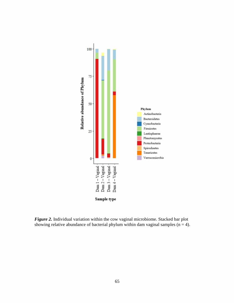
65
Figure 2. Individual variation within the cow vaginal microbiome. Stacked bar plot
showing relative abundance of bacterial phylum within dam vaginal samples (n = 4).

66
Figure 3. Bacterial composition of the dam vaginal and corresponding calf fecal microbiome at phylum level

67
Figure 4. Relative abundance of bacterial genera present in cow and calf microbiomes.
Heatmap color (dark red to dark blue) displays the relative abundance of each taxon
across samples. Each sample is identified at the bottom of the heatmap by the sample
name. Colors on top of each heatmap represent the time point to which each sample
belongs.

68
Figure 5. Similarity of cow and calf samples. Principal component analyses (PCA) of Beta diversity at the phylum level. Variable
contributions are labeled by color (A), where arrow direction indicates quality of representation on the factor map. Arrow length
indicates variable contribution to the principal components (A). Points represent individual samples from the different sample
locations and were colored by sample type.
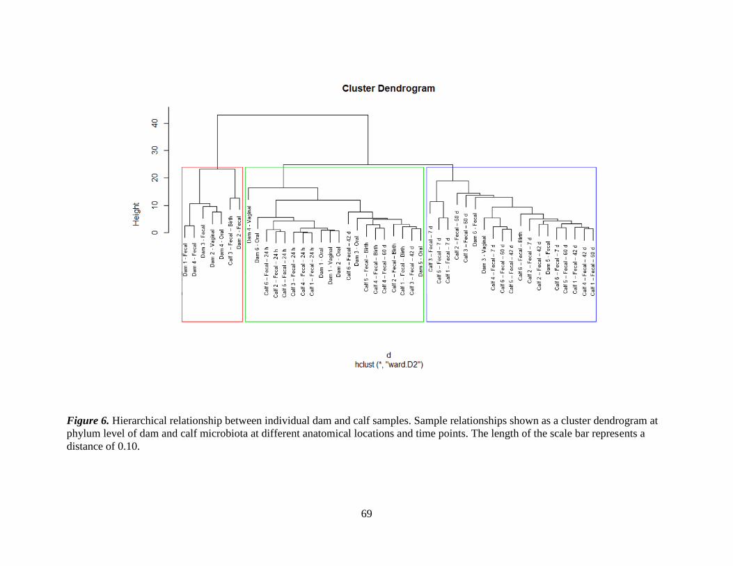
69
Figure 6. Hierarchical relationship between individual dam and calf samples. Sample relationships shown as a cluster dendrogram at
phylum level of dam and calf microbiota at different anatomical locations and time points. The length of the scale bar represents a
distance of 0.10.

70
REFERNCES
Aagaard, K., J. Ma, K. M. Antony, R. Ganu, J. Petrosino, and J. Versalovic. 2014. The
Placenta Harbors a Unique Microbiome. Science Translational Medicine
6(237):237ra265-237ra265.
Alipour, M. J., J. Jalanka, T. Pessa-Morikawa, T. Kokkonen, R. Satokari, U. Hynönen, A.
Iivanainen, and M. Niku. 2018. The composition of the perinatal intestinal microbiota in
cattle. Scientific Reports 8(1):10437.
Bäckhed, F., J. Roswall, Y. Peng, Q. Feng, H. Jia, P. Kovatcheva-Datchary, Y. Li, Y.
Xia, H. Xie, H. Zhong, Muhammad T. Khan, J. Zhang, J. Li, L. Xiao, J. Al-Aama, D.
Zhang, Ying S. Lee, D. Kotowska, C. Colding, V. Tremaroli, Y. Yin, S. Bergman, X. Xu,
L. Madsen, K. Kristiansen, J. Dahlgren, and J. Wang. 2015. Dynamics and Stabilization
of the Human Gut Microbiome during the First Year of Life. Cell Host & Microbe
17(5):690-703.
Bonder, M. J., A. Kurilshikov, E. F. Tigchelaar, Z. Mujagic, F. Imhann, A. V. Vila, P.
Deelen, T. Vatanen, M. Schirmer, S. P. Smeekens, D. V. Zhernakova, S. A.
Jankipersadsing, M. Jaeger, M. Oosting, M. C. Cenit, A. A. M. Masclee, M. A. Swertz,
Y. Li, V. Kumar, L. Joosten, H. Harmsen, R. K. Weersma, L. Franke, M. H. Hofker, R. J.
Xavier, D. Jonkers, M. G. Netea, C. Wijmenga, J. Fu, and A. Zhernakova. 2016. The
effect of host genetics on the gut microbiome. Nature Genetics 48:1407.
Chow, W. L. and Y.-K. Lee. 2006. Mucosal interactions and gastrointestinal microbiota.
Gastrointestinal microbiology:123-136.
Collado, M. C., S. Rautava, J. Aakko, E. Isolauri, and S. Salminen. 2016. Human gut
colonisation may be initiated in utero by distinct microbial communities in the placenta
and amniotic fluid. Scientific Reports 6:23129.
Dill-McFarland, K. A., J. D. Breaker, and G. Suen. 2017. Microbial succession in the
gastrointestinal tract of dairy cows from 2 weeks to first lactation. Scientific reports
7:40864.
Dominguez-Bello, M. G., E. K. Costello, M. Contreras, M. Magris, G. Hidalgo, N. Fierer,
and R. Knight. 2010. Delivery mode shapes the acquisition and structure of the initial
microbiota across multiple body habitats in newborns. Proceedings of the National
Academy of Sciences 107(26):11971-11975.
Gilbert, W. and R. Brace. 1993. Amniotic fluid volume and normal flows to and from the
amniotic cavity. Pages 150-157 in Proc. Seminars in perinatology.

71
Goodrich, J. K., E. R. Davenport, M. Beaumont, M. A. Jackson, R. Knight, C. Ober, T.
D. Spector, J. T. Bell, A. G. Clark, and R. E. Ley. 2016. Genetic Determinants of the Gut
Microbiome in UK Twins. Cell host & microbe 19(5):731-743.
Guzman, C. E., L. T. Bereza-Malcolm, B. De Groef, and A. E. Franks. 2015. Presence of
selected methanogens, fibrolytic bacteria, and proteobacteria in the gastrointestinal tract
of neonatal dairy calves from birth to 72 hours. PLoS One 10(7):e0133048.
Hall, A. B., A. C. Tolonen, and R. J. Xavier. 2017. Human genetic variation and the gut
microbiome in disease. Nature Reviews Genetics 18:690.
Hill, C. J., D. B. Lynch, K. Murphy, M. Ulaszewska, I. B. Jeffery, C. A. O’Shea, C.
Watkins, E. Dempsey, F. Mattivi, K. Tuohy, R. P. Ross, C. A. Ryan, P. W. O’ Toole, and
C. Stanton. 2017. Evolution of gut microbiota composition from birth to 24 weeks in the
INFANTMET Cohort. Microbiome 5(1):4.
Karstrup, C. C., K. Klitgaard, T. K. Jensen, J. S. Agerholm, and H. G. Pedersen. 2017.
Presence of bacteria in the endometrium and placentomes of pregnant cows.
Theriogenology 99:41-47.
Laguardia-Nascimento, M., K. M. G. R. Branco, M. R. Gasparini, S. Giannattasio-Ferraz,
L. R. Leite, F. M. G. Araujo, A. C. d. M. Salim, J. R. Nicoli, G. C. de Oliveira, and E. F.
Barbosa-Stancioli. 2015. Vaginal Microbiome Characterization of Nellore Cattle Using
Metagenomic Analysis. PLOS ONE 10(11):e0143294.
Lauber, C. L., M. Hamady, R. Knight, and N. Fierer. 2009. Pyrosequencing-Based
Assessment of Soil pH as a Predictor of Soil Bacterial Community Structure at the
Continental Scale. Applied and Environmental Microbiology 75(15):5111.
Lima, S. F., A. G. V. Teixeira, F. S. Lima, E. K. Ganda, C. H. Higgins, G. Oikonomou,
and R. C. Bicalho. 2017. The bovine colostrum microbiome and its association with
clinical mastitis. Journal of Dairy Science 100(4):3031-3042.
Lindner, J. D. D., M. Santarelli, C. T. Yamaguishi, C. R. Soccol, and E. Neviani. 2011.
Recovery and identification of bovine colostrum microflora using traditional and
molecular approaches. Food Technology and Biotechnology 49(3):364.
Lozupone, C. A., J. I. Stombaugh, J. I. Gordon, J. K. Jansson, and R. Knight. 2012.
Diversity, stability and resilience of the human gut microbiota. Nature 489:220.
Malmuthuge, N. 2016. Role of commensal microbiota in neonatal calf gut development.
Malmuthuge, N., P. J. Griebel, and L. Guan le. 2015. The Gut Microbiome and Its
Potential Role in the Development and Function of Newborn Calf Gastrointestinal Tract.
Front Vet Sci 2:36.
72
Malmuthuge, N., P. J. Griebel, and L. L. Guan. 2014. Taxonomic Identification of
Commensal Bacteria Associated with the Mucosa and Digesta throughout the
Gastrointestinal Tracts of Preweaned Calves. Applied and Environmental Microbiology
80(6):2021.
Malmuthuge, N. and L. L. Guan. 2016. Gut microbiome and omics: a new definition to
ruminant production and health. Animal Frontiers 6(2):8-12.
Mayer, M., A. Abenthum, J. Matthes, D. Kleeberger, M. Ege, C. Hölzel, J. Bauer, and K.
Schwaiger. 2012. Development and genetic influence of the rectal bacterial flora of
newborn calves. Veterinary microbiology 161(1-2):179-185.
McGuirk, S. M. and S. F. Peek. 2014. Timely diagnosis of dairy calf respiratory disease
using a standardized scoring system. Animal health research reviews 15(2):145-147.
Nagpal, R., H. Tsuji, T. Takahashi, K. Nomoto, K. Kawashima, S. Nagata, and Y.
Yamashiro. 2017. Ontogenesis of the Gut Microbiota Composition in Healthy, Full-
Term, Vaginally Born and Breast-Fed Infants over the First 3 Years of Life: A
Quantitative Bird’s-Eye View. Frontiers in Microbiology 8(1388).
Oikonomou, G., A. G. V. Teixeira, C. Foditsch, M. L. Bicalho, V. S. Machado, and R. C.
Bicalho. 2013. Fecal Microbial Diversity in Pre-Weaned Dairy Calves as Described by
Pyrosequencing of Metagenomic 16S rDNA. Associations of Faecalibacterium Species
with Health and Growth. PLOS ONE 8(4):e63157.
Pascault, N., S. Roux, J. Artigas, S. Pesce, J. Leloup, R. D. Tadonleke, D. Debroas, A.
Bouchez, and J.-F. Humbert. 2014. A high-throughput sequencing ecotoxicology study of
freshwater bacterial communities and their responses to tebuconazole. FEMS
Microbiology Ecology 90(3):563-574.
Redford, A. J. and N. Fierer. 2009. Bacterial Succession on the Leaf Surface: A Novel
System for Studying Successional Dynamics. Microbial Ecology 58(1):189-198.
Romano-Keeler, J. and J.-H. Weitkamp. 2015. Maternal influences on fetal microbial
colonization and immune development. Pediatric research 77(1-2):189.
Sharma, R., U. Schumacher, V. Ronaasen, and M. Coates. 1995. Rat intestinal mucosal
responses to a microbial flora and different diets. Gut 36(2):209-214.
Shin, N.-R., T. W. Whon, and J.-W. Bae. 2015. Proteobacteria: microbial signature of
dysbiosis in gut microbiota. Trends in Biotechnology 33(9):496-503.
Teeling, H., B. M. Fuchs, D. Becher, C. Klockow, A. Gardebrecht, C. M. Bennke, M.
Kassabgy, S. Huang, A. J. Mann, J. Waldmann, M. Weber, A. Klindworth, A. Otto, J.
Lange, J. Bernhardt, C. Reinsch, M. Hecker, J. Peplies, F. D. Bockelmann, U. Callies, G.
Gerdts, A. Wichels, K. H. Wiltshire, F. O. Glöckner, T. Schweder, and R. Amann. 2012.

73
Substrate-Controlled Succession of Marine Bacterioplankton Populations Induced by a
Phytoplankton Bloom. Science 336(6081):608.
The Human Microbiome Project, C. and C. Huttenhower and D. Gevers and R. Knight
and S. Abubucker and J. H. Badger and A. T. Chinwalla and H. H. Creasy and A. M. Earl
and M. G. FitzGerald and R. S. Fulton and M. G. Giglio and K. Hallsworth-Pepin and E.
A. Lobos and R. Madupu and V. Magrini and J. C. Martin and M. Mitreva and D. M.
Muzny and E. J. Sodergren and J. Versalovic and A. M. Wollam and K. C. Worley and J.
R. Wortman and S. K. Young and Q. Zeng and K. M. Aagaard and O. O. Abolude and E.
Allen-Vercoe and E. J. Alm and L. Alvarado and G. L. Andersen and S. Anderson and E.
Appelbaum and H. M. Arachchi and G. Armitage and C. A. Arze and T. Ayvaz and C. C.
Baker and L. Begg and T. Belachew and V. Bhonagiri and M. Bihan and M. J. Blaser and
T. Bloom and V. Bonazzi and J. Paul Brooks and G. A. Buck and C. J. Buhay and D. A.
Busam and J. L. Campbell and S. R. Canon and B. L. Cantarel and P. S. G. Chain and I.
M. A. Chen and L. Chen and S. Chhibba and K. Chu and D. M. Ciulla and J. C. Clemente
and S. W. Clifton and S. Conlan and J. Crabtree and M. A. Cutting and N. J. Davidovics
and C. C. Davis and T. Z. DeSantis and C. Deal and K. D. Delehaunty and F. E. Dewhirst
and E. Deych and Y. Ding and D. J. Dooling and S. P. Dugan and W. Michael Dunne and
A. Scott Durkin and R. C. Edgar and R. L. Erlich and C. N. Farmer and R. M. Farrell and
K. Faust and M. Feldgarden and V. M. Felix and S. Fisher and A. A. Fodor and L. J.
Forney and L. Foster and V. Di Francesco and J. Friedman and D. C. Friedrich and C. C.
Fronick and L. L. Fulton and H. Gao and N. Garcia and G. Giannoukos and C. Giblin and
M. Y. Giovanni and J. M. Goldberg and J. Goll and A. Gonzalez and A. Griggs and S.
Gujja and S. Kinder Haake and B. J. Haas and H. A. Hamilton and E. L. Harris and T. A.
Hepburn and B. Herter and D. E. Hoffmann and M. E. Holder and C. Howarth and K. H.
Huang and S. M. Huse and J. Izard and J. K. Jansson and H. Jiang and C. Jordan and V.
Joshi and J. A. Katancik and W. A. Keitel and S. T. Kelley and C. Kells and N. B. King
and D. Knights and H. H. Kong and O. Koren and S. Koren and K. C. Kota and C. L.
Kovar and N. C. Kyrpides and P. S. La Rosa and S. L. Lee and K. P. Lemon and N.
Lennon and C. M. Lewis and L. Lewis and R. E. Ley and K. Li and K. Liolios and B. Liu
and Y. Liu and C.-C. Lo and C. A. Lozupone and R. Dwayne Lunsford and T. Madden
and A. A. Mahurkar and P. J. Mannon and E. R. Mardis and V. M. Markowitz and K.
Mavromatis and J. M. McCorrison and D. McDonald and J. McEwen and A. L. McGuire
and P. McInnes and T. Mehta and K. A. Mihindukulasuriya and J. R. Miller and P. J.
Minx and I. Newsham and C. Nusbaum and M. O’Laughlin and J. Orvis and I. Pagani
and K. Palaniappan and S. M. Patel and M. Pearson and J. Peterson and M. Podar and C.
Pohl and K. S. Pollard and M. Pop and M. E. Priest and L. M. Proctor and X. Qin and J.
Raes and J. Ravel and J. G. Reid and M. Rho and R. Rhodes and K. P. Riehle and M. C.
Rivera and B. Rodriguez-Mueller and Y.-H. Rogers and M. C. Ross and C. Russ and R.
K. Sanka and P. Sankar and J. Fah Sathirapongsasuti and J. A. Schloss and P. D. Schloss
and T. M. Schmidt and M. Scholz and L. Schriml and A. M. Schubert and N. Segata and
J. A. Segre and W. D. Shannon and R. R. Sharp and T. J. Sharpton and N. Shenoy and N.
U. Sheth and G. A. Simone and I. Singh and C. S. Smillie and J. D. Sobel and D. D.
Sommer and P. Spicer and G. G. Sutton and S. M. Sykes and D. G. Tabbaa and M.
Thiagarajan and C. M. Tomlinson and M. Torralba and T. J. Treangen and R. M. Truty
and T. A. Vishnivetskaya and J. Walker and L. Wang and Z. Wang and D. V. Ward and
W. Warren and M. A. Watson and C. Wellington and K. A. Wetterstrand and J. R. White

74
and K. Wilczek-Boney and Y. Wu and K. M. Wylie and T. Wylie and C. Yandava and L.
Ye and Y. Ye and S. Yooseph and B. P. Youmans and L. Zhang and Y. Zhou and Y. Zhu
and L. Zoloth and J. D. Zucker and B. W. Birren and R. A. Gibbs and S. K. Highlander
and B. A. Methé and K. E. Nelson and J. F. Petrosino and G. M. Weinstock and R. K.
Wilson and O. White. 2012. Structure, function and diversity of the healthy human
microbiome. Nature 486:207.
Whon, T. W., M.-S. Kim, S. W. Roh, N.-R. Shin, H.-W. Lee, and J.-W. Bae. 2012.
Metagenomic Characterization of Airborne Viral DNA Diversity in the Near-Surface
Atmosphere. Journal of Virology 86(15):8221.
Wilson, M. 2005. Microbial inhabitants of humans: their ecology and role in health and
disease. Cambridge University Press.

75
CHAPTER 3
CONCLUSION AND IMPLICATIONS
Summary and Future Studies
Despite the extensive use of management protocols aimed at optimizing calf health,
neonatal diarrhea remains a major cause of calf mortality and morbidity (USDA, 2007,
Malmuthuge, 2016). Pre-weaned dairy heifers are highly susceptible to many viral and
bacterial enteric infections within the first few weeks of life and neonatal calf diarrhea
accounts for more than 50% of pre-weaned dairy heifer deaths. Enteric pathogens such as
Escherichia coli, Cryptosporidium parvum, and rotavirus are some of the most common
causes of NCD and mortality (Meganck et al., 2015). More than half of all neonatal diarrheas
appear during the first week of life, with an additional 15% occurring during the second week
(Uetake, 2013). In cases of infection in dairy calves, disease is often managed though the
use of antibiotics, however, the use of antibiotics in young ruminants is not always
effective and does not come without adverse effects to the neonatal gut microbiome
(Tamburini et al., 2016). With the growing concern of antibiotic resistance there has been
a strong push to reduce the use of antibiotics in animal production, presenting the need to
identify alternate treatment strategies. Modification of the gut microbiota could be utilized
as an effective alternative method for disease management, providing a new strategy to
suppress calf mortality (Willing et al., 2018).
This study investigated the maternal sources of bacteria to the neonatal calf and
the composition of bacterial communities of the Holstein dam and calf. Our hypothesis
was that the dam’s vaginal, fecal, and oral microbiomes will influence the early
microbiome of the calf. The microbiota of pre-weaned calves is dominated by 10
76
bacterial phyla. Three phyla, Proteobacteria, Firmicutes, and Bifidobacteria dominated
the fecal and vaginal microbiomes of the cow. Similarities between the vaginal and fecal
microbiome of the dam and the fecal microbiome of the calf indicate that the transference
of maternal microbes prior to and during birth play a role in the inoculation of the
neonatal calf gut microbiome. Further examinations are necessary to gain a complete
understanding of the maternal reproductive tract microbiome and the microbial
colonization patterns that occur in utero.
Although microbial communities of the same sample location and time point were
similar, we observed individual variation between each of the subjects. Individual
variation within the microbiome could be attributable to genetic or environmental factors.
Further examinations of the maternal, environmental, and genetic effects on the
microbiome of neonatal calves are necessary to gain a complete understanding of the
development of the gut microbiome. Microbial interventions applied to the intestinal
microbiome of neonatal calves could be used as a targeted strategy to improve immune
function, health, and growth of dairy calves. An in-depth understanding of host-microbial
interactions could lead to the development of disease management protocols through
manipulation of the gut microbiome (Malmuthuge and Guan, 2017).

77
REFERNCES
Malmuthuge, N. 2016. Role of commensal microbiota in neonatal calf gut development.
Malmuthuge, N. 2017. Understanding the gut microbiome of dairy calves: opportunities
to improve early-life gut health. Journal of dairy science 100(7):5996-6005.
Meganck, V., G. Hoflack, S. Piepers, and G. Opsomer. 2015. Evaluation of a protocol to
reduce the incidence of neonatal calf diarrhoea on dairy herds. Preventive Veterinary
Medicine 118(1):64-70.
Tamburini, S., N. Shen, H. C. Wu, and J. C. Clemente. 2016. The microbiome in early
life: implications for health outcomes. Nature Medicine 22:713.
Uetake, K. 2013. Newborn calf welfare: a review focusing on mortality rates. Anim Sci J
84(2):101-105.
USDA, U. 2007. Heifer Calf Health and Management Practices on US Dairy Operations,
2007. APHIS, ed. Dairy.
Willing, B. P., D. M. Pepin, C. S. Marcolla, A. J. Forgie, N. E. Diether, and B. C.
Bourrie. 2018. Bacterial resistance to antibiotic alternatives: a wolf in sheep’s clothing?