sos in pharmaceutical sciences, jiwaji university, gwalior
TRANSCRIPT
SOS in Pharmaceutical Sciences, Jiwaji University, Gwalior
Relevant Physiology of Urine FormationSUBJECT- PHARMACEUTICAL CHEMISTRY-VII (4T2)
PRESENTED BY:
JAGDEESH AHIRWAR
ASST. PROF. (CONTRACT)
Physiology of Urine Formation
• Urine formation starts from glomerular filtration (g.f.) in aprodigal way. Normally, about 180 L of fluid is filteredeveryday: all soluble constituents of blood minus the plasmaproteins (along with substances bound to them) and lipids,are filtered at the glomerulus.
• More than 99% of the glomerular filtrate is reabsorbed in thetubules; about 1.5 L urine is produced in 24 hours.
• The diuretics act primarily by inhibiting tubular reabsorption:just 1% decrease in tubular reabsorption would more thandouble urine output.
Physiology of Urine Formation
• The mechanisms that carryout ion movement across tubularcells are complex and involve a variety of energy dependenttransmembrane pumps as well as channels in between theloose fitting cells of the proximal tubule (PT).
• All Na+ that enters tubular cells through the luminalmembrane is pumped out of it into the renal interstitium atthe basolateral membrane by Na+K+ATPase energised Na+-K+
antiporter. Because there is a large intracellular toextracellular gradient for K+, it diffuses out through K+
channels to be recirculated by the Na+-K+ antiporter.
• For simplification, tubular reabsorption can be divided intofour sites.
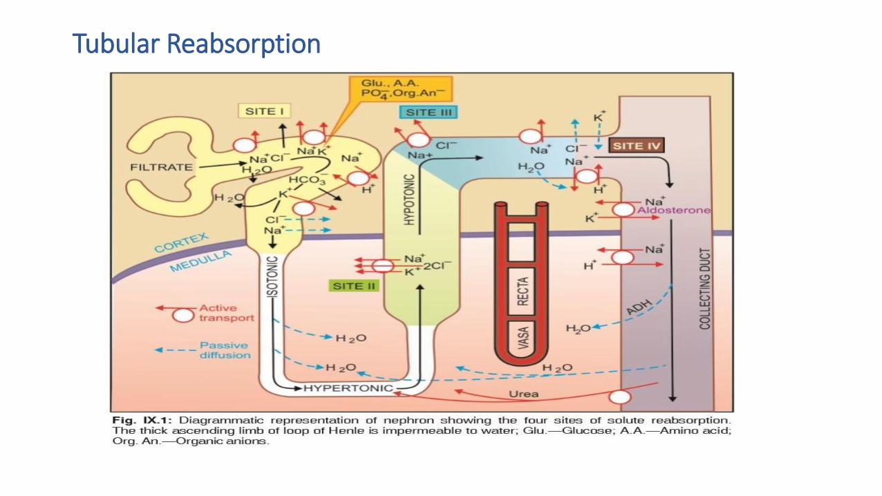
Tubular Reabsorption
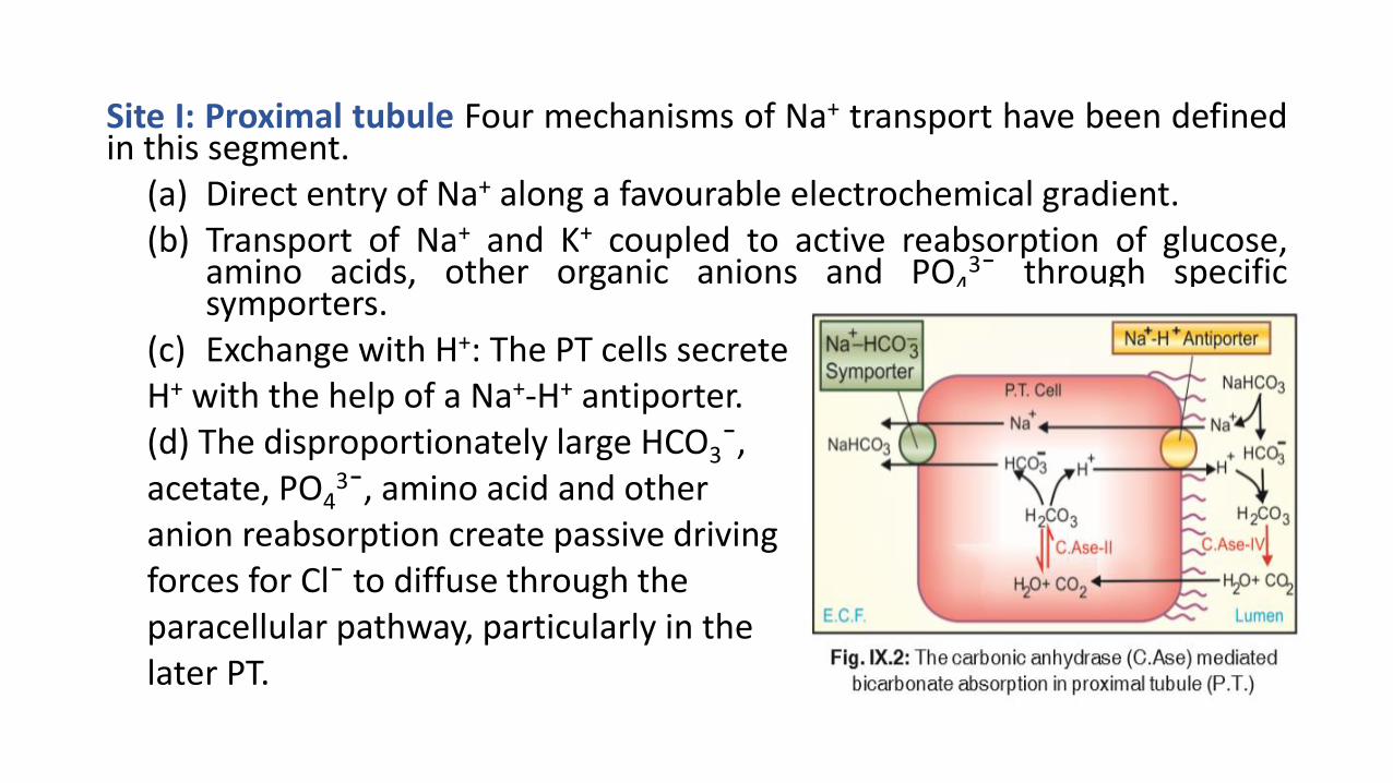
Site I: Proximal tubule Four mechanisms of Na+ transport have been definedin this segment.
(a) Direct entry of Na+ along a favourable electrochemical gradient.(b) Transport of Na+ and K+ coupled to active reabsorption of glucose,
amino acids, other organic anions and PO43¯ through specific
symporters.(c) Exchange with H+: The PT cells secreteH+ with the help of a Na+-H+ antiporter.(d) The disproportionately large HCO3¯,acetate, PO4
3¯, amino acid and otheranion reabsorption create passive drivingforces for Cl¯ to diffuse through theparacellular pathway, particularly in thelater PT.
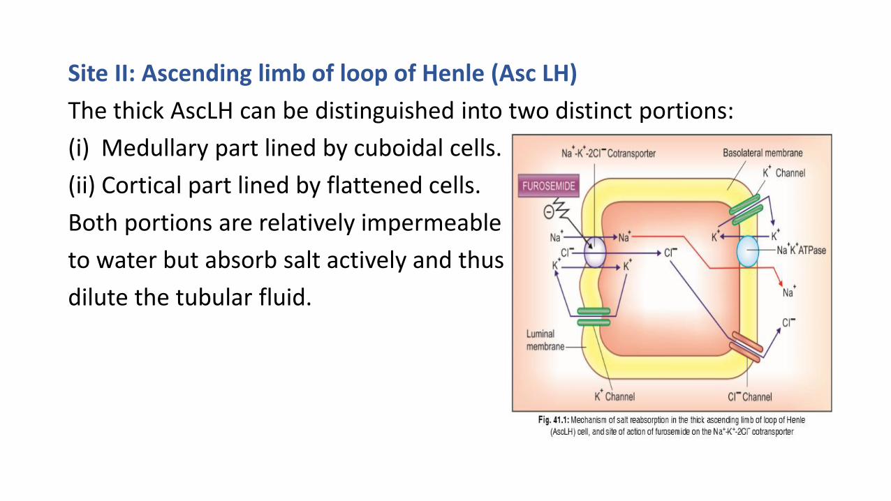
Site II: Ascending limb of loop of Henle (Asc LH)
The thick AscLH can be distinguished into two distinct portions:
(i) Medullary part lined by cuboidal cells.
(ii) Cortical part lined by flattened cells.
Both portions are relatively impermeable
to water but absorb salt actively and thus
dilute the tubular fluid.
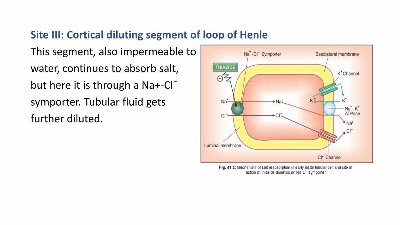
Site III: Cortical diluting segment of loop of Henle
This segment, also impermeable to
water, continues to absorb salt,
but here it is through a Na+-Cl¯
symporter. Tubular fluid gets
further diluted.
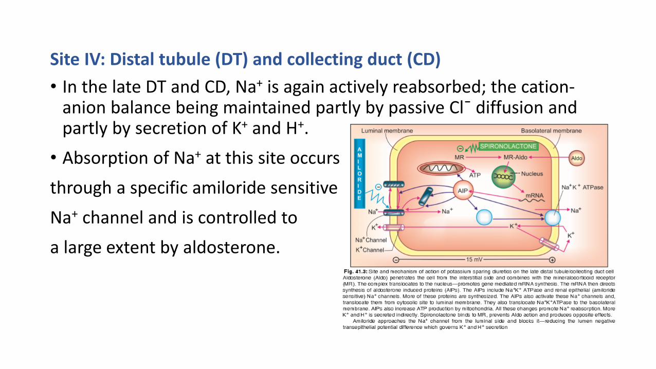
Site IV: Distal tubule (DT) and collecting duct (CD)
• In the late DT and CD, Na+ is again actively reabsorbed; the cation-anion balance being maintained partly by passive Cl¯ diffusion and partly by secretion of K+ and H+.
• Absorption of Na+ at this site occurs
through a specific amiloride sensitive
Na+ channel and is controlled to
a large extent by aldosterone.
Regulation of renal function
• Glomerular filtration rate (g.f.r.) is dependent on the pumping actionof heart, the magnitude of renal blood flow and the relativedimensions of afferent and efferent glomerular vessels. Thus,systemic and intrarenal haemodynamic changes can reflect in g.f.r.
• About 80% nephrons lie in outer cortex, have short loops of Henleand low Na+ reabsorptive capacity; while 20% or so arejuxtamedullary, possess long loops of Henle and are largelyresponsible for creating the corticomedullary osmotic gradient.
• Redistribution of blood flow between these two types of nephronscan alter salt and water excretion. Further, haemodynamic changeswithin different segments of renal vasculature can alter pressurerelationships which govern flow of solute and water.
• The renin-angiotensin-aldosterone system has a profound bearing ondistal tubular reabsorption of Na+ and secretion of K+/H+.
• Angiotensin II produced locally in the kidney has direct effects onintrarenal vascular beds as well as on salt and water reabsorption.
• Prostaglandins (PGs) are produced locally in kidney; act as modulatorsof renal circulation and renin release. PGE2 inhibits the action of ADHand has direct effects on tubular reabsorption.
• Natriuretic hormone produced by the atrium (atrial natriureticpeptide: ANP) and may be other sites also has been found to beimportant in inducing natriuresis in response to salt and volumeoverload. It mediates ‘escape’ from long term aldosterone action.
Relation to diuretic action
• The relative magnitudes of Na+ reabsorption at different tubular sitesare:
PT 65–70%; Asc LH 20–25%;
DT 8–9%; CD 1–2%.
• The maximal natriuretic response to a diuretic can give a clue to itssite of action. It may appear that diuretics acting on PT should be themost efficacious. However, these agents are either too weak or causedistortion of acid-base balance (CAse inhibitors). Moreover, theireffect may be obscured by compensatory increase in reabsorptionfurther down the nephron, because the reserve reabsorptive capacityof diluting segments is considerable and can overshadow moreproximal actions.

Thank You…