sonic hedgehog is a regulator of extracellular glutamate...
TRANSCRIPT
Scientific Report
Sonic hedgehog is a regulator of extracellularglutamate levels and epilepsyShengjie Feng1,2,†, Shaorong Ma1,2,†, Caixia Jia1,†, Yujuan Su1,2, Shenglian Yang1, Kechun Zhou1,
Yani Liu3, Ju Cheng1,2, Dunguo Lu1, Liu Fan1 & Yizheng Wang1,*
Abstract
Sonic hedgehog (Shh), both as a mitogen and as a morphogen,plays an important role in cell proliferation and differentiationduring early development. Here, we show that Shh inhibits gluta-mate transporter activities in neurons, rapidly enhances extra-cellular glutamate levels, and affects the development of epilepsy.Shh is quickly released in response to epileptic, but not physiologi-cal, stimuli. Inhibition of neuronal glutamate transporters by Shhdepends on heterotrimeric G protein subunit Gai and enhancesextracellular glutamate levels. Inhibiting Shh signaling greatlyreduces epileptiform activities in both cell cultures and hippo-campal slices. Moreover, pharmacological or genetic inhibition ofShh signaling markedly suppresses epileptic phenotypes in kindlingor pilocarpine models. Our results suggest that Shh contributes tothe development of epilepsy and suppression of its signalingprevents the development of the disease. Thus, Shh can act as amodulator of neuronal activity, rapidly regulating glutamate levelsand promoting epilepsy.
Keywords Epilepsy; extracellular glutamate; glutamate transporter; neuronal
activity; sonic hedgehog
Subject Categories Neuroscience; Signal Transduction
DOI 10.15252/embr.201541569 | Received 14 October 2015 | Revised 1 March
2016 | Accepted 7 March 2016
Introduction
The hedgehog family of secreted proteins controls a wide variety of
processes in embryonic development, stem cell maintenance, and
cancer cell proliferation [1]. Shh is one of the three members in the
family and has important roles in early neuronal development,
including axon guidance [2], neuronal differentiation [3], and corti-
cal microcircuit formation [4]. When Shh is released by the secre-
tion cells, it binds to Patched (Ptch1) to relieve its inhibition on
Smoothened (Smo), a transmembrane protein homologous with
members of G-protein-coupled receptors (GPCRs), and thus trigger
responses, leading to enhanced Gli or Ci (in Drosophila) expression
and Gli/Ci-dependent transcription [5]. As most of the effects of Shh
are mediated by Gli transcription, its expression levels are always
used as an indicator of the activation of this signaling [5,6]. It has
been reported that in mouse embryonic stem cells and developing
spinal neurons [3,7], Shh can induce an intracellular Ca2+ eleva-
tion. Shh and its signaling molecules are also expressed in mature
central nervous system (CNS) though their functions are not clear
[8–10].
Epilepsy is one of the most common chronic neurological disor-
ders with the characteristic of recurrent unprovoked seizures. About
one percent of population worldwide has been diagnosed with
epilepsy [11]. Individuals who got the first unprovoked seizure have
a high risk to expect a recurrence within 2 years of the initial
seizure. Understanding of the mechanism underlying epileptogene-
sis is critical for prevention and treatment of epilepsy. Generally,
epileptic seizures result from an imbalance between neuronal exci-
tation and inhibition. As the principal excitatory neurotransmitter in
the mammalian brain, glutamate depolarizes neurons, which inevi-
tably plays a role in the initiation and spreading of seizure activity,
even when the primary defect is not of glutamatergic origin [11,12].
Therefore, it is of vital importance that the extracellular glutamate
level is kept low [13]. Since there seem to be no extracellular
enzymes which can efficiently metabolize glutamate, the only rapid
way to clear glutamate from the extracellular fluid is by cellular
uptake [14], a task predominantly operated by the glutamate trans-
porters. It has been known for decades that glutamate uptake activ-
ity is not constant, but subject to regulation [14]. Down-regulation
of glutamate transporters in mice was found to cause spontaneous
seizures [15–17].
Five members of excitatory amino acid transporter proteins
(EAATs) have been identified and named as EAAT1-5 in mammals
[14,18]. Among them, EAAT1 (GLAST), EAAT2 (GLT-1), and
EAAT3 (EAAC1) are expressed in hippocampus and cortex
throughout the developing and adult stage [14,18,19]. The EAAC1
is localized to neurons, whereas GLAST and GLT-1 predominantly
to glia [14,18,19]. When a glutamate is taken into the cell by the
1 Laboratory of Neural Signal Transduction, Institute of Neuroscience, State Key Laboratory of Neuroscience, Shanghai Institutes for Biological Sciences, Chinese Academy ofSciences, Shanghai, China
2 University of Chinese Academy of Sciences, Shanghai, China3 Center of Cognition and Brain Science, AMMS, Beijing, China
*Corresponding author. Tel: +86 21 5492 1793; Fax: +86 21 5492 1735; E-mail: [email protected]†These authors contributed equally to this work
ª 2016 The Authors EMBO reports 1
Published online: April 4, 2016
glutamate transporters, 3Na+ and 1H+ influx and 1K+ efflux are
associated to generate a depolarization current [14,19]. The
expression of these transporters can be regulated by growth
factors to affect the homeostasis of synaptic glutamate levels,
though the regulation of their activities remains controversial
[14,20].
It has been reported that Shh expression in temporal lobe epilep-
tic foci of the patients is greatly increased, indicating that Shh may
participate in the changes that occur during temporal lobe epileptic
development [21]. Here, we report that Shh can enhance the extra-
cellular glutamate levels and control epileptogenesis.
Results and Discussion
Shh release and its pathway activation underepileptic stimulation
We initially studied the expression pattern of molecules associated
with Shh pathway in rat hippocampus, including Ptch1, Smo, and
Gli, and found that they were expressed from early stages to adult-
hood (Fig EV1A and B) and functional in cultures (Fig EV1C and
D). We then examined the influence of limbic seizures on Shh
pathway and found that a 2-h status epilepticus (SE) induced by
pilocarpine (Fig 1A–C) or one electrographic seizure induced by
kindling stimuli (Fig 1D–F) all increased Gli1 protein levels in the
cortex and hippocampus within 24 h (Figs 1A–F and EV1F–I). By
contrast, Shh expression was not changed in the same period
(Figs 1A–F and EV1E and G). These results suggest that Shh path-
way is activated likely due to Shh release in epilepsy rather than
enhanced expression level of Shh. To test whether Shh is indeed
released under epileptic stimulation, we examined Shh levels
in vivo from hippocampi and cortex in pilocarpine model. The
levels of Shh detected by enzyme-linked immunosorbent assay
(ELISA) were significantly increased 0.5, 1, and 1.5 h after the
seizure induction (Fig 1G). Also, Shh levels in the medium of
slices or hippocampal neurons incubated in the medium with
picrotoxin (Pic) or Mg2+-free (0Mg), conditions known to induce
epileptiform activities in slices or cells [22–24], were markedly
enhanced within 1 h in hippocampal slices (Fig 1H) or 15 min in
hippocampal neurons (Fig 1I). Thus, epileptic neuronal activity
rapidly increases Shh release. Consistently, up-regulation of Gli1
in neurons was found 4 h after the incubation in 0Mg for 30 min
(Fig 1J), suggesting that Shh pathway was activated by the
secreted Shh.
However, increase of Shh release was not found under physio-
logical conditions, such as theta-burst stimulation (TBS), 20-Hz
stimulation, or 50-mM-KCl stimulation (Fig EV2A–C). Moreover,
inhibition of Shh signaling did not change neuronal transmission in
physiological conditions, including basal synaptic transmission,
AMPA receptor-mediated excitatory postsynaptic current (AMPA-
EPSC), paired-pulse facilitation, or long-term potentiation (LTP)
(Fig EV2D–G). These results suggest that Shh did not have any role
in the physiological conditions. Therefore, compared with other
factors (BDNF, NGF or NT-3, etc.) [11,25] whose secretion can be
increased in response to both LTP and epileptic stimulations, Shh is
unique in its release in epilepsy and in regulation of neural activity,
but not synaptic efficacy.
Inhibiting Shh pathway reduces the epileptiform activity
We next investigated whether Shh is involved in the formation of
epileptiform activities. Neurons incubated in 0Mg medium for
30 min showed the robust epileptiform activity with a train of
high-frequency action potentials overlaying on the plateau of large
depolarization shifts with the frequency of 7.38 � 0.56 or
6.52 � 0.58 burst/min (Fig 2A–C, E left panel or Fig EV2H). In the
presence of cyclopamine or Sant-1, agents known to specifically
inhibit Smo [26,27], the frequency was 0.22 � 0.09 or 0.64 � 0.18
burst/min, respectively (Fig 2A–C and E left panel, and Fig EV2H).
All neurons incubated with 0Mg medium showed epileptiform
activities, whereas in the presence of cyclopamine or Sant-1,
26.32% or 54.55% exhibited such activities (Fig 2C and D left
panel). Moreover, robotnikinin, an agent targeting Shh [28], and
5E1, a Shh-neutralizing antibody [29,30], both reduced the epilep-
tiform activities (middle and right panels of Fig 2D and E; Fig
EV2I and J). Additionally, in the hippocampal slices the overall
frequency of spontaneous epileptiform bursts induced by picro-
toxin was reduced from 8.07 � 0.52 bursts/min to 6.09 � 0.55 by
cyclopamine (Fig 2F–H). Meanwhile, the expression of Gli1 or Gli2
was not changed when neurons or slices were treated with Shh or
the inhibitors in Mg2+-free or Pic conditions in 30 min
(Figs EV2K–M and 1J). Thus, inhibition of Shh pathway
suppresses the formation of epileptiform activity independent of
Gli transcription.
Shh increases the extracellular glutamate levels
To explore the mechanism by which Shh regulates epileptic activi-
ties, we assessed its effect on Ca2+ changes in hippocampal
neurons. Shh induced a slow and sustained intracellular Ca2+
elevation in an NMDA receptor (NMDAR)-dependent manner
(Fig EV3A–D), but it did not influence NMDA- or AMPA-induced
currents, suggesting that Shh did not directly affect these receptors
(Fig EV3E and F). Under Mg2+-free or Pic conditions, whole-cell
recording can detect a large and slow NMDA receptor current, the
tonic NMDA receptor current, an indication of ambient glutamate
in the extracellular space [31]. By application of D-CPP, an antago-
nist to the NMDA receptors, the amplitude of the tonic NMDA
receptor currents can be measured (Fig EV3G and H). Cyclopamine
greatly reduced the tonic NMDA receptor currents in both
hippocampal slices and neurons (Fig 2I and J). However, the
NMDA-induced currents were not affected by cyclopamine
(Fig EV3I). Together, these results support an explanation that Shh
increases extracellular glutamate to induce the NMDAR currents.
Indeed, Shh markedly increased the glutamate levels in the culture
medium in a cyclopamine-sensitive manner (Fig 3A). In contrast,
Shh did not affect the neuronal excitability (Fig EV3J–L). We then
explored whether Shh increases glutamate release or inhibits its
uptake. In the paired-pulse facilitation experiments, the paired-
pulse ratio was not different between vehicle- and Shh-incubated
hippocampal slices (Fig 3B). Therefore, it is unlikely that Shh
affects glutamate release. In contrast, Shh greatly inhibited 3H-
glutamate uptake in hippocampal neurons (Fig 3C). This inhibition
was reversed by cyclopamine (Fig 3C). Together, these results
suggest that Shh inhibits glutamate uptake to increase extracellular
glutamate levels.
EMBO reports ª 2016 The Authors
EMBO reports Shh is a regulator of glutamate and epilepsy Shengjie Feng et al
2
Published online: April 4, 2016
A B C
D E F
G H I J
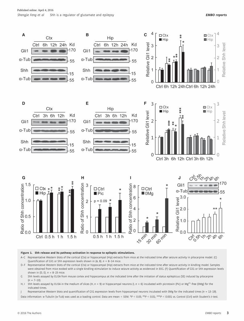
Figure 1. Shh release and its pathway activation in response to epileptic stimulations.
A–C Representative Western blots of the cortical (Ctx) or hippocampal (Hip) extracts from mice at the indicated time after seizure activity in pilocarpine model. (C)Quantification of Gli1 or Shh expression levels shown in (A, B). n = 8–14 mice.
D–F Representative Western blots of the cortical (Ctx) or hippocampal (Hip) extracts from mice at the indicated time after seizure activity in kindling model. Sampleswere obtained from mice evoked with a single kindling stimulation to induce seizure activity as evidenced in EEG. (F) Quantification of Gli1 or Shh expression levelsshown in (D, E). n = 8–23 mice.
G Shh levels assayed by ELISA from mouse cortex and hippocampus at the indicated time after the initiation of status epilepticus (SE) induced by pilocarpine(n = 7–10).
H, I Shh levels assayed by ELISA in the medium of slices (H, n = 9) or hippocampal neurons (I, n = 6) incubated with picrotoxin (Pic) or Mg2+-free (0Mg) for theindicated times.
J Representative Western blots and quantification of Gli1 expression levels from hippocampal neurons incubated with 0Mg for the indicated times (n = 13–19).
Data information: a-Tubulin (a-Tub) was used as a loading control. Data are mean + SEM. *P < 0.05; **P < 0.01; ***P < 0.001 vs. Control (Ctrl) with Student’s t-test.
ª 2016 The Authors EMBO reports
Shengjie Feng et al Shh is a regulator of glutamate and epilepsy EMBO reports
3
Published online: April 4, 2016
A B
C
D
E
F G H
I J
Figure 2.
EMBO reports ª 2016 The Authors
EMBO reports Shh is a regulator of glutamate and epilepsy Shengjie Feng et al
4
Published online: April 4, 2016
Shh rapidly inhibits the activity of glutamate transportersthrough Gai/o proteins
We then studied whether Shh regulates glutamate transporter activi-
ties. We found that cyclopamine no longer inhibited the epileptiform
activity in the presence of TBOA, a non-selective antagonist of gluta-
mate transporters (Fig EV4A–C), suggesting that cyclopamine and
TBOA might act on the similar pathway to regulate the epileptic
activities. It has been reported that glial transporters are barely
expressed in culture systems [32,33]. Consistently, the glutamate
uptake in neuronal cultures was slightly inhibited by dihydrokainate
(DHK), a selective inhibitor of GLT-1 [14] (Fig EV4D). In contrast,
expressing EAAC1 RNAi, but not GLAST RNAi, inhibited the gluta-
mate uptake (Figs 3D and EV4E–G), suggesting that EAAC1 is the
main glutamate transporter in the cultured neurons. Moreover, Shh
no longer suppressed the glutamate uptake when the EAAC1 RNAi
was expressed (Fig 3D). Additionally, aspartate (Asp), a non-
selective agonist of glutamate transporters, induced an inward
current in neurons, which was blocked by EAAC1 RNAi (Fig 3E).
Inhibiting GLT-1 and GLAST did not change the inward current
(Fig EV4H). Therefore, EAAC1-dependent current was the major
component of Asp-induced current in the cultures. Further, Shh
reduced the Asp-induced inward current density in a manner that
was sensitive to cyclopamine (Figs 3F and EV4I). In the presence of
TBOA, cyclopamine no longer affected Shh inhibition of EAAC1
current (Fig EV4J). In addition, Shh also increased extracellular
glutamate and reduced the EAAC1 current in the cultures treated
with ARA-C (Fig EV4K and L), suggesting that neuronal EAAC1 was
inhibited by Shh. To further confirm Shh inhibition of EAAC1, we
studied the effect of SmoA1, a constitutively active form of Smo
[34], on EAAC1 activities in HEK293 cells. Expressing SmoA1
eliminated the increase in 3H-glutamate uptake caused by
expressing EAAC1 (Fig 3G). Collectively, these results suggest
that Shh inhibits EAAC1 activities to elevate the extracellular
glutamate.
It has been reported that Smo can signal through Gai/o, but notmembers of Gs, Gq, and G12 families, to regulate downstream effec-
tors [35–37]. Pretreatment of the neurons with pertussis toxin
(PTX), an agent known to selectively inhibit Gai/o [35], blocked the
increase in extracellular glutamate levels, prevented Shh inhibition
of glutamate uptake, and reversed the inhibition of EAAC1 current
(Figs EV4M and 3H and I), suggesting that Gai/o protein is necessary
for Shh to inhibit EAAC1 activity. To detail Shh regulation of EAAC1
activity, we examined the I–V curve or Asp dose–response curve of
EAAC1 (Fig EV4N and O) and found that neither of them was
affected by Shh. Further, the expression level of EAAC1 was
unchanged 30 min after treatment with Shh (Fig EV4P). Using the
sucrose gradient centrifugation (0.3–2.0 M gradient) to isolate the
subcellular fractions from hippocampal neurons, we found that
Smo, Gai1-3, Gb, and EAAC1 were mainly presented in the same
fractions (Fig EV4Q). Together, these results point to a possibility
that Shh inhibits EAAC1 activity by suppressing its surface expres-
sion to elevate extracellular glutamate in a manner that is dependent
on Gai.
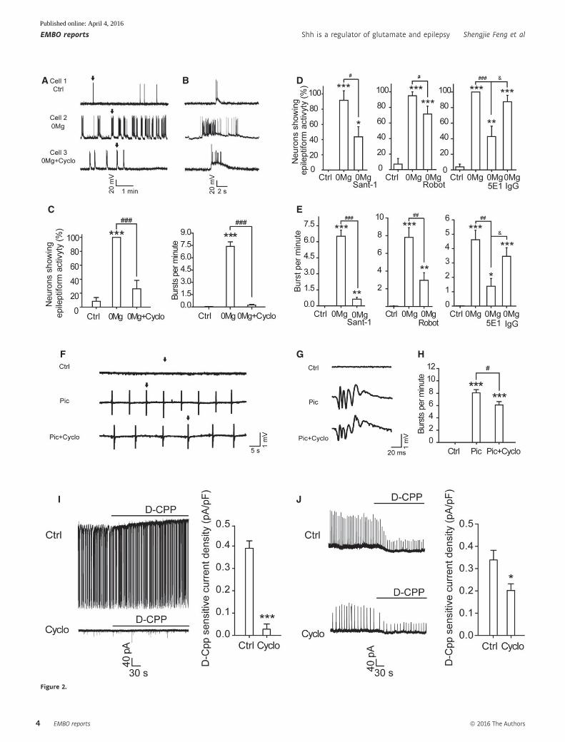
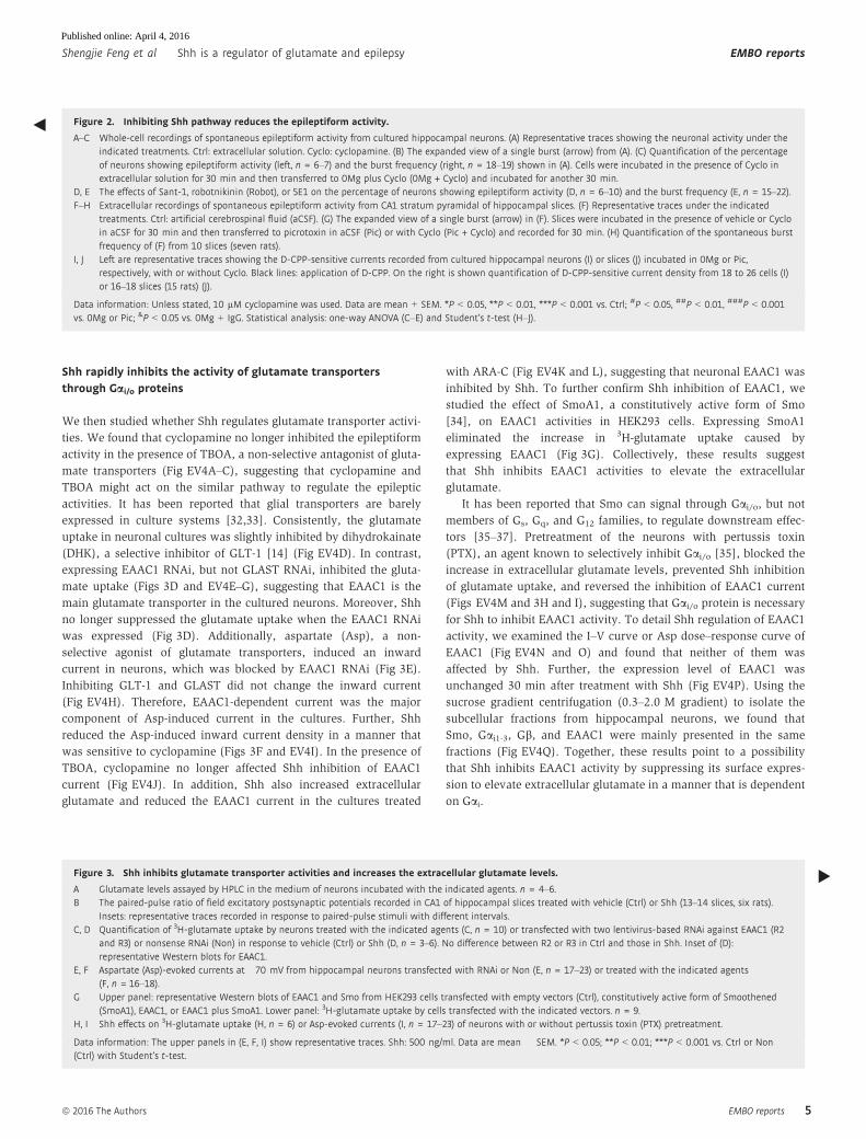
◀ Figure 2. Inhibiting Shh pathway reduces the epileptiform activity.
A–C Whole-cell recordings of spontaneous epileptiform activity from cultured hippocampal neurons. (A) Representative traces showing the neuronal activity under theindicated treatments. Ctrl: extracellular solution. Cyclo: cyclopamine. (B) The expanded view of a single burst (arrow) from (A). (C) Quantification of the percentageof neurons showing epileptiform activity (left, n = 6–7) and the burst frequency (right, n = 18–19) shown in (A). Cells were incubated in the presence of Cyclo inextracellular solution for 30 min and then transferred to 0Mg plus Cyclo (0Mg + Cyclo) and incubated for another 30 min.
D, E The effects of Sant-1, robotnikinin (Robot), or 5E1 on the percentage of neurons showing epileptiform activity (D, n = 6–10) and the burst frequency (E, n = 15–22).F–H Extracellular recordings of spontaneous epileptiform activity from CA1 stratum pyramidal of hippocampal slices. (F) Representative traces under the indicated
treatments. Ctrl: artificial cerebrospinal fluid (aCSF). (G) The expanded view of a single burst (arrow) in (F). Slices were incubated in the presence of vehicle or Cycloin aCSF for 30 min and then transferred to picrotoxin in aCSF (Pic) or with Cyclo (Pic + Cyclo) and recorded for 30 min. (H) Quantification of the spontaneous burstfrequency of (F) from 10 slices (seven rats).
I, J Left are representative traces showing the D-CPP-sensitive currents recorded from cultured hippocampal neurons (I) or slices (J) incubated in 0Mg or Pic,respectively, with or without Cyclo. Black lines: application of D-CPP. On the right is shown quantification of D-CPP-sensitive current density from 18 to 26 cells (I)or 16–18 slices (15 rats) (J).
Data information: Unless stated, 10 lM cyclopamine was used. Data are mean + SEM. *P < 0.05, **P < 0.01, ***P < 0.001 vs. Ctrl; #P < 0.05, ##P < 0.01, ###P < 0.001vs. 0Mg or Pic; &P < 0.05 vs. 0Mg + IgG. Statistical analysis: one-way ANOVA (C–E) and Student’s t-test (H–J).
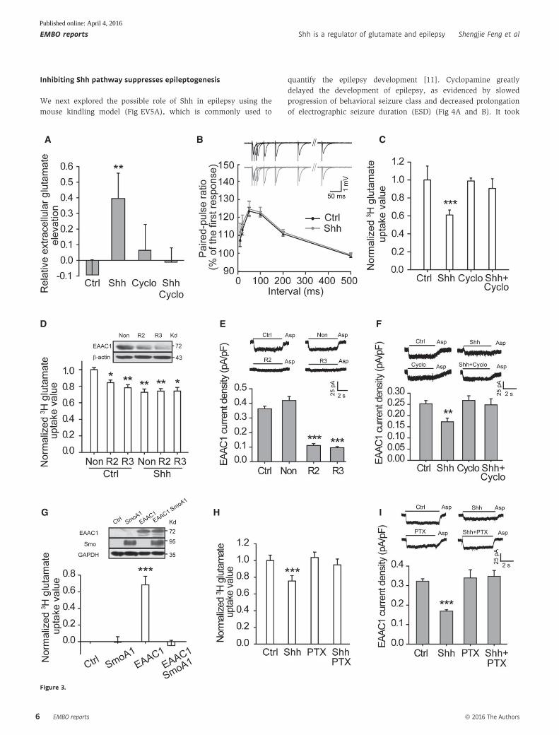
Figure 3. Shh inhibits glutamate transporter activities and increases the extracellular glutamate levels.
A Glutamate levels assayed by HPLC in the medium of neurons incubated with the indicated agents. n = 4–6.B The paired-pulse ratio of field excitatory postsynaptic potentials recorded in CA1 of hippocampal slices treated with vehicle (Ctrl) or Shh (13–14 slices, six rats).
Insets: representative traces recorded in response to paired-pulse stimuli with different intervals.C, D Quantification of 3H-glutamate uptake by neurons treated with the indicated agents (C, n = 10) or transfected with two lentivirus-based RNAi against EAAC1 (R2
and R3) or nonsense RNAi (Non) in response to vehicle (Ctrl) or Shh (D, n = 3–6). No difference between R2 or R3 in Ctrl and those in Shh. Inset of (D):representative Western blots for EAAC1.
E, F Aspartate (Asp)-evoked currents at �70 mV from hippocampal neurons transfected with RNAi or Non (E, n = 17–23) or treated with the indicated agents(F, n = 16–18).
G Upper panel: representative Western blots of EAAC1 and Smo from HEK293 cells transfected with empty vectors (Ctrl), constitutively active form of Smoothened(SmoA1), EAAC1, or EAAC1 plus SmoA1. Lower panel: 3H-glutamate uptake by cells transfected with the indicated vectors. n = 9.
H, I Shh effects on 3H-glutamate uptake (H, n = 6) or Asp-evoked currents (I, n = 17–23) of neurons with or without pertussis toxin (PTX) pretreatment.
Data information: The upper panels in (E, F, I) show representative traces. Shh: 500 ng/ml. Data are mean � SEM. *P < 0.05; **P < 0.01; ***P < 0.001 vs. Ctrl or Non(Ctrl) with Student’s t-test.
▸
ª 2016 The Authors EMBO reports
Shengjie Feng et al Shh is a regulator of glutamate and epilepsy EMBO reports
5
Published online: April 4, 2016
Inhibiting Shh pathway suppresses epileptogenesis
We next explored the possible role of Shh in epilepsy using the
mouse kindling model (Fig EV5A), which is commonly used to
quantify the epilepsy development [11]. Cyclopamine greatly
delayed the development of epilepsy, as evidenced by slowed
progression of behavioral seizure class and decreased prolongation
of electrographic seizure duration (ESD) (Fig 4A and B). It took
A B C
D E F
G H I
Figure 3.
EMBO reports ª 2016 The Authors
EMBO reports Shh is a regulator of glutamate and epilepsy Shengjie Feng et al
6
Published online: April 4, 2016
A B C
D E F
G H I
J K L
M N O
Figure 4.
ª 2016 The Authors EMBO reports
Shengjie Feng et al Shh is a regulator of glutamate and epilepsy EMBO reports
7
Published online: April 4, 2016
more stimulation to reach the fully kindled state in cyclopamine-
treated mice (16.5 � 1.2 stimulations; Fig 4C) than its controls
(9.07 � 1.2 stimulations). Then, we inhibited endogenous Shh
using its neutralizing antibody 5E1 to see whether Shh pathway was
indeed involved in the development of kindling-induced epilepsy.
Similar to the effect of cyclopamine, the administration of 5E1
greatly reduced the severity of epilepsy with delayed behavioral
seizure class and decreased prolongation of ESD (Fig 4D and E).
More stimulation was needed to reach the fully kindled state in 5E1-
treated mice (18.7 � 1.61 stimulations; Fig 4F) than in its controls
(12.94 � 1.3 stimulations; Fig 4F).
To clearly show the role of Shh signaling in epilepsy develop-
ment, we generated Smofl/flCaMKIIa-CreERT2 mice, which specifically
lack Smo in CaMKIIa-positive neurons after tamoxifen treatment
(Fig EV5B). The epilepsy development in Smofl/flCaMKIIa-CreERT2
mice was notably delayed (Figs 4G–I and EV5D–F), in a pattern
reminiscent of that seen in cyclopamine-treated mice. Since in addi-
tion to the neuronal glutamate transporter EAAC1, astrocytic trans-
porter GLT-1 is the main transporter to control glutamate uptake in
hippocampus [14,15,38], we then generated Smo+/flAldh1l1-Cre
mice, in which Smo was specifically ablated in Aldh1l1-positive
astrocytes (Fig EV5C). Down-regulation of Smo in astrocytes also
suppressed the epilepsy development (Fig 4J–L). No difference in
the basal electrographic seizure threshold was observed between
genetically modified mice and their controls (Fig EV5G and H).
Therefore, Smo in both neurons and astrocytes participates in
epilepsy development in mouse kindling models. Since stimulation
of Shh pathway increased extracellular glutamate in vivo (Fig EV4R)
and glutamate transporter family displays a considerable homology
(50–60% at the amino acid level) [39], it is likely that the activity of
transporters, including GLT-1 and GLAST, can be also regulated by
Shh to contribute to the enhancement in extracellular glutamate.
It has been reported that elevation of glutamate levels plays a
crucial role in spontaneous seizures induced by pilocarpine [40,41].
To further test the effects of Shh inhibition on epileptogenesis, we
examined the frequency of spontaneous seizures in pilocarpine-
induced spontaneous seizures. The frequency of spontaneous
seizures (class 4–5) was markedly inhibited in cyclopamine-treated
group (2.44 � 0.47, 1.74 � 0.27, or 2.39 � 0.39 in the 4th, 6th, or
8th week, respectively) than in the control group (7.05 � 1.02,
5.46 � 0.67, or 6.91 � 0.87 in the 4th, 6th, or 8th week, respectively)
in every week of time (Fig 4M). Overall, 68.18% of control mice
developed class 5 seizure within 8 weeks, whereas 21.74% of
cyclopamine-treated mice did (Fig 4N). Together, these results are
consistent with an explanation that Shh pathway contributes to the
development of epilepsy. Therefore, Smo could be a novel target for
anti-epileptogenesis therapy.
Here, we report a novel function of Shh in CNS. Our data show
that Shh is specifically released under epileptic stimulations and
regulates the extracellular glutamate levels independent of enhanced
Gli expression in the hippocampal neurons. The current findings led
us to propose a model that epileptic activity induces Shh release to
activate Smo and trigger subsequent responses, including
stimulation of Gai proteins, inhibition of surface expression of the
neuronal glutamate transporter EAAC1, increase in extracellular
glutamate levels, and enhancement in neuronal activities,
contributing to epileptogenesis (Fig 4O). Therefore, Shh–glutamate
signaling likely initiates a positive feedback to amplify the network
excitation to promote the development of epilepsy. Our findings
thus provide evidence to explain the fact that people face much
higher risk of permanent epilepsy after one experience of seizure.
Thus, Smo can induce a long-term effect through the Gli-expression
pathway and also induce a short-term effect through the Gaiproteins in neurons. Furthermore, because of the close relationship
between glutamate transporters and excitotoxicity, our work also
suggests a novel candidate for further study of excitotoxicity-related
diseases.
In conclusion, our findings point to a previously unknown role
of Shh in modulating extracellular glutamate and in contributing to
epileptogenesis independent of Gli transcription and to the existence
of Shh-regulated Gi protein and EAAC1 activities that is essential
for Shh-controlled neuronal activity. The demonstration of Shh-
dependent epileptiform activity in cultures and slices and
epileptogenesis in animal models further expands our understanding
of the diverse functions of Shh.
Materials and Methods
Animals
The 129SV Smofl/fl mice were from Jackson Lab, CaMKIIa-CreERT2
mice from European Mouse Mutant Archive, and Aldh1l1-Cre mice
from Mutant Mouse Resource & Research Centers. We crossed these
Cre mice with Smofl/fl mice to generate Smo-conditional knockout
mice. Smofl/flCaMKIIa-CreERT2 recombination was induced by tamo-
xifen (Tam, intraperitoneally, once a day for 7 consecutive days) at
adulthood. Tam (10 mg/ml, Sigma, T-5648) solution was prepared
in corn oil containing 10% ethanol. Corn oil containing 10% ethanol
(Oil) served as the control for Tam. We performed experiments
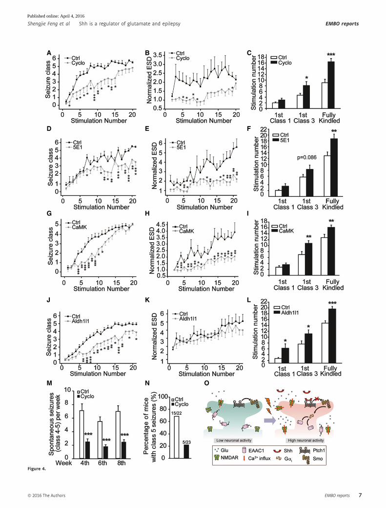
▸Figure 4. Inhibiting Shh pathway suppresses epileptogenesis in mouse epilepsy models.
A–L Effects of Cyclo (A–C, n = 14), 5E1 (D–F, n = 16–20), or ablation of Smo in CaMKIIa-positive neurons (G–I), or ablation of Smo in Aldh1l1-positive astrocytes (J–L) onthe progression of kindling including seizure class (A, D, G, J), evoked electrographic seizure duration (ESD) (B, E, H, K), and the number of stimulations required toreach equivalent seizure intensity (C, F, I, L). In (A–F), Ctrl are vehicle-treated mice. In (G–I), Ctrl: Smofl/fl induced by tamoxifen; CaMK: Smofl/flCaMKIIa-CreERT2 inducedby tamoxifen, n = 14-20. In (J–L), Ctrl: Smo+/fl; Aldh1l1: Smo+/flAldh1l1-Cre, n = 19–20.
M Frequency of spontaneous seizures (class 4–5) in mice administrated with Cyclo or vehicle (Ctrl) at 4th, 6th, and 8th week after pilocarpine SE induction. n = 22–23.N Percentage of mice with class 5 seizures within 8 weeks after pilocarpine SE induction. n = 22–23.O Schematic diagram depicting a working model for Shh regulation of epileptogenesis. Epileptic activity triggers sequential responses, including Shh release,
inhibition of glutamate transporter activity, and increase in extracellular glutamate, leading to epilepsy development.
Data information: Cyclo: 10 mg/kg. 5E1: 900 ng/mouse. Data are mean � SEM from at least three independent experiments. *P < 0.05; **P < 0.01; ***P < 0.001 vs. Ctrlwith Student’s t-test.
◀
EMBO reports ª 2016 The Authors
EMBO reports Shh is a regulator of glutamate and epilepsy Shengjie Feng et al
8
Published online: April 4, 2016
4 weeks after the induction. Oil-treated Smofl/flCaMKIIa-CreERT2
mice, oil-treated Smofl/fl mice, and tamoxifen-treated Smofl/fl mice
served as controls for Tam-treated Smofl/flCaMKIIa-CreERT2 mice.
Homozygous Smofl/flAldh1l1-Cre mice were lethal during the embryo-
nic development, so Smo+/flAldh1l1-Cre mice were used (Smo+/fl as
controls). We used adult male C57BL/6 mice in pilocarpine or
kindling epilepsy models. The C57BL/6 mice were from SLAK Labo-
ratory Animal Shanghai China. For transgenic mice, both male and
female mice were used in epilepsy experiments. For C57BL/6 mice,
male mice were used in the experiments. All animal studies
followed the animal welfare guidelines of Institute of Neuroscience,
CAS.
Reagents and antibodies
Rabbit anti-Gli1 (2534), anti-Shh (2287), Gli1 blocking peptide
(1641S), Shh blocking peptides (13937S), and anti-Gai1 antibodies
were from Cell Signaling; rabbit anti-Gli2, anti-EAAC1, anti-GLAST,
and anti-Gai2 antibodies from Abcam; rabbit anti-Gai3 from Milli-
pore; and mouse anti-b-actin, anti-a-tubulin, and anti-GAPDH anti-
bodies from Sigma-Aldrich. Fura-2 AM, Alexa Fluor 488-conjugated
goat anti-rabbit and Texas-Red-conjugated goat anti-mouse
secondary antibodies were from Molecular Probes, and HRP-
conjugated goat anti-rabbit and anti-mouse secondary antibodies
from Amersham. All other reagents were purchased from Sigma-
Aldrich.
Animal models for seizures and epilepsy
Pilocarpine model
Pilocarpine hydrochloride (Sigma-Aldrich) dissolved in 0.9%
(wt/vol) sterile saline was intraperitoneally (i.p.) administered to
adult male mice (C57BL/6) at a dosage of 300 mg/kg (body weight).
Scopolamine methyl nitrate (2 mg/kg, i.p.; Sigma-Aldrich) was
injected 30 min before pilocarpine to block peripheral side effects.
Diazepam (4 mg/kg, i.p.; Sigma-Aldrich) was used to terminate
status epilepticus (SE, 2 h) of continuous seizures to standardize the
duration of seizure activity.
To detect Gli1 and Shh levels, mice were killed at varying inter-
vals (6, 12, or 24 h after the termination of SE) and their expres-
sion levels in cortex and hippocampi were analyzed. Vehicle
(saline, 0.9% wt/vol sterile saline)-injected mice served as
controls.
To detect the spontaneous seizures, mice were divided into two
groups after SE induction, vehicle (HBC, 45% wt/vol HBC (2-hydroxy-
propyl-b-cyclodextrin, Sigma-Aldrich) in PBS)- or cyclopamine
(Cyclo, 10 mg/kg, Selleckchem, Sigma-Aldrich, Abcam)-treated
group. A video monitoring system was used for recording the
behaviors at the 4th, 6th, and 8th week (7 h/day, 5 days/week)
after SE induction. HBC or Cyclo was administrated every other
day from the first day after SE induction to the end of the
8th week.
To examine Shh release in vivo, mice were killed at varying inter-
vals (0.5, 1, or 1.5 h after the initiation of SE) and their cortex and
hippocampi were dissected and ground gently by grinding rod with
heparin. The samples were centrifuged at 100 g at 4°C and Shh in
the supernatant was determined by ELISA. Animals were divided
into groups randomly.
Kindling model
Kindling model was established according to the method described
previously with a little modification [42]. We implanted a bipolar
electrode used for stimulating and recording stereotactically in the
left amygdala of adult mice under sodium pentobarbital anesthe-
sia, at the following coordinates (with bregma as the reference):
1.2 mm posterior, 2.8 mm lateral, and 4.6 mm below dura. Four
screws were also inserted into the skull through a drilled hole
without piercing the dura. One of the screws served as a ground
electrode. The electrodes and screws were fixed with a mixture of
acrylic and dental cement. After a recovery period of 5–7 days, we
determined the electrographic seizure threshold (EST) for each
animal by applying a 1-s train of 1-ms biphasic rectangular pulses
at 60 Hz beginning at 60 lA. More stimulation, increasing by
20 lA steps, were delivered at 10 min intervals until an electro-
graphic seizure with a duration of no less than 3 s was detected
by the electroencephalogram (EEG) recording from the bipolar
electrode. Both EEG and behavioral seizures were recorded. We
scored the behavioral progression of stimulation-evoked seizures
according to Racine’s standard classification [43] with a little
modification: 0, no behavioral change; 1, eye blinking and/or
facial clonus; 2, head nodding; 3, unilateral forelimb clonus; 4,
rearing with bilateral forelimb clonus; 5, generalized clonic convul-
sions with loss of hind limb control; and 6, severe whole body
convulsions with continuous jumping behavior. We defined the
fully kindled state as the occurrence of three consecutive class 5 or
6 seizures. We stimulated and recorded each mouse once a day.
To detect Gli1 and Shh expression, mice with an electrographic
seizure detected were divided into groups randomly and killed at
varying intervals (3, 6, or 12 h after the stimuli) and their protein
levels in cortex and hippocampi were analyzed. Mice undergone
surgery but without stimulation were used as controls.
To test the effect of inhibition of Shh pathway on kindling
development, Cyclo (10 mg/kg) and its vehicle (HBC), or 5E1
(900 ng/mouse, DSHB) and its control IgG were delivered intraperi-
toneally (i.p.) or intracerebroventricularly (i.c.v.), respectively,
30 min before each stimulation. Animals were divided into groups
randomly.
Cell culture and transfection
Primary hippocampal neurons were isolated and cultured as
described previously [44]. Briefly, the neurons were obtained by
dissociating the hippocampus from SD rat brains of embryonic day
18 and then seeded at a density of 5 × 10⁴/cm2 onto coverslips (No.
1 Glass, Warner Instruments, Connecticut, USA) which had been
coated with 50 lg/ml poly-D-lysine (Sigma-Aldrich). Cells were
cultured in Neurobasal supplemented with B-27 and 0.5 mM gluta-
max for 9–12 days before use. In ARA-C (cytosine b-D-arabinofurano-side, Sigma C1768, 10 lM) experiments, the ARA-C was added
24–48 h after plating.
The HEK293 cells cultured in DMEM supplemented with 10%
FBS were transfected with control (1 lg pCIG and 3 lg pEGFP),
pEGFP-smoothened (1 lg pCIG and 3 lg pEGFP-constitutively
active smoothened), pCIG-EAAC1 (1 lg pCIG-EAAC1 and 3 lgpEGFP), or pCIG-EAAC1 plus pEGFP-smoothened (1 lg pCIG-
EAAC1 and 3 lg pEGFP-constitutively active form of smoothened)
using Lipofectamine 2000 (Invitrogen).
ª 2016 The Authors EMBO reports
Shengjie Feng et al Shh is a regulator of glutamate and epilepsy EMBO reports
9
Published online: April 4, 2016
Electrophysiology
Whole-cell recordings in cultured neurons
Whole-cell patch-clamp recordings were carried out at room temper-
ature (22–25°C) at DIV (days in vitro) 9–12. Patch electrodes were
pulled with a Flaming/Brown micropipette puller (P-97, Sutter
Instruments, USA) and fire-polished. The recording electrodes had a
resistance of 4–6 MΩ when filled with different internal solutions.
The liquid junction potential was auto-adjusted each time by pipette
offset. After the formation of whole-cell recording, access resis-
tances were generally < 20 MΩ.To record NMDA-activated currents, the pipette solution A (in
mM: CsCl 140, EGTA 10, Mg2ATP 0.3, CaCl2 0.3, and HEPES 10, pH
adjusted to 7.3 with CsOH) and the external solution (ES) A (in
mM: NaCl 140, KCl 5, CaCl2 1, MgCl2 1, glucose 10, and HEPES 10,
pH adjusted to 7.4 with NaOH) were used. The membrane potential
was held at +40 mV. To record AMPA-activated currents, the pipette
solution B (in mM: potassium gluconate 120, KCl 20, MgCl2 2,
HEPES 10, EGTA 10, and Na2ATP 2, pH adjusted to 7.3 with KOH)
and ES A were used. The membrane potential was held at �70 mV.
To record epileptiform discharge or tonic NMDA receptor-
mediated currents, the pipette solution B and the ES B (in mM: NaCl
145, KCl 3, CaCl2 2, MgCl2 2, glucose 10, and HEPES 10, pH
adjusted to 7.4 with NaOH) were used. In recording of epileptiform
discharge, Mg2+-free ES (0Mg) was prepared from the external solu-
tion B, in which MgCl2 was omitted. ES was used as a control. Cells
were incubated in various ES for 30 min before recordings. In
0Mg+Cyclo group, cells were incubated in ES with Cyclo (10 lM in
DMSO, used in all in vitro experiments) for 30 min and then trans-
ferred to 0Mg plus Cyclo and incubated for another 30 min. Record-
ings were performed for 10 min. The epileptiform burst is defined
as a large depolarization shift with ≥ 10 mV depolarization and
≥ 300 ms in duration and at least five action potentials as described
in the previous study [45]. Neurons with at least two epileptiform
bursts during 10 min recording were defined as “neurons showing
epileptiform activity” [45].
In recording of D-CPP (50 lM)-sensitive tonic NMDA currents,
0Mg was used as control. The cells were incubated in 0Mg for
30 min before recording. In 0Mg+Cyclo group, the cells were incu-
bated in the presence of Cyclo for 30 min and then transferred to
0Mg plus Cyclo and incubated for another 30 min. The membrane
potential was held at �70 mV.
To record aspartate (Asp, 100 lM)-evoked EAAC1 currents, the
pipette solution C (in mM: KNO3 140, MgCl2 2.5, HEPES 10, EGTA
11, Na2ATP 5, and HEPES 10, pH adjusted to 7.3 with KOH) and the
ES C (in mM: NaCl 135, KCl 5.4, CaCl2 1.8, MgCl2 1.3, glucose 10,
HEPES 10, D-CPPene (NMDA receptor antagonist) 0.05, CNQX
(AMPA/KA receptor antagonist) 0.02, and bicuculline (GABAA
receptor antagonist) 0.02, pH adjusted to 7.4 with NaOH) were
used. The membrane potential was held at �80 mV.
To test the effect of Shh on neuronal excitability, a series of depo-
larizing currents from 0 to 200 in 5 pA step increment was injected
[46] and the resting membrane potential, the averaged current
threshold to induce the first action potential, and frequencies of fir-
ing in response to 70-, 100-, and 150-pA current injections with or
without Shh were determined with pipette solution B and the ES B.
Drug solutions were prepared in external solutions and applied
to neurons by pressure using the 8-Channel Focal Perfusion System
(ALA Scientific Instruments, USA). Neurons were bathed constantly
in external solutions between drug applications. Drug solution
exchange was accomplished by electronic control.
Recordings in hippocampal slices
Hippocampal slices were prepared from male SD rats (90–140 g).
Animals were anesthetized with 1% pentobarbital sodium, and their
brains were rapidly removed. Transverse slices were cut 400 lmthick on a vibration microtome (Leica VT1000S, Leica Micro-
systems, Wetzlar, Germany) in ice-cold dissection buffer (in mM:
sucrose 213.26, KCl 2.5, MgSO4 2, CaCl2 0.5, NaH2PO4 1.25,
NaHCO3 26, and D-glucose 10). Before use, the slices were equili-
brated in artificial cerebrospinal fluid (aCSF, in mM: NaCl 124, KCl
2.5, MgSO4 1, CaCl2 2, NaH2PO4 1.25, NaHCO3 26, and D-glucose
10) saturated with 95% O2/5% CO2 for at least 1 h. Recordings
were in a submerge chamber at a flow rate of 1.5–2 ml/min with
aCSF.
To record spontaneous epileptiform discharge, the aCSF contain-
ing 6.5 mM KCl and 100 lM picrotoxin (Pic) was used and record-
ings were performed at 30°C with aCSF as a control. Slices were
pre-incubated with DMSO or Cyclo for 30 min and then perfused
with Pic or Pic plus Cyclo. Borosilicate glass microelectrodes with
1–3 MΩ resistance filled with aCSF were positioned at CA1 stratum
pyramidal. Recordings were performed for 30 min after the
frequency of discharge was stabilized.
In paired-pulse experiments, microelectrodes were positioned at
CA1 stratum radiatum to record field excitatory postsynaptic poten-
tial (fEPSP). Paired-pulse stimuli were delivered at 10, 20, 50, 100,
200, and 500-ms intervals every 20 s at 30–40% of maximal
response. Paired-pulse ratio was the percentage of the second stimu-
lus-evoked fEPSP amplitude divided by the first stimulus-evoked
fEPSP amplitude in a given paired-pulse stimuli in individual slices.
In recording of AMPA-EPSC, picrotoxin (100 lM) was added
with a holding at �70 mV and pipette solution B was used. In LTP
experiment, theta-burst stimulation (TBS, five trains of stimuli
which contain five burst (five stimuli at 100 Hz) at 5 Hz repeated at
0.1 Hz) was given by a tungsten electrode positioned at striatum
radiatum of CA1.
To record D-CPP-sensitive tonic NMDA receptor currents, Pic
was used as a control. Whole-cell recordings were performed at CA1
pyramidal neurons with a holding at +40 mV. The inner solution (in
mM: CsCl 140, EGTA 10, Mg2ATP 0.3, CaCl2 0.3, and HEPES 10)
was used. The cells were incubated in Pic for 30 min before record-
ing. In Pic+Cyclo group, the cells were incubated in the presence of
Cyclo for 30 min and then transferred to Pic plus Cyclo and incu-
bated for another 30 min.
Data were acquired using MultiClamp700A and 700B and Digi-
data1322AA and 1440A (Axon Instruments, California, USA),
sampled at 10 kHz, and filtered at 2 kHz. Offline analysis was done
by Clampfit 9.0 and 10.2 software (Axon Instruments, California,
USA).
ELISA
Briefly, hippocampal neurons (2 × 10⁶ per 3.5-cm dish) were
cultured for 10 days and replaced with the medium with 450 llMg2+-free external solution (0Mg) (as in electrophysiology). After
incubation for different times (15, 30, or 45 min), all supernatant
EMBO reports ª 2016 The Authors
EMBO reports Shh is a regulator of glutamate and epilepsy Shengjie Feng et al
10
Published online: April 4, 2016
was immediately collected and assayed in the plate coated with
anti-Shh antibody. In the TTX experiment, cells were treated with
50 mM KCl, CNQX, 10 lM; APV, 100 lM with or without TTX
(1 lM) for 30 min. In the electro-stimulation experiments, the cells
were stimulated at 20 Hz for 30 min [47,48]. All cell experiments
used ES as a control. For determination of Shh secretion from
slices, acute hippocampal slices (400 lm thick) were transferred to
900 ll oxygen-bubbled aCSF (as a control) or Pic (as in electro-
physiology) in 3.5-cm dish and incubated for different times (0.5,
1, or 1.5 h). After incubation, slices were rinsed with manual stir
bars, and the supernatant was collected and centrifuged at 4°C for
10 min. To detect the Shh levels under physiological condition,
TBS (as in electrophysiology) was given by a tungsten electrode
positioned at striatum radiatum of CA1. All the supernatant was
applied in one anti-Shh-coated well with a two-step incubation.
Shh levels were determined by the ELISA kit (R&D MSHH00) and
calculated from the standard curve prepared for each plate, using
Origin 7.5 software. The standard curves were linear within the
range used (0–500 pg/ml Shh). The quantities of Shh in experi-
mental samples were always within the linear range of the stan-
dard curve.
Cytosolic Ca2+ measurement
Changes in [Ca2+]i concentration were measured using Fura-2 AM
(Invitrogen). Briefly, a total of 1 × 10⁵ primary cultured neurons
were seeded on coverslips and incubated with 2 lM Fura-2 AM at
37°C for 25 min. Cells were washed three times with normal exter-
nal solution (in mM: NaCl 120, KCl 16, CaCl2 2, MgCl2 2, glucose
12, sucrose 12, and HEPES-free acid 10, pH 7.4) and imaged using a
Nikon Eclipse Ti microscope (Nikon, Japan) with dual excitation
wavelengths for Fura-2 AM at 340 and 380 nm and detection of flu-
orescent emission at 500 nm.
High performance liquid chromatography (HPLC) assay
After two washes with external solution (in mM: NaCl 145, KCl 3,
CaCl2 2, MgCl2 2, glucose 10, and HEPES 10, pH adjusted to 7.4 with
NaOH), cells were incubated with vehicle (bovine serum albumin in
external solution, BSA) for 30 min. Then, the incubating solution
was collected for HPLC assay as a baseline control. Immediately
after removing the solution, cells were treated with either BSA or
Shh (Sigma-Aldrich, Selleckchem, 500 ng/ml used in all experi-
ments) for another 30 min. The incubating solution was also
collected for HPLC test.
The HPLC system (Agilent Jordax Eclipse Plus C18 2.1 mm
I.D × 150 mm analytical column, 3.5/micron) and fluorescence
detector (Agilent G1321A HPLC-FLD) with excitation wavelength at
340 nm and emission wavelength at 450 nm were used. Column
temperature was maintained at 36°C. The mobile phase was formed
by methanol, acetonitrile, and water.
3H-glutamate uptake assay
After two washes with the uptake buffer (in mM: glucose 6, KCl 4,
NaCl 130, CaCl2 1.3, MgSO4 1.2, KH2PO4 1, and HEPES [pH 7.3]
25), cells were pre-incubated with vehicle (BSA), Shh, or inhibitors
for 10 min. Uptake assays were started by adding [3H] L-glutamic
acid at 10�6 M final concentration (specific activity 25 Ci/mmol,
PerkinElmer) diluted in the uptake buffer. Incubation was at 37°C
for 6 min. The reaction was stopped by rapidly adding 1 ml of cold
uptake buffer and followed by two washes with the cold medium.
Then, 1 N NaOH was added to cells and the radioactivity was
assessed by liquid scintillation counting.
Microdialysis
The mice were implanted with CMA 7 guide cannula (CMA micro-
dialysis AB, Kista, Sweden) with the following coordinates (to
bregma): 2.5 mm posterior, 3.1 mm lateral, and 2.4 mm below
dura. Seven days after implantation, the guide cannula were
replaced with CAM 7 microdialysis probes (CMA microdialysis
AB, Kista, Sweden), the tip of which was covered with a 1.0 mm
length of permeable hollow fiber. The dialysis cannula was
connected to a microinfusion pump (CMA/100 microdialysis
pump, CAM Microdialysis) and continuously perfused with aCSF
at 1.0 ll/min. Following a 2-h stabilization period (the
concentration of glutamate is between 2 and 5 lM), the dialysates
were collected in tubes at 15-min intervals. The baseline lasted
for 60 min before administration of SAG (Smoothened agonist,
4 mM via the microdialysis probe). The HPLC system was used
for analysis of glutamate.
RNAi constructs and lentiviral vectors
Specific sequences of short hairpin RNA (shRNA) targeting rat
EAAC1, GLAST mRNA sequence and a nonsense shRNA were
designed and constructed into the pLentiLox3.7 (pLL3.7) lentiviral
vector, which has a GFP tag [49]. The lentivirus was packaged and
amplified in HEK293T cells. The cultured hippocampal neurons
were infected at an MOI of 5, unless otherwise noted. The shRNA
sequences are described below.
Nonsense shRNA forward:
T-(Gttctccgaacgtgtcacg)-(TTCAAGA)-(gacgtgacacgttcggagaaC)-TTT
TTTC;
Nonsense shRNA reverse:
TCGA GAAAAAA (Gttctccgaacgtgtcacg)-(TCTCTTGAA)-(cgtga
cacgttcggagaaC)-A
Rat shEAAC1-2 forward:
T-(Gccgtggcagctgtgttca)-(TTCAAGAGA)-(tgaacacagctgccacggC)-T
TTTTTC
Rat shEAAC1-2 reverse:
TCGAGAAAAAA (Gccgtggcagctgtgttca)-(TCTCTTGAA)-(tgaaca
cagctgccacggC)-A
Rat shEAAC1-3 forward:
T-(Gtcaacattgtgaacccct)-(TTCAAGAGA)-(aggggttcacaatgttgaC)-TT
TTTTC
Rat shEAAC1-3 reverse:
TCGA GAAAAAA (Gtcaacattgtgaacccct)-(TCTCTTGAA)-(aggggtt
cacaatgttgaC)-A
Rat shGLAST-3 forward:
T-(Ggatgtgaagagctacctg)-(TTCAAGAGA)-(caggtagctcttcacatcC)-TT
TTTTC
Rat shGLAST-3 reverse:
TCGA GAAAAAA (Ggatgtgaagagctacctg)-(TCTCTTGAA)-(caggtag
ctcttcacatcC)-A
ª 2016 The Authors EMBO reports
Shengjie Feng et al Shh is a regulator of glutamate and epilepsy EMBO reports
11
Published online: April 4, 2016
Rat shGLAST-4 forward:
T-(Gaagcctgctttaaacagt)-(TTCAAGAGA)-(actgtttaaagcaggcttC)-TT
TTTTC
Rat shGLAST-4 reverse:
TCGA GAAAAAA (Gaagcctgctttaaacagt)-(TCTCTTGAA)-(actgttt
aaagcaggcttC)-A.
Statistical analysis
Data were expressed as mean � SEM. Statistical analysis for Ca2+
imaging, electrophysiology, HPLC, 3H-glutamate uptake and ELISA
was evaluated using Student’s t-test. P-values less than 0.05 were
considered statistically significant. All statistical analysis was
performed using Office Excel 2004 (Microsoft Corporation,
Redmond, WA) or Origin 7.5 (OriginLab). The results of the percent-
age of neurons showing epileptiform activity and burst frequency in
hippocampal neurons used one-way ANOVA. Other results used
Student’s t-test. The F-test was done always before Student’s t-test.
Expanded View for this article is available online.
AcknowledgementsThe work was supported by the grant (81130081) from NNSF of China. The
authors thank Y. Zhao for Shh, Gli1, and SmoA1 constructs, ZJ. Fan for techni-
cal assistance, and YF. Li for diagram modification.
Author contributionsSF and SM conducted experiments and wrote the manuscript. CJ did whole-
cell recording in cultures and epilepsy model experiments. YS and SY did ELISA
work and epilepsy model experiments. KZ, DL, and LF did whole-cell recording
in slices. YL did whole-cell recording in cultures. JC did Ca2+ imaging analysis.
All authors discussed the results and commented on the manuscript. YW
supervised the study and wrote the manuscript.
Conflict of interestThe authors declare that they have no conflict of interest.
Reference
1. Jiang J, Hui CC (2008) Hedgehog signaling in development and cancer.
Dev Cell 15: 801 – 812
2. Charron F, Stein E, Jeong J, McMahon AP, Tessier-Lavigne M (2003) The
morphogen sonic hedgehog is an axonal chemoattractant that collabo-
rates with netrin-1 in midline axon guidance. Cell 113: 11 – 23
3. Belgacem YH, Borodinsky LN (2011) Sonic hedgehog signaling is decoded
by calcium spike activity in the developing spinal cord. Proc Natl Acad
Sci U S A 108: 4482 – 4487
4. Harwell CC, Parker PR, Gee SM, Okada A, McConnell SK, Kreitzer AC,
Kriegstein AR (2012) Sonic hedgehog expression in corticofugal projec-
tion neurons directs cortical microcircuit formation. Neuron 73:
1116 – 1126
5. Varjosalo M, Taipale J (2008) Hedgehog: functions and mechanisms.
Genes Dev 22: 2454 – 2472
6. Robbins DJ, Fei DL, Riobo NA (2012) The Hedgehog signal transduction
network. Sci Signal 5: re6
7. Heo JS, Lee MY, Han HJ (2007) Sonic hedgehog stimulates mouse embry-
onic stem cell proliferation by cooperation of Ca2+/protein kinase C and
epidermal growth factor receptor as well as Gli1 activation. Stem Cells
25: 3069 – 3080
8. Traiffort E, Charytoniuk D, Watroba L, Faure H, Sales N, Ruat M (1999)
Discrete localizations of hedgehog signalling components in the devel-
oping and adult rat nervous system. Eur J Neurosci 11: 3199 – 3214
9. Sasaki N, Kurisu J, Kengaku M (2010) Sonic hedgehog signaling regulates
actin cytoskeleton via Tiam1-Rac1 cascade during spine formation. Mol
Cell Neurosci 45: 335 – 344
10. Ihrie RA, Shah JK, Harwell CC, Levine JH, Guinto CD, Lezameta M, Krieg-
stein AR, Alvarez-Buylla A (2011) Persistent sonic hedgehog signaling in
adult brain determines neural stem cell positional identity. Neuron 71:
250 – 262
11. Morimoto K, Fahnestock M, Racine RJ (2004) Kindling and status epilep-
ticus models of epilepsy: rewiring the brain. Prog Neurobiol 73: 1 – 60
12. During MJ, Spencer DD (1993) Extracellular hippocampal glutamate and
spontaneous seizure in the conscious human brain. Lancet 341:
1607 – 1610
13. Fonnum F (1984) Glutamate: a neurotransmitter in mammalian brain. J
Neurochem 42: 1 – 11
14. Danbolt NC (2001) Glutamate uptake. Prog Neurobiol 65: 1 – 105
15. Rothstein JD, Dykes-Hoberg M, Pardo CA, Bristol LA, Jin L, Kuncl RW,
Kanai Y, Hediger MA, Wang Y, Schielke JP, et al (1996) Knockout of
glutamate transporters reveals a major role for astroglial transport in
excitotoxicity and clearance of glutamate. Neuron 16: 675 – 686
16. Sepkuty JP, Cohen AS, Eccles C, Rafiq A, Behar K, Ganel R, Coulter DA,
Rothstein JD (2002) A neuronal glutamate transporter contributes to
neurotransmitter GABA synthesis and epilepsy. J Neurosci 22: 6372 – 6379
17. Tanaka K, Watase K, Manabe T, Yamada K, Watanabe M, Takahashi K,
Iwama H, Nishikawa T, Ichihara N, Kikuchi T, et al (1997) Epilepsy and
exacerbation of brain injury in mice lacking the glutamate transporter
GLT-1. Science 276: 1699 – 1702
18. Maragakis NJ, Rothstein JD (2004) Glutamate transporters: animal
models to neurologic disease. Neurobiol Dis 15: 461 – 473
19. Storck T, Schulte S, Hofmann K, Stoffel W (1992) Structure, expression,
and functional analysis of a Na(+)-dependent glutamate/aspartate
transporter from rat brain. Proc Natl Acad Sci U S A 89: 10955 – 10959
20. Figiel M, Maucher T, Rozyczka J, Bayatti N, Engele J (2003) Regulation of
glial glutamate transporter expression by growth factors. Exp Neurol
183: 124 – 135
21. Fang M, Lu Y, Chen GJ, Shen L, Pan YM, Wang XF (2011) Increased
expression of Sonic hedgehog in temporal lobe epileptic foci in humans
and experimental rats. Neuroscience 182: 62 – 70
22. Cao HY, Jiang YW, Liu ZW, Wu XR (2003) Effect of recurrent epileptiform
discharges induced by magnesium-free treatment on developing cortical
neurons in vitro. Brain Res Dev Brain Res 142: 1 – 6
23. DeLorenzo RJ, Pal S, Sombati S (1998) Prolonged activation of the N-
methyl-D-aspartate receptor-Ca2+ transduction pathway causes sponta-
neous recurrent epileptiform discharges in hippocampal neurons in
culture. Proc Natl Acad Sci U S A 95: 14482 – 14487
24. Salazar P, Tapia R, Rogawski MA (2003) Effects of neurosteroids on
epileptiform activity induced by picrotoxin and 4-aminopyridine in the
rat hippocampal slice. Epilepsy Res 55: 71 – 82
25. Morimoto K, Sato K, Sato S, Yamada N, Hayabara T (1998) Time-depen-
dent changes in neurotrophic factor mRNA expression after kindling and
long-term potentiation in rats. Brain Res Bull 45: 599 – 605
26. Schmutz M, Portet C, Jeker A, Klebs K, Vassout A, Allgeier H, Heckendorn
R, Fagg GE, Olpe HR, van Riezen H (1990) The competitive NMDA recep-
tor antagonists CGP 37849 and CGP 39551 are potent, orally-active
EMBO reports ª 2016 The Authors
EMBO reports Shh is a regulator of glutamate and epilepsy Shengjie Feng et al
12
Published online: April 4, 2016
anticonvulsants in rodents. Naunyn Schmiedebergs Arch Pharmacol 342:
61 – 66
27. Chen JK, Taipale J, Young KE, Maiti T, Beachy PA (2002) Small molecule
modulation of Smoothened activity. Proc Natl Acad Sci U S A 99:
14071 – 14076
28. Stanton BZ, Peng LF, Maloof N, Nakai K, Wang X, Duffner JL, Taveras
KM, Hyman JM, Lee SW, Koehler AN, et al (2009) A small molecule that
binds Hedgehog and blocks its signaling in human cells. Nat Chem Biol
5: 154 – 156
29. Hall JM, Bell ML, Finger TE (2003) Disruption of sonic hedgehog signal-
ing alters growth and patterning of lingual taste papillae. Dev Biol 255:
263 – 277
30. Parra LM, Zou Y (2010) Sonic hedgehog induces response of commis-
sural axons to Semaphorin repulsion during midline crossing. Nat
Neurosci 13: 29 – 35
31. Herman MA, Jahr CE (2007) Extracellular glutamate concentration in
hippocampal slice. J Neurosci 27: 9736 – 9741
32. Guo Y, Wei Q, Huang Y, Xia W, Zhou Y, Wang S (2013) The effects of astro-
cytes on differentiation of neural stem cells are influenced by knock-down
of the glutamate transporter, GLT-1. Neurochem Int 63: 498 – 506
33. Pita-Almenar JD, Zou S, Colbert CM, Eskin A (2012) Relationship between
increase in astrocytic GLT-1 glutamate transport and late-LTP. Learn
Mem 19: 615 – 626
34. Taipale J, Chen JK, Cooper MK, Wang B, Mann RK, Milenkovic L, Scott
MP, Beachy PA (2000) Effects of oncogenic mutations in Smoothened
and Patched can be reversed by cyclopamine. Nature 406: 1005 – 1009
35. Riobo NA, Saucy B, Dilizio C, Manning DR (2006) Activation of heterotri-
meric G proteins by Smoothened. Proc Natl Acad Sci U S A 103:
12607 – 12612
36. Polizio AH, Chinchilla P, Chen X, Kim S, Manning DR, Riobo NA (2011)
Heterotrimeric Gi proteins link Hedgehog signaling to activation of Rho
small GTPases to promote fibroblast migration. J Biol Chem 286:
19589 – 19596
37. Barzi M, Kostrz D, Menendez A, Pons S (2011) Sonic Hedgehog-induced
proliferation requires specific Galpha inhibitory proteins. J Biol Chem
286: 8067 – 8074
38. Gaillet S, Plachez C, Malaval F, Bezine MF, Recasens M (2001) Transient
increase in the high affinity [3H]-L-glutamate uptake activity during
in vitro development of hippocampal neurons in culture. Neurochem Int
38: 293 – 301
39. Beart PM, O’Shea RD (2007) Transporters for L-glutamate: an update on
their molecular pharmacology and pathological involvement. Br J Phar-
macol 150: 5 – 17
40. Smolders I, Khan GM, Manil J, Ebinger G, Michotte Y (1997) NMDA
receptor-mediated pilocarpine-induced seizures: characterization
in freely moving rats by microdialysis. Br J Pharmacol 121:
1171 – 1179
41. Curia G, Longo D, Biagini G, Jones RS, Avoli M (2008) The
pilocarpine model of temporal lobe epilepsy. J Neurosci Methods 172:
143 – 157
42. He XP, Minichiello L, Klein R, McNamara JO (2002) Immunohistochemi-
cal evidence of seizure-induced activation of trkB receptors in the
mossy fiber pathway of adult mouse hippocampus. J Neurosci 22:
7502 – 7508
43. Racine RJ (1972) Modification of seizure activity by electrical
stimulation. II. Motor seizure. Electroencephalogr Clin Neurophysiol 32:
281 – 294
44. Brewer GJ, Torricelli JR, Evege EK, Price PJ (1993) Optimized survival of
hippocampal neurons in B27-supplemented Neurobasal, a new serum-
free medium combination. J Neurosci Res 35: 567 – 576
45. Sun Y, Wu Z, Kong S, Jiang D, Pitre A, Wang Y, Chen G (2013) Regulation
of epileptiform activity by two distinct subtypes of extrasynaptic GABAA
receptors. Mol Brain 6: 21
46. Cummins TR, Rush AM, Estacion M, Dib-Hajj SD, Waxman SG (2009)
Voltage-clamp and current-clamp recordings from mammalian DRG
neurons. Nat Protoc 4: 1103 – 1112
47. Hirase H, Leinekugel X, Czurko A, Csicsvari J, Buzsaki G (2001) Firing
rates of hippocampal neurons are preserved during subsequent sleep
episodes and modified by novel awake experience. Proc Natl Acad Sci U
S A 98: 9386 – 9390
48. Louie K, Wilson MA (2001) Temporally structured replay of awake
hippocampal ensemble activity during rapid eye movement sleep.
Neuron 29: 145 – 156
49. Rubinson DA, Dillon CP, Kwiatkowski AV, Sievers C, Yang L, Kopinja J,
Rooney DL, Zhang M, Ihrig MM, McManus MT, et al (2003) A lentivirus-
based system to functionally silence genes in primary mammalian cells,
stem cells and transgenic mice by RNA interference. Nat Genet 33:
401 – 406
50. Zhang XM, Ramalho-Santos M, McMahon AP (2001) Smoothened
mutants reveal redundant roles for Shh and Ihh signaling
including regulation of L/R symmetry by the mouse node. Cell 106:
781 – 792
51. Long F, Zhang XM, Karp S, Yang Y, McMahon AP (2001) Genetic manipu-
lation of hedgehog signaling in the endochondral skeleton reveals a
direct role in the regulation of chondrocyte proliferation. Development
128: 5099 – 5108
ª 2016 The Authors EMBO reports
Shengjie Feng et al Shh is a regulator of glutamate and epilepsy EMBO reports
13
Published online: April 4, 2016