some factors affecting the lipid secretory phase of fat ... · pdf filesome factors affecting...
TRANSCRIPT

Some factors affecting the lipid secretory phase of fat absorption by intestine in vitro from golden hamster
Elliott W. Strauss and Jules S. Jacob
Division of Biology and Medicine, Brown University, Providence, RI 029 12
Abstract We studied the secretory phase of fat absorption by sacs of the everted intestine in vitro when long chain fatty acid and monoglyceride had been taken u p from a physicochemically defined bile salt micellar solution. After uptake, a sac received a supplemental incubation in a saline solution without added lipid. T h e presence of calcium ion in the medium dur ing supplemental incubation was essential for the production and release of resynthesized tri- glyceride in the form of chylomicrons. T h e particles appeared to enter the serosal fluid via the lacteals. T h e rate of secretion of the lipid varied directly with the concentration of calcium ion in the mucosal fluid in the range 100-900 PM. At a given concentration in the serosal o r mucosal fluid, the calcium ion in the latter fluid had the greater effect. T h e study of additional factors included the original location of the segment in the intestine of the living animal, temperature, use of D,O instead of water, and the effects of Ba2+, Sr2+, Mg2+, and La3+ o n lipid sect.etioii.-Strauss, E. W., and J. S. Jacob. Some factors affecting the lipid secretory phase of fat absorption by intestine in vitro f rom golden hamster.J. Lipid Re-7. 1981. 22: 147-136.
Supplementary key words chylomicrons . secretlon . sacs o t everted intestine . calcium ion . lanthanum ion
T h e incubation of everted hamster intestine in a bile acid-lipid mixed micellar solution resulted in the cellular uptake of lipid, the resynthesis of TG, and the formation of cytoplasmic lipid droplets (1,2), but not a quantitatively significant production and release of chylomicrons during the secretory stage of fat absorp- tion. T h e addition of calcium ion in the incubation medium resulted in the efflux of chylomicron-like particles from the intestine of hamster (3) and the rat (4). Saunders and Sillery (4) found that apoprotein A-1 was secreted with the TG, suggesting that the intestine was producing lipoprotein. However, the precise physicochemical state of the medium contain- ing lipid and calcium was ill-defined. The addition of calcium ion might have affected the lipid in the medium, the uptake of that lipid, as well as the secretion of the newly taken-up lipid by the cell.
In the present work, the medium contained lipid in a known physicochemical state. Sacs, uniformly taking up this lipid, were subjected to a supplemental incubation in fresh media without added lipid when calcium ion, as well as other experimental variables, might affect the secretory process. The results suggested that the addition of calcium ion was essen- tial for the secretory phase of fat absorption. The divalent cation may promote exocytosis, as it seems to do in other types of secretory cells.
MATERIALS AND METHODS
NaTDC (Sigma Chemical Co., St. Louis, MO) was recrystallized ten times in 95% EtOH (5). Recrystal- lized bile salt and standards in amounts of 100 p g were applied separately onto a thin-layer plate which was developed according to the method by Hofmann (6). A single spot became visible for the recrystallized bile salt and standard. [ l-14C:]Oleic acid (Applied Science Division, Milton Roy Company Laboratory Group, State College, PA) (sp act 50 mCi/mmol) was used as a tracer and was 99.2% pure FFA, as deter- mined by quantitative TLC ( 7 ) . Unlabeled oleic acid (Sigma) showed as a single spot of FFA upon TLC. Monoolein (Sigma) was resolved as a mixture of the 1 - and 2-isomers, on boric acid-impregnated silica gel plates (8). Electrolytes and other reagents were of analytical grade. Deuterium oxide had a stated purity of 99.8% and was used as supplied (Sigma). Water was deionized and redistilled in glass.
Solutions Each ml of bile acid-lipid, mixed micellar medium
contained: 0.6 mM [1-14C]01eic acid (sp act 1.7 mCi/
Abbreviations: TLC, thin-layer chromatography; NaTDC, so- dium taurodeoxycholate; TG, triacylglycerol; FFA, free fatty acid; CE, cholesteryl ester; MG, monoacylglycerol; PL, phospholipid; DG, diacylgl ycerol; Tris, 2-amino-2-( hydroxymethy1)- 1,3-pro- panediol.
Journal of Lipid Research Volume 22, 1981 147
by guest, on May 22, 2018
ww
w.jlr.org
Dow
nloaded from

mmol), 0.3 mM monoolein, and 2.9 mM NaTDC in a bicarbonate-saline solution. T h e bicarbonate-saline solution consisted of Na+, 149.1 mM; K+, 4.9 mM; C1-, 128.1 mM; HCO,-, 25.9 mM; and glucose, 11 mM. T h e pH was 7.4 after gassing the solution for 1 hr with a mixture of 95% 02-5% COP at a flow rate of 2 l i t edmin . Ca2+, when present, was added as iso- tonic CaCI, to the bicarbonate-saline. In experiments with lanthanum ions, Tris (Sigma) was used to buffer a solution of the following ionic composition: Na+, 125 mM; K+, 5.0 mM; CI-, 130 mM; Tris, 5 mM; and glucose, 11 mM; the p H was 7.45 at 35°C. When isotonic LaCI, solution was included in the buffer, the pH was 7.30 at 35°C. T h e Tris-saline was gassed with O2 for 1 hr at a flow rate of 2 l i tedmin. T h e saline was also used as the serosal fluid and in the prepara- tion of the micellar incubation medium.
Experimental procedures Male golden hamsters weighing 100 g were allowed
free access to water and the regular diet (RMH 3000 Agway, Inc., Syracuse, NY). An animal was killed by a blow to the head, the peritoneal cavity opened, and the contents of the small intestine were washed out in situ with 50 ml of 0.9% NaCI. T h e intestine was stripped from the mesentery and placed in a Petri dish containing 0.9% NaCl. The gut was everted with a stainless steel rod and 50 ml of saline flushed through the “lumen”. T h e intestine was washed for 30 sec in each of three dishes containing 200 ml of 0.9% NaCI, and for 15 sec in each of two beakers con- taining 100 ml of 0.2% NaTDC in 0.9% saline. Bile salt solution adhering to the tissue was removed by washing in 1000 ml of 0.9% NaCI, swirling with the aid of a magnetic stirrer. The everted jejunum was divided into three segments, each weighing about 100 mg, which were fashioned into sacs containing about 0.4 ml of bicarbonate-saline, with OI- without added CaCl,, as serosal fluid. Each sac was washed vigorously for 15 sec in a beaker containing 100 ml of 0.2% NaTDC in 0.9% NaCI. Adhering bile salt solution was removed from the sac by washing in 1000 ml o f 0.9 NaCl, swirling with the aid of a magnetic stirrer.
T h e sacs were subjected t o a two-stage incubation. T h e first stage permitted the cells t o take up ‘”C- labeled oleic acid from a bile salt micellar medium. T h e second stage was used to test the effects of calcium ion o r other experimental variables upon the secre- tory phase of fat absorption.
A sac was placed in a 25-ml Erlenmeyer flask coil-
taining 3 ml of a bile salt micellar solution. The container was gassed for 30 sec with a mixture of 95% O2 and 5% CO, at a flow rate of‘ 5 litersimin. T h e flask was stoppered tightly and incubated in a Dubnoff shaker, 95 oscillationsirnin, a t 35°C. At the
148 Journal of Lipid Research Volume 2 2 , 1981
end of 5 min, the sac was removed from the flask, washed lightly for 5 sec in 1000 ml of 0.9% NaCl to remove adhering micellar solution, and drained on a hard filter paper. T h e serosal fluid was not changed. T h e sac was placed in a second flask of 25 ml o r 1 liter capacity containing 3 or 300 ml, respectively, of bicarbonate buffer, plus 11 mM glucose, with or without added CaC1,. The container was gassed for 1 min with the 0,-CO, mixture, and placed in the Dubnoff incubator for a supplemental period (“post- incubation”) at 35°C. After 30 min, the sac was re- moved and the serosal fluid was aspirated by means of a 3-ml plastic syringe having a #26 gauge needle. Measurements of ionized calcium in the final mucosal fluids of sacs were performed at 23°C.
Chemical analyses
T h e serosal fluids were immediately extracted with chloroform-methanol 2: 1 (v/v) (9). Solvents in the lipid-rich chloroform layer were evaporated under a gentle stream of N,. The dried lipids were redissolved in 0.6 ml of benzene. Aliquots of 100 ~1 were added in duplicate to scintillation vials and, with TLC stand- ards, to the origin of an activated 0.75 mm layer of silica gel G (E. Merck, Darmstadt, Germany) on a 20 x 20 cm glass plate. The plate was developed using the solvent system of Brown and Johnston (7). TG, FFA, and CE were identified and transferred into scintillation vials along with 10 ml of Aquasol ( N e w England Nuclear, Inc., Boston MA). T h e amount of I4C-label was determined in a liquid scintillation counter (Beckman LS 100, Beckman Instruments, Inc., Palo Alto, CA). T h e results in counts per min were equally quenched in part, making correction unnecessary. T h e recovery of ‘“C-label from a TLC plate exceeded 95% of the amount applied originally.
Free ionized calcium determinations
A calcium-selective microelectrode and an MI-402 reference microelectrode (Microelectrodes, Inc., Lon- donderry, NH) were used with an Orion 801 digital pH meter in the mV mode (Orion Instruments, Inc., Cambridge, MA) to determine the concentration of free, ionized calcium.
Electron microscopy In additional experiments, linolenic. acid was sub-
stituted for oleic acid in the mucosal fluid for the first stage of incubation. The tissue was fixed in 2% osmium tetroxide in veronal-acetate buffer and proc- essed for electron microscopy.
Statistical analyses Differences between means were tested f o r statis-
tical significance using Student’s t test ( 10). Correla-
by guest, on May 22, 2018
ww
w.jlr.org
Dow
nloaded from
tion coefficients and regression line equations were calculated using regression analysis (10) and the statis- tical program of a TI-55 hand calculator (Texas ln- struments, Inc., Dallas, TX). Statistical differences be- tween regression lines were examined by an analysis of covariance using the F test (10).
RESULTS
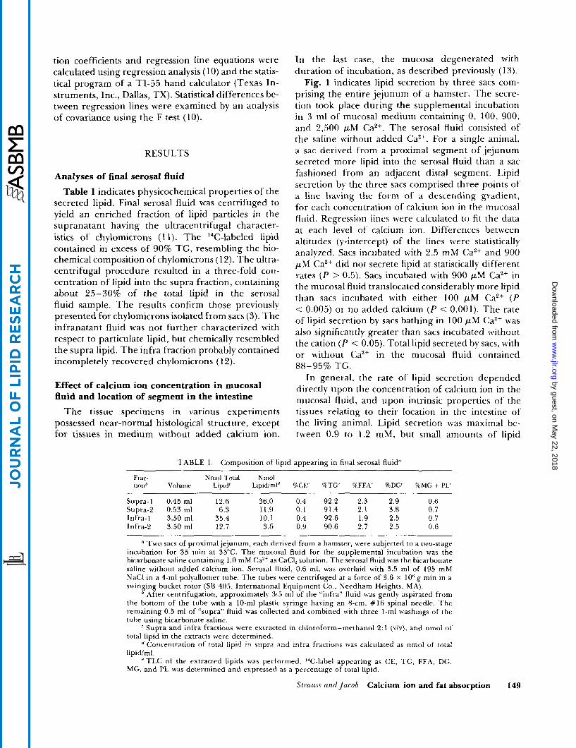
Analyses of final serosal fluid Table 1 indicates physicochemical properties of the
secreted lipid. Final serosal fluid was centrifuged to yield an enriched fraction of lipid particles in the supranatant having the ultracentrifugal character- istics of chylomicrons (1 1). The 14C-labeled lipid contained in excess of 90% TG, resembling the bio- chemical composition of chylomicrons (12). The ultra- centrifugal procedure resulted in a three-fold con- centration of lipid into the supra fraction, containing about 25-30% of the total lipid in the serosal fluid sample. The results confirm those previously presented for chylomicrons isolated from sacs (3). The infranatant fluid was not further characterized with respect to particulate lipid, but chemically resembled the supra lipid. The infra fraction probably contained incompletely recovered chylomicrons ( 12).
Effect of calcium ion concentration in mucosal fluid and location of segment in the intestine
The tissue specimens in various experiments possessed near-normal histological structure, except for tissues in medium without added calcium ion.
In the last case, the mucosa degenerated writh duration of incubation, as described previously (13).
Fig. 1 indicates lipid secretion by three sacs com- prising the entire jejunum of a hamster. The secre- tion took place during the supplemental incubation in 3 ml of mucosal medium containing 0, 100, 900, and 2,500 p M CaZ+. The serosal fluid consisted of the saline without added ea2+. For a single animal, a sac derived from a proximal segment of jejunum secreted more lipid into the serosal fluid than a sac fashioned from an adjacent distal segment. Lipid secretion by the three sacs comprised three points of a line having the form of a descending gradient, for each concentration of calcium ion in the mucosal fluid. Regression lines were calculated to fit the data at each level of calcium ion. Differences between altitudes (y-intercept) of the lines were statistically analyzed. Sacs incubated with 2.5 mM Ca2+ and 900 p M Ca2+ did not secrete lipid at statistically different rates ( P > 0.5). Sacs incubated with 900 pM Ca2+ in the mucosal fluid translocated considerably more lipid than sacs incubated with either 100 p M CaZf ( P < 0.005) or no added calcium ( P < 0.001). The rate of lipid secretion by sacs bathing in 100 p M Ca2+ was also significantly greater than sacs incubated without the cation ( P < 0.05). Total lipid secreted by sacs, with or without Ca2+ in the mucosal fluid contained
In general, the rate of lipid secretion depended directly upon the concentration of calcium ion in the mucosal fluid, and upon intrinsic properties of the tissues relating to their location in the intestine of the living animal. Lipid secretion was maximal be- tween 0.9 to 1.2 mM, but small amounts of lipid
88-95% TG.
TABLE 1. Composition of lipid appearing in final serosal fluid"
Frac- Nmol Total Nmol cionb Volume Lipid' L~pidiml" %CE' %TU RFFA' %DG' %MG + PI.'
Supra-] 0.45 ml 12.6 36.0 0.4 92.2 2.3 2.9 0.6
Infra-1 3.50 ml 35.4 10.1 0.4 92.6 1.9 2.5 0.7 Infra-2 3.50 ml 12.7 3.6 0.9 90.6 2.7 2.5 0.6
Supra-2 0.53 ml 6.3 11.9 0.1 91.4 2.1 3.8 0.7
Two sacs of proximal jejunum, each derived from a hamster, were subjected to a two-stage incubation for 35 min at 35°C. The mucosal fluid for the supplemental incubation was the bicarbonate saline containing 1.0 mM Caz+ as CaClZ solution. The serosal fluid was the bicarbonate saline without added calcium ion. Serosal fluid, 0.6 ml, was overlaid with 3.5 ml of 195 mM NaCl in a 4-ml polyallomer tube. The tubes were centrifuged at a force of 3.6 x lo6 g min in a swinging bucket rotor (SB 405, International Equipment Co., Needham Heights, M A ) .
' After centrifugation, approximately 3:s ml of the "infra" fluid was gently aspirated from the bottom of the tube with a 10-ml plastic syringe having an 8-cm, # I 6 spinal needle. The remaining 0.5 ml of "supra" fluid was collected and combined with three I-ml washings o f the tube using bicarbonate saline.
Supra and infra fractions were extracted in chloroform-methanol 2: 1 (v /v ) , and nmol of total lipid in the extracts were determined.
Concentration of total lipid in supra and infra fractions was calculated as nmol of total lipid/ml.
TLC of the extracted lipids was performed. I4C-label appearing as CE, TG, FF.4. DG, MG, and PL was determined and expressed as a percentage of total lipid.
Strauss ctndjncnb Calcium ion and fat absorption 149
by guest, on May 22, 2018
ww
w.jlr.org
Dow
nloaded from

[ c O z + ] EQUATION r P
T [ "0"prn IY=-1.36x+6.7 I-0.761L.01 1
ILL I O O p m ~ = - 2 . 3 8 ~ + 1 2 . 4 -0.50 L.05 900pm Y=-9.25x+33.7 -0.70 L.OOO1
$,, , 2.500 p m Y = - 5 . 9 4 ~ +27.0 -0.96 L.OOO1
\ W
2 2 0 - 2 t- c, z l 5 -
0 D (L l 0 -
. J
J
2 I- '1 v) w -1 I
I I I z o 1 I z 2 3 SAC
Fig. 1. Lipid secretion by everted jejunal sacs incubated with Ca2+ in the mucosal bathing fluid. Sac 1 was from more proximal intestine than sac 2 and sac 2 was more proximal than sac 3. Sacs were filled with bicarbonate-saline, thoroughly washed, and sub- jected to a two-stage incubation at 35°C under a gas phase of 95% 02/5% C02. First, the sacs were incubated in a bile salt micellar solution containing [l-14C}oleic acid and monoolein. After 5 min, sacs were lightly washed for 30 sec in 0.9% NaC1 and then incubated for a supplemental period of 30 min. The mucosal bathing fluid during the postincubation was bicarbonate saline containing 0, 100, 900, and 2,500 pM CaC1,. Il-14C]Oleic acid appearing in the final serosal fluids was determined. Lipid secre- tion was expressed as nmol total lipids secreted by 100 mg wet weight of tissue in 30 min. Regression lines were calculated for lipid secretion by three sacs incubated with four levels of Ca2+ in the postincubation period. Correlation coefficients ( r ) for regression lines were all statistically significant at P < 0.05 or less. The number of points ( N ) used to calculate the regression lines were as follows: 0 p M Cazf, N = 6; 100 p M Ca2+, N = 16; 900 p M Ca*+, N = 15; and 2,500 p M CaZ+, N = 6. Vertical bars represent 1 S.E.M.
appeared in the serosal fluid even when the mucosal fluid contained no added calcium ion. The ileum se- creted very little lipid.
cluded in the micellar medium, then translocation might have occurred during the early minutes after uptake, as indicated previously (3). The current data suggest that secretion was maximal at 15-25 min of incubation (10-20 min of the supplemental incuba- tion). The time required for the appearance of chylo- microns in the serosal fluid might be partly accounted for by the time required for transport via the lacteals (Fig. 3).
The present results differ from the previously dem- onstrated time course, when lipid secretion increased progressively over a 1-hr period (3). The earlier experiment, however, used Ca2+ in a single-stage incubation having continuous uptake of lipid.
Criteria of effectiveness of an experimental variable
In the following experiments, we wanted to find out the effect of a specific variable or condition during "postincubation" on lipid secretion. The assay de- pended upon showing that, for a single animal, secre- tion rates of sacs 1, 2, and 3 did not have the relationship: sac 1 > sac 2 > sac 3 (see Fig. 1). If the variable significantly affected lipid secretion, then the
- t-
52 70 g -d TOTAL
INCUBATION + MICELLARI.~~~~~-SUPPLEMENTAL W
INCUBATION
3 60. A W 3 v) p 50- a I 8 40-
0 MEAN, TOTAL LIPIDS
MEAN,TG - , D
W 07
_1
J
0 t
2 IO
I
Time course for lipid secretion z TIME, MIN
Fig. 2 demonstrates the rates at which 14C-labeled total lipid and TG appeared in the serosal fluid during the initial incubation, successive 10-min periods of supplemental incubation, and the total translocation for the entire period. Approximately 3.5% of the 14C-labeled oleic acid in the initial micellar solution was translocated into the final serosal fluid, even though the period for uptake was only 5 min. Almost no lipid appeared in the serosal fluid during the first stage of incubation. The earliest appearance of TG in the serosal fluid was during the first 10 min of the supplemental incubation. If calcium had been in-
Fig. 2. Time course for secretion of total lipids and triglycerides. A segment of everted jejunum, corresponding in size and location to Sac 1, was cannulated with glass tubing and incubated in two stages as described in Experimental Procedures. The mucosal fluid during the supplemental incubation consisted of the bicarbonate saline with 1.0 mM CaCI,. The serosal fluid was bicarbonate saline without added calcium ion. The serosal compartment was drained of its contents, washed three times with 1.0-ml volumes of bicarbonate saline, and filled with fresh serosal medium at the end of the preliminary incubation period and at consecutive 10-min intervals during the supplemental incubation period. Total lipids and triglycerides in the serosal fluids and washings were determined as detailed in Chemical Analyses. Lipid secretion was expressed as nmol of total lipids and triglycerides trans- located by 100 mg wet weight of tissues 2 1 S.E.M. The experiment was performed three times.
150 Journal of Lipid Research Volume 22, 1981
by guest, on May 22, 2018
ww
w.jlr.org
Dow
nloaded from

Fig. 3. (:hylomicron-likr particles ( C ) within the confines of a lymphatic capillary (L) in the lamina propria mucosae of a villus of hamster jrjunum. .]'he lacteal is distingrrishahle from a blood capillary hecause i t has no basement lamina, and the wall is more attenuated and may have larger pores. Particles entered the lumen o f the lacteal from the surrounding intercellular fluid where clumps of the particles o f varying density ( a r r o w ) intermingle between lymphocytes and plasma cells. T h e particles may flow along the length of the vessel to enter the serosal fluid. The micrograph was taken from a sac of hamster.jcjunum that was initially incuhated with linolenic acid-NaTDC micellar solution and supplementally incuhatetl in medium containing added calcium ion at Si"<;. Linolenate was used instead of oleate in the micellar medium t o increase the electron density of the lipid particles after osmification. Magnification is approximately ~8,400. The inset is an enlarged region in Fig. 3 depicting thc particles Ixtving a dcnse. granular rim (approximately ~70 ,000) .
by guest, on May 22, 2018
ww
w.jlr.org
Dow
nloaded from

relationships between the sacs would be: sac 1 < sac 2 > sac 3, o r sac 1 > sac 2 < sac 3.
Effect of temperature on lipid secretion
T h e optimal temperature for lipid secretion by the everted sac was 35°C. Lipid secretion also took place at 30"C, but at a lower rate. Practically no T G release took place at 23°C or lower temperatures.
Effect of D,O upon lipid secretion
T h e effect of deuterium oxide upon the lipid secretory mechanism was tested by including nearly 100% DzO in the mucosal and serosal fluids of a supplemental incubation. A bicarbonate-saline solu- tion containing 1 mM CaClz was prepared, as de- scribed in Materials and Methods, except that the solution used either DzO or HzO as solvents. D,O severely impaired lipid secretion. T h e average lipid se- cretion for sacs 1 and 3, incubated with D 2 0 , was 1.3 4 0.4 nmol of total lipid/100 mg wet weight of tissue (n = 4), compared with 14.4 ? 1.8 nmol (12.6 and 16.2) of total lipid1100 mg of tissue for sac 2, incubated with H 2 0 .
Effect of serosal calcium ion on lipid secretion Addition of calcium ion to the seroszl fluid en-
hanced lipid secretion by sacs (Table 2). Sacs incu- bated with 2.5 mM Ca2+ o r 100 pM Ca2+ in the serosal fluid secreted considerably more lipid than sacs incu- bated without added calcium ion ( P < 0.02 and P < 0.005, respectively). However, lipid secretion by sacs incubated with calcium ion in the serosal fluid was always less than that by sacs incubated with a similar concentration of calcium ion in the mucosal fluid (Table 2). T h e findings appeared to indicate that the mucosal surface was more sensitive to calcium ion than the serosal surface during lipid secretion.
It was unlikely that the lesser effect of Ca2+ in serosal
fluid resulted from leakage of CaZ+ from serosal to mucosal fluid during the first incubation period. Using 4sCa as tracer in the serosal fluid, less than 3% entered the micellar solution.
Conjecturally, the serosal insensitivity might have been absolute, with only the mucosal surface, pre- sumably the microvilli, displaying sensitivity to calcium ion during lipid secretion. That is, leakage of calcium ion into the mucosal compartment might have been the mechanism of the serosal effect. T o clarify the problem, we increased the mucosal volume from 3 ml to 300 ml. If there were leakage, then the large mucosal volume would minimize changes in calcium ion concentration that might enhance lipid secretion. Lipid secretion, under these conditions, would be due to an action of serosal calcium ion on the baso- lateral aspects of the absorptive cell.
Using 2.5 and 0.9 mM calcium ion in the serosal fluid, stimulation of lipid secretion was clear-cut, even though mucosal calcium ion concentrations were less than 10 p M (Table 3). When the serosal calcium ion concentration was 100 p M , there was very little lipid secretion and the final concentration of calcium ion in the mucosal fluid was again much less than 10 p M (Table 3). Histologically, the tissue deteriorated like the specimens that were incubated in mucosal fluid without added calcium ion. The observations indicated that the serosal surface was sensitive t o calcium ion during lipid secretion. T h e reasons were not clear for the greater sensitivity of the mucosal surface.
Effects of barium, magnesium and strontium on lipid secretion
Barium chloride, when added to the mucosal bath- ing medium without CaCl,, in concentrations of 0.09, 0.24, and 2.55 mM did not enhance lipid secretion. In levels above 0.39 mM, BaZ+ in the mucosal fluid,
TABLE 2. Effect of calcium ion in mucosal or serosal fluids
Sac" [Ca'+] 1.ocation" Lipid Secretion'
1 2.5 mM Serosal 23.8 5 8.8 (9.4,22.3,39.9)
3 2.5 m M Serosal 11.6 5 4.4 (6.1,8.4,20.2)
2 0.1 mM Sei-osal 7.1 i 1.1 (6.0,8.2)
2 2.5 mM Mucosal 29.8 2 10.1 (11.4,31.6,46.4)
1 0.1 m M Mucosal 19.1 -+ 1.1 (18.0,20.1)
3 0.1 m M Mucosal 16.3 -C 0.5 (15.8,16.8)
" Sac 1 was more proximal than Sac 2, and Sac 2 more proximal than Sac 3.
CaZ+ was included in the mucosal postincubation fluid or in the serosal fluid.
Lipid translocation by sacs is expressed in mean nmol of total lipid/100 mg wet weight of tissue/30 min t S . E . M . Individual values for lipid secretion are given in parentheses.
152 Journal of Lipid Research Volume 22, 1981
by guest, on May 22, 2018
ww
w.jlr.org
Dow
nloaded from

Sac"
TABLE 3. Lipid secretion with 300 ml mucosal volumes
Initial [Caz+Ib Location' Lipid Secretiond Final [CaZ+IP
2.5 mM Mucosal 41.7 f 0.3 (41.4,41.9) 2.4 t 0.1 mM(2.4,2.4,2.5) 2.5 mM Serosal 13.4 2 5.6 (7.5,19.2) 1 1 0 pM [3] 2.5 mM Mucosal 12.9 2.5 t 0.1 mM (2.4,2.5,2.5) 0.9 mM Mucosal 53.4 2 4.4 (49.0,57.8) - 0.9 mM Serosal 14.5 2 4.3 (10.2,18.8) - 0.9 mM Mucosal 17.0 f 7.9 (9.1,24.8) - 0.1 mM Mucosal 22.0 2 5.0 [5] 5 5 ? 15 p M (40,70) 0.1 mM Serosal 1.9 ? 0.6 [5] 9 1 0 pM [2] 0.1 mM Mucosal 16.7 f 6.8 [5] 83 f 3 p M (80,85)
'' Sac 1 was more proximal than Sac 2 , and Sac 2 was more proximal than Sac 3. Initial concentration of calcium ion present in the mucosal fluid at the be-
ginning of the supplemental incubation, and in the serosal fluid at the beginning of the preliminary incubation.
The calcium-bicarbonate medium was either the mucosal or serosal fluid. Lipid secretion was expressed as mean nmol of total lipids transported by
100 mg wet weight of tissue in 30 min ? 1 S.E.M. Individual values for lipid secretion are given in parentheses. Number of experiments is given in brackets.
Concentration of calcium ion in 300 tnl of mucosal fluid at the end of 30 min of supplemental incubation. as mean 2 1 S.E.M. Individual values are given in parentheses. Number of experiments is given in brackets.
without added calcium ion, caused severe degenera- tive changes in intestinal absorptive cells. Low con- centrations of Ba2+ (0.09 mM) did not inhibit the secretory response to 1.2 mM Ca2+.
Strontium ion enhanced the secretion of lipid, but less so than equimolar calcium ion (1.2 mM). Mor- phology of intestine incubated with strontium was normal.
Substitution of 1.0 mM Mg2+ for Ca+ did not en- hance lipid secretion. Mg2+ (1.0 mM) and 0.1 mM Ca2+ did enhance lipid secretion, but the effect of the ion mixture was attributable to calcium ion.
Effect of lanthanum on lipid secretion
Lanthanum ion, in concentrations of 0.1, 0.5, and 1.0 mM in the mucosal medium, strongly inhibited the secretory response to 1.0 mM Ca2+ (Table 4). The trivalent cation alone did not enhance lipid secretion in concentrations of 0.5 or 1.0 mM in the mucosal fluid. Incubations with lanthanum were performed with a Tris-buffered saline to avoid the precipitation of lanthanum carbonate. Secretion of lipid by intestine incubated with Tris-saline was similar to that of tissue treated with the normal bicarbonate saline. Tissue incubated in Tris-buffered saline containing calcium or calcium and 0.1 mM La3+ had near-normal his- tology. The morphology of the villi that had been treated with 1.0 mM La3+ and CaZ+ suffered mildly.
Lipid esterification The absorptive mucosa and final incubation media
were combined and extracted for total lipids and
analyzed for their content of 14C-labeled TG after a supplemental incubation in Hepes-buffered saline with the following additions: 1 ) 1.0 mM Ca", 2 ) no additions, 3 ) 1.0 mM Sr2+ 4 ) 1.0 mM Mg2+, 5 ) 1.0 mM Ca2+ + 100% DzO, 6 ) 0.3 mM La3+ + 1.0 mM Ca2+, and 7 ) 0.3 mM Ba2+. The degree of esterifica- tion was nearly constant under all conditions with 84 -+ 2.5% (S.E.M.) of the total tracer as TG, suggest- ing that the ions affected transport rather than esterification.
Electron microscopy indicated many lipid droplets in enterocytes during the first stage of incubation. When there was secretion, the droplets disappeared.
TABLE 4. Effect of added lanthanum and calcium ions on lipid secretion
Sac" I La3 +I" Lipid Secretion'
1 1.0 mM 5.1 2 2.6 (2.5,7.6) 3 1.0 mM 1.9 i 0.1 (1.8,Z.O) 1 0.5 mM 3.7 t 1.2 [4] 3 0.5 mM 1.0 t 0.2 [4] 1 0.1 mM 13.9 t 2.0 (11.9,15,9) 3 0.1 mM 10.7 ? 6.4 (4.3,17.1) 2 0 mM (1.0 mM Ca2+) 16.5 2 3.7 [6]
"Sac 1 was more proximal than Sac 2, and Sac 2 was more proximal than Sac 3.
" The concentration of lanthanum added t o the mucosal fluid during the supplemental incubation only. The fluids also contained 1.0 mM CaZ+. ' Lipid secretion was expressed as mean nmol of total lipids
translocated by 100 mg wet weight of tissue in 30 min 2 1 S.E.M. lndividual values for lipid secretion are given in parentheses. Number of experiments is given in brackets.
S/rcrucc r i d JocoD Calcium ion and fat absorption 153
by guest, on May 22, 2018
ww
w.jlr.org
Dow
nloaded from

DISCUSSION
T h e secretory phase of fat absorption was analyzed advantageously by incubating the everted jejunum from hamster in entirely synthetic media. Perhaps the most important of the results was that the presence of calcium ion in the medium was essential for the secretion of T G in chylomicron-like particles. When Ca2+ was deleted, secretion ceased and cells of the absorptive epithelium deteriorated (13). It was clear that the calcium ion played a general supportive role for the intestinal mucosa. Did the calcium ion also exert a specific effect upon lipid secretory mecha- nisms in the cell? Certain of the data suggest an affirmative reply.
The structure of the intestine was microscopically near-normal when the medium contained calcium ion over the range 0.1-5.0 mM. Electron micrographs were typical for the secreting enterocyte, including the exocytosis of chylomicron-like particles (14). T h e concentration of Ca2+ was related directly to the lipid secretory rate, over the range 0.1-0.9 mM. This sug- gested that Ca2+ might have specifically affected the cellular mechanisms for transferring lipid. T h e ef- fective range for secretion was approximately similar to the range, 0.5-1.3 mM, determining directly the contraction of the isolated heart of the frog (15). T h e location of an intestinal segment in the living animal also determined the rate of secretion, even at concen- trations of calcium which were optimal for secretion (Fig. 1). This influence of location must have resulted from intrinsic properties of the tissues (16).
The experiments using La3+ provided a second indi- cation that calcium ion stimulated lipid-transfer mechanisms in the cell. When the intestine was incubated in medium containing the antagonistic calcium and lanthanum ions, lipid secretion was sharply curtailed, while the tissue integrity was main- tained. Of the assayed ions, only La3+ inhibited the action of Ca2+. T h e specific and general effects of Ca2+ were dissociated. T h e lanthanum ion might have prevented uptake of Ca'+ from the medium or bind- ing of the divalent cation to the plasmalemma. Theoretically, La3+ and caz+ acted similarly on mem- branes, but the binding of lanthanum was much stronger ( 17). Experimentally, lanthanum ion stops flux of Ca2+ across various biological (18-2 1) and artificial (22,23) membranes, and displaces Ca2+ from binding sites on the plasmalemma ( 18, 19, 2 1, 24, 2 5 ) , apparently without the lanthanum entering the cell
T h e stimulation of secretion by calcium ion in other types of cells provides a third reason favoring a specific role of CaZ+ in the secretion of lipid by intestinal cells. Douglas and Rubin (27) call the action of the ion on cells of the adrenal medulla, "stimulus- secretion coupling". One class of secretory cell took u p external Ca2+ after neural o r hormonal stimulation (28). Conversely, pancreatic acinar cells utilized the calcium in cytoplasmic stores after stimulation (28- 33). The lipid secretion by intestine appears unique, utilizing external calcium ion for secretion without any apparent neural o r hormonal stimulus.
Lipid secretion also seemed unique, judging by the effects of other divalent cations. The intestinal pro- duction was not inhibited by Mg2+ or stimulated by Ba2+. In contrast, Mg'+ inhibited and Ba2+ stimulated secretion of catecholamines, acetylcholine, vaso- pressin, oxytocin, 'ISH, LH, ACTH, prolactin, and insulin (34). O n the other hand, Mg2+ and Ba2+ apparently had similar effects on lipid secretion by the enterocyte and protein secretion by the pancreatic acinar cell (30). La3+ inhibited secretion by cells of adrenal medulla ( 3 5 ) , exocrine pancreas (21), and intestine.
Reducing the temperature of incubation to less than 35"C, or substituting deuterium oxide for water in the incubation medium impaired lipid secretion. How- ever, more than one component of the lipid secretory mechanism in the enterocytes might have been af- fected. Electron microscopy of absorptive cells at 23°C indicated that the temperature did not permit the transfer of lipid from endoplasmic reticulum to Golgi complex.' This transfer was a prerequisite for secretion. Microtubules are important for fat absorp- tion in the living rat (36, 37). T h e inhibitory action of deuterium oxide 011 lipid secretion may be ex- plained by the stabilizing action of DzO on a micro- tubular-microfilamentous network mediating exo- cytosis (38), although actions of deuterium oxide on the endoplasmic reticulum (39) and cytosol (40) have been observed.
T h e action o f calcium was t o facilitate exocytosis in a number of secretory cells (41-43). A calcium- binding protein, such as calmodulin, may link calcium- ion shifts with secretion (44, 45). Intestinal cells con- tain calmodulin (46). Whether calcium directly ef- fected exocytosis of lipid particles by the absorptive cell awaits future investigati0n.m This work was supported by grant AM-20392 from the National Institutes of Health, Public Health Service.
(26). Therefore, the lanthanum experiments sug- ~
' Strauss, E. W. Lipid droplet formation injejunum of the golden hamster in vitro during incubations with bile salt micellar solutions. Submitted for publication.
gested that within or on the enterocyte.
lipid secretion by acting
154 Journal of Lipid Research Volume 22, 1981
by guest, on May 22, 2018
ww
w.jlr.org
Dow
nloaded from

Manuscript received 25 ApnlI980, in revwrd form 3 I July 1980, and in measuring cellular calcium influx. Czrc. Rev. 30: re-revwed form 8 Septemhfr 1980. 44-54.
1.
2.
3.
4.
D.
6.
7.
8.
9.
10.
1 1 .
12.
13.
14.
1.5.
16.
17.
18.
REFERENCES
Johnston, J . M., and B. Borgstriim. 1964. T h e in- testinal absorption and metabolism of micellar solutions of lipids. Biochim. Biophys. Acta. 84: 412-423. Strauss, E. W. 1966. Electron microscopic study of intestinal fat absorption in vitro from mixed micelles containing linolenic acid, monoolein, and bile salt. J . Lipid Res. 7: 307-323. Strauss, E. W. 1977. Effects of calcium and magnesium ions upon Fat absorption by sacs of everted hamster intestine. Gastroenterology. 73: 42 1-424. Saunders. D. R., and J . Sillery. 1979. Effect of calcium on absorption of fatty acid by rat jejunum in vitro. Lipidv. 14: 703-706. Pope, J. L., T. M. Parkinson, and J . A. Olson. 1966. Action of bile salts on the metabolism and transport of water-soluble nutrients by perfused rat ,je,junum in vitro. Bzorhim. Biophys. Acta. 130: 2 18-232. Hofmann, A. F. 1962. Thin-layer adsorption chro- matography of free and conjugated bile acids on silicic acid. J . Lipid Re.\. 3: 127-128. Brown, J. L., and J . M. Johnston. 1962. Radioassay of lipid components separated by thin-layer chromatog- raphy. J . Lipid Res. 3: 480-481. Thomas, A. E. 111, J . E. Scharoun, and H. Ralston. 1965. Quantitative estimation of isomeric mono- glycerides by thin-layer chromatography. J. Am. Oil Chem. Soc. 42: 789-792. Bragdon, J. H . 1960. Extraction of lipids from serum. In Lipids and the Steroid Hormones in Clinical Medicine. F. M'. Sunderman and F. W. Sunderman, J r . , editors. J . B. Lippincott, Philadelphia, PA. 6-8. Snedecor, G. W., and b'. G. Cochran. 1967. Statis- tical Methods. Iowa State University Press, Ames, IA.
Dole, V. P., and J. T. Hamlin 111. 1962. Particulate fat in lymph and blood. Physiol. Rev. 42: 674-701. Zilversniit, D. B. 1969. Chylomicrons. In Structural and Functional Aspects of Lipoproteins in Living Sys- tems. E. Tria and A. M. Scanu, editors. Academic Press, London and New York. 329-368. Jacob, J . S., E. W. Strauss, and W. M. Strauss. 1976. Calcium and magnesium requirements by intestine dur- ing fat absorption. J . Cell B i d . 70: 158 a (Abstract). Strauss, E. M'. 1975. Importance of Ca2+ and Mg2+ dur- ing absorption of micellar lipids by intestine in vitro. Gmtroenterolqy. 68: 992. (Abstract). McLean, F. C . , and A. B. Hastings. 1934. A biological method for the estimation of calcium ion concentra- tion. J . B i d . Chem. 107: 337-350. M'u, A-L., S. B. Clark, and P. R. Holt. 1975. Trans- mucosal triglyceride transport rates in proximal and distal rat intestine in vivo. J . Lipid Res. 16: 251-257. Lettvin. J. Y., W. F. Pickard, W. S. McCulloch, and W. Pitts. 1964. A theory of passive ion flux through axon membranes. Nature. 202: 1338- 1339. van Breeman, C., B. R. Farinas, P. Gerba, and E. D. McNaughton. 1972. Excitation-contraction coupling in rabbit aorta studied by the lanthanum method for
91- 106, 419-438.
19. van Breemen, C., B. R. Farinas, R. Casteels, P. Gerba, F. Wuytack, and R. Deth. 1973. Factors controlling cytoplasmic Ca2+ concentration. Philos. Trans. R . Sor. Lond. Ser. B . 265: 57-71.
20. Mela, L. 1968. Interactions of La3+ and local anaes- thetic drugs with mitochondrial Ca2+ and Mn2+ uptake. Arch. Biochem. Biophys. 123: 286-293.
21. Chandler, D. E., and J. A. Williams. 1974. Pancreatic acinar cells: effects of lanthanum ions on amylase release and calcium ion fluxes. J . Physiol. 243: 831- 846.
22. van Breemen, C. 1968. Permselectivity of a porous phospholipid-cholesterol artificial membrane. Calcium and lanthanum effects. Biochem. Biophys. Res. Commun. 32: 977-983.
23. van Brecmen, D., and C. van Breemen. 1969. Calcium exchange diffusion in a porous phospholipid ion- exchange membrane. Nature. 223: 898-900.
24. Langer, G. A., and .J. S. Frank. 1972. Lanthanum in heart cell cu1ture.J. Cell Biol. 54: 441 -455.
2.5. Takata, M., W. F. Pickard, J . Y. Lettvin, and J . W. Moore. 1966. Ionic conductance changes in lobster axon membrane when lanthanum is substituted for calcium. J. Gcn. Physiol. 50: 46 1-47 1.
26. Lesseps, Roland J. 1967. T h e removal by phospho- lipase C of a layer of lanthanum-staining material external to the cell membrane in embryonic chick cells. J. Cell Biol. 34: 173- 183.
27. Douglas, M'. W., and R. P. Rubin. 1961. T h e role of calcium in the secretory response of the adrenal medulla to acetycholine. J . Physiol. 159: 40-57.
28. Douglas, W. W. 1976. T h e role of calcium in stimulus- secretion coupling. In Stimulus-Secretion Coupling in the Gastrointestinal Tract. R. M. Case and H. Goebell, editors. University Park Press, Baltimore, MD. 17-31,
29. Argent, B. E., R. K . Smith, and R. M. Case. 1976. T h e role of calcium in pancreatic enzyme and electrolyte secretion. In Stimulus-Secretion Coupling in the Gastrointestinal Tract. R. M. Case and H. Goebell, editors. University Park Press, Baltimore, MD. 237- 254.
30. Argent, B. E., R. M. Case, and T . Scratcherd. 1973. Amylase secretion by the perfused cat pancreas in re!ation to the secretion of calcium and other electro- lytes and as influenced by the external ionic environ- ment. J . Physiol. 230: 575-593.
31. Case, R. M., and T . Clausen. 1973. T h e relationship between calcium exchange and enzyme secretion in the isolated rat pancreas. J . Physiol. 235: 75- 102.
32. Chandler, D. E., and J. A. Williams. 1978. Intra- cellular divalent cation release in pancreatic acinar cells during stimulus-secretion coupling. I. Use of chloro- tetracycline as fluorescent probe. J . Cell Biol. 76: 37 1-385.
33. Chandler, D. E., and J. A. Williams. 1978. Intra- cellular divalent cation release in pancreatic acinar cells during stimulus-secretion coupling. 11. Subcellular localization of the fluorescent probe chlorotetracycline.
J . Cell B i d . 76: 386-399. 34. Rubin, R. P. 1970. T h e role of calcium in the release
of neurotransmitter substances and hormones. Phnr- mncol. RUJ. 22: 389-428.
S h " s and Jacob Calcium ion and fat absorption 155
by guest, on May 22, 2018
ww
w.jlr.org
Dow
nloaded from

35. Borowitz, J. L. 1972. Effect of lanthanum on catechol- amine release from adrenal medulla. L f e Sci. 11: 959-964.
36. Glickman, R. M., J. L. Perrotto, and K. Kirsch. 1976. Intestinal lipoprotein formation: effect of colchicine. Gastroenterology. 70: 347 - 352.
37. Reaven, E. P., and G. M. Reaven. 1977. Distribution and contents of microtubules in relation to the trans- port of lipid. J . Cell Biol. 75: 559-572.
38. van Obberghen, E., G. Somers, G. Devis, M. Ravazzola, F. Malaise-Lagae, L. Orci, and W. J. Malaise. 1974. Dynamics of insulin release and microtubular-micro- filamentous system. VI, Effects of D 2 0 . Endocrinology. 95: 1518-1528.
39. Matty, A. J., and K. Deutsch. 1968. The effect of 'H,O on the membranous structures of the proximal and distal convoluted tubules of the mouse. Biochim. Biophys.
40. Marsland, D., and A. M. Zimmerman. 1963. Cell division: differential effects of heavy water upon the
Acta. 163: 14-19.
mechanisms of cytokinesis and karyokinesis in the eggs of Arbacia punctulata. Exp. Cell Res. 30: 23-35.
41. Satir, B. H., and S. G. Oberg. 1978. Paramecium fusion rosettes: possible function as Ca'+ gates. Science. 199:
42. Kagayama, M., and W. W. Douglas. 1974. Electron microscope evidence of calcium-induced exocytosis in mast cells treated with 48/80 or the ionophores A- 23187 and X-537A. J . Cell Biol. 62: 519-526.
43. Schramm, M . , and Z. Selinger. 1975. The functions of cycIic AMP and calcium as alternative second mes- sengers in parotid gland and pancreas. J . Cyclic Nucleo- tide Res. 1: 181-192.
44. Cheung, W. Y. 1980. Calmodulin plays a pivotal role in cellular regulation. Science. 207: 19-27.
45. Ilundain, A., and R. J. Naftalin. 1979. Role of Ca2+- dependent regulator protein in intestinal secretion. Nature. 279: 446-448.
46. Howe, C. L., M. S. Mooseker, and T . A. Graves. 1980. Brush-border calmodulin. J . Cell Biol. 85: 916-923.
536-538.
156 Journal of Lipid Research Volume 22, 1981
by guest, on May 22, 2018
ww
w.jlr.org
Dow
nloaded from