solution to gas phase
TRANSCRIPT
Solution to Gas PhaseSolution to Gas Phase
•• DNADNA–– Secondary StructureSecondary Structure–– QuadruplexQuadruplex FormationFormation
•• Protein ComplexesProtein Complexes–– pH dependencepH dependence–– NoncovalentNoncovalent interactionsinteractions
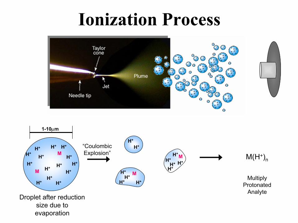
H+
H+
H+
H+
H+ M
H+
H+
H+
H+
H+ MH+
M(H+)n
Droplet after reduction size due to evaporation
“CoulombicExplosion”
Multiply Protonated
Analyte
Ionization Process
H+
H+
H+
H+
H+H+
H+H+
H+
H+M
M
1-10µm
H+
H+
H+
H+
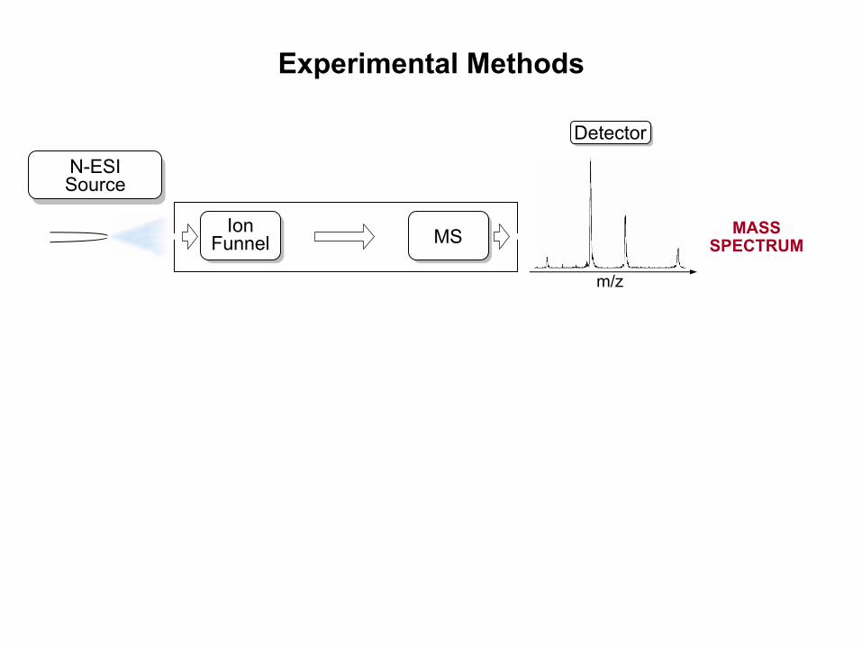
Experimental Methods
Detector
m/z
MSIonFunnel
MASS SPECTRUM
N-ESI Source

Experimental Methods
Detector
m/z
MSIonFunnel
1 – 5 Torr HeE
MSDriftCell
IonFunnel
time
ARRIVAL-TIME DISTRIBUTION
MASS SPECTRUM
N-ESI Source

DNA
• Secondary Structure–AT Helix Stability
• Quadruplex Formation–Telomer
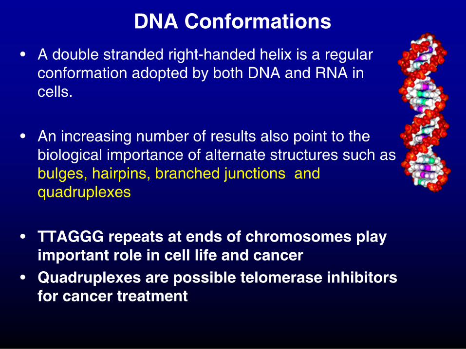
DNA Conformations
• A double stranded right-handed helix is a regular conformation adopted by both DNA and RNA in cells.
• An increasing number of results also point to the biological importance of alternate structures such as bulges, hairpins, branched junctions and quadruplexes
• TTAGGG repeats at ends of chromosomes play important role in cell life and cancer
• Quadruplexes are possible telomerase inhibitors for cancer treatment

• F. Rosu, V. Gabelica, C. Houssier, P. Colson, E. De Pauw Rapid Commun. Mass Spectrom. 2002, 16, 1729.
Observed triplexes and quadruplexes cationized by NH4+ in ESI-MS
MS/MS: triplex → duplex + antigene (similar to solution dissociation)
• T. Aggerholm, S.C. Nanita, K.J. Koch, R.G. Cooks J. Mass Spectrom. 2003, 38, 87
Observed “magic number” G-quartet adducts in ESI-MS
Quadruplexes in the Gas Phase
• S.A. Ho stadler, R.H. Griffey, Chem. Rev. 2001, 101, 377
Review of evidence that noncovalent RNA and DNA complexes are transferred from solution into the gas phase
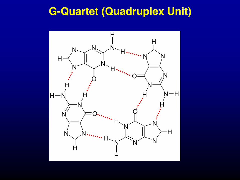
G-Quartet (Quadruplex Unit)
N
N
N
NH
OH
N
H
H
NN
N N
H
O
HNHH
N
N
N
NH
OH
N
H
H
NN
NN
H
O
H N HH
G
G
G
G
G
G
G
G
G
G
G
G
5´ 3´
3´5´
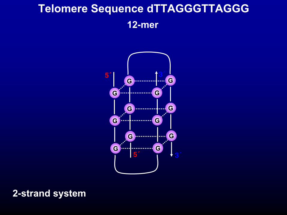
Telomere Sequence dTTAGGGTTAGGG
2-strand system
12-mer
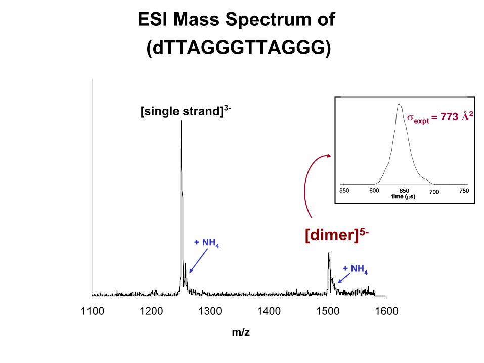
ESI Mass Spectrum of (dTTAGGGTTAGGG)
m/z
1100 1200 1300 1400 1500 1600
[single strand]3-
+ NH4[dimer]5-
+ NH4
time (µs)550 600 650 700 750
time (µs)550 600 650 700 750
σexpt = 773 Å2

Theoretical Structures of [dimer]5-
Quadruplex
σexpt = 773 Å2
Globular
σtheory = 780 Å2 σtheory = 748 Å2

dTTAGGGTTAGGGTTAGGGTTAGGG
intramolecular quadruplex
G
G
G
G
G
G
G
G
G
G
G
G
5´
3´
24-mer
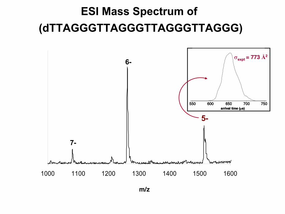
m/z
1000 1100 1200 1300 1400 1500 1600
7-
6-
5-
ESI Mass Spectrum of (dTTAGGGTTAGGGTTAGGGTTAGGG)
arrival time (µs)550 600 650 700 750
arrival time (µs)550 600 650 700 750
arrival time (µs)550 600 650 700 750
σexpt = 773 Å2
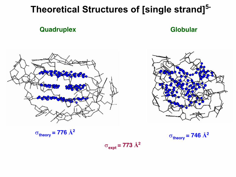
Theoretical Structures of [single strand]5-
Quadruplex Globular
σexpt = 773 Å2
σtheory = 776 Å2σtheory = 746 Å2

Duplexes in the Gas Phase
• V. Gabelica and E. DePauw, Int. J. Mass Spectrom. 2002, 219, 151.
• P.D. Schnier, J.S. Klassen, E.F. Strittmatter and E.R. Williams,J. Am.Chem. Soc. 1998, 120, 9605-9613.
Higher Ea for complimentary duplexes and Ea correlated to –∆Hd in solutionEvidence of Watson-Crick pairing in vacuo
CID yields correlate with number of GC pairs and ∆Hdiss in solutionSuggests structure conserved in gas phase
• M. Rueda, S.G. Kalko, F.J. Luque, M. Orozco J. Am. Chem. Soc. 2003, 125, 8007
Gas-phase MD simulations indicate 12- and 16-mer duplexes retain major
conformational features as the double helix in aqueous solution

W. Fuller, et al
J. Mol. Biol. 1965, 12, 60
75% relative humidity
3 Common Helical Duplexes
A.H.J. Wang, et al
Nature 1979, 282, 680
CGCGCG in high salt conc.
A-Form B-Form Z-Form
R. Langridge, et al
J. Mol. Biol. 1960, 2, 19
92% relative humidity
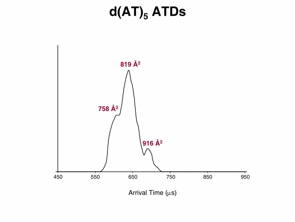
d(AT)5 ATDs
450
Arrival Time (µs)
550 650 750 850 950
819 Å2
758 Å2
916 Å2

time (ps)
Cro
ss-s
ectio
n (Å
2 )
dAT 10-mer (A-form) 300K dynamics
σEXPT = 758, 819, 916 Å2
820 Å2
760 Å2
920 Å2

d(AT)5 ATDs
450
Arrival Time (µs)
550 650 750 850 950
σexp = 819 Å2
σ theory = 820 Å2σexp = 758 Å2
σ theory = 760 Å2
σexp = 916 Å2
σ theory = 920 Å2

ESI spray
dehydrate dehydrate
B-DNA(90% humidity)
A-DNA(75% humidity)
Solution vs. Solvent-Free Structures
Ion Funnel
920 Å2
760 Å2
820 Å2
DNA Summary
• DNA helices are observed in the gas phase• B-DNA → A-DNA transitions upon
dehydration• Quad structures conserved
– In absence of solvent– In absence of salt

Protein Complexes
Aβα-synuclein
Solution → Gas Phase
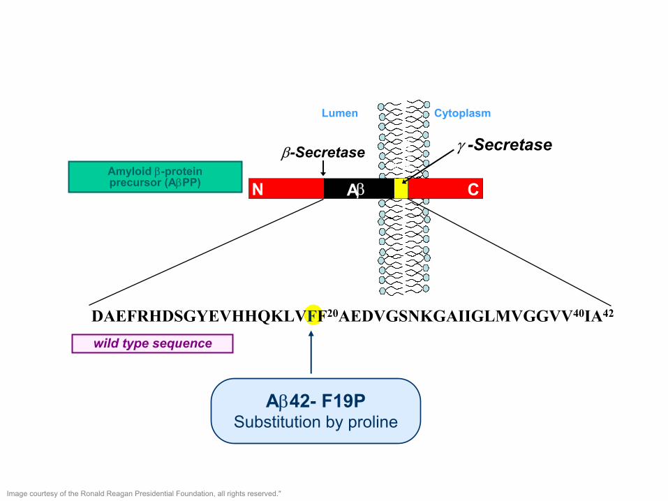
DAEFRHDSGYEVHHQKLVFF20AEDVGSNKGAIIGLMVGGVV40IA42
N CAβ
ER
Mem
bran
e
CytoplasmLumen
Amyloid β-proteinprecursor (AβPP)
β-Secretase
wild type sequence
γ -Secretase
Aβ42- F19PSubstitution by proline
Image courtesy of the Ronald Reagan Presidential Foundation, all rights reserved."
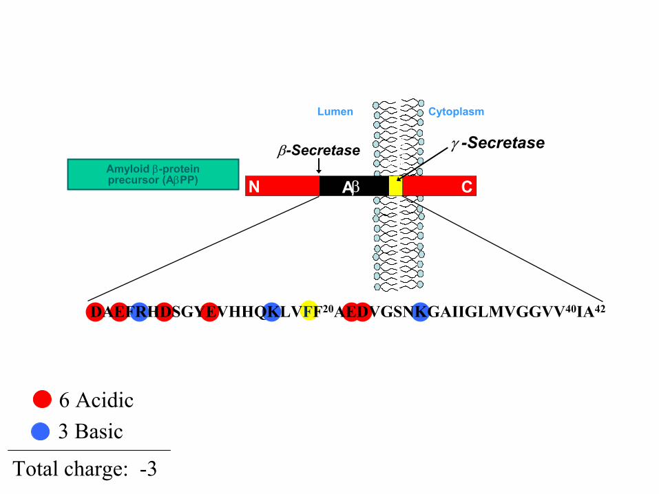
6 Acidic 3 Basic
Total charge: -3
N CAβ
ER
Mem
bran
e
CytoplasmLumen
Amyloid β-proteinprecursor (AβPP)
β-Secretase γ -Secretase
DAEFRHDSGYEVHHQKLVFF20AEDVGSNKGAIIGLMVGGVV40IA42

2
0800
M (-3)
D (-4)D (-5)
M (-4)
M (-5)T (-7)
1000 1200 1400 1600 1800 2000 2200 24000
100
%
m/z
2
0800
M (-3)
D (-4)D (-5)
M (-4)
M (-5)T (-7)
1000 1200 1400 1600 1800 2000 2200 24000
100
%
m/z
800 1000 1200 1400 1600 1800 2000 2200 24000
100
%
M (-3)
M (-4)
M (-5)
0800 1000 1200 1400 1600 1800 2000 2200 2400
0
100
%
M (-3)
M (-4)
M (-5)
0
Aβ42-wt
Aβ42-F19P
time (µs)550 650 750 850 950
time (µs)550 650 750 850 950
time (µs)1000600 700 800 900
time (µs)1000600 700 800 900
time (µs)550 650 750 850 950
time (µs)550 650 750 850 950
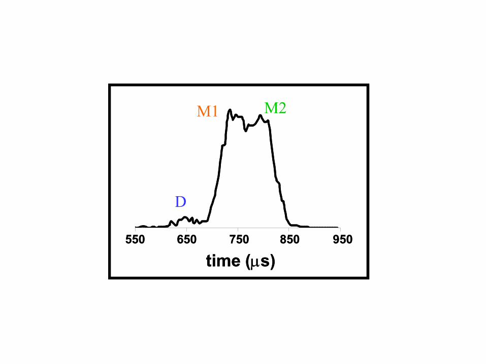
D
M1 M2

Radius of gyration (Å)
Cro
ss s
ectio
n (Å
2 )
Gas phase
550
600
650
700
750
800
850
900
950
1000
7 9 11 13 15 17 197
experimentexperiment
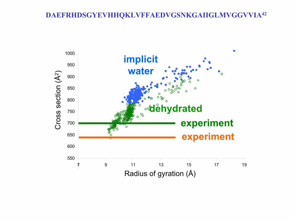
DAEFRHDSGYEVHHQKLVFFAEDVGSNKGAIIGLMVGGVVIA42

• Generate 100s of model structures using:Molecular mechanics (CHARMM22)Replica exchange algorithmImplicit solvent (GB/SA) and Gas Phase
• Calculate for each structure:Cross sectionRadius of gyration
• in collaboration with Andriy Baumketner and Joan Shea (UCSB)
Model structures of Aβ42-wt monomers

Radius of gyration (Å)
Cro
ss s
ectio
n (Å
2 )
Gas phase
550
600
650
700
750
800
850
900
950
1000
7 9 11 13 15 17 197
implicitwater
experimentexperiment
DAEFRHDSGYEVHHQKLVFFAEDVGSNKGAIIGLMVGGVVIA42
7 9 11 13 15 17 19
Radius of gyration (Å)
Cro
ss s
ectio
n (Å
2 )
dehydrated
550
600
650
700
750
800
850
900
950
1000
7
implicitwater
experimentexperiment
DAEFRHDSGYEVHHQKLVFFAEDVGSNKGAIIGLMVGGVVIA42

9 11 13 15 17 19
Radius of gyration (Å)
Cro
ss s
ectio
n (Å
2 )
Gas phase
implicitwater
experiment
dehydratedexperiment
550
600
650
700
750
800
850
900
950
1000
7
DAEFRHDSGYEVHHQKLVFFAEDVGSNKGAIIGLMVGGVVIA42
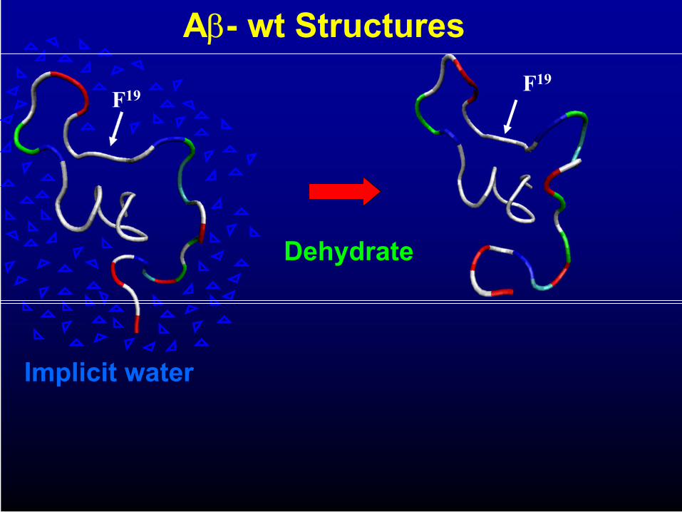
Dehydrate
Implicit water
F19 F19
Aβ- wt Structures
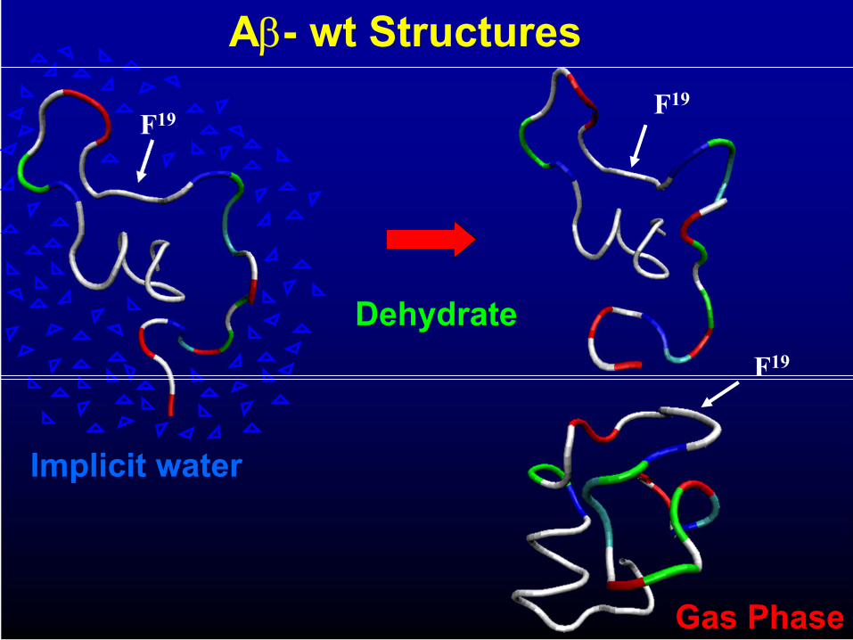
Dehydrate
Implicit water
F19 F19
Gas Phase
Aβ- wt Structures
F19

1 10 20 30 40MDVFMKGLS KAKEGVVAAA EKTKQGVAEA AGKTKEGVLY VGSKTKEGVV 50 60 70 80 90HGVATVAEKT KEQVTNVGGA VVTGVTAVAQ KTVEGAGSIA AATGFVKKDQ 100 110 120 130 140LGKNEEGAPQ EGILEDMPVD PDNEAYEMPS EEGYQDYEPE A
Amino-acid sequence of human α-synuclein. The seven imperfect repeats are underlined.
Parkinson’s Disease: α-synuclein
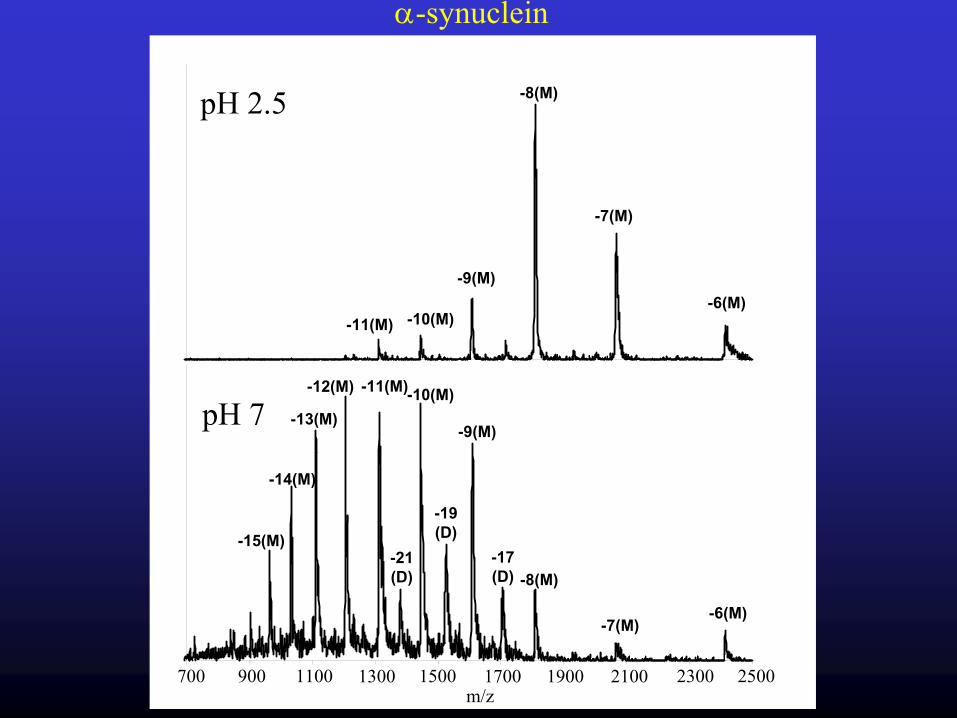
700 900 1100 1300 1500 1700 1900 2100 2300 2500
pH 7
pH 2.5
α-synuclein
-9(M)
-12(M)
-13(M)
-14(M)
-15(M)
-10(M)
-8(M)-17(D)
-19(D)
-11(M)
-21(D)
-7(M)-6(M)
-9(M)
-8(M)
-7(M)
-6(M)-10(M)-11(M)
m/z

α-synuclein

-9 -8 -7
500 1300700 900 1100arrival time (µs)
90 V
40 V
20 V
500 1300700 900 1100 arrival time (µs)
500 1300700 900 1100arrival time (µs)
(a) (b) (c)
α-synuclein
Open Open Open
Compact Compact Open

α-synuclein Summary
• Dimers form under specific solution condition
• Gas phase structures mimic solution structures
• Compact soln. structures open up with injection energy and charge

α-synucleinNMR and X-ray diffraction ineffectictive
Undergoes environmentally induced conformational changes• α-helical in Acidic Phospholipid Vesicles• Exhibits β-structure prior to fibril formation
Rg for small angle X-ray scatteringpH 7: Globular (15 Å) < 40Å α-syn < random coil (52 Å)Rg reduces in value 33% from pH 7 → pH 3

Conclusions
• Small Systems– Rearrange on Desolvation
• Large Systems– Retain Solution Structures Small Systems– Rearrange on Desolvation
• Intermediate Systems– Case depends on solvent stabilization
• Less in nucleotides• More in peptides/proteins
– Depends on Rearrangement Barriers

AcknowledgementsErin BakerDr. Jennifer GiddenAlexandra Fezuco
Erin BakerSummer Bernstein
Summer BernsteinDr. Thomas WyttenbachDr. Andrij BaumketnerDr. Joan Shea
AT
Thomas
Summer
Erin
AlyAly
JenJen