solid lipid particle-based inhalable sustained drug delivery system against experimental...
TRANSCRIPT

ARTICLE IN PRESS
Tuberculosis (2005) 85, 227–234
Tuberculosis
KEYWORDAntitubercdrugs;TuberculosNebulizatiBioavailab
1472-9792/$ - sdoi:10.1016/j.t
$Authors dopresented here�Correspondi
fax: +91 172 27E-mail addr
http://intl.elsevierhealth.com/journals/tube
Solid lipid particle-based inhalable sustained drugdelivery system against experimentaltuberculosis$
Rajesh Pandey, G.K. Khuller�
Department of Biochemistry, Postgraduate Institute of Medical Education & Research,Chandigarh 160 012, India
Accepted 30 November 2004
Sular
is;on;ility
ee front matter & 2005ube.2004.11.003
not have conflict of reson any other financialng author. Tel.: +91 17244 401.ess: [email protected]
Summary The present study was planned to evaluate the chemotherapeuticpotential of nebulized solid lipid particles (SLPs) incorporating rifampicin, isoniazidand pyrazinamide against experimental tuberculosis. The SLPs prepared by the‘‘emulsion solvent diffusion’’ technique possessed a favourable mass medianaerodynamic diameter suitable for bronchoalveolar drug delivery. Following a singlenebulization to guinea pigs, therapeutic drug concentrations were maintained in theplasma for 5 days and in the organs (lungs, liver and spleen) for 7 days whereas freedrugs were cleared by 1–2 days. The mean residence time and drug bioavailabilitywere improved several-fold in the case of drug-loaded SLPs. A similar pharmaco-kinetic profile was observed in Mycobacterium tuberculosis-infected guinea pigs. Onnebulization of drug-loaded SLPs to infected guinea pigs at every 7th day, notubercle bacilli could be detected in the lungs/spleen after 7 doses of treatmentwhereas 46 daily doses of orally administered drugs were required to obtain anequivalent therapeutic benefit. Further, there was no evidence of any biochemicalhepatotoxicity. Thus, nebulization of SLP-based antitubercular drugs forms a soundbasis for improving drug bioavailability and reducing the dosing frequency for bettermanagement of pulmonary tuberculosis.& 2005 Elsevier Ltd. All rights reserved.
Elsevier Ltd. All rights reserv
ults in regards to research/commercial interest.2747 585x517475;
o.in (G.K. Khuller).
Introduction
Solid lipid nanoparticles (SLNs) are nanocrystallinesuspensions in water, prepared from lipids, whichare solid at room temperature.1 Nanoparticles areknown to hold promise as therapeutic drug carriersand SLNs are a new form of particulate carriers
ed.

ARTICLE IN PRESS
R. Pandey, G.K. Khuller228
besides the more conventional ones such as lipo-somes, lipid emulsions and polymeric nanoparti-cles.2 The SLNs possess good tolerability (beingderived from physiological lipids), scaling-up feasi-bility, the ability to incorporate hydrophobic/hydrophilic drugs and an enhanced stability ofincorporated drugs.3,4 Thus, SLNs are unique inthe sense that they combine the virtues oftraditional nanoparticles while eliminating someof their demerits. It is not surprising that SLNs havebeen researched as adjuvants,5 as stabilizers forlabile compounds,6 for the incorporation of drugs toimprove their bioavailability7 as well as for tar-geted drug delivery.8 However, SLNs have not beenexplored as antitubercular drug (ATD) carriers todate, though recently liposomes9 and polymericnanoparticles10 proved to be successful ATD carriersin experimental tuberculosis (TB) models. Aspulmonary TB is the commonest form of thedisease, the present study was planned to co-incorporate three frontline ATDs (rifampicin, iso-niazid and pyrazinamide) in solid lipid particles(SLPs) and to evaluate its chemotherapeutic po-tential via the respiratory route in guinea pigs, inwhich the pathophysiology of TB resembles that ofhuman disease.
Materials and methods
Chemicals and drugs
Stearic acid (Mr 284.5), rifampicin, isoniazid,pyrazinamide and polyvinyl alcohol (PVA; Mr
13,000–23,000, 85% hydrolysed) were obtainedfrom Sigma Chemical Co. (St. Louis, MO, USA). Allother reagents were obtained from standardcompanies.
Animals
Dunkin Hartley guinea pigs (300–400 g body weight)were obtained from Hisar Agricultural University,Hisar, India. The animals were fed standard pelletdiet and water ad libitum. The study was approvedby the Institute’s Ethical Committee.
Culture
The culture of Mycobacterium tuberculosis H37Rvoriginally obtained from National Collection ofType Cultures (NCTC, London) was maintained onYouman’s modified medium.
Preparation of SLPs
The preparation of SLPs was based on the principleof ‘emulsion solvent diffusion method in water’11
with slight modifications. Briefly, 10mg of eachdrug (rifampicin, isoniazid and pyrazinamide) and30mg of stearic acid (as compared to monostearinin the original method) were put in a mixture ofacetone/ethanol (12ml each) and heated to60–70 1C in a water bath. The total drug:lipid ratiowas maintained at 1:1w/w. The resulting solutionwas poured into 25ml (as compared to 240ml in theoriginal method) of 1% w/v aqueous PVA at 4–8 1Cunder mechanical stirring. The SLPs formed in-stantaneously and were recovered by centrifuga-tion at 35,000g for 30min at 4–8 1C. The pellet waswashed thrice with distilled water and vacuumdried. It should be noted that the three drugs wereco-incorporated in the SLPs. Drug-free/emptyparticles were prepared by substituting sodiumchloride in place of the ATDs.
Characterization of SLPs
Five milligrams of SLPs were put in 5ml of ethanoland heated to 50 1C to lyse/dissolve the particlesand release the drugs. The percentage of drugincorporation efficiency with respect to initialamount of drug taken was calculated by theformula: (amount of drug released from the lysedSLPs/amount of drug initially taken to prepare theSLPs)� 100. Rifampicin was analysed by a micro-biological assay which was specific for the drug,using Bacillus subtilis (MTCC 441) as the indicatorstrain (sensitivity 0.25 mg/ml, linearity 0.25–10 mg/ml).12 Isoniazid was estimated by a spectrofluori-metric method based on the principle that inpresence of salicylaldehyde, the drug formed ahydrazone which was soluble in isobutanol and ahighly fluorescent compound which could be mea-sured at an excitation wavelength of 392 nm andemission of 478 nm (sensitivity 0.1 mg/ml, linearity0.1–10 mg/ml).13 Pyrazinamide was analysed spec-trophotometrically, based on the formation of acoloured complex between the drug and sodiumnitroprusside at alkaline pH, which could bemeasured at 495 nm (sensitivity 5.0 mg/ml, linearity5–70 mg/ml).14 Residual PVA was analysed color-imetrically based on the reaction between twoadjacent hydroxyl groups of PVA and an iodinemolecule to form a coloured complex which couldbe measured at 695 nm (sensitivity 25 mg/ml,linearity 25–550 mg/ml).15 Residual acetone/etha-nol were analysed by head space GC.

ARTICLE IN PRESS
Inhalable drug delivery system for TB chemotherapy 229
In vitro drug release studies
Five milligrams of drug-loaded SLPs were put in50ml of simulated gastric fluid (SGF; 0.1M HCl, pH1.2) or simulated intestinal fluid (SIF; phosphatebuffer, pH 6.8), both without enzymes, preparedaccording to the USP.16 At various time points(30min, 1, 2, 3, 4, 6, 12, 24, 48, 72 h), 1ml aliquotswere drawn for drug analysis and replaced with anequal volume of buffer. The results were expressedas the percentage of drug released with respect tothe theoretical value.
Nebulization
The dose for each guinea pig was suspended in 4mlof isotonic saline and put in the chamber of acompressor-nebulizer (Medel Aerofamily, Italy; aclosed vent jet nebulizer with 250 kPa pressure,nebulizer airflow 5.5 l/min). The formulation re-quired a nebulization time of 4min while a suitablydesigned face mask was applied to the mouth of theanimal during the process.17
Aerodynamic characterization of nebulizedSLPs
The nebulized SLPs were sized on a 7-stageAndersen Cascade Impactor (Andersen SamplersInc., Atlanta, GA, USA) and evaluated for massmedian aerodynamic diameter (MMAD) as well asgeometric standard deviation (GSD).
Preparation of SLPs for in vivo studies
The drug doses used throughout the study wererifampicin 12mg/kg + isoniazid 10mg/kg + pyrazi-namide 25mg/kg body weight, as per the standardhuman doses used in our previous reports.10,17
Since the doses were different for the three drugs,the initial amount of each drug required for SLPpreparation was calculated by the formula:(amount of drug required per animal/mean drugincorporation efficiency) � 100. Once the totaldrug quantities required were known, an equiva-lent amount of stearic acid was used in thepreparation (to maintain drug/lipid ratio at unity).The basic procedure for SLP preparation remainedthe same as discussed above. For a 400 g guinea pig,50mg of dried SLPs constituted a therapeutic dose(containing 4.8mg rifampicin + 4mg isoniazid +10mg pyrazinamide). This calculation is as perstandard methodology.18
In vivo drug disposition studies
Guinea pigs were divided into the followinggroups of eight animals each—Group 1, drug-loaded SLPs, aerosol; Group 2, free drugs, aerosol;Group 3, drug-free SLPs, aerosol; Group 4, freedrugs, oral; and Group 5, free drugs, intravenous(iv). Following the drug administration, theanimals were bled at different time points and0.5–0.7ml of blood collected (at each timepoint) was processed to analyse the drugs in theplasma by the methods mentioned above. Inaddition, the animals were sacrificed at varioustime points to analyse the drug content in20% organ homogenates (100mg tissue in 500 mlisotonic saline) of lungs, liver and spleen. Theresults were expressed as mg drug/ml plasma/gorgan weight.
Pharmacokinetic analysis
The plasma drug concentration versus time datawere used to determine the area under theconcentration–time curve (AUC0�t) by the trape-zoidal rule. The terminal AUCt�N was obtained bydividing the last measurable plasma drug concen-tration by the elimination rate constant (obtainedby regression analysis). The sum of AUC0�t andAUCt�N gave the total AUC0�N while the areaunder moment curve/area under curve gave themean residence time (MRT). Relative bioavailabilityof nebulized drug was computed by the formula(AUCaerosol/AUCoral)� (doseoral/doseaerosol) whereas(AUCaerosol/AUCiv)� (doseiv/doseaerosol) yielded theabsolute bioavailability.
Experimental infection and chemotherapy
Guinea pigs were infected intramuscularly with1� 105 viable bacilli of M. tuberculosis H37Rv in0.1ml of sterile isotonic saline (standardizedprocedure in our laboratory in lieu of the absenceof aerosol infection facilities).17 The animalswere maintained in biological safety cabinets. 20days later, six animals were sacrificed, the caudallobe of right lung and spleen were removedaseptically and homogenized in sterile isotonicsaline. The establishment of infection was con-firmed by Ziehl–Neelsen staining of the homoge-nates. In addition, the homogenates were platedon Middlebrook 7H10 agar base (supplementedwith OADC) to obtain the basal infection load.The remaining animals were divided into thefollowing groups of six animals each—Group 1,untreated control; Group 2, nebulized drug-free

ARTICLE IN PRESS
R. Pandey, G.K. Khuller230
SLPs, weekly (7 doses); Group 3, nebulized freedrugs, weekly (7 doses); Group 4, nebulizeddrug-loaded SLPs, weekly (7 doses); and Group 5,oral free drugs daily (46 doses). The drugdoses were the same as mentioned above.Free drugs were suspended freshly in isotonicsaline just before oral/aerosol administration.At day 46 following the initiation of chemo-therapy, the animals were killed and lung/spleen homogenates were prepared in sterileisotonic saline. The homogenates (50 ml of undi-luted and 1:10 diluted) were inoculated onMiddlebrook medium for enumeration of colonyforming units (CFU). The colonies were countedon day 21 post inoculation and the CFU datawere analysed by one-way analysis of variance(ANOVA).
During the 5th week (days 35–42) of chemother-apy, the infected animals in groups 3–5 were bledto obtain the plasma drug concentration profile andthe pharmacokinetic data. The values were com-pared with those of their healthy counterparts andthe data were analysed by Student’s unpaired t-test.
0
1
2
3
1 2 3 4 5 6
Oral free drugs
0
5
10
15
20
25
30
35
40
Time (Days)
0.04 0.08 0.12 0.16 0.25 0.5
1 2 3 4 5 6
Time (Days)
0.04 0.08 0.12 0.16 0.25 0.5
1 2 3 4 5 6
Time (Days)
0.04 0.08 0.12 0.16 0.25 0.5
2.5
1.5
0.5
Pla
sma
rifam
pici
n (µ
g/m
l)
0
1
2
2.5
1.5
0.5Pla
sma
ison
iazi
d (µ
g/m
l)P
lasm
a py
razi
nam
ide
(µg/
ml)
Figure 1 Plasma drug profile following the nebulization of Apigs. Figures in the right-hand panel indicate the profile in M
Hepatotoxicity studies
Blood was collected from each animal sacrificed atthe end of chemotherapy and the sera wereanalysed immediately for total bilirubin, alanineaminotransferase (ALT) and alkaline phosphatase(ALP) using standard kits.
Results
Characterization of SLPs
The drug incorporation efficiency was 5175% forrifampicin, 4574% for isoniazid and 4174% forpyrazinamide. The amount of residual PVA was10.5–12.5% w/w of vacuum-dried particles. Noresidual acetone/ethanol could be detected.
In vitro drug release
In case of isoniazid/pyrazinamide, the drug re-leased in SGF was o15% in the first 6 h and 12–15%
Nebulized SLNs
0
5
10
15
20
25
30
35
1 2 3 4 5 6
Time (Days)
0.04 0.08 0.12 0.16 0.25 0.5
1 2 3 4 5 6
Time (Days)
0.04 0.08 0.12 0.16 0.25 0.5
1 2 3 4 5 6
Time (Days)
0.04 0.08 0.12 0.16 0.25 0.5
0
1
2
2.5
1.5
0.5
Pla
sma
ison
iazi
d (µ
g/m
l)P
lasm
a py
razi
nam
ide
(µg/
ml)
0
1
2
3
2.5
1.5
0.5
Pla
sma
rifam
pici
n (µ
g/m
l)
TD-loaded SLPs as compared to oral free drugs in guinea. tuberculosis H37Rv-infected animals.

ARTICLE IN PRESS
Inhalable drug delivery system for TB chemotherapy 231
during 6–72 h. Rifampicin was released to a lesserextent, i.e. 9% in the first 6 h and 11% during 6–72 h.The drug released in SIF was no more than 20% up to6 h and 11% from 6–72 h, in the case of isoniazid/pyrazinamide while rifampicin release was in therange of 8–12% during the entire study period.
Aerodynamic characterization
Approximately 95% of the generated aerosol was inthe respirable range (o6 mm). For the majority(35.873.2%) of nebulized SLPs the size ranged from1.1–2.1 mm, with an MMAD of 1.770.1 mm and GSDof 1.970.1 mm, suitable for bronchoalveolar drugdelivery.
Drug distribution profile in plasma/organs
Following a single nebulization of ATD-loaded SLPsto guinea pigs, all the drugs were detectable in theplasma from 45min onwards up to day 5 (Fig. 1). Ateach time point the drug concentrations were at orabove the minimum inhibitory concentration(MIC90) already established in our laboratory(rifampicin 0.2 mg/ml, isoniazid 0.3 mg/ml, pyrazi-
0
2
4
0
2
4
0
50
Day 5
Day 5
Day 5
Lung L
Rif
amp
icin
(µµg
/g)
Iso
nia
zid
(µg
/g)
Pyr
azin
amid
e (µ
g/g
)
100
Figure 2 Tissue drug levels following the nebulization of ATDiv/aerosol administration.
namide 8 mg/ml).18 On the other hand, free drugswere cleared from the circulation within 12 hirrespective of the route of administration. It wasfurther demonstrated that the drug distributionprofile remained unchanged in healthy as well as M.tuberculosis H37Rv-infected guinea pigs (Fig. 1).
All the three drugs could be detected in thelungs, liver and spleen of the animals up to day 7following the nebulization of ATD-loaded SLPs(Fig. 2) whereas free drugs were cleared by24–48 h from the tissues.
Pharmacokinetics
In the case of nebulized drug-loaded SLPs, the MRTwas increased by 14–21-fold, 7–8-fold and 13–21-fold compared to iv, oral or nebulized free drugs,respectively (Table 1). Thus, the relative bioavail-ability was increased 10–16-fold, whereas theabsolute bioavailability was increased by 8–10-fold.Further, the pharmacokinetic parameters did notdiffer significantly (p40.05) between healthy andTB-infected guinea pigs so that a 11–17-foldincrease in relative bioavailability was observed inthe case of infected animals.
Day 7
Day 7
Day 7
Spleeniver
-loaded SLPs. Free drugs were cleared by 48 h after oral/

ARTICLE IN PRESS
Table 1 Salient pharmacokinetic parameters following the nebulization of ATD-loaded SLPs to healthy/TB-infected guinea pigs, as compared to free drugs.
Rifampicin Isoniazid Pyrazinamide
Healthy guineapigs (n ¼ 8)
TB-infectedguinea pigs(n ¼ 6)
Healthy guineapigs (n ¼ 8)
TB-infectedguinea pigs(n ¼ 6)
Healthy guineapigs (n ¼ 8)
TB-infectedguinea pigs(n ¼ 6)
MRT (h)Iv free drugs 2.5170.60 nd 2.4170.70 nd 3.8670.73 ndOral free drugs 6.2071.00 6.0070.85� 5.5071.10 5.6170.60� 6.6071.00 6.8170.80�
Nebulized free drugs 2.5070.80 2.5570.68� 3.1070.70 2.9870.66� 3.5070.60 3.4270.38�
Nebulized SLPs 52.7675.10 54.1174.80� 40.9176.77 37.2072.95� 56.0474.22 52.5073.91�
AUC0–N (mg.h/ml)Iv free drugs 16.5073.00 nd 18.8072.90 nd 202.0078.16 ndOral free drugs 8.4071.10 8.0570.60� 10.9071.80 11.1071.60� 185.0076.50 190.0073.00�
Nebulized free drugs 2.1070.30 2.1170.20� 18.0072.00 17.5071.50� 98.0074.00 95.1074.10�
Nebulized SLPs 135.3076.81 138.8176.81� 112.5479.00 108.1177.21� 2121.00745.00 2091.00722.00�
Relative bioavailabilityIv free drugs — — — — — —
Oral free drugs 1.00 1.00 1.00 1.00 1.00 1.00Nebulized free drugs 0.25 0.26 1.65 1.58 0.53 0.50Nebulized SLPs 16.11 17.24 10.32 9.74 11.46 11.01
Absolute bioavailabilityIv free drugs 1.00 — 1.00 — 1.00Oral free drugs 0.51 — 0.58 — 0.92 —
Nebulized free drugs 0.13 — 0.96 — 0.49 —
Nebulized SLPs 8.20 — 5.99 — 10.50 —
nd, not done (iv free drugs were not administered to infected guinea pigs).�p40.05 (Student’s t-test) with respect to healthy animals.
R. Pandey, G.K. Khuller232
Chemotherapy
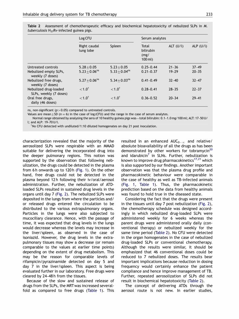
Prior to the start of chemotherapy an infection loadof 2.870.2 log CFU was observed. Seven doses ofweekly nebulized drug-loaded SLPs resulted inundetectable CFU in the organs of M. tuberculosisH37Rv-infected guinea pigs whereas 46 conven-tional doses were required to achieve the sametherapeutic benefit. Untreated control and animalsreceiving empty SLPs or weekly free drugs viaaerosol route showed comparable bacterial load(Table 2).
Biochemical hepatotoxicity
No alterations in serum bilirubin, ALT or ALP wereobserved in animals receiving drug-loaded or emptySLPs which indicated the safety of the formulationas far as biochemical hepatotoxicity is concerned(Table 2).
Discussion
The direct pulmonary delivery of ATDs remains atherapeutic challenge and hence, the present study
was designed to evaluate the potential of nebulizedSLP-based ATD delivery in a guinea pig TB model.The SLPs prepared according to the ‘‘emulsionsolvent diffusion’’ method showed a drug incor-poration efficiency of 5175%, 4574% and 4174%for rifampicin, isoniazid and pyrazinamide, respec-tively. Rifampicin showed the highest incorporationowing to the lipid-based nature of the formulation.The stability of the particles was indicated by theslow drug release observed in SGF/SIF which couldbe attributed to the residual PVA (10.5–12.5% w/w)associated with SLPs. Particle stability is known tobe enhanced by PVA which forms a barrier to thediffusional release of incorporated compounds.19
Because SLPs are a lipid-based formulation, itis expected that lipophilic drugs would remainincorporated for a longer time period whereashydrophilic drugs would be released more. Hence,rifampicin (a hydrophobic drug) was released toa lesser extent in SGF/SIF as compared withisoniazid/pyrazinamide which are hydrophilicdrugs.
The MMAD refers to the diameter above and belowwhich 50% of the number of nebulized particles aredistributed. The lower the MMAD, the morethe number of nebulized particles available forbronchoalveolar deposition. Aerodynamic particle
ARTICLE IN PRESS
Table 2 Assessment of chemotherapeutic efficacy and biochemical hepatotoxicity of nebulized SLPs in M.tuberculosis H37Rv-infected guinea pigs.
Log CFU Serum analytes�
Right caudallung lobe
Spleen Totalbilirubin(mg/100ml)
ALT (U/l) ALP (U/l)
Untreated controls 5.2870.05 5.2370.05 0.25–0.44 21–36 37–49Nebulized empty SLPs,weekly (7 doses)
5.2370.06ns 5.3370.04ns 0.21–0.37 19–29 20–35
Nebulized free drugs,weekly (7 doses)
5.2770.06ns 5.3470.03ns 0.41–0.49 32–40 32–47
Nebulized drug-loadedSLPs, weekly (7 doses)
o1.0y o1.0y 0.28–0.41 28–35 22–37
Oral free drugs,daily (46 doses)
o1.0y o1.0y 0.36–0.52 20–34 29–41
ns, non-significant (p40.05) compared to untreated controls.Values are mean7SD (n ¼ 6) in the case of log (CFU) and the range in the case of serum analytes.�Normal range obtained by analysing the sera of 10 healthy guinea pigs was—total bilirubin: 0.1–1.0mg/100ml; ALT: 17–50U/
l; and ALP: 19–70U/l.yNo CFU detected with undiluted/1:10 diluted homogenates on day 21 post inoculation.
Inhalable drug delivery system for TB chemotherapy 233
characterization revealed that the majority of theaerosolized SLPs were respirable with an MMADsuitable for delivering the incorporated drug intothe deeper pulmonary regions. This notion wassupported by the observation that following neb-ulization, the drugs could be detected in the plasmafrom 6h onwards up to 120h (Fig. 1). On the otherhand, free drugs could not be detected in theplasma beyond 12h following their iv/oral/aerosoladministration. Further, the nebulization of ATD-loaded SLPs resulted in sustained drug levels in theorgans until day 7 (Fig. 2). The nebulized SLPs weredeposited in the lungs from where the particles and/or released drugs entered the circulation to bedistributed to the various extrapulmonary organs.Particles in the lungs were also subjected tomucociliary clearance. Hence, with the passage oftime, it was expected that drug levels in the lungswould decrease whereas the levels may increase inthe liver/spleen, as observed in the case ofisoniazid. However, the drug levels in the extra-pulmonary tissues may show a decrease (or remaincomparable to the values at earlier time points)depending on the extent of drug metabolism. Thismay be the reason for comparable levels ofrifampicin/pyrazinamide detected on day 5 andday 7 in the liver/spleen. This aspect is beingevaluated further in our laboratory. Free drugs werecleared by 24–48h from the tissues.
Because of the slow and sustained release ofdrugs from the SLPs, the MRTwas increased several-fold as compared to free drugs (Table 1). This
resulted in an enhanced AUC0�N and relative/absolute bioavailability of all the drugs as has beendemonstrated by other workers for tobramycin20
and idarubicin7 in SLNs. Further, nebulization isknown to improve drug pharmacokinetics17,21 whichis also supported by our findings. Another importantobservation was that the plasma drug profile andpharmacokinetic behaviour were comparable inthe case of healthy as well as TB-infected animals(Fig. 1, Table 1). Thus, the pharmacokineticprediction based on the data from healthy animalswas found to hold true in the diseased state.
Considering the fact that the drugs were presentin the tissues until day 7 post nebulization (Fig. 2),the chemotherapy schedule was designed accord-ingly in which nebulized drug-loaded SLPs wereadministered weekly for 6 weeks whereas theparent drugs were administered orally daily (con-ventional therapy) or nebulized weekly for thesame time period (Table 2). No CFU were detectedin the organ homogenates in the case of nebulizeddrug-loaded SLPs or conventional chemotherapy.Although the results were similar, it should beemphasized that 46 conventional doses could bereduced to 7 nebulized doses. The results bearimportant implications because reduction in dosingfrequency would certainly enhance the patientcompliance and hence improve management of TB.Further, repeated aerosolization of SLPs did notresult in biochemical hepatotoxicity (Table 2).
The concept of delivering ATDs through theaerosol route is not new. In earlier studies,

ARTICLE IN PRESS
R. Pandey, G.K. Khuller234
microparticles encapsulating rifampicin22 or rifam-picin plus isoniazid23 were used for this purpose.Recently, inhalable polymeric nanoparticles17 andliposomes9 were shown to be efficient ATD carriers.The advantage with SLNs is that unlike liposomes,their long-term stability as well as drug incorpora-tion efficiency is better, whereas in contrast topolymeric formulations, the risk of residual organicsolvents is minimum.3 However, SLNs have not yetbeen explored for the respiratory delivery of ATDs.In fact, the nebulization of SLNs is a new andupcoming area of research.24 Our findings suggestthat SLPs offer an economical and patient-friendlyapproach for the direct pulmonary administrationof anti-TB drugs, bearing a high chemotherapeuticpotential.
Acknowledgement
This research was partially funded by a grant fromthe Department of Science and Technology, Govt.of India, New Delhi.
References
1. Jenning V, Lippacher A, Gohla SH. Medium scale productionof solid lipid nanoparticles (SLN) by high pressure homo-genization. J Microencapsul 2002;19:1–10.
2. Rao GC, Kumar MS, Mathivanan N, Rao ME. Nanosuspensionsas the most promising approach in nanoparticulate drugdelivery systems. Pharmazie 2004;59:5–9.
3. Schwarz C, Mehnert W, Lucks JS, Muller RH. Solid lipidnanoparticles (SLN) for controlled drug delivery. I. Produc-tion, characterization and sterilization. J Control Rel1994;30:83–96.
4. Muller RH, Maassen S, Schwarz C, Mehnert W. Solid lipidnanoparticles (SLN) as potential carrier for human use:interaction with human granulocytes. J Control Rel1997;47:261–9.
5. Olbrich C, Muller RH, Tabatt K, Kayser O, Schulze C, SchadeR. Stable biocompatible adjuvants—a new type of adjuvantbased on solid lipid nanoparticles: a study on cytotoxicity,compatibility and efficacy in chicken. Altern Lab Anim2002;30:443–8.
6. Kristl J, Volk B, Gasperlin M, Sentjure M, Jurkovic P. Effectof colloidal carriers on ascorbyl palmitate stability. Eur JPharm Sci 2003;19:181–9.
7. Zara GP, Bargoni A, Cavalli R, Fundaro A, Vighatto D,Gasco MR. Pharmacokinetics and tissue distributionof idarubicin-loaded solid lipid nanoparticles afterduodenal administration to rats. J Pharm Sci 2002;91:1324–33.
8. Yu BT, Sun X, Zhang ZR. Enhanced liver targeting by synthesisof N1-stearyl-5-Fu and incorporation into solid lipid nano-particles. Arch Pharm Res 2003;26:1096–101.
9. Pandey R, Sharma S, Khuller GK. Nebulization of liposomeencapsulated antitubercular drugs in guinea pigs. Int JAntimicrob Agents 2004;24:93–4.
10. Pandey R, Zahoor A, Sadhna S, Khuller GK. Nanoparticleencapsulated antitubercular drugs as a potential oral drugdelivery system against murine tuberculosis. Tuberculosis(Edinb) 2003;83:373–8.
11. Hu FQ, Yuan H, Zhang HH, Fang M. Preparation of solidlipid nanoparticles with clobetasol propionate by anovel solvent diffusion method in aqueous system andphysiochemical characterization. Int J Pharm 2002;239:121–8.
12. Saito H, Tomioka H. Therapeutic efficacy of liposomeentrapped rifampicin against Mycobacterium avium complexinfection induced in mice. Antimicrob Agents Chemother1989;33:429–31.
13. Scott EH, Wright RC. Fluorimetric determination of iso-nicotinic acid hydrazide in plasma. J Lab Clin Med 1967;70:355–60.
14. Gurumurthy P, Nair NGK, Sarma GR. Methods for theestimation of pyrazinamide and pyrazinoic acid in bodyfluids. Indian J Med Res 1980;71:129–34.
15. Joshi DP, Lan-Chun-Fung YL, Pritchard JW. Determination ofpoly (vinyl alcohol) via its complex with boric acid andiodine. Anal Chim Acta 1979;104:153–60.
16. United States Pharmacopeia. USP 26/NF21, UnitedStates pharmacopeial convention, Rockville, MD, USA,2003. 2528 pp.
17. Pandey R, Sharma A, Zahoor A, Sharma S, Khuller GK,Prasad B. Poly (DL-lactide-co-glycolide) nanoparticle-based inhalable sustained drug delivery system for experi-mental tuberculosis. J Antimicrob Chemother 2003;52:981–6.
18. Pandey R, Khuller GK. Chemotherapeutic potential ofalginate–chitosan microspheres as anti-tubercular drugcarriers. J Antimicrob Chemother 2004;53:635–40.
19. Sahoo SK, Panyam J, Prabha S, Labhasetwar V. Residualpolyvinyl alcohol associated with poly (DL-lactide-co-glyco-lide) nanoparticles affects their physical properties andcellular uptake. J Control Rel 2002;82:105–14.
20. Cavalli R, Bargoni A, Podio V, Muntoni E, Zara GP, Gasco MR.Duodenal administration of solid lipid nanoparticles loadedwith different percentages of tobramycin. J Pharm Sci2003;92:1085–94.
21. Dhuley JN. Aerosolized liposomal hamycin for treatment ofsystemic Candida infections in mice. FEMS Immunol MedMicrobiol 1999;25:231–6.
22. Suarez S, O’Hara P, Kazantseva M, et al. Airways delivery ofrifampicin microparticles for the treatment of tuberculosis.J Antimicrob Chemother 2001;48:431–4.
23. Sharma R, Saxena D, Dwivedi AK, Misra A. Inhalablemicroparticles containing drug combinations to targetalveolar macrophages for treatment of pulmonary tubercu-losis. Pharm Res 2001;18:1405–10.
24. Videira MA, Botelho MF, Santos AC, Gouveia LF, de Lima JJ,Almeida AJ. Lymphatic uptake of pulmonary delivered solidlipid nanoparticles. J Drug Target 2002;10:607–13.