soft tissue metastasis in patients with primary malignancies; … · 2018-05-02 · between a soft...
TRANSCRIPT

Copyrights © 2018 The Korean Society of Radiology 321
Original ArticlepISSN 1738-2637 / eISSN 2288-2928J Korean Soc Radiol 2018;78(5):321-329https://doi.org/10.3348/jksr.2018.78.5.321
INTRODUCTION
Metastatic tumors presenting as soft tissue masses are rela-tively rare compared to bony metastasis or direct invasion by carcinoma (1-3). They are usually misdiagnosed as a soft tissue sarcoma on imaging studies, and can be the source of diagnostic confusion both clinically and pathologically. The distinction between a soft tissue metastasis and primary soft tissue tumor or inflammation is important, because the treatment and prog-nosis are significantly different (1, 4, 5). Moreover, the metastatic tumors to soft tissue are known to have poor prognosis (1, 6).
However, in most cases, the distinction between a primary soft tissue sarcoma and metastatic carcinoma is difficult without bi-opsy (6).
Although there have been several case reports of skeletal mus-cle metastases from lung, breast, colonic, renal, ovarian, gastric, esophagus, melanoma, and sarcoma (3, 5, 7-12), imaging find-ings of soft tissue metastases, including magnetic resonance imaging (MRI) and computed tomography (CT) appearance are usually non-specific (13). Also, to our knowledge, there are few original reports about the features of MRI for soft tissue metastasis involving muscle or subcutaneous fat layer.
Soft Tissue Metastasis in Patients with Primary Malignancies; Magnetic Resonance Imaging and Clinical Evaluations원발암이 있는 환자에서 연조직 전이; 자기공명영상과 임상적 평가
So Min Park, MD1,2, In Sook Lee, MD1,2*, You Seon Song, MD1,2, Shin Young Park, MD1,2, Hoseok Lee, MD3, Jae Hyuck Yi, MD4, Jong Woon Song, MD5
1Department of Radiology, Pusan National University Hospital Biomedical Research Institute, Busan, Korea2Department of Radiology, Pusan National University School of Medicine, Busan, Korea3Department of Radiology, Kyungpook National University Hospital, Daegu, Korea4Department of Radiology, Keimyung University, Dongsan Medical Center, Daegue, Korea5Department of Radiology, Inje University Haeundae Paik Hospital, Busan, Korea
Purpose: The purpose of this study was to evaluate the clinical and magnetic reso-nance imaging (MRI) findings of soft tissue metastases distinct from benign soft tissue lesions.Materials and Methods: We retrospectively analyzed the MRI findings of soft tis-sue lesions found incidentally in patients with primary carcinoma and those with-out primary carcinoma from 2002–2015. To evaluate the features of soft tissue me-tastases distinct from benign soft tissue lesions, patients with benign soft tissue lesions were randomly selected and statistically analyzed for the distinctive features of the two groups.Results: A total of 47 patients (mean age 46.2 years) and 36 controls (mean age 46.2 years) were enrolled. Thirty six of the 47 patients were diagnosed with soft tissue metastasis, most commonly as the primary cancer (31%). The most common site of soft tissue metastasis was the lower extremities (36%) followed by the upper extremi-ties (31%). Soft tissue metastasis was statistically significantly different from benign soft tissue lesions according to patient age, lesion size, margin, presence of degener-ative changes in lesions, and presence of edema around the mass.Conclusion: If the incident soft tissue lesion shows malignant features on MRI in patients with primary carcinoma or in patients over 40 years of age, the radiologist should consider the possibility of metastatic cancer.
Index termsSoft Tissue NeoplasmsMetastasisCarcinomaMagnetic Resonance Imaging
Received June 23, 2017Revised August 14, 2017Accepted November 2, 2017*Corresponding author: In Sook Lee, MDDepartment of Radiology, Pusan National University School of Medicine, 179 Gudeok-ro, Seo-gu, Busan 49241, Korea.Tel. 82-51-240-7354 Fax. 82-51-244-7534E-mail: [email protected]
This is an Open Access article distributed under the terms of the Creative Commons Attribution Non-Commercial License (http://creativecommons.org/licenses/by-nc/4.0) which permits unrestricted non-commercial use, distri-bution, and reproduction in any medium, provided the original work is properly cited.

322
Incidentally Detected Soft Tissue Metastasis
jksronline.orgJ Korean Soc Radiol 2018;78(5):321-329
In this study, we evaluated clinical data and MRI findings of soft tissue metastases distinct from benign soft tissue lesions.
MATERIALS AND METHODS
Patients
This study was approved by our Institutional Review Board (No. E-2017011), and informed consent was waived due to ret-rospective study.
We retrospectively evaluated the soft tissue lesions inciden-tally found in patients with a primary malignancy between 2002 and 2015. Initially, we obtained a list of patients with pathologi-cally proven metastases to the muscle or subcutaneous fat layer during the same period from the pathology department. We searched the patients with soft tissue tumor who already have known primary cancer in our radiologic report searching system. We combined the data and excluded patients who do not un-dergo MRI for the soft tissue lesions. Lesions without patholog-ic confirmation were excluded. Also, cases that represented me-tastases to lymph nodes and with soft tissue involvements of lymphoma or leukemia were excluded. Finally, 47 patients (25 females, 22 males; age range 36–89 years, mean age 60.7 years) were enrolled. For comparison with soft tissue metastases, 36 pa-tients (19 females, 17 males; age range 7–82 years, mean age 46.2 years) with pathologically proven benign soft tissue tumors with-in the same period were selected through an imaging readout da-tabase and these patients were classified as control group.
Clinical data investigated included patient age, gender, patho-logic type of primary cancer, clinical symptoms at the time of soft tissue lesion discovery (initial presenting symptoms), period be-tween diagnosis of primary cancer and detection of soft tissue lesions, and histologic findings of incidentally detected soft tissue lesions.
Distant metastasis to other sites, such as bone or solid organs, was investigated based on the patient’s clinical records and im-ages of other departments, including bone scan or positron emis-sion tomography (PET)-CT, or images of other areas, such as ab-domen or chest CT scans.
Image Analysis
Two radiologists with 4 and 12 years experience in musculo-skeletal imaging, respectively, retrospectively reviewed the MRI
scans using a picture archiving and communication system. De-cisions were reached by discussion and consensus.
Conventional MRI data contained T1- and T2-weighted fast spin-echo sequences with/without fat-suppression using essen-tially axial planes and alternative sagittal or coronal planes. De-layed enhanced images were also obtained in three planes by fatsuppressed, fast spin-echo T1-weighted imaging.
Evaluations of the MRI scans included number of lesions, le-sion size, margins, locations (anatomical location and depth of the lesion), lesion homogeneity, presence of soft tissue edema around the lesion, presence of degenerative changes within the lesion, and presence of cortical erosion or bony involvement by expansion or infiltration of soft tissue lesion.
We defined multiple lesions as having more than one soft tis-sue lesion within the scan range. The size of the lesion was de-fined as the longest diameter by measuring the long axis. In a patient with multiple lesions, the largest lesion was analyzed. The margin of a lesion was simply classified as well- or ill-de-fined. Well-defined margin was defined as the case where the entire lesion was clearly bounded to the surrounding normal structures and ill-defined margin was defined as a case where some or all the lesion was unclear or ambiguous to the surround-ing normal structure. The locations of a lesion were basically investigated based on anatomical locations and classified as deep (deep to compartmental fascia) or superficial (involving skin and subcutaneous fat).
The homogeneity of the lesion was simply classified as homo-geneous and heterogeneous, and assessed on both T1 weighted images (T1WIs) and T2 weighted images (T2WIs). The soft tis-sue edema was defined as the appearance of infiltrative high signal intensity around the lesion on T2WI and contrast-en-hanced image. The degenerative change within the lesion was defined as a lesion with high signal intensity on T2WIs that was not enhanced on the contrast-enhanced image or high signal in-tensities on T1WIs and T2WIs representing hemorrhage. De-generative changes included cystic necrosis and hemorrhage.
Statistical Analyses
Fisher’s exact test and Mann-Whitney U test were used to determine whether gender, patient symptoms, lesion location (deep or superficial), and margin, lesion homogeneity, presence of soft tissue edema around the lesion, degenerative change with-

323
So Min Park, et al
jksronline.org J Korean Soc Radiol 2018;78(5):321-329
in the lesion, and presence of cortical erosion or bony involve-ment may affect the distinction between soft tissue metastases and benign soft tissue lesions. For age and lesion size, we used two-sample t-test. The analysis was performed using SPSS sta-tistics version 21 (IBM Corp., Armonk, NY, USA). Statistical significance was accepted for p < 0.05.
RESULTS
Thirty-six (15 females, 21 males, mean age 63 years, age range 36–89 years) of 47 patients (77%) were diagnosed with soft tis-sue metastases. Ten patients (9 females, 1 male, mean age 53 years, age range 40–72 years) had benign soft tissue lesions. In the remaining patient with thyroid cancer, the soft tissue lesion was diagnosed as myxofibrosarcoma. Thirty-three patients had a single soft tissue lesion (Fig. 1) and the remaining 14 had mul-tiple lesions (Fig. 2). Of the 36 patients with soft tissue metasta-ses, 14 had multiple lesions and 12 had a single lesion. Of the 10 patients with benign soft tissue lesions, only one patient with hemangiomas had multiple lesions. All patients in the control group had a single mass. On the other hand, in 17 of 47 patients, soft tissue lesions were found incidentally without knowledge of the presence of primary cancer (Fig. 3). Among those 17 pa-tients, we interpreted primary malignant tumors in 9 patients
with single soft tissue lesion and metastases in 8 patients with multiple soft tissue lesions in the first MRI scans. The benign soft tissue tumors of control group included schwannoma (n = 13), hemangioma (n = 6), benign lipomatous tumor (n = 4), gi-ant cell tumor (n = 3), nodular fasciitis (n = 3), glomus tumor (n = 2), epidermal cyst (n = 2), leiomyoma (n = 1), myxoma (n = 1), and pilomatricoma (n = 1).
The types of primary malignancies and the number of pa-tients diagnosed with soft tissue metastasis according to each primary malignancy are summarized in Table 1. Among all primary tumors, lung cancer was the most common (26%) and the majority of soft tissue metastasis originated from lung can-cer (31%). The pathologic results of three unknown primary origin were poorly differentiated squamous cell carcinoma, melanoma, and primitive neuroectodermal tumor (PNET).
The anatomical locations of soft tissue lesions are summarized in Table 2. Metastatic soft tissue lesions (n = 36) were the most common in the lower extremities (thigh, lower leg, ankle, and toe; 13/36, 36%), followed by the shoulder (6/36, 17%) and the upper extremities (upperarm, forearm, finger, hand, and wrist; 5/36, 14%).
Among 36 patients with soft tissue metastases, 11 complained of pain. In the control group, 14 patients had pain. The symptom was not significant for differentiating between metastatic and
Fig. 1. A 50-year-old male patient diagnosed with oropharyngeal cancer two years previously and presently complaining of buttock pain.A. On coronal fat-suppressed T2-weighted image of the pelvis, a hyperintense soft tissue lesion with ill-defined margin (arrow) is seen in the muscular layer of the right buttock. Peritumoral edema (arrowheads) is also noted.B. The lesion shows homogeneous enhancement without degenerative change within the lesion (arrow) on axial fat-suppressed, contrast-en-hanced T1-weighted image.
A B
324
Incidentally Detected Soft Tissue Metastasis
jksronline.orgJ Korean Soc Radiol 2018;78(5):321-329
benign lesions (p > 0.05).Thirteen of 36 patients (36%) diagnosed with soft tissue metas-
tasis had distant metastasis to bone or other solid organs (Fig. 2). Results of statistical analyses todifferentiate between soft tis-
sue metastasis and benign soft tissue lesion are summarized in Table 3. Age, lesion size, margin, degenerative change within
the mass, and the presence of edema around soft tissue lesions (Fig. 1) were statistically significant.
DISCUSSION
Distant metastases to soft tissue are relatively uncommon, even
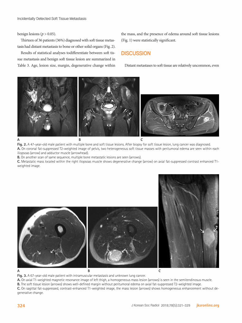
Fig. 2. A 47-year-old male patient with multiple bone and soft tissue lesions. After biopsy for soft tissue lesion, lung cancer was diagnosed.A. On coronal fat-suppressed T2-weighted image of pelvis, two heterogeneous soft tissue masses with peritumoral edema are seen within each iliopsoas (arrow) and adductor muscle (arrowhead).B. On another scan of same sequence, multiple bone metastatic lesions are seen (arrows).C. Metastatic mass located within the right iliopsoas muscle shows degenerative change (arrow) on axial fat-suppressed contrast enhanced T1-weighted image.
A B C
Fig. 3. A 67-year-old male patient with intramuscular metastasis and unknown lung cancer.A. On axial T1-weighted magnetic resonance image of left thigh, a homogeneous mass lesion (arrows) is seen in the semitendinosus muscle.B. The soft tissue lesion (arrows) shows well-defined margin without peritumoral edema on axial fat-suppressed T2-weighted image.C. On sagittal fat-suppressed, contrast-enhanced T1-weighted image, the mass lesion (arrows) shows homogeneous enhancement without de-generative change.
A B C

325
So Min Park, et al
jksronline.org J Korean Soc Radiol 2018;78(5):321-329
in patients with known cancers and despite comprising approxi-mately 55% of our body mass (2, 14). Some previous studies re-ported a relatively broad range of frequency, and these frequen-cy differences might be considered to depend on the procedures used for the examinations or active examinations or autopsy cases (1, 3, 4). Several factors may contribute to the rarity of he-matogenous metastases to soft tissue areas. For example, organs with a high incidence for metastatic carcinomas, such as the liver, lung, or bone, are rich in capillary vasculature and have a constant blood flow, whereas in soft tissues, such as skeletal
muscle, the blood flow is variable and subject to variations in tissue pressure affecting tumor implantation (6, 8, 14, 15). Also, some authors suggested that lactic acid production by muscle inhibits growth of tumor studies, and uncommon hematoge-nous metastases to muscles are thought to be due to muscle motion, muscle pH, and the muscle’s ability to remove tumor-produced lactic acid (11, 14, 16, 17). Leukemia and lymphoma are the most frequent cancer of metastases to muscle (11).
With regard to the primary malignant tumor, several studies (1, 6, 13, 18, 19) reported that the most frequent tumor of ori-gin was lung cancer, similar to our series. However, many other studies reported different tendencies for the common source caus-ing soft tissue metastases (1, 2, 13), and a variety of malignant
Table 1. The Types of Primary Malignancies and the Number of Pa-tients Diagnosed with Soft Tissue Metastasis According to Each Pri-mary Malignancy
Primary MalignancyNumber of Patients
Soft Tissue Metastasis
Lung cancer 12 11Breast cancer 6 2Thyroid cancer 5 2Colorectal cancer 5 5Stomach cancer 2 2Hepatocellular cell carcinoma 2 2Oropharyngeal cancer 2 0Melanoma 2 2Cholangiocarcinoma 2 2
Bowen’s disease (Squamous cell cancer in situ)
1 1
Hemangiopericytoma 1 1Renal cell carcinoma 1 1Gall bladder cancer 1 1Uterine cervical cancer 1 1Plasmacytoma (multiple myeloma) 1 0Unknown origin 3 3Total 47 36
Table 2. The Anatomical Locations of Soft Tissue LesionsLocations Metastasis Control Group
Neck area 0 3Shoulder 6 0Upperarm 2 5Elbow 1 0Forearm 1 1Finger/hand/wrist 1 6Pelvic area 4 3Thigh 7 4Lower leg 6 5Ankle 0 1Toe 0 2Back area (muscle & subcutaneous fat) 1 4Psoas muscle 3 2Chest wall 3 0Abdominal wall 1 0Total 36 36
The location of one new malignant tumor (myxofibrosarcoma) was thigh.
Table 3. Clinical and Magnetic Resonance Imaging Analysis Results for Differentiating between Soft Tissue Metastasis and Benign Soft Tissue Lesion
Soft Tissue Metastasis Benign Lesion p-ValueAge (years) 62.83 ± 2.45 46.22 ± 3.05 < 0.0001Gender (M:F) 21:15 17:19 0.348Size (cm) 7.5 (2.8–26) 3.5 (0.4–11.6) 0.001Symptom n = 11 n = 14 0.461Location (deep: superficial) 30:6 25:11 0.168Margin (well:ill) 10:26 32:4 < 0.0001Homogeneity 16 11 0.227Degenerative change within the mass 16 8 0.047Bone erosion 6 5 0.745Peritumoral edema 28 8 < 0.0001Mineralization 2 2 1

326
Incidentally Detected Soft Tissue Metastasis
jksronline.orgJ Korean Soc Radiol 2018;78(5):321-329
tumors including skin cancer, renal cell carcinoma, melanoma, breast cancer, and colon cancer can cause soft tissue metastases (13, 20-23). In our study, the most commones tumors metasta-sizing to soft tissue were carcinomas of the lung and colon. In one study (2), malignant melanoma was the most frequently spe-cific tumor type that metastasized to soft tissue. However, there were only two cases of melanoma metastasis to soft tissue in our study.
Many metastatic tumors to soft tissue present as occult me-tastases from an unrecognized primary (2, 7, 24). Also, in this study, 17 soft tissue metastatic lesions were found incidentally without knowledge of the presence of primary cancer. Among the 17 incidental metastases, 9 had solitary soft tissue metasta-sis. Because the imaging characteristics of these lesions are nonspecific and there is considerable overlap in the appearance of metastatic lesions and primary soft tissue sarcomas, the diag-nosis of solitary soft tissue metastasis in patients with unknown primary malignancy is very difficult. On MRI, large, multinod-ular, and hypervascular soft-tissue sarcomas may have central areas of hemorrhage, necrosis, and calcification (25). However, hemorrhage may occur within a primary or metastatic soft-tis-sue tumor. Also, peri-lesional edema may occur in soft tissue sarcoma, metastasis and infection.
The frequent metastatic site varies (1, 2, 13), with the thigh muscles, iliopsoas, and paraspinous muscles reported as com-mon sites (3, 6, 20, 21, 26). In this study, the most frequent site of metastasis to soft tissue area was the lower extremity, followed by the shoulder. These results contrast with previous studies (1, 2, 11, 13). These differences might be caused by the composition of the primary cancer. In this study, among 36 patients with soft tissue metastasis, 11 patients had lung cancer. In these patients the lesions were located in a lower leg (n = 4), shoulder (n = 3), thigh (n = 2), pelvic area (n = 1), and psoas muscles (n = 1). Giv-en the ease of arterial metastasis of lung cancer, lesions can eas-ily metastasize to distal extremities. In our study, the proportion of the lung cancer in primary malignancies was high, which might affect the anatomical site of the soft tissue metastasis. The anatomic distribution of metastases to soft tissue area in this study was similar to the distribution of soft tissue sarcoma, with 36% occurring in the lower extremity. Thus, metastatic carcino-ma can often be confused clinically and histologically with pri-mary soft tissue sarcoma. Soft tissue metastases can occur in
muscles and subcutaneous sites, but the incidence of subcuta-neous fat involvement has been reported to be lower than that of muscle involvement. It is believed that subcutaneous metas-tases might be underreported in the literature (13).
Several authors (13, 27) suggested that soft tissue metastases, especially those in skeletal muscle, are frequently painful or pal-pable, and a painful soft tissue mass is more commonly noted in patients with soft tissue metastasis than in primary sarcoma (19). In this study, 20 of 36 patients (56%) with soft tissue metas-tases complained of pain or palpable mass. However, the pres-ence of symptom was not significant, and we could not com-pare symptoms between sarcoma and soft tissue metastases because our study did not include sarcomas. Clinical presenta-tion, anatomic distribution, and radiographic imaging studies of metastases to soft tissue area are similar to those of soft tissue sarcomas (6). Because the treatment and prognosis are differ-ent, differentiating between these diseases is important, but dif-ficult (3, 6). Moreover, the metastases of carcinomas to soft tis-sue appears to be a late event in the progression of the disease and the overall prognosis is poor (2, 6, 18). However, distant metastases to other sites occurred only in 36% of our cases.
MRI has become the preferred technique for distinguishing soft tissue metastases from other tumorous processes (28). In several previous studies (11, 13, 19, 24, 29), MRI revealed soft tissue metastases to have poorly defined margins, large areas of central necrosis, and extensive peritumoral edema, but rare erosion of the adjacent bone. As noted presently, to compare the benign soft tissue lesions and soft tissue metastasis, an ill-defined margin, presence of degenerative change within the le-sion, and peritumoral edema are statistically significant find-ings in soft tissue metastasis.
Soft tissue metastasis was significantly larger than benign soft tissue tumors, and cut-off values were 7.5 cm in diameter. As reported (30), cutaneous metastasis occurs as multiple, small skin nodules. Presently, the proportion of deep locations was high (30/36) and the size of the lesion was large. If the lesions locate deep in the skin layer, the size of the lesion may help to differentiate the benign and malignant tumors. In the same con-text, as the lesion becomes larger the prevalence of degenerative change within the lesion may increase. Thus, this finding sug-gests the presence of metastatic lesions than benign soft tissue tumors in patients with known malignancy. Ill-defined margin

327
So Min Park, et al
jksronline.org J Korean Soc Radiol 2018;78(5):321-329
with peritumoral edema were the significant findings to differ-entiate the benign and soft tissue metastasis in this study, but it is present in other benign conditions including infection or in-flammatory lesions. So, clinical and radiological findings to ex-clude the possibility of infection or inflammation may be im-portant. The statistically significant imaging findings in this study were somewhat similar to malignant soft tissue tumors, such as sarcoma or other primary soft tissue malignancies. There-fore, MRI findings of soft tissue metastases are not pathogno-monic, and thus, needle or excisional biopsy is mandatory for diagnosis (1, 11, 19, 30). However, MRI evaluation of soft tissue metastases is advisable before biopsy (13). Also, confirmation of past history is important for the early diagnosis (1). Moreover, the diagnosis of soft tissue metastasis should be considered in the differential diagnosis of any painful soft tissue mass with ex-tensive peritumoral enhancement pattern in MRI that would otherwise be most suggestive of a soft tissue sarcoma (13, 19).
There were some limitations in this study. First was the small number of patients. This reflects the low incidence of soft tissue metastasis and exclusion of cases that represented metastases to lymph nodes. We included only cases involving skeletal muscles and subcutaneous fat layers. Second, we could not prospectively perform the recent functional MRI for a more advanced imag-ing analysis because this study was retrospectively analyzed. However, even functional MRI might be difficult to differenti-ate between soft tissue metastasis and primary sarcoma because these will show similar malignant imaging findings.
In summary, there were no specific imaging findings to diag-nose soft tissue metastasis in this study. Although there were some significant imaging findings differentiating between the benign soft tissue tumors and soft tissue metastasis, these find-ings were almost the same as in the malignant soft tissue tumors reported in previous studies. Therefore, it is difficult to distin-guish between soft tissue metastases and primary malignant tu-mors. Patients included in our study were all over 40 years of age, except two. When a patient 40 years or older shows nonspecific imaging findings, especially aggressive findings in the bones, radiologists often prefer bone metastasis due to its frequency. On the contrary, because of the rarity of the soft tissue metastasis, we often misdiagnose these diseases. Therefore, in the same con-text, in patients over age 40 years of age with primary malignan-cies or who are unaware of the primary malignancies, when there
is non-specific soft-tissue lesion with malignant features are seen on MRI, the possibility of metastasis should be considered, as in the case of bone metastasis, rather than primary malignant tumors. Also, radiologists should try to make a definitive diag-nosis through biopsy or special imaging methods such as func-tional MRI before treatments.
REfERENCES
1. Torigoe T, Terakado A, Suehara Y, Okubo T, Takagi T, Kaneko
K, et al. Metastatic soft tissue tumors. J Cancer Ther 2011;
5:746-751
2. Plaza JA, Perez-Montiel D, Mayerson J, Morrison C, Suster S.
Metastases to soft tissue: a review of 118 cases over a 30-
year period. Cancer 2008;112:193-203
3. Sudo A, Ogihara Y, Shiokawa Y, Fujinami S, Sekiguchi S. In-
tramuscular metastasis of carcinoma. Clin Orthop Relat Res
1993;296:213-217
4. Pearson CM. Incidence and type of pathologic alterations
observed in muscle in a routine autopsy survey. Neurology
1959;9:757-766
5. Viswanathan N, Khanna A. Skeletal muscle metastasis from
malignant melanoma. Br J Plast Surg 2005;58:855-858
6. Herring CL Jr, Harrelson JM, Scully SP. Metastatic carcinoma
to skeletal muscle. a report of 15 patients. Clin Orthop Relat
Res 1998;355:272-281
7. Sridhar KS, Rao RK, Kunhardt B. Skeletal muscle metastases
from lung cancer. Cancer 1987;59:1530-1534
8. Stulc JP, Petrelli NJ, Herrera L, Lopez CL, Mittelman A. Iso-
lated metachronous metastases to soft tissues of the but-
tock from a colonic adenocarcinoma. Dis Colon Rectum
1985;28:117-121
9. Yoshioka H, Itai Y, Niitsu M, Fujiwara M, Watanabe T, Sato-
mi H, et al. Intramuscular metastasis from malignant mela-
noma: MR findings. Skeletal Radiol 1999;28:714-716
10. Tochigi H, Nakao Y, Horiuchi Y, Toyama Y. Metastatic ma-
lignant melanoma in the hand muscle--a case report. Hand
Surg 2000;5:69-72
11. Williams JB, Youngberg RA, Bui-Mansfield LT, Pitcher JD.
MR imaging of skeletal muscle metastases. AJR Am J Roent-
genol 1997;168:555-557
12. Chand M, Thomas RJ, Dabbas N, Bateman AC, Royle GT. Soft

328
Incidentally Detected Soft Tissue Metastasis
jksronline.orgJ Korean Soc Radiol 2018;78(5):321-329
tissue metastases as the first clinical manifestation of squa-
mous cell carcinoma of the esophagus: case report. World
J Oncol 2010;1;135-137
13. Damron TA, Heiner J. Distant soft tissue metastases: a series
of 30 new patients and 91 cases from the literature. Ann
Surg Oncol 2000;7:526-534
14. Seely S. Possible reasons for the high resistance of muscle
to cancer. Med Hypotheses 1980;6:133-137
15. Fidler IJ, Hart IR. Principles of cancer biology, biology of
cancer metastasis. In: Devita VT, Hellman S, Rosenberg SA,
eds. Cancer: principles and practice of oncology. Philadel-
phia: JB Lippincortt 1982:80-92
16. Acinas García O, Fernández FA, Satué EG, Buelta L, Val-
Bernal JF. Metastasis of malignant neoplasms to skeletal
muscle. Rev Esp Oncol 1984;31:57-67
17. Pretorius ES, Fishman EK. Helical CT of skeletal muscle me-
tastases from primary carcinomas. AJR Am J Roentgenol
2000;174:401-404
18. Glockner JF, White LM, Sundaram M, McDonald DJ. Unsus-
pected metastases presenting as solitary soft tissue lesions:
a fourteen-year review. Skeletal Radiol 2000;29:270-274
19. Tuoheti Y, Okada K, Osanai T, Nishida J, Ehara S, Hashmoto
M, et al. Skeletal muscle metastases of carcinoma: a clini-
copahtological study of 12 cases. Jpn J Clin Oncol 2004;34:
210-214
20. Torosian MH, Botet JF, Paglia M. Colon carcinoma metastat-
ic to the thigh--an unusual site of metastasis. report of a
case. Dis Colon Rectum 1987;30:805-808
21. Bibi C, Benmeir P, Maor F, Sagi A. Hand metastasis from re-
nal cell carcinoma with no bone involvement. Ann Plast
Surg 1993;31:377-378
22. Laurence AE, Murray AJ. Metastasis in skeletal muscle sec-
ondary to carcinoma of the colon--presentation of 2 cases.
Br J Surg 1970;57:529-530
23. McKeown PP, Conant P, Auerbach LE. Squamous cell carci-
noma of the lung: an unusual metastasis to pectoralis mus-
cle. Ann Thorac Surg 1996;61:1525-1526
24. Alburquerque TL, Ortin A, Cacho J. Metastasis in deep calf
muscles as first manifestation of bronchus adenocarcinoma.
Am J Med 1987;83:606-607
25. Stoller DW, Steinkirchner TM, Porter B. Bone and soft-tis-
sue tumors. In: Stoller DW, eds. Magnetic resonance imag-
ing in orthopaedics and sports medicine, 1st ed. Philadel-
phia: Lippincott 1993:1094-1116
26. Mignani G, McDonald DJ, Boriani S, Avella M, Gaiani L,
Campanacci M. Soft tissue metastasis from carcinoma. a
case report. Tumori 1989;75:630-633
27. Schultz SR, Bree RL, Schwab RE, Raiss G. CT detection of
skeletal muscle metastases. J Comput Assist Tomogr 1986;
10:81-83
28. O’Keefe D, Gholkar A. Metastatic adenocarcinoma of the
paraspinal muscles. Br J Radiol 1988;61:849-851
29. Munk PL, Gock S, Gee R, Connell DG, Quenville NF. Case re-
port 708: metastasis of renal cell carcinoma to skeletal
muscle (right trapezius). Skeletal Radiol 1992;21:56-59
30. Kransdorf MJ, Jelinek JS, Moser RP Jr, Utz JA, Brower AC,
Hudson TM, et al. Soft-tissue masses: diagnosis using MR
imaging. AJR Am J Roentgenol 1989;153:541-547

329
So Min Park, et al
jksronline.org J Korean Soc Radiol 2018;78(5):321-329
원발암이 있는 환자에서 연조직 전이; 자기공명영상과 임상적 평가적 평가
박소민1,2 · 이인숙1,2* · 송유선1,2 · 박신영1,2 · 이호석3 · 이재혁4 · 송종운5
목적: 본 연구는 양성 연조직 병변과 구분되는 연조직 전이의 임상 소견과 자기공명영상 소견을 찾고자 한다.
대상과 방법: 2002~2015년 동안 원발암이 있는 환자 및 원발암을 모르고 있던 환자에서 우연히 발견된 연조직 병변의 자
기공명영상소견을 후향적으로 분석하였다. 양성 연조직 병변과 구분되는 연조직 전이의 특징을 평가하기 위해 같은 기간
내 진단된 양성 연조직 병변 대조군을 무작위로 선정하였고, 두 군의 구별되는 특징에 대해 통계학적으로 분석하였다.
결과: 총 47명(평균연령 60.7세)의 환자와 대조군 36명(평균연령 46.2세)을 본 연구에 포함하였다. 47명 중에 36명이
연조직 전이로 진단되었다. 전체 연조직 전이 중 원발암이 폐암인 경우가 가장 많았다(31%). 연조직 전이의 가장 흔한 위치
는 하지(36%)였으며, 다음으로는 상지(31%)였다. 연조직 전이는 나이, 병변의 크기, 경계, 병변 내 퇴행성 변화의 유무,
종괴주위 부종의 유무에서 양성 연조직 병변과 통계학적으로 유의한 차이를 보였다.
결론: 원발암이 있거나 혹은 원발암의 존재를 모르고 있는 40세 이상의 환자에서, 우연히 발견된 연조직 병변이 자기공명
영상에서 악성의 특징을 보일 때, 영상의학과 의사는 전이암의 가능성을 생각해야 한다.
1부산대학교병원 의생명연구원 영상의학과, 2부산대학교 의학전문대학원 영상의학교실, 3경북대학교병원 영상의학과, 4계명대학교 동산의료원 영상의학과, 5인제대학교 부산해운대백병원 영상의학과