smart and safe - olympus · 13818 outstanding image quality and maximum detection as you would...
TRANSCRIPT

SMART AND SAFEENDOCAPSULE 10
1551
5
2 3
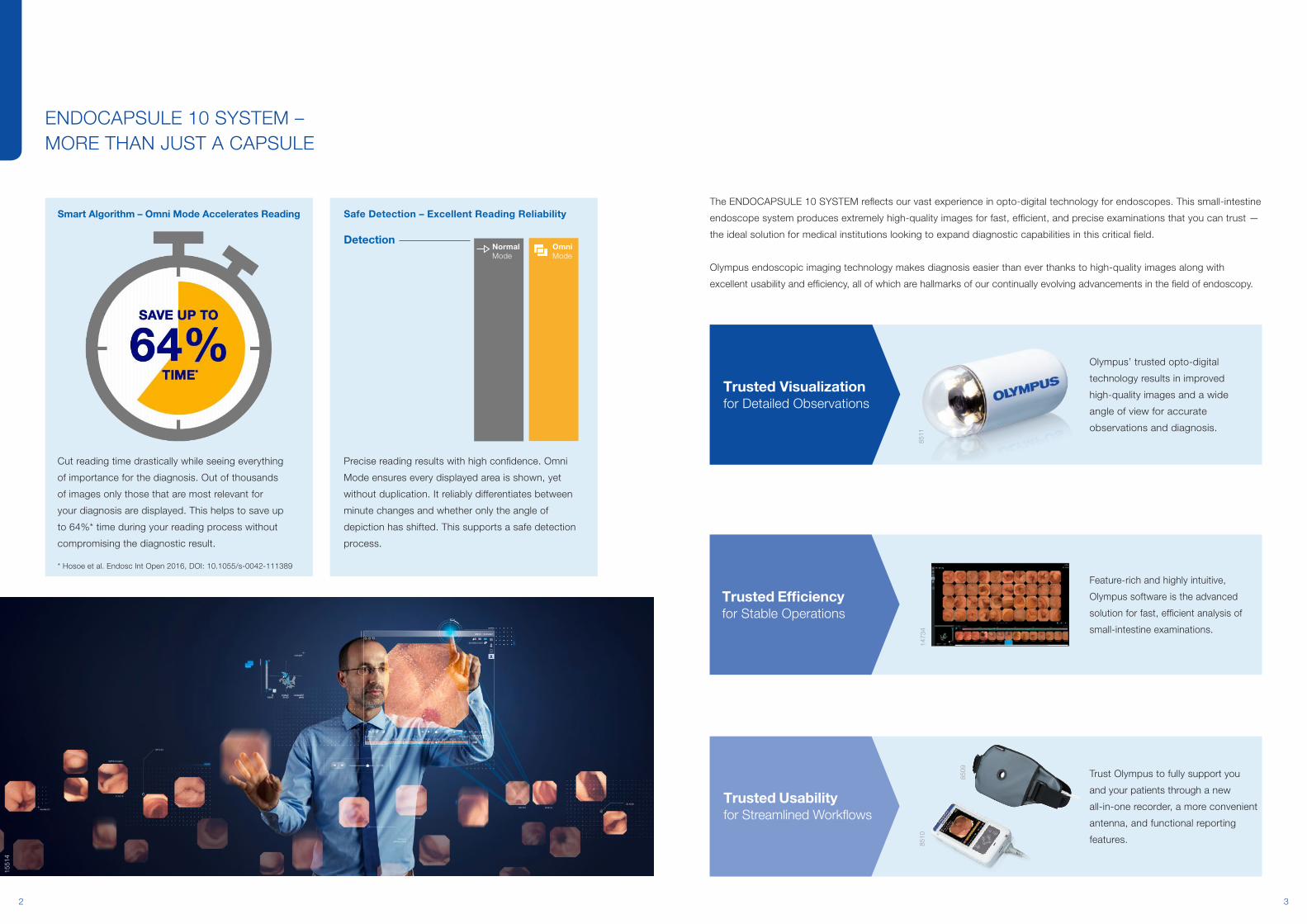
The ENDOCAPSULE 10 SYSTEM reflects our vast experience in opto-digital technology for endoscopes. This small-intestine
endoscope system produces extremely high-quality images for fast, efficient, and precise examinations that you can trust —
the ideal solution for medical institutions looking to expand diagnostic capabilities in this critical field.
Olympus endoscopic imaging technology makes diagnosis easier than ever thanks to high-quality images along with
excellent usability and efficiency, all of which are hallmarks of our continually evolving advancements in the field of endoscopy.
ENDOCAPSULE 10 SYSTEM –MORE THAN JUST A CAPSULE
Smart Algorithm – Omni Mode Accelerates Reading
Cut reading time drastically while seeing everything
of importance for the diagnosis. Out of thousands
of images only those that are most relevant for
your diagnosis are displayed. This helps to save up
to 64%* time during your reading process without
compromising the diagnostic result.
1551
4
* Hosoe et al. Endosc Int Open 2016, DOI: 10.1055/s-0042-111389
Safe Detection – Excellent Reading Reliability
Precise reading results with high confidence. Omni
Mode ensures every displayed area is shown, yet
without duplication. It reliably differentiates between
minute changes and whether only the angle of
depiction has shifted. This supports a safe detection
process.
8510
8509
8511
Olympus’ trusted opto-digital
technology results in improved
high-quality images and a wide
angle of view for accurate
observations and diagnosis.
Trust Olympus to fully support you
and your patients through a new
all-in-one recorder, a more convenient
antenna, and functional reporting
features.
Trusted Usability for Streamlined Workflows
Feature-rich and highly intuitive,
Olympus software is the advanced
solution for fast, efficient analysis of
small-intestine examinations.
1473
4
Trusted Efficiency for Stable Operations
Trusted Visualization for Detailed Observations
DetectionNormal Mode
OmniMode
54
Structure Enhancement Color Tone
8522
8523
8524
8525
8526
Level 1 Level 3 Level 8 Red −3 / Blue +3Red 0 / Blue 0
8519
EC-S10
8515
Angioma(bleeding)
8514
Multiple inflammations withCrohn disease stenosis
8512
Normal
8513
Angioma(no bleeding)
Observable Findings
8518
Previous model
Less Halation
8516
Previous model
8517
EC-S10
Less Noise
EC-S10
8521
The white line indicates the angle of view of the
previous model.
8520
Previous model
Previousmodel
50% longer observation
12 hEC-S10160°
8 h145° Previousmodel
EC-S10
8511
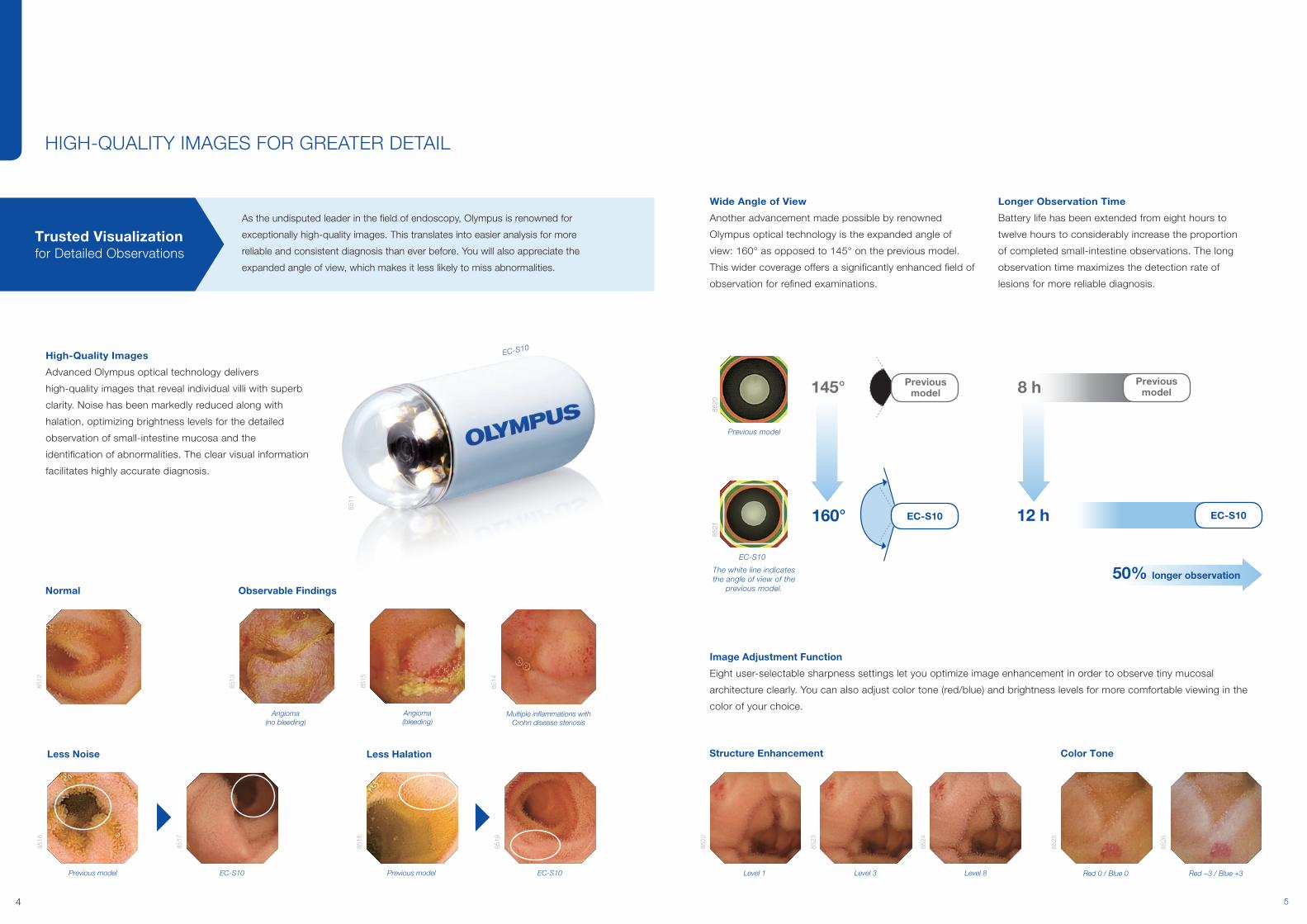
High-Quality Images
Advanced Olympus optical technology delivers
high-quality images that reveal individual villi with superb
clarity. Noise has been markedly reduced along with
halation, optimizing brightness levels for the detailed
observation of small-intestine mucosa and the
identification of abnormalities. The clear visual information
facilitates highly accurate diagnosis.
As the undisputed leader in the field of endoscopy, Olympus is renowned for
exceptionally high-quality images. This translates into easier analysis for more
reliable and consistent diagnosis than ever before. You will also appreciate the
expanded angle of view, which makes it less likely to miss abnormalities.
Trusted Visualization for Detailed Observations
Wide Angle of View
Another advancement made possible by renowned
Olympus optical technology is the expanded angle of
view: 160° as opposed to 145° on the previous model.
This wider coverage offers a significantly enhanced field of
observation for refined examinations.
Longer Observation Time
Battery life has been extended from eight hours to
twelve hours to considerably increase the proportion
of completed small-intestine observations. The long
observation time maximizes the detection rate of
lesions for more reliable diagnosis.
Image Adjustment Function
Eight user-selectable sharpness settings let you optimize image enhancement in order to observe tiny mucosal
architecture clearly. You can also adjust color tone (red/blue) and brightness levels for more comfortable viewing in the
color of your choice.
HIGH-QUALITY IMAGES FOR GREATER DETAIL
EC-S10
76
1473
2
Playback view tabRed color overview tab
Overview tab
“Normal” button
“Adjust” button
“Omni” button3D Track area
Normal mode Play back all images.
Omni-selectedmode
Play back onlyselected images.
Adjust modePlay back all images
at optimal speed.
High speed
Normal speed
1053
8
Trusted Efficiency for Stable Operations
ENDOCAPSULE 10 SYSTEM software facilitates reading with a variety of
unique functions to detect images requiring closer inspection, providing the
means for the fast reviewing of results to ultimately speed up diagnosis.
3D Track Function
Track the capsule as it moves through the small intestine
with the 3D Track function. A high-precision antenna
recognizes the detailed signals from the capsule, allowing
the system to display the capsule track in 3D. The track
progress bar is useful for estimating capsule location in
the small intestine. It also indicates on the 3D tracking
screen where each thumbnail image was captured in
order to assess the locations of abnormalities. The 3D
Track function operates intuitively, showing capsule
location to help you decide what approach should be
taken for subsequent procedures.
Overview Function
This function displays a library of characteristic images. The
new Adjacent image display and Enlarging image functions
provide a quick way for further observation without having
to switch to Playback view mode. In addition, the new Red
color overview function gives you a quick overview only of
images showing an excessive amount of red.
Bubble and Debris Image Detection Algorithm
Bubbles and debris can sometimes adhere to the capsule
and degrade image quality. The ENDOCAPSULE 10
SYSTEM automatically detects poor-quality images and
displays only those that can be accurately read. This
algorithm also enhances the performance of Adjust mode
and the Overview function.
1473
3
Track progress bar
3D Track area
INTELLIGENT READING FUNCTIONS SIMPLIFY ANALYSIS
Adjust Mode
Change playback speed depending on differences in
images. In Adjust mode, images showing no change
are superimposed on each other, and review speed is
optimized to move quickly past images indicating no
characteristic differences compared to preceding images.
This mode vastly reduces playback time to increase
reading effectiveness.
Omni Mode
Images that overlap with previous ones are skipped, and
new images are selected even when only minute changes
are present. This algorithm can recognize that an image
is identical, even when the capsule is displaying the same
section of small intestine from a different angle. This
intelligent approach helps to speed up diagnosis by
analyzing a larger number of attributes than ever before.*
* Compared to ENDOCAPSULE 10 SYSTEM Express-selected mode
1473
7
Adjacent Image
Display Function
Click selected image.
Enlarging Image
Function
Briefly place mouse
over an image.
1473
6
8531
Previous Model
Display of Overview
mode of previous
ENDOCAPSULE system.
New Model
ENDOCAPSULE 10
SYSTEM with new
algorithm.14
735
98
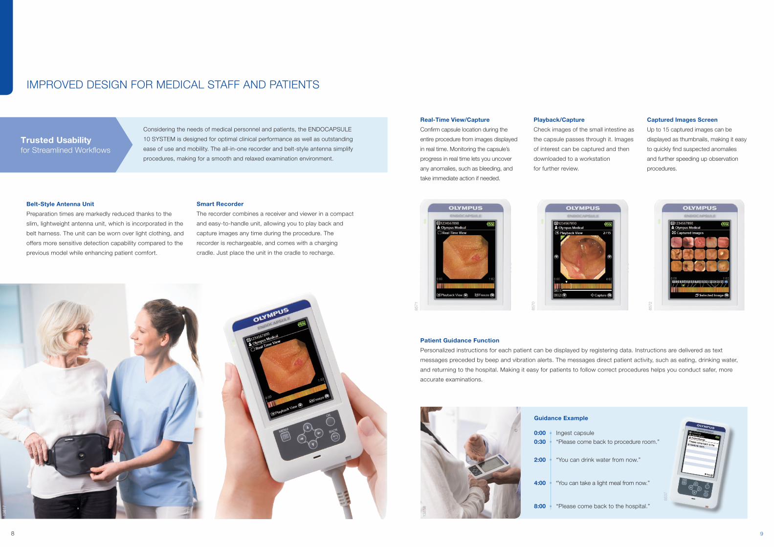
IMPROVED DESIGN FOR MEDICAL STAFF AND PATIENTS
Smart Recorder
The recorder combines a receiver and viewer in a compact
and easy-to-handle unit, allowing you to play back and
capture images any time during the procedure. The
recorder is rechargeable, and comes with a charging
cradle. Just place the unit in the cradle to recharge.
Belt-Style Antenna Unit
Preparation times are markedly reduced thanks to the
slim, lightweight antenna unit, which is incorporated in the
belt harness. The unit can be worn over light clothing, and
offers more sensitive detection capability compared to the
previous model while enhancing patient comfort.
Trusted Usability for Streamlined Workflows
Considering the needs of medical personnel and patients, the ENDOCAPSULE
10 SYSTEM is designed for optimal clinical performance as well as outstanding
ease of use and mobility. The all-in-one recorder and belt-style antenna simplify
procedures, making for a smooth and relaxed examination environment.
Real-Time View/Capture
Confirm capsule location during the
entire procedure from images displayed
in real time. Monitoring the capsule’s
progress in real time lets you uncover
any anomalies, such as bleeding, and
take immediate action if needed.
Playback/Capture
Check images of the small intestine as
the capsule passes through it. Images
of interest can be captured and then
downloaded to a workstation
for further review.
Captured Images Screen
Up to 15 captured images can be
displayed as thumbnails, making it easy
to quickly find suspected anomalies
and further speeding up observation
procedures.
Guidance Example
0:00 Ingest capsule
0:30 “Please come back to procedure room.”
2:00 “You can drink water from now.”
4:00 “You can take a light meal from now.”
8:00 “Please come back to the hospital.”
8537
1381
8
Patient Guidance Function
Personalized instructions for each patient can be displayed by registering data. Instructions are delivered as text
messages preceded by beep and vibration alerts. The messages direct patient activity, such as eating, drinking water,
and returning to the hospital. Making it easy for patients to follow correct procedures helps you conduct safer, more
accurate examinations.
8572
8570
8571
1381
7

11 1210
1474
0
1474
1
Report
Fast and intuitive reporting of
findings is possible. You can
view and annotate images
without disrupting your workflow.
Repeatedly used words and
phrases can be recorded in the
user dictionar y, which reduces
the time required to compile reports.
Report Template
Findings known from previous capsule examinations can
be saved as report templates. If your report consists of a
common diagnosis, a report template can be applied with
just a few clicks, eliminating the need to write the report
from scratch.
Examination Data Management
Each phase of a procedure is displayed in an easy-to-read
format to visualize at a glance the status of individual
examination procedures.
DATA MANAGEMENT MADE EASY –EFFORTLESSLY SHARE RESULTS AND CREATE REPORTS
Trusted Usability for Streamlined Workflows
The ENDOCAPSULE 10 SYSTEM includes several intuitive report templates to
further streamline examinations, analysis, and diagnosis. Moreover, the system
connects seamlessly to existing networks to facilitate the sharing of patient
information when a consensus diagnosis is desired.
1473
9
During the ProcedureBefore the Procedure
IMPROVEMENTS AT EVERY STEP OF THE WORKFLOW
Simple User Interface
The latest software generation means
you need 30% less interaction with
the system to complete the entire
procedure.
1382
0
Complete Coverage of the Small Bowel
A prolonged battery life of a minimum of 12 hours
considerably increases the proportion of complete
examinations of the small intestine.
1381
8
Outstanding Image Quality and Maximum Detection
As you would expect from Olympus, the capsule captures
images in outstanding quality, ensuring maximum detection.
Patient-Friendly Procedure
The belt-style antenna means patients can go about their
normal daily lives. The recorder presents the patient with useful
support messages throughout the examination.
Real-Time Decision-Making
If necessary, patients can be
monitored using the real-time
view of the recorder to enable
an immediate decision about
the follow-on procedure.
4
5
5
7
1
4744
1381
9
Fast Patient Setup
The belt-style antenna makes it easy to set
the procedure up.
3
Quick Patient Data Entry
Full ENDOBASE compatibility means that
you can easily register new patients using
your existing Olympus IT solution.
2
After the Procedure
3D tracking area
Tracking progress bar
Colors identify anatomical areas
1381
3
Plan Follow-on Procedures Effectively
3D tracking shows the location of the lesion within the
small intestine in 3D and lets you plan the optimum
approach for follow-on treatment.
9
10
1381
5
Images capturedby EC-10 A
AA A A
Omni Mode recognizes change in orientation
A
Previous modesselect more images
A AA AA1
Images capturedby EC-10 A B
A A A`
Omni Mode selects these images BA`A
Previous modes select more images BAA
A2
Reducing the number of images thanks to improved recognition of identicality Safeguarding detection through recognition of minute changes
Significantly Reducing Reading Time While Safeguarding Detection
Omni Mode analyzes a greater number of image attributes compared to previous
algorithms, meaning you are only presented with the most important clinical data
during reading.
8
Secure On-the-Go Reporting
ENDOCAPSULE SOFTWARE 10 LIGHT means that you
can create reports when and where it suits you, without
compromising on data security.
Reporting Design Exactly Matched to Your Needs
The reporting of findings is fast and intuitive thanks
to customized report design templates. Seamless
ENDOBASE integration makes reporting even faster.
Less Fatigue during Reading
The 16:9 HDTV software display format means you have
more space to examine images and enter your findings
into the system.
Easy Data Analysis
Intelligent export functions help you to prepare and
analyze data for later presentation.
11
12
13
14Secure Network Data Storage
Procedure data can be easily shared with satellite
workstations attached to the hospital network.
www.olympus.eu/capsule

13
8547
8547
System Integration
The workstation of the ENDOCAPSULE 10 SYSTEM integrates easily into existing hospital information systems for fast and
easy data sharing. All examination data for patients – including results from ENDOCAPSULE – can be managed centrally,
making collaboration inside the facility easier. Note: Network performance may vary depending on the network environment.
Note: Access to ENDOCAPSULE Atlas varies depending on the security policy of your network.
ENDOCAPSULE Atlas
Select ENDOCAPSULE Atlas from the menu to automatically
open the ENDOCAPSULE Atlas website.
This gives you one-click access to a library of clinical data
regarding capsule endoscopy to assist in observation in small-
intestine diseases.
1574
1
ENDOCAPSULE SOFTWARE 10 LIGHT
For added convenience, ENDOCAPSULE SOFTWARE 10 LIGHT
gives you the ability to continue post-examination procedures even
without direct access to the hospital network.
Hospital-Wide Network
* Note: Olympus Documentation System is not available in some regions.
Outside Hospital
8580
8580
EC-10 Workstation
EC-10 Workstation (online)
ECSL (online)
ECSL outside hospital (off-network)
Network Storage
8544
Olympus Documentation System*
8549
8545
4774
4774

E0
42
93
07 ·
x.0
00
· 0
8/1
6 ·
PA
· H
B
SMART AND SAFE – ENDOCAPSULE 10
ENDOCAPSULE Recorder Set: MAJ-2029
Components
1. ENDOCAPSULE Recorder: Olympus RE-10 1 piece
2. Battery Pack: MAJ-2030 1 piece
3. Antenna Unit: MAJ-2031 1 piece
4. Recorder Holder: MAJ-2033 1 piece
5. Cradle: MAJ-2032 1 piece
6. Antenna Unit Holder: MAJ-2034 1 piece
7. Capsule Activator: MAJ-1478 2 pieces
ENDOCAPSULE Recorder: Olympus RE-10
Battery Life Typ. 12 hours
Size Weight 320 g
Dimensions(W/H/D)
87 mm × 154 mm × 33 mm
LCD Display Size 3.5 inches
Battery Pack: MAJ-2030
Type Lithium-ion storage cell
Capacity 2860 mAh
Voltage 3.7 V
Recharging Time Approx. 2 hours
Size Weight 70 g
Dimensions(W/H/D)
70 mm × 10 mm × 55 mm (without projection parts)
Antenna Unit: MAJ-2031
Size Weight 150 g
Dimensions (W/H/D)
87 mm × 51 mm × 15 mm (without projection parts)
Recorder Holder: MAJ-2033
Size Weight 110 g (incl. strap)
Dimensions (W/H/D)
Pouch: 100 mm × 175 mm × 45 mm
Antenna Unit Holder: MAJ-2034
Size Weight 190 g
Dimensions Pouch: 340 mm (W) × 160 mm (H) × 15 mm (D)
Long belt: 50 mm (W) × 1000 mm (L)
Short belt: 50 mm (W) × 700 mm (L)
Cradle: MAJ-2032
Power Supply DC 6 V/2 A
Size Weight Main body: 315 g
Dimensions (W/H/D)
142 mm × 79 mm × 85 mm
Components Cradle, AC adapter, AC cable, USB cable
ENDOCAPSULE SOFTWARE 10 LIGHT: MAJ-2189
Components
ENDOCAPSULE SOFTWARE 10 LIGHT (DVD-R) 1 piece
ENDOCAPSULE SOFTWARE 10: MAJ-2188
Components
ENDOCAPSULE SOFTWARE 10 (DVD-R) 1 piece
Specifications
ENDOCAPSULE Small Intestinal Capsule ENDOCAPSULE Small Intestinal Capsule
Endoscope Set: MAJ-2027 Endoscope: Olympus EC-S10
Components ENDOCAPSULE Small Intestinal Capsule Endoscope: Olympus EC-S10
5 pieces Optics Field of view 160 degrees
Depth of field 0–20 mm
Sampling Rate 2 fps
Battery Life 12 hours
Size Weight 3.3 g
Dimensions ∅ 11 mm (diameter) × 26 mm (length)
Specifications, design, and accessories are subject to change without any notice or obligation on the part of the manufacturer.
Postbox 10 49 08, 20034 Hamburg, GermanyWendenstrasse 14–18, 20097 Hamburg, GermanyPhone: +49 40 23773-0, Fax: +49 40 233765www.olympus-europa.com
1474
2
➋➊
➐➍➎
➎ ➎
➏➏
➏
➏
➏
➌
8550
8551
Note: EC-S10 is not sold as a single product but as MAJ-2027
www.olympus.eu/capsule