small rna-induced mrna degradation achieved through...
TRANSCRIPT

Small RNA-induced mRNA degradationachieved through both translationblock and activated cleavage
Karine Prevost, Guillaume Desnoyers, Jean-Francxois Jacques, Francxois Lavoie, and Eric Masse1
Department of Biochemistry, RNA Group, University of Sherbrooke, Sherbrooke, Quebec J1H 5N4, Canada
Small RNA (sRNA)-induced mRNA degradation occurs through binding of an sRNA to a target mRNA with theconcomitant action of the RNA degradosome, which induces an endoribonuclease E (RNase E)-dependent cleavageand degradation of the targeted mRNA. Because many sRNAs bind at the ribosome-binding site (RBS), it ispossible that the resulting translation block is sufficient to promote the rapid degradation of the targeted mRNA.Contrary to this mechanism, we report here that the pairing of the sRNA RyhB to the target mRNA sodB initiatesmRNA degradation even in the absence of translation on the mRNA target. Remarkably, even though it pairs atthe RBS, the sRNA RyhB induces mRNA cleavage in vivo at a distal site located >350 nucleotides (nt) downstreamfrom the RBS, ruling out local cleavage near the pairing site. Both the RNA chaperone Hfq and the RNAdegradosome are required for efficient cleavage at the distal site. Thus, beyond translation initiation block, sRNA-induced mRNA cleavage requires several unexpected steps, many of which are determined by structural featuresof the target mRNA.
[Keywords: RyhB; small RNA; RNAse E; RNA degradosome; mRNA decay; translation initiation; Hfq]
Supplemental material is available for this article.
Received October 9, 2010; revised version accepted January 4, 2011.
The degradation of mRNAs in bacteria has been exhaus-tively studied for more than four decades. It is now wellestablished that mRNA stability depends on several fac-tors, such as, but not limited to, translation initiation andelongation, secondary structures in the 59-untranslatedregion (UTR) and 39-UTR, and polyadenylation (for review,see Coburn and Mackie 1999; Kushner 2004; Carpousiset al. 2009; Belasco 2010). In the bacterium Escherichiacoli, the main pathway of mRNA degradation depends onthe RNA degradosome, which is a protein complex com-posed of endoribonuclease E (RNase E), a 39–59 polynucle-otide phosphorylase (PNPase), an RNA helicase (RhlB),and enolase. Although many enzymes can initiate mRNAdecay, the most important is RNase E, which cleaveswithin accessible RNA structures such as the 59-UTRor the intercistronic region of polycistronic messages(Carpousis 2007). Following initial mRNA cleavage byRNase E, the resulting RNA fragments are degraded byexoribonucleases such as PNPase. When the RNA frag-ments are highly structured, exoribonucleases depend onthe ATP-dependent RNA helicase RhlB to efficientlyremove these structures (Py et al. 1996). Interestingly,
RNase E is an enzyme that lacks sequence specificityand, while it cleaves generally in AU-rich single-strandregions, it is dependent on the region adjacent to thecleavage site (Mackie and Genereaux 1993; McDowallet al. 1995). Although several RNase E cleavage sites havebeen identified so far (Kaberdin 2003), it has been impos-sible to predict RNase E cleavage sites based on RNAsequences or structures.
Whereas each step of mRNA decay is important fornormal turnover, the initial cleavage of mRNAs by RNaseE is considered to be the limiting factor (Kushner 2002).This is explained by the fact that RNase E activity in-creases in the presence of monophosphorylated 59 ends,which are generated by the initial cleavage (Mackie 1998,2000). Until recently, RNase E was thought to initiate thedegradation of the targeted RNA. However, a newly dis-covered pyrophosphatase, named RppH, was demonstratedin E. coli to dephosphorylate the 59-end triphosphate ofthe first nucleotide in the mRNA, similar to the decap-ping of mRNAs in eukaryotes (Celesnik et al. 2007; Deanaet al. 2008). Such types of enzymes are likely to causedephosphorylation of some mRNAs first, thereby creat-ing a monophosphorylated 59 end that stimulates RNaseE cleavage within the mRNA.
Although RNase E is the central component of the RNAdegradosome, the composition of this protein complex canvary considerably. Recently, the RNA degradosome was
1Corresponding author.E-MAIL [email protected]; FAX (819) 564-5340.Article published online ahead of print. Article and publication date areonline at http://www.genesdev.org/cgi/doi/10.1101/gad.2001711.
GENES & DEVELOPMENT 25:385–396 � 2011 by Cold Spring Harbor Laboratory Press ISSN 0890-9369/11; www.genesdev.org 385
Cold Spring Harbor Laboratory Press on May 30, 2018 - Published by genesdev.cshlp.orgDownloaded from

shown to interact with the protein Hfq, an RNA chaperoneinvolved in mRNA and small RNA (sRNA) interaction(Morita et al. 2005). The same study also demonstratedthat two sRNAs—SgrS and RyhB, both involved in rapiddegradation of specific target mRNAs—were bound to Hfqin a complex formed by Hfq and the RNA degradosome(Morita et al. 2005). These observations suggested that theRNA degradosome, the RNA chaperone Hfq, and the sRNAtogether form a ribonucleoprotein specialized in sRNA-induced mRNA degradation.
sRNA-induced mRNA degradation was first demon-strated by the trans-expressed sRNA RyhB acting on targetmRNAs such as sodB, acnB, fumA, and sdhCDAB (Masseand Gottesman 2002; Masse et al. 2003a). A follow-up ofthese experiments indicated that RyhB triggered the deg-radation of at least 18 transcripts, which made RyhB thesRNA with the most known target mRNAs (Masse et al.2005). Furthermore, it was demonstrated that the RNAdegradosome and the RNA chaperone Hfq play essen-tial roles in this mechanism (Masse and Gottesman2002; Masse et al. 2003b; Kawamoto et al. 2005; Moritaet al. 2005). We also demonstrated that removal of theC-terminal region of RNase E resulted in inhibition ofsRNA-induced mRNA degradation (Masse et al. 2003a).It was later shown that the C-terminal region of RNase Ewas responsible for the binding to Hfq RNA chaperone(Morita et al. 2005). A recent study has shown that RyhBalone was sufficient to stop mRNA translation in theabsence of mRNA degradation (Morita et al. 2006). Be-cause these experiments were performed in the absence ofmRNA degradation, when the RNA degradosome is inac-tivated, these results suggested that the target mRNAdegradation was only secondary to the sRNA action.
A previous attempt at describing the RyhB-inducedcleavage in vitro suggested the presence of two RNaseE sites in the upstream part of sodB target mRNA(Afonyushkin et al. 2005). Remarkably, the addition ofRyhB did not induce significant cleavage of sodB during invitro cleavage assays (Afonyushkin et al. 2005). The in-terpretation of these puzzling results suggested that RyhBdid not act as an inducer of cleavage but merely blockedinitiation of translation. Recently, another sRNA, SgrS,has been shown to act, to some extent, similarly to RyhB.The sRNA SgrS regulates the levels of the glucose trans-porter ptsG mRNA according to glucose availability(Morita et al. 2005). Notably, the target mRNA ptsG mustbe localized at the membrane for the SgrS sRNA to act(Kawamoto et al. 2005). While it is still unclear why SgrSaction depends on membrane localization, one likely ex-planation is the observation that the RNA degradosome isassociated with the cytoplasmic membrane (Liou et al.2002). However, none these studies indicate how or wherethe initial sRNA-induced cleavage occurred on targetmRNAs. Beside RyhB and SgrS, other sRNAs such asRybB (Papenfort et al. 2006), SraD or MicA (Rasmussenet al. 2005, Udekwu et al. 2005), OmrA, and OmrB(Guillier and Gottesman 2008) use RNase E to promote thedecay of their respective targets. Similar sRNAs have beencharacterized in Vibrio sp. (Lenz et al. 2004). Notably, all ofthese sRNAs regulate their targets by binding at the
ribosome-binding site (RBS). The bulk of these observationssuggests that, at least for these sRNAs, blocking theinitiation of translation is a prerequisite for efficientdegradation of mRNA.
Here, we report that RyhB induces mRNA degradationby promoting a distal downstream RNase E-dependentcleavage site within the ORF of the target mRNA sodB.Using in vivo and in vitro techniques, we mapped a prom-inent cleavage site located 350 nucleotides (nt) down-stream from the sodB pairing site with RyhB. Mutagenesisof this cleavage site protects the target mRNA sodB fromRyhB-induced cleavage. Using a modified sodB mRNAwith prematurely stopped translation, we still observed aRyhB-induced mRNA cleavage within sodB, at the samecleavage site. Thus, blocking translation alone is not sufficientfor sRNA-induced degradation of a target mRNA. Theseresults shed light on some long-standing questions regard-ing the mechanism of sRNA-induced mRNA degradation.
Results
Minimal sequence requirement for sRNA-inducedmRNA degradation in vivo
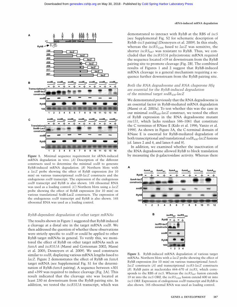
The sRNA RyhB pairs with its target mRNAs at the RBS,thereby blocking the initiation of translation (Morita et al.2006). Whereas the effect of RyhB pairing on translation iswell established, the subsequent steps leading to theinitiation of mRNA degradation still remain obscure.Thus, we sought to determine the minimal target mRNAsequence necessary for RyhB-induced mRNA degradation.We designed several transcriptional sodB-lacZ fusions (Fig.1A), with various lengths that spanned the sodB transcriptfrom nucleotide +130 to +607 (relative to the transcrip-tional +1). Each fusion was tested for RyhB sensitivity, asshown in Figure 1B. While the fusions of 430 nt and longerwere sensitive to RyhB expression, fusions of 400 nt andshorter demonstrated resistance to RyhB expression. Asa control, the endogenous sodB mRNA was monitoredunder the same conditions (Fig. 1B, second panel). Thesedata indicate that the region between +400 and +430 ofsodB mRNA encompasses a region that is required forRyhB-dependent mRNA degradation (Fig. 1A).
We then performed a similar experiment on transla-tional SodB-LacZ fusions. Contrary to transcriptionalfusions, in which both sodB and lacZ ORFs have in-dependent translation initiation starts, the correspondingtranslation fusions contained a single translation initiationsite (from sodB RBS) and produced single-hybrid SodB-LacZ peptides. These SodB-LacZ fusions were assayedunder the same conditions as described above for sensitiv-ity to RyhB. Results showed that each SodB-LacZ fusionassayed, from +130 to +607, was sensitive to RyhBexpression (Fig. 1C). Even though constructs shorter thansodB430 (sodB130, sodB289, and sodB400) did not harbor aputative cleavage site within sodB, they remained sensi-tive to RyhB-induced cleavage. These results suggest thepresence of additional cleavage sites downstream fromnucleotide 430 of sodB, most likely in the lacZ section ofthe fusion (see the Discussion for details).
Prevost et al.
386 GENES & DEVELOPMENT
Cold Spring Harbor Laboratory Press on May 30, 2018 - Published by genesdev.cshlp.orgDownloaded from
RyhB-dependent degradation of other target mRNAs
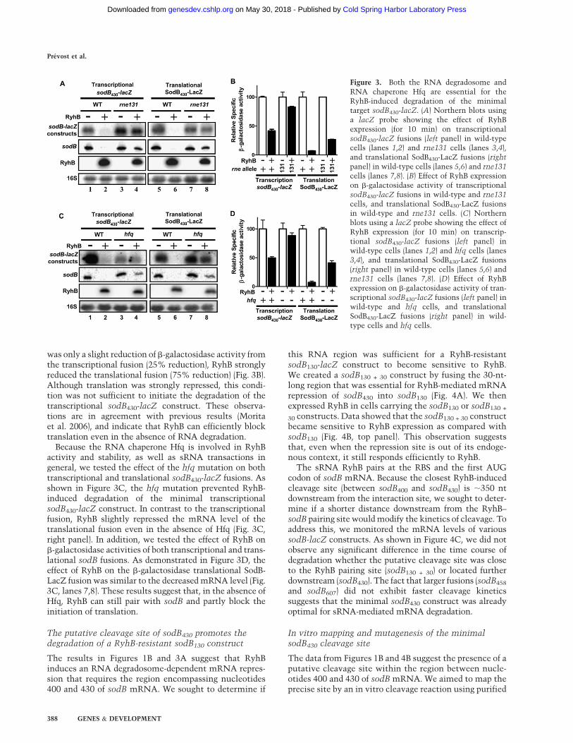
The results shown in Figure 1 suggested that RyhB induceda cleavage at a distal site in the target mRNA sodB. Wethen addressed the question of whether these observationswere strictly specific to sodB or could be applied to otherRyhB target mRNAs in general. To verify this, we moni-tored the effect of RyhB on other target mRNAs such asfumA and iscRSUA (Masse and Gottesman 2002; Masseet al. 2005; Desnoyers et al. 2009). We used constructssimilar to sodB, displaying various mRNA lengths fused tolacZ. Figure 2 demonstrates the effect of RyhB on fumAtarget mRNA (see Supplemental Fig. S1 for the determi-nation of RyhB–fumA pairing). A sequence between +301and +399 was required to induce cleavage (Fig. 2A). Thisresult indicated that the cleavage site was located atleast 230 nt downstream from the RyhB pairing site. Inaddition, we tested the iscRSUA transcript, which was
demonstrated to interact with RyhB at the RBS of iscS(see Supplemental Fig. S2 for schematic description ofRyhB–iscS pairing) (Desnoyers et al. 2009). In this study,whereas the iscRS1268 fused to lacZ was sensitive, theshorter iscRS687 was resistant to RyhB. Thus, we con-cluded that the iscRSUA polycistronic mRNA requiredthe sequence located >19 nt downstream from the RyhBpairing site to promote cleavage (Fig. 2B). The combinedresults of Figures 1 and 2 suggest that RyhB-inducedmRNA cleavage is a general mechanism requiring a se-quence further downstream from the RyhB pairing site.
Both the RNA degradosome and RNA chaperone Hfqare essential for the RyhB-induced degradationof the minimal target sodB430-lacZ
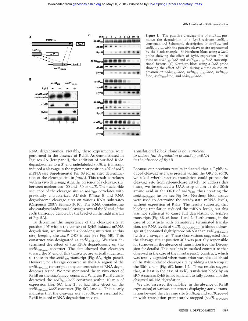
We demonstrated previously that the RNA degradosome isan essential factor in RyhB-mediated mRNA degradation(Masse et al. 2003a). To test whether this was the case inour minimal sodB430-lacZ construct, we tested the effectof RyhB expression in the RNA degradosome mutantrne131, which lacks residues 586–1061 that constitutethe C terminus of RNase E (Kido et al. 1996; Vanzo et al.1998). As shown in Figure 3A, the C-terminal domain ofRNase E is essential for RyhB-mediated degradation ofboth transcriptional and translational sodB430-lacZ fusions(cf. lanes 2 and 4, and lanes 6 and 8).
In addition, we examined whether the inactivation ofthe RNA degradosome allowed RyhB to block translationby measuring the b-galactosidase activity. Whereas thereFigure 1. Minimal sequence requirement for sRNA-induced
mRNA degradation in vivo. (A) Description of the differentconstructs used to determine the minimal sodB to generateRyhB-induced mRNA degradation. (B) Northern blots witha lacZ probe showing the effect of RyhB expression (for 10min) on various transcriptional sodB-lacZ constructs and theendogenous sodB transcript. The expression of the endogenoussodB transcript and RyhB is also shown. 16S ribosomal RNAwas used as a loading control. (C) Northern blots using a lacZ
probe showing the effect of RyhB expression (for 10 min) onvarious translational SodB-LacZ constructs. The expression ofthe endogenous sodB transcript and RyhB is also shown. 16Sribosomal RNA was used as a loading control.
Figure 2. RyhB-induced mRNA degradation of various targetmRNAs. Northern blots with a lacZ probe showing the effect ofRyhB expression (for 10 min) on various transcriptional fumA-lacZ constructs (A) and transcriptional iscRS-lacZ constructs(B). RyhB pairs at nucleotides 664–670 of iscRS, which corre-sponds to the RBS of iscS. Whereas the iscRS687 fusion extends19 nt into the iscS ORF, the iscRS1268 fusion extend 600 nt intoiscS ORF. Expression of endogenous sodB transcript and RyhB isalso shown. 16S ribosomal RNA was used as loading control.
sRNA-induced mRNA degradation
GENES & DEVELOPMENT 387
Cold Spring Harbor Laboratory Press on May 30, 2018 - Published by genesdev.cshlp.orgDownloaded from
was only a slight reduction of b-galactosidase activity fromthe transcriptional fusion (25% reduction), RyhB stronglyreduced the translational fusion (75% reduction) (Fig. 3B).Although translation was strongly repressed, this condi-tion was not sufficient to initiate the degradation of thetranscriptional sodB430-lacZ construct. These observa-tions are in agreement with previous results (Moritaet al. 2006), and indicate that RyhB can efficiently blocktranslation even in the absence of RNA degradation.
Because the RNA chaperone Hfq is involved in RyhBactivity and stability, as well as sRNA transactions ingeneral, we tested the effect of the hfq mutation on bothtranscriptional and translational sodB430-lacZ fusions. Asshown in Figure 3C, the hfq mutation prevented RyhB-induced degradation of the minimal transcriptionalsodB430-lacZ construct. In contrast to the transcriptionalfusion, RyhB slightly repressed the mRNA level of thetranslational fusion even in the absence of Hfq (Fig. 3C,right panel). In addition, we tested the effect of RyhB onb-galactosidase activities of both transcriptional and trans-lational sodB fusions. As demonstrated in Figure 3D, theeffect of RyhB on the b-galactosidase translational SodB-LacZ fusion was similar to the decreased mRNA level (Fig.3C, lanes 7,8). These results suggest that, in the absence ofHfq, RyhB can still pair with sodB and partly block theinitiation of translation.
The putative cleavage site of sodB430 promotes thedegradation of a RyhB-resistant sodB130 construct
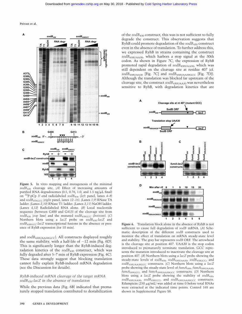
The results in Figures 1B and 3A suggest that RyhBinduces an RNA degradosome-dependent mRNA repres-sion that requires the region encompassing nucleotides400 and 430 of sodB mRNA. We sought to determine if
this RNA region was sufficient for a RyhB-resistantsodB130-lacZ construct to become sensitive to RyhB.We created a sodB130 + 30 construct by fusing the 30-nt-long region that was essential for RyhB-mediated mRNArepression of sodB430 into sodB130 (Fig. 4A). We thenexpressed RyhB in cells carrying the sodB130 or sodB130 +
30 constructs. Data showed that the sodB130 + 30 constructbecame sensitive to RyhB expression as compared withsodB130 (Fig. 4B, top panel). This observation suggeststhat, even when the repression site is out of its endoge-nous context, it still responds efficiently to RyhB.
The sRNA RyhB pairs at the RBS and the first AUGcodon of sodB mRNA. Because the closest RyhB-inducedcleavage site (between sodB400 and sodB430) is ;350 ntdownstream from the interaction site, we sought to deter-mine if a shorter distance downstream from the RyhB–sodB pairing site would modify the kinetics of cleavage. Toaddress this, we monitored the mRNA levels of varioussodB-lacZ constructs. As shown in Figure 4C, we did notobserve any significant difference in the time course ofdegradation whether the putative cleavage site was closeto the RyhB pairing site (sodB130 + 30) or located furtherdownstream (sodB430). The fact that larger fusions (sodB458
and sodB607) did not exhibit faster cleavage kineticssuggests that the minimal sodB430 construct was alreadyoptimal for sRNA-mediated mRNA degradation.
In vitro mapping and mutagenesis of the minimalsodB430 cleavage site
The data from Figures 1B and 4B suggest the presence of aputative cleavage site within the region between nucle-otides 400 and 430 of sodB mRNA. We aimed to map theprecise site by an in vitro cleavage reaction using purified
Figure 3. Both the RNA degradosome andRNA chaperone Hfq are essential for theRyhB-induced degradation of the minimaltarget sodB430-lacZ. (A) Northern blots usinga lacZ probe showing the effect of RyhBexpression (for 10 min) on transcriptionalsodB430-lacZ fusions (left panel) in wild-typecells (lanes 1,2) and rne131 cells (lanes 3,4),and translational SodB430-LacZ fusions (right
panel) in wild-type cells (lanes 5,6) and rne131cells (lanes 7,8). (B) Effect of RyhB expressionon b-galactosidase activity of transcriptionalsodB430-lacZ fusions in wild-type and rne131
cells, and translational SodB430-LacZ fusionsin wild-type and rne131 cells. (C) Northernblots using a lacZ probe showing the effect ofRyhB expression (for 10 min) on transcrip-tional sodB430-lacZ fusions (left panel) inwild-type cells (lanes 1,2) and hfq cells (lanes3,4), and translational SodB430-LacZ fusions(right panel) in wild-type cells (lanes 5,6) andrne131 cells (lanes 7,8). (D) Effect of RyhBexpression on b-galactosidase activity of tran-scriptional sodB430-lacZ fusions (left panel) inwild-type and hfq cells, and translationalSodB430-LacZ fusions (right panel) in wild-type cells and hfq cells.
Prevost et al.
388 GENES & DEVELOPMENT
Cold Spring Harbor Laboratory Press on May 30, 2018 - Published by genesdev.cshlp.orgDownloaded from
RNA degradosomes. Notably, these experiments wereperformed in the absence of RyhB. As demonstrated inFigures 5A (left panel), the addition of purified RNAdegradosomes to a 39-end radiolabeled sodB458 transcriptinduced a cleavage in the region near position 407 of sodBmRNA (see Supplemental Fig. S3 for in vitro determina-tion of the cleavage site in fumA). This result correlateswith in vivo data suggesting the presence of a cleavage sitebetween nucleotides 400 and 430 of sodB. The nucleotidesequence of the cleavage site at sodB407 correlates withpreviously characterized AU-rich RNase E and RNAdegradosome cleavage sites on various RNA substrates(Carpousis 2007; Belasco 2010). The RNA degradosomealso catalyzed additional cleavages toward the 59 end of thesodB transcript (denoted by the bracket in the right marginof Fig. 5A).
To determine the importance of the cleavage site atposition 407 within the context of RyhB-induced mRNAdegradation, we introduced a 9-nt-long mutation at thissite, keeping the sodB ORF intact (see Fig. 5B). Thisconstruct was designated as sodB430GCC. We then de-termined the effect of the RNA degradosome on thesodB430GCC construct. The data showed that cleavagestoward the 59 end of this transcript are virtually identicalto those in the sodB430 transcript (Fig. 5A, right panel).However, no cleavage occurred in the 407 region of thesodB430GCC transcript at any concentration of RNA degra-dosomes tested. We next monitored the in vivo effect ofRyhB on the sodB430GCC construct. Whereas RyhB clearlydestroyed the sodB430-lacZ construct within 10 min ofexpression (Fig. 5C, lane 2), it had little effect on thesodB430GCC-lacZ construct (Fig. 5C, lane 4). This clearlyindicates that the cleavage site at sodB407 is essential forRyhB-induced mRNA degradation in vivo.
Translational block alone is not sufficientto induce full degradation of sodB430 mRNAin the absence of RyhB
Because our previous results indicated that a RyhB-in-duced cleavage site was present within the ORF of sodB,we asked whether active translation could protect thecleavage site from ribonuclease attack. To address thisissue, we introduced a UAA stop codon at the 30thamino acid in the ORF of sodB430, thus creating thesodB430UAA30 fusion (see Fig 6A). Northern blots assayswere used to determine the steady-state mRNA levels,without expression of RyhB. The results suggested thatblocking translation reduced the mRNA levels, but thiswas not sufficient to cause full degradation of sodB430
transcripts (Fig. 6B, cf. lanes 1 and 2). Furthermore, in thecase of constructs with prematurely terminated transla-tion, the RNA levels of sodB430UAA30GCC (without a cleav-age site) contained slightly more mRNA than sodB430UAA30
(with a cleavage site). These observations suggested thatthe cleavage site at position 407 was partially responsiblefor turnover in the absence of translation (see the Discus-sion for details). This result is in marked contrast to thatobserved in the case of the fumA399-lacZ construct, whichwas totally degraded when translation was blocked aheadof the RyhB-induced cleavage site by adding a UAA stop atthe 30th codon (Fig. 6C, lanes 1,2). These results suggestthat, at least in the case of sodB, translation block by ansRNA such as RyhB is not sufficient to fully account for theobserved mRNA degradation.
We also assessed the half-life (in the absence of RyhBexpression) of various constructs displaying active trans-lation beyond the cleavage site (sodB430 and sodB430GCC)or with translation prematurely stopped (sodB430UAA30
Figure 4. The putative cleavage site of sodB430 pro-motes the degradation of a RyhB-resistant sodB130
construct. (A) Schematic description of sodB430 andsodB130 + 30, with the putative cleavage site representedby the black triangle. (B) Northern blots using a lacZ
probe showing the effect of RyhB expression (for 10min) on sodB130-lacZ and sodB130 + 30-lacZ transcrip-tional fusions. (C) Northern blots using a lacZ probeshowing the effect of RyhB during a time-course ex-pression on sodB130-lacZ, sodB130 + 30-lacZ, sodB430-lacZ, sodB458-lacZ, and sodB607-lacZ.
sRNA-induced mRNA degradation
GENES & DEVELOPMENT 389
Cold Spring Harbor Laboratory Press on May 30, 2018 - Published by genesdev.cshlp.orgDownloaded from
and sodB430UAA30GCC). All constructs displayed roughlythe same stability, with a half-life of ;12 min (Fig. 6D).This is significantly longer than the RyhB-induced deg-radation kinetics of the sodB430 construct, which wasfully degraded after 5–7 min of RyhB expression (Fig. 4C).These data strongly suggest that blocking translationcannot fully explain RyhB-induced mRNA degradation(see the Discussion for details).
RyhB-induced mRNA cleavage of the target mRNAsodB430-lacZ in the absence of translation
While the previous data (Fig. 6B) indicated that prema-turely stopped translation contributed to destabilization
of the sodB430 construct, this was is not sufficient to fullydegrade the construct. This observation suggests thatRyhB could promote degradation of the sodB430 constructeven in the absence of translation. To further address this,we expressed RyhB in strains containing the constructsodB430UAA30, which harbors a stop signal at the 30thcodon. As shown in Figure 7C, the expression of RyhBpromoted rapid degradation of sodB430UAA30, which wasstill dependent on the cleavage site at residue 407 (cf.sodB430UAA30 [Fig. 7C] and sodB430UAA30GCC [Fig. 7D]).Although the translation was blocked far upstream of thecleavage site, the construct sodB430UAA30 was neverthelesssensitive to RyhB, with degradation kinetics that are
Figure 5. In vitro mapping and mutagenesis of the minimalsodB430 cleavage site. (A) Effect of increasing amounts ofpurified RNA degradosomes (0.5, 0.76, 1.0, and 1.5 ng/mL final)on 32P-pCp 39-end radiolabeled sodB458 (left panel, lanes 4–8)and sodB458GCC (right panel, lanes 12–16). (Lanes 1,9) RNase TAladder. (Lanes 2,10) RNase T1 ladder. (Lanes 3,11) NaOH ladder.(Lanes 4,12) Radiolabeled RNA alone. (B) Local nucleotidesequence (between G400 and G413) of the cleavage site fromsodB430 (top line) and the mutated sodB430GCC (bottom). (C)Northern blots using a lacZ probe on sodB430-lacZ andsodB430GCC-lacZ transcriptional fusions in the absence or pres-ence of RyhB expression (for 10 min).
Figure 6. Translation block alone in the absence of RyhB is notsufficient to cause full degradation of sodB mRNA. (A) Sche-matic description of the different sodB constructs used tomonitor the effect of translation on mRNA steady-state leveland stability. The gray bar represents sodB ORF. The arrowheadis the cleavage site at position 407. UAA30 is the stop codonintroduced to prematurely terminate translation. GCC repre-sents the mutation introduced to inactivate the cleavage site atposition 407. (B) Northern blots using a lacZ probe showing thesteady-state levels of sodB430, sodB430UAA30, sodB430GCC, andsodB430UAA30GCC constructs. (C) Northern blots using a lacZ
probe showing the steady-state level of fumA399, fumA399UAA30,fumA399GCC, and fumA399UAA30GCC constructs. (D) Northernblots using a lacZ probe showing the stability of sodB430,sodB430UAA30, sodB430GCC, and sodB430UAA30GCC constructs.Rifampicin (250 mg/mL) was added at time 0 before total RNAswere extracted at the indicated time points. Control 16S areshown in Supplemental Figure S8.
Prevost et al.
390 GENES & DEVELOPMENT
Cold Spring Harbor Laboratory Press on May 30, 2018 - Published by genesdev.cshlp.orgDownloaded from

similar to those of the wild-type sodB430 construct (half-life#7.5 min) (Fig. 7A) and the endogenous sodB mRNA.These rapid degradation kinetics of the sodB430UAA30
construct by RyhB suggest an active mechanism of degra-dation as compared with passive degradation. Indeed, whenwe monitored the effect of RyhB on sodB430UAA30 inrne131 cells (Fig. 7E), the transcript was not degraded.These data demonstrate that both the sRNA RyhB and theRNA degradosome promote mRNA cleavage in the ab-sence of translation elongation at the cleavage site.
Discussion
Our work unveils key determinants in the mechanism ofsRNA-induced mRNA degradation. First, we demonstratethat pairing of the sRNA promotes the cleavage at a distalsite, sometimes hundreds of nucleotides downstreamwithin the target mRNA. Second, prematurely terminatedtranslation of a target mRNA is not sufficient to induce thefull degradation of a target mRNA. Third, the sRNA-induced cleavage can initiate even in the absence of
translation on the target mRNA. We propose a model(Fig. 8) in which RyhB and Hfq act first on the target mRNAby competing with 30S ribosome subunits and blockingthe initiation of translation. The following steps includethe recruitment of the RNA degradosome and cleavage at thedistal site deep within the ORF. Surprisingly, the cleavagesite is located as far as 350 nt downstream from the RyhBpairing site. This suggests a mechanism that allows trans-lating ribosomes to clear off the ORF before the RNAdegradosome proceeds to cleave the mRNA (see the modelin Fig. 8). In this way, the mRNA cleavage happens afterthe passage of elongating ribosomes, thereby preventingaccumulation of cleaved transcripts harboring stalled ribo-somes. Such a mechanism seems widely employed, sincewe describe three different target mRNAs (sodB, fumA,and iscRSUA) where RyhB induces distal mRNA degrada-tion (Figs. 1, 2). We assume that a significant fraction ofsRNAs that binds at the RBS of their target mRNAs willfunction similarly to RyhB.
In fact, a large number of Hfq-binding sRNAs such asMicA (Rasmussen et al. 2005; Udekwu et al. 2005), SgrS(Morita et al. 2005), RybB (Papenfort et al. 2006), OmrA,and OmrB (Guillier and Gottesman 2008) bind to the RBSof their specific target mRNAs to repress translation anddecrease the mRNA level. We think that these sRNAsshare, at least in part, the same mechanism as the onedescribed here for RyhB. The effects of RyhB-inducedtarget mRNA degradation have been widely investigated
Figure 7. RyhB-induced mRNA cleavage in prior absence oftranslation on the target mRNA sodB430-lacZ. Northern blotsusing a lacZ probe showing the effect of RyhB expression inwild-type cells on sodB430 (A), sodB430GCC (B), sodB430UAA30 (C),and sodB430UAA30GCC (D). (E) Northern blots using a lacZ probeshowing the effect of RyhB expression on sodB430UAA30 inrne131 cells.
Figure 8. Model for sRNA-induced mRNA degradation (see thetext for details).
sRNA-induced mRNA degradation
GENES & DEVELOPMENT 391
Cold Spring Harbor Laboratory Press on May 30, 2018 - Published by genesdev.cshlp.orgDownloaded from

(Masse et al. 2003a, 2005; Geissmann and Touati 2004;Afonyushkin et al. 2005; Morita et al. 2005; Desnoyerset al. 2009). However, none of these studies have in-vestigated whether RyhB sRNA acts solely by blockingtranslation initiation, thereby inducing mRNA degradation(passive degradation model), or whether RyhB promotesthe cleavage and degradation of the target mRNA indepen-dently of translation (Fig. 8, active cleavage model).
The distant location of downstream cleavage sites maycontribute to optimal sRNA-induced mRNA decay. In-deed, when RyhB pairs with a target mRNA and blockstranslation initiation, the last translating ribosome canreach and pass the cleavage site before the degradosomeinitiates mRNA cleavage. Once the mRNA is cleaved,the resulting generation of a 59-P (monophosphorylated)end accelerates by a 30-fold factor the RNase E activitytoward the 39 end of the transcript (Mackie 1998; Jiangand Belasco 2004; Garrey et al. 2009). Consequently, thecleaved mRNA will be fully degraded more rapidly. Whenthe mRNA is cleaved once, the following rapid RNaseE cleavages, concomitantly with exonuclease action,fully destroy the remaining RNA (Carpousis 2007; Belasco2010). In this context, if RNase E cleavage sites are locatedupstream of the ORF, the activated RNase E could cleavewithin the ORF before ribosomes have completed trans-lation, thus trapping ribosomes in a prematurely termi-nated ORF. If this were the case, the tmRNA (SsrA) wouldlikely help such a stalled ribosome (Keiler et al. 1996). Incontrast, a distal cleavage site may prevent RNase Ecleavage before ribosomes have cleared the upstream partof the mRNA.
We show here that, even with a prematurely termi-nated translation of the target mRNA (Fig. 7C, constructsodB430UAA30), the sRNA RyhB promotes an RNA degra-dosome-dependent cleavage. A similar result was demon-strated previously for the sRNA SgrS and the target mRNAptsG (Morita et al. 2006). However, in the previous study,Morita et al. (2006) used the antibiotic kasugamycin toblock cell-wide translation and then express the sRNASgrS. This is in contrast with our results, where a stopcodon was inserted in the ORF of sodB mRNA, thusspecifically blocking sodB translation. The UAA stopcodon was used because it is the natural stop codon ofthe sodB ORF as well as being the most abundant andstrongest termination signal in E. coli (Poole et al. 1995;Crawford et al. 1999). By terminating translation at the30th codon, we prevented any bias in the sequence andstructure of sodB mRNA at both the RyhB pairing site (atthe RBS) and the cleavage site at position 407. A point ofconcern would be the possibility that the AUU codon atthe 30th amino acid of sodB430UAA30 was leaky. If this werethe case, then ribosomes reading through the stop codonwould contribute to the stabilization of the mRNA. Toverify this possibility, we introduced a stop codon followedby a frameshift, which would fully prevent any ribosomeread-through. As shown in Supplemental Figure S4, weobserved the same results as presented in Figure 6 with thesingle AUU stop at the 30th codon. Thus, both of the sodBconstructs with blocked translation were most likely freeof ribosomes at the position 407. Furthermore, even though
translation was terminated upstream, the cleavage site atposition 407 of sodB was still critical for efficient RyhB-induced degradation (Fig. 7D).
An argument suggesting the importance of the cleavagesite at residue 407 was the significant effect that inactivat-ing the sodB407 cleavage site had on the steady-state levelof sodB, whether the transcript was translated (cf. sodB430
and sodB430GCC in Fig. 6B) or not (cf. sodB430UAA30 andsodB430UAA30GCC in Fig. 6B). Nonetheless, in the absenceof translation, the sodB430UAA30 transcript was present at asignificant level, which was in contrast to the fully de-graded fumA399UAA construct (Fig. 6C). One reasonexplaining why the untranslated sodB430UAA30 constructwas not fully degraded is the presence of secondarystructures in the RNA in the vicinity of residue 407(Supplemental Fig. S5). Because RNase E requires ssRNA,the presence of secondary structures may hinder theribonuclease attack. To test this, we performed in vitroRNA degradosome cleavage using an oligonucleotidelocated 5 nt downstream from the cleavage site to removethe RNA structure. The presence of the oligonucleotideclearly improved the specific cleavage at position 407(Supplemental Fig. S5), which suggests that local RNAstructures hinder the action of the RNA degradosome.Thus, the presence of inhibitory RNA structures in thevicinity of position 407 in sodB may explain why this sitewas not fully attacked in the absence of translation ascompared with fumA.
Surprisingly, while the steady-state levels of the con-structs varied significantly (Fig. 6B), they all displayed thesame stability (Fig. 6D). To explain this, we suggest thatthe construct with prematurely terminated translation(sodB430UAA30) comprised two distinct populations oftranscripts: a first population of transcripts with a rapidturnover (<1 min), and a second population that is morestable. The rapid decay of the first population of tran-scripts likely depends on cleavage at nucleotide 407, sincethe construct sodB430UAA30GCC displayed a steady-statelevel similar to that of the wild-type sodB430. Thissuggests that, when translation is prematurely stopped(sodB430UAA30), the cleavage site at nucleotide 407 isexposed and cleavage occurs rapidly.
While the group of short transcriptional sodB-lacZ fu-sions (sodB130, sodB289, and sodB400) displayed resistanceto RyhB (Fig. 1B), the equivalent SodB-LacZ translationalfusions were all RyhB-sensitive. These results suggest thatthe RyhB-induced cleavage within SodB-LacZ translationalfusions occurs in the lacZ gene regardless of the sodBlength. Thus, these data indicate that the sRNA pairs to anRNA sequence and promotes cleavage within a foreigngene other than the original target mRNA. In addition, it isvery likely that the sRNA-induced translation block aloneis sufficient to destabilize the lacZ transcript, as observedpreviously (Iost and Dreyfus 1995).
Earlier studies with sodB-lacZ fusions suggested thatRyhB triggered degradation of sodB within the regionupstream of nucleotide 192 (Geissmann and Touati2004). Our data are in disagreement with this observation.We explain these previous results by the use of a lacZtranscriptional fusion that contained a potential RNase
Prevost et al.
392 GENES & DEVELOPMENT
Cold Spring Harbor Laboratory Press on May 30, 2018 - Published by genesdev.cshlp.orgDownloaded from

E-dependent cleavage site in the linker region betweenthe sodB and lacZ mRNAs. Here, the use of a shorterlinker (pFRD construct) that did not contain an RNaseE-dependent cleavage provided a tool to focus on thesequence in sodB mRNA.
In another study performed by a different group,Afonyushkin et al. (2005) attempted to reproduce RyhB-induced mRNA degradation in vitro. They used a sodBtranscript identical to the transcript used by Geissmannand Touati (2004) (sodB192) to characterize a RyhB-inducedcleavage site at nucleotide +67, which is just 6 nt down-stream from the RyhB pairing site. These in vitro resultsdo not corroborate our findings. Whereas we also detectcleavage within the 59 end of the sodB458 substrate (Fig. 5A,in vitro-specific cleavages), we did not consider themsignificant, as these cleavages did not correlate with theRyhB-induced in vivo cleavage. Our results indicate thatcleavages in the 59 end of sodB are not significant in vivo,at least within the context of RyhB-induced cleavage. Totest whether RyhB was pairing at position 407 to inducelocal cleavage, we tested in vitro pairing between sodB andRyhB. The results clearly showed (Supplemental Fig. S6)that, whereas RyhB bound to the previously known pair-ing site (Supplemental Fig. S6, left panel), there was noevidence suggesting RyhB pairing at position 407 (Supple-mental Fig. S6, right panel).
Another possible reason explaining the RyhB-inducedcleavage of sodB192 in vitro (Afonyushkin et al. 2005) is theuse of a 59-monophosphate-labeled target mRNA, whichdiffers from the in vivo 59-triphosphate substrate. Asmentioned above, 59-monophosphate substrate has beenshown to promote significantly RNase E-dependent cleav-age (Mackie 1998, 2000). In contrast to this, the in vitromRNA substrates used here were labeled at their 39 end(pCp) (see the Materials and Methods), which keeps the59-triphosphate intact and prevents degradation by the39–59 PNPase of the RNA degradosome. Thus, we believethat this type of in vitro target mRNA probably reproducesmore closely the in vivo substrate that is cleaved throughRNase E (Masse et al. 2003a). To confirm the cleavage siteat sodB407, we performed one additional in vitro approach.We incubated a T7 RNAp-generated sodB458 transcriptwith purified RNA degradosome followed by primerextension, which stops at any RNase E-generated cleavage.As shown in Supplemental Figure S7, whereas the absenceof RNA degradosome prevented any cleavage, incubationof sodB458 in the presence of the RNA degradosomeresulted in a major cleavage at the sodB407 site. Althoughthis approach still did not prevent in vitro-specific cleavagein the upstream region of the mRNA, it confirmed our dataon the specific cleavage at sodB407.
Whereas RyhB and all other previously mentionedsRNAs bind on their targets at or close to the RBS, a recentstudy has provided evidence that pairing at the RBS wasnot a universal step in the mechanism of sRNA-dependentmRNA silencing. Indeed, the sRNA MicC in Salmonellatyphimurium induces an RNase E-dependent mRNAcleavage in the ompD mRNA even though the sRNApairs within the ORF, far downstream from the RBS(Pfeiffer et al. 2009). Hfq binds in vitro to the target ompD
close to MicC pairing within the ORF. This is similar toa previous study where the RNA chaperone Hfq wasshown to bind close to the sRNA pairing site to melt themRNA structure and promote sRNA pairing (Geissmannand Touati 2004). In contrast to previously describedsRNA systems, MicC does not alter translation of thetarget (Pfeiffer et al. 2009). These results may be explainedby the low translation rate of the target mRNA ompD,which would allow the sRNA MicC to pair at the ORFwithout competing with ribosomes.
Our study uncovered many critical determinants ofsRNA-induced mRNA degradation. However, additionalsteps have to be elucidated to reproduce the completereaction in vitro. Additional factors such as protein com-ponents or RNA structures still need to be characterized toprovide a comprehensive view of the whole process. Thisis not surprising, given the complexity of the system. Amajor question that remains to be addressed is whether thesRNA must block translation in order to induce mRNAdegradation. This question is of importance in view of therecent description of the sRNA MicC, which promotesdegradation of ompC mRNA without blocking its trans-lation (Pfeiffer et al. 2009). Further work is needed tounravel the relationship between the activity of sRNAsand the translation activity of their target mRNAs. Studiesalong these lines of research are currently ongoing in ourlaboratory.
Materials and methods
Bacterial strains and plasmids
Derivatives of E. coli MG1655 were used in all experiments. TheDH5a bacterial strain was used for routine cloning procedures.Strains constructed by P1 transduction were selected for theappropriate antibiotic-resistant marker. Except as otherwise in-dicated, for cells carrying pFRD, pRS1551, pNM12, and pBAD-ryhB, ampicillin was used at a final concentration of 50 mg/mL.See Supplemental Tables S1 and S2 for a complete description ofstrains and oligonucleotides used in this study.
For the construction of sodB-lacZ transcriptional and trans-lational fusions, a PCR fragment made with the oligonucleotidesEM423 (forward for all sodB constructions), EM424 (sodB130),EM516 (sodB289), EM515 (sodB400), EM527 (sodB430), EM528(sodB458), EM514 (sodB517), and EM511 (sodB607) was digestedby EcoRI and BamHI and ligated into EcoRI/BamHI-digested pFRD
(for transcriptional fusions) and EcoRI/BamHI-digested pRS1551(for in-frame translational fusions). The vector pFRD is a derivativeof the original transcriptional vector, pRS1553 (Simons et al.1987), which harbors a transcriptional terminator in the linkerbetween the cloned gene and the lacZ reporter gene (Repoila andGottesman 2001). All of the transcriptional fusions were con-structed from the pFRD vector (without transcriptional terminator)and were in-frame with a TAA translation stop (gatccGGCATTTTAA), which was located 26 nt upstream of the RBS of lacZ.
To generate sodB130 + 30, a PCR product of 30 nt was madewith oligonucleotides EM625–EM626, then digested withBamHI and ligated into BamHI-digested pFRD-sodB130. To pro-duce sodB430 modifications, two independent PCR reactionswere performed using the sodB430 or sodB430GCC (to constructsodB430UAA30GCC) fusions as a template with the oligonucleo-tides sodB430UAA30 (EM1050–EM195 and EM194–EM1051) andsodB430GCC (EM630–EM195 and EM194–EM631). The two PCR
sRNA-induced mRNA degradation
GENES & DEVELOPMENT 393
Cold Spring Harbor Laboratory Press on May 30, 2018 - Published by genesdev.cshlp.orgDownloaded from

products were then mixed to serve as the template for a third PCR(EM194–EM195, oligonucleotides in the pFRD). The resultingPCR product was then digested by BamHI and EcoRI and ligatedinto EcoRI/BamHI-digested pFRD (for transcriptional fusions).
For the construction of fumA-lacZ transcriptional fusions,a PCR fragment made with the following oligonucleotides:EM533 (forward for all fumA constructions), EM534 (fumA126),EM560 (fumA301), EM559 (fumA399), EM558 (fumA507), EM556(fumA703), and EM555 (fumA801). A PCR fragment for the iscRS-
lacZ fusion was made with EM359 (forward for iscRS construc-tions), EM629 (iscRS687), and EM587 (iscRS1268). These PCRproducts were digested by EcoRI and BamHI and ligated intoEcoRI/BamHI-digested pFRD. To generate fumA399 modifications,two independent PCR reactions were performed using thefumA399 or fumA399GCC (to construct fumA399UAA30GCC) fusionas template with the following oligonucleotides: fumA399UAA30
(EM1028–EM195 and EM194–EM1029) and fumA399GCC
(EM1114–EM195 and EM194–EM1115). The two PCR productswere then mixed to serve as the template for a third PCR (EM194–EM195, oligonucleotides in the pFRD sequence). The resultingPCR product was then digested by BamHI and EcoRI and ligatedinto EcoRI/BamHI-digested pFRD (for transcriptional fusions).
The transcriptional and translational fusions were deliveredin a single copy into the bacterial chromosome of differentstrains—EM1455 (Dara714 leu+ DryhBTcat) or JF133 (rne131
zce-726TTn10 DryhBTcat)—at the l att site as described pre-viously (Simons et al. 1987). Stable lysogens were screened forsingle insertion of recombinant l by PCR (Powell et al. 1994).
E. coli strain KP604 [BL21 (DE3) pLysS/pET21b-rne-Flag] wasused for purification of RNase E-Flag degradosomes. The plas-mid pET21b-rne-Flag was constructed by PCR amplification ofGM402 (harboring pGM102 with the complete rne gene; a giftfrom George Mackie) with primers EM476 and EM478, and thendigested with NdeI–XhoI. The resulting fragment containing therne gene was then inserted into pET21b digested with NdeI–XhoI.
Purification of RNase E-Flag degradosomes
RNase E-Flag degradosomes were prepared as described pre-viously (Regonesi et al. 2006), with some modifications. E. coli.KP604 (BL21 [DE3] pLysS/pET21b-rne-Flag) culture was grown in500 mL of Luria-Bertani (LB) broth with ampicillin (50 mg/mL)and chloramphenicol (30 mg/mL) at 30°C until it reached anOD600 value of 0.5–0.6. Expression of RNase E-Flag was inducedwith 1mM IPTG for 3 h at 30°C.
RNA extraction and Northern blot analysis
Cells were grown at 37°C on LB medium and total RNA wasextracted using the hot-phenol procedure (Aiba et al. 1981).Arabinose (0.1%) was added when indicated. Half-life determina-tion of RNA was performed by addition of 250 mg/mL rifampicin.After total RNA extraction, 5 mg of total RNA was loaded ona polyacrylamide gel (5%–10% acrylamide 29:1, 8 M urea) and20–30 mg was loaded on an agarose gel (1%, 13 MOPS). Aftermigration, the RNA was transferred to a Hybond-XL membrane(Amersham Biosciences) or a Biodyne B membrane (Pall) andcross-linked by UV (1200 J). The membrane was prehybridizedwith 50% formamide, 53 SSC, 53 Denhardt’s reagent, 1% SDS,and 100 mg/mL sheared salmon sperm DNA for 4 h at 60°C. Then,the radiolabeled RNA probe was added directly in the prehybrid-ization buffer with the membrane and incubated 16 h at 60°C.Before exposure to a phosphor screen, the membrane was washedthree times with 13 SSC/0.1% SDS and once with 0.13 SSC/0.1% SDS. The phosphor screen was analyzed on a Storm 860(Molecular Dynamics) or a Typhoon Trio (GE Healthcare), and
quantification was performed using the ImageQuant software(Molecular Dynamics).
Internally radiolabeled RNAs generated by in vitroRNA synthesis
The radiolabeled probes used for Northern blot analysis weretranscribed with a purified T7 RNA polymerase from a PCRproduct to generate the antisense transcript of the gene of interest.Transcription was performed in T7 transcription buffer (80 mMHEPES-KOH at pH 7.5, 24 mM MgCl2, 40 mM DTT, 2 mMspermidine), 400 mM NTPs (A, C, and G), 10 mM UTP, 3 mL ofa-32P-UTP (3000 Ci/mmol), 20 U of RNase OUT (Invitrogen), 5 mgof T7 RNA polymerase, and 1 mg of DNA template. After 4 h ofincubation at 37°C, the mixture was treated with 2 U of TurboDNase (Ambion) and extracted once with phenol-chloroform.Nonincorporated nucleotides were removed with a G-50 Sephadexcolumn. The primers used for generating DNA templates for invitro RNA synthesis were EM470–EM471 (lacZ), EM188–EM189(sodB), EM190–EM191 (ryhB), and EM293–EM294 (16S rRNA).
RNAs generated by in vitro RNA synthesis
Unlabeled RNAs were transcribed in vitro with purified T7 RNApolymerase from a PCR product. Transcription was performed inT7 transcription buffer (80 mM HEPES-KOH at pH 7.5, 24 mMMgCl2, 40 mM DTT, 2 mM spermidine), 500 mM NTPs (A, C, Gand U), 40 U of RNase OUT (Invitrogen), 2 U of inorganicpyrophosphatase (Roche), 10 mg of T7 RNA polymerase, and 2mg of DNA template. After 4 h of incubation at 37°C, the mixturewas treated with 2 U of Turbo DNase (Ambion), and the RNA wasextracted once with phenol-chloroform and precipitated withisopropanol. The RNA products were purified from a denaturing6% polyacrylamide, 8 M urea gel and precipitated before use. Theprimers used for generating DNA templates for in vitro RNAsynthesis were EM90–EM528 (sodB458), and the primers weredigested with BamHI. To produce sodB458GCC, two independentPCR reactions were performed using the sodB458 DNA as tem-plate with the following oligonucleotides: EM630–EM528 andEM90–EM631. The two PCR products were then mixed to serveas the template for a third PCR (EM90–EM528). The resultingPCR product was then digested by BamHI.
39-end labeling of RNA
For the 39-end labeling of RNA with pCp, we transcribed RNAwith T7 RNA polymerase from a PCR product as describedabove. Then, 60 pmol of in vitro transcribed RNA was mixed with10% DMSO, 20 U of RNase OUT (Invitrogen), 1 mM ATP(Fermentas), 20 U of T4 RNA Ligase (Fermentas), 13 T4 RNALigase buffer (Fermentas), and 4 mL of cytidine 39, 59-bis(phos-phate) (59-32P) at 3000 Ci/mmol in a final volume of 40 mL. Themixture was incubated for 60 min at 37°C. The reaction wasstopped by addition of 1 vol of loading buffer II (95% formamide,18 mM EDTA, 0.025% SDS, xylene cyanol, bromophenol blue)(Ambion). The labeled RNA was then purified from a denaturing6% polyacrylamide, 8 M urea gel and precipitated before use.
RNA degradosome degradation assay
To determine the RNase E cleavage sites on an RNA transcript, 5nM 39end-labeled sodB and 0.0066 mg/mL tRNAs were mixed andheated for 2 min at 90°C. The mixture was slowly cooled untilit reached 37°C before adding the degradation buffer (13.3 mMTris-Cl at pH 7.5, 0.33 mM DTT, 73.34 mM NH4Cl, 3.33 mMmagnesium acetate, 0.1 mM EDTA, 0.7% glycerol), followed by
Prevost et al.
394 GENES & DEVELOPMENT
Cold Spring Harbor Laboratory Press on May 30, 2018 - Published by genesdev.cshlp.orgDownloaded from

incubation for 40 min at 37°C. Then, purified RNase E-Flagdegradosomes (0.5, 0.76, 1.0, and 1.5 ng/mL final) or RNAdegradosome buffer only (5 mM Tris-Cl at pH 7.5, 50% glycerol,75 mM NaCl) was added, followed by incubation for 30 min at37°C. The reaction was stopped with 1 vol of phenol, and sampleswere separated on an 8% polyacrylamide, 8 M urea gel.
Ribonuclease-generated RNA ladder
Ribonucleases T1 (0.1 U) (Ambion) and TA (0.5 U) (Jena Bio-sciences) were used for 5 min and 0.5 min, respectively, at 37°C inthe sequence buffer (Ambion). The alkaline ladder was performedin the alkaline buffer (Ambion) for 5 min at 90°C.
b-Galactosidase assays
Kinetic assays for b-galactosidase activity were performed asdescribed previously (Prevost et al. 2007) using a SpectraMax 250microtitre plate reader (Molecular Devices). Briefly, overnightbacterial cultures were incubated at 37°C in LB medium withampicillin at a final concentration of 50 mg/mL and diluted 1000-fold into 50 mL of fresh LB medium with ampicillin at 37°C.Cultures were grown with agitation to an OD600 of 0.1 beforeinducing RyhB expression by adding arabinose to a final concen-tration of 0.1% (strains carrying pBAD-ryhB or the control vectorpNM12). Specific b-galactosidase activities (around OD600 of 0.8and 1.0) were calculated using the formula Vmax/OD600. Thereported results represent data from at least three independentexperiments.
Acknowledgments
We thank George Mackie, Francxois Bachand, Sherif Abou-Elela,Daniel Lafontaine, Frederieke Brouwers, and Gilles Dupuis forhelpful suggestions throughout the course of this work. This workwas funded by operating grant MOP69005 to E.M. from theCanadian Institutes for Health Research (CIHR). G.D. is a PhDscholar from the FQRNT (Fonds Quebecois de la Recherche sur laNature et les Technologies). E.M. is a CIHR New Investigator andFRSQ (Fonds de la Recherche en Sante du Quebec) Junior II scholar.
References
Afonyushkin T, Vecerek B, Moll I, Blasi U, Kaberdin VR. 2005.Both RNase E and RNase III control the stability of sodBmRNA upon translational inhibition by the small regulatoryRNA RyhB. Nucleic Acids Res 33: 1678–1689.
Aiba H, Adhya S, de Crombrugghe B. 1981. Evidence for twofunctional gal promoters in intact Escherichia coli cells. J Biol
Chem 256: 11905–11910.Belasco JG. 2010. All things must pass: Contrasts and common-
alities in eukaryotic and bacterial mRNA decay. Nat RevMol Cell Biol 11: 467–478.
Carpousis AJ. 2007. The RNA degradosome of Escherichia coli:An mRNA-degrading machine assembled on RNase E. Annu
Rev Microbiol 61: 71–87.Carpousis AJ, Luisi BF, McDowall KJ. 2009. Endonucleolytic
initiation of mRNA decay in Escherichia coli. Prog Mol Biol
Transl Sci 85: 91–135.Celesnik H, Deana A, Belasco JG. 2007. Initiation of RNA decay
in Escherichia coli by 59 pyrophosphate removal. Mol Cell
27: 79–90.Coburn GA, Mackie GA. 1999. Degradation of mRNA in
Escherichia coli: An old problem with some new twists.Prog Nucleic Acid Res Mol Biol 62: 55–108.
Crawford DJ, Ito K, Nakamura Y, Tate WP. 1999. Indirect regula-tion of translational termination efficiency at highly expressedgenes and recoding sites by the factor recycling function ofEscherichia coli release factor RF3. EMBO J 18: 727–732.
Deana A, Celesnik H, Belasco JG. 2008. The bacterial enzymeRppH triggers messenger RNA degradation by 59 pyrophos-phate removal. Nature 451: 355–358.
Desnoyers G, Morissette A, Prevost K, Masse E. 2009. SmallRNA-induced differential degradation of the polycistronicmRNA iscRSUA. EMBO J 28: 1551–1561.
Garrey SM, Blech M, Riffell JL, Hankins JS, Stickney LM, DiverM, Hsu YH, Kunanithy V, Mackie GA. 2009. Substratebinding and active site residues in RNases E and G: Role ofthe 59-sensor. J Biol Chem 284: 31843–31850.
Geissmann TA, Touati D. 2004. Hfq, a new chaperoning role:Binding to messenger RNA determines access for small RNAregulator. EMBO J 23: 396–405.
Guillier M, Gottesman S. 2008. The 59 end of two redundantsRNAs is involved in the regulation of multiple targets, in-cluding their own regulator. Nucleic Acids Res 36: 6781–6794.
Iost I, Dreyfus M. 1995. The stability of Escherichia coli lacZ
mRNA depends upon the simultaneity of its synthesis andtranslation. EMBO J 14: 3252–3261.
Jiang X, Belasco JG. 2004. Catalytic activation of multimericRNase E and RNase G by 59-monophosphorylated RNA. Proc
Natl Acad Sci 101: 9211–9216.Kaberdin VR. 2003. Probing the substrate specificity of Escher-
ichia coli RNase E using a novel oligonucleotide-based assay.Nucleic Acids Res 31: 4710–4716.
Kawamoto H, Morita T, Shimizu A, Inada T, Aiba H. 2005.Implication of membrane localization of target mRNA in theaction of a small RNA: Mechanism of post-transcriptionalregulation of glucose transporter in Escherichia coli. Genes
& Dev 19: 328–338.Keiler KC, Waller PR, Sauer RT. 1996. Role of a peptide tagging
system in degradation of proteins synthesized from damagedmessenger RNA. Science 271: 990–993.
Kido M, Yamanaka K, Mitani T, Niki H, Ogura T, Hiraga S.1996. RNase E polypeptides lacking a carboxyl-terminal halfsuppress a mukB mutation in Escherichia coli. J Bacteriol
178: 3917–3925.Kushner SR. 2002. mRNA decay in Escherichia coli comes of
age. J Bacteriol 184: 4658–4665, discussion 4657.Kushner SR. 2004. mRNA decay in prokaryotes and eukaryotes:
Different approaches to a similar problem. IUBMB Life 56:585–594.
Lenz DH, Mok KC, Lilley BN, Kulkarni RV, Wingreen NS,Bassler BL. 2004. The small RNA chaperone hfq and multiplesmall RNAs control quorum sensing in Vibrio harveyi andVibrio cholerae. Cell 118: 69–82.
Liou GG, Chang HY, Lin CS, Lin-Chao S. 2002. DEAD box RhlBRNA helicase physically associates with exoribonucleasePNPase to degrade double-stranded RNA independent ofthe degradosome-assembling region of RNase E. J Biol Chem
277: 41157–41162.Mackie GA. 1998. Ribonuclease E is a 59-end-dependent endo-
nuclease. Nature 395: 720–723.Mackie GA. 2000. Stabilization of circular rpsT mRNA demon-
strates the 59-end dependence of RNase E action in vivo. J Biol
Chem 275: 25069–25072.Mackie GA, Genereaux JL. 1993. The role of RNA structure in
determining RNase E-dependent cleavage sites in the mRNAfor ribosomal protein S20 in vitro. J Mol Biol 234: 998–1012.
Masse E, Gottesman S. 2002. A small RNA regulates theexpression of genes involved in iron metabolism in Escher-ichia coli. Proc Natl Acad Sci 99: 4620–4625.
sRNA-induced mRNA degradation
GENES & DEVELOPMENT 395
Cold Spring Harbor Laboratory Press on May 30, 2018 - Published by genesdev.cshlp.orgDownloaded from
Masse E, Escorcia FE, Gottesman S. 2003a. Coupled degradationof a small regulatory RNA and its mRNA targets in Escher-ichia coli. Genes Dev 17: 2374–2383.
Masse E, Majdalani N, Gottesman S. 2003b. Regulatory roles forsmall RNAs in bacteria. Curr Opin Microbiol 6: 120–124.
Masse E, Vanderpool CK, Gottesman S. 2005. Effect of RyhBsmall RNA on global iron use in Escherichia coli. J Bacteriol
187: 6962–6971.McDowall KJ, Kaberdin VR, Wu SW, Cohen SN, Lin-Chao S.
1995. Site-specific RNase E cleavage of oligonucleotides andinhibition by stem–loops. Nature 374: 287–290.
Morita T, Maki K, Aiba H. 2005. RNase E-based ribonucleopro-tein complexes: Mechanical basis of mRNA destabilizationmediated by bacterial noncoding RNAs. Genes Dev 19:2176–2186.
Morita T, Mochizuki Y, Aiba H. 2006. Translational repressionis sufficient for gene silencing by bacterial small noncodingRNAs in the absence of mRNA destruction. Proc Natl Acad
Sci 103: 4858–4863.Papenfort K, Pfeiffer V, Mika F, Lucchini S, Hinton JC, Vogel J.
2006. sE-dependent small RNAs of Salmonella respond tomembrane stress by accelerating global omp mRNA decay.Mol Microbiol 62: 1674–1688.
Pfeiffer V, Papenfort K, Lucchini S, Hinton JC, Vogel J. 2009.Coding sequence targeting by MicC RNA reveals bacterialmRNA silencing downstream of translational initiation. Nat
Struct Mol Biol 16: 840–846.Poole ES, Brown CM, Tate WP. 1995. The identity of the base
following the stop codon determines the efficiency of in vivotranslational termination in Escherichia coli. EMBO J 14:151–158.
Powell BS, Rivas MP, Court DL, Nakamura Y, Turnbough CL Jr.1994. Rapid confirmation of single copy l prophage integra-tion by PCR. Nucleic Acids Res 22: 5765–5766.
Prevost K, Salvail H, Desnoyers G, Jacques JF, Phaneuf E, MasseE. 2007. The small RNA RyhB activates the translation ofshiA mRNA encoding a permease of shikimate, a compoundinvolved in siderophore synthesis. Mol Microbiol 64: 1260–1273.
Py B, Higgins CF, Krisch HM, Carpousis AJ. 1996. A DEAD-boxRNA helicase in the Escherichia coli RNA degradosome.Nature 381: 169–172.
Rasmussen AA, Eriksen M, Gilany K, Udesen C, Franch T,Petersen C, Valentin-Hansen P. 2005. Regulation of ompAmRNA stability: The role of a small regulatory RNA in growthphase-dependent control. Mol Microbiol 58: 1421–1429.
Regonesi ME, Del Favero M, Basilico F, Briani F, Benazzi L,Tortora P, Mauri P, Deho G. 2006. Analysis of the Escher-
ichia coli RNA degradosome composition by a proteomicapproach. Biochimie 88: 151–161.
Repoila F, Gottesman S. 2001. Signal transduction cascade forregulation of RpoS: Temperature regulation of DsrA. J Bacter-
iol 183: 4012–4023.Simons RW, Houman F, Kleckner N. 1987. Improved single and
multicopy lac-based cloning vectors for protein and operonfusions. Gene 53: 85–96.
Udekwu KI, Darfeuille F, Vogel J, Reimegard J, Holmqvist E,Wagner EG. 2005. Hfq-dependent regulation of OmpA syn-thesis is mediated by an antisense RNA. Genes Dev 19:2355–2366.
Vanzo NF, Li YS, Py B, Blum E, Higgins CF, Raynal LC, KrischHM, Carpousis AJ. 1998. Ribonuclease E organizes the pro-tein interactions in the Escherichia coli RNA degradosome.Genes Dev 12: 2770–2781.
Prevost et al.
396 GENES & DEVELOPMENT
Cold Spring Harbor Laboratory Press on May 30, 2018 - Published by genesdev.cshlp.orgDownloaded from

10.1101/gad.2001711Access the most recent version at doi: originally published online February 2, 201125:2011, Genes Dev.
Karine Prévost, Guillaume Desnoyers, Jean-François Jacques, et al. translation block and activated cleavageSmall RNA-induced mRNA degradation achieved through both
Material
Supplemental
http://genesdev.cshlp.org/content/suppl/2011/01/26/gad.2001711.DC1
Related Content
Genes Dev. February , 2011 25: 294-298
Teppei Morita and Hiroji AibaHfq-binding small RNA in bacteriaRNase E action at a distance: degradation of target mRNAs mediated by an
References
http://genesdev.cshlp.org/content/25/4/385.full.html#related-urls
Articles cited in:
http://genesdev.cshlp.org/content/25/4/385.full.html#ref-list-1This article cites 45 articles, 19 of which can be accessed free at:
License
ServiceEmail Alerting
click here.right corner of the article or
Receive free email alerts when new articles cite this article - sign up in the box at the top
Copyright © 2011 by Cold Spring Harbor Laboratory Press
Cold Spring Harbor Laboratory Press on May 30, 2018 - Published by genesdev.cshlp.orgDownloaded from