small animal physiological monitoring system manuals... · small animal physiological monitoring...
TRANSCRIPT

Small Animal Physiological Monitoring System
USER’S MANUAL
Small Animal Physiological Monitoring System 75-1500
75-1501
Publication 5424-001 REV1.1

1Publication 5424-001 REV1.1
Table of Contents
SUBJECT PAGE #
Warranty & Repair Information 2
Manual Description 2
Warranty 2
General Safety Summary 3-4
Technical Specifications 5
System Information 6
Introduction 7
Operation 8-23
MINIMUM REQUIREMENTS 8
GETTING STARTED 9
ANDROID APPLICATION 10-15
THEORY OF OPERATION 16-21
ECG 16
Respiration 17
Temperature 17
Oxygen Saturation 18
Blood Pressure 19
Exhaled CO2 20
Stereotaxic Device 21
Analog Outputs 21
Cleaning of the Device 21
DATA TRANSFER AND ANALYSIS 22-23
Troubleshooting Guide 24
Ordering Information 25

2 Publication 5424-001 REV1.1
Manual DescriptionThis manual is designed to provide all operational and program information required to operate the Physiological Monitoring System. The functions and features are described in the Technical Specifications section.
WarrantyHarvard Apparatus warranties this instrument for a period of one year from date of purchase. At its option, Harvard Apparatus will repair or replace the unit if it is found to be defective as to workmanship or materials. This warranty does not extend to damage resulting from misuse, neglect or abuse, normal wear and tear, or accident. This warranty extends only to the original consumer purchaser.
IN NO EVENT SHALL HARVARD APPARATUS BE LIABLE FOR INCIDENTAL OR CONSEQUENTIAL DAMAGES. Some states do not allow the exclusion or limitation of incidental or consequential damages so the above limitation or exclusion may not apply to you. THERE ARE NO IMPLIED WARRANTIES OF MERCHANTABILITY, OR FITNESS FOR A PARTICULAR USE, OR OF ANY OTHER NATURE. Some states do not allow this limitation on an implied warranty, so the above limitation may not apply to you.
If a defect arises within the one-year warranty period, promptly contact Harvard Apparatus, 84 October Hill Road, Holliston, Massachusetts 01746 using our toll free number 1–800–272–2775, or outside the U.S. call 508-893-8999. Our E-mail address is [email protected]. Goods will not be accepted for return unless an RMA (returned materials authorization) number has been issued by our customer service department. The customer is responsible for shipping charges. Please allow a reasonable period of time for completion of repairs or replacement. If the unit is replaced, the replacement unit is covered only for the remainder of the original warranty period dating from the purchase of the original device.
This warranty gives you specific rights, and you may also have other rights which vary from state to state.
Warranty & Repair Information

3Publication 5424-001 REV1.1
Please read the following safety precautions to ensure proper use of your Physiological Monitoring System. If the equipment is used in a manner not specified, the protection provided by the equipment may be impaired.
To Prevent Hazard or InjuryUse Proper Line Cord Use only the specified line cord for this product and make sure the line cord is certified for the country of use.
Use Proper Power Supply The unit is supplied with an approved power supply.
Always use the provided power supply for the monitoring system. Cheaper power supplies have been found to add noise in the ECG, SpO2 and blood pressure signals.
A protection circuit is present on the system. If the detected supply is not between 11V and 14V, the system will not start.
The Android tablet should be kept plugged into its wall charger. Most computer’s USB ports don’t provide sufficient power to keep a tablet running and the battery will slowly drain.
Ground the Product This product is grounded through the grounding conductor of the power cord. To avoid electric shock, the grounding conductor must be connected to earth ground. Before making any connections to the input or output terminals of the product, ensure that the product is properly grounded.
Make Proper Connections Make sure all connections are made properly and securely. Any signal wire connections to the unit must be no longer than 3 meters.
Observe All Terminal Ratings Review the operating manual to learn the ratings on all connections.
Orient the Equipment Properly Do not orient the equipment so that it is difficult to operate the disconnection device.
Place Product in Proper Environment Review the operating manual for guidelines for proper operating environments.
Avoid Exposed Circuitry Do not touch any electronic circuitry inside of the product.
General Safety Summary
4 Publication 5424-001 REV1.1
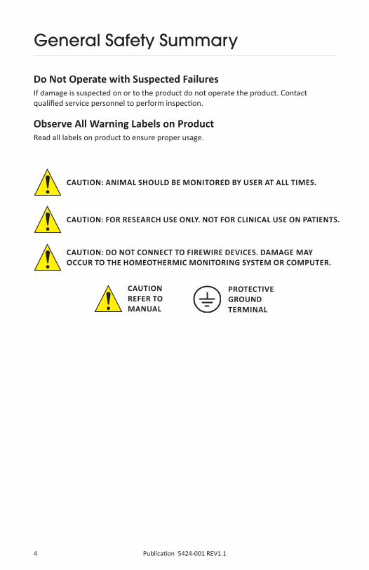
CAUTION: ANIMAL SHOULD BE MONITORED BY USER AT ALL TIMES.
Do Not Operate with Suspected FailuresIf damage is suspected on or to the product do not operate the product. Contact qualified service personnel to perform inspection.
Observe All Warning Labels on ProductRead all labels on product to ensure proper usage.
General Safety Summary
CAUTIONREFER TO MANUAL
PROTECTIVE GROUND TERMINAL
CAUTION: FOR RESEARCH USE ONLY. NOT FOR CLINICAL USE ON PATIENTS.
CAUTION: DO NOT CONNECT TO FIREWIRE DEVICES. DAMAGE MAY OCCUR TO THE HOMEOTHERMIC MONITORING SYSTEM OR COMPUTER.

5Publication 5424-001 REV1.1
Technical Specifications
ECG Display Leads I, II, III, aVL, aVR & aVFSupine or prone position1 kHz, 24-bit acquisition3 external electrodes connections (2 mm)
Respiration 250 Hz acquisition
Heart & Breath Rate 200 to 800 beats/min25 to 330 breaths/min Calculated every second
Warming & Temperature Heated surface up to 45°C0.1°C resolutionClosed loop PID controller keeps the animal within ±0.1°C of set temperature
Blood Pressure Full waveform displaySystolic and diastolic numeric values
Exhaled CO2 Connect ventilator directly to the platform (1/8” tubing)1 Hz refresh rate0.1% accuracy
SpO2 80 to 100% saturation250 Hz, 24-bit acquisitionRed & infrared channel display

6 Publication 5424-001 REV1.1
System Information
Heated Platform Easy to clean surface
10 cm X 21 cm X 2.5 cm (mouse)
12 cm X 30 cm X 2.5 cm (rat & mouse)
Tablet 8” Android tablet (10.1” available)
High resolution touchscreen display
Capacity of over 400 hours of saved data
5-8 hours battery life
USB cable and charger
Communication Module Bluetooth and analog outputs are in a separate enclosure to avoid interference
9 cm X 11 cm X 2.5 cm
100-240V power supply
Bluetooth 15 to 25 meters typical range between the communication module and the display unit
Analog Outputs 4 configurable outputs
BNC connectors, ±5V range
1 kHz, 16-bit refresh rate
Data Analysis .csv conversion tool
MATLAB® & Excel® import and display scripts
Compatible with third-party analysis software (LabChart)

7Publication 5424-001 REV1.1
Introduction
The Small Animal Physiological Monitoring System from Harvard Apparatus is an instrument integrating multiple physiology parameters on one single small platform.
The platform integrates monitoring of the rectal temperature, electrocardiogram (ECG), respiration, oxygen saturation (SpO2), blood pressure and exhaled CO2 (EtCO2). It also includes a controlled heating surface to maintain the animal’s body temperature at the desired level.
The system has many benefits regarding surgery procedures. It demands less installation time at the beginning of procedures and it reduces the required space and the number of wires around the animal. A single data/power cable connects to a small wireless communication module. All data is transferred to an Android tablet by Bluetooth for display and saving.
The Physiological Monitoring System features a real-time display which provides numeric values as well as waveforms that can be customized by the user. Multiple signals can be displayed on each of the three graphs.
The Physiological Monitoring System comes complete with a heated platform, an Android tablet with a protective sleeve and stand, Bluetooth communication module, a rectal probe and electrode gel. These components are all supplied in a sturdy case for storage and transport.
At the end of experiments, recorded data can easily be transferred to any computer for analysis. Scripts and utilities are provided to convert data in LabChart or CSV format and to display signals in Excel and Matlab
Physiological Monitoring System

8 Publication 5424-001 REV1.1
Operation
MINIMUM REQUIREMENTSMain VoltageThe monitoring system must be plugged to a 100-240 VAC 50-60 Hz main supply. The Android tablet comes with a USB charger, requiring a main supply as well. As stated in the Warning section, only use the power supplies sent with the system.
Android DeviceWhile an Android tablet is supplied with the system, any other Android device can be used, as a replacement or in conjunction. It must have Android 4.0.3 or above, a Bluetooth antenna and a high resolution display (720 x 1280 and above).
ComputerA computer is required only for advanced data analysis.
A USB port is required to connect the Android tablet for data transfer. Drivers are already installed for Windows Vista and above.
Java (JDK 1.6 and above) is required to run the LabChart and .csv conversion script.
Microsoft Excel on Windows is required to run the Excel display script.
Matlab is required to run the Matlab display script.

9Publication 5424-001 REV1.1
Operation
GETTING STARTEDInstalling the Monitoring SystemThe power switch is located on the communication module to power up the whole system. Plug in the power supply and make sure that the cable between the communication module and the monitoring platform is screwed tightly on both sides.
It is recommended to start the system 5 to 10 minutes prior to positioning the animal, to ensure that the platform is fully heated. It is important to note that the platform remembers the last target temperature, even if it was unplugged. This means that if the last user turned off the heating, the platform will not heat by default and the heating feature must be turned back on.
Android TabletThe Android tablet should be kept plugged into its wall charger. The majority of tablets have their USB port under the tablet. To use the provided stand, put the tablet on the side or upside down and the screen will automatically rotate.
The Android tablet comes with the “Monitoring Station” application pre-installed. When started, it will automatically connect to the monitoring platform.
Positioning the AnimalAfter anesthesia induction, place the animal on the monitoring platform with its paws on the metal pads and make sure to have a good contact by covering them with conductive gel. Insert the rectal temperature probe and set the desired temperature from the “Monitoring Station” application’s settings menu. See the section Theory of Operations for more information.

10 Publication 5424-001 REV1.1
Operation
ANDROID APPLICATIONWindowsThe Android application has two main windows:
A: Connection interface The first window appears at software opening. It informs user about connection status with the monitoring station. At the top of this window, there is a tool menu ( ) to access all the settings. The help menu (“?”) links to the User Manual that can be found on the tablet.
B: Monitoring interface The second window automatically appears after the connection is established. User can find all the information about the animal’s physiology, displayed either as numbers at the bottom of the screen, or on three configurable graphs. Furthermore, multiple options are available at the top of the window.
Reminder: This user manual can be accessed with the “?” button at the top of the window.

11Publication 5424-001 REV1.1
Operation
To select which signals to be displayed on the graphs, select the “Curve” menu at the top of the window.
GraphsEach of the 3 graphs can display up to 4 different signals each: 2 on the left axis and 2 on the right axis. The bottom graph displays low frequency signals and the two others are for high frequency signals.
It is possible to zoom on the signals by pinching on the graphs with two fingers:
• On the top 2 graphs, the duration can be adjusted from 1 to 5 seconds, by increments of 1 second.
• For the bottom graph, it can be adjusted from 30 seconds to 30 minutes, by increments of 30 seconds.

12 Publication 5424-001 REV1.1
Operation
Closing the ApplicationTo close the application, press the back button at the bottom of the window. It is also possible to close the application from the notification bar, when swiping down from the top of the screen, as shown below.
The application will automatically be closed if the system is turned off.
Pressing the home button will just put the application in background and will not close it. This is useful to navigate to other apps, while monitoring an animal and saving data.
Settings ModificationsWhile data saving is activated, advanced settings under the “Tool” menu cannot be modified for stability considerations and to preserve data integrity.
Temperature settings can be modified from the display window, even while data saving is on.

13Publication 5424-001 REV1.1
Operation
Signal RangeBy default, the range of the signals displayed is automatically set. For ECG, respiration, blood pressure, heart rate, respiration rate, temperatures and CO2, the range menu offers the possibility to set axis limits manually and avoid automatic rescaling.
SavingOn the display page, there is a button to save the results. As soon as the button is pressed, data starts to be saved. When pressed a second time, a popup will ask user to confirm if they really want to stop saving data. One minute of data uses approximately 0.5 MByte. Data is saved as a stream, meaning that unexpected shutdowns (e.g.: drained battery) will not affect prior acquired data.
Adding CommentsComments can be linked to the saved data file during acquisition. When adding a comment, a new file named comments.txt is created in the results folder. Each comment is preceded by a time marker, in order to refer the comment with the saved data. If a comment is added when saving is not enabled, this comment is added to the previous saved results.
In the dialog window that appears when adding a comment, there will be a list of the last 10 comments added during the acquisition.

14 Publication 5424-001 REV1.1
Operation
AlarmsMultiple alarms are available to inform the user when physiological parameters are out of the desired ranges.
When an alarm is ON, a popup window is shown on the screen and a noise signal can be played by the tablet. The sound on the Android tablet must be activated to hear the sound signal. Also, numerical values of these off limit parameters, at the bottom of the screen, turn red and blink as long as their value is abnormal, as shown in the following image.
To prevent false alarms, fault condition must last at least 10 seconds before alarm activation.
BeeperIn the Alarm menu, an option enables a sound marker following either heart or respiration rate. This setting is used when user might not be able to keep an eye on the tablet’s screen (e.g. during surgery or experimentation). With this feature, users can be notified of changes in physiology without looking at the display. The sound on the Android tablet must be activated to hear the noise.
15Publication 5424-001 REV1.1
Operation
LanguageMonitoring software can be set to either English or French. To change language, go in “Settings Menu” -> “General Settings”.
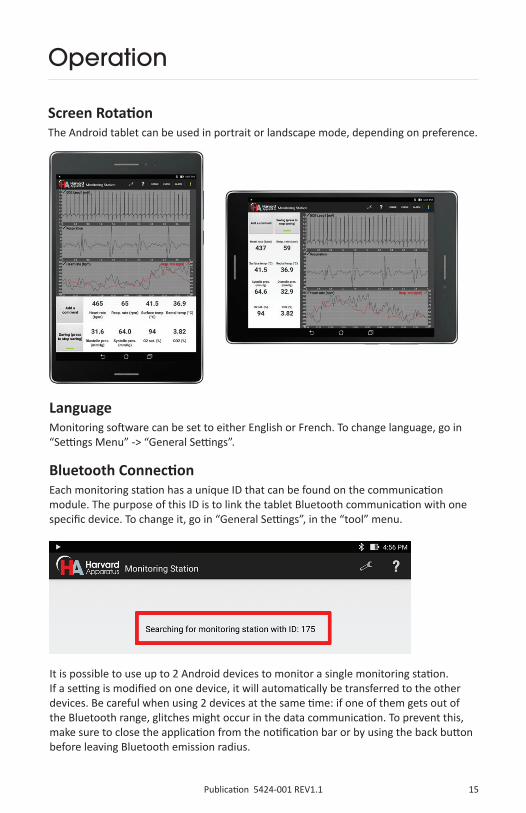
Bluetooth ConnectionEach monitoring station has a unique ID that can be found on the communication module. The purpose of this ID is to link the tablet Bluetooth communication with one specific device. To change it, go in “General Settings”, in the “tool” menu.
It is possible to use up to 2 Android devices to monitor a single monitoring station. If a setting is modified on one device, it will automatically be transferred to the other devices. Be careful when using 2 devices at the same time: if one of them gets out of the Bluetooth range, glitches might occur in the data communication. To prevent this, make sure to close the application from the notification bar or by using the back button before leaving Bluetooth emission radius.
Screen RotationThe Android tablet can be used in portrait or landscape mode, depending on preference.

16 Publication 5424-001 REV1.1
Operation
THEORY OF OPERATIONECGTo improve the signal, place a small amount of the provided conductive gel on each of the surface ECG sensors before positioning the animal; no additional adhesive is necessary, the gel is adequate to ensure a good electrical connection. All four of the animal’s paws much be placed on the corresponding sensor, even if only Lead I (Left Arm – Right Arm) is measured. This is important to ensure that the Right Leg Drive circuit which helps with 50/60 Hz noise reduction properly works.
If the animal is in the supine position, it is strongly urged that that external ECG leads be used rather than the surface ECG sensors. The external ECG lead connectors follow the American Heart Association (AHA) color coding. Below is a pictorial and table description of this color coding and how it compares to the European (IEC) color coding. The external ECG ports on the platform are 2 mm; adapters are available to 1.5 mm touch proof connectors. When the animal is in the supine position it is recommended that the ECG Electrodes Inversion Option be activated in the advanced setting menu. By turning on this option, hardware switches are activated that ensure that the correct leads are acquired and displayed.
AHA IEC
Right Arm RA White R Red
Left Arm LA Black L Yellow
Right Leg RL Green N Black
Left Leg LL Red F Green
Six main derivations are available (leads I, II & III, aVR, aVL, aVF), as well as externalleads. Right Leg Drive (RLD) is activated by default in the settings, to reduce 50/60 Hznoise. When using external leads, RLD should be changed to “External Leads”. RLD canalso be turned off, but it would usually add 50/60 Hz noise.

17Publication 5424-001 REV1.1
Operation
RespirationThe respiration sensor is the black diamond on the middle of the device. To work efficiently, it must be placed under the abdomen of the animal.Two filters are available for the respiration signal before display: a low-pass filter (25 Hz, 4th order Butterworth), and a high-pass filter (2 Hz, 4th order Butterworth). Both filters can be individually disabled and they have no effect on saved data. It also has no effect on breathing rate, which is computed from raw data.As an alternative to the respiration sensor, the respiration waveform can be derived from the ECG leads (both internal and external) by selecting the desired option in the respiratory settings. ECG derived respiration is useful during a surgery when the sensor detects noise originating from the surgeon.
TemperatureThe rectal probe should be inserted in the rectum of the animal so that the metal part is completely inside it. A lubricant is recommended to ease the insertion. Also, for better stability, the probe’s cable should be taped to the platform.
The ECG gain can also be changed. In the unlikely event where the ECG signal would be saturated and not visible on the screen, the gain can be reduced. The default gain value is 3. The ECG display and saved data represents the value before the amplifier. Thus, the signal amplitude will not change when changing the gain.
An additional cut-band (50/60 Hz, 4th order Butterworth) filter can be applied on all ECG signals before display. It should be enabled when the RLD is not sufficient to reduce the noise. It has no effect on saved data nor heart rate calculation since they use raw data.
The larger platform for rat and mouse includes two sets of surface ECG electrodes – one for adult rat and tone for mouse and rat under 20 days old. Select the correct set of electrodes in the ECG settings.

18 Publication 5424-001 REV1.1
Operation
Oxygen SaturationOxygen saturation measurement is one of the options available on the monitoring platform. A sensor clip with an optical source – detector pair is supplied.
It is recommend to place the sensor on the left hind leg of the animal. Prior to placement of the sensor, the animal’s leg should be carefully shaved so that any fur in the area of contact with the sensor is removed. The clip should be taped to the platform to avoid movement artifacts in the signal. In the same way, the force applied on the spring (through the adjustment of the screw) should be sufficient to stop any leg movements without blocking blood circulation. ECG acquisition of Lead I will continue to work even if the left paw is not on the sensor.
To make sure that the clip is installed properly, display ECG Lead I, SpO2 IR (infrared) and SpO2 R (red) on the graphs, as shown on the tablet on the next page. The DC amplitude of the SpO2 signals should be between 1000 and 10,000 with the default parameters. The AC amplitude should be 2 or more.
For long experiments, it is recommended to cover the animal with a few gauze pads to keep the temperature stable across its body.The temperature of the heating platform can be controlled in two different modes:
1. A target surface temperature is set, which will fix the temperature of the surface of the platform.
2. A target rectal temperature is set, and the platform temperature will be automatically controlled. Note that in this mode, if the rectal temperature is under 28°C, the system will consider that there is no animal on the platform and the surface temperature will be fixed to 40°C. The controller will maintain the rectal temperature within a tolerance of ±0.2°C.
In all modes, the surface temperature is limited to 45°C.

19Publication 5424-001 REV1.1
Operation
A low-pass filter (45 Hz, 4th order Butterworth) is applied by default on the SpO2 signals before display. It can be disabled and it has no effect on saved data. It also has no effect on oxygen saturation calculation, which is computed from raw data. Multiple parameters are available in the settings to change gains, filters and LED amplitudes. These parameters should only be modified by experienced users.
Blood PressureBlood Pressure is one of the options available on the monitoring platform. A bridge amplifier is built into the platform and the connector is a Redel 5-pin, used on Millar bridge amplifiers. A pressure transducer is not included with this option and must be purchased separately. The APT300 Blood Pressure transducer is available with the appropriate connector to the interface with the system (73-4905). Alternatively, adapters are available for DIN 8-pin connectors (used on AD Instruments bridge amplifiers) and for Millar’s Low Profile (used on Millar catheters).
Blood pressure catheters can have a zero offset of up to ±50 mmHg. For this reason, it is recommended to perform a calibration each time the catheter is changed by placing it at room pressure. An option is available in the advanced settings for calibration.

20 Publication 5424-001 REV1.1
Operation
Exhaled CO2
Exhaled CO2 is one of the options available on the physiological monitoring platform. If this option is added on after the initial purchase of the system, the platform must be returned for modification.
To measure the exhaled CO2, an external ventilator must the used. The ventilator must be connected in series with the CO2 module. The exhaled gas from the animal must be connected to the input port of the CO2 module on the platform, which is the one on the right side of the animal. The other port, on the left side, must be connected to the ventilator.
The CO2 module is factory calibrated. However, a manual calibration can be performed in the advanced settings. To do so, supply a flow of 10 mL/minute of nitrogen to the sensor and activate the calibration option. It is recommended to do a manual calibration every year to get the most accurate CO2 level.
The CO2 measurement can be displayed in either percentage or mm Hg, as selected in the CO2 setting.
21Publication 5424-001 REV1.1
Operation
Analog OutputsThere are 4 BNC analog outputs on the communication module. The outputs are short circuit protected.
ECG signals are encoded from -5V to 5V, where 5V is 12.5 mV at the default ECG gain of 3. If the ECG gain is changed, the ratio will also change. For example, at a gain of 6, 5V will represent 6.25 mV.
Temperatures are encoded from -5V to 5V where -5V is 25°C, 0V is 35°C and 5V is 45°C. They increase by 50mV per 0.1°C.
Expired CO2 is encoded from 0 to 5V, where 0V is 0% and 5V is 10%.
Blood pressure is encoded from -5V to 5V, where 0V is 0 mmHg and 5V is 400 mmHg.
The rest of the signals (respiration and SpO2) are in arbitrary units.
In the advanced settings on the Android tablet, it is possible to select the signals for each the analog outputs.
Cleaning of the DeviceThe platform’s surface and the rectal probe can be cleaned with alcohol based solutions, such as ethanol and isopropanol, and peroxide solutions.
The system is not watertight, but is splash proof. Avoid spilling liquids on the side of the platform as it could leak inside.
The communication module, the cables, the oxygen saturation sensors can be cleaned with warm water and a small amount of light detergent.
Stereotaxic DeviceTwo Stereotaxic Kits are options available on the monitoring platform; Stereotaxic Kit with Gas Anesthesia Mask and Stereotaxic Kit with Nose Clamp. Each of those options are available for the mouse platform and the rat and mouse platform. The options that fit on the rat and mouse platform are for use with mouse only. The stereotaxic device is fixed to the platform with M3 screws and it can be easily removed depending on the application.
Other accessories can be used with the stereotaxic device, such as jaw holders and cages for awaken monitoring. Contact Technical Service for information about additional accessories.

22 Publication 5424-001 REV1.1
Operation
DATA TRANSFER AND ANALYSISTransfer Saved DataTo recover saved data, plug the device on a computer and browse to the “MonitoringStation” folder. In this folder, there will be multiple folders named with the date: YYMMDD. In this sub-folder, folders are named with the starting time: HHMMSS. The whole folder should be copied on the computer. It contains all the saved data in multiple binary files.
If an external SD card is present, data is automatically saved to it.
The following table shows information about each saved file.
File name (.bin) Corresponding data Data format Sampling frequency
ECGAux ECG external leads (µV) Int16 1000 Hz
ECGLeadI ECG LA-RA (Lead I) (µV) Int16 1000 Hz
ECGLeadII ECG LL-RA (Lead II) (µV) Int16 1000 Hz
Respiration Respiration Int16 250 Hz
SpO2Infrared SpO2 Infrared Int32 250 Hz
SpO2Red SpO2 Red Int32 250 Hz
BloodPressure Blood pressure (1/100th mmHg) Int16 250 Hz
CO2 Exhaled CO2 (1/1000th %) Int16 1 Hz
SurfaceTemperature Surface temperature (1/100th °C) Int16 10 Hz
RectalTemperature Rectal temperature (1/100th °C) Int16 10 Hz
RespirationRate Calculated breathing rate (BPM) Int16 1 Hz
HeartRate Calculated heart rate (BPM) Int16 1 Hz
SpO2 Calculated saturation (1/10th %) Int16 1 Hz
ECG data is in µV. ECG lead III can be obtained by subtracting lead I from lead II. Respiration vary from -512 to 511. Temperatures can be converted in °C by dividing by 100. Blood pressure can be converted in mmHg by dividing by 100. Exhaled CO2 can be converted in % by dividing by 1000 or in ppm by multiplying by 10.
Matlab and Excel ScriptsScripts are available to load, analyze and display all the saved signals in Excel and Matlab. Contact Technical Service for more information.
The Excel script will load binary files saved by the tablet into separate worksheets of an Excel document. Data can then easily be exported in .csv or displayed on a graph. A simple display code is also provided. This script can import up to 90 minutes of data.
Matlab is a powerful and flexible mathematic tool that can analyze complex waveforms. The provided script will load binary files in Matlab workspace and will display signals chosen by the user

23Publication 5424-001 REV1.1
Operation
LabChart and .csv Conversion ToolA cross-platform Java program (Windows, Mac & Linux) is also provided to automatically convert the saved files into a LabChart binary file (.adibin) or .csv files. Java must be installed on the computer. The software asks the user to choose a folder containing the saved files and it creates the desired files inside a subfolder called “csv”. A batch conversion can also be done when choosing a folder containing multiple folders of results.
On average, data will use 4 MB/minute for csv conversion and 3 MB/minute for LabChart conversion. An option is available to select certain signals to convert, in order to reduce file size.
When all the signals are selected, the conversion is limited to 3.5 hours of data. Note that Excel will not be able to load very large .csv files with more than 1 million lines (for example, when loading raw ECG signals of more than 17 minutes). In these situations, use the provided Excel script.
Android LimitationsOn some devices, if the application is not displayed on the screen, Android might stop it at any moment to free up memory. When saving important results, it is important not to turn off the screen or navigate to other apps. When displaying results, the application prevents the device to go to sleep and the screen will not automatically turn off.
Bluetooth RangeThe Bluetooth connection is typically limited to a range of 15 to 30 meters, depending on the environment. Note that Wi-Fi signals can interfere with Bluetooth. If there is wireless router close to the system or a high Wi-Fi activity, the range could be decreased. For these reasons, especially when saving important results, it is recommended to keep a maximum distance of 5 meters between the Android device and the communication module.
Data will be lost if the system is too far away. Display and saving will be paused until the system is brought closer. At a certain distance the connection can also be lost and the application will be closed.
There is a 10 second buffer on the system, which prevents any data loss even if the tablet is out of range for some time.
Demo ModeA demonstration mode is available on the Android application. It allows displaying previously acquired data when the platform is not connected. Contact Technical Service for more information.

24 Publication 5424-001 REV1.1
Troubleshooting Guide
ECG signal is weak The most common reason for this problem is that there is not enough conductive gel between the paws and the pads of the platform. The gel can dry out during long experiments, so it is recommended to use more gel. Even if only Lead I is measured, it is important to connect all the paws of the animal to the platform, because the Right Leg Drive circuit that helps reduce 50/60 Hz noise will not work properly.
Respiration signal is weak The sensor on the platform must be placed under the abdomen of the animal for better results. If it is placed under the rib cage, the sensor might also detect the heartbeat.
The tablet is not connecting to the system
Each system has an ID, which is written on top of the communication module. Make sure that the tablet is searching for that ID when trying to connect. If not, change it in Settings ͢ General. If the problem is still present, make sure the system is powered on. There should be a green LED on the communication module. Check that the cable between the communication module and the platform is connected and screwed in. If this problem occurs after a software update or with a new tablet, the Bluetooth device needs to be paired. On some devices, a “Smart Lock” notification will appear asking to add the Bluetooth device as a trusted device. Simply confirm this request. Pairing can also be performed from the Android’s Bluetooth settings. Note that pairing sometimes cannot be done while another Bluetooth device is connected to the monitoring system
The system is not powering Make sure you are using the correct power supply. A protection circuit is present on the system. If the detected supply is not between 11V and 14V, the system will not start.
The tablet is not recognized by the computer
On the tablet, go in Settings ͢ Storage, touch the Menu icon and then choose MTP connection. Also, make sure that the tablet is not in USB debugging mode (an icon would appear in the notification bar when the tablet is connected to a computer). If you are using a Mac, you will need to install Android File Transfer. If you are using Windows XP or Linux, ask technical services for assistance.
There is 50/60 Hz noise in the signals
Using a power bar or an extension cable has been found to increase the background noise in critical signals, such as ECG, SpO2 and blood pressure. Try connecting the system directly in the wall. Make sure the outlet has three prongs, with an earth ground connection. Also make sure you are using the provided power supply. For noise in the ECG signal, make sure that the option Right Leg Drive is activated in the Settings.

25Publication 5424-001 REV1.1
Order # Product
System and Options
75-15001 Physiological Monitoring System for Mouse (10 g to 100 g)
75-15011 Physiological Monitoring System for Mouse & Rat ( 10 g to 600 g)
75-15022 Blood Pressure Option
75-1503 End Tidal CO2 Option
75-1504 SpO2 Option
Accessories73-4905 APT3000 Pressure Transducer for Physiological Monitoring System
77-0207 ECG 3-Needle Electrodes, 2 mm pin
75-1539 Nose Cone Holder
75-1540 Stereotaxic Kit with Gas Anesthesia Mask for 75-1500 Platform
75-1541 Stereotaxic Kit with Nose Cone for 75-1500 Platform
75-1542 Stereotaxic Kit with Gas Anesthesia Mask for 75-1501 Platform
75-1543 Stereotaxic Kit with Nose Cone for 75-1501 Platform
75-1552 BP Adapter to DIN-8
Ordering Information
Note1: At the end of the part number add EU for Europe, UK for United Kingdom and CN for China versions.Note2: Blood pressure transducer needs to be purchased separately.For more complete product details and part number listings please visit our website www.harvardapparatus.com

26 Publication 5424-001 REV1.1
Notes

27Publication 5424-001 REV1.1
Notes

28 Publication 5424-001 REV1.1
Notes

Hugo Sachs Elektronik / Harvard Apparatus, GmbH
Gruenstrasse 1 March-Hugstetten D-79232, Germany
phone +49.0.7665.9200.0fax +49.0.7665.9200.90e-mail [email protected] www.hugo-sachs.de
Biochrom Limited - Harvard Apparatus UK
East Wing, Building 1020 Cambourne Business Park, Cambourne Cambridge, CB23 6DW, United Kingdom
phone +44.1732.864001fax +44.1732.863356e-mail [email protected] www.harvardapparatus.co.uk
Harvard Apparatus
84 October Hill Road, Holliston, MA 01746, USA
phone +1.508.893.8999toll free +1.800.272.2775 (USA Only)fax +1.508.429.5732e-mail [email protected] www.harvardapparatus.com
Panlab, S.L. / Harvard Apparatus Spain
C/Energia, 112 08940 Cornellà Barcelona, Spain
phone +34.934.750.697 (International Sales) + 34.934.190.709 (Sales in Spain)fax +34.934.750.699e-mail [email protected] www.panlab.com
CMA Microdialysis, AB
Torshamnsgatan 30A SE-164 07 Kista, Sweden
phone +46.8.470.10.00e-mail [email protected] www.microdialysis.se
Harvard Apparatus Canada
6010 Vanden Abeele Saint-Laurent, Quebec H4S 1R9, Canada
phone +1.514.335.0792toll free +1.800.361.1905 (CAN Only)fax +1.514.335.3482e-mail [email protected] www.harvardapparatus.ca
Harvard Apparatus, S.A.R.L.
6 Avenue des AndesMiniparc Building 8 91952 Les Ulis Cedex, France
phone +33.1.64.46.00.85fax +33.1.64.46.94.38e-mail [email protected] www.harvardapparatus.fr
Harvard Apparatus China
Room 1902E, 19F, Building B Zhong Shan Plaza 1065 West Zhong Shan Road Changning DistrictShanghai, China
phone +86.21.2230.5128e-mail [email protected]
France
U.K.
divisions of Harvard Bioscience, Inc.