slurring pen and falling brain - cpa chennai€¢ no neurocutaneous markers • fundus examination...
TRANSCRIPT

SLURRING PEN AND FALLING BRAIN
SLURRING PEN AND FALLING BRAIN

PRESENTING COMPLAINTS
• Master Kishore, 10 yr old male ,first born child of non consanguineous marriage
• C/O disturbances in handwriting noticed for the past 1 month in the form of slowness of writing and writing big letters.
• H/o frequent vomiting after feeds


H/O PRESENTING ILLNESS
• No H/o recent fever, local trauma
• Speech was normal. Able to recollect recent and remote events
• Able to grasp objects firmly.
• No H/o involuntary movements,No h/o frequent falls
• No H/o loss of consciousness, seizures, gait disturbances
• No H/o regression of milestones
• No similar history in family members

EXAMINATION
• He was a well looking child .
• His present handwriting was compared with his previous handwriting.
• The letters were big, badly spaced and incoherent.

• Intentional tremors found.
• No other cerebellar signs
• No muscle wasting/hand deformity
• No neurocutaneous markers
• Fundus examination -Normal
• Rest of the neurological examination was normal.
• Other systems were also normal
EXAMINATION
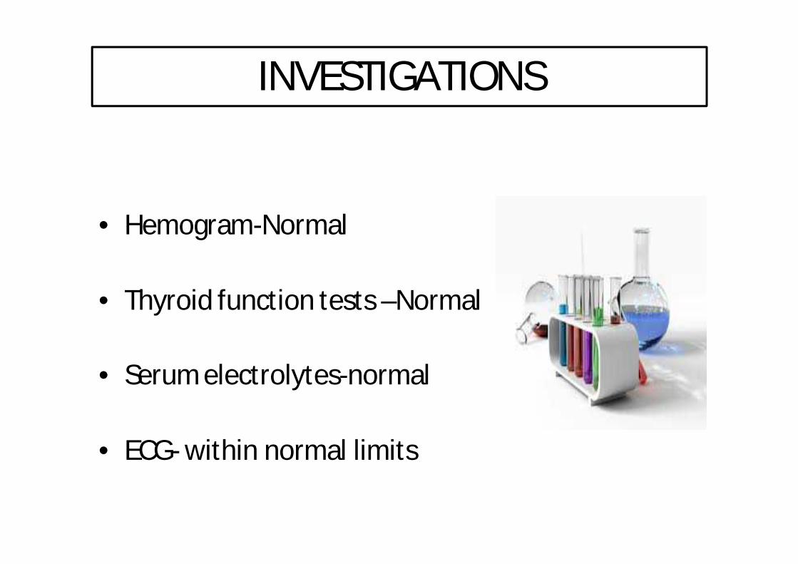
INVESTIGATIONS
• Hemogram-Normal
• Thyroid function tests –Normal
• Serum electrolytes-normal
• ECG- within normal limits

MRI BRAIN

MRI BRAIN

MRI BRAIN
• Bilateral cerebellar tonsils appear peg like and are seen to herniate below the level of cerebellum for a length of 1 cm.Nocervicomedullary compression seen.
ARNOLD CHIARI MALFORMATION TYPE 1
MRI SPINE –no detectable lesion

CASE 2

HISTORY
• A 11 yr old girl was brought for c/o not growing adequately as compared to peers of her age group.
• No H/o chronic illnesses ,drug intake
• Neonatal history – not significant
• NIL family history of short stature

EXAMINATION
• General examination –No facial dysmorphism
• Systemic examination-Normal

EXAMINATION
Height for age Z score less than -3Weight for age Z score -1 and -2US:LS ratio 1.2:1Inference:Proportionate short stature with normal development and no co-morbidites.
Ø ?CONSTITUTIONALØ ?IDIOPATHIC SHORT STATUREØ ?GROWTH HORMONE DEFCIENCY

EVALUATION
• BONE AGE -11 YEARS (=Chronological age)

INVESTIGATIONS
• Baseline investigations-normal
• Vitamin-D
• Calcium Normal
• Phosphorus
• PTH levels

FURTHER…
• Growth hormone deficiency detected.
• Prior to starting GH replacement therapy,Neuroimaging done.


MRI BRAIN AND SPINE
• Cerebellar tonsil herniation 5 to 6 mm
• MRI SPINE:
– Focal intramedullary CSF intensity area 6*2 cm at C5 level
– 10*3 cm at L6 level
– Irregular CSF intensity 14 cm long extending from D4 to L1.
S/O SYRINGOMYELIA

• ARNOLD CHIARI MALFORMATION WITH SYRINGOMYELIA

TREATMENT
• The child has undergone Foramen Magnum decompression ,C1 laminectomy and duraplasty.

CAUSES
• Congenital:
– Hydrocephalus
– Craniosynostosis
– Hyperostosis ( craniometaphyseal dysplasia, osteopetrosis, erythroid hyperplasia)
– X-linked vitamin D-resistant rickets
– Neurofibromatosis type I

CAUSES
• Acquired:
– Head trauma- cerebellar tonsillar ectopia, possibly because of dural strain.
– Posterior fossa hypoplasia causes reduced cerebral and spinal compliance


CLASSIFICATIONTYPE TYPE 1 TYPE 2
Age at diagnosis Adults ,older children Infants and younger children
Clinical features HeadacheMyelopathy,central cord symptoms
Swalloing,feedingdifficulties,apnea,weak cry
Anatomical abnormality Cerebellar herniation Cerebellar & brain stem herniation
Myelomeningocele No Always
Shared anamolies Craniocervical hypermobilitysyndromes
Agenesis of corpus callosum,heterotopia
Klippel fel anamolyBasilar invaginationBifida of C1 posterior arch
Basilar invaginationOccipitalisation of atlasBifida of C1 posterior arch

TYPE-I
• Extension of the cerebellar tonsils (the lower part of the cerebellum) into the foramen magnum, without involving the brain stem.
• Most common • Recurrent headache, neck pain, urinary frequency and
progressive lower leg spasticity.• Asymptomatic till adolescence.• No hydrocephalus.
• Acquired

ARNOLD CHIARI MALFORMATION TYPE 1

TYPE-II
• Classic CM, involves the extension of both cerebellar and brain stem tissue into the foramen magnum.– Onset at early infancy– Hydrocephalus– Presents as stridor ,weak cry, apnea, gait abnormalities and
incoordination.– Occurs due to failure of pontine flexure development during
embryogenesis and elongation of the fourth ventricle .• Also, the cerebellar vermis (the nerve tissue that connects the two
halves of the cerebellum) may be only partially complete or absent. Type II is usually accompanied by a myelomeningocele.

ARNOLD CHIARI MALFORMATION TYPE 2

TYPE-III
• Most serious form of CM.The cerebellum and brain stem protrude, or herniate, through the foramen magnum and into the spinal cord. Part of the brain’s fourth ventricle, a cavity that connects with the upper parts of the brain and circulates CSF, may also protrude through the hole and into the spinal cord.

TYPE-IV & TYPE-0
• Type IV- Involves an incomplete or underdeveloped cerebellum—a condition known as cerebellar hypoplasia. In this rare form of CM, the cerebellar tonsils are located in a normal position but parts of the cerebellum are missing.
• Type 0- no protrusion of the cerebellum but symptoms of CM are present.

TREATMENT
• Asymptomatic or mild symptoms-conservative management
• Symptomatic – urgent surgical decompression

REVIEW OF LITERATURE
• Chiari I malformation is not directly associated with other congenital brain malformations, However, craniovertebral malformations are common in patients with Chiari I malformation.Distention with cerebrospinal fluid (CSF) of the central canal of spinal cord (ie, hydromyelia) or paracentral cavities (ie, syringomyelia) is present in approximately 25% of patients with Chiari I malformation.

• In one study, a rate 0.6% was reported in all age groups, and a rate of 0.9% was reported in a study of only pediatric patients. Therefore, this condition is more common in both the adult and pediatric populations than was recognized previously. A female predominance has been reported in some large case series, with a male-to-female ratio of 2:3.
REVIEW OF LITERATURE

BMC Pediatr. 2014 Dec 10;14:294
-A 7 yr -old boy was evaluated for a two month history of
atypical movements which would occur in the evening, and
last for an hour after eating. These stereotypical movements
with the head and chest bending forward and to the left
side, accompanied by a grimace, were associated with
sensation of breath locking without cyanosis.
• Oesophageal ph s/o severe GER.
• No improvement with prokinetics,PPI.
• Eeg,polysomnography normal
REVIEW OF LITERATURE

• A brain magnetic resonance imaging (MRI) was performed and revealed a CM I: cerebellar tonsils extending to 12 mm, with syringomyelia (D4-D5).
• He underwent posterior fossa decompression with upper cervical laminectomy and expansion duroplasty.
REVIEW OF LITERATURE

• Dysphagia occurs in 5-15% of patients
REVIEW OF LITERATURE

A case of a 27-year-old woman who presented with a three-year history of dysphagia, chest pain, and weight loss.
• Esophageal manometrics revealed markedly disordered esophageal motility and gastroesophageal reflux.
• The subsequent onset of neurological symptoms led to the diagnosis of Chiari type I malformation. Following posterior craniotomy with decompression, her dysphagia and chest discomfort completely resolved.
REVIEW OF LITERATURE

Childs Nerv Syst. 2001 May
• Cervical encephalocele in a newborn--Chiari III malformation. Case report and review of the literature.
• Chiari malformations are brain stem anomalies with or without spinal canal involvement.
• The combination of brain stem and cerebellar anomaly with a cervical cele is named Chiari III malformation.
• Patients with this extremely rare defect frequently present with severe neurological defects and can have a poor prognosis.
REVIEW OF LITERATURE

Beier AD1, Barrett RJ, Burke K, Kole B, Soo TM.
A 31-year-old man with a Chiari type I malformation (CM-1) occurring in conjunction with LEOPARD syndrome.
• He presented with severe dysphagia
MRI) brain and cervical spine revealed CM-1 with an extensive cervical syrinx.
• The patient underwent a suboccipitalcraniectomy with C1 laminectomy and duraplasty.
REVIEW OF LITERATURE

• DISCUSSION: – The occurrence of a CM-1 with LEOPARD syndrome
has only been reported once, whereas CM-1 and Noonan syndrome have been linked in several cases. The similarity between LEOPARD and Noonan syndromes has been reported and many propose they represent 2 entities along a spectrum.
• CONCLUSION: – In light of this spectrum, we propose that CM-1
should be considered in all patients presenting with LEOPARD-Noonan syndrome.
REVIEW OF LITERATURE

WHY THIS PRESENTATION?
• It is asymptomatic or produces rare symptoms like GER.
• Presence of cervicomedullary compression is an indication for immediate surgery.
• Presence of associated anamolies carries poor prognosis and sometimes fatal.

REFERENCES
• 1.Ital J Neurol Sci. 1994 Feb;15(1):57-60.• Monosymptomatic presentation of type I Arnold-Chiari malformation:
report of two cases.Cammalleri R1, D'Amelio M, Gangitano M, Raimondo D, Rossetti M, Camarda R.
• 2. BMC Pediatr. 2014 Dec 10;14:294. doi: 10.1186/s12887-014-0294-Abnormal movements associated with oropharyngeal dysfunction in a child with Chiari I malformation.Berthet S1, Crevier L2, Deslandres C3,4.
• 3. 1996 Mar;41(3):512-5.Esophageal dysphagia as the sole symptom in type I Chiari malformation.Elta GH1, Caldwell CA, Nostrant TT.
• 4. Leopard syndrome and Chiari type I malformation: a case report and review of the literature.
• Beier AD1, Barrett RJ, Burke K, Kole B, Soo TM.• 5. Childs Nerv Syst. 2001 May;17(6):373-5.• Cervical encephalocele in a newborn--Chiari III malformation. Case report
and review of the literature.• Häberle J1, Hülskamp G, Harms E, Krasemann T.