slovenian society of human genetics
TRANSCRIPT

5th COLLOQUIUM OF GENETICS
Proceedings
PIRAN
September 23th, 2016
GENETIC SOCIETY SLOVENIAin collaboration with
SLOVENIAN SOCIETY OF HUMAN GENETICS

Colloquium of Genetics 2016
GENETIC SOCIETY OF SLOVENIA IN COLLABORATION WITH
THE SLOVENIAN SOCIETY OF HUMAN GENETICS
5th COLLOQUIUM OF GENETICS
Proceedings
Marine Biology Station Piran National Institute of Biology
Piran September 23rd 2016

Colloquium of Genetics 2016
2 | P a g e
Organizers
Genetic Society Slovenia in collaboration with The Slovenian Society of Human Genetics National Institute of Biology Marine Biology Station Piran University of Ljubljana Biotehnical Faculty
Faculty of Medicine University of Maribor
Faculty of Medicine Faculty of Chemistry and Chemical Technology
Editors Uroš Potočnik Katja Repnik Reviewers Branka Javornik Tanja Kunej Uroš Potočnik Maja Čemažar Jernej Jakše Damjan Glavač Design: Staša Jurgec Publisher: Genetic Society Slovenia, Ljubljana, September 2016 Number of issues: 60 USB keys
Contributing authors are responsible for proof‐reading corrections.

Colloquium of Genetics 2016
3 | P a g e
CIP - Kataložni zapis o publikaciji
Narodna in univerzitetna knjižnica, Ljubljana
575.111(082)(0.034.2)
616-056.7(082)(0.034.2)
COLLOQUIUM of Genetics (5 ; 2016 ; Piran)
Proceedings [Elektronski vir] / 5th Colloquium of Genetics, Piran, September
23rd 2016 ; [organizers] Genetic Society of Slovenia in collaboration with Slovenian
Society of Human Genetics ... [et al.] ; editors Uroš Potočnik, Katja Repnik. -
Ljubljana : Genetic Society of Slovenia, 2016
ISBN 978-961-93545-3-7
1. Potočnik, Uroš, 1969- 2. Slovensko genetsko društvo 3. Slovensko društvo za
humano genetiko
286511872

Colloquium of Genetics 2016
4 | P a g e
Members of boards
Scientific board Peter Dovč Damjan Glavač Branka Javornik Uroš Potočnik Jernej Jakše Darja Žgur Bertok Tanja Kunej Maja Čemažar
Organization board Katja Repnik Staša Jurgec Uroš Potočnik Andreja Ramšak Nataša Štajner
Colloquium of Genetics 2016
5 | P a g e
CONTENT
PROGRAM OF MEETING 7
LECTURES 8
OPENING LECTURE 9
Branka Javornik: STUDIES OF HOP-VERTICILLIUM PATHOSYSTEM 9
OPENING LECTURE 10
Marjanca Starčič Erjavec: GENETIC DIVERSITY OF Escherichia coli FROM GUT MICROBIOTA 10
ABSTRACTS AND PAPERS OF LECTURES 11
BIOTECHNOLOGY 11
Špela Kos: GENE ELECTROTRANSFER FOR DNA VACCINATION 12
Helena Volk: PRODUCTION OF RECOMBINANT EFFECTOR PROTEINS FROM Verticillium nonalfalfa 13
Mojca Juteršek: TOWARDS MOLECULAR GENETIC DELINIATION OF Synechocystis SPECIES 14
Ester Stajič: DEVELOPMENT OF CABBAGE HAPLOID INDUCER LINE WITH GENOME EDITING 20
Katja Guček: CRISPR/Cas9 GENOME EDITING IN RAPESEED 21
Sabina Belc: DISTRIBUTION ANALYSIS AND PRODUCTION OF FUNGAL ACTINOPORIN- AND PERFORIN-
LIKE PROTEINS 22
MOLECULAR BASIS OF DISEASES & GENOMICS 23
Miha Modic: TDP-43 SAFEGUARDS PLURIPOTENCY BY REGULATING ALTERNATIVE
POLYADENYLATION AND REPRESSING PARASPECKLES 24
Urša Lampreht Tratar: GENE ELECTROTRANSFER OF ANTIBIOTIC-FREE IL-12 PLASMID INDUCES
ANTITUMOR EFFECT IN B16F10 MELANOMAS 25
Katja Uršič: PERITUMORAL GENE ELECTROTRANSFER OF IL-12 AS ADJUVANT IMMUNOTHERAPY TO
INTRATUMORAL ELECTROCHEMOTHERAPY WITH CISPLATIN FOR TREATMENT OF MURINE B16F10
MELANOMA 26
Katarina Žnidar: THE EFFECT OF GENE ELECTROTRANSFER OF BLANK pDNA ON TUMOR CELLS'
SURVIVAL AND EXPRESSION OF CYTOSOLIC DNA SENSORS 27
Vasja Progar: FUNCTIONAL ENRICHMENT OF DIFFERENTIALLY EXPRESSED GENES IN HOP INFECTED
BY V. nonalfalfae 28
Kristina Marton: Verticillium nonalfalfae AND ITS CANDIDATE SECRETED EFFECTOR PROTEINS 29
Marija Rogar: POLYMORPHISMS IN SEGREGATION GENES INVOLVED IN THE DEVELOPMENT OF
GASTRIC CANCER IN SLOVENIAN POPULATION 30
Alja Zottel: GLIOBLASTOMA MULTIFORME AND NANOBODIES 31

Colloquium of Genetics 2016
6 | P a g e
POSTERS 32
ABSTRACTS AND PAPERS OF POSTERS 33
MOLECULAR BASIS OF DISEASES 33
Marko Vidak, Damjana Rozman, Mirjana Liović, Radovan Komel: SEARCH OF NEW GLIOBLASTOMA
STEM CELL MARKERS BY COMPARING MICROARRAYS DATA FROM THE NCBI GEO DATABASE, AND
EXPERIMENTAL VALIDATION OF THOSE MARKERS IN GLIOMA CELL LINES 34
Matej Rebek, Eva Jakljevič, Darja Žgur-Bertok, Marjanca Starčič Erjavec: PHYLOGENETIC GROUPS
AND clbAQ, usp, hlyA, cnf1 GENES AMONG COMMENSAL Escherichia coli STRAINS ISOLATED FROM
FECES OF HEALTHY HUMANS OF BOTH GENDERS AND BOTH AGE GROUPS 35
Tinkara Prijatelj, Danijela Vočanec, Tadej Pajič, Martina Fink, Irena Preložnik Zupan, Peter Černelč,
Radovan Komel, Tanja Kunej, Nataša Debeljak: NOVEL MOLECULAR-GENETIC DIAGNOSTIC TEST FOR
JAK2 NEGATIVE ERYTHROCYTOSIS 40
Rok Končnik, Barbara Krajnc, Jovan Krsteski, Uroš Potočnik: POLYMORPHISM rs1056837 IN CYP1B1
ENCODING AN ESTROGEN METABOLIZING ENZYME PRESENTS A RISK FOR ULM AMONG SLOVENIAN
WOMEN 41
Matjaž Deželak, Žan Hribar, Carina E. P. Kozmus, Uroš Potočnik: A POLYMORPHISM LOCATED IN
3’UTR OF PITHD1 NEAR CANNABINOID RECEPTOR 2 GENE CNR2 IS ASSOCIATED WITH SEVERE FORMS
OF CROHN’S DISEASE AND CNR1 GENE EXPRESSION 42
POPULATION GENETICS & GENOMICS 44
Taja Jeseničnik, Nataša Štajner, Sebastjan Radišek, Branka Javornik, Jernej Jakše: DISCOVERY OF
SMALL RNAs IN Verticilliun nonalfalfae 44
Matej Babič, Tanja Kunej, Borka Jerman-Blažič: NEW METHOD FOR STATISTICAL PATTERN
RECOGNITION USAGE IN CANCER ASSOCIATED microRNAs 45
Blaz Skrlj, Nina Pirih, Tanja Kunej: IDENTIFICATION OF NON-SYNONYMOUS POLYMORPHISMS
WITHIN REGIONS CORRESPONDING TO PROTEIN INTERACTION SITES 46
Marko Flajšman, Nataša Štajner, Igor Šantavec, Branka Javornik, Darja Kocjan Ačko: GENOTYPIZATION AND MORPHOLOGICAL CHARACTERIZATION OF SIX SLOVENIAN LANDRACES OF PROSO MILLET (Panicum miliaceum L.) 52
SPONSORS 53

Colloquium of Genetics 2016
7 | P a g e
PROGRAM OF MEETING
Registration 8.30 – 9.00
Opening of the 5th COLLOQUIUM OF GENETICS
9.00 – 10.00
OPENING LECTURE:
Chairman: Uroš Potočnik
Branka Javornik: Studies of hop-Verticillium pathosystem
Marjanca Starčič Erjavec: Genetic diversity of Escherichia coli from gut microbiota
9.10 – 10.00
9.10 – 9.35 9.35 – 10.00
Biotechnology
Chairmen: Sabina Berne in Jernej Jakše
10.00 – 11.30
Špela Kos: Gene electrotransfer for DNA vaccination 10.00 – 10.15
Helena Volk: Production of recombinant effector proteins from Verticillium nonalfalfae 10.15 – 10.30
Mojca Juteršek: Towards molecular genetics deliniation of Synechocystis species 10.30 – 10.45
Ester Stajič: Development of cabbage haploid inducer line with genome editing 10.45 – 11.00
Katja Guček: CRISPR/Cas9 genome editing in rapeseed 11.00 – 11.15
Sabina Belc: Distribution analysis and production of fungal actinoporin- and perforin-like proteins
11.15 – 11.30
Coffee break and poster viewing 11.30 – 11.50
Molecular Basis of Diseases and Genomics
Chairmen: Tanja Kunej in Marjanca Starčič Erjavec
11.50 – 13.50
Miha Modic: TDP-43 safeguards pluripotency by regulating alternative polyadenylation
and repressing paraspeckles
11.50 – 12.05
Urša Lampreht Tratar: Gene electrotransfer of antibiotic-free IL-12 plasmid induces
antitumor effect in B16F10 melanomas
12.05 – 12.20
Katja Uršič: Peritumoral gene electrotransfer of IL-12 as adjuvant immunotherapy to
intratumoral electrochemotherapy with cisplatin for treatment of murine B16F10
melanoma
12.20 – 12.35
Katarina Žnidar: The effect of gene electrotransfer of blank pDNA on tumor cells' survival
and expression of cytosolic DNA sensors
12.35 – 12.50
Vasja Progar: Functional enrichment of differentially expressed genes in hop infected by
V. nonalfalfae
12.50 – 13.05
Kristina Marton: Verticillium nonalfalfae and its candidate secreted effector proteins 13.05 – 13.20
Marija Rogar: Polymorphisms in segregation genes involved in the development of gastric cancer in Slovenian population
13.20 – 13.35
Alja Zottel: Glioblastoma multiforme and nanobodies 13.35 – 13.50
Lunch (self-service bar), poster viewing 13.50 – 15.00
Meeting of the Genetic Society Slovenia with announcement of the best lecture and the best poster awards
15.00 – 16.00

Colloquium of Genetics 2016
8 | P a g e
LECTURES

Colloquium of Genetics 2016
9 | P a g e
OPENING LECTURE
STUDIES OF HOP-VERTICILLIUM PATHOSYSTEM Branka Javornik
University of Ljubljana, Biotechnical Faculty, Ljubljana, Slovenia
ABSTRACT
The studies address Verticillium resistance in plants, working on the hop-Verticillium nonalfalfea (Vna)
pathosystem. This work includes two broad aims.
First, to improve hop resistance to Verticillium wilt. This work includes mapping of gene(s) or QTLs for
Verticillium resistance and the development of molecular markers linked to resistance for application
in hop breeding.
The second aim is to improve the management of wilt disease based on an understanding of the
mechanisms of host- pathogen interaction and the pathogenicity of Vna. This work has focused on
studies of the hop-Vna interaction on proteome and transcriptome levels to search for hop factors
implicated in resistance and Vna factors related to virulence. On the pathogen side, Vna has been being
further studied by comparison of proteomes and whole genome sequences of Vna isolates with
different virulence, and functional analysis of predicted candidate genes.

Colloquium of Genetics 2016
10 | P a g e
OPENING LECTURE
GENETIC DIVERSITY OF Escherichia coli FROM GUT MICROBIOTA
Marjanca Starčič Erjavec
University of Ljubljana, Biotechnical Faculty, Department of Biology, Jamnikarjeva 101, 1000 Ljubljana, Slovenia
ABSTRACT
Mammals have a complex gut microbiota shaped by intestinal anatomy, function and diet1.
Escherichia coli (E. coli) is one of the first colonizers of the mammalian gut and afterwards part of the
normal gut microbiota. It usually coexists with its host in mutual benefit. Nevertheless, it is known
that the intestinal microbiota can also be a reservoir of extraintestinal pathogenic variants of E. coli
(ExPEC) that can cause disease infections at different extraintestinal anatomic sites (e. g. urinary
tract, skin and soft-tissue infections)2. Further, it is known that animals (e. g. cats, dogs and birds) can
be a potential reservoir of ExPEC3. In last years our studies have been focused on the virulence
potential for ExPEC among E. coli from gut microbiota from different hosts. The lecture will present
an overview of the published4, 5 and recently obtained data.
REFERENCES 1. Stevens CE, Hume ID. The mammalian gastrointestinal tract. In: Comparative physiology of the vertebrate digestive
system. 2nd ed. Cambridge University Press, Cambridge, 2004, 46-92.
2. Leimbach A, Hacker J, Dobrindt U. E. coli as an all-rounder: the thin line between commensalism and pathogenicity. Curr.
Top. Microbiol. Immunol. 2013, 358, 3-32.
3.Bélanger L, Garenaux A, Harel J, Boulianne M, Nadeau E, Dozois CM. Escherichia coli from animal reservoirs as a potential
source of human extraintestinal pathogenic E. coli. FEMS Immunol. Med. Microbiol. 2011, 62, 1-10.
4. Starčič Erjavec M, Žgur-Bertok D. Virulence potential for extraintestinal infections among commensal Escherichia coli
isolated from healthy humans – the Trojan horse whitin our gut. FEMS Microbiol. Lett. 2015, 362, 1-9.
5. Vadnov M, Barbič D., Žgur-Bertok D, Starčič Erjavec M. Escherichia coli isolated from feces of brown bears (Ursus arctos)
have a lower prevalence of human extraintestinal pathogenic E. coli virulence-associated genes. Can. J. Vet. Res. 2016, In
press.

Colloquium of Genetics 2016
11 | P a g e
ABSTRACTS and PAPERS of LECTURES
BIOTECHNOLOGY
Špela Kos
Gene electrotransfer for DNA vaccination
Helena Volk
Production of recombinant effector proteins from Verticillium nonalfalfae
Mojca Juteršek
Towards molecular genetics deliniation of Synechocystis species
Ester Stajič
Development of cabbage haploid inducer line with genome editing
Katja Guček
CRISPR/Cas9 genome editing in rapeseed
Sabina Belc
Distribution analysis and production of fungal actinoporin- and perforin-like proteins

Colloquium of Genetics 2016
12 | P a g e
Corresponding author: Špela Kos, [email protected]
Abstract
GENE ELECTROTRANSFER FOR DNA VACCINATION
Spela Kos1, Kevin Vanvarenberg2, Tanja Dolinsek1, Maja Cemazar1, 3, Jure Jelenc4, Véronique Préat2, Gregor Sersa1, Gaëlle Vandermeulen2
1Institute of Oncology Ljubljana, Department of Experimental Oncology, Slovenia 2Université Catholique de Louvain, Louvain Drug Research Institute, Advanced Drug Delivery and Biomaterials, Avenue E.,
Belgium 3University of Primorska, Faculty of Health Sciences, Slovenia 4Iskra Medical d.d.o., Slovenia
ABSTRACT
A number of notable technology advances in DNA vaccination over the past few years have led to the
resurgence of this field as a promising treatment modality. Among these advancements are improved
physical methods of naked DNA delivery to cells. Of these, gene electrotransfer is considered as a safe
and efficient method to deliver the plasmids to target cells or tissues1. Among the potential targets,
skin is an easy accessible and immunocompetent tissue, which makes it an attractive target for gene
electrotransfer2. Our study focused on gene electrotransfer of model DNA vaccine coding for the
ovalbumin (OVA) protein, delivered into the mouse skin. For this purposes the non-invasive multi-
electrode array (MEA) was used3. The efficiency of the gene expression and the activation of immune
response against delivered antigen were followed. The results demonstrated strong gene expression
and an efficient delivery of DNA vaccine. The use of MEA to deliver the ovalbumin plasmid generated
a strong immune response, as evidenced by the presence of antibodies in the serum, the IFN-gamma
response and the delayed tumor growth when the mice were challenged with B16-OVA cells. The
described method of gene electrotransfer by MEA electrode to skin proved as a promising approach
to deliver the plasmids coding for the therapeutic molecules. It sets the stage for the further
development of electroporation mediated delivery of DNA vaccines against infectious agents,
autoimmunity or for cancer therapy.
REFERENCES 1. Lambricht L, Lopes A, Kos S, Sersa G, Preat V, Vandermeulen G. Clinical potential of electroporation for gene therapy and
DNA vaccine delivery. Expert Opinion Drug Deliv. 2015, 13, 295-310.
2. Gothelf A, Gehl J. Gene electrotransfer to skin; review of existing literature and clinical perspectives. Curr Gene Ther.
2010, 10, 287-299.
3. Kos S, Blagus T, Cemazar M, Jelenc J, Sersa G. Utilization of multi-array electrodes for delivery of drugs and genes in the
mouse skin. IFMBE proceedings. 2015, 53, 321-324.
Colloquium of Genetics 2016
13 | P a g e
Corresponding author: Helena Volk, [email protected]
Abstract
PRODUCTION OF RECOMBINANT EFFECTOR PROTEINS FROM Verticillium nonalfalfae
Helena Volk 1, Marko Dolinar 2, Branka Javornik 1, Sabina Berne 1
1Department of Agronomy, Biotechnical Faculty, University of Ljubljana, Slovenia 2Department of Chemistry and Biochemistry, Faculty of Chemistry and Chemical Technology, University of Ljubljana, Slovenia
ABSTRACT
Verticillium nonalfalfae (Vna) is a phytopathogenic fungus causing Verticillium wilt on various plants,
including hop (Humulus lupulus L.). The fungus enters the plant through the roots and continues to
spread into the stem of the plant. To colonise the host plant successfully, Vna secrets a repertoire of
effector proteins that protect the fungus from the plant immune system or modulate the host plant
physiology.
Unravelling how effectors function in the host organism leads to better understanding of the plant-
pathogen interactions and may facilitate the discovery of host plant proteins involved in resistance to
the pathogen. Recombinant proteins expressed in Escherichia coli and Pichia pastoris have been used
for biochemical characterisation and functional analysis of effectors.
The current study focuses on two effector proteins, VnaSSP4.2 and VnaCBP8.213, that are highly
expressed during infection of hop. The two effectors without signal peptide were cloned to pET32a
expression vector and produced in E. coli. Various parameters were tested to obtain a high amount of
soluble recombinant proteins: bacterial strains Shuffle (optimized for the formation and maintenance
of disulphide bonds) and BL21(DE3)pLysS (drives protein production in a reducing environment),
optimal expression temperature and concentration of IPTG inducer. Protein expression was examined
by Western blot analysis, and the optimal parameters for purification of recombinant protein using Ni-
NTA affinity chromatography on FPLC system were chosen.
Our results confirm that the parameters for efficacious recombinant protein production are protein to
protein dependent, with bacterial strain and temperature during expression being the most significant
factors.

Colloquium of Genetics 2016
14 | P a g e
Corresponding author: Marko Dolinar, [email protected]
Article
TOWARDS MOLECULAR GENETIC DELINIATION OF Synechocystis SPECIES
Mojca Juteršek, Marina Klemenčič, Marko Dolinar
Faculty of Chemistry and Chemical Technology, University of Ljubljana, Slovenia
Identification and classification of cyanobacteria have been traditionally based on their morphological features, which
often lead to misidentifications and false classifications. To overcome variable morphological criteria, DNA sequencing is
becoming the most widely applied molecular method in the identification and cataloguing of cyanobacteria. We focused
on the unicellular genus Synechocystis that includes the model cyanobacterium Synechocystis sp. PCC 6803, broadly used
in biotechnology and synthetic biology. Little is known about the distribution of this strain and of its relatives in the
environment, which might be important for biosafety assessments in biotechnology and synthetic biology. Two genomic
regions, the variable part of the 16S rRNA coding region and the 16S – 23S internal transcribed spacer (ITS), were
amplified by PCR from 11 Synechocystis members, cloned and sequenced. Comparison of our sequencing data with
previously published sequences indicates that the current Synechocystis genus is highly heterogeneous, but that the ITS
region alone reflects the phylogenetic positioning of strains very well.
INTRODUCTION
Cyanobacteria represent an evolutionary and ecologically important group of photosynthetic
microorganisms. Furthermore they are also becoming increasingly studied for their use in
biotechnology, e. g. for production of biofuels. This creates the need for use of recombination
technology or synthetic biology, resulting in GMOs that pose potential biosafety hazards that need to
be identified and minimized. One of such hazards is the possibility of horizontal gene transfer and
homologous recombination, two phenomena proven to be significant among prokaryotes1. Since the
most broadly used cyanobacterial strain in biotechnology and synthetic biology is Synechocystis sp.
PCC 6803 which is also naturally competent for genetic transformation2, we wished to know whether
there are any close relatives of the strain present in aquatic environments and planned to develop a
DNA barcoding approach specifically for these unicellular cyanobacteria.
Most widely used DNA-based method for identification of cyanobacterial species is 16S rRNA
gene analysis, using the assumption that individuals of the same species share greater sequence
similarity than individuals of different species3. Although overall evolution of 16S rRNA gene is rather
slow, there are regions that are more variable and enable discrimination of distantly as well as
closely related organisms. With its variable length and number, rRNA ITS region has become a
popular tool in identification and classification of cyanobacteria4. Our goal was therefore to
investigate possibilities for eventual ITS-based molecular tool for discrimination between species and
strains of the Synechocystis genus.
METHODS
Eleven Synechocystis strains were obtained from different culture collections (except for
Synechocystis nigrescens that was obtained from a supplier of teaching consumables), as listed in
Table 1.
Table 1: List of analysed strains. SAG - Sammlung von Algenkulturen Göttingen, CCAP -
Culture Collection of Algae and Protozoa, CCALA - Culture Collection of Autotrophic Organisms, PCC -
Pasteur Culture Collection, Carolina = Carolina Biological Supply Company.

Colloquium of Genetics 2016
15 | P a g e
Species Strain label
Synechocystis aquatilis SAG 90.79
Synechocystis bourrellyi CCAP 1480/1
Synechocystis fuscopigmentosa CCALA 810
Synechocystis limnetica CCAP 1480/5
Synechocystis minuscula SAG 258.80
Synechocystis nigrescens Carolina
Synechocystis pevalekii SAG 90.79
Synechocystis salina CCALA 192
Synechocystis sp. CCAP 1480/4
Synechocystis sp. PCC 6714
Synechocystis sp. PCC 6803
Cells from 1 ml of culture were pelleted at 5000 g for 5 min and boiled for 10 min at 95°C.
Lysates were used for PCR using Taq polymerase. For amplification of the variable region of 16S
rDNA, primers CYA106F (5'-CGGACGGGTGAGTAACGGTGA-3'),
CYA781Ra (5'-GACTACTGGGGTATCTAAATCTTATT-3') and
CYA781Rb (5'-GACTACAGGGGTATCTAATCCCTTT-3') were used. For amplification of ITS regions we
used primers CSIF (5'-GTCACGCCCGAAGTCGTTAC-3') and ULR (5'- CCTCTGTGTGCCTAGGTATC-3').
Reverse primers for 16S rDNA amplification (CYA781Ra in CYA781Rb) were used in equimolar
quantities. All used primers are marked on ribosomal RNA operon scheme in Figure 1.
Figure 1: Scheme of partial rrn operon with marked primer positions. Nucleotide positions
are labelled for reference as deduced from Synechocystis sp. PCC 6803 genome.
PCR products were resolved on 1.2% or 1.5% agarose gels and visualized using ethidium
bromide. After electrophoresis, PCR products were excised from agarose gels and purified using
GeneJet Gel Extraction Kit (Thermo Scientific). Purified products were ligated into pJET1.2 using
CloneJET PCR Cloning Kit (Thermo Scientific). After transformation of competent Escherichia coli
DH5α cells and plating onto selective media, plasmid DNA was isolated from overnight cultures of
one to several independent clones using Plasmid MiniPrep Kit (Thermo Scientific). Sequencing was
performed by Macrogen Europe.
All the sequences were compared to the non-redundant dataset of the GenBank collection
using BLASTN. For other sequence analyses EMBOSS Water and Clustal Omega were used, both at
the EMBL-EBI web server. For multiple alignments of 16S sequences we utilized RDP Aligner. We built

Colloquium of Genetics 2016
16 | P a g e
neighbourhood-joining tree using MEGA version 6 applying the Jukes-Cantor model. Bootstrap
resampling using 500 replicates was performed to test the robustness of the trees.
RESULTS
Agarose gel electrophoresis of ITS amplicons showed great diversity of lengths as can be seen
from Figure 2. According to ITS lengths, Synechocystis members can be roughly divided into four
groups. The shortest regions were found in S. aquatilis and S. fuscopigmentosa (group A). Most of the
analysed representatives belong to the group with lengths similar to PCC 6803 and PCC 6714 (S.
minuscula, S. salina, S. sp. CCAP 1480/4). Group C with an intermediate size ITS region was
represented by S. pevalekii and group D with very long ITS regions by S. limnetica and S. bourrellyi.
These differences allow for a rapid PCR-based discrimination between some of the Synechocystis
members without sequencing.
Figure 1: PCR amplification products of ITS regions for the 10 Synechocystis strains using CSIF
and ULR primers, resolved on 1% agarose gel. (1) Synechocystis sp. PCC 6803, (2) Synechocystis sp.
PCC 6714, (3) Synechocystis sp. CCAP 1480/4, (4) Synechocystis salina CCALA 192, (5) Synechocystis
minuscula SAG 258.80, (6) Synechocystis aquatilis SAG 90.79, (7) Synechocystis fuscopigmentosa
CCALA 810, (8) Synechocystis pevalekii SAG 91.79, (9) Synechocystis limnetica CCAP 1480/5, (10)
Synechocystis bourrellyi CCAP 1480/1. ITS region of Synechocystis nigrescens could not be amplified
using CSIF and ULR primers.
Since the observed ITS length heterogeneity exceeded expected differences among species
of the same genus, we performed sequencing and BLAST searches. We found that Synechocystis
members whose ITS lengths differed from the length obtained with PCC 6803, showed higher
relatedness to members of different genera. Group B seemed to be related to Geminocystis and
Cyanobacterium genera, group C to Chamaesiphon and group D to Synechococcus. Results of BLAST
search with highest scoring strains and their identity to our sequences are presented in Table 2.
To confirm results obtained with ITS regions, we amplified, sequenced and performed a
BLAST search of 16S rRNA gene variable regions and found results supportive of previous findings.
With concatenated 16S and ITS sequences we prepared a phylogenetic tree that shows clustering
into 4 groups as can be seen in Figure 3.

Colloquium of Genetics 2016
17 | P a g e
Figure 2: A phylogenetic tree for the analysed members of the Synechocystis genus. Tree is
based on our sequencing results of the 16S rRNA and ITS regions combined (S. nigrescens is not
included as its ITS region could not be amplified). For comparison, sequence data for PCC 6714 and
PCC 7509 as deposited in GenBank were included.

Colloquium of Genetics 2016
18 | P a g e
Table 1: Results of BLAST search with highest scoring strains (GenBank ID numbers of each strain are in brackets) and their identity to our 16S rRNA
and ITS sequences. When the highest score was that of the same strain and collection, the second highest score is listed. Results for fully sequenced S. sp.
PCC 6803 and S. sp. PCC 6714 strains are also not shown, since they showed sequences from genomes of both strains as highest scores, as expected.
Different results for 16S and ITS for one strain are due to differences in deposited sequences, since for some strains only either 16S or ITS sequence is
available. * S. salina shows highest identity to Gloecapsa alpicola FACHB-400, which has been recently reclassified twice, firstly to Synechocystis and then to
Geminocystis genus.
Species/Strain
Highest BLAST scores
16S ITS
Strain and GenBank ID Identity Strain and GenBank ID Identity
S. aquatilis Cyanobacterium aponinum KSU-WH-5 (KT807478.1),
lklSCC30 (KM438201.1) and PCC 10605 (CP003947.1)
97.0% Cyanobacterium aponinum PCC 10605 (CP003947.1) 95.0%
S. bourrellyi Synechococcus elongatus CCAP 1479/1B (KM020008.1),
Synechococcus sp. CCAP 1479/10 (HE975006.1), PCC 7009
(AM709628.1), EW15 (DQ275602.1) and BO8806
(AF317072.1)
99.6% Synechococcus sp. PCC 7009 (AM709628.1) 99.8%
S. fuscopigmentosa Geminocystis sp. NIES-3709 (AP014821.1) 98.9% Geminocystis sp. NIES-3709 (AP014821.1) 96.8%
S. limnetica Synechococcus sp. MA0607K (FJ763779.1) 99.2% Prochlorococcus marinus MIT9313 (BX548175.1) 87.6%
S. minuscula Synechocystis salina LEGE 06155 (HQ832911.1) and
Synechocystis cf. salina LEGE 07073 (HM217083.1)
97.4% Gloeothece sp. PCC 6909 (CCAP 1480/4,
(HE975009.1)
80.0%
S. nigrescens Synechocystis sp. SAG 37.92 (KM020010.1) 99.8% / /
S. pevalekii Chamaesiphon subglobosus PCC 7430 (AY170472.1) 99.6% Chamaesiphon minutus PCC 6605 (CP003600.1) 90.0%
S. salina* Gloeocapsa alpicola FACH-400 (JX872524.1) and
Gloeothece sp. PCC 6909 (HE975009.1)
99.6% Gloeothece sp. PCC 6909 (HE975009.1) 98.8%
S. sp CCAP 1480/4 Gloeocapsa alpicola FACHB-400 (JX872524.1) 99.7% Synechocystis sp. PAK12 (EF555570.1) 93.2%
Colloquium of Genetics 2016
19 | P a g e
DISCUSSION
Our original goal was to develop DNA barcoding for Synechocystis genus. The present analysis
showed great genetic heterogeneity the of current Synechocystis genus especially in their ITS regions,
which is a good basis for development of discrimination methods, either by agarose gel
electrophoresis or with use of ITS-specific primers or for developing sequence-based approach.
Also, our research greatly expanded the range of Synechocystis members with available
sequences which could eventually contribute to a more precise taxonomic delineation of the genus.
Namely, variability in 16S rRNA and ITS genomic regions indicates that some current Synechocystis
members are too genetically different to be assigned to this genus. Taxonomic inconsistencies are
not new in Cyanobacteria phylum since cyanobacterial systematics was traditionally based on
morphology alone. This turned out to be problematic because of cryptic variability, lack of
morphological differences and convergent evolution5. Also, cells can change morphology in varying
growth conditions or when transferred from natural to laboratory growth conditions6,7. Mislabelling
of strains in culture collections therefore cannot be excluded as it has also been noted before that
more than a half of the strains in culture collections are probably incorrectly identified8.
Since the introduction of genetic methods to the field of cyanobacterial taxonomy, many
systematic reconsiderations were needed, but no extensive research has been done for
Synechocystis genus so far. Our current results represent a solid basis for taxonomic reconsideration
of Synechocystis and related cyanobacterial genera.
REFERENCES 1. Achtman, M. and Wagner, M. Microbial diversity and the genetic nature of microbial species. Nat. Rev. Microbiol. 2008,
6, 431-440.
2. Yu, Y., You, L., Liu, D., Hollinshead, W., Tang, Y. J. and Zhang, F. Development of Synechocystis sp. PCC 6803 as a
phototrophic cell factory. Marine Drugs 2013, 11, 2894-2916.
3. Eckert, E. M., Coates, R. C., Coci, M. and Callieri, C. Does a barcoding gap exist in prokayotes? Evidences from species
delimitation in cyanobacteria. Life 2015, 5, 50-64.
4. Smit, S., Widmann, J. and Knight, R. Evolutionary rates vary among rRNA structural elements. Nucl. Acids Res. 2007, 35,
3339-3354.
5. Dvořák, P., Poulíčková, A., Hašler, P., Belli, M., Casamatta, D. A. and Papini, A. Species concepts and speciation factors in
cyanobacteria, with connection to the problems of diversity and classification. Biodiver. Conserv. 2015, 24, 739-757.
6. Castenholz, R. W. and Waterbury, J. B. Oxygenic phototrophic bacteria. Group I. Cyanobacteria. In: Staley, J. T., Bryant, M.
P., Pfenning, N. and Holt, J. G (eds) Bergey's manual of systematic bacteriology. 1989. Baltimore: Williams and Wilkins,
1710-1789.
7. Moro, I., Rascio, N., La Rocca, N., Di Bella, M and Andreoli C. Cyanobacterium aponinum, a new cyanoprokaryote from the
microbial mat of Euganean thermal springs (Padua, Italy). Algol. Studies 2007, 123, 1-15.
8. Komárek, J. and Anagnostidis, K. Modern approach to the classification system of Cyanophytes 4 – Nostocales. Algol.
Studies 1989, 56, 247-345.
Colloquium of Genetics 2016
20 | P a g e
Corresponding author: Ester Stajič, [email protected]
Abstract
DEVELOPMENT OF CABBAGE HAPLOID INDUCER LINE WITH GENOME EDITING
Ester Stajič1, Jana Murovec1, Borut Bohanec1
1 University of Ljubljana, Biotechnical Faculty, Department of Agronomy, Slovenia
ABSTRACT
For plant breeding, induction of haploids is one of the fastest techniques for the production of hybrids
because homozygous lines can be made in one generation. One of techniques for production of
parental homozygous plants is pollination with inducer lines which can be produced with different
point mutations in the centromere-specific histone H3 variant (CENH3)1. Because CENH3 is highly
conserved, the same point mutations could be generated by genome editing also in cabbage.
Therefore, our aim was to develop a protocol for genome editing of CENH3 gene in cabbage using
CRISPR/Cas9 system. For that purpose, three sgRNAs targeting CENH3 in cabbage were designed and
expression vectors were constructed that would be introduced in cabbage cells through two different
transformation techniques.
We developed a protocol for Agrobacterium tumefaciens mediated transformation and tested
different transformation parameters, like the type of explant (hypocotyl, leaf, petiole), concentration
of selective antibiotic and duration of selection (1- 4 weeks).
For protoplast transformation, a protocol for protoplast isolation and regeneration was first developed
using immobilization in calcium alginate layers2. We tested the effect of incubation time and source of
protoplasts on protoplasts isolation efficiency. For optimization of protoplasts regeneration protocol,
protoplasts were cultured at different densities and after formation of calli, they were transferred to
medium supplemented with or without different growth regulators.
In the future, we plan to use the developed techniques to first edit cabbage CENH3 gene and later to
use them for editing of other targets.
REFERENCES 1. Karimi-Ashtiyani R, Ishii T et al. Point mutation impairs centromeric CENH3 loading and induces haploid plants. PNAS.
2015, 112, 36, 11211-11216. 2. Kielkowska A, Adamus A. An alginate-layer technique for culture of Brassica oleracea L. protoplasts. In vitro Cell.Dev.Biol.-
Plant. 2012, 48, 265-273.

Colloquium of Genetics 2016
21 | P a g e
Corresponding author: Katja Guček, [email protected]
Abstract
CRISPR/Cas9 genome editing in rapeseed
Katja Guček1, Sabina Mazinjanin1, Jana Murovec1
1University of Ljubljana, Biotechnical Faculty, Agronomy Department, Slovenia
ABSTRACT
Rapeseed (Brassica napus L.) is one of the most important crops for oil production and a model
organism for microspore embryogenesis studies. The aim of our work was to develop delivery methods
for ribonucleoprotein-guided endonucleases (RNPs) into rapeseed cells which could be later used for
genome editing without integrating foreign genetic material1 into the rapeseed genome. For this
purpose, two different cell types (microspores and protoplasts) and two different delivery methods
were tested.
First, isolation protocols for highly viable protoplasts and highly embryogenic microspores were
optimized. Up to 84% viable protoplasts were produced with overnight incubations of etiolated
hypocotyls and the most embryogenic microspores (at late uninucleate to early binucleate
developmental stage) were obtained from 3-4 mm flower buds. Several sgRNAs were constructed and
in vitro synthesised targeting exons of phytoene desaturase gene (pds) encoding an important enzyme
in the carotenoid biosynthetic pathway which mutation results in albino and dwarf phenotypes4.
Before transformation, sgRNA and Cas9 were preassembled in molar ratios from 1:10 to 1:205. They
were introduced in microspores with electroporation and in protoplasts with PEG. After 24 hours, on
target mutations were detected using T7E1 endonuclease assay, restriction enzymes site loss assay
and Sanger sequencing. Preliminary results showed targeted mutagenesis of protoplasts in the region
of exon 1 of the pds gene using the T7E1 assay. New experiments are currently underway. With further
optimization, these techniques could be used for basic research and for plant breeding.
REFERENCES 1. Kanchiswamy CD. DNA-free genome editing methods for targeted crop improvement, Plant Cell Rep.2016, 35,
1469–1474
2. Custers J. “Microspore culture in rapeseed (Brassica napus L.),” in Doubled Haploid Production in Crop Plants, eds
Maluszynski M, Kasha KJ, Forster BP, Szarejko I. Dordrecht,Kluwer Academic Publishers 2003, 185–193.
3. Wang YP, Sonntag K, Rudlogg E. Development of rapeseed with high erucic acid content by asymmetric somatic
hybridization between Brassica napus and Crambe abyssinica. Theoretical and Applied Genetics 2003, 106, 1147-
1155.
4. Qin G, Gu H, Ma L, Peng Y, Deng XW, Chen Z, Qu LJ. Disruption of phytoene desaturase gene results in albino and
dwarf phenotypes in Arabidopsis by impairing chlorophyll, carotenoid, and gibberellin biosynthesis. Cell Res. 2007,
17, 471-82.
5. Woo JW, Kim J, Kwon SI, Corvalan C, Cho SW, Kim H, Kim SG, Kim ST, Choe S, Kim JS. DNA-free genome editing in
plants with preassembled CRISPR-Cas9 ribonucleoproteins. Nat Biotechnol.2015 33(11):1162–1164.

Colloquium of Genetics 2016
22 | P a g e
Corresponding author: Sabina Belc, [email protected]
Abstract
DISTRIBUTION ANALYSIS AND PRODUCTION OF FUNGAL ACTINOPORIN- AND PERFORIN-
LIKE PROTEINS
Sabina Belc1,2, Nada Kraševec1, Gregor Anderluh1
1National Institute of Chemistry, Laboratory for Molecular Biology and Nanobiotechnology, Slovenia 2University of Ljubljana, Biotechnical Faculty, Biotechnology, Slovenia
ABSTRACT
Pore-forming proteins are currently of great interest due to their role in fungal pathogenicity, which is
recognized as a serious threat to food safety1 and public health2. The aim of our study was to analyze
the prevalence of aegerolysins, MACPF (Membrane Attack Complex component/PerForin) and NLP
(NEP-1-like protein) proteins within the fungi kingdom. We attempted to express proteins B473 (a PlyB
homolog) and NLP from fungus Aspergillus niger in bacterium Escherichia coli, in order to confirm the
results of bioinformatic analysis, and characterize the properties of the proteins.
We found that the prevalence of the proteins does not correlate with the fungal lifestyle. Individual
organisms possess several different copies of the genes encoding the proteins under study, which are
spread throughout the genome and the phylogenetic tree. This suggests the possibility of different
gene donors. Synteny analysis showed that in 38 cases, homolog PlyA is found adjacent to homolog
PlyB, like in the fungus Pleurotus ostreatus.
There is an increasing body of evidence to suggest that HGT is an important mechanism in eukaryotic
genome evolution3. Researchers connected HGT to emergence of new diseases2 and expansion of host
range4. We used in silico methods to discover a potential horizontal gene transfer event in fungus A.
niger, and noted repeated sequences in its vicinity that could be due to transposon activity.
This study revealed some interesting starting points for further research. It would be advantageous to
optimize the production and isolation of target pore-forming proteins, as they offer a great potential
for biotechnological innovations5,6.
REFERENCES 1. Fisher MC, Henk DA, Briggs CJ, Brownstein JS, Madoff LC, McCraw SL, Gurr SJ. Emerging fungal threats to animal, plant
and ecosystem health. Nature. 2012, 484, 7393: 186–194.
2. Pfaller MA, Diekema DJ. Rare and emerging opportunistic fungal pathogens: concern for resistance beyond Candida
albicans and Aspergillus fumigatus. Journal of clinical microbiology. 2004, 42, 10: 4419–4431.
3. Fitzpatrick DA. Horizontal gene transfer in fungi. FEMS Microbiology Letters. 2012, 329, 1: 1–8.
4. Mehrabi R, Bahkali AH, Abd-Elsalam KA, Moslem M, Ben M’barek S, Gohari AM, Jashni MK, Stergiopoulos I, Kema GHJ,
de Wit PJGM. Horizontal gene and chromosome transfer in plant pathogenic fungi affecting host range. FEMS
Microbiology Reviews. 2011, 35, 3: 542–554.
5. Majd S, Yusko EC, Billeh YN, Macrae MX, Yang J, Mayer M. Applications of biological pores in nanomedicine, sensing,
and nanoelectronics. Current Opinion in Biotechnology. 2010, 21, 4: 439–476
6. Provoda CJ, Lee KD. Bacterial pore-forming hemolysins and their use in the cytosolic delivery of macromolecules.
Advanced Drug Delivery Reviews. 2010, 41, 2: 209–221.

Colloquium of Genetics 2016
23 | P a g e
ABSTRACTS of LECTURES
MOLECULAR BASIS OF DISEASES & GENOMICS
Miha Modic
TDP-43 safeguards pluripotency by regulating alternative polyadenylation and
repressing paraspeckles
Urša Lampreht Tratar
Gene electrotransfer of antibiotic-free IL-12 plasmid induces antitumor effect in
B16F10 melanomas
Katja Uršič
Peritumoral gene electrotransfer of IL-12 as adjuvant immunotherapy to intratumoral
electrochemotherapy with cisplatin for treatment of murine B16F10 melanoma
Katarina Žnidar
The effect of gene electrotransfer of blank pDNA on tumor cells' survival and
expression of cytosolic DNA sensors
Vasja Progar
Functional enrichment of differentially expressed genes in hop infected by V.
nonalfalfae
Kristina Marton
Verticillium nonalfalfae and its candidate secreted effector proteins
Marija Rogar
Polymorphisms in segregation genes involved in the development of gastric cancer in
Slovenian population
Alja Zottel
Glioblastoma multiforme and nanobodies

Colloquium of Genetics 2016
24 | P a g e
Corresponding author: Miha Modic, [email protected]
Abstract
TDP-43 SAFEGUARDS PLURIPOTENCY BY REGULATING ALTERNATIVE POLYADENYLATION
AND REPRESSING PARASPECKLES
Miha Modic1,2, Gregor Rot3, Markus Grosch1, Tjasa Lepko1, Dmitry Shaposhnikov1, Ejona Rusha1, Davide Cacchiarelli4, Boris Rogelj5,6, Christian von Mering3, Alexander Meissner4, Jernej Ule7,8*, Micha Drukker1* 1Institute of Stem Cell Research, Helmholtz Zentrum München, Munich, Germany 2International Max Planck Research School, Max Planck Institute of Biochemistry, Munich, Germany 3Institute of Molecular Life Sciences and Swiss Bioinformatic Institute, Zurich Switzerland 4Broad Institute of Harvard University/MIT, Boston, USA 5Department of Biotechnology, Jožef Stefan Institute, and Biomedical Research Institute BRIS, Ljubljana, Slovenia 6Faculty of Chemistry and Chemical Technology, University of Ljubljana, Ljubljana, Slovenia 7Institute of Neurology, Department for Molecular Neuroscience, University College London, United Kingdom 8Francis Crick Institute, London, United Kingdom
ABSTRACT
A fundamental question of stem cell biology is what dissolves the self-renewal program to facilitate
exit from pluripotency. Here we show that TDP-43 regulates alternative polyadenylation (APA) during
early differentiation, including of the pluripotency factor SOX2. A decrease of TDP-43 during early
differentiation leads to loss of SOX2 due to the coupled action of miR-21. Hence, overexpression or
reduction of TDP-43 promotes somatic cell reprogramming/pluripotency or differentiation,
respectively. Moreover, TDP-43 stimulates production of an unstable short isoform of the lncRNA
NEAT1. Reduced TDP-43 allows formation of the stable full-length isoform of NEAT1, which is required
for efficient differentiation by scaffolding the nuclear domains named paraspeckles. Taken together,
our work reveals a post-transcriptional axis that stabilizes the states of pluripotency and differentiation
independently of any specific lineage.

Colloquium of Genetics 2016
25 | P a g e
Corresponding author: Ursa Lampreht Tratar, ulamprehtonko-i.si
Abstract
GENE ELECTROTRANSFER OF ANTIBIOTIC-FREE IL-12 PLASMID INDUCES ANTITUMOR
EFFECT IN B16F10 MELANOMAS
Ursa Lampreht Tratar1, Luisa Loiacono2,3, Maja Cemazar1,4, Urska Kamensek1, Gregor Sersa1, Emanuela
Signori2,5
1Department of Experimental Oncology, Institute of Oncology Ljubljana, Zaloska 2, Ljubljana, Slovenia 2Laboratory of Genetic and Clinical Pathology, University Campus Bio-Medico of Rome, via Álvaro
del Portillo 21, 00128 Rome, Italy. 3Laboratory of Oncology, IRCCS “Casa Sollievo della Sofferenza”, viale dei Cappuccini, 71013 San
Giovanni Rotondo (Fg), Italy. 4Faculty of Health Sciences, University of Primorska, Polje 42, Izola, Slovenia 5National Research Council - Institute of Translational Pharmacology (CNR-IFT), Via Fosso del Cavaliere 100, Rome, Italy
ABSTRACT
The use of plasmids encoding interleukin-12 (IL-12) in gene electrotransfer studies has shown great
promise in treating tumors induced in experimental animals, and in human and veterinary clinical
studies1. These studies were using plasmids carrying the gene for antibiotic resistance, which is not
recommended for human therapy. Therefore, the aim of this study was to construct an antibiotic-free
plasmid and to evaluate its antitumor effectiveness in murine B16F10 melanoma. The antibiotic-free
plasmid encoding IL-12 (pORFmIL-12ORT) was successfully constructed using ORT technology (Cobra
bio, Keele, UK). Gene electrotransfer of pORFmIL-12ORT was performed in B16F10 tumors growing
subcutaneously in C57Bl/6 mice. The antitumor effectiveness was evaluated by tumor growth delay
assay and local immune response was detected by hemotoxylin eosin (HE) staining and
immunohistochemical (IHC) staining of F4/80 and MHCII. Systemic effect was evaluated by the IL-12
and IFN determination in blood. All the IL-12 treated tumors responded completely to the therapy by
the day 14. HE staining showed infiltration of immune cells, which lasted from day 4 to day 14. IHC
staining for F4/80 and MHCII showed higher intensity of positive cells at day 4, which lasted until day
14, compared to control. Serum IL-12 and IFN showed increased levels of IL-12 at days 1 and 4 and of
IFN at days 4 and 8. Our results showed complete tumor response after gene electrotransfer of
pORFmIL-12ORT and increased immune cells infiltration into the tumors. Also, high infiltration of
MHCII positive cells in the IL-12 treated tumors proposes a M1 polarization of macrophages, that is
important for the tumor elimination2.
REFERENCES 1. Cemazar M, Jarm T, Sersa G. Cancer electrogene therapy with interleukin-12. Curr Gene Ther. 2010, 10(4), 300 - 11
2. Chanmee T, Ontong P, Konno K, Itano N. Tumor-Associated Macrophages as Major Players in the Tumor
Microenvironment. Cancers. 2014, 6(3), 1670 - 90

Colloquium of Genetics 2016
26 | P a g e
Corresponding author: Katja Uršič ([email protected])
Abstract
PERITUMORAL GENE ELECTROTRANSFER OF IL-12 AS ADJUVANT IMMUNOTHERAPY TO
INTRATUMORAL ELECTROCHEMOTHERAPY WITH CISPLATIN FOR TREATMENT OF MURINE
B16F10 MELANOMA
Katja Uršič1, Špela Kos1, Urška Kamenšek1, Maja Čemažar1,2, Gregor Serša1
1Institute of Oncology Ljubljana, Department of Experimental Oncology, Slovenia
2University of Primorska, Faculty of Health Sciences, Slovenia
ABSTRACT
Treatment of metastatic melanoma has undergone rapid renovation, especially in the light of
combining established therapies with recently discovered immunotherapies. Our research group has
been studying intratumoral electrochemotherapy (i.t. ECT; a combination of chemotherapeutic drug
and electric pulses) as a local ablative therapy which is a well-established therapy in preclinical models
as well as in clinics.1 ECT with cisplatin (CDDP) results not only in direct cytotoxic effect on cancer cells,
but also indirectly affects the immune system.2,3 However, after applying low-dose i.t. ECT with CDDP
(2 mg/kg) to murine B16F10 melanoma, we observed only a delayed growth of primary tumors with
no complete responses. To achieve both better local tumor growth control and an effect on distant
non-treated metastasis (abscopal effect), we applied peritumoral gene electrotransfer (p.t. GET) of
plasmid encoding interleukin-12 (IL-12) as an adjuvant immunotherapy.4 IL-12 is a proinflammatory
cytokine which monitors inflammation via innate and adaptive immune system responses.5 With p.t.
GET of therapeutic plasmid encoding mouse IL-12 under constitutive EF-1α/HTLV hybrid promoter, we
achieved low dose expression of the transgene in surrounded (p.t.) skin tissue, without any systemic
cytotoxicity. Treatment by p.t. GET (50 µg/tumor) alone resulted in only tumor growth delay but no
complete responses, whereas with combined therapy (i.t. ECT with CDDP and p.t. GET with IL-12), we
cured 37.5% of animals. Further, we also observed abscopal effect in combined therapy. Thus we
conclude that p.t. GET of IL-12 potentiates the effect of i.t. ECT with CDDP on local and systemic level.
REFERENCES 1. Yarmush ML, Golberg A, Serša G, Kotnik T, and Miklavčič D. Electroporation-Based Technologies for Medicine: Principles,
Applications, and Challenges. Annu. Rev. Biomed. Eng. 2014, 16, 295–320.
2. Kelland L. The resurgence of platinum-based cancer chemotherapy. Nat. Rev. Cancer. 2007, 7, 573–584.
3. Hato SV, Khong A, De Vries IJM, and Lesterhuis WJ. Molecular pathways: The immunogenic effects of platinum-based
chemotherapeutics. Clin. Cancer Res. 2014, 20, 2831–2837.
4. Sersa G, Teissie J, Cemazar M, Signori E, Kamensek U, Marshall G, and Miklavcic D. Electrochemotherapy of tumors as in
situ vaccination boosted by immunogene electrotransfer. Cancer Immunol. Immunother. 2015, 64, 1315–1327.
5. Tugues S, Burkhard SH, Ohs I, Vrohlings M, Nussbaum K, Vom Berg J, Kulig P, and Becher B. New insights into IL-12-
mediated tumor suppression. Cell Death Differ. 2015, 22, 237–246.

Colloquium of Genetics 2016
27 | P a g e
Corresponding author: Katarina Žnidar, [email protected]
Abstract
THE EFFECT OF GENE ELECTROTRANSFER OF BLANK pDNA ON TUMOR CELLS' SURVIVAL
AND EXPRESSION OF CYTOSOLIC DNA SENSORS
Katarina Žnidar1, Maša Bošnjak2, Nina Semenova3, Loree Heller3, 4, Maja Čemažar1, 2
1University of Primorska, Faculty of Health Sciences, Slovenia 2Institut of Oncology Ljubljana, Slovenia 3Old Dominion University, Frank Reidy Research Center for Bioelectrics, USA 4Old Dominion University, College of Health Sciences, USA
ABSTRACT
Gene electrotransfer (GET) is an effective non-viral delivery method for introducing plasmid DNA
(pDNA) into cells and tissues. After exposure of cells to the electric pulses, pDNA enters the cells and
reaches the cytosol via endocytosis or through electroporation induced pores [1]. Preclinical studies
showed tumor growth delay and regression after GET of the blank pDNA, however the possible
mechanisms responsible for tumor regression are not known [2,3]. One of the possible mechanism
could be the activation of DNA-specific receptors in the cytosol referred to as cytosolic DNA sensors.
Their response to DNA leads to upregulation of interferons and cytokines as well as cell death [4].
In our study, the presence of cytosolic DNA sensors in different mouse tumor cells (B16F10, TS/A, WEHI
164) were demonstrated using RT-PCR and western blot. After GET of control pDNA several DNA
sensors (DDX60, DAI, p204) and Interferon β (IFN β) were upregulated on mRNA and protein level.
pDNA concentration-dependent decrease in cell survival after pDNA GET was obtained in all cell lines,
with TS/A cell line the least susceptible. After GET of control pDNA, a higher number of necrotic cells
were observed in two cell lines (B16F10 and WEHI 164) compared to TS/A cells as determined by flow
cytometry using annexin V and 7AAD assay to characterize apoptotic or necrotic cell death.
To conclude, the increased expression of several DNA sensors and IFNβ after GET of blank pDNA
indicate that this might be the possible mechanism for observed cell death of tumor cells.
REFERENCES 1. Rosazza C, Phez E, Escoffre JM, Cezanne L, Zumbusch A, and Rols MP. Cholesterol implications in plasmid DNA
electrotransfer: Evidence for the involvement of endocytotic pathways. Int J Pharm 2012, 423: 134-143.
2. Heller L, Todorovic V, Cemazar M. Electrotransfer of single-stranded or double-stranded DNA induces complete
regression of palpable B16.F10 mouse melanomas. Cancer Gene Ther 2013, 20:695-700.
3. Ugen KE, Kutzler MA, Marrero B, Westover J, Coppola D, Weiner DB, et al. Regression of subcutaneous B16 melanoma
tumors after intratumoral delivery of an IL-15-expressing plasmid followed by in vivo electroporation. Cancer Gene Therapy
2006, 13: 969-974.
4. Desmet CJ, Ishii KJ . Nucleic acid sensing at the interface between innate and adaptive immunity in vaccination. Nat Rev
Immunol 2012, 12:479-491.

Colloquium of Genetics 2016
28 | P a g e
Corresponding author: Vasja Progar, [email protected]
Abstract
FUNCTIONAL ENRICHMENT OF DIFFERENTIALLY EXPRESSED GENES IN HOP INFECTED BY V.
nonalfalfae
Vasja Progar1, Sabina Berne1, Jernej Jakše1, Nataša Štajner1, Sebastjan Radišek2, Branka Javornik1
1University of Ljubljana, Biotechnical Faculty - Department of Agronomy, Slovenia 2Slovenian Institute for Hop Research and Brewing, Plant Protection Department, Slovenia
ABSTRACT
Hop (Humulus lupulus L.) is an agriculturally important plant with uses in brewing and pharmacy. Its
production is hampered by Verticillium wilt, predominantly caused by a soil-borne pathogenic fungus
Verticillium nonalfalfae. Some hop cultivars exhibit resistance against this disease but underlying
molecular mechanisms of resistance remain to be determined.
In this study, resistant and susceptible hop plants were infected with a lethal strain of V. nonalfalfae.
RNA-Seq was performed on roots and shoots of infected and control plants harvested at 6, 12, 18 and
30 days post inoculation.
Sequenced reads were mapped to the gene models of hop draft genome1, which were then assigned
Arabidopsis gene IDs based on sequence similarity. Differentially expressed genes (DEGs) were
determined using FunPat2 and subjected to functional network analysis using ClueGO3.
The susceptible hop cultivar mounted a strong basal defence response against V. nonalfalfae, with
enhanced chitinase and oxidoreductase activity and downregulation of photosynthetic processes in
shoots. In the resistant cultivar, cutin metabolism and cell wall biogenesis were enriched in DEGs in
shoots and terpenoid biosynthesis and polysaccharide catabolism in roots. Additionally, the network
analysis exposed several DEGs at intersections of the enriched functional groups. These potentially
code for hub proteins coordinating the hop response to V. nonalfalfae and identification of their exact
roles in this pathosystem should be pursued in further studies.
REFERENCES 1. HopBase v1.0. http://hopbase.org/ 2. Sanavia T., Finotello F., Di Camillo B. FunPat: function-based pattern analysis on RNA-seq time series data. BMC Genomics
2015, 16(Suppl 6), S2. 3. Bindea G., Mlecnik B., Hackl H., Charoentong P., Tosolini M., Kirilovsky A., Fridman W.H., Pages F., Trajanoski Z., Galon J.
ClueGO: a Cytoscape plug-in to decipher functionally grouped gene ontology and pathway annotation networks. 2009.
Bioinformatics 2009, 25(8), 1091-1093.

Colloquium of Genetics 2016
29 | P a g e
Corresponding author: Kristina Marton, [email protected]
Abstract
Verticillium nonalfalfae AND ITS CANDIDATE SECRETED EFFECTOR PROTEINS
Marton Kristina1, Berne Sabina1, Progar Vasja1, Štajner Nataša1, Flajšman Marko1, Radišek Sebastjan2,
Javornik Branka 1
1Biotechnical faculty, Department of Agronomy, Slovenia 2Slovenian Institute for Hop Research and Brewing, Plant protection department, Slovenia Country
ABSTRACT
Verticillium nonalfalfae (Vna)1 is a soil born phytopathogenic ascomycete that causes verticillium
wilting on a wide range of crops, including hop. Apart from phytosanitary measures, the only effective
approach is planting resistant varieties. In modern resistance breeding programs, plant pathogen
effectors are exploited as a tool to detect resistance (R) genes and functionally characterize R proteins
that recognize different effectors2. Effectors are small secreted proteins that modulate host immune
responses and can also serve as avirulence factors. We report here an in-house pipeline for effector
detection of phytopathogenic fungi.
The putative Vna proteome containing 9,269 gene models was functionally annotated with a wide
range of analyses using blast and hmmer searches against different databases. Based on signal peptide,
transmembrane domain predictions and putative cellular localization, we determined the Vna
secretome, comprising 962 gene models. Three hundred and thirty CSEPs (candidate secreted effector
proteins) were identified after omitting gene models with carbohydrate-active enzyme annotation and
retaining gene models with none or only effector-specific PFAM domains. For the 49 best ranked CSEPs
(based on in planta expression, transcriptomic data of differentially expressed gene models, no
sequence homology to other known genes, lineage-specificity for highly virulent Vna etc.), we
examined their temporal and spatial expression during hop infection using RT-qPCR and identified the
six best effector candidates. Two of them were knocked out from Vna conidia, which were used for
inoculation of susceptible hop plants. Disease symptoms evaluation revealed reduced virulence of
knock outs.
REFERENCES 1. Inderbitzin P, Bostock RM, Davis RM, Usami T, Platt HW, Subbarao K V. Phylogenetics and taxonomy of the fungal
vascular wilt pathogen Verticillium, with the descriptions of five new species. PLoS One. 2011, 6(12), e28341.
2. Klosterman SJ, Subbarao K V, Kang S, et al. Comparative genomics yields insights into niche adaptation of plant vascular
wilt pathogens. Dangl JL, ed. PLoS Pathog. 2011, 7(7), e1002137.

Colloquium of Genetics 2016
30 | P a g e
Corresponding author: Marija Rogar, [email protected]
Abstract
POLYMORPHISMS IN SEGREGATION GENES INVOLVED IN THE DEVELOPMENT OF GASTRIC
CANCER IN SLOVENIAN POPULATION
Marija Rogar1, Petra Hudler1, Radovan Komel1
1 Medical Centre for Molecular Biology, Faculty of Medicine UL, Vrazov trg 2, 1000 Ljubljana
ABSTRACT
Gastric cancer is very complex and heterogeneous disease, and while numerous studies focusing on its
development were carried out, the detailed mechanisms still remain unclear1. Genomic instability is
one of the main topics of gastric cancer genetic studies2. Single nucleotide polymorphisms (SNPs) in
genes encoding mitotic kinases as well as of other segregation genes, or their combinations, may result
in the development of cancer3.
Polymorphisms were determined using quantitative real-time PCR (qPCR), restriction fragment length
polymorphism (RFLP) and DNA sequencing.
The association between polymorphisms rs2277559 (BUB1B), rs2241666 (ZWINT), rs11858113 (CASC5)
and rs11855334 (CASC5) and increased risk of developing gastric cancer in male population was
determined. Genotypes of six polymorphisms (in BUB1B gene) rs2277559, rs2290551, rs1801376,
rs1047130, rs1565866, rs2277560 were associated with Lauren classification. On the other hand, the
link between genotypes of polymorphisms rs1801376 (BUB1B), rs11855334 (CASC5), rs2241666
(ZWINT), rs2910101 (PTTG1) and rs1047130 (BUB1B) and tumour differentiation was observed.
Survival analysis revealed association between the lymph node involvement and perineural invasion.
Statistically higher frequencies of haplotypes G-A-G-T-G-G-A, G-G-A-G-A-A-G and A-G-G-T-A-G-A in
gene BUB1B and of haplotypes A-A-A-C and C-C-G-T in gene ESPL1 were observed in gastric cancer
patients, while haplotypes A-C-A-T and C-A-G-T in gene ESPL1 were found frequently present in the
group of controls.
Specific genotypes of selected polymorphisms in chromosome segregation genes were found
associated with development of gastric cancer in Slovenian population of gastric cancer patients.
Polymorphisms linked to gastric cancer could serve as potential diagnostic and prognostic biomarkers.
REFERENCES 1. Hudler P.: Genetic aspects of gastric cancer instability. The Scientific World Journal. 2012; 2012: 761909.
2. Lengauer C., Kinzler K. W., Vogelstein B.: Genetic instability in colorectal cancers. Nature. 1997; 386(6625): 623-7.
3. Frank S. A.: Genetic predisposition to cancer - insights from population genetics. Nature reviews Genetics. 2004;
5(10): 764-72.

Colloquium of Genetics 2016
31 | P a g e
Corresponding author: Alja Zottel, [email protected]
Abstract
GLIOBLASTOMA MULTIFORME AND NANOBODIES
Alja Zottel1, Ivana Jovčevska1, Neja Zupanec1, Radovan Komel1
1 Medical Centre for Molecular Biology, Institute of Biochemistry, Faculty of Medicine, University of Ljubljana
ABSTRACT
Glioblastoma multiforme (GBM) is an astrocytoma type of brain cancer and is one of the deadliest
cancers.1 In spite of the standard therapy that includes surgery, radiotherapy and chemotherapy, most
of the patients do not survive more than two years.1 There are many reasons why the therapies fail.
One of the biggest reasons is the existence of glioblastoma stem cells that are resistant to
chemotherapy and radiotherapy and are able to form a new tumor.2 The other reason is that it is
difficult to surgically remove tumor completely.3 One of most promising devices for targeting GBM are
nanobodies, small antigen-recognizing part of heavy chain antibody, that is mostly found in llamas,
camels and sharks.4 The are many advantages of nanobodies regarding classical antibodies such as high
yield in production, low immunogenicity, solubility and stability at high temperature as extreme pH.4,5
The aim of our work was to evaluate the effect of four nanobodies on U87MG, U251MG and NCH
glioblastoma cells lines. Furthermore, we have determined the location of specific antigens using FITC
bound nanobodies and have verified our results with commercial antibodies. We confirmed the results
for three nanobodies and one has to be confirmed in future. Nanobodies for different antigens effect
cells in different way. We conducted two different metabolic assays: WST test and AlamarBlue test.
The results show that the most cytotoxic for NCH cells is Nb206 and for U87MG and U251MG are the
most cytotoxic Nb10, Nb79 and Nb206 nanobodies.
REFERENCES
1. Stupp R, Mason WP, van den Bent MJ, Weller M, Fisher B, Taphoorn MJ, et al. Radiotherapy plus concomitant and adjuvant
temozolomide for glioblastoma. N Engl J Med. 2005;352(10):987-96.
2. Bao S, Wu Q, McLendon RE, Hao Y, Shi Q, Hjelmeland AB, et al. Glioma stem cells promote radioresistance by preferential
activation of the DNA damage response. Nature. 2006;444(7120):756-60.
3. Van Meir EG, Hadjipanayis CG, Norden AD, Shu HK, Wen PY, Olson JJ. Exciting new advances in neuro-oncology: the avenue
to a cure for malignant glioma. CA Cancer J Clin. 2010;60(3):166-93.
4. Deffar K, Shi H, Li L, Wang X, Zhu X. Nanobodies - the new concept in antibody engineering. African Journal of Biotechnology.
2009;8(12):2645-52
5. Oliveira S, Heukers R, Sornkom J, Kok RJ, van Bergen En Henegouwen PM. Targeting tumors with nanobodies for cancer
imaging and therapy. J Control Release. 2013;172(3):607-17.

Colloquium of Genetics 2016
32 | P a g e
POSTERS

Colloquium of Genetics 2016
33 | P a g e
ABSTRACTS and PAPERS of POSTERS
MOLECULAR BASIS OF DISEASES
Marko Vidak, Damjana Rozman, Mirjana Liović, Radovan Komel
Search of new glioblastoma stem cell markers by comparing microarrays data from the
NCBI GEO database, and experimental validation of those markers in glioma cell lines
Matej Rebek, Eva Jakljevič, Darja Žgur-Bertok, Marjanca Starčič Erjavec
Phylogenetic groups and clbAQ, usp, hlyA, cnf1 genes among commensal escherichia
coli strains isolated from feces of healthy humans of both genders and both age groups
Tinkara Prijatelj, Danijela Vočanec, Tadej Pajič, Martina Fink, Irena Preložnik Zupan, Peter
Černelč, Radovan Komel, Tanja Kunej, Nataša Debeljak
Novel molecular-genetic diagnostic test for JAK2 negative erythrocytosis
Rok Končnik, Barbara Krajnc, Jovan Krsteski, Uroš Potočnik
Polymorphism rs1056837 in CYP1B1 encoding an estrogen metabolizing enzyme
presents a risk for ULM among Slovenian women
Matjaž Deželak, Žan Hribar, Carina E. P. Kozmus, Uroš Potočnik
A polymorphism located in 3’UTR of PITHD1 near cannabinoid receptor 2 gene CNR2
is associated with severe forms of Crohn’s disease and CNR1 gene expression

Colloquium of Genetics 2016
34 | P a g e
Corresponding author: Marko Vidak ([email protected])
Abstract
SEARCH OF NEW GLIOBLASTOMA STEM CELL MARKERS BY COMPARING MICROARRAYS
DATA FROM THE NCBI GEO DATABASE, AND EXPERIMENTAL VALIDATION OF THOSE
MARKERS IN GLIOMA CELL LINES
Marko Vidak1, Damjana Rozman1 , Mirjana Liović1 and Radovan Komel1
1University of Ljubljana, Faculty of Medicine, Institute of Biochemistry, Vrazov Trg 2, SI-1000 Ljubljana, Slovenia
ABSTRACT
Glioblastoma multiforme (GBM) is the most lethal brain tumor. Glioblastoma stem cells are believed
to be the reason for the tumor's malignancy. Identification of stem cells among other cells in the tumor
mass would therefore facilitate GBM therapy. Most likely there are different groups of stem cells
within a single tumor, each such group expressing its own set of markers1. Our goal is to find new
markers that would enable targeting of stem cell populations not expressing any of already known
markers.
Gene expression profiles of glioblastoma cell lines will be compared with the profiles of non-malignant
brain cells from the same dataset. All the profiles have been obtained from microarrays and uploaded
to the NCBI GEO database. The goal is to find genes that are significantly overexpressed in malignant
cells in most or all the analyzed datasets. The proteins coded by these genes will be assessed by their
function, intracellular location and expression frequency in brain and other tissues. Most interesting
candidates, i.e. surface proteins that are not too commonly expressed outside the brain and whose
function is related to growth and proliferation, will have their expression investigated in the U87
glioma cell line and the NCH CD133+ purported glioblastoma stem cell line, as well as in a new cell line
derived from the U87. Compared to the original U87 line, this new cell line is believed to be enriched
in stem-cell like cells, including CD133- stem cells not present in the NCH line.
REFERENCES 1. Wang J, Sakariassen P, Tsinkalovsky O, Immervoll H, Bøe SO, Svendsen A, et al. CD133 negative glioma cells form tumors in nude rats and give rise to CD133 positive cells. Int J Cancer. 2008, 122(4), 761-768.

Colloquium of Genetics 2016
35 | P a g e
Corresponding author: Matej Rebek ([email protected]):
Article
PHYLOGENETIC GROUPS AND clbAQ, usp, hlyA, cnf1 GENES AMONG COMMENSAL
Escherichia coli STRAINS ISOLATED FROM FECES OF HEALTHY HUMANS OF BOTH GENDERS
AND BOTH AGE GROUPS
Matej REBEK1, Eva JAKLJEVIČ2, Darja ŽGUR-BERTOK1, Marjanca STARČIČ ERJAVEC1
1Department of Biology, Biotechnical Faculty, University of Ljubljana, Jamnikarjeva 101, 1000 Ljubljana, Slovenia 2Grammar school Poljane, Strossmayerjeva 1, 1000 Ljubljana, Slovenia
The bacterium E. coli and its host usually coexist in mutual benefit; E. coli is known to be an important member of the gut
microbiota. But there are several E. coli strains that have specific virulence factors and can cause a variety of different
intestinal and extraintestinal infections. It is known that the gut microbiota can be the reservoir of such strains. The aim
of our study was to reveal if carriage of such potential pathogenic E. coli strains in the gut differs among females and
males and among children (0-18 years) and adults (>18 years). To fulfill this aim we isolated on MacConkey plates E. coli
from feces of healthy humans of both genders and both age groups. To confirm the identification of E. coli the indole
test, URISELECT plates and LAMP assay were used. ERIC-PCR was performed to distinguish different isolates/strains. We
obtained a collection of 56 different E. coli strains. All strains were screened for phylogenetic groups by the triplex PCR as
well as the extended quadruplex PCR phylotyping method and the following virulence-associated genes: clbA, clbQ, cnf1,
hlyA and usp. The results showed that around 50% of all tested strains belonged to the phylogenetic group B2. The genes
clbA, clbQ were detected in 62%,usp in 55%, hlyA in 20% and cnf1 in 18% of all tested E. coli strains. There was no
statistical significant difference in carriage of potential pathogenic E. coli strains among females and males and among
children and adults. Our results showed that in the intestine of healthy humans of both genders and age groups there is a
high virulence potential for extraintestinal pathogenicE. coli strains.
INTRODUCTION
Escherichia coli (E. coli) is the commensal species of the facultative anaerobic microbiota in the lower
intestine of healthy warm-blooded organisms. It colonizes the gastrointestinal tract of infants within
a few hours after birth and mostly coexists with its host in mutual benefit for decades1. However, it is
assumed that the intestinal microbiota is also a reservoir of the so called extraintestinal pathogenic E.
coli (ExPEC) strains that can, due to specific virulence factors instigate an impressive variety of
extraintestinal infections2. Characteristic virulence traits that are present in most ExPEC include
various adhesins, factors to avoid or subvert host defence systems (e.g. capsule, Toll/interleukin-1
receptor domain–containing protein), mechanisms for nutrient acquisition (e.g. siderophores), and
toxins (e.g. hemolysin, cytotoxic necrotizing factor 1)3. It is known that ExPEC mainly belong to
phylogenetic group B2 and to a lesser extent to the group D. In addition, many virulence factors are
associated with the B2 group3, 4.
Yet, data on the prevalence of potential ExPEC strains among the bowel microbiota from healthy
humans are very limited2. To further our understanding of the potential of commensal E. coli to
cause extraintestinal infections and to compare their virulence potential in relation to host
gender/age we screened a collection of 56 E. coli strains, isolated from feces of healthy humans of
both genders and both groups (0-18 and >18), for the prevalence of several extraintestinal virulence-
associated genes. Statistical analysis was performed to reveal a possible difference in ExPEC carriage
among females and males and both age groups.

Colloquium of Genetics 2016
36 | P a g e
METHODS
E. coli strains were isolated as lactose-positive colonies on MacConkey agar plates from feces of 56
individuals. The indole test was performed to ascertain the E. coli species. The investigated strains
were cultivated in Luria Bertani medium or agar at 37˚C.
We used ERIC-PCR to differentiate E. coli isolates/strains. The ERIC-PCR profiles, following agarose gel
electrophoresis, were determined manually. We used the same method as described in Fajs et al.,
20135. Prior to profiling, boiled lysates from isolates were prepared6 to be used in PCR reactions.
To verify the identification of E. coli we used the LAMP assay. LAMP assay method was performed as
described in Hill et al., 20087. The lysates, as well as PCR reactions, were prepared in duplicates.
E. coli strains were screened for phylogenetic groups with the triplex PCR method differentiating the
phylogenetic groups A, B1, B2, D8 and with the extended quadruplex PCR method differentiating the
phylogenetic groups A, B1, B2, C, D, E, F9. We also performed a screening for the following virulence-
associated genes: clbAQ, cnf1, hlyA and usp. The primers and PCR programs used were described
previously3, 10, 11, 12, 13. The lysates, as well as PCR reactions, were prepared in duplicates.
To analyze the data we used Fisher's exact test (two-tailed). The threshold for statistical significance
was set at p values of < 0.05.
RESULTS
ERIC-PCR was performed on all 56 isolates. ERIC-PCR produced profiles with bands ranging from less
than 250 bp to approximately 3000 bp. Based on analysis of ERIC-PCR profiles, all the isolates had
different ERIC-PCR profiles and were thus considered genetically distinct.
Products of the LAMP assay were analyzed with agarose gel electrophoresis (Figure 1) and we were
able to verify the identification of E. coli in all but two isolates. These isolates were not E. coli,
therefore we excluded them from this research.
Figure 1: Agarose gel electrophoresis of LAMP products. LAMP products were analyzed on a 1% agarose gel. 1 – GeneRulerTM 50 bp DNA ladder (Fermentas, St. Leon-Rot, Germany); 2,3 – isolate 2001 ž; 4,5 – isolate 1999ž/8; 6,7 – not E. coli; 8,9 – isolate 1998ž/2; 10,11 – isolate 1999ž/5; 12,13 – isolate 1999ž/4; 14, 15 – isolate 1969m; 16 – negative control.
Our analysis showed that the majority of the strains, studied with the triplex PCR method1, belonged
to groups B2 and A, 27 (48%) were assigned to the group B2 and 11 (20%) were assigned to group A.
Ten (18%) isolates were grouped as D and 8 (14%) belonged to the B1 group (Figure 2). As expected,
when tested with the extended quadruplex PCR method2, the results were quite similar. Twenty-six

Colloquium of Genetics 2016
37 | P a g e
(46%) of the studied strains belonged to the group B2, 14 (25%) of them belonged to the group A and
8 (14%) of the strains belonged to the group B1. Only a few of the strains belonged to groups C, D, E
and F (Figure 2).
Figure 2: Prevalence of E. coli strains by phylogenetic groups using the triplex PCR method (A) and the extended quadruplex PCR method (B).
The most prevalent virulence-associated genes among the studied strains were clbAQ, which was
found in 35 (62%) of the tested strains, followed by usp, detected in 31 (55%) strains, cnf1 was found
in 10 (18%) strains and hlyA in 52 (58%) strains (Figure 3).
Figure 3: Prevalence of virulence-associated genes, found among studied E. coli strains
Analysis of the distribution of virulence-associated genes in relation to age revealed that no
statistically significant differences were found between the age group 0-18 years and the age group
>18 years (Table 1).

Colloquium of Genetics 2016
38 | P a g e
Table 1: Positive E. coli strains for virulence-associated genes in relation to age
No. (%) of positive strains
0–18 years >18 years p value1
(n = 27) (n = 29)
Virulence genes
clbAQ 20 (74) 15 (52) 0.104
cnf1 5 (19) 5 (17) 1
hlyA 6 (22) 5 (17) 0.742
usp 17 (63) 14 (48) 0.296 1 Calculated by Fisher’s exact test.
The performed analysis of the distribution of virulence-associated genes in relation to gender
showed that the studied traits were rather equally distributed among the E. coli isolates from males
and females (Table 2).
Table 2: Positive E. coli strains for virulence-associated genes in relation to gender
No. (%) of positive strains
Female Male p value1
(n = 41) (n = 15)
Virulence genes
clbAQ 25 (61) 10 (67) 0.764
cnf1 9 (22) 1 (7) 0.259
hlyA 10 (24) 1 (7) 0.255
usp 21 (51) 9 (60) 0.763 1 Calculated by Fisher’s exact test.
DISCUSSION
As our analysis showed that more than 60% of the studied isolates belonged to either the B2 or D
phylogenetic groups when studied with triplex PCR method8, and almost 50% of isolates belonged to
the B2 phylogenetic group when studied with extended quadruplex PCR method9, it can be assumed
that the majority of the tested isolates exhibit a high virulence potential4. Accordingly, a high
prevalence of several virulence-associated genes associated with the B2 group was detected. Thus,
many of the studied strains harbored genes characteristic of ExPEC. Due to morphological differences
and differences in dynamics (transit time), related to gender as well as age14, 15, we expected to find
several statistically significant associations in relation to host gender and age. However, in our study,
among isolates from males and females no statistically significant differences were found (Table 1
and Table 2). To conclude, the review article Starčič Erjavec and Žgur-Bertok, 201516 showed that
there is high virulence potential for extraintestinal infections among commensal E. coli isolated from
healthy humans and our study showed that healthy females and males and both age groups have no
statistical significant difference in carriage of potential ExPEC.

Colloquium of Genetics 2016
39 | P a g e
REFERENCES
1. Kaper J, Nataro J, Mobley H. Pathogenic Escherichia coli.Nat. Rev. Microbiol. 2004, 2, 123–140.
2. Tenaillon O, Skurnik D, Picard B, Denamur E. The population genetics of commensal Escherichia coli. Nat. Rev. Microbiol.
2010, 8, 207–217.
3. Russo T, Carlino U, Johnson J. Identification of a new iron-regulated virulence gene, ireA, in an extraintestinal pathogenic
isolate of Escherichia coli. Infect. Immun. 2001, 69, 6209–6216.
4. Picard B, Garcia J, Gouriou S, Duriez P, Brahimi N, Bingen E, Elion J, Denamur E. The link between phylogeny and virulence
in Escherichia coli extraintestinal infection. Infect. Immun. 1999, 67, 546–553.
5. Fajs L, Jelen M, Borić M, Đapa T, Žgur-Bertok D, Starčič-Erjavec M. The discriminative power in determining genetic
diversity of Escherchia coli isolates: Comparing ERIC-PCR with AFLP. Afr. J. Microbiol. Res. 2013, 70, 20, 2416-2419.
6. Le Bouguenec C, Archambaud M, Labigne A. Rapid and specific detection of the pap, afa, and sfa adhesin-encoding
operons in uropathogenic Escherichia coli strains by polymerase chain reaction. J. Clin. Microbiol. 2009, 30, 1189–1193.
7. Hill J, Beriwal S, Chandra I, Paul V, Kapil A, Singh T, Wadowsky R, Singh V, Goyal A, Jahnukainen T, Johnson J, Tarr P, Vats
A. Loop-mediated isothermal amplification assay for rapid detection of common strains of Escherichia coli. J. Clin. Microbiol.
2008, 46, 8, 2800-2804.
8. Clermont O, Bonacorsi S, Bingen A. Rapid and simple determination of the Escherichia coli phylogenetic group. App.
Environ. Microbiol. 2000, 66, 10, 4555-4558.
9. Clermont O, Christenson J, Denamur E, Gordon D. The Clermont Escherichia coli phylo-typing method revisited:
improvement of specificity and detection of new phylo-groups. Environ. Microbiol. Rep. 2013,5, 1, 58-65.
10. Johnson J, Russo T, Tarr P, Carlino U, Bilge S, Vary Jr. J, Stell A. Molecular epidemiological and phylogenetic associations
of two novel putative virulence genes, iha and iroN(E. coli), among Escherichia coli isolates from patients with urosepsis,
Infect. Immun. 2000, 68, 3040–3047.
11. Johnson J, Stell A, Extended virulence genotypes of Escherichia coli strains from patients with urosepsis in relation to
phylogeny and host compromise., J. Infect. Dis. 2000,181, 261–272.
12. Schubert S, Rakin A, Karch H, Carniel E, Heesemann J. Prevalence of the "high-pathogenicity island" of Yersinia species
among Escherichia coli strains that are pathogenic to humans. Infect. Immun. 1998, 66, 480–485.
13. Starčič Erjavec M, Arbiter T, Žgur-Bertok D. Pathogenicity islands, plasmids and iron uptake systems in extraintestinal
pathogenic Escherichia coli strains. Acta biol. slov. 2009, 52, 73–83.
14. Graff J, Brinch K, Madsen J. Gastrointestinal mean transit times in young and middle-aged healthy subjects. Clin. Physiol.
2001, 21, 253–259.
15. Hounnou G, Destrieux C, Desmé J, Bertrand P, Velut S. Anatomical study of the length of the human intestine. Surg.
Radiol. Anat. 2002, 24, 290–294.
16. Starčič Erjavec M, Žgur-Bertok D. Virulence potential for extraintestinal infections among commensal Escherichia coli
isolated from healthy humans – the Trojan horse whitin our gut. FEMS Microbiol. Lett. 2015, 362, 1-9.

Colloquium of Genetics 2016
40 | P a g e
Corresponding author: Prof.dr. Nataša Debeljak, [email protected]
Abstract
NOVEL MOLECULAR-GENETIC DIAGNOSTIC TEST FOR JAK2 NEGATIVE ERYTHROCYTOSIS
Tinkara Prijatelj1,2 & Danijela Vočanec1,2, Tadej Pajič3, Martina Fink3, Irena Preložnik Zupan3, Peter
Černelč3, Radovan Komel1, Tanja Kunej2 and Nataša Debeljak1
¹ Institute of Biochemistry, Faculty of Medicine, University of Ljubljana
² Department of Animal Science, Biotechnical Faculty, University of Ljubljana
³ Clinical department of Haematology, University Medical Centre Ljubljana
ABSTRACT
Erythrocytosis is heterogeneous group of disorders characterized by the expansion of the erythrocyte
compartment including elevated red blood cell (RBC) number, haematocrit, and haemoglobin content
in the peripheral blood. Familial erythrocytosis (FE) is a group of rare congenital disorders with various
genetic background. Erythropoietin receptor gene (EPOR) mutations are the indicator for primary
familial erythrocytosis. Secondary erythrocytosis syndromes are typically associated with a defect in
various genes included in oxygen sensing pathway that leads to the increased erythropoietin
production (1). Current diagnostic procedure in Slovenia enables exclusion of JAK2 gene mutations,
the cause of polycythaemia vera (PV). Our study was the basis of new molecular-genetic test for EPO
and EPOR genes in JAK2 negative erythrocytosis.
Sequence analysis of EPO promoter and 3’ enhancer and EPOR exon 8 was performed on two related
patients with erythrocytosis with unknown cause.
Primary familial erythrocytosis due to EPOR mutation was excluded by sequence analysis in both
patients. So far, 24 mutations in EPOR, located in exon 8, have been associated with erythrocytosis.
Mutations are leading to cytoplasmic truncation of the receptor and loss of the C-terminal negative
regulatory domain (2).
However, sequence analysis revealed mutation in 3’ enhancer region of the EPO gene in both patients,
previously described in blood donors with upper limit haematocrit. The role of erythropoietin in
erythrocytosis is indirect and previously had not been linked to the disease.
New molecular-genetic test for analysis of the EPO (promoter and 3’ enhancer) and EPOR (exon 8)
mutations was successfully implemented in clinical use.
REFERENCES
1. G. Lee, M. O. Arcasoy, The clinical and laboratory evaluation of the patient with erythrocytosis. Eur J Intern Med 26,
297-302 (2015).
2. C. Bento et al., Genetic basis of congenital erythrocytosis: mutation update and online databases. Hum Mutat 35,
15-26 (2014).
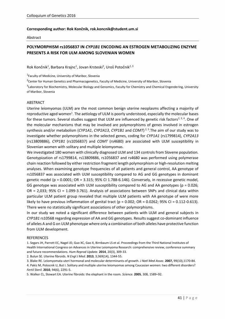
Colloquium of Genetics 2016
41 | P a g e
Corresponding author: Rok Končnik, [email protected]
Abstract
POLYMORPHISM rs1056837 IN CYP1B1 ENCODING AN ESTROGEN METABOLIZING ENZYME
PRESENTS A RISK FOR ULM AMONG SLOVENIAN WOMEN
Rok Končnik1, Barbara Krajnc1, Jovan Krsteski2, Uroš Potočnik2, 3
1Faculty of Medicine, University of Maribor, Slovenia 2Center for Human Genetics and Pharmacogenetics, Faculty of Medicine, University of Maribor, Slovenia 3Laboratory for Biochemistry, Molecular Biology and Genomics, Faculty for Chemistry and Chemical Engendering, University
of Maribor, Slovenia
ABSTRACT
Uterine leiomyomas (ULM) are the most common benign uterine neoplasms affecting a majority of
reproductive aged women1. The aetiology of ULM is poorly understood, especially the molecular bases
for these tumors. Several studies suggest that ULM are influenced by genetic risk factors2, 3, 4. One of
the molecular mechanisms that may be involved are polymorphisms of genes involved in estrogen
synthesis and/or metabolism (CYP1A1, CYP2A13, CYP1B1 and COMT) 2, 5.The aim of our study was to
investigate whether polymorphisms in the selected genes, coding for CYP1A1 (rs1799814), CYP2A13
(rs13809886), CYP1B1 (rs1056837) and COMT (rs4680) are associated with ULM susceptibility in
Slovenian women with solitary and multiple leiomyomas.
We investigated 180 women with clinically diagnosed ULM and 134 controls from Slovene population.
Genotypization of rs1799814, rs13809886, rs1056837 and rs4680 was performed using polymerase
chain reaction followed by either restriction fragment length polymorphism or high-resolution melting
analyses. When examining genotype frequencies of all patients and general control, AA genotype of
rs1056837 was associated with ULM susceptibility compared to AG and GG genotypes in dominant
genetic model (p = 0.0001; OR = 3.315; 95% CI 1.788-6.146). Conversely, in recessive genetic model,
GG genotype was associated with ULM susceptibility compared to AG and AA genotypes (p = 0.026;
OR = 2,033; 95% CI = 1.099-3.761). Analysis of associations between SNPs and clinical data within
particular ULM patient group revealed that multiple ULM patients with AA genotype of were more
likely to have previous inflammation of genital tract (p = 0.002; OR = 0.0262; 95% CI = 0.112-0.613).
There were no statistically significant associations of other polymorphisms.
In our study we noted a significant difference between patients with ULM and general subjects in
CYP1B1 rs10568 regarding expression of AA and GG genotypes. Results suggest co-dominant influence
of alleles A and G on ULM phenotype where only a combination of both alleles have protective function
from ULM development.
REFERENCES 1. Segars JH, Parrott EC, Nagel JD, Guo XC, Gao X, Birnbaum LS et al. Proceedings from the Third National Institutes of
Health International Congress on Advances in Uterine Leiomyoma Research: comprehensive review, conference summary
and future recommendations. Hum Reprod Update. 2014, 20(3), 309-33.
2. Bulun SE. Uterine fibroids. N Engl J Med. 2013, 3;369(14), 1344-55.
3. Blake RE. Leiomyomata uteri hormonal and molecular determinants of growth. J Natl Med Assoc. 2007, 99(10),1170-84.
4. Pakiz M, Potocnik U, But I. Solitary and multiple uterine leiomyomas among Caucasian women: two different disorders?
Fertil Steril. 2010, 94(6), 2291-5.
5. Walker CL, Stewart EA. Uterine fibroids: the elephant in the room. Science. 2005, 308, 1589–92.

Colloquium of Genetics 2016
42 | P a g e
Corresponding author: Matjaž Deželak, [email protected]
Abstract
A POLYMORPHISM LOCATED IN 3’UTR OF PITHD1 NEAR CANNABINOID RECEPTOR 2 GENE CNR2 IS
ASSOCIATED WITH SEVERE FORMS OF CROHN’S DISEASE AND CNR1 GENE EXPRESSION
Matjaž Deželak1, Žan Hribar1, Carina E. P. Kozmus1,2, Uroš Potočnik1,3
1Centre for Human Molecular Genetics and Pharmacogenomics, Faculty of Medicine, University of Maribor, Slovenia 2Institute for Molecular and Cell Biology, University of Porto, Porto, Portugal 3Laboratory for Biochemistry Molecular Biology and Genomics, Faculty for Chemistry and Chemical Engineering, University of
Maribor, Maribor, Slovenia
ABSTRACT
Inflammatory bowel disease (IBD) is a chronic inflammatory and autoimmune disease of gastrointestinal tract. Crohn's disease (CD) and ulcerative colitis (UC) are the principal types of inflammatory bowel disease. CD patients that do not respond to standard therapy (corticosteroids, aminosalicylates, immunosuppressives, etc.) need treatment with biological drugs. The so-called ‘refractory CD patients’ can be regarded as a yet another subtype of IBD. The search for molecular biomarkers that would identify individuals refractory to standard therapy is mandatory to save costs on therapeutics as well as to improve patient’s well-being. Several reports have suggested that the endocannabinoid system (ECS) is involved in pathology of intestinal inflammation1 and that mRNA expression of cannabinoid receptor 2 gene CNR2 correlates with the seriousness of symptoms2. A single nucleotide polymorphism (SNP) rs4237 located in the 3’ untranslated region of PITHD1 and in the vicinity of CNR2, was reported to be an expression quantitative trait locus (eQTL) of CNR2 mRNA expression levels3. Therefore, we tested the hypothesis that rs4237 and CNR2 mRNA expression are specifically associated with refractory CD but not with other types of IBD and that rs4237 is associated with CNR2 mRNA expression. We studied case-control cohorts of 276 healthy individuals, 61 UC and 113 CD patients on standard therapy and 119 refractory CD patients on adalimumab (Humira®, Abbot Laboratories) therapy. Relative mRNA expressions of CNR1 and CNR2 were measured in PBMCs with qPCR using -ΔΔCq method4. Genotyping was performed by High Resolution Melting (HRM) analysis.
Between-cohort analyses confirmed that rs4237 was associated only with refractory CD (p = 3.50 10-2; odds ratio = 0.435; 95% confidence interval: 0.204-0.924) but not with other types of IBD. According to the recessive genetic model, individuals with CT or TT genotypes were more likely to be refractory to standard therapy (28.9%) compared to individuals with CC genotype (15.0%). CNR1 and CNR2 mRNA expressions
differed significantly between healthy individuals and refractory CD patients (p = 1.66 10-9 and p = 5.37 10-21, respectively). In refractory patients, median CNR1 and CNR2 mRNA expressions were lower (-1.236 ± 2.006 and -1.586 ± 2.071, respectively) compared to healthy individuals. Within-cohort analyses confirmed
eQTL relation between rs4237 and CNR1 mRNA expression (p = 2.10 10-2) specifically for refractory CD but not for other types of IBD. According to the recessive genetic model, refractory CD patients with CT or TT genotypes had lower median CNR1 mRNA expression (-1.510 ± 1.795) compared to patients with CC genotype (-0.266 ± 4.064). Our results suggest that CT or TT genotype of rs4237 in PITHD1 as well as 2- and 3-fold lower CNR1 and CNR2 mRNA expressions, respectively, increase the risk for refractory CD. In addition, eQTL relation between rs4237 and CNR1 mRNA expression was established for refractory CD patients.
REFERENCES 1. Fichna J, Bawa M, Thakur GA, et al. Cannabinoids alleviate experimentally induced intestinal inflammation by acting at central
and peripheral receptors. PLoS ONE 2007, 9(10), e109115.
2. Wright KL, Duncan M, Sharkey KA. Cannabinoid CB2 receptors in the gastrointestinal tract: a regulatory system in states of
inflammation. Br. J. Pharmacol. 2008, 153, 263-270.
3. Dixon AL, Liang L, Moffatt MF et al. A genome-wide association study of global gene expression. Nat. Genet. 2007, 39, 1202-7.
4. Livak KJ, Schmittgen TD. Analysis of relative gene expression data using real-time quantitative PCR and the 2(-Delta Delta C(T))
method. Methods 2001, 25, 402-408.
Colloquium of Genetics 2016
43 | P a g e
ABSTRACTS and PAPERS of POSTERS
POPULATION GENETICS & GENOMICS
Taja Jeseničnik, Nataša Štajner, Sebastjan Radišek, Branka Javornik, Jernej Jakše
Discovery of small RNAs in Verticilliun nonalfalfae
Matej Babič, Tanja Kunej, Borka Jerman-Blažič
New method for statistical pattern recognition usage in cancer associated microRNAs
Blaž Skrlj, Nina Pirih, Tanja Kunej
Identification of non-synonymous polymorphisms within regions corresponding to
protein interaction sites
Marko Flajšman, Nataša Štajner, Igor Šantavec, Branka Javornik, Darja Kocjan Ačko
Genotypization and morphological characterization of six Slovenian landraces of proso
millet (Panicum miliaceum L.)

Colloquium of Genetics 2016
44 | P a g e
Corresponding author: Taja Jeseničnik, [email protected]
Abstract
DISCOVERY OF SMALL RNAs IN Verticilliun nonalfalfae
Taja Jeseničnik1, Nataša Štajner1, Sebastjan Radišek2, Branka Javornik1, Jernej Jakše1
1 University of Ljubljana, Biotechnical Faculty, Jamnikarjeva 101, SI-1000, Ljubljana, Slovenia 2 Plant Protection Department, Slovenian Institute for Hop Research and Brewing, Cesta Žalskega tabora 2, SI-3310, Žalec,
Slovenia
ABSTRACT
RNA interference is an evolutionary conserved eukaryotic mechanism regulating gene expression post-
transcriptionally1. The process of gene regulation is mediated by small RNAs2, including microRNA-like
small RNAs (milRNAs), recently shown to exist in filamentous fungi3,4,5,6. However, to date no milRNAs
have been reported in the phyto-pathogenic fungus Verticillium nonalfalfae, a soil borne plant
pathogen causing vascular wilt in many important crops worldwide. Two pathotypes of V. nonalfalfae,
mild strain and lethal strain, have been isolated from Slovenian hop fields, with the lethal strain causing
severe symptoms in hop plants, resulting in complete dieback of the plant7. With the accessible
genome sequence and detailed transcriptome of the two strains, we aim to identify milRNAs in of V.
nonalfalfae and to elucidate their possible involvement in the pathogenesis process. Discoveries of
novel microRNAs correlate with the advent of new NGS sequencing and bioinformatics technologies,
also applied for the purposes of this study. Two Slovenian isolates, Rec (mild) and T2 (lethal), were
acquired from the fungal genebank of the Slovenian Institute for Hop Research and Brewing. Small
RNA fractions were isolated from four different sources: spores, mycelia, mycelia grown on simulated
xylem fluid medium (SXM) and resting mycelia and used for small RNA library construction and
sequenced using the Ion Proton sequencing platform. Fungal milRNA precursors were predicted using
MIReNA software8 and the results were further validated and candidates selected manually using
criteria for plant miRNA candidates. Several candidate milRNA precursors were selected for both
pathotypes and have now been validated with stem-loop RT-PCR.
REFERENCES 1. Hannon GJ. RNA interference. Nature 2002, 418, 6894, 244-51.
2. Hamilton AJ, Blaucombe DC. A species of small antisense RNA in posttranscriptional gene silencing in plants. Science
1999, 286, 5441, 950-2.
3. Zhou J, Fu Y, Xie J, Li B, Jiang D, Li G, Cheng J. Identification of microRNA-like RNAs in a plant pathogenic fungus
Sclerotinia sclerotiorum by high-throughput sequencing. Mol Genet Genomics 2012, 287, 4, 275-82.
4. Chen R, Jiang N, Jiang Q, Sun X, Wang Y, Zhang H, Hu Z. Exploring microRNA-like small RNAs in the filamentous fungus
Fusarium oxysporum. PLoS One 2014, 9, 8, e104956.
5. Mueth NA, Ramachandran SR, Hulbert SH. Small RNAs from the wheat stripe rust fungus (Puccinia striiformis f.sp. tritici).
BMC Genomics 2015, 16, 718.
6. Liu T, Hu J, Zuo Y, Jin Y, Hou J. Identification of microRNA-like RNAs from Curvularia lunata associated with maize leaf
spot by bioinformation analysis and deep sequencing. Mol Genet Genomics 2016, 291, 2, 587-96.
7. Radišek S, Jakše J, Simončič A, Javornik B.Characterization of Verticillium albo-atrum Field Isolates Using Pathogenicity
Data and AFLP Analysis. Plant Disease 2003, 87, 6, 633-638.
8. Mathelier A ,Carbone A. MIReNA: finding microRNAs with high accuracy and no learning at genome scale and from deep
sequencing data. Bioinformatics 2010, 26, 18, 2226-34.

Colloquium of Genetics 2016
45 | P a g e
Corresponding author: Matej Babič, [email protected]
Abstract
NEW METHOD FOR STATISTICAL PATTERN RECOGNITION USAGE IN CANCER ASSOCIATED
microRNAs
Matej Babič1, Tanja Kunej2, Borka Jerman-Blažič1
1Jozef Stefan Institute, Laboratory for Open Systems and Networks, Slovenia 2University of Ljubljana, Biotechnical Faculty, Department of Animal Science, Slovenia
ABSTRACT
MicroRNAs (miRNAs) are small 22-25 nucleotides long noncoding RNA molecules that regulate gene
expression and that are present and stable in the bloodstream. They are evolutionally conserved, and
control gene expression in metazoan animals, plants, viruses, and bacteria primarily at post-
transcriptional and transcriptional levels. In this study, we present a novel 2D graphical representation
of miRNAs sequences based on graph theory. Graph theory is one of most tools in mathematics, which
are used in many cases, because we can simple present very complex problems. In recent years, a very
large variety of statistical methodologies, at various levels of complexity, have been put forward to
analyse genotype data and detect genetic variations that may be responsible for increasing the
susceptibility to cancer. In this article, we present new method for statistical pattern recognition.
Statistical pattern recognition relates to the use of statistical techniques for analysing data
measurements in order to extract information and make justified decisions. We considered
construction of n × 4 matrix to represent DNA primary sequences. Four column of matrix present 4
nucleotides of DNA base. First column present A, second C, third G and last column present T. We
traveled through DNA sequences to determinate each nucleotide from DNA base in matrix. Denoted
nucleotides present vertex of new graph. We calculated topological properties of this graph and
compared it for different genomic miRNAs sequences. The novelty of graphical representations of
miRNAs sequences is very useful for prediction cancer association. We tested the proposed method
on hsa-mir-423, hsa-mir-27a. The importance of topological modeling of miRNAs is distinguished, as
it presents simplified visual characteristics for the rapid and efficient modeling, comparison of miRNAs
and researching effort to establish their roles in cancer.
REFERENCES
1. Gregory, SG; Barlow, KF; McLay, KE; Kaul, R; Swarbreck, D; Dunham, A; Scott, CE; Howe, KL; et al. (2006). "The DNA
sequence and biological annotation of human chromosome 1". Nature. 441 (7091): 315–21. Bibcode:2006Natur.441..315G.
doi:10.1038/nature04727. PMID 16710414.

Colloquium of Genetics 2016
46 | P a g e
Corresponding author: Tanja Kunej, PhD, [email protected]
Article
IDENTIFICATION OF NON-SYNONYMOUS POLYMORPHISMS WITHIN REGIONS
CORRESPONDING TO PROTEIN INTERACTION SITES
Blaz Skrlj1, Nina Pirih1, Tanja Kunej1
1Department of Animal Science, Biotechnical Faculty, University of Ljubljana
Background: Protein-protein interactions (PPI) play an important role in function of all organisms and enable
understanding underlying metabolic processes. Predictions of protein interactions are an important aspect in proteomics,
as experimental methods may result in high degree of false positive results and are more expensive than computational
predictions. Prediction of PPIs has been a topic of increased interest in the last few years. Although there are many
databases collecting predicted PPIs, exploration of genetics information underlying PPI interactions has not been
investigated thoroughly. The aim of the present study was to identify: 1. genomic locations corresponding to regions
involved in predicted interaction sites, 2. Non-synonymous polymorphisms within interacting sites (PPI-SNPs), 3. examples
in which predicted PPIs sites overlap with previously reported experimentally validated interactions. Methods: Datasets
representing predicted PPIs were obtained from PiSITE database (http://pisite.hgc.jp). Non-synonymous polymorphisms
mapped on protein structural data (PDBs) were obtained from the UCSC’s ftp server. Polymorphism locations on protein
structures were mapped to predicted PPI regions in order to identify PPI-SNPs. Locations of experimentally validated PPIs
were extracted from published literature (http://www.ncbi.nlm.nih.gov/pubmed). Location of polymorphisms on 3D
protein structures was visualized using PyMol software. Results: We developed a scalable method for identification of PPI-
SNPs, which was further used to identify 662 polymorphisms located within the predicted PPI sites that map to 196 genes
that are associated with different diseases, mainly with different types of cancer. Additionally, 13 polymorphisms in seven
genes within predicted PPIs are also located within experimentally validated interaction regions. Conclusions: The present
study presents novel SNP prioritization approach for functional studies and potential candidates for disease biomarkers. INTRODUCTION
The study of protein interactions (PIs) provides insights into biological processes. Proteins can interact
with DNA, smaller ligands, RNA or with other proteins (protein-protein interactions; PPIs). Protein
interaction malfunctions can result in loss of phenotypic trait, which many times results in a disease1.
Relevance of protein interaction prediction is situated on the fact, that obtaining empirical evidence in
great quantity is currently a challenging task. Additionally, accuracy of high-throughput experiments
still yields many false-positive and false-negative results2. Using the data of predicted protein
interactions many a times provides additional evidence in explaining various biological phenomena3,
or facilitate planning experimental validation of experimental PPIs4. Use of modern prediction
techniques such as machine learning, graph theory, Bayesian prediction and pattern recognition have
been used in tools, such as Struct2Net5, PiPs6, HitPredict7, PRISM8, DOMINE9, ProBis10, and PiSITE11.
Previous studies on domain altering SNPs in human proteome12 identified many polymorphisms which
alter specific protein domains and can therefore have clinically significant effects. Non-synonymous
polymorphisms (nsSNP) have been shown to cause pathological effects, for example in
hypercholesterolemia13, incident coronary heart disease14, breast cancer15 and many other types of
cancer.
Prediction of the effect of amino acid substitution on protein function is an important task in systems
biology. One of the most widely used tools in this field is SIFT algorithm, which uses homology based
approach to identify amino acid loci, which could potentially affect protein function. SIFT tool has
already been successfully used to analyze effects of possible cancer mutations16; yet identified
mutations were not directly associated with protein-protein interaction sites. However, significant 3D
clustering of missense mutations in several previously known oncoproteins has been detected17. In our
previous study we reanalyzed genomic regions involved in interactions between transcription factor
and its targets and identified polymorphisms located within hypoxia response elements (HREs)18.

Colloquium of Genetics 2016
47 | P a g e
Although there are already many datasets providing predicted PPI interactions, those predictions
remain in the protein domain – they are not associated with underlying nucleotide sequence and
genetic variability. To our knowledge, there are no reported studies related with polymorphisms
located within regions corresponding to predicted protein-protein interaction information performed
for nsSNPs, mapped onto protein structure (PDB)16.
Protein-protein interaction data used in this study was obtained from the PiSITE database. Interactions
were predicted using BLAST queries, residue specific interactions were identified based on distance
criteria, where distance between residues needed to be smaller than 4 Å11. The aim of this study was
to identify nsSNPs, occurring within predicted protein-protein interaction sites.
METHODS
Non-synonymous polymorphisms mapped onto PDB structures (n=11551) were obtained from UCSC
institute’s ftp server19. Regions involved in predicted protein-protein interactions were obtained from
PiSITE11. Mutations’ datasets were preprocessed and further merged using algorithms, implemented
in R language (https://www.r-project.org/). Using Python scripting (https://www.python.org/), we
extracted all interactions from the PiSITE dataset, consisting of 110325 chains. Association of genes
with diseases was analyzed using functional annotation tool DAVID
(https://david.ncifcrf.gov/home.jsp). The predicted protein-protein interaction interfaces were further
analyzed for overlap with experimentally validated interactions in previously published literature
(http://www.ncbi.nlm.nih.gov/pubmed). In addition, we selected nsSNPs that are located within
predicted as well as experimentally validated PPI regions. Selected results were visualized using open-
source PyMol software (https://sourceforge.net/projects/pymol/).
RESULTS
In the present study we identified nsSNPs located within regions corresponding to predicted protein-
protein interaction interfaces. For some selected cancer genes we also confirmed that predicted
interaction sites overlap with previously reported experimentally validated interaction sites. Main
workflow of the process is presented in Figure 1.
Figure 1: Main workflow of the study.
Mapping of nsSNPs to predicted interaction sites resulted in the catalog of 662 nsSNPs with matching
reference SNP ID (rs) accession numbers from dbSNP. Collected 662 PPI-SNPs within predicted
interaction sites map to 196 protein-coding genes. Further data processing revealed that from 196
identified genes, 48 are statistically significant associated with at least one type of cancer. Additionally,
genes were also associated with Alzheimer’s disease, rheumatoid arthritis, diabetes type 1 and type 2,
asthma, cardiovascular disease, cholesterol, depression, and Crohn’s disease. Ten cancer associated
genes with the highest number of SNPs located within predicted PPIs are presented in Table 1.
Table 1: Ten genes with the highest number of SNPs, located within predicted PPIs..

Colloquium of Genetics 2016
48 | P a g e
Gene name CFTR TCAP XRCC5 FGFR2 MICA SERPINA1 VHL TP53 BRCA1
Number of PPI-SNPs
5 5 5 6 6 8 8 10 15
rs SNP ID numbers
(examples)
rs74571530
rs35032490
rs1800100
rs80055610
rs11531593
rs45495192
rs45458802
rs17851031
rs45614536
rs45513698
rs1805380
rs11558396
rs2287558
rs11575909
rs61758380
rs55977237
rs755793
rs77543610
rs4647921
rs79184941
rs55689343
rs3819268
rs17200179
rs17206575
rs17206589
rs17200207
rs17200186
rs17580
rs28929471
rs28929473
rs11575873
rs1131154
rs55819880
rs72552401
rs12077
rs17855706
rs5030812
rs5030818
rs28940297
rs5030811
rs5030804
rs5030805
rs5030822
rs28934875
rs28934572
rs28934575
rs11540652
rs28934573
rs28934576
rs28934578
rs28934571
etc.
rs80357316
rs80357031
rs80357054
rs80357017
rs80357406
rs80357438
rs1800062
rs80356929
etc.
Legend: CFTR: Cystic fibrosis transmembrane conductance regulator; TCAP: Telethonin, XRCC5: X-ray repair
cross-complementing protein 5, FGFR2: Fibroblast growth factor receptor 2, MICA: MHC class I polypeptide-
related sequence A, SERPINA1: Alpha-1-antitrypsin, VHL: von Hippel-Lindau tumor suppressor; TP53: tumor
protein p53; BRCA1: BRCA1, DNA repair associated.
Using data from the literature we found 7 genes in which predicted PPI sites also corresponded to
experimentally validated interaction sites: TCAP, APOA1, CRP, ESR1, TP53, VHL, and SHFM1. Out of 662
PPI-SNPs 13 nsSNPs are present within predicted and validated PPIs (Table 2).
Table 2: Identified nsSNPs in genes associated with cancer that are located within predicted as well as experimentally
validated protein-protein interaction interfaces.
Gene name rs SNP ID numbers
Associated cancer type (According to DAVID functional annotation analysis).
Interaction sites found in the literature
TCAP rs45495192 rs45614536 rs45513698
breast cancer 1–53 (binding to the very N-terminus of titin20)
APOA1 rs28931573 colorectal cancer 161–240 (binding to NDRG121)
CRP rs34200896 lung cancer 109-132 (constitutive binding region22)
ESR1 rs79374934
bone cancer, breast cancer, colon cancer, endometrial cancer, prostate cancer, uterine cancer etc.
185–595 (binding to MUC1-CD23) 410–731 (binding to HPIP24)
TP53 rs28934576
bladder cancer, leukemia, lung cancer, brain cancer, breast cancer, cervical cancer, stomach cancer etc.
256 – 294 (Binding to p120E4F25)
VHL rs17855706 rs5030812 rs5030818 rs28940297
brain cancer, breast cancer, renal cancer, kidney cancer
100-155 (binding to TRiC26) 157–172 (binding to elongin BC26),
SHFM1 rs11553943 rs1802882
breast cancer 7-25, 37-63 (binding to BRCA227)
For example, in VHL gene there are four nsSNPs within predicted PPI and also located within previously
experimentally validated interaction regions. Graphic presentation for VHL gene is presented in Figure
2. Polymorphisms are located on locus 110 (rs17855706), 115 (rs5030812), 161 (rs5030818) and 163
(rs28940297). Experimentally validated PPI sites encompass amino acids 100-155 and 157-17226.

Colloquium of Genetics 2016
49 | P a g e
Figure 2: Visualization of polymorphisms within predicted and experimental interaction regions in VHL gene (PDB accession
1lm8, chain V) on modified figure from PiSITE server. Predicted PPI region is marked with green color and
experimental PPI region with blue line. Polymorphisms are marked with red boxes.
Three dimensional visualization of VHL gene and four polymorphisms located within predicted and
validated interaction sites are presented in Figure 3.
Figure 3: Representation of different layers of interaction information in this study on example protein VHL. Four
polymorphisms located within both, predicted and experimentally validated interaction sites are marked in pink
color: rs17855706 (His110Tyr), rs5030812 (His115Arg), rs5030818 (Arg161Ter) and rs28940297 (Leu163Pro).
DISCUSSION
Identification of nsSNPs located within predicted protein-protein interaction sites provides additional
information on biological phenomena and enables more detailed explanation of disease development.
Within locations of predicted protein interactions we identified 662 polymorphisms mapping to 196
genes, which showed significant clustering in terms of cancer and other disease associations. The

Colloquium of Genetics 2016
50 | P a g e
results of the present study are in concordance with an observation by Kamburov et al. (2015), who
proved significant clustering patterns of polymorphisms emerged in cancer related protein
interactions. Oncoproteins VHL, TP53 and BRCA1 were in the present study additionally confirmed as
proteins with the highest number of predicted PPI-SNPs. In addition, regions involved in predicted PPI
also overlapped with experimentally validated interactions in seven proteins, including VHL and TP53.
By literature mining we found two interaction sites for protein VHL. VHL binds to elongin BC via a linear
sequence known as “BC box”, consist of amino acids 157–17226. We have detected two nsSNPs
(rs5030818, rs28940297) located within this region, which is known as a hot spot for disease-causing
inherited point mutations26. Furthermore we have identified two nsSNPs (rs17855706, rs5030812) in
the region of VHL comprising amino acids 100-155, that mediates TRiC binding26. Within previously
reported experimentally validated interaction region of oncoprotein TP53, we have identified one
nsSNP (rs28934576). This TP53 region is important for binding to the E1A-regulated transcription factor
p120E4F, a transcriptional repressor of the adenovirus E4 promoter. The interaction involves carboxy-
terminal half of p120E4F and sequences located at the end of the sequence-specific DNA-binding
domain of TP53, which encompassing amino acids 256 – 29425.
Similarly to our previous SNP prioritization studies in which we collected polymorphisms located within
miRNA genes28 and within transcription factor-target interaction18 in the present study we collected
polymorphisms, located within predicted PPI sites, associated with cancer risk. Additionally we have
also confirmed that predicted interaction sites overlap with experimentally validated interactions in
previously published literature. In conclusion, we identified 13 nsSNPs located within both, predicted
and previously experimentally validated interaction sites that map to seven cancer associated genes.
In addition, an investigation whether other identified nsSNPs within PPI sites are associated with
cancer should also follow. The results of the present preliminary study could serve as a basis for
planning more targeted experimental designs, functional experiments and might present novel drug
targets and therapeutic strategies for cancer.
REFERENCES
1. Gonzalez M W, Kann M. G. Chapter 4: Protein interactions and disease. PLoS Comput Biol. 2012, 8, 12.
2. Rao V S, Srinivas K, Sujini G N, Kumar G N. Protein-protein interaction detection: methods and analysis. Int J Proteomics.
2014.
3. Zahiri J, Bozorgmehr J H, Masoudi-Nejad A. Computational prediction of protein-protein interaction networks: algo-
rithms and resources. Curr Genomics. 2013, 14, 6, 397-414.
4. Jessulat M, Pitre S, Gui Y, Hooshyar M, Omidi K, Samanfar B et al. Recent advances in protein-protein interaction
prediction: experimental and computational methods. Expert opinion on drug discovery. 2011, 6, 9, 921-935.
5. Singh R, Park D, Xu J, Hosur R, Berger B. Struct2Net: a web service to predict protein-protein interactions using a
structure-based approach. Nucleic Acids Res. 2010, 38, 508-515.
6. McDowall M D, Scott M S, Barton G J. PIPs: human protein-protein interaction prediction database. Nucleic Acids Res.
2009, 37, 651-656.
7. López Y, Nakai K, Patil A. HitPredict version 4: comprehensive reliability scoring of physical protein-protein interactions
from more than 100 species. Database (Oxford). 2015.
8. Baspinar A, Cukuroglu E, Nussinov R, Keskin O, Gursoy A. PRISM: a web server and repository for prediction of protein-
protein interactions and modeling their 3D complexes. Nucleic Acids Res. 2014, 42, 285-289.
9. Yellaboina S, Tasneem A, Zaykin D V, Raghavachari B, Jothi R. DOMINE: a comprehensive collection of known and
predicted domain-domain interactions. Nucleic Acids Res. 2011, 39, 730-735.
10. Konc J, Janežič D. ProBiS-ligands: a web server for prediction of ligands by examination of protein binding sites. Nucleic
Acids Res. 2014, 42, 215-220.
11. Higurashi M, Ishida T, Kinoshita K. PiSite: a database of protein interaction sites using multiple binding states in the
PDB." Nucleic acids research. 2009, 37, 360-364.
12. Liu Y, Tozeren A. Domain altering SNPs in the human proteome and their impact on signaling pathways. PLoS One. 2010,
5, 9.

Colloquium of Genetics 2016
51 | P a g e
13. Angarica V E, Orozco M, Sancho J. Exploring the complete mutational space of the LDL receptor LA5 domain using
molecular dynamics: linking SNPs with disease phenotypes in familial hypercholesterolemia. Hum Mol Genet. 2016, 25,
6, 1233-1246.
14. Jensen M K, Pers T H, Dworzynski P, Girman C J, Brunak S, Rimm E B. Protein interaction-based genome-wide analysis of
incident coronary heart disease. Circ Cardiovasc Genet. 2011, 4, 5, 549-556.
15. Milne R L, Burwinkel B, Michailidou K, Arias-Perez J I, Zamora M P, Menéndez-Rodríguez P et al. Common non-
synonymous SNPs associated with breast cancer susceptibility: findings from the Breast Cancer Association Consortium.
Hum Mol Genet. 2014, 23, 22, 6096-6111.
16. Kumar P, Henikoff S, Ng P C. Predicting the effects of coding non-synonymous variants on protein function using the
SIFT algorithm. Nature protocols. 2009, 4, 7, 1073-1081.
17. Kamburov A, Lawrence M S, Polak P, Leshchiner I, Lage K, Golub T R et al. Comprehensive assessment of cancer
missense mutation clustering in protein structures. Proc Natl Acad Sci U S A. 2015, 112, 40, 5486-5495.
18. Slemc L, Kunej T. Transcription factor HIF1A: downstream targets, associated pathways, polymorphic hypoxia response
element (HRE) sites and initiative for standardization of reporting in scientific literature. Tumor Biology. In press.
19. Ryan M, Diekhans M, Lien S, Liu Y, Karchin R. LS-SNP/PDB: annotated non-synonymous SNPs mapped to Protein Data
Bank structures. Bioinformatics. 2009, 25, 11, 1431-1432.
20. Pinotsis N, Petoukhov M, Lange S, Svergun D, Zou P, Gautel M et al. Evidence for a dimeric assembly of two
titin/telethonin complexes induced by the telethonin C-terminus. J Struct Biol. 2006, 155, 2, 239-250.
21. Hunter M., Angelicheva D, Tournev I, Ingley E, Chan D C, Watts G F et al. NDRG1 interacts with APO A-I and A-II and is a
functional candidate for the HDL-C QTL on 8q24. Biochem Biophys Res Commun. 2005, 332, 4, 982-992.
22. Zhang J, Yang L, Ang Z, Yoong S L, Tran T T, Anand G S et al. Secreted M-ficolin anchors onto monocyte transmembrane
G protein-coupled receptor 43 and cross talks with plasma C-reactive protein to mediate immune signaling and regulate
host defense. J Immunol. 2010, 185, 11, 6899-6910.
23. Wei X, Xu H, Kufe D. MUC1 oncoprotein stabilizes and activates estrogen receptor alpha. Mol Cell. 2006, 21, 2, 295-305.
24. Manavathi B, Acconcia F, Rayala S K, Kumar R. An inherent role of microtubule network in the action of nuclear
receptor. Proc Natl Acad Sci U S A. 2006, 103, 43, 15981-15986.
25. Sandy P, Gostissa M, Fogal V, Cecco L D, Szalay K, Rooney R J et al. p53 is involved in the p120E4F-mediated growth
arrest. Oncogene. 2000, 19, 2, 188-199.
26. Feldman D E, Thulasiraman V, Ferreyra R G, Frydman J. Formation of the VHL-elongin BC tumor suppressor complex is
mediated by the chaperonin TRiC. Mol Cell. 1999, 4, 6, 1051-1061.
27. Yang, H, Jeffrey P D, Miller J, Kinnucan E, Sun Y, Thoma, N H et al. BRCA2 function in DNA binding and recombination
from a BRCA2-DSS1-ssDNA structure. Science. 2002, 297, 5588, 1837-1848.
28. Zorc M, Obsteter J, Dovc P, Kunej T. Genetic Variability of MicroRNA Genes in 15 Animal Species. J Genomics. 2015, 3,
51-56.

Colloquium of Genetics 2016
52 | P a g e
Corresponding author: Darja Kocjan Ačko, [email protected]
Abstract
GENOTYPIZATION AND MORPHOLOGICAL CHARACTERIZATION OF SIX SLOVENIAN
LANDRACES OF PROSO MILLET (Panicum miliaceum L.)
Marko Flajšman1, Nataša Štajner1, Igor Šantavec1, Branka Javornik1, Darja Kocjan Ačko1 1University of Ljubljana, Biotechnical Faculty, Agronomy Department, Jamnikarjeva 101, Ljubljana SI-1000, Slovenia
ABSTRACT
Proso millet (Panicum miliaceum L.) is one of the world's oldest cultivated cereals. It appeared as a staple crop in northern China 10,000 years ago1 and later spread to other parts of the world, including Slovene territory where it was grown as early as a thousand years B.C. by the Celts2. Proso millet has two main advantages over other cereals: firstly, it is considered a health-food crop owing to its unique nutritional value3 and secondly, it is very tolerant of various abiotic stresses, e.g., salinity and drought. In addition, it can grow well in poor soils4. In Slovenia, the cultivation of millet reached its peak in the 19th century, covering 18,000 ha. In 2016, proso millet is cultivated on only around 200 ha, mostly by organic farmers, who use landraces of yellow and white proso millet from Prekmurje and Gorenjska regions, the Austrian cultivar 'Kornberger Mittelfrühe' domesticated in Slovenia as 'Kornberg millet' and, in recent years, also the cultivar 'Sonček'2. In the current study, we analysed the genetic diversity of six Slovenian landraces of proso millet together with the cultivar 'Sonček' by utilizing both molecular markers and morphological traits. The use of microsatellites (SSRs), which have proved to be a powerful tool for genetic diversity analyses in various crops, also resulted in high discrimination with proso millet. We successfully amplified 11 SSR loci and, of these, 7 loci were polymorphic. Only accessions of the cultivar 'Sonček' formed a uniform cluster, all other landraces were very diverse. Nevertheless, we were able to differentiate six different clusters of genotypes, representing six landraces, which differ also in the morphology of the panicle. Moreover, yield stability and 1000-seed weight, as well as other morphological characteristics, were determined in a 2-year field experiment, also showing very diverse results between years as well as among samples within some landraces. 1. Lu H, Zhang J, Liu K-B, Wu N, Li Y, Zhou K, Ye M, Zhang T, Zhang H, Yang X. Earliest domestication of common millet (Panicum miliaceum) in East Asia extended to 10,000 years ago. Proc Natl Acad Sci USA. 2009, 106, 7367–7372. 2. Kocjan Ačko D. Importance and possibilities of proso millet (Panicum miliaceum L.) production for human nutrition and animal feed in Slovenia. Int. j. food, agric. environment. 2012, 10(2), 636-640. 3. Liu M, Qiao Z, Zhang S, Wang Y, Lu P. Response of broomcorn millet (Panicum miliaceumL.) genotypes from semiarid regions of China to salt stress. Crop J. 2015, 3, 57–66. 4. Sabir P, Ashraf M, Akram NA. Accession variation for salt tolerance in proso millet (Panicum miliaceum L.) using leaf proline content and activities of some key antioxidant enzymes. J. Agron. Crop Sci. 2011, 197, 340–347.

Colloquium of Genetics 2016
53 | P a g e
SPONSORS