slides 1 10
TRANSCRIPT
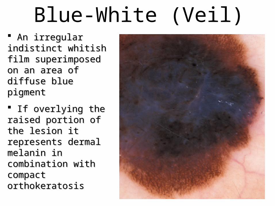
Blue-White (Veil) An irregular indistinct An irregular indistinct whitish film whitish film superimposed on an area superimposed on an area of diffuse blue pigmentof diffuse blue pigment
If overlying the raised If overlying the raised portion of the lesion it portion of the lesion it represents dermal represents dermal melanin in combination melanin in combination with compact with compact orthokeratosisorthokeratosis
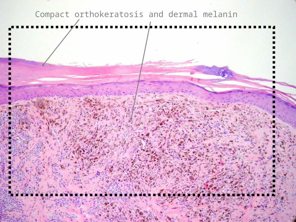
Compact orthokeratosis and dermal melanin

Blue-white veil
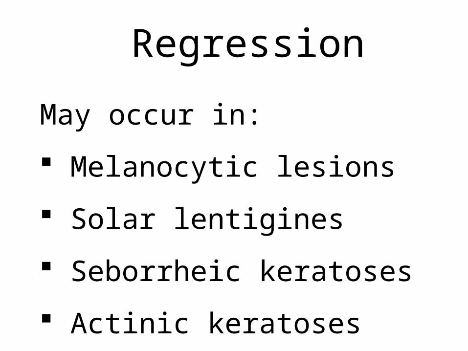
Regression
May occur in:
Melanocytic lesions
Solar lentigines
Seborrheic keratoses
Actinic keratoses

Well localized blue-gray dots or granules Diffuse multiple blue-gray dots or granules on a background of hypomelanosis Histologically represents melanin and fibrosis in the papillary dermis
Regression characterized by:
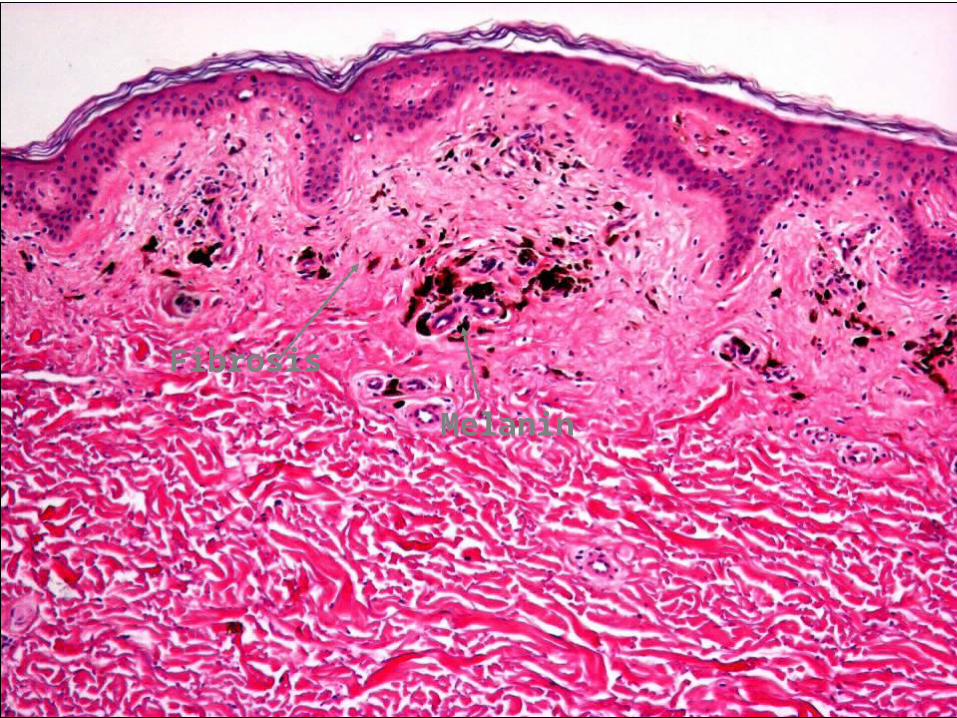
Melanin
Fibrosis

Regression
Pattern AnalysisPattern AnalysisThe differentiation of The differentiation of
melanocytic nevi from melanocytic nevi from melanomamelanoma
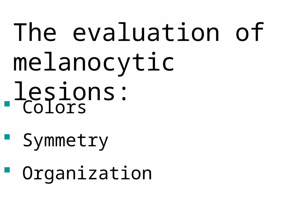
Colors
Symmetry
Organization
The evaluation of melanocytic lesions:

BlackBlack
Brown Brown
GrayGray BlueBlue
RedRed
WhiteWhite YellowYellow
Benign lesions generally have 3 or less colors. The most common colors being light brown, dark brown, and black.
Malignant lesions generally have more than 3 colors.