skx - ucl discoverydiscovery.ucl.ac.uk/1446588/1/u602513.pdf · single voxel proton magnetic...
TRANSCRIPT

THE EARLY DIAGNOSIS AND MANAGEMENT OF
CREUTZFELDT-JAKOB DISEASE
Submitted to the University of London
for the degree of Doctor of Medicine (MD)
Rebecca Jane Cordery BSc MB BS MRCP
University College London
Gower Street, London WC1N 3BG
March 2003
/SKX( LCM.) \ i m /
1
UMI Number: U602513
All rights reserved
INFORMATION TO ALL USERS The quality of this reproduction is dependent upon the quality of the copy submitted.
In the unlikely event that the author did not send a com plete manuscript and there are missing pages, th ese will be noted. Also, if material had to be removed,
a note will indicate the deletion.
Dissertation Publishing
UMI U602513Published by ProQuest LLC 2014. Copyright in the Dissertation held by the Author.
Microform Edition © ProQuest LLC.All rights reserved. This work is protected against
unauthorized copying under Title 17, United States Code.
ProQuest LLC 789 East Eisenhower Parkway
P.O. Box 1346 Ann Arbor, Ml 48106-1346

ABSTRACT
This thesis describes work undertaken to improve the early diagnosis of variant
Creutzfeldt-Jakob disease (vCJD), using existing clinical and research tools.
Twenty-one cases referred to the National Hospital for Neurology and Neurosurgery
and St. Mary’s Hospital, London with suspected vCJD completed participation in the
study. Fifteen cases were confirmed with definite or probable vCJD and six were
given alternative diagnoses. These six cases with alternative diagnoses formed a
control group. Further controls were recruited from patients referred with sporadic
and familial forms of prion disease.
A neuropsychiatry questionnaire comprising a battery of standardised tests was
formulated. Of those with definite or probable vCJD, 86% exhibited anxiety, 93%
irritability, 64% agitation and 79% displayed evidence of severe depressive
symptoms. Fifty seven percent experienced simple delusions, most commonly of theft
and suspicion and 36% described misidentifications (mean 8 months from illness
onset). Behavioural change was common to all cases, 79% with aggression, 71%
emotional lability and 79% sleep problems.
Comprehensive neuropsychology assessments from those with vCJD were compared
with sporadic and familial cases. Moderate to severe intellectual decline is
characteristic of vCJD and impairment affects all cognitive domains. Only a minority
of the vCJD cases presented with perceptual impairment compared with 50% of
sporadic and familial cases. The proportion of cases with nominal impairment in the
familial disease group was significantly lower than in the variant and sporadic groups.
2

Serial volumetric MR imaging was only possible in a subgroup of cases with familial
CJD. The annual mean rate of whole brain atrophy was 2.05% compared to 0.25% in
normal controls. Single voxel proton magnetic spectroscopy performed in three cases
with vCJD showed a 2.5 fold (150%) increase in the mean myo-inositol concentration
and 50% reduction in N-acetylaspartate in the pulvinar region. Similar changes were
seen in the caudate nucleus where no signal change was detected on T2 weighted
images.
The key to early diagnosis still relies on a high index of suspicion for vCJD and early
referral to the appropriate specialist services. First hand experience of the problems
faced by patients prompted a second, parallel project to be undertaken. A survey was
conducted of all UK consultant neurologists and old age psychiatrists to assess current
practices in the diagnosis and management of young people with dementia. It was
concluded that young people may be under investigated if managed solely by an old
age psychiatrist and may not receive adequate follow up services if managed solely by
a neurologist.
3

DEDICATION
This thesis is dedicated to my husband Roger and our daughters, Rachel and Justine.
Roger has provided untiring support with patience and encouragement, always giving
sound judgement and advice. Rachel and Justine have both been born since I started
this work. Their happy, inquisitive natures and excitement for life are an amazing
new source o f inspiration.
“The way to love anything is to realize that it might be lost”
G.K. Chesterton (1874-1936).
TABLE OF CONTENTS
ABSTRACT.........................................................................................................................2
DEDICATION.
LIST OF TABLES............................................................................................................. 8
LIST OF FIGURES......................................................................................................... 10
AIMS AND OBJECTIVES............................................................................................. 11
AIMS AND OBJECTIVES............................................................................................. 11
Characterisation of a new disease................................................................................ 11
The need for an early diagnosis.................................................................................... 12
Study outline I: Using psychiatric, cognitive and neuroimaging tools in the early diagnosis of vCJD............................................................................................................... 13Study outline II: National survey to assess current practices in the diagnosis and management of young people with dementia...................................................................13
INTRODUCTION............................................................................................................ 15
The prion diseases........................................................................................................... 15The nature of the infectious agent........................................................................................................16Prion biology..........................................................................................................................................17Prion strain diversity..............................................................................................................................18The species barrier and susceptibility to prion disease...................................................................... 19
Classification of prion diseases...................................................................................... - 19
Sporadic C J D .................................................................................................................... 20
Familial C JD ................................................................................................................... - 21
Acquired forms of CJD ................................................................................................... .. 22Bovine Spongiform Encephalopathy................................................................................................... 23The emergence of variant CJD.............................................................................................................24Sporadic CJD in young people.............................................................................................................25Evidence for transmission of BSE to humans.....................................................................................27
Variant C JD ....................................................................................................................... 29Epidemiology.........................................................................................................................................29Predicting the size of a future epidemic.............................................................................................. 30Investigating the possible underascertainment o f vCJD................................................................... 32Clinical features o f variant CJD...........................................................................................................34Diagnostic criteria for variant CJD...................................................................................................... 35Tonsillar biopsy......................................................................................................................................37
STUDY I.............................................................................................................................39
Methods...............................................................................................................................39
5

Case selection........................................................................................................................................ 39Consent and ethical considerations..................................................................................................... 41Inclusion criteria.................................................................................................................................... 42
Case Summaries.................................................................................................................44
Patient V I........................................................................................................................... 44
Results..................................................................................................................................62
STUDY IA: A STUDY OF THE PSYCHIATRIC MANIFESTATIONS OF CJD. 81
Introduction........................................................................................................................ 81
Methods...............................................................................................................................86Assessment tools................................................................................................................................... 87Definitions of psychiatric terms...........................................................................................................89
Results..................................................................................................................................91Disorders o f thought content: Delusions and persecutory ideation................................................ 94Disorders o f perception: Hallucinations and misidentifications......................................................96Behavioural change............................................................................................................................... 97Mood disturbances................................................................................................................................ 99Activities o f daily living..................................................................................................................... 100The assessment o f premorbid personality traits................................................................................101
Discussion..........................................................................................................................101
STUDY IB: A STUDY OF THE COGNITIVE FEATURES OF CJD....................107
Introduction.......................................................................................................................107
Methods.............................................................................................................................110
Results................................................................................................................................113
Discussion..........................................................................................................................119
STUDY 1C: NEUROIMAGING IN CJD ....................................................................124
Introduction.......................................................................................................................124
Quantification of cerebral and cerebellar atrophy....................................................... 127Methods................................................................................................................................................ 129Results................................................................................................................................................... 136Discussion............................................................................................................................................. 143
MR spectroscopy in CJD.................................................................................................146Introduction.......................................................................................................................................... 146Methods................................................................................................................................................ 148Results....................................................................................................................................................150Discussion............................................................................................................................................. 158
STUDY II: THE MANAGEMENT OF YOUNG PEOPLE WITH DEMENTIA. 161
Introduction.......................................................................................................................161
Method............................................................................................................................... 163Statistical Analysis...............................................................................................................................163
Results................................................................................................................................ 163
Discussion.......................................................................................................................... 169
6

CONCLUSIONS............................................................................................................173
REFERENCES..............................................................................................................178
APPENDICES............................................................................................... 195
PUBLICATIONS RELATING TO THIS THESIS...................................................202
ACKNOWLEDGEMENTS.......................................................................................... 204
7

LIST OF TABLES
Table 1 The number of cases of variant CJD assessed for the study with the total
deaths from definite or probable variant CJD in the UK over the same time
period..........................................................................................................................39
Table 2: Assessment stage, confirmed cases of variant CJD.........................................66
Table 3: Assessment characteristics, alternative diagnoses...........................................66
Table 4: Final alternative diagnoses................................................................................ 66
Table 5: Presenting symptoms, confirmed cases of variant C JD ................................. 67
Table 6: Presenting symptoms, group with alternative diagnoses................................ 68
Table 7: Symptoms experienced throughout the course of the illness, confirmed cases
of variant CJD........................................................................................................... 69
Table 8: Symptoms experienced throughout the course of the illness, group with
alternative diagnoses.................................................................................................70
Table 9: Clinical signs, confirmed cases of variant CJD................................................71
Table 10: Clinical signs, cases with alternative diagnoses............................................ 72
Table 11: CSF examination, confirmed cases of variant CJD.......................................73
Table 12: CSF examination, cases with other diagnoses................................................74
Table 13: MRI examination, confirmed cases of variant C JD ......................................75
Table 14: MRI examination, other diagnoses.................................................................76
Table 15: EEG examination, variant CJD cases..............................................................77
Table 16: EEG examination, other diagnoses.................................................................78
Table 17: Biopsy and histology, variant CJD cases....................................................... 79
Table 18: Biopsy and histology, other diagnoses........................................................... 80
Table 19: Summary of classification of sporadic CJD based on molecular and
phenotypic analysis of 300 subjects; Parchi et al, 199927...................................... 83
Table 20: Milestones in diagnosis (Variant CJD ).......................................................... 92
Table 21: Milestones in diagnosis (other diagnoses)......................................................92
Table 22: Psychiatric Milestones (Variant CJD).............................................................93
Table 23: Psychiatric Milestones (Other Diagnoses).....................................................94
Table 24: Disorders of Thought; vCJD group................................................................95
Table 25: Disorders of Perception; vCJD group.............................................................96
Table 26: Behavioural Features; vCJD group (14 cases with definite or probable
vCJD)......................................................................................................................... 98
Table 27: Mood Disturbance; vCJD group................................................................... 100
8
Table 28: Neuropsychology assessment: Mean age and sex distribution of the cases
..................................................................................................................................I l l
Table 29: Clinical features of all cases undergoing neuropsychology assessment... 114
Table 30: Level of intellectual decline per patient group............................................ 115
Table 31: Number of patients impaired in each cognitive domain per group (compared
using Fisher’s exact test)........................................................................................ 117
Table 32: Three pairwise comparisons of the proportions of patients in each group
with impaired nominal skills..................................................................................117
Table 33: Longitudinal data showing both severity of intellectual decline in familial
cases (n=8), and the number of cases with impairment in each cognitive domain
at baseline assessment.............................................................................................118
Table 34: MR study: Clinical symptoms and signs...................................................... 137
Table 35: Mean volumes corrected for Total Intracranial Volume (TIV)..................138
Table 36: Percentage rate of change of BBSI per year.................................................142
Table 37: MR spectroscopy: Clinical characteristics of the cases...............................151
Table 38: Metabolite concentrations in the left thalamus (institutional units (iu) given)
..................................................................................................................................154
Table 39: Metabolite concentrations in the right frontal white m atter....................... 154
Table 40: Metabolite concentrations in the right caudate nucleus (institutional units
(iu) given)................................................................................................................155
Table 41: Summary of the key responses from consultant neurologists and old age
psychiatrists.............................................................................................................168
9

LIST OF FIGURES
Figure 1: Brain segmentation, illustration of one coronal slice..................................131
Figure 2: Cerebellar segmentation, illustrated by one coronal slice.......................... 132
Figure 3: Cerebellar segmentation illustrated by one sagittal slice............................133
Figure 4: Corrected cerebellar volumes adjusted for age and sex..............................139
Figure 5: Corrected (brain - cerebellum) adjusted for age and sex ............................140
Figure 6: Percentage rate of change of BBSI per year................................................143
Figure 7: MR Spectroscopy Case V2 (vCJD) left thalamus....................................... 156
Figure 8: MR Spectroscopy Normal control left thalamus..........................................157
10

AIMS AND OBJECTIVES
Characterisation o f a new disease
The first cases of vCJD in humans were recognised in 19951'3. The young age at
onset of the original cases, the clinical phenotype and appearance of certain
pathological characteristics distinguished these cases from other forms of CJD. These
considerations, together with the knowledge from surveillance (re-instigated in 1991
following the BSE epidemic in cattle) of the rarity of sporadic CJD in people under
the age of 40 years, led to the suggestion of a new disease appearing in humans. Our
knowledge of these very rare diseases is rapidly evolving, drawing on aspects of
protein biology, neurogenetics and epidemiology. This together with the experience
of clinicians and pathologists involved in the study of sporadic, familial and acquired
cases (including the study of Kuru in the Fore people of Papua New Guinea), is
assisting the characterisation of this new disease.
CJD is often difficult to diagnose in the early stages due to its insidious onset, with
prominent psychiatric features including personality and behavioural change. Certain
psychiatric features appear to be characteristic including symptoms of depression, the
occurrence of delusions and hallucinations, and the prominence of behavioural
symptoms including problems with sleeping and aggressive behaviour4. Other early
features include sensory changes, dysaesthesiae, gait disturbance and chorea or
myoclonus5. Although there are characteristic features of vCJD, there are case reports
in the literature of unusual presentations of the disease6. Cases have now been
reported in childhood and the elderly and these need to be carefully documented to
establish the clinical phenotypes in these different age groups7. Similarly, there needs
11

to be a high index of suspicion of cases with variations in the methionine/valine
polymorphism at codon 129 of the prion gene that is linked to susceptibility to the
disease. To date all cases of vCJD have been methionine homozygous at codon 129
of the prion gene. The appearance of CJD in population groups with other
polymorphisms at this location (MV or W ) may be very different. It is vital that
there is further detailed characterisation of the early clinical, psychiatric and cognitive
aspects of this new and evolving disease.
The need for an early diagnosis
An early diagnosis is important in vCJD for several reasons. The burden of illness
falling on the young patient and their family has profound implications on family life,
employment prospects, family finances and life within the home. An early diagnosis
may allow the patient to discuss the implications with their loved ones and to be
involved in helping to make financial/legal arrangements. The illness is rapidly
progressive, with the patient rapidly becoming fully dependent for all aspects of care.
Being aware of the diagnosis allows the appropriate services and support to be in
place early, predicting problems before they occur. With the emergence of possible
disease modifying agents for prion diseases, there is new impetus to instigate therapy
at the earliest opportunity.
It is very difficult to assess the risk of transmission of vCJD from a person with
unsuspected, sub-clinical infection who undergoes surgery, donates blood or organs,
or who visits the dentist, for example. Confirmation of the diagnosis allows
appropriate infection control measures to be implemented to reduce the possible risk
of further transmission of the disease.
12

Study outline I: Using psychiatric, cognitive and neuroimaging tools in the early
diagnosis o f vCJD
The aim of the work performed in this study was to assess the application of the
available, reliable, non-invasive tools of psychiatry, neuropsychology and
neuroimaging in the early diagnosis and further characterisation of, vCJD. It involved
the prospective study of all patients referred to the National Hospital for Neurology
and Neurosurgery and St. Mary’s Hospital, London with a possible diagnosis of vCJD
between May 1998 and May 2002. Serial clinical, psychiatric, cognitive and imaging
studies were performed at baseline and over the subsequent months in all cases,
irrespective of the final diagnosis. Results of investigations could therefore be
compared between positive cases of vCJD and those with an alternative diagnosis.
Results of investigations were also compared with a group of patients with a
confirmed diagnosis of a prion disease, of the sporadic, familial or iatrogenic forms.
Most cases referred with possible vCJD underwent tonsil biopsy. Pathological
confirmation of the disease and correlation with clinical and imaging features was
attempted.
Study outline II: National survey to assess current practices in the diagnosis and
management o f young people with dementia
Seeing at first hand the experience of patients and their families through the
diagnostic process and the subsequent follow up, support and services offered to
young people with dementia prompted a second, parallel project to be undertaken.
There is often a delay in patients with young onset dementia being referred to
13
specialist services due to the insidious onset of these illnesses, including CJD. There
are also concerns however that referral between specialists, particularly neurologists
and old age psychiatrists is incomplete and that patients may be under-investigated or
inappropriately followed. A survey was conducted of all consultant neurologists and
old age psychiatrists in the UK, to assess current practices in the diagnosis and
management of young people with dementia. It was designed to look at the current
level of referral between specialists, how each investigates their patients and details of
follow up care. The results are discussed in the light of the recommendations of both
the task force set up by the European Federation of Neurological Societies (EFNS) to
look at the management of patients with dementia and the guidelines of the American
Association of Neurologists.
14

INTRODUCTION
The prion diseases
The prion diseases or transmissible spongiform encephalopathies (TSEs) are a group
of rare neurodegenerative diseases affecting animals and humans. The earliestiL
records, dating back to the 18 Century, describe Scrapie, a naturally occurring prion
disease of sheep and goats, which is now endemic in many parts of the World. Other
TSEs include chronic wasting disease (CWD), which is naturally occurring in native
North American deer and wapiti . It was first recognised by biologists in the 1960’s
as a disease syndrome of captive deer held in wildlife research facilities in Colorado,
but was not recognised as a TSE until the 1970’s9. The TSEs were dramatically
brought to the attention of the wider scientific community and the public with the
emergence of the epidemic of bovine spongiform encephalopathy (BSE) in the UK in
198610. Coincident with this, spongiform encephalopathies have been described in a
wide variety of captive cats and other zoo animals. Some of these have been shown
to be caused by a BSE like prion strain11&12 and is seems likely that the vast expansion
in the host range of the spongiform encephalopathies is due to transmission of BSE
from cattle to other species. Surveillance for the human prion diseases was re
instituted in 1991, when concerns were raised about the possibility of transmission to
humans of BSE. The occurrence of five cases of apparently sporadic CJD in young
people in the UK in 1995/1996 warranted further investigation, leading to the
discovery of the new disease, vCJD1'3.
15

The human prion diseases
The human prion diseases were first described by Creutzfeldt and Jakob, with the
eponym being introduced by Spielmeyer in 192213. The traditional classification of
the prion diseases included, CJD, Gerstmann-Straussler-Scheinker syndrome (GSS)
and Kura, but very little was known about the cause of these diseases. It had been
known since 1936 that scrapie could be transmitted via inoculation, between sheep
and goats14 but it was not until the recognition of Kura in the 1950s and its similarities
to scrapie, that transmission of first Kura and then CJD and GSS to primates by
intracerebral inoculation was attempted15'17. This led to the description of the
transmissible dementias and new diagnostic criteria were created. Cases could be
diagnosed on their transmissibility and the occurrence of common, classical
histopathological features of spongiform change, neuronal loss and astrocytic
proliferation.
The nature of the infectious agent
Over this time, there was much debate over the nature of the transmissible agent,
which was assumed to be a “slow virus”, although no systemic reaction or clinical
markers of infection were seen and no virus was ever isolated. The agent did not
behave as if it contained nuclei acid and it was suggested that it might simply be a
1 QjPr IQ AA
protein . Griffith, in 1967 proposed the protein-only hypothesis , suggesting that
the infectious agent may be a modified form of a normal cellular protein. Prasiner
purified the scrapie infectious agent in 198221. This was termed the prion protein.
The prion gene has subsequently been cloned, on chromosome 20.
16

Prion biology
The prion protein (PrPc) is a normal, host encoded, glycosylphosphatidylinositol
(GPI)-anchored glycoprotein. This is converted into an abnormal disease forming
isoform, PrPSc, by a post-translational conformational change22. PrPc contains about
40% alpha helix and very little beta pleated sheet, whereas PrPSc is composed of 30%
alpha helix and 45% beta sheet. This conformational change is accompanied by
marked changes in the physicochemical properties of the prion protein such that it
becomes partially resistant to proteolytic degradation and insoluble in nondenaturing
detergents23. There are several models for the propagation of PrPSo. One model, the
“refolding model” suggests that PrPSc comes into contact with a normal PrPc molecule
and induces it to change shape and form more PrPSc24. This process leads to an
exponential conversion cascade. The “seeding” model suggests that PrPc and PrPSc
are in equilibrium (heavily weighted to PrP°). PrPSc is stabilized when it adds onto a
seed or aggregate of PrPSc. The seed slowly grows and gradually more PrPSc
aggregates swinging the equilibrium towards producing more PrPSc from PrP°25. It is
now generally believed that PrP is delicately balanced between the native alpha and
beta forms with a high-energy barrier between them. The balance could be altered by
a mutation in the gene producing an abnormal gene product in familial forms of the
disease, by inoculation of a seed of beta PrP as in acquired forms or by a rare
stoichastic conformational change leading to sporadic forms, thus accounting for all
of the possible aetiologies of prion disease.
The role of PrPc in the normal cell remains unclear. The gene is well conserved in
mammalian species and there is greatest expression in the central nervous system and
immune system8. It is thought that it may play a role in cell signalling or adhesion.
17

The N terminal of the molecule has a segment of five repeats of an eight amino acid
sequence (known as the octarepeat section). This contains one of the binding sites for
copper on the molecule. It may be that PrP° has a role in copper transport and that a
conformational change leads to disruption of this leading to neurotoxicity.
Prion strain diversity
There is species variation in the gene encoding the prion protein. On transmission of
prions between or within species, PrP sequence is specified by the host (the recipient).
The prion strain is enciphered in the conformation of the PrPSc, from the source. This
interacts with recipient PrP° to determine the tertiary structure of the host (recipient)
PrPSc. The PrPSc molecular types were traditionally shown to be maintained on
passage in transgenic mice with human PrP and they could be distinguished by their
biological properties12. The diseases varied in their incubation periods and
neuropathological features. It has been possible recently, to associate several human
PrP molecular types with certain phenotypes of prion disease. For example, Hill
and colleagues describe three PrPSc subtypes among cases of sporadic and iatrogenic
CJD and a distinct type 4 pattern in vCJD12&26. Parchi et al describe two PrPSc
01molecular types in classical CJD . Molecular strain typing has greatly refined the
diagnosis of vCJD and may allow transmission to other species to be determined more
easily.
18

The species barrier and susceptibility to prion disease
There is a species barrier to the transmission of prions such that on inoculation of
prions from one species to another, not all of the animals will succumb and those that
do will have longer and more variable incubation periods. A second passage to the
recipient species resembles a within species transmission with most animals affected
with the original, shorter incubation period. It is suggested that this is due to
differences in the tertiary structure of PrP in different species (in turn affected by
PrPSc conformation and primary amino acid sequence), which makes their direct
interaction less efficient. Certainly there are a higher proportion of individuals
homozygous for methionine or valine at codon 129 in those with sporadic and
iatrogenic CJD, suggesting that an identical prion primary structure makes interaction
more efficient28. A species-strain barrier may now be referred to, recognising that
prion PrPSc molecular subtype affects transmission properties between species. Hill et
al, 1997 showed that vCJD prions (human PrP) transmit to wild type mice more
effectively than those in classical CJD but they transmit less efficiently to transgenic
mice expressing only human PrP26.
Classification o f prion diseases
The prion diseases are now classified into sporadic, familial, and acquired forms.
Sporadic CJD is the most common form and is found Worldwide with an incidence of
one case per million per year. A mutation in the Prion gene is found in approximately
15% of cases of human prion disease. There are over 20 different mutations now
recognised with great diversity of clinical and pathological features. Prion diseases
can be acquired by iatrogenic means or from oral exposure through for example,
19
cannibalism (Kuru) or an infected food chain (BSE). It is assumed that BSE has been
transmitted through the oral route but the exact source is not known.
Sporadic CJD
Classical, sporadic CJD is a rapidly progressive neurodegenerative disorder with
multifocal dementia and often myoclonus. It has onset usually between 45 and 75
years of age, with peak onset at 60-65 years. Prodromal features of depression,
malaise, insomnia, weight loss, headaches and other non-specific pains occur in about
one third of cases. This is followed by progressive cognitive impairment, myoclonus,
pyramidal, extrapyramidal, cerebellar signs and sometimes cortical blindness.
Progression to akinetic mutism can occur within a matter of weeks and approximately
70% of cases die in less than six months. The pathological hallmarks of spongiform
change, neuronal loss and reactive gliosis may vary greatly in their degree and
distribution8.
Recent analysis of molecular and clinicopathological features of a large series of
sCJD patients identified six distinct clinicopathological groups, which could be
distinguished by the codon 129 polymorphism (methionine or valine) and the prion
strain type (MM1, MV1, W l , MM2-C, MM2-T, MV2, W 2 )27. The classical
myoclonic or Heidenhain variants constituted about 70% of the series and were
accounted for by the MM1 and MV1 groups. They were characterised by a rapidly
progressive dementia with early myoclonus, and 40% of cases had visual impairment
or unilateral signs at onset. This group usually have characteristic EEG features. The
characteristic spongiform change, neuronal loss and gliosis were often prominent in
the occipital cortex and PrP staining was of the “synaptic type”. The W 2 group
20
included patients previously classified as the ataxic variant and accounted for 16% of
the cases. They tended to have ataxia at the onset with dementia occurring late and no
typical EEG features. This is supported by more prominent involvement
pathologically of the subcortical/brain stem nuclei with spongiosis in the deeper
layers of the neocortex. PrP staining tended to be in plaque like focal deposits. 9% of
the cases were of the Kuru - plaque variant, or MV2 group. This group had a longer
mean duration of symptoms (17.1 months) and cognitive impairment more commonly
from the outset. There were usually no typical EEG features. The distinguishing
feature of this group was the presence of amyloid-Kuru plaques particularly in the
cerebellum. The other groups represent 5% of cases and were much rarer. The MM2
- thalamic group included those cases previously described as the thalamic form of
CJD and Familial Fatal Insomnia (FFI). Insomnia and hyperactivity occurred in most
cases with ataxia and cognitive impairment. There was characteristically prominent
atrophy of the thalamus and inferior olive. Spongiosis may be focal or absent. The
MM2 and W l groups show cortical signs and progressive dementia, without
cerebellar signs or classical EEG features. These groups are distinguished by their
pathological features. In the MM2 groups, there are usually large confluent vacuoles
in all cortical layers.
Familial CJD
Hereditary prion disease is diagnosed by the presence of a mutation in the prion gene,
leading to an autosomal dominant pattern of inheritance. There are over 20 known
mutations, which are either point mutations or insertions encoding additional copies
of the octapeptide repeat present at the N terminal end of the prion molecule. There is
a wide spectrum of diseases with very varied clinical phenotypes and pathological
21
features. Classically, GSS presents in the third to fourth decade with chronic
cerebellar ataxia and pyramidal signs, with dementia occurring late in the illness.
There are multicentric PrP-amyloid plaques histologically29. These cases have been
difficult to diagnose in the past as there may be great variability in the phenotype of
the illness even within one family with the same mutation and some unusual cases
may not have the characteristic pathological features of prion disease . Some
mutations, for example the 144 base pair insertion, characteristically have a prodrome
of behavioural and personality problems from an early age. The illness duration is
usually long compared to that of sporadic cases and common features include
progressive cerebellar ataxia, chorea, myoclonus, pyramidal and extrapyramidal signs,
dementia and rarely amyotrophic features31.
Acquired forms o f CJD
The iatrogenic transmission of Creutzfeldt-Jakob disease (CJD) was first recognised
in 1974 in a recipient of a corneal graft from a donor who had died of undiagnosed
CJD32. Transmission has subsequently been demonstrated following neurosurgery33,
stereotactic electroencephalography34, dura-mater implants35 and after the
administration of human pituitary-derived growth hormone and gonadotrophin36*37.
The incubation period and clinical phenotype of the ensuing illnesses vary with the
route of inoculation. When infection is introduced directly into the central nervous
system, the incubation period is short (months) and the disease resembles classical
sporadic CJD with a progressive dementia syndrome. Inoculation via a peripheral
route produces an illness with an incubation period of years (or decades) and a
T ft JPr T Qpredominantly cerebellar onset .
22

Kuru is a prion disease discovered in a tribe living in the Eastern Highlands of Papua
New Guinea in the 1950’s. The Kuru epidemic is thought to have started from the
random occurrence of a case of sporadic CJD in a tribe member, and spread by oral
inoculation at the time of his or her death after ritualistic cannibalism. The disease
affects males and females, but with a preponderance for women and children. The
women and children were given the brain and internal organs at such feasts. The
disease onset ranged from 5 years to over 60 years and incubation periods are
estimated as lying between 4.5 years and over 40 years (mean 12 years). The disease
has a distinctive clinical phenotype with a predominant cerebellar ataxia, and
dementia occurring only late in the illness8.
Bovine Spongiform Encephalopathy
Although some scientists believe that BSE may have first appeared in the 1970’s, it
was not until 1986 that it was formally described by Gerald Wells (neuropathologist
at the Central Veterinary Laboratory (CVL))10. It was presumed that the infection
could have resulted from the feeding of scrapie infected meat and bone meal (MBM)
to cattle. It is now felt that the epidemic may have started from a novel source, such
as rare sporadic cases of BSE in cows that entered the food chain8. The source of
infection to humans i.e. the infectious material is still not defined, but it is known that
sheep are susceptible to BSE and that there is a risk not only from bovine materials
but also of transmission of BSE through sheep to humans (WHO Consultation on
Public Health and Animal TSEs Epidemiology, Risk and Research Requirements,
2000)9.
23
A ban was put on the use of specified bovine offal (SBO) in 1989 and in the same
year, many restrictions were put on UK beef exports. In 1990, the government set up
a new scientific advisory committee, the Spongiform Encephalopathy Advisory
Committee (SEAC), to advise the Ministry of Agriculture, Food and Fisheries
(MAFF) and the Department of Health (DOH). The CJD Surveillance Centre was set
up in Edinburgh.
The emergence of variant CJD
In October, 1995 there were two cases reported in the Lancet of apparently sporadic
CJD aged 16 and 181&2. In April 1996 the CJD surveillance unit reported 10 young
cases with certain clinical and pathological characteristics, which distinguished them
from cases of sporadic CJD and it was considered likely that this was a new disease,
termed new variant CJD40. By this time there had been over 30,000 suspected cases
of BSE-infected cattle in the UK. Although the size of the epidemic of BSE in cattle
has been by far the greatest in the UK, it is a European problem. By 2000, the
number of confirmed cases of BSE were as follows; UK 1,337; France 138;
Switzerland 33; Ireland 57; Portugal 136; Germany 7; Belgium 941. The World
Health Organisation (Fact Sheet No. 113) report that since 1989, cases have been
reported in native cattle in Austria, Belgium, Czech Republic, Denmark, Finland,
France, Germany, Greece, Ireland, Israel, Italy, Japan, Liechtenstein, Luxembourg,
Netherlands, Poland, Portugal, Slovakia, Spain and Switzerland.
24

Sporadic CJD in young people
There have been rare reports of sporadic CJD in teenagers in the literature. In 1981,
Monreal et al reported the case of a sixteen-year-old boy from the USA, with a triad
of progressive dementia, stimulus sensitive myoclonus, a characteristic EEG and a
spongiform encephalopathy confirmed at post mortem42. Brown et al, 1985 describe a
19-year-old girl from France presenting with headaches, lethargy, somnolence,
personality change, cognitive decline and a progressive cerebellar syndrome with
myoclonus and abnormal movements43. EEG showed bilateral, symmetric 1-2 cycle
per second pseudoperiodic sharp wave spike activity. Histological examination of a
fragment of temporal cortex confirmed the presence of spongiosis, neuronal loss and
reactive gliosis. There is a report from Canada, of a 14 year old, English bom girl,
with pathologically confirmed sporadic CJD, who presented with slowly progressive
clumsiness, unsteady gait, personality change and cognitive decline44. Kulczycki et al
report three young cases from Poland, one of whom was age 19, presenting with
memory loss, confusion, and evidence of dementia, spasticity and tremor on
examination45. There was pathological confirmation of a spongiform encephalopathy.
However, these cases of CJD in teenagers were extremely rare, which increased the
significance of the reporting of two cases of apparent sporadic CJD in teenagers in the
UK in October 1995. Britton et al describe a 16-year-old girl who presented in 1994
with backache, numbness in the fingertips and face and subsequently, dysarthria, poor
balance, clumsiness of the limbs and urinaiy frequency1. On examination, she had
poor recall and dyscalculia. She was dysarthric, had an intention tremor of the left
upper limb and gait ataxia. Initial MR imaging was reported as normal (early reports
in vCJD may have overlooked an abnormality in the thalamus). An EEG was normal.
Her cognition deteriorated and she developed myoclonus. A frontal brain biopsy
25

revealed spongiform change and numerous cortical plaques with an eosinophilic
centre and a vacuolated rim. Immunohistochemistry was positive for prion protein.
There were no known mutations detected in the prion gene. Bateman et al describe an
<218-year-old male with a six-month history of memory loss, apathy and confusion .
He subsequently developed visual hallucinations, delusions of reference, and an
excessive fear of water. There was also deterioration in his gait. On examination, he
was disorientated in time, place and person, dysarthric, with myoclonus, pyramidal
signs and ataxia. MR imaging of the brain was reported as normal (signal
abnormality in early reporting in vCJD may have been overlooked) but an EEG
showed generalised non-specific slow wave activity. The illness was rapidly
progressive, with overall duration 9-12 months. Post mortem examination confirmed
the presence of spongiform change, astrocytosis and neuronal loss, most severe in the
deep grey structures. Screening for PRNP excluded all known mutations and direct
sequencing of the open reading frame excluded novel coding mutations. Both of
these cases were noted to be homozygous for methionine at codon 129 of the prion
gene.
By April 1996, the CJD Surveillance Unit reported ten cases of CJD in young people
in the UK, with the unusual young age of onset, clinical features that varied from
those of sporadic CJD, the absence of EEG changes characteristic of sporadic CJD
and a unique neuropathological profile40. It was proposed that these were cases o f a
“new variant” of CJD and raised the possibility that they could be causally linked to
BSE.
26

Evidence for transmission of BSE to humans
There is evidence that PrPSc molecular types, distinguished by their physicochemical
properties are responsible for the different forms of CJD. PrPSc types may differ in
their primary PrP sequence, the degree of glycosylation of the molecule and the final
tertiary structure or conformation. This variety can be illustrated in the different
patterns seen on molecular analysis by Western blot. Two distinct clinical groups
have been described in sporadic CJD i.e. those with a Type 1 banding pattern on
Western blot, with homozygosity for methionine at codon 129 of PrP, and those with
a Type 2 banding pattern, in a minority of MM cases and all MV and W cases27.
Types 1 and 2 are also seen in some iatrogenic cases, although a third type is seen in
peripherally acquired (growth hormone associated) iatrogenic CJD cases. All of the
cases of vCJD have been MM at polymorphic residue 129 of PrP and all cases were
associated with a unique Type 4 banding appearance on Western blot with a
characteristic pattern of glycosylation (vCJD had band sizes similar to type 3 CJD but
had a very different pattern of band intensities).
Transmission studies of all types of PrPSc, in transgenic mice (expressing only human
PrP, the transgene homozygous for valine at codon 129) and non-transgenic mice,
have provided further evidence that the same prion strain causes BSE and vCJD ’ .
Almost all transgenic mice inoculated with PrPSc types reported in sporadic and
iatrogenic CJD contracted disease with similar short incubation periods. Far fewer
transmissions were seen in non-transgenic mice with longer and more variable
incubations periods. In contrast to this transmission of vCJD to non-transgenic mice
was far more efficient, albeit with long incubation periods and transmission to
transgenic mice was much reduced with variable incubation periods. The transgenic
27

mice inoculated with vCJD also showed unusual clinical features. BSE transmitted
efficiently to nontransgenic mice with long incubation periods. BSE transmission was
demonstrated to transgenic mice but with very long incubation periods and the
appearance of the unusual clinical features seen with the transmission of vCJD to this
species. The patterns of neuropathology were very similar in the vCJD and BSE
inoculated animals (variant CJD and BSE inoculated nontransgenic mice had PrP
plaques and diffuse PrP deposition whereas transgenic mice had a pericellular pattern
of immunostaining). Further analysis by Western blot showed that vCJD inoculated
non-transgenic mice produced mouse PrPSc with type 4 strain patterns
indistinguishable from BSE inoculated nontransgenic mice. Transmission of vCJD in
transgenic mice produced type 4 like glycoform ratios but with different fragment
sizes (type 2 PrPSc pattern) and these were termed type 5 (conversion of fragment
size on passage in mice with a different genotype at codon 129 has been well
documented). Bruce et al conducted transmission studies of sporadic CJD and vCJD
to mice, looking for the BSE signature, based on incubation periods and pathology,
only ever seen in transmissions from animals suspected or known to have been
infected with BSE11. They report a striking similarity between the vCJD and BSE in
the mice in terms of both incubation period and neuropathology. This is further
evidence that the same prion strain is involved in both diseases.
28
Variant CJD
Epidemiology
Concerns for the possible spread of BSE to the human population led to the detailed
analysis of epidemiological data for all forms of CJD in the UK. Comparison of data
for sporadic CJD (1970-1996) showed that the yearly number of deaths from CJD
rose from an average of 24.8 in the pre-BSE period 1980-84 to 33.6 in the period
1990-9646. One of the greatest increases was seen in the over 70-age group. It was
thought that this increase might reflect improved case ascertainment in this group. It
has been estimated that only about 4% of elderly patients dying with dementia come
to autopsy and therefore that many cases of CJD may be potentially missed.
A European Union collaborative study of CJD was initiated, to identify any change in
the epidemiological characteristics of CJD from 1993-1995, which may have resulted
from exposure of the human population to BSE across Europe47. Data from the
national registries of the UK, France, Germany, Italy, The Netherlands and Slovakia
were compared and the overall annual mortality rate for CJD in Europe was shown to
be 0.71 cases per million, with remarkable, relative consistency in mortality rates
noted, both with time and between countries. The data confirmed a high relative
incidence rate of CJD in the youngest age group (<39 years) in the UK, coinciding
with the emergence of vCJD. The geographical distribution of vCJD has been
analysed48. The incidence is higher in the north of the UK compared to the south (rate
ratio north versus south 1.94). There is no evidence of a link with social class and
attempts to link these data to those on the consumption of meat and meat products as
recorded in the Household Food Consumption and Expenditure Survey and the
Dietary and Nutritional Survey of British Adults have given inconsistent results. A
29

cluster of five cases has been confirmed in Leicestershire. Investigations by the local
public health teams have led to suggestions that people in this area with vCJD were
15 times more likely to have purchased and consumed beef from a butcher who
removed the brain from a cow compared with control group relatives who purchased
meat from retailers where cross contamination was not a risk49. Attention has also
been drawn to a further possible cluster of cases in Kent, all living within 50 km of
two rendering factories50. It has been postulated that another possible route of
infection in Kent may have been via drinking water abstracted less than 2 miles from
one of these rendering plants51. There have also been studies to look at the possible
increased risk of infection with CJD in people working with animals or animal
products. Aylin et al studied the records from people dying aged between 20-74
years, during 1979 and 1996 in England and Wales, with occupations including
butchers, abattoir workers, farmers, farm workers or veterinarians52. No increase in
deaths from CJD or other dementias was found among these occupational groups.
Predicting the size of a future epidemic
There have been 121 deaths due to definite or probable vCJD to 31st December 2002.
Predicting the size of the vCJD epidemic is very difficult as the number of people
likely to be infected is unknown, there being no diagnostic test to detect subclinical
infection, and the range of incubation period is similarly unknown. Any projections
therefore have to rely on assumptions about these missing quantities. There are
estimates that 450,000 infected cattle entered the food chain in the UK prior to the
SBO ban in 1989 and a further 280,000 after the ban53. Estimates would have to take
many factors into consideration such as, the infectiousness of various bovine tissues,
the patterns of consumption of bovine products in the general population and the
30

efficiency of transmission of prions to humans via consumption of beef products.
These limitations mean that very large numbers of infections cannot be ruled out54.
The age distribution of the cases may be explained by either greater exposure, greater
susceptibility to infection or shorter incubation periods in young people. It should
also be considered that if incubation periods are very long, some infected people
might not develop vCJD in their lifetime.
In one patient, prion protein was detected in an appendix removed in 1995, 8 months
prior to onset of symptoms of vCJD55. In another case, no prion protein was detected
in an appendicectomy specimen removed in 1990, 9 years before symptom onset56.
The fact that prions could be detected in lymphatic tissue of a case with subclinical
vCJD led to the proposal that this may be one way of determining the level of
subclinical infection in the general population. Over 3000 specimens from surgically
resected appendices and tonsils were screened for the presence of prion proein56. No
tonsil or appendix specimens were positive for prion protein. Using the mathematical
model generating epidemic scenarios consistent with age stratified disease incidence
and assuming that the tests can detect infection in the last 75% of the incubation
period (with 100% sensitivity and specificity), then the upper bound of the epidemic
size is reduced from several million cases to approximately 150 000 cases by this
negative result. However, if the test could only detect infection in the last 50% of the
incubation period, then there is no reduction in the predictions of an uncertain but
very large epidemic. Interpretation of the results is hampered by uncertainty as to
what a negative result implies for the probability of future disease A further screen of
large numbers of tonsils and appendices for prion protein, to determine the number of
people with preclinical vCJD has been performed57. One appendix specimen out of
31

8318 specimens showed lymphoreticular accumulation of prion protein with
immunohistochemistry using monoclonal antibodies. This gives the estimated
detectable prevalence of prion protein accumulation among people aged 10-50
(between 1995 and 1999) as 120 per million. The authors recognise that large-scale
prospective screening of tissue from tonsillectomies is needed to give more precise
data.
The National CJD Surveillance Unit monitors the incidence of and mortality from
vCJD. The annual death rate from vCJD remained relatively constant up to the last
quarter of 1998. The total number of deaths over the three years 1996-1998 was 35,
but nine deaths occurred towards the end of 199858. However, the most recent
analysis of the increasing trend in deaths showed that the increase was not exponential
and that it is now slowing59. The authors of this report support the need for continued
surveillance as it is possible there may be future epidemics. The disease may still
occur in those who are homozygous for methionine at codon 129 of the prion protein
gene but within subgroups with longer incubation periods than have been seen so far.
The disease may occur in those with other genotypes ( W or MV) or those infected
with other strains of BSE. The transmission of vCJD between people following
surgical procedures with contaminated instruments or from blood products also has
the potential to alter the incidence of the disease and the disease phenotype may differ
if the route of infection is different
Investigating the possible underascertainment of vCJD
One important factor to consider when assessing the possible size of a future epidemic
of vCJD is the possibility of underascertainment of cases prior to the recognition of
32

the disease in 1995/96. This has been investigated in two large studies in England and
Wales60&61. Majeed et al performed a structured review of the clinical records of
1485 people who died age 15-44 years in England during 1979-1996. Sufficient
information was retrieved in 91% of cases to exclude CJD as the cause of death. It
was therefore concluded that it was unlikely that significant numbers of cases were
misclassified in this age group. Hillier et al studied all certified deaths (excluding
external injury and poisoning) in residents of Wales aged 15-45, between 1985 and
199561. Those considered to fall into the category of “Non-specific fatal disorders
compatible with vCJD” (a category decided by a steering committee looking at which
ICD-9 diagnoses might be compatible with a diagnosis of vCJD at any stage of the
illness) were examined further. These illnesses included suicide, transport accidents,
neurological diseases (including encephalitis, encephalomyelitis, cerebral
degenerations manifest in childhood, degenerative dementias, extrapyramidal disease,
spinocerebellar ataxia, anterior horn cell disease, diseases of the autonomic nervous
system, multiple sclerosis, epilepsy and coeliac disease), psychiatric diseases
(discussed in detail later) and those due to substance abuse. Clinical data were
reviewed and histological tissue re-examined and no new cases of vCJD were
detected supporting the view that vCJD was a new disease and not simply the result of
better case ascertainment.
Since May 1997 there has also been active surveillance for patients younger than 16
years old with progressive intellectual and neurological deterioration (PIND) in the
UK. This has taken the form of a card reporting system by consultant paediatricians
with follow up of cases by telephone interviews or site visits62. This was set up due to
concerns that children may develop the disease and that it may look different
33

clinically in a younger age group. The study has confirmed the presence of two cases
of definite vCJD and one probable case in the three-year study period and surveillance
continues.
Clinical features of variant CJD
Detailed reports of the neurological, psychiatric and investigative features of the first
fourteen cases of variant CJD were published by Zeidler et al from the CJD
surveillance unit4&5. The clinical features were noted to be distinct from those of
other forms of CJD. First the mean age of onset was only 29 years (range 16-48
years) and the median duration of illness was long, at 14 months, compared to that
expected with sporadic CJD (mean age of onset 65 years, median illness duration 4.5
months). Secondly, there was a preponderance of psychiatric symptoms, early in the
illness course. Most cases were depressed, withdrawn and lethargic and insomnia and
weight loss were common. Sensory disturbance was another striking early feature.
These ranged from paraesthesia, dysaesthesia, pain or a sensation of coldness,
particularly in the lower limbs and feet. Some cases suffered from memory loss or
mild unsteadiness from an early stage in the illness but further neurological signs
were usually not apparent for a median of 6.25 months. Ataxia, involuntary
movements, marked cognitive impairment, and urinary incontinence were common
leading to akinetic mutism, sometimes with cortical blindness. The illness was
rapidly progressive once neurological features appeared with the mean delay from
unsteadiness and becoming bedbound, approximately 6 months.
The most common clinical signs included cerebellar limb or gait ataxia. Other signs
seen in isolation or in combination with these included involuntary movements
34

(chorea, myoclonus), pyramidal signs, rigidity, sensory symptoms, upgaze paresis and
the appearance of primitive reflexes. Some cases were noted to have a longer
prodrome of personality change and sensory disturbance before the appearance of
neurological signs.
No patients showed the characteristic EEG patterns associated with sporadic CJD
(periodic sharp wave complexes). However, 12 out of 14 cases did have abnormal
EEG readings, with slow-wave activity that deteriorated as the illness progressed. No
cases showed a leucocyte response in the CSF, although 4 out of 14 cases had a raised
protein level. Oligoclonal bands were not detected in any samples. 2 out of 5 cases
tested for protein 14-3-3 were positive. MR imaging was reported as normal in 8 out
of 14 cases (in early cases of vCJD signal abnormality may have been overlooked in
the thalamus). Four were reported to have mild generalised atrophy. Two cases had
high signal on T2 weighted images in the posterior thalamus.
Diagnostic criteria for variant CJD
Diagnostic criteria for vCJD have been proposed, based on the analysis of 33
pathologically confirmed cases63 (see appendix I for updated criteria from the
Department of Health, February 2003). Neuropathology is currently mandatory for
the diagnosis of definite vCJD. The sensitivity of the diagnostic criteria for probable
vCJD lies between 64 and 77%, depending on the availability of MR imaging for
review, with 100% specificity.
Cases of vCJD usually present with psychiatric symptoms. Indications of the true
aetiology of the disease include limb pain or sensory symptoms, cognitive decline, or
even visual symptoms. Often the diagnosis is not considered however, until the onset
35

of frank neurological signs e.g. ataxia, a median of 8 months into the illness and
diagnosis may be particularly delayed if the prodrome of psychiatric features and
personality change is prolonged. Unusual presentations of vCJD have been
documented, for example, with a nocturnal seizure disorder64, or with loss of taste and
smell65. The importance of thorough investigation is paramount as the differential
diagnosis is wide and includes treatable causes e.g. cerebral vasculitis, Wilson’s
disease and Hashimoto’s encephalitis66. MR imaging is the most useful non-invasive
investigation to date and is discussed in detail later. The presence of CSF 14-3-3 is a
useful marker in the diagnosis of sporadic CJD. One study shows a correlation with a
diagnosis of sporadic CJD with 94% sensitivity and 84% specificity67. However the
test will not distinguish sporadic and variant forms and false negative results have
been documented in definite cases of vCJD. This may have been partly due to
suboptimally stored CSF samples68.
Alternative aids to diagnosis vCJD are under investigation. For example, the
detection of loss of respiratory sinus arrhythmia by simple high-resolution ECG
recordings has been shown to successfully predict BSE infection in cows69.
A recent retrospective case note review of the first one hundred cases of vCJD
confirmed the dominance of psychiatric features in the early stages, including
dysphoria, withdrawl, anxiety, insomnia and loss of interest. Interestingly, it was
noted that a significant proportion did exhibit neurological symptoms within four
months of illness onset (memory loss, sensory disturbance, ataxia and dysarthria) and
that a certain combination of psychiatric and neurological symptoms and signs mightnr\
expediate the diagnosis in a proportion of patients .
36

Tonsillar biopsy
A definite diagnosis of vCJD can only be confirmed by brain biopsy or post mortem
examination. However since PrP is widely expressed outside the CNS, the biopsy of
alternative, more accessible tissues has been investigated as a diagnostic investigation
for vCJD. Necropsy samples of lymphoreticular tissues (tonsil, spleen and lymph
nodes) from patients dying of CJD and tonsil biopsy samples from patients suspected
to have the disease have been analysed by Western blot and immunohistochemistry
techniques to detect PrPSc71&72. All lymphoreticular tissues obtained at post mortem
from patients with confirmed vCJD were positive for PrPSc but not those from
patients with other forms of CJD or control subjects. Tonsil biopsy tissue was
positive in all eight patients with an adequate tonsil biopsy specimen and with
confirmed or probable vCJD. The test was negative in all patients subsequently found
to have alternative diagnoses. Although the importance of a negative test has not yet
been fully explored and the stage at which PrPSc may be detectable in tonsil tissue is
not yet known, this test has the potential to be a highly sensitive and specific test in
advanced disease.
Neuropathology remains essential for the diagnosis of vCJD. Large fibrillary PrP
amyloid plaques surrounded by a halo of spongiform change are characteristic of
vCJD. Other characteristic features include spongiform change which is more
pronounced often in the basal ganglia, abundant PrP deposition in the occipital cortex
and cerebellar molecular layer with perineuronal and perivascular deposits, and
marked thalamic gliosis 73. Direct comparison of vCJD with Kuru shows some
similarities and differences in neuropathology74. In Kuru, spongiform change,
37

astrocytosis and neuronal loss were more severe in the frontal cortex, hippocampus
(CA1 area) and the cerebellum. In the caudate nucleus and putamen, these changes
were of equal severity to those seen in vCJD. The type and distribution of PrP
deposition were also similar in vCJD and Kuru. In the same comparison, it was noted
that PrP deposition was often seen in well-preserved areas, with a tendency for proper
plaques as well as diffuse deposits. Some of the plaques were multicentric in both
diseases, but florid plaques were only rarely seen in Kuru.
38

STUDYI
Methods
Case selection
The study was nested within the comprehensive clinical assessment and diagnostic
services for patients presenting with suspected pre-senile dementia to the National
Hospital for Neurology and Neurology (NHNN) and the Prion Clinic at S t Mary’s
Hospital, London, the latter established shortly before commencement of the project
Between eight and seventeen confirmed new cases of all forms of prion disease have
been seen at these two centres each year since 1998. Many more are assessed and
alternative diagnoses reached. 15 cases of confirmed vCJD participated in the study
over a four-year time period (May 1998 - May 2002).
Table 1 The number of cases of variant CJD assessed for the study with the total deaths from
definite or probable variant CJD in the UK over the same time period.
YearNumber o f new cases of
variant CJD assessed
Deaths from definite or probable Variant
CJD in the UK (CJD Surveillance Centre,
Edinburgh)
1998
(From May)5 8
1999 2 15
2000 2 28
2001 4 20
2002
(End May)2 9
The number of new cases seen at the two centres, as a percentage of the deaths from
definite and probable vCJD in the UK for each year, was therefore: 28% for 1998;
13% for 1999; 7% for 2000; 20% for 2001; 22% for 2002.
39
Both the NHNN and the St. Mary’s Prion Clinic are national, tertiary referral centres.
In the first year of the project, approximately 28% of the total national cases were
seen at one of these two sites. This was lower than expected and this figure fell to 7%
in 2000. The prion diseases are difficult to diagnose, particularly in the early stages
of the illness as they often have an insidious onset. Lengthy investigations are often
undertaken by referring hospitals to rule out other diagnostic possibilities, before
variant CJD is considered. The rapid progression of psychiatric, cognitive and motor
problems in young people with a delay in reaching a diagnosis is particularly
distressing for relatives. By the time the diagnosis was discussed the relatives often
felt that travelling a distance with their spouse or child, to a specialist centre, was a
further unnecessary trial, if no treatment was available. With the formulation of
diagnostic criteria for the disease, neurologists were able to make the diagnosis of
vCJD with increasing confidence. Referral to the Prion Clinic was encouraged as this
allowed a histological diagnosis to be made by tonsillar biopsy. This is particularly
important, as post mortem is not compulsory for all cases of probable or possible
vCJD.
During the first year of the study it became clear that referrals would be limited and
cases were presenting with moderated to advanced disease. One way to increase the
size of our control group would be to increase the index of suspicion amongst general
practitioners, with a request to refer more young people with symptoms of depression
and personality change. However, the complaints associated with these conditions
were extremely common and it was considered inappropriate to worry vulnerable
young people about a very rare illness where no treatment was available. Recruitment
into the neuropsychology and imaging sections of the study was broadened to include
40

cases with familial and sporadic forms of prion disease referred to the centre. With
hindsight it would have been informative to include a group of controls with young
onset Alzheimer’s disease (AD), as AD forms part of the differential diagnosis of
vCJD.
The number of cases of vCJD referred to the NHNN and St. Mary’s hospital did
increase to 25% of the National figure in 2001 with the commencement of therapeutic
trials for disease modifying agents.
Consent and ethical considerations
Ethics approval for all aspects of the study was obtained from the National Hospital
Joint Ethics Committee (ref: 97/N076) and St. Mary’s Local Research Ethics
Committee. An information sheet was given to each subject and their next of kin
prior to consent being obtained. Written consent was obtained from the subject if
they were felt to be competent to understand the implications of the study. The assent
of the next of kin was also sought. In the case of a child under the age of 16, consent
of the subject and their next of kin were sought.
In accordance with the requirements of the Data Protection Act 1998, we informed all
subjects that clinical details would be held on the UCLH NHS computer system.
Anonymized research data were also held on a secure database, on a UCL system,
with restricted access within the Dementia Research Group. Data were stored for the
purpose of providing health care, and the research and statistical analysis outlined in
the project.
41

Following the guidelines published by the General Medical Council concerning the
publication of clinical data pertaining to patients with rare conditions, we further
consulted the relatives of cases, at the end of the study, for consent to publish
anonymized information. Consent was given in 11 cases; two declined and 10 were
no longer contactable by telephone or post. There follows therefore, detailed
description of 9 of the 21 cases enrolled. For the remaining cases, a tabulated
summary of the clinical features is presented.
Inclusion criteria
All subjects referred to the NHNN and the Prion Clinic at St. Mary’s Hospital,
London, for the further investigation of possible variant CJD, were approached for
entry into the study. A proportion of these cases, were ultimately given alternative
diagnoses and therefore served as a control group in the analysis of the results of the
psychiatric, psychology and imaging studies.
Further comparison is made with data collected from similar psychology and imaging
studies performed in subjects referred to the centres with known familial, sporadic or
iatrogenic CJD over the study period.
Clinical Data Collection
A detailed history was taken from each patient and their family. Multiple family
members were involved to corroborate dates. The clinical notes were examined for
extra information. A full general and neurological examination was performed. The
42

patient was classified as having probable or possible vCJD, if appropriate and a
further working differential diagnosis was given. This quantity of clinical data was
collected so that the clinical phenotype of vCJD could be established and compared to
early reports in the literature. It was important to watch for unusual features of the
disease. It was also important to be able to put the results of the psychiatric,
neuropsychology and imaging studies into a clinical context for each patient. This
allowed the suggestion of links between symptoms and signs and abnormalities found
on neuropsychology testing and imaging.
43

Case Summaries
Patient VI
This 38-year-old gentleman presented with a 9-month history of, initially change in
personality i.e. becoming short tempered and irritable with mood swings, depression
of mood and insecurity. He later developed paranoid ideas of reference in relation to
his work colleagues. His wife noticed involuntary jerky movements in his sleep that
progressed to similar involuntary movements of the limbs whilst awake and he had
complained of a sensation of heat and burning in his feet. The sensory symptoms
progressed to involve both legs, face and scalp and to such an extent that he was
unable to shave. Other features that developed included cognitive impairment, with
noticeable difficulty with route finding in familiar environments, occasional
incontinence of urine and problems with balance. By 11 months into the illness there
were increasing problems with confusion and aggressive behaviour especially at
night.
He was adopted and so had no information about his biological parents. He had two
children by his first marriage, both well. He drank moderate amounts of alcohol and
had recently given up a mild smoking habit. On systemic enquiry he had recently
suffered with bouts of diarrhoea.
General examination was normal. The patient was restless, agitated and disorientated
in time and place. His gait was broad based and mildly ataxic. There was some
dystonic posturing of both feet with some choreiform movements, (which later
became generalised). No myoclonus was seen during his inpatient stay (the history
44

suggested that this had been present previously). Examination of the cranial nerves
was normal except for a mild orofacial dyspraxia and spontaneous speech appeared
normal. Tone was increased in the lower limbs with some clonus at the ankle, power
was normal. There was some hypersensitivity to pin prick sensation in both feet.
There was heel-shin dysmetria in both lower limbs. There was symmetrical
hyperreflexia with probable flexor plantar responses.
The complete open reading frame of the prion protein gene was sequenced and no
mutations were detected. The codon 129-genotype was methionine homozygous.
Western blot of tonsil tissue demonstrated PrPSc of the type seen in all other cases of
vCJD. PrP immunohistochemistry was positive.
The patient died 14 months into the illness. At post mortem mild focal spongiform
change was seen in the cerebral cortex (most marked in the occipital lobes) with
scattered plaques, some “florid” in appearance. Spongiform change, gliosis and
neuronal loss were marked in the globus pallidus, putamen, caudate nuclei and the
thalamus. Immunocytochemistry showed extensive PrP deposition throughout the
brain including the cerebellum. In the cortex, there were scattered discrete plaques
and granular deposits. PrP deposition in the basal ganglia was prominent around
vacuoles and in linear “beaded” chains. In the thalamus, there were granular and
linear deposits and occasional plaques. There was also granular staining in the dorsal
root entry zones and gracile columns of the spinal cord. The diagnosis of variant CJD
was confirmed.
45

Patient V2
This 38-year-old gentleman presented with a 14-month history that began with sudden
onset o f back pain on holiday and no other symptoms. This was investigated by an
orthopaedic team in the UK and tests included MR imaging. The pain persisted but
changed in character over the next six months, radiating down the legs (right greater
than left) and was not improved by an epidural. Further sensory symptoms
developed, 10 months from the illness onset with tingling in both hands and feet, with
the skin feeling raw as if being rubbed off. The pains extended into the face, head and
abdomen and became increasingly severe. By 11 months from the illness onset, the
patient had noticed some slurring of his speech and he was experiencing difficulty
with walking, his right leg giving way, and some stiffness. Over this time it was
noticed that he was increasingly short tempered and aggressive. He had been
depressed as a consequence of his pain and had noticed some cognitive problems,
with particular difficulty doing mechanical things or using equipment e.g. his mobile
phone. He had an increased tendency to eat sweet things.
There was no family history of neurological problems or dementia. There was no past
medical history of note.
The patient scored 17/30 on the mini mental state examination. His speech was slow
and mildly dysarthric. Eye movements were broken with no nystagmus. There was
stimulus sensitive myoclonus. His gait was broad based and ataxic. There was drift
of the right arm and tone was increased particularly in the right leg. The reflexes
46
were brisk (it was not possible to elicit the plantar responses due to sensitivity of the
feet). There was hypersensitivity to pinprick sensation in the lower limbs. There
were no primitive reflexes.
The complete open reading frame of the prion protein gene was sequenced and no
mutations were detected. The codon 129-genotype was methionine homozygous.
Western blot of tonsil tissue demonstrated PrPS° in a pattern seen with all other tested
cases of variant CJD. Immunohistochemistry for PrP was also positive.
Patient V3
This 28-year-old woman presented with symptoms dating back 13 months, with initial
behavioural change i.e. becoming increasingly short tempered and aggressive, and
emotional lability. She was diagnosed with depression by her GP and treated with
antidepressant therapy (fluoxetine, thioridazine and venlafaxine). She was also
treated with antibiotics for swellings in her breast and groin. Six months later, her
family noticed deterioration in her balance and walking. She went on to develop
choreiform movements of her head and limbs, some slurring of her speech and
amenorrhoea. Subsequent deterioration was rapid with disorientation in time and
place, a childlike character, marked cognitive decline with an inability to read or
concentrate on the television and urinary incontinence. Although she had not
complained of any abnormal sensory symptoms, she would get very distressed if
anyone attempted to touch her lower limbs.
47
There was no medical or surgical history of note. She smoked 20 cigarettes per day.
There was no family history of neurological illness. On systemic enquiry, she had
previously complained of occasional “panic attacks” with shortness of breath and
palpitations.
The patient arrived in a wheelchair. General examination was unremarkable. There
were marked choreiform movements of her head and limbs. No myoclonus was seen.
She had a broad based gait with marked ataxia. Her right pupil was smaller than the
left and poorly reactive. Eye movements were full with no nystagmus. There was
some dyspraxia of eye opening and tongue movements. No primitive reflexes could
be elicited. There was mild increased tone in the lower limbs. Power was intact.
Coordination was poor in the lower limbs. Reflexes were symmetrical (it was not
possible to test ankle reflexes and plantar responses due to hypersensitivity of the
lower limbs). Sensation was intact, though it was not possible to test the lower limbs,
again due to hyperpathia.
The entire open reading frame of the prion protein gene was sequenced and no
mutations were detected. The codon 129-genotype was methionine homozygous.
Western blot of tonsil tissue was positive for PrPSc in the pattern seen in other cases
of variant CJD. PrP immunohistochemistry was also strongly positive.
The patient died 16 months from illness onset. At post mortem, on macroscopic
examination of the brain, the lateral ventricles appeared minimally, evenly dilated.
Sections of the frontal, frontoparietal, temporal and occipital lobes, basal ganglia,
48

thalamus, cerebellum and brainstem showed evidence of a spongiform
encephalopathy. The most affected areas included the thalamus, hypothalamus,
amygdala, frontal boundary zone, tectal plate, periaqueductal grey, substantia nigra,
pontine tegmentum and the occipital cortex. In the neocortex, the rather patchy
spongiform change was associated with oval prion amyloid deposits. The white
matter was generally gliotic and there was fibrosis of the meninges. Prion
immunohistochemistry was strongly positive. The neuropathological diagnosis of
definite variant CJD was given.
Patient VS
This 13-year-old girl presented with a five-month history commencing with
behavioural change and subsequent dysarthia and a progressive movement disorder.
Her mother reported that she had an ingrowing toenail infection five months prior to
this and a urinary tract infection that had led to her being “run down”. She was
becoming tired at school during the autumn term and finding it difficult to find the
energy to do her homework. She seemed to have an abnormal fear of her teachers and
feared going to school because she had not completed her homework. She developed
a sudden onset of behavioural change in December of that year and was both verbally
and physically aggressive to her family, throwing dangerous objects and using
abusive language. There were no complaints from her school until February the
following year, at which time she first complained that her legs felt “funny”. Her
family noted that her speech was slurred and gait unsteady. One of her teachers noted
jerky movements. Over the subsequent two months her speech, handwriting,
49

cognition and balance deteriorated and she became childish, dependent and
emotionally labile. She complained of bilateral leg pain and myoclonus was noted.
She latterly had disturbed nights, often crying for long periods and had tactile and
visual hallucinations. There was no past medical history of note. Her parents and
older sister were all well. Her maternal grandfather was reported to have “fits” and an
aunt, multiple sclerosis.
General examination was unremarkable. There were marked generalised chorea
(upper limbs > lower limbs) and orobulbar movements. Eye movments were normal
and the cranial nerves were intact. There was an intention tremor in the upperlimbs.
Tone and power were normal. There was some past pointing in the upper limbs.
Reflexes were symmetrical. The gait was ataxic and the patient could not sit or stand
unaided.
The entire open reading frame of the prion protein gene was sequenced and no
mutations were detected. The codon 129-genotype was methionine homozygous.
PrPS° was demonstrated by Western blot of tonsil tissue and immunohistochemistry
for PrPSc was positive.
Patient VI0
This 25-year-old gentleman first developed symptoms, 11 months prior to referral to
the Prion unit. His father noticed a change in his behaviour and personality around
the time that he went on holiday with his long-term girlfriend and her children. His
50

father felt it was very out of character that he informed his girlfriend on the first day
of their holiday that he was involved with another woman. He was depressed and
complained of multiple physical problems e.g. pain in his arms, difficulty sleeping
and difficulty passing urine. He was prescribed antidepressants by his GP but only
took them for two weeks. Four months later he arranged a trip abroad and mixed up
the bookings and became verbally aggressive to the airport staff. His father also
remembers that he had to write down instructions on how to drive home when taking
his father to hospital, as he could not remember the route by car. The patient had
previously been active but was scuffing his feet when walking by this time and was
mildly unsteady. There was a rapid deterioration after approximately 8 months of
symptoms, and the patient needed help with all the ADLs including toileting.
There was a family history of polyposis coli and depression. His paternal
grandmother had a psychiatric illness and died age 73 years. The patient had a
moderate alcohol intake and was a nonsmoker, though he had possibly smoked
cannabis at university.
General examination was unremarkable. The patient was orientated in person only.
The patient was able to engage but with fluctuating attention. He had a vacant
expression with chewing movements of the jaw (orofacial dyskinesia) and occasional
dystonic movements and myoclonic jerks were seen in the upper limbs. Eye pursuit
movements were broken and jerky, with gaze impersistence. There was an intention
tremor. Tone was increased more prominently in the lower limbs, with clonus at the
ankle. Power was full proximally (cooperation was poor with further testing).
51

Coordination was mildly impaired in the upper limbs. Reflexes were brisk with
extensor plantar responses. There was no evidence of hypersensitivity in the limbs.
The complete open reading frame of the prion protein gene was sequenced and no
mutations were found. The codon 129-genotype was methionine homozygous.
Western blot of tonsil tissue was positive for PrPSc in the pattern seen in other cases
of variant CJD. PrP immunohistochemistry was also positive.
Patient VI4
This 19-year-old gentleman was referred to the Prion unit with 18 months of
symptoms. With hindsight there may have been a suggestion of deterioration in his
work and handwriting in particular and he was struggling with maths. His friends
noticed that his football skills had deteriorated over the next few months. His father
really noticed a change about four months after this when he seemed to go “off
balance” and to stagger when walking. The patient also complained of dizziness
when going down stairs at school. He was admitted to hospital for investigation and
he decided to defer his A levels. Over the next few months his speech deteriorated
with word finding difficulties in particular. He had frequent awakenings from sleep,
screaming, vivid dreams and occasional hallucinations. He complained of seeing
people outside the window or people coming to beat him up. Over this time motor
function deteriorated. He developed a tremor in both hands then jerky movments.
Walking became increasingly difficult, and he used a wheelchair, 10 months from
52

illness onset Six months prior to this assessment he developed dysphagia and by the
time of admission he required NG feeding and was doubly incontinent. The patient
had a hernia operation age 7. There was no family history of neurological illness.
The patient was mute and drowsy. He was seated in a wheelchair with a head support
and required a hoist to transfer. He was not able to cooperate with the examination
and attempted to grab an approaching tendon hammer. He had hypomimic facies.
There was difficulty following movements with his eyes. Tone was generally
increased in the limbs. There were spontaneous slow movements of all four limbs
(left>right). Myoclonic jerks had been previously documented. Tendon reflexes were
present and brisk with flexor plantar responses. Abdominal reflexes were absent.
The complete open reading frame of the prion protein gene was sequenced and no
mutations were found. The codon 129-genotype was methionine homozygous.
Western blot of tonsil tissue was positive for PrPSc in the pattern seen in other cases
of variant CJD. PrP immunohistochemistry was also positive.
Patient VI5
This 21-year-old woman presented with a nine-month history of initially malaise, and
behavioural change. Her parents described her as argumentative, irritable and over
tired, which was out of character and they encouraged her to visit her GP. Over the
next few months she complained of aching legs and a cold left foot. By six months
her walking and coordination had deteriorated. Her parents went on holiday for two
weeks that highlighted her inability to look after herself and prompted an emergency
53

admission to hospital. She further developed emotional lability, cognitive problems,
dysarthria and choreiform movements. She had a past medical history of tonsillitis.
She smoked 10 cigarettes per day and did not drink alcohol. There was no family
history of neurological illness.
The patient was emotionally labile, with a cheerful affect but bursting into tears
intermittently. She was orientated in all but place. General examination was
unremarkable. No abnormalities were detected on cranial nerve examination. Tone
was increased in the lower limbs with clonus at both ankles (L>R). Power was
preserved. Coordination was reduced in the upper and lower limbs. Tendon reflexes
were symmetrically brisk with positive finger jerks and extensor plantar responses.
There was patchy reduction of pinprick sensation (left lower leg and L3, right lower
leg, right arm). Gait was broad based and unsteady.
The complete open reading frame of the prion protein gene was sequenced and no
mutations were found. The codon 129-genotype was methionine homozygous.
Western blot analysis and PrP immunohistochemistry of a tonsil tissue specimen were
both positive.
Patient 01
This 36-year-old woman was noted by her family to have personality change, about
seven years ago. She became apathetic and indifferent, stopped doing the household
54

chores for fear of contamination and performed obsessional rituals. A long-term
relationship broke up about five years ago and she moved to live with her brother.
About one year later she returned to her home area but was unable to cope with a
house and moved to a flat. Her obsessional behaviour had, by this time disappeared.
Over the last two years she had become increasingly reclusive and her GP prescribed
antidepressants. Approximately eighteen months ago she developed poor balance
with a tendency to fall to the right, and occasional twitching of the limbs. Her
condition progressed such that she became increasingly emotionally labile,
incontinent of urine and faeces, and complained of spasms in her legs. She had been
in a nursing home for one year at the time of referral.
She had a road traffic accident age 17, in which her boyfriend died. She suffered a
head injury with concussion. She cut her arms for two years following this and
“drifted” for a while, with periods of heavy drinking and taking drugs. There is no
family history of any neurological illness.
The patient was in a wheelchair and was cheerful and engaging. She scored 9/30 on
the MMSE. General examination was unremarkable. There were frontal release signs
and prominent dyspraxia. She had limited upgaze with impaired voluntary saccades
and jerky horizontal pursuit movements. She was dysarthric and her tongue
movements were slow and hesitant. There was generalised myoclonus and stimulus
sensitive myoclonus. Tone was increased in all four limbs, legs>arms, with
extrapyramidal rigidity and a spastic paraparesis. Power was normal in the upper
limbs. There was bilateral finger nose ataxia. Reflexes were brisk but symmetrical,
with extensor plantar responses. There was frontal executive dysfunction.
55

The complete open reading frame of the prion gene was sequenced and a mutation,
P105L, was found. This is usually associated with a GSS “spastic paraparetic”
phenotype. The codon 129--genotype was valine homozygous and there was a
polymorphism at codon 209, which, to current knowledge, has no known affect on the
disease phenotype. A diagnosis of familial CJD was given.
Patient 02
This 39-year-old female first developed episodes of blurred vision and mild
photophobia, 7 months prior to assessment. About one month later, mild unsteadiness
was noted which gradually progressed over the next few months, with the
development of dysarthria and memory loss. There was some fluctuation in the
severity of symptoms and the patient was often worse when tired. She found it hard
to get up in the morning, became restless and lost her appetite. After six months of
symptoms there was a more rapid deterioration in mood and behaviour, with
dysarthria, abnormal eye movements, marked ataxia, cognitive decline and possible
paranoid beliefs. Over the four weeks, prior to assessment at St. Mary’s hospital, she
stopped speaking and walking and became more jerky and incontinent.
There was a past history of tonsillectomy. Her father had died aged 76 after a twenty-
year illness starting with dysarthria, ataxia and nystagmus and progressing to an
ataxic, wasted, areflexic state. Her paternal grandmother had a possible diagnosis of
56

motor neurone disease with cerebellar degeneration and a paternal uncle may have
been ataxic.
General examination was unremarkable. The patient was mute but could sometimes
obey commands. Her eyes were open with roving movements and there may have
been a supranuclear gaze palsy. Her face was impassive with some facial twitching
and she had brisk facial reflexes. Tone was mildly increased in the limbs and there
were some jerky movements. The tendon reflexes were brisk and symmetrical and
the plantar responses were flexor.
The complete open reading frame of the prion protein gene was sequenced and no
mutation was found. The codon 129-genotype was valine homozygous.
Tonsillar biopsy was not performed due to systemic sepsis.
The patient died 15 months from symptom onset. At post mortem macroscopically
there was severe diffuse atrophy, including the cerebellum but with relative
preservation of the occipital lobes. On microscopic examination the changes were
extreme with some unusual features. There was severe degeneration of both the grey
and white matter. Both the cortex and deep grey matter were affected. The white
matter showed marked degeneration and extensive loss of myelin. Prion deposition
was extensive in the grey and white matter. There was a diffuse punctuate deposition
in association with vacuolation in the cortex, dense deposits resembling plaques were
also noted and fine granular deposition within the cells. Dense deposits of varying
sizes were seen in the white matter. There were no florid plaques.
57

A diagnosis of sporadic CJD was given.
Patient 06
This 35-year-old, righthanded gentleman was assessed at the NHNN after a 13-month
history starting with intermittent myoclonic jerks of the upper and lower limbs and
head. He had a generalized tonic-clonic seizure four months later and since that time
the myoclonic jerks increased in frequency. Around this time he also complained of
memory impairment, behavioural disturbance and emotional lability. Subsequently
he had cramping dysaesthesiae and paraesthesiae of the lower limbs and a couple of
months prior to assessment his walking became unsteady. His myoclonic jerks
responded well to treatment with sodium valproate and clonazepam and his cognitive
impairment was relatively unchanged. He had several absence seizures a week and
his walking deteriorated to the point that he needed a walking stick. He was recently
diagnosed with diabetes mellitus. His mother (age 75) has a diagnosis of probable
Alzheimer’s disease. There is no other family history of note. The patient had
pneumonia as a child and a tonsillectomy. He is a smoker and drinks little alcohol.
On general examination the patient was noted to be overweight, with bilateral ptosis
and myopathic facies. There was mildly limited abduction of the left eye and
saccadic intrusions on horizontal pursuit and hypometric saccades. There was a mild
tremor of the hands and mild ataxia of the left upper limb. Reflexes were normal
apart from a depressed left ankle jerk. Plantar responses were flexor. The gait was
mildly ataxic. He scored 28/30 on the MMSE.
58

A tonsil biopsy was not performed. The codon 129-genotype was methionine
homozygous.
Microscopic examination of a skin biopsy revealed round subnuclear intracytoplasmic
in clusions in several of the myoepithelial cells surrounding the apocrine glands. The
inclusions stained with PAS and Lugols stain. Electron microscopy showed
occasional intracytoplasmic inclusions, consisting of fibrillary material and non-
emcapsulated.
Diagnosis: Lafora Body disease.
Patient 07
This 25-year-old, Caucasian gentleman presented 14 months prior to assessment with
an acute onset schizophreniform psychosis. He initially reported feeling “stressed
out” and took several weeks off work, but on returning to work he was promptly sent
back home being described as vacant, and making mistakes, such that he could not
perform his job as a warehouse man properly. Approximately two months later, he
went away for a long weekend with a friend and took cocaine, drank alcohol and
smoked cannabis. His mother reports that he was markedly different on his return,
with paranoid beliefs, for example believing that passers-by and neighbours were out
to do him harm. He was observed to have conversations with the TV and a mobile
phone that was not connected. He complained of second person auditory and visual
hallucinations. He exhibited bizarre behaviour, such as carrying around a scarecrow,
59

which he explained was his soulmate. His sleep was reduced to 1-3 hours per night.
His behaviour became increasingly agitated and threatening.
He was admitted to a psychiatric ITU and treated with chlorpromazine and droperidol.
As an inpatient he was noted to have frequent fits, starting with repetitive twitching of
the right hand, with a slow Jacksonian march of the right arm, followed by his eyes
rolling up and his head being thrown back and loss of consciousness. On one
occasion, clonic movements of the legs were noted. Carbamazepine was commenced
with little effect, followed by zuclopenthixol and procyclidine and later olanzapine
and sodium valproate. His behaviour became increasingly withdrawn; he stopped
eating and displayed self-injurious behaviour. He had some response to ECT.
After about five months of symptoms, he was transferred to another hospital, with a
coincident improvement in his behaviour. His self-care, eating and drinking
normalised and his behaviour normalised. Seizure control also improved with sodium
valproate. The patient was initially discharged five months later, but his mother
found his insomnia, disorientation and memory disturbance difficult to manage.
There was evidence of a continuing psychosis, with sexual disinhibition and he was
readmitted to hospital.
There was a past medical history of psoriasis age 13 and idiopathic thrombocytopenic
purpura age 15, treated with steroids. The patient regularly smoked cannabis. He
sniffed 1 gram of cocaine at weekends, for five years. He regularly took ecstasy
tablets and had also tried LSD and amphetamines.
60

The patient obeyed simple commands but only spoke a few words, tending to
perseverate. There was severe cognitive impairment. His eye movements were full,
although the saccades were rather slow. There was possible visual distortion. He was
not ataxic and could walk unaided. Tone in the limbs was normal, with power
preserved. The reflexes were brisk and the plantar responses were extensor.
The complete open reading frame of the prion protein gene was sequenced and no
mutations were found. The codon 129-genotype was methionine homozygous.
Western blot analysis of tonsil biopsy material was negative for PrPSc.
Imrminohistochemistry for PrPSc was negative.
The patient continues to need treatment for psychotic symptoms but no longer
requires anti-convulsants. His cognitive decline was at its worse at the time of review
for the project with slow but definite improvement since then. The aetiology of his
illness remains unclear despite extensive investigations. A diagnosis of a post
encephalitic illness of unknown aetiology is most likely.
61
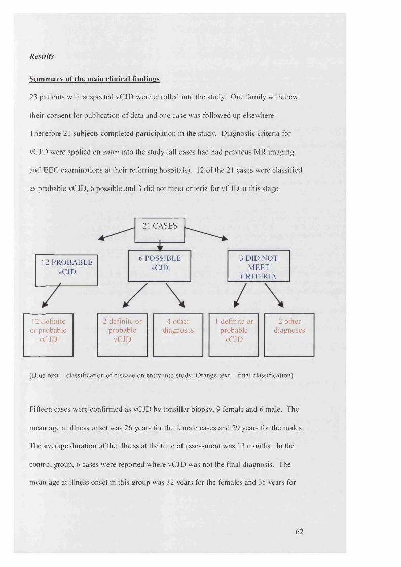
Results
Sumimarv of the main clinical findings.
23 patients with suspected vCJD were enrolled into the study. One family withdrew
their consent for publication o f data and one case was followed up elsewhere.
Therefore 21 subjects com pleted participation in the study. D iagnostic criteria for
vC JD were applied on entry into the study (all cases had had previous MR imaging
and E E G examinations at their referring hospitals). 12 o f the 21 cases were classified
as probable vCJD, 6 possible and 3 did not m eet criteria for vCJD at this stage.
21 CASES
6 POSSIBLE vCJD
3 DID NOT MEET
CRITERIA
12 PROBABLE vCJD
12 definite 2 definite or 4 other 1 definite or 2 otheror probable probable diagnoses probable diagnoses
vCJD vCJD vCJD
(Blue text = classification of disease on entry into study; Orange text = final classification)
Fifteen cases were confirm ed as vCJD by tonsillar biopsy, 9 female and 6 male. The
mean age at illness onset was 26 years for the female cases and 29 years for the males.
The average duration o f the illness at the tim e o f assessm ent was 13 months. In the
control group, 6 cases were reported where vCJD was not the final diagnosis. The
mean age at illness onset in this group was 32 years for the females and 35 years for
62

the male cases. The mean duration in months of the illness at the time of assessment
was longer in this group at 26 months (tables 2,3 and 4).
It was realised that the control group would be small (n=6) with varied diagnoses,
which would not permit the meaningful application of statistics for comparisons with
the patient group. The control group was therefore expanded, as explained in the
methods, for the imaging and neuropsychology parts of the study.
Fourteen of the fifteen cases with vCJD presented, in the first instance to their GP.
One case mainly involved hospital physicians/ surgeons (A& E and orthopaedics).
Four cases were referred to a psychiatrist prior to a neurologist, at a mean of 10
months from illness onset. Five out of the 15 cases saw a psychiatrist at any time. All
cases were referred to a neurologist at some time in the illness course, at a mean of 11
months from illness onset. The mean time to confirming the final diagnosis by tonsil
biopsy was 14 months (tables 19 and 20).
Six out of the 15 cases of vCJD complained of symptoms of depression at
presentation and 10 out of 15 during the first four months of the illness. Personality
change was noted initially in 8 out of 15 cases, and in 11 out of 15 cases within four
months. Three of these cases complained of cognitive problems from the outset and
in seven cases this was an early feature (within 4 months). Complaints of difficulty
walking or with poor balance were made by four cases at presentation, and seven by
four months. Other symptoms complained of at presentation included sensory
disturbance in seven cases, two cases with altered vision and six with sleep
abnormalities. By comparison, four of the six cases with alternative diagnoses had
63
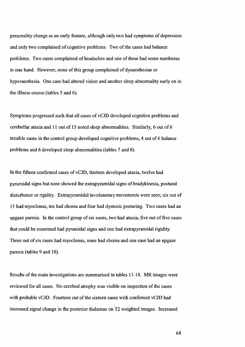
personality change as an early feature, although only two had symptoms of depression
and only two complained of cognitive problems. Two of the cases had balance
problems. Two cases complained of headaches and one of these had some numbness
in one hand. However, none of this group complained of dysaesthesiae or
hyperaesthesia. One case had altered vision and another sleep abnormality early on in
the illness course (tables 5 and 6).
Symptoms progressed such that all cases of vCJD developed cognitive problems and
cerebellar ataxia and 11 out of 15 noted sleep abnormalities. Similarly, 6 out of 6
testable cases in the control group developed cognitive problems, 4 out of 6 balance
problems and 6 developed sleep abnormalities (tables 7 and 8).
In the fifteen confirmed cases of vCJD, thirteen developed ataxia, twelve had
pyramidal signs but none showed the extrapyramidal signs of bradykinesia, postural
disturbance or rigidity. Extrapyramidal involunatary movements were seen; six out of
15 had myoclonus, ten had chorea and four had dystonic posturing. Two cases had an
upgaze paresis. In the control group of six cases, two had ataxia, five out of five cases
that could be examined had pyramidal signs and one had extrapyramidal rigidity.
Three out of six cases had myoclonus, none had chorea and one case had an upgaze
paresis (tables 9 and 10).
Results of the main investigations are summarised in tables 11-18. MR images were
reviewed for all cases. No cerebral atrophy was visible on inspection of the cases
with probable vCJD. Fourteen out of the sixteen cases with confirmed vCJD had
increased signal change in the posterior thalamus on T2 weighted images. Increased
64

signal was also seen in different cases in the head of the caudate nucleus, the upper
midbrain, the parietal cortex and subcortical areas, the centrum semi-ovale, the
cortical white matter, the middle thalamus and the occipital white matter extending
into the globus pallidus on the right. In the control group, one case showed global
cerebral and cerebellar atrophy. No signal change was seen in the posterior thalamus
but one case had altered signal in the mesial frontal lobes and another in the caudate
and lentiform nuclei.
For an overview of the clinical symptoms, signs and investigations see tables 5-20.
These data are discussed in the context of the psychiatric features, neuropsychology
profiles and imaging characteristics in the following chapters.
65

Table 2: Assessment stage, confirmed cases of variant CJD
Subject Assessment Stage
(x mths into illness)VI 10V2 14V3 13V4 13V5 16V6 25V7 12V8 13V9 11
V10 10V I1 10V I3 13V14 18V15 9V16 20
Table 3: Assessment characteristics, alternative diagnoses
Subject Assessment Stage
(x mths into illness)Ol 8402 80 4 2405 90 6 140 7 16
Table 4: Final alternative diagnoses
Subject Final diagnosis
Ol familial CJD
02 sporadic CJD
04 familial CJD
05 sporadic CJD
06 Lafora Body disease
0 7 psychotic illness

Table 5: Presenting symptoms, confirmed cases of variant CJD
Subject Early Symptoms < 4 months
Depression Change in personality or behaviour
Cognitiveproblems
Cerebellar (ataxia or dysarthria)
Paraesthesiae or numbness
DysaesthesiaeAlteredvision Sleep
abnormalor
malaiseVI + + + - - + - +V2 + + - + + + - -
V3 + + - - - - - -
V4 + + - - - - - -
V5 + + + - - - - -
V6 + + - - - - - -
V7 - + + - + + - -
V8 - + - - - - - +V9 + + - + + + + +
V10 + + + + - + - +V I1 - - + + + - - -
V13 + - + + + - +V14 - - + + - - - -
V15 - - - + + - - +V16 + + - - - - - -
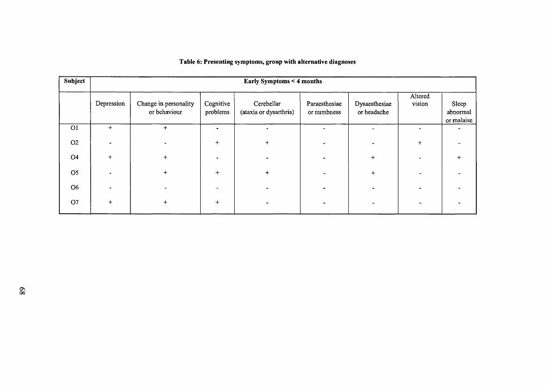
Table 6: Presenting symptoms, group with alternative diagnoses
Subject Early Symptoms < 4 months
Depression Change in personality or behaviour
Cognitiveproblems
Cerebellar (ataxia or dysarthria)
Paraesthesiae or numbness
Dysaesthesiae or headache
Alteredvision Sleep
abnormal or malaise
0 1 + + - - - - - -
0 2 - - + + - - + -
0 4 + + - - - + - +
0 5 - + + + - + - -
0 6 - - - - - - - -
0 7 + + + - - - - -
ON00

Table 7: Symptoms experienced throughout the course of the illness, confirmed cases of variant CJD
Subject All Symptoms
Depression Change in personality or behaviour
Cognitiveproblems
Cerebellar (ataxia or dysarthria)
Paraesthesiae or numbness
Dysaesthesiae Alteredvision
Sleep abnormal or malaise
VI + + + + - + - +
V2 + + + + + + - +
V3 + + + + + + - +
V4 + + + + - + + +
V5 + + + + - - - -
V6 + + + + + + - +
V7 - + + + + + - -
V8 - + + + - - - +
V9 + + + + + + + +
V10 + + + + - + - +
V ll - - + + - - - +
V13 + - + + + + + +
V14 - + + + - - - +
V15 - + + + - + - -
V16 + + + + - - - -
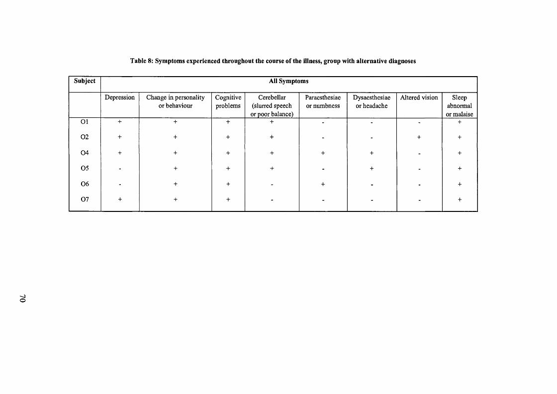
Table 8: Symptoms experienced throughout the course of the illness, group with alternative diagnoses
Subject All Symptoms
Depression Change in personality or behaviour
Cognitiveproblems
Cerebellar (slurred speech
or poor balance)
Paraesthesiae or numbness
Dysaesthesiae or headache
Altered vision Sleep abnormal or malaise
0 1 + + + + - - - +
0 2 + + + + - - + +
0 4 + + + + + + - +
0 5 - + + + - + - +
0 6 - + + - + - - +
0 7 + + + - - - - +

Table 9: Clinical signs, confirmed cases of variant CJD
Subject Clinical signs
Myoclonus Chorea Dystonia Ataxia Pyramidal Extrapyramidal* Upgaze paresis Sensoryabnormality
VI - + + + + - - +V2 - - - + + - - +
V3 - + - + - - - +
V4 + + + + + - - -
V5 + + - NT + - - -
V6 - + - + + - + +V7 - + - + - - - +
V8 - + - + - - - NTV9 - - - + + - - -
V10 + - + + + - + -
V ll + + - + + - - +
V13 - - + - + - - -
V14 + + - + + - - NTV15 - + - + + - - +
V16 + - - + + - - NT
N T = Not tested or unable to test; *extrapyramidal signs excluding involuntary movements (myoclonus, chorea and dystonia)

Table 10: Clinical signs, cases with alternative diagnoses
Subject Clinical signs
Myoclonus Chorea Dystonia Ataxia Pyramidal Extrapyramidal Upgazeparesis
Sensoryabnormality
0 1 + - - NT + + - -
0 2 + - - NT + - + NT
0 4 - - - + + - - -
0 5 + - - + + - - NT
0 6 - - - - + - - -
0 7 - - - - NT NT NT NT
NT = Not Tested or unable to test

Table 11: CSF examination, confirmed cases of variant CJD
mthsfrom
illnessonset
proteinSfi
glucose(serum)mmol/1
cellcount
oligoclonalbands
S100ng/ml
NSE PI 4-3-3
VI 9 0.48 4.8 (5.8) 0 0 na na na
V2 na na na na na na na na
V3 13 0.57 3.1 1 0 1.36 45 positive
V4 13 0.20 3.7 (5.6) 1 na 0.92 22 trace
V5 8 1.60-2.90 normal 0 0 0.69 16 positive
V6 24 normal normal 1 0 na na positive
V7 11 0.34 3.0 (4.8) 0 na 0.61 na negative
V8 12 0.23 3.5 0 na 1.42 na negative
V9 11 >3.00 normal 0 0 0.82 15 positive
V10 8 0.27 3.3 0 na 0.59 nt negative
V ll 9 0.75 normal 0 1* 0.33 18 negative
V13 13 normal normal 0 0 0.55 na negative
V14 5 0.40 3.7(4.4) 0 0 0.40* na negative*
V15 9 0.25 3.9 (5.4) 0 0 0.31 12 trace
V16 11 0.61 4.0 2 + serum/CSF 0.4 12 negative
na - not available; * 9 months la ter;r one unmatched CSFIgG band; +matched serum & CSF bands
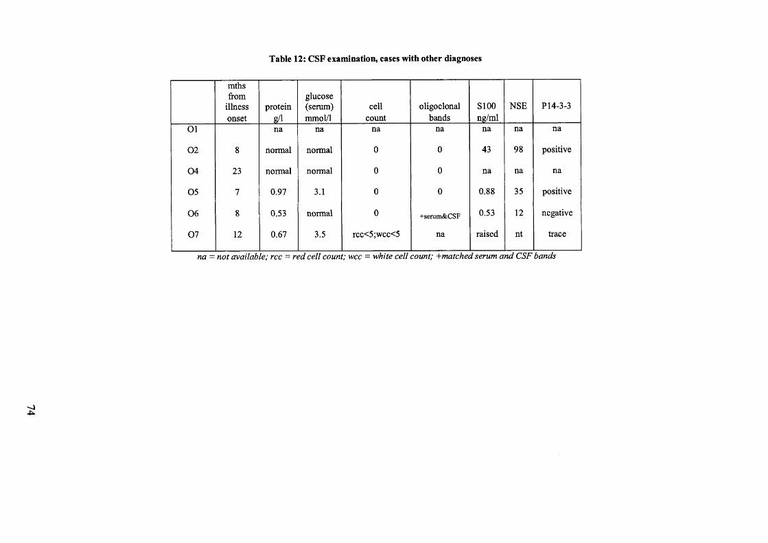
Table 12: CSF examination, cases with other diagnoses
mthsfrom
illnessonset
proteing/1
glucose(serum)mmol/1
cellcount
oligoclonalbands
S100ng/ml
NSE P14-3-3
01 na na na na na na na
02 8 normal normal 0 0 43 98 positive
04 23 normal normal 0 0 na na na
05 7 0.97 3.1 0 0 0.88 35 positive
06 8 0.53 normal 0 +serum&CSF 0.53 12 negative
07 12 0.67 3.5 rcc<5;wcc<5 na raised nt trace
na = not available; rcc = red cell count; wcc = white cell count; +matched serum and CSF bands

Table 13: MRI examination, confirmed cases of variant CJD
mthsfrom
illnessonset
atrophy signal changecerebrum cerebellum posterior
thalamusother
VI 7 0 0 0 0
VI 9 (DWI) 0 0 1 1 (head caudate)
V2 14 0 0 1 1 (upper midbrain)
V3 13 0 0 1 0
V4 11 0 0 1 0
V5 8 0 0 1 l(subcortical white matter lesions)
V6 22 0 0 1 0
V7 11 0 0 1 0
V8 15 0 0 1 0
V8 16 0 0 1 1 (aqueduct,caudate, putamen bilaterally)
V9 11 0 0 1 1 (centrum semi-ovale)
V10 10 0 0 1 infarct left trigone
V ll 9 0 0 1 1 (cortical white matter)
V13 13 0 0 1 1 (medial thalamus)
V14 16 0 0 1 1 (medial thalamus)
V15 9 0 0 0 1 (occipital white matter, into globus pallidus R)
V16 9 0 0 1 0
DWI = Diffusion Weighted Imaging
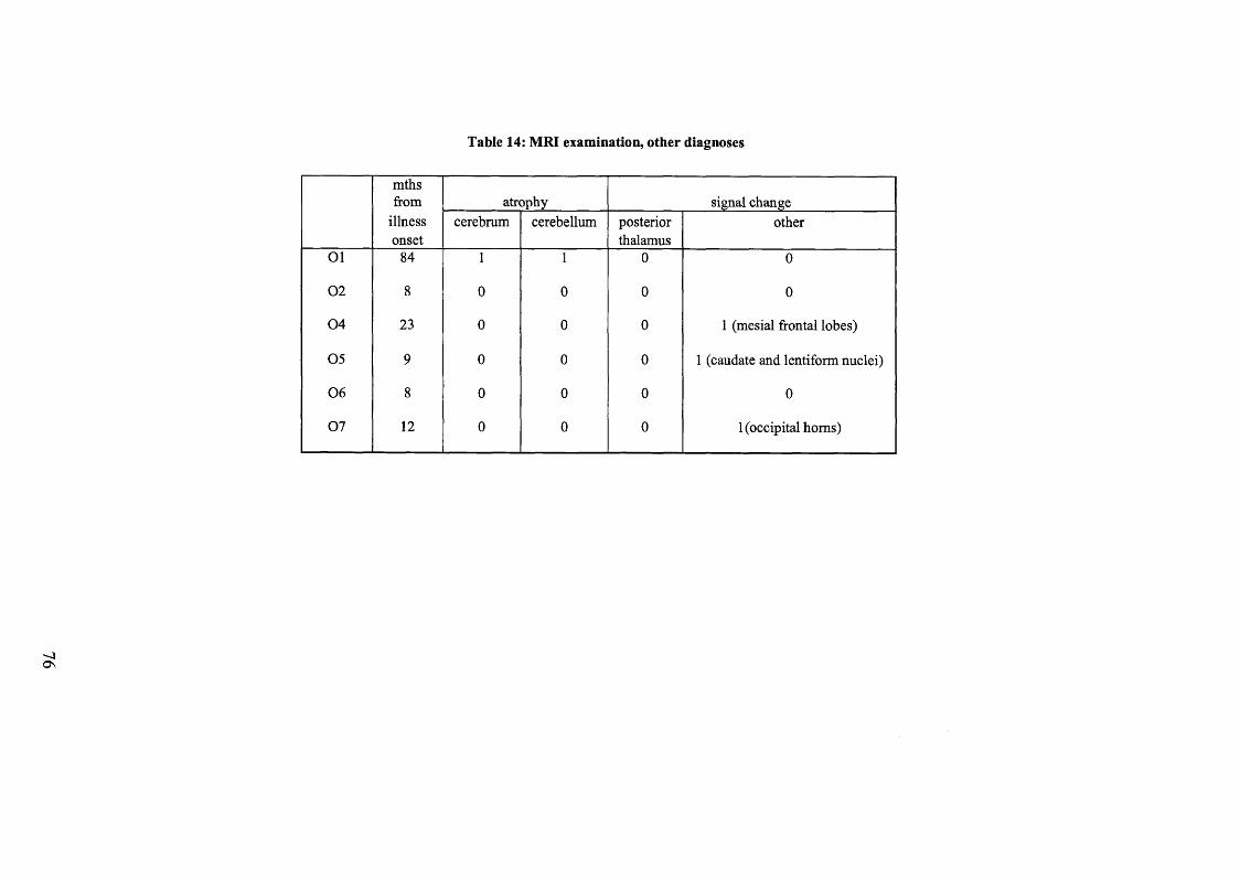
Table 14: MRI examination, other diagnoses
mthsfrom
illnessonset
atrophy signal changecerebrum cerebellum posterior
thalamusother
01 84 1 1 0 0
02 8 0 0 0 0
04 23 0 0 0 1 (mesial frontal lobes)
05 9 0 0 0 1 (caudate and lentiform nuclei)
06 8 0 0 0 0
07 12 0 0 0 1 (occipital homs)

Table 15: EEG examination, variant CJD cases
mthsfrom
illnessonset
periodiccomplexes
epileptiformactivity
VI 7 0 0 Disorganised background activity. Diffuse slow wave excess and irregular waveforms.
VI 9 0 0 Normal background activity. Intermittent excess theta over both hemispheres.
V2 14 0 0 Normal record
V3 13 0 0 Marked and diffuse slow activity over both hemispheres.
V4 10 0 0 Bilateral slow wave activity
V4 13 0 0 Non-specific mild to moderate disturbances.
V5 8 0 0 Bilateral slow wave activity
V6 22 0 0 Polyrhythmic low amp. Background, excess of fast act., increase o f intermittent slow act..
V7 11 0 0 Normal.
V8 15 0 0 Diffuse slow activity, featureless periods, attenuated background.
V9 11 0 0 Widespread non-specific abnormality.
V10 10 0 0 Slowing of background rhythm, runs of sharp waves intermittently.
V ll 9 0 0 Normal.
V13 15 0 0 No alpha rhythm, EEG dominated by theta and slow activity.
V14 16 0 0 Diffuse and nonspecific excess of slow and theta without definite alpha rhythm.
V15 9 0 0 Nonspecific abnormalities with widespread theta rhythms.
V16 9 0 0 Diffuse theta waves and periodic generalised delta waves.

Table 16: EEG examination, other diagnoses
mthsfrom
illnessonset
periodiccomplexes
epileptiformactivity
summary
01 84 0 0 normal
02 8 0 0 nonspecific, mildly abnormal
04 23 0 0 nonspecific excess of theta activity
05 9 0 0 anterocentral excess of slow activity. Mild excess theta and slow widespread over cerebrum.
06 8 0 0 mild and diffuse slowing
07 14 0 0 Diffusely slow background with theta and delta activity.
oo

Table 17: Biopsy and histology, variant CJD cases
WB IH Frontal Parietal Temporal Occipital Thalamus GlobusPallidus
Putamen Caudate Cerebellum Brainstem SpinalCord
VI + + S,F,P S,F,P S,F,P S,F,P S,G,N,P S,G,N,P S,G,N S,G,N P P P
V2 + + nt nt nt nt nt nt nt nt nt nt nt
V3 + + SP SP SP SP SP S S S SP SP
V4 + + nt nt nt nt nt nt nt nt nt nt nt
V5 nt + S,F,P S,F,P S,F,P S,F,P G,N,F,P F,P F,P S,F,P S,F,P
V6 + + nt nt nt nt nt nt nt nt S,F P P
V7 + nt nt nt nt nt nt nt nt nt nt nt nt
V8 + + nt nt nt nt nt nt nt nt nt nt nt
V9 + + S,G,N,F,P S,G,N,F,P S,G,N,F,P S,G,N,F,P S,G,F,P S,P SsP SC,PP S,N,F,P S
V10 + + nt nt nt nt nt nt nt nt nt nt nt
V I1 + + S,G,N,F,P S,G,N,F,P S,G,N,F,P S,G,N,F,P S,G,N,P S,G,N,P S,G,N,P S,G,N,P S,N,F,P S,N,G,P
V13 + + nt nt nt nt nt nt nt nt nt nt nt
V14 + + nt nt nt nt nt nt nt nt nt nt nt
V15 + + nt nt nt nt nt nt nt nt nt nt nt
V16 + + nt nt nt nt nt nt nt nt nt nt nt
WB - Western Blot; IH-Immunohistochemistry; S - Spongiform change; F -F lo r id plaques; P - Prion protein deposition; G - Gliosis; N - Neuronal loss; nt - not tested
as no post mortem

Table 18: Biopsy and histology, other diagnoses
WB IH Frontal Parietal Temporal Occipital Thalamus GlobusPallidus
Putamen Caudate Cerebellum BrainstemSpinalCord
01 nt nt NA NA NA NA NA NA NA NA NA NA NA
02 nt nt S,G,N,P S,G,N,P S,G,N,P S,G,N,P S,G,N,P S,G,N,P S,G,N,P S,G,N,P S,G,N,P S,G,N,P S,G,N,P
03 - - NA NA NA NA NA NA NA NA NA NA NA
04 nt nt NA NA NA NA NA NA NA NA NA NA NA
05 - - nt nt nt nt nt nt nt nt nt nt nt
06 nt nt NA NA NA NA NA NA NA NA NA NA NA
07 - - NA NA NA NA NA NA NA NA NA NA NA
WB - Western Blot; IH - Immunohistochemistry; S - Spongiform change; P - Prion protein deposition; G - Gliosis; N - Neuronal loss; nt - not tested as no post mortem;
NA- not applicable (still alive)
ooo

STUDY IA: A STUDY OF THE PSYCHIATRIC MANIFESTATIONS OF CJD
Introduction
The psychiatric features of CJD have been recognised for many years. In sporadic
CJD, original large-scale studies of the clinical features have confirmed the presence
of prodromal symptoms in 39% of “typical cases”, consisting of asthenia, weight loss
and sleep disorders, with two thirds showing some mental deterioration at the onset75.
94% show evidence of dementia, 45% behavioural change and 39% deficits of higher
cognitive function as the disease progressed. Will et al classified three presentations
of sporadic CJD, sub-acute, intermediate and an amyotrophic form76. In the sub-acute
form, 33% had a prodrome of altered personality, or malaise with some anorexia and
weight loss, with less frequent sleep disturbance. In 29% of cases, depression or
emotional lability led to psychiatric referral. In fact at presentation, the commonest
symptoms were of dementia (21%) and behavioural disturbance (18%), with visual
hallucinations occurring in just 1%. Throughout the course of the illness, 17%
experienced visual hallucinations and 100% developed dementia.
However, these “typical” cases of sporadic CJD are easier to diagnose and Will et al
noted that it was the intermediate cases that caused problems i.e. 2 young cases
presenting with emotional lability and ataxia and one presenting with personality
change. Since these earlier studies, the characterisation of PrPSc molecular types has
evolved and now sporadic CJD is classified into six phenotypic subgroups, by PrP
molecular type and the polymorphism for methionine and valine at codon i2927&28.
70% of subjects showed the classic CJD phenotype (short illness duration, typical
EEG and classical pathology), with PrPSc type 1 and at least one methionine allele at
81

codon 129 i.e. MM1 or MV1. However, 5 other subtypes were recognised with
younger onset on average, a prolonged disease course, and an absence of EEG
features. These are more difficult to diagnose and may be more difficult to
distinguish from vCJD cases clinically. The presence or absence of psychiatric
features or insomnia, for example, may aid in the distinction as in W 1 and MM2-C
cases, insomnia, sensory symptoms and psychiatric features are not a feature.
Psychiatric features and insomnia are characteristic of MM2-T subgroup, which is
indistinct from FFI and to a lesser extent of MV2 and W 2 subgroups. The following
table gives a summary of the Parchi classification. This was designed following the
examination of a series of 300 patients.
82

Table 19: Summary of classification of sporadic CJD based on molecular and phenotypic analysis of 300 subjects; Parchi et al, 199927
sCJD clinical
group
MM1 MV1 W 1 MM2-C MM2-T MV2 W 2
codon 129 MM MV W MM MM MV W
PrPSc type 1 1 1 2-C 2-T 2 2
Clinical features Rapidly progressive
dementia, early,
prominent
myoclonus. Visual
impairment or
unilateral signs at
onset in 40%
Rapidly progressive
dementia, early,
prominent
myoclonus. Visual
impairment or
unilateral signs at
onset in 40%
Progressive
dementia
Progressive
dementia
Insomnia and
psychomotor
hyperactivity in most
cases in addition to
ataxia and cognitive
impairment
Ataxia,
progressive
dementia, long
duration of
illness (>2
years) in some
cases
Ataxia at onset,
late dementia
Sensory(% cases with signs or
symptoms)
7 25 0 0 0 7 15
Psychiatric(% cases with signs or
symptoms)
34 12 0 0 67 44 21
Insomnia(% cases with signs or
symptoms)
8 0 0 0 67 15 15
EEG typical* typical no typical EEG no typical EEG no typical EEG no typical EEG no typical EEG
in most cases
Source: Parchi et al, Ann Neurol 1999; 46:224-233. SCJD = sporadic CJD Typical = typical periodic sharp wave complexes
oou>

The psychiatric features of vCJD have been documented. Two of the original three
reported cases presented with early psychiatric features with referral to psychiatric
services1'3. In a review of the clinical characteristics of the first 14 cases it was noted
that 9/14 had psychiatric symptoms at the disease onset, all developing psychiatric
features early in the illness. 13/14 were referred to psychiatric services with a
diagnosis of depression or depression secondary to an organic cause made4.
Nine out of fourteen had insomnia; most had excessive sleepiness in the day, early in
the illness. In those with depression, all but one had early weight loss and anorexia.
7/14 had symptoms of depression prior to any cognitive impairment and 2/14 had one
episode of suicide ideation. Most had drug therapy, three with transient improvement.
2/14 had first rank symptoms of schizophrenia. Importantly, 12/14 had unsustained
delusions that occurred within a few months of illness onset and were fleeting, lasting
hours or days at most. 8/14 had visual, and 5/14 had auditory hallucinations.
However, it was noted that ultimately a diagnosis of CJD was made by the
development of sensory or other neurological symptoms or signs.
Although these early features are characteristic of vCJD there is still marked
variability of presentation. Of three cases reported in Leicester, each had behavioural
change and personality change at the onset and two were diagnosed with depression,
but none had sensory symptoms, delusions or hallucinations6.
In the review of the first 35 cases of vCJD, 34 had early persistent psychiatric
features, most commonly depression, anxiety and withdrawl . Five had first rank
symptoms. Twenty-five were seen by a psychiatrist early in the illness and were
given treatment. A later retrospective case note review of the first one hundred cases
84

confirmed that 38% of the cases were initially referred to psychiatry services, with
63% seen by a psychiatrist at some time. The most common psychiatric symptoms
were dysphoria, anergia, loss of interest, anxiety and withdrawl. A small proportion
developed hallucinations and paranoid behaviour or delusions. The fleeting delusions
noted in the early reviews were shown to be relatively rare compared to other
symptoms70.
The difficulty with diagnosing vCJD early lies with the insidious onset and
presentation with symptoms that could be attributed to many other illnesses. As part
of the study to detect under-ascertainment of vCJD in Wales61, a steering committee
decided which ICD-9 diagnoses could be compatible with a diagnosis of vCJD at any
stage of the illness and which may therefore constitute the differential diagnosis in the
early stages of the disease. These included, alcoholic psychoses, drug psychoses,
transient organic psychotic conditions (including acute confusional and subacute
confusional states), schizophrenic psychoses, affective psychoses, paranoid states,
neurotic disorders (anxiety states, hysteria, phobic states, obsessive-compulsive
disorders, neurotic depression, neurasthenia, depersonalisation syndrome and
hypochondriasis), personality disorders, sexual deviations and disorders, alcohol
dependence syndrome, drug dependence, acute reaction to stress, adjustment reaction,
depressive disorder and disturbance of conduct. The differential diagnosis is therefore
broad and it is important to realise that sharing the possibility of prion disease with a
patient who may be suffering from depression or anxiety may have far reaching
consequences.
85

One window of opportunity for reaching an early diagnosis lies in the mean of 6
months before clear-cut neurological signs occur. It was felt that if further features of
the psychiatric presentation could be distinguished early, this might aid in the
identification of those with a progressive neurodegenerative illness.
Aim o f the neuropsychiatric examination ofpatients with suspected vCJD
This was to test the hypothesis that there would be early distinguishing features in the
psychiatric presentation of vCJD, in particular, a characteristic profile of depressive
symptoms. These symptoms should be readily determined by formally assessing and
quantifying changes in thought content, mood, symptoms of anxiety and personality
change. The presence of a characteristic psychiatric profile may distinguish cases
with vCJD from those with other causes of depressive symptoms.
Methods
A behavioural assessment was completed with 20 out of the 21 families enrolled in
the study (14 families where a case of vCJD was confirmed and six with alternate
diagnoses. The family of one case with probable vCJD (V5), were unavailable for
completion of the tests). A battery of five tests was used in the form of an interview
with the carer. In all cases the patient was too unwell and cognitively impaired to
contribute. Each carer was interviewed personally by RJC and additional information
was obtained from relatives, friends and nursing staff caring for the patient. The
patients’ medical notes were examined.
86

Assessment tools
The following assessment tools were used:
1. MOUSEPAD: Manchester and Oxford Universities Scale for the
Psychopathological Assessment of Dementia77. This test is based on the Present
Behavioural Examination, though it is shorter and places equal emphasis on
psychiatric and behavioural features in people with dementia. It was chosen as a tool
to assess the non-cognitive features of dementia. It gives the benefit of allowing the
measurement of phenomena in the last month and since the onset of dementia, with
separate ratings for severity and frequency. This was scored with the carer in each
case, as the patient was too impaired to give sufficient information.• 7 0
2. CORNELL scale for depression in dementia . This scale and the Clinical
Anxiety Scale (CAS) were used because the MOUSEPAD does not include any items
for depression or anxiety. It is a rating scale for depression in dementia that relies on
observed and informant based questions rather than reports from the patient. It is a
19-item scale, rated absent, mild, intermediate or severe and a score of > 8 implies the
presence of significant depressive symptoms.
3. CLINICAL ANXIETY SCALE (CAS)79. The CAS is an interview based
measure which is a shorter version of the Hamilton Anxiety Scale. The person is
rated by a clinician, who gauges how anxious the subject has been feeling over the
past two days, including the present day, using six areas. The clinician is guided by
detailed criteria for scoring each area. The scores are graded in four ranges i.e.
recovered or absent; mild, moderate and severe. Validity is based on that of the
Hamilton Anxiety Scale.
4. MEMORY FUNCTIONING QUESTIONNAIRE (MFQ)80 This tool was
used to detect memory complaints and it was hoped that information from this would
87
supplement that obtained from formal neuropsychology testing. The MFQ was
designed to detect memory complaints in the elderly. This tests 64 items on 7 scales
of memory, rating retrospective functioning, frequency of forgetting, frequency of
forgetting when reading, remembering past events, seriousness of forgetting and
mnemonics usage. This scale should be self rated and also completed with an
informant. However, only the latter was possible and therefore only an idea of the
functioning “in the eyes of the carer” was obtained.
Q 1
5. IDDD . Interview to determine deterioration in daily functioning in
dementia. This was used to assess the ability to perform the activities of daily living.
The questionnaire refers to 33 self care activities and scores each on a three point
scale i.e. independent, needing prompting and dependent, referring to behaviour in the
last month compared to their normal level of function.
6. STANDARDISED ASSESSMENT OF PERSONALITY (SAP)82. The SAP
was used to classify the subjects’ premorbid personality. It was expected that our
control group would consist of many young people without vCJD, but with some
psychiatric features in common with the early stages of vCJD. It was important to
establish whether people with certain personality traits experienced these symptoms.
This test did not assess personality change as a result of illness. The assessment was
based on an interview with an informant to classify the patients’ premorbid
personality in clinical terms. The result is classification into one of eleven types:
self-conscious, schizoid, paranoid, cyclothymic, obsessional, anxious, neurasthenic,
explosive, sociopathic, hysterical or “normal”. Two grades of severity are noted i.e.
the presence of a trait and a severer level if the subject is handicapped in day to day
life by the personality trait. The test is performed in three stages. Firstly, the
relationship and length of acquaintance with the patient is established. An
88

exploratory section follows this, when a description of personality is requested.
Seven standard questions are used if the response is not helpful. Key descriptive
words e.g. houseproud, shy etc are circled on a list in the proforma. Finally, more
detailed questions relating to an emerging personality trait are explored.
Definitions of psychiatric terms
Delusions
Delusions were defined according to Goldberg (1987) and Cummings (1985) as
“mistaken beliefs which are held with conviction, which are not shared by others of
the same cultural or social background and intellect and which persist despite
evidence to the contrary”83&84. The presence of delusions was based on evidence
given by the carer, through information given in the MOUSEPAD. Delusions were
measured a) if they had occurred at any time since the onset of the illness and b) in the
last month. They were divided into simple delusions (Cummings, 1985) e.g. of theft
or suspicion, or complex. The presence of persecutory ideation was also noted where
ideas of persecution were not held with delusional intensity. The presence of
delusions was also rated as 0, absent, 1 mild (less than once per week), 2 moderate (<
4 out of 7 days per week, and 3, severe (greater than four times per week).
Disorders o f perception: Hallucinations and misidentijications.
Hallucinations were defined as “perceptions that are not based on external stimuli”.
The presence of visual hallucinations was noted if the subject reported seeing
someone or something without an external stimulus or if they had been observed
89
interacting with such a non-existent person or object Misidentifications were defined
using the classification of Rubin (1988) with additional categories85*87:
a) people in the house - a belief based on misrecognition, that others are living in the
house
b) misidentification of mirror image whereby the subject has indicated an inability to
recognise the subjects own reflection in the mirror or “someone else is in the mirror”
c) misidentification of TV images - such that the patient is talking to the TV or is
fearful that an event on screen is taking place in the room
d) misidentification of people - mistaking a relative or friend for another e.g. spouse
for a daughter or brother.
Details were taken from information given by the carer during the MOUSEPAD test.
Behavioural disorders
Details of wandering, following the carer about the house, eating behaviour (increased
amount or eating more quickly) sleep patterns, sexual behaviour, aggression (verbal
and physical) and emotional lability were all taken during the MOUSEPAD. The
presence of symptom, the stage of the illness it occurred at and its severity were
scored for each case from details given by the carer.
Mood disorders
The presence of symptoms of depression was recorded as positive responses to items
on the Cornell Scale for depression performed with the carer. The carer was therefore
asked to judge the subjects inner feelings e.g. depressed mood, anxiety, feelings of
90

hopelessness and vegetative symptoms e.g. sleep disturbance, weight loss and diumal
variation in symptoms.
Personality
This describes acceptable attributes that have been present since adolescence, are
stable overtime despite fluctuations in mental state, which are manifest in different
environments and which are recognisable to friends and acquaintances82.
Results
The time of disease onset is taken as the time when the family, sometimes with
hindsight, felt that there had been a change in personality or behaviour, if this
preceded other clinical symptoms. The mean time to seeking medical attention in all
confirmed cases of vCJD was five months (range 0-18 months) and all cases, except
two visited their general practitioner in the first instance. The time taken to seek
medical attention in the comparative group, with alternative diagnoses ranged from 1-
60 months (data not available for one case). Of the two cases presenting within one
month of symptom onset, one presented with a psychotic episode and so made
immediate contact with medical services and the other had a rapidly progressive
course, later consistent with sporadic CJD (tables 20 and 21).
91

Table 20: Milestones in diagnosis (Variant CJD)
Time to Who did patient first
see?
Time to Time to Time tomedicalattention
neurologyopinion
psychiatricopinion
finaldiagnosis
(months) (months) (months) (months)VI 3 GP 9 8 11
V2 0 GP 12 NA 14
V3 2 GP 13 9 13
V4 9 GP 12 10 13
V5 11 GP 16 NA 16
V6 18 GP 20 21 25
V7 na na 11 NA 12
V8 5 GP 8 NA 13
V9 3 GP 5 NA 11
V10 3 GP 8 NA 11
V I1 8 GP 8 NA 15
V13 0-4 GP 5 NA 15
V14 5 na 5 NA 18
V15 0 GP 7 NA 9
V16 7 GP 20 14 20
GP - General Practitioner; NA - Not applicable; na - not available
Table 21: Milestones in diagnosis (other diagnoses)
Time to medical attention (months)
Who did patient first
see?
Time to neurology
opinion (months)
Time to psychiatric
opinion (months
Time to final
diagnosis (months)
Ol 60 GP 72 NA 84
02 na GP 5 NA 8
04 7 GP 23 12 23
05 1 na 7(UK) NA 7
06 6 Physician 9 NA 21
07 1 GP NK 1 undiagnosed
GP - General Practitioner; NA - Not applicable; na- not available
92

Personality change or a change in behaviour was present at the onset of disease in 11
out of 15 cases. Seven out of 15 cases were diagnosed by their GP with depression
and at least 7 out of 15 were treated with antidepressant medication (In many
instances there were conflicting reports from relatives as to whether there was a
response to antidepressant therapy. As insufficient supportive evidence was available,
a figure for the response rate has not been included). Five out of 15 cases were
referred to a psychiatrist with appointments on average 12 months from disease onset
(range 8-21 months). All cases were referred to neurologists during the disease
course at mean time 11 months from disease onset (range 5-20 months). The time to
reach a diagnosis (taken as the time to the availability of the result of a tonsil biopsy)
ranged from 9-25 months; mean 14 months from disease onset (table 22).
Table 22: Psychiatric Milestones (Variant CJD)
VI
Diagnosedwith
depressionY
Who made diagnosis?
GP
Treated with antidepressants?
Y
Referred to a psychiatrist?
Y
Any other psychiatric diagnosis?
N
V2 N NA NA N N
V3 Y GP Y Y N
V4 Y GP Y Y N
V5 N NA NA N N
V6 Y NK Y Y Y
V7 N NA NA N N
V8 N NA NA N N
V9 Y GP NK N N
V10 Y GP Y N N
V I1 N NA NA N N
V13 Y GP Y N N
V14 N NA NA N N
V15 N NA NA N N
V16 Y GP Y Y N
GP - General Practitioner; NA - Not available; NK - Not known
93
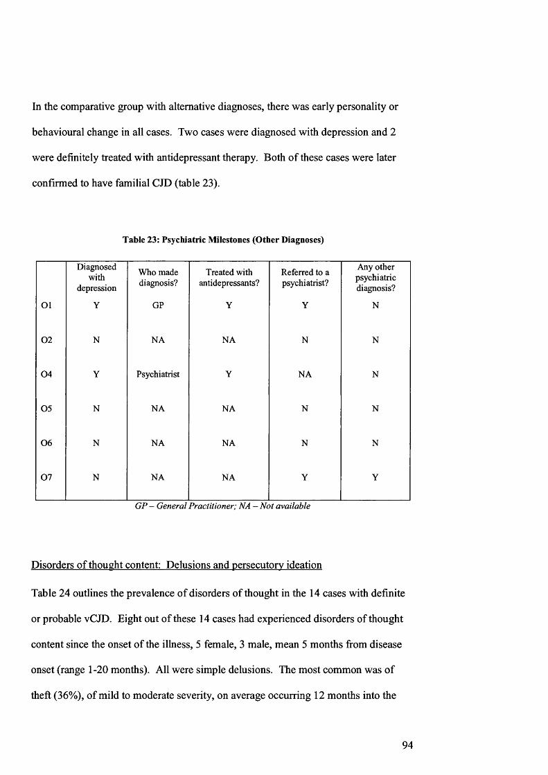
In the comparative group with alternative diagnoses, there was early personality or
behavioural change in all cases. Two cases were diagnosed with depression and 2
were definitely treated with antidepressant therapy. Both of these cases were later
confirmed to have familial CJD (table 23).
Table 23: Psychiatric Milestones (Other Diagnoses)
Diagnosedwith
depression
Who made diagnosis?
Treated with antidepressants?
Referred to a psychiatrist?
Any other psychiatric diagnosis?
Ol Y GP Y Y N
02 N NA NA N N
04 Y Psychiatrist Y NA N
05 N NA NA N N
06 N NA NA N N
07 N NA NA Y Y
GP - General Practitioner; NA - Not available
Disorders of thought content: Delusions and persecutory ideation
Table 24 outlines the prevalence of disorders of thought in the 14 cases with definite
or probable vCJD. Eight out of these 14 cases had experienced disorders of thought
content since the onset of the illness, 5 female, 3 male, mean 5 months from disease
onset (range 1-20 months). All were simple delusions. The most common was of
theft (36%), of mild to moderate severity, on average occurring 12 months into the
94

illness (earliest 5 months from illness onset). Four out of 14 cases expressed
delusions of suspicion, three of which experienced severe delusions of being watched.
Other delusions were of abandonment (3/14 cases), the house not their own (2/14
cases) and someone else in the house (2/14 cases). The fears of abandonment were
severe in two cases and in one case occurred early in the disease (within one month).
This was notably the case with a young age at onset (<16 years of age).
In the comparative group with alternative diagnoses, four out of the six cases tested
had disorders of thought, at mean time 22 months from illness onset (range 2-72
months). The most common was of suspicion (50% of cases) i.e. of poisoning, being
followed and that their spouse was having an affair. One case (07) presented with
florid delusions on two occasions, linked to drug ingestion.
Table 24: Disorders of Thought; vCJD group
Months fromNumber of cases Severity of illness onset(%) symptoms mean (range)
Any 8 1-3(57)
Theft 5 1-2 12(36) (5-25)
Suspicion 4 1-3 10(29) (6-20)
Abandonment 3 3 12(21) (1-25)
House not own 2 nk 6home(14) (5-7)
Someone else in 9 1 f \house z X J 1 O
(14)nk - not known
95

Disorders of perception: Hallucinations and misidentifications
Only one case of the 14 cases with definite or probable vCJD had experienced
auditory hallucinations and two, visual hallucinations, by the time of referral. The
first case described two statues of dogs on the mantelpiece talking to each other. The
first with visual hallucinations would hold things that were not there and try to give
them to her parents. The second complained of seeing people outside the window.
Misidentifications of any type occurred in 5/14 cases. The most common was of
misinterpreting TV images as real events in four cases with definite or probable vCJD
(in one case just one such episode was reported), on average eight months from illness
onset, 2 with mild and one with severe symptoms (table 25).
In the comparative group, case 07 had marked auditory hallucinations during
psychotic episodes. Case 05, developed disturbing, visual hallucinations 8-9 months
from the onset of his illness. These were of mermaids, snakes on his bed and fire,
which were very distressing to the patient. Case 01, who was later diagnosed with
familial CJD, spent time conversing with herself in the mirror about six to seven years
into her illness. Case 07 accused his sister and aunt of not being who they claimed to
be and misinterpreted TV images as real events.
Table 25: Disorders of Perception; vCJD group
Months fromMISIDENTIFICATIONS Number of cases Severity of illness onset(%) symptoms (range)
Any 5 1-3 8(36) (5-10)
Mirror Image 0 nk nk(0)
People 3 1-2 8(21) (5-10)
Television 4 1-3 8(29) (7-10)
96

Behavioural change
Behavioural disturbance as distinguished from responses from the MOUSEPAD
showed that all 14 cases with definite or probable vCJD had some behavioural change
anywhere from one to 23 months from illness onset (table 26). The most common
features were, aggression, sleep disturbance and emotional lability. Aggression was
seen in 79% of cases, on average appearing 7 months into the illness (range 1-15).
Eleven out of 14 expressed verbal and 3/14 physical aggression. Sleep problems
were seen in 11/14 cases, on average 9 months into the illness. Nine out of 14 were
restless at night (on average 9 months from illness onset) and 9/14 dosed in the day
(mean 8 months from illness onset). Seventy-one percent showed emotional lability 8
months on average into the illness (1-19 months range); four of fourteen did so by
inappropriate laughing and 9/14 by inappropriate crying.
Six out of 14 cases with definite or probable vCJD had a tendency to wander or
follow on average six months into the illness but this may have been curtailed by the
onset of balance and therefore walking problems. Changes in eating and sexual
behaviour were much less frequent.
In the comparative group, all cases showed some behavioural change, 2 months to 6
years from illness onset. All cases had become aggressive (3 both verbally and
physically, 4/6 physically aggressive). All cases also had sleep disturbance. Four out
of six cases had become more emotionally labile.
97

Table 26: Behavioural Features; vCJD group (14 cases with definite or probable vCJD)
Months fromNumber of cases Severity of
illness onset(%) symptoms
mean (range)
Any 14 1-3
(100) (1-23)
Aggression - any 11 1-3 7
(79) (1-10)
Aggression - verbal 11 3 7
(79) (1-15)
Aggression - physical 3 1-3 6
(21) (4-10)
Wandering or following 6 1-3 7
(43) (1-13)
Eating more/more2 2 8
quickly
(14) (6-10)
Sweet tooth 4 2 6
(29) (5-8)
Sleep disturbance - any 11 1-3 9
(79) (1-23)
Restless/ wakeful 9 1-3 8
(64) (1-23)
Sleeping in day 9 1-3 8
(64) (6-15)
Sexual disinhibition 2 1 8
(14) (6-10)
Emotional lability - any 10 1-3 8
(71) (1-19)
Emotional lability -4 1-3 10
laugh
(29) (1-19)
Emotional lability - cry 9 1-3 8
(64) (1-19)
98

Mood disturbances
In 10 out of the 14 cases with definite or probable vCJD, the families reported that
they felt that the subject had symptoms that could be attributed to depression early in
the illness. Only one case had a past medical history of depression. The Cornell scale
for depression should be scored for symptoms experienced in the last week. The
average score for the Cornell was 15.6 (range 5-31) with 11/14 (79%) cases scoring
greater than or equal to 8, indicating evidence of severe depression. Breakdown of
the Cornell scores shows that symptoms covered all aspects i.e. mood related,
behavioural disturbance, physical signs and cyclic aspects of depression, though
ideational disturbance was less common. Only 5/14 cases demonstrated suicidal
ideation at any point in the illness. Of particular interest, 12/14 cases showed
evidence of anxiety, 13/14 irritability and 9/14 showed agitation (table 27).
In the comparative group, 3 of the cases were considered to have symptoms of
depression at some point in their illness. Scores on the Cornell scale for depression
ranged from 11 to 24. Four out of five cases tested with the Cornell in this group,
showed symptoms of anxiety, 4/5 had symptoms of agitation, and all (5/5) were
irritable.
99

Table 27: Mood Disturbance; vCJD group
CORNELL Number of cases (%)
Mean score (range)
Mood related signs 14 4.5(100) (1-8)
Behavioural 12 4.0disturbance(86) (0-7)
Physical signs 13 3.5(93) (1-6)
Cyclical functions 11 3.8(79) d-8)
Ideational disturbance 7 3.1(50) (1-7)
Overall mean score 15.6(range of scores) (2-31)
Activities of daily living
In these reports by the caregivers, all of the 14 patients with definite or probable vCJD
were more dependent in daily activities than before the onset of the illness. Scores on
the IDDD, ranged from 49-139, mean 96, indicating moderate to severe disability.
The mean score for complex activities was far greater than that for self care activities,
but this was due to a much greater use of score 9 i.e. caregiver not able to judge the
initiative of performance, mainly in this patient group because of the movement or
balance disorder prohibiting this activity or action.
Most help was needed with actually performing self-care activities such as getting
washed, dressed, making food and eating it. Less assistance was needed with being
reminded to eat or use the toilet. The patient needed more help with complex tasks
such as expressing themselves and understanding what people said. Scores were high
100

for paying for items in shops, help with payment, switching off the cooker etc as these
were scored as 9 by the carer as the patient no longer did the shopping or cooking for
example.
In the comparative group, scores on the IDDD ranged from 47 - 194, indicating that
these cases were also very dependent on carers for the activities of daily living.
The assessment of premorbid personality traits
On examination using the SAP, all of the cases were classified by informants as
having a “normal” premorbid personality.
Discussion
The occurrence of psychiatric disorders, especially depression has been well
documented in CJD. Behavioural disturbance, including a change in personality i.e.
self-neglect, apathy, or irritability, depression, paranoia, a schizophrenia-like illness,
euphoria and hypersexuality are documented. Psychiatric problems are among the
more difficult problems in a differential diagnosis and often in CJD many alternative
psychiatric diagnoses may be considered before the true situation becomes apparent90.
This is further complicated by the additional diagnosis of depressive pseudodementia.
Depression is found in over 30% of cases with CJD and it is not infrequent for a
diagnosis of depressive pseudodementia to precede one of CJD91. This is thought to
be in part due to some cases having a long clinical course before neurological signs
occur, the associated locomotor or visual symptoms which are liable to be taken as
conversion symptoms, that the disease may seem to be precipitated by life events and
101

that periods of stability can occur. Other “hysterical” presentations of CJD are
documented. One case was initially described as a hysterical aphonia with anxiety
and irritability (with a transient hallucinatory psychosis), prior to a diagnosis of
CJD92. Other presentations include for example features of an obsessive-compulsive
disorder with anorexia nervosa and mirthless laughter93&94.
The presentation of patients with vCJD is characterised by the onset of symptoms of
depression with associated fleeting delusions, personality and behavioural change.
However, the presentation is not uniform and we are yet to confirm the disease
phenotype in different age ranges and within different genetic subgroups of the
population. The aim of this study was to characterise the early psychiatric features of
the illness in more detail.
From an initial review of the notes from the GP and psychiatrists and from a
retrospective view with the families, all cases would fulfil the criteria for major
depression at some stage in the illness (except for the incompatability of these criteria
when considering depression with an organic cause). All cases scored highly on the
Cornell scale for depression indicating a high level of depression in the patient group
at the later stage of the illness. These results need to be interpreted with caution as the
test was scored with a carer and not the patient in the first instance. The carers all
expressed the difficulty in limiting their answers to their experience with the patient
over the last week and tended to score the case on the evidence of depressive
symptoms over the duration of the illness. Also, the sleep disturbance, agitation and
cognitive deficit, including a lack of concentration and lack of interest, features of the
dementia syndrome, are common to those of depression and so a high score may not
102

be a true indicator of the level of depression in this patient group. All of the patientsO / Q Q
did lack insight to a certain degree. In the study of AD patients ' it was noted that
patients with severe cognitive impairment had fewer complaints of depression but did
not differ in terms of observation of depression (by interviewer or relative) or past
medical history of depression. Whilst no particular profile of symptoms of depression
predominated, there were additional symptoms experienced by all 14 subjects with
definite or probable vCJD, which were persistent and would not be considered as
compatible with a single diagnosis of depression, even early in the illness. 14/14 had
mood or behavioural change, and of these many had additional features: 6/14 had an
unsteady gait, 9/14 had sensory limb symptoms of pain or paraesthesiae, 6/14
cognitive problems, 2/14 had visual problems, one had difficulty passing urine and
one case, urinary incontinence.
Other psychiatric features of vCJD include delusions, fleeting and complex in vCJD.
86In the AD group a significant proportion of subjects suffered from associated
delusions or persecutory ideas. Theft and suspicion were the most common, the
former more common in men. Cognitive function bore no relationship to the presence
of symptoms. In this study 8 out of 14 cases with definite or probable vCJD were
found to have disorders of thought content, mostly simple delusions, again theft and
suspicion the most common.
Disorders of perception occur commonly in AD. Misrecognition and
• • • • • £7misidentification occurred in 30% of Bums’sample . The most common in men are
the misidentification of people and thinking that people are in the house, more
commonly in younger patients. In the same study hallucinations were found to be
103
associated with a more rapid decline in AD. In CJD, 10-15% of cases have visual
hallucinations, but these were often felt to represent misperceptions. However, it is
thought in vCJD, that they are most likely to be true hallucinations. In this study
group, there were only three cases with definite or probable vCJD who experienced
hallucinations (two with visual and one auditory hallucinations). Misidentifications
were more commonly encountered (36% of cases), the most common, misidentifiying
TV images as real events.
Behavioural problems in patients with AD are common, with 20% of Bums’ group
aggressive, 19% wandering, 10% binge eating, 6% hyperorality, 48% with urinary
incontinence, 7% sensory disturbance, all greater with increased severity of dementia.
Behavioural problems were also seen in all of the cases with definite or probable
vCJD in our study group. The most common was aggression (11/14 verbal, 3/14
physical), sleep problems i.e. restless at night or dosing in the day. 71% of our cases
showed emotional lability, most commonly of inappropriate laughing. Changes in
eating behaviour or sexual disinhibition were far less common.
The overall severity of dementia cannot be assessed without looking at cognitive
impairment, disability in daily life and behavioural disturbance. Often it is assumed
that impaired functioning in daily life can be estimated from the cognitive impairment
assessed during formal psychology testing. However, this is tested in an unfamiliar
situation. The IDDD was devised to look at performance during daily living activities
that are familiar. In our study, scores obtained from the IDDD reveal a high level of
dependence of the person on the caregiver covering a general spread of activities of
104

daily living, cases tending to score more highly on needing assistance with tasks
rather than needing reminding to carry them out.
Premorbid personality is an important factor in both the diagnosis and prognosis of
psychiatric illness. It is interesting to establish that all cases tested, whatever the final
diagnosis, were classified as having “normal” premorbid personalities. At the end of
the study we did not have any young people in the control group with an abnormal
personality trait, with psychiatric symptoms suggestive of vCJD.
The comparison group had a range of alternative diagnoses i.e. 2 with sporadic CJD, 2
with familial CJD, one with Lafora Body disease and one case with a psychotic
disorder. The results of the behavioural study in these cases only illustrate that the
constellation of disorders of thought, perception and mood can occur in a variety of
rare illness, seen in young people. However, the cases are ultimately distinguished
from those with vCJD by their lack of progression to further neurological signs
compatible with the diagnosis.
Conclusion
The hypothesis that it would be possible to elicit distinguishing features in the
psychiatric presentation of patients with vCJD was simply not realised. It was
possible to highlight the additional sensory, movement and cognitive symptoms
experienced by patients that would not be compatible with a single diagnosis of
depression. It was also possible to give details regarding the proportions of patients
with definite or probable vCJD who experienced disorders of thought, perception and
105

behavioural problems. Novel information was collected regarding the range and type
of disorders documented.
106
STUDY IB: A STUDY OF THE COGNITIVE FEATURES OF CJD
Introduction
Dementia characterizes all but a small group of patients with sporadic CJD. MM1
and MV1 patients can lapse into coma abruptly after showing neurological signs at
the onset without the appearance of dementia27. Cognitive impairment is invariably
present at onset in W 1 and MM2-C subtypes but occurs later in the W 2 subtype (in
this group only 27% of cases had cognitive impairment at the onset of the illness).
The MM2-T subgroup is indistinguishable from cases previously described as having
the thalamic form of CJD and indeed fatal familial insomnia (FFI). The behavioural
and cognitive features of FFI are well described95. FFI is characterized by a picture of
cognitive decline, with fluctuations in attention and vigilance levels, associated with
disturbances of memory and temporal organization and may also occur on a sporadic
basis96.
Dementia is a variable feature of familial prion disease. Kindreds have been
described with phenotypes of classical CJD and Gerstmann-Straussler Scheinker
(GSS) disease but also with a range of neurodegenerative syndromes. There is often
marked phenotypic variability even within one family31. Progressive dementia is seen
to a variable extent and at a variable time in the illness with different mutations.
In some forms of iatrogenic prion disease such as those where the infectious material
is inoculated centrally in the nervous system, for example following corneal
transplantation or the use of inadequately sterilized neurosurgical or
electrophysiological instruments32'34, the ensuing illness resembles classical sporadic
107

CJD with a rapid dementia syndrome. Other iatrogenic forms for example, following
the peripheral administration of human cadaveric - pituitary derived growth hormone,
have a cerebellar onset and dementia occurs later in the illness and as a minor
component36&37. However, in this group of young cases, mild cognitive impairment
(in the form of a significant decline in general intelligence) is detectable at an early
07stage of the illness (mean 4.5 months from illness onset).
There is experience of the oral inoculation of humans with prions in the study of
Kuru, a prion disease thought to have been spread by cannibalism in a tribe in Papua
New Guinea. In this disease dementia is often absent, although in the terminal stages,QO
it has been reported that the patients faculties are often obtunded .
Pronounced cognitive decline appears to be a feature of vCJD. This may aid in its
differential diagnosis from other forms of prion diseases and other neuropsychiatric
conditions. However, a neuropsychology profile providing the distinguishing features
of vCJD and the stage in the illness at which it occurs, has yet to be established; only
qualitative and anecdotal accounts of cognitive decline have been reported to date.
Two of the first three published cases of vCJD presented with symptoms of
forgetfulness and intermittent confusion from the onset of the illness. Bateman et al,
describe an 18 year old male, who complained he had “gone nutty” . He complained
of a reduction in short term memory and was found to be disorientated in time, place
and person. Tabrizi et al, describe a 28-year-old female presenting with increasing
forgetfulness and confusion, who on examination three months into the illness was
found to have a reduced short-term memory3. Progressive deterioration in cognition
108

was reported in all three cases. A further report of the first 14 cases of vCJD revealed
a rapid progression to global cognitive impairment in most cases5. Fluctuations in
individuals’ performance over hours or days were noted. Three cases were reported
from Leicestershire with more atypical clinical phenotypes and significant cognitive
impairment6. In particular, patient 1 showed impairments of memory, verbal fluency,
spatial judgement and a decline in language functions; patient 2 showed severe
behavioural change, characterised by emotional lability and aggressiveness and
patient 3, memory impairment and personality change.
In a review of the first 35 cases of vCJD, 6 out of 35 had early symptomatic cognitive
impairment and all progressed to a dementia syndrome later in the illness63. In ten
patients the neuropsychology assessment occurred after the development of
neurological symptoms. Cognitive impairment was reported in all cases. In the
retrospective case note review of the first one hundred cases, it was suggested that
often the possibility of the diagnosis of only realised with the onset of cognitive
impairment. From this analysis it was noted that cognitive decline developed a
median 4-7.5 months from illness onset and included poor memory, concentration,
disorientation or even overt confusion. In a very small proportion these features were
present at the earliest stage .
Detailed quantitative neuropsychology data have been reported in only one previous
study. This single case study reports a detailed profile showing intellectual decline,
severe memory impairment and executive dysfunction". Some longitudinal data are
included showing progressive decline over a four-month period.
109

In this section of the study, the detailed neuropsychology profiles of 12 cases are
described. The data are compared with cross sectional data obtained from patients
with histologically confirmed sporadic CJD and cases with familial CJD with
confirmed mutations in the prion protein gene.
Aim
This was to test the hypothesis that there is a characteristic neuropsychology profile of
patients with vCJD and that this would be distinct from the profiles of cases with
sporadic and familial prion diseases in the control group.
Methods
In this study, formal neuropsychology agreed to by 10 out of the 15 cases enrolled
with definite or probable vCJD. Data from two cases of definite vCJD seen prior to
the study were added to this patient group. The control group was formed from 10
patients with sporadic disease and 18 cases with familial CJD all seen at the NHNN
and the Prion Unit, St. Mary’s Hospital, London during the study period. All of the
familial cases had a confirmed mutation in the PRNP gene and symptoms attributable
to the disease. There was histological confirmation of disease for all ten sporadic
cases, 8 from post mortem examination and two from brain biopsy. Case 04 with
familial disease was included in the familial prion group and 05 was included in the
sporadic prion disease group. The mean age and sex distribution of these study
groups are given in table 28. The clinical details are given in table 29.
110

Table 28: Neuropsychology assessment: Mean age and sex distribution of the cases
Variant CJD Sporadic CJD Familial CJD
N Mean age (years) SD N Mean age
(years) SD N Mean age (years) SD
Males 5 28 7.3 8 55 8.7 12 4210.
0
Females 7 23 4.6 2 56 6.0 6 40 4.2
Total 12 25 6.3 10 55 8.7 18 42 9.0
The following neuropsychology test battery was used:
General Intelligence Wechsler Adult Intelligence Scale
Revised, WAIS-R100
Coloured Progressive Matrices101
Verbal and Visual Memory Recognition Memory Test, RMT102
Nominal Skills Graded Difficulty Naming Test, GNT103
Oldfield Picture Naming Test104
Literacy Skills National Adult Reading Test, NART105
Visual Perception and
Visuospatial Function
Fragmented Letters
Object Decision
Dot Counting
Cube Analysis
Position Discrimination106
Executive Function Wisconsin Card Sorting Test, WCST107
Weigl Sorting Test108
111

The WAIS-R was used to calculate an intelligence quotient (IQ) for each person i.e. a
number used to express the person’s performance in intelligence tests and represents
the ratio of the subject’s score to the statistical norm for a population group (IQ=100
being average). A verbal IQ (VIQ), performance IQ (PIQ) and a lull IQ (FIQ) were
calculated. The NART provided, not only a measure of reading skills, but also an
estimate of the premorbid optimal level of functioning (reading IQ equivalent).
Values for the full, verbal and performance IQ were predicted from the number of
errors made on the NART. The intellectual functioning score was calculated from the
difference between the NART expected IQ values and the observed IQ values. (A
difference of 10 indicates mild intellectual decline, 11-20 moderate decline and
greater than 20, severe intellectual decline). In individuals who presented with
acquired dyslexia, education and occupational background were used to provide
evidence of premorbid ability.
Test scores for memory, naming and perceptual function are difficult to interpret in
isolation. Age specific conversion tables are available which allow the conversion of
raw scores to percentile scores. Scores at or below the 5th percentile indicate memory,
naming or perceptual impairment. Performance on the Weigl sorting test was taken as
impaired if the subject could reach only one or none of the two solutions.
Statistical Analysis
As the NART was not used as an estimate of premorbid intellectual function in some
cases, it was not possible to apply a statistical method to compare the degree of
intellectual decline between the three disease groups. However, the proportions of
patients presenting with deficits in each of the five cognitive domains (verbal and
112

visual memory, naming, visuoperception and frontal executive function) were
compared using Fisher’s exact test. Fisher’s exact test is a signicance test for two or
more groups with different subjects but small samples. Data were analysed using
STATA, release 6.0 (Stata Corperation, College Station, Texas). This programme can
readily compute Fisher’s exact test for larger tables (i.e. larger than the 2x2 tables for
which the test was originally devised). The test was applied first to a 3x2 table as
there were three comparisons being made e.g. the proportion of patients with verbal
memory impairment in each group was compared between 1) variant versus sporadic;
2) variant versus familial and 3) sporadic versus familial. Where the p value is > 0.05
it implies that there is no evidence against the null hypothesis that all 3 proportions
are equal. One p value in the 3x2 table was significant and so three different 2x2
Fisher’s exact tests were then done relating to each of the 3 comparisons given above,
to see where the difference lay.
Results
Comparing first, the vCJD with the sporadic and familial cases, it was noted that the
patients with vCJD were significantly younger. There was a preponderance of males
in the familial and sporadic groups. The mean duration of illness at the time of
assessment was 12.4 months (range 9-25) in the variant group, 41.7 months (range 1-
132) in the familial group and 7.7 months (range 1-15) in the sporadic group.
The clinical characteristics, imaging and EEG findings from this selection of patients
can be compared using table 29.
113

Table 29: Clinical features of all cases undergoing neuropsychology assessment
PRNPmutation
personalitych’ge/dep’sion
cognitivedecline
sensorysymptoms
choreadystonia myoclonus ataxia pyramidal extrapyramidal MR
atrophy MRI signal change EEG p’dic complexes
EEG nonspec. abnormalities
VI - + + + + - 4- + . - thalamus _ +V3 - + + + + - + - - - thalamus - +V4 - + + + + + + + - - thalamus - +V5 - + + - + + - + - - thalamus - +V6 - + + + + - + + - - thalamus - +V8 - + + - + - + - - - thalamus - +V9 - + + + - - + + - - thalamus - +
V10 - + + + + + + + - - thalamus - +V I1 - - + - + + + + - - thalamus - _
V15 - + + + + - + + - . occipital white . +V17 - + + - + + + + . _ _ +V18 - + + - - . . + _ _ _ . +FI 144 - + . . . + . + + +F2 P102L + + na na . + + . _ . +F3 P102L + + - . . + + . + _ _ +F4 Al 17V - + - - + + + + + _ nt ntF5 A117V + + - - . + . + white matter _ +F6 E200K + + . + + + + +F7 E200K - - . + + + . . _ . _ +F8 144 + + na na - + + + _ _ +
F9(04) novel* + + + - - + + - . mesial frontal _ +F10 144 + + na na + + . + . +FI 1 144 + + - . . + . + _ _ +F12 144 - + . _ + + . + + +F13 144 + + . . . + _ + . +F14 144 + + . . . . _ _ + +F15 144 - + . + _ . + . +F16 144 + + . . + + . + + . na naF17 144 + + . . . + . + .
F18 144 - + - - - - . . + . _ +SI - - + - + + + . . . _ +S2 - + + - + - - + + na na na naS3 - + + - - + - + + - cortical 4- -
S4 - - + - - - - - - + . _ +S5 - + + - - + + - - + - na naS6 - - + - - - + . + _ _ +
S7(05) - + + - - + + + - . caudate,lentiform _ +S8 - + + - - - - - - - - na naS9 - + + + + + . + + . _ + _
S10 - - + - + + + + - nt nt - -
(nt = not tested; na = not available; *7 octarepeat insertion)

Ten patients could not be formally assessed, as they were unable to cope with the test
demands (2 vCJD (VI0 & V I8), 6 sporadic and 2 familial cases). There were 8
patients with familial CJD who were assessed on more than one occasion (mean
difference in months between assessments = 8.8 months); all were symptomatic at the
initial assessment.
Table 30: Level of intellectual decline per patient group
Variant CJD N=10
Sporadic CJD N=4
Familial CJD N=16
VIQ
None 1 2
Mild Decline 1 - 1
Moderate Decline 2 - 3
Severe Decline 7 3 10
PIQ
None 1 1
Mild Decline - - -
Moderate decline - - 2
Severe Decline 10 3 13FIQ
None _ 1 1
Mild Decline - - -
Moderate Decline - - 2
Severe Decline 10 3 13
The prevalence and degree of intellectual decline in the three patient groups are given
in table 30. Significant intellectual impairment was seen in all three groups with the
majority of cases presenting with severe decline in both the VIQ and PIQ. All of the
vCJD cases had some degree of intellectual decline, severe in 70%. Two cases with
familial disease were unimpaired.
115

Table 31 shows performance in each of the five cognitive domains. All of the vCJD
cases had impairment of verbal and visual memory. This was true of all of the
sporadic cases, with one exception. The patient with normal memory also had intact
intellectual function. The prevalence of memory impairment was also high in the
group with familial disease although there were cases with no impairment or selective
modality specific memory impairment. Nominal skills were severely impaired in all
patients with variant and sporadic CJD with only one exception in each group. This
was in contrast to the group with familial disease where only a minority of patients
had nominal impairment. This difference in the proportion of cases with nominal
impairment, compared across the disease groups was statistically significant
(P = 0.006). Three further pairwise comparisons of the proportions in each group was
done to see where the difference lay. The proportion of patients with nominal
impairment was statistically significantly different in the variant and familial groups
(P = 0.005).
Fifty percent of the sporadic and familial cases had perceptual impairment.
Strikingly, only 2 cases with vCJD (20%), both with severe, global cognitive decline
had impaired perception. This difference in the proportion of cases in each disease
group with perceptual impairment was not statistically significant (P = 0.31).
Frontal executive impairment was prominent in all three disease groups. All of the
variant and sporadic cases tested showed frontal executive dysfunction. All except
two of the familial cases also showed frontal executive impairment.
116

Table 31: Number of patients impaired in each cognitive domain per group (compared using
Fisher’s exact test)
Variant CJD N=10
Sporadic CJD N=4
Familial CJD N=16
P value 3 way FE test
Verbal Memory! 10 3 11 0.13
Visual Memory! 10 3 13 0.32
Nominal skills! 9 3 5 0.006
Visual Perception! 2 2 8 0.31
Frontal 9* 3* 14 0.24
* Only 9 variant CJD patients tested
* Only 3 sporadic CJD patients tested
Standardised test performances converted to percentile scores or cut off points:
! < 5th percentile indicating memory, naming and perceptual impairment
FE test = Fisher’s exact test
Table 32: Three pairwise comparisons of the proportions of patients in each group with impaired
nominal skills
NOMINAL SKILLS Variant vs familial Variant vs sporadic Sporadic vs familial
PVALUE 0.005 0.505 0.255
Table 33 gives the longitudinal data for the eight cases with familial disease. All
cases with intellectual decline at the first assessment showed evidence of progression
at follow up. One individual, with no intellectual decline initially, remained
117

unchanged one year later. Similarly, all eight patients with impairment in one of the
five domains at initial assessment also showed further progression on subsequent
testing.
Table 33: Longitudinal data showing both severity of intellectual decline in familial cases (n=8),
and the number of cases with impairment in each cognitive domain at baseline assessment
1st Assessment 2nd Assessment
VIQ
None 2 1
Mild Decline 1 -
Moderate Decline 1 3
Severe Decline 4 4
PIQ
None 1 1
Mild Decline - -
Moderate Decline 2 2
Severe Decline 5 5
FIQ
None 1 1
Mild Decline - -
Moderate Decline 2 2
Severe Decline 5 5
Cognitive domains
Verbal Memory 4 6
Visual Memory 6 7
Nominal skills 3 4
Visual Perception 4 4
Frontal 7 8
Case 06, diagnosed with Lafora Body disease, underwent two assessments, 9 months
apart. At the initial assessment, he was functioning in the borderline defective range
on both the verbal and performance subscales of the WAIS-R, reflecting a marked
118

decline in his intellectual functions. He also had selective visual memory impairment
and his nominal and frontal executive functions also impaired. This pattern was
repeated at the follow up assessment, although there was some mild improvement in
performance based on intellectual skills.
Post mortem histological examination of brain tissue was performed in five of the
variant CJD cases in this part of the study. The disease had advanced significantly in
all cases prior to death and so the pattern of histology would not directly reflect that at
the time of testing. All five cases showed severe spongiform change, neuronal loss
and astrogliosis in the basal ganglia and thalamus. These changes and florid plaques
were seen to a lesser extent, throughout the cerebral cortex, most severely in the
occipital cortex. Widespread prion protein deposition was demonstrated by
immunohistochemistry. In three cases the cerebellum was noted to be severely
affected (VI, 5 & 9). In one case where the hippocampus was studied, it was
relatively spared (V I1).
Discussion
These results indicate that moderate and severe intellectual decline is a characteristic
of variant CJD. This is also true for sporadic CJD, but familial CJD patients are
generally less severely impaired at early stages of an illness with a longer time course.
The presence of cognitive impairment was high in all patient groups. Specifically,
verbal and visual memory impairments and frontal executive dysfunction were
pervasive in all patient groups. Nominal skills were similarly impaired in patients
with variant and sporadic CJD. However, there was a statistically significant
119

difference in the proportion of cases with nominal impairment in the variant CJD
group compared to the proportion in the familial CJD group.
In the vCJD group only a minority of patients presented with a perceptual impairment
compared with half the patients in the sporadic and familial CJD groups, although the
difference in the proportions affected in each group was not statistically significant.
Clinically, the diseases differ mainly in their tempo and this is reflected in the
cognitive profiles of the groups. This is also a limitation of the study in that it is
difficult to match patients at comparable stages or severity of disease. The time
course of familial CJD differs with each mutation type and the individual, but is
usually longer than that of sporadic disease. The longitudinal data provide further
evidence of this. Cases with classical sporadic CJD characteristically have a rapidly
progressive dementia syndrome. Six out of ten of the sporadic cases in our study
were already untestable at the time of referral to the National Hospital.
The patients in this study with vCJD showed rapid cognitive decline. At the time of
assessment, (mean 12.4 months from illness onset), the majority had developed a
profile of moderate to severe intellectual impairment, verbal and visual memory and
nominal impairment and frontal executive dysfunction, with in many cases sparing of
visuoperceptual functions. This was at a late stage in the illness when there were
overt neurological signs. However, our experience of a further case, not part of this
study, shows that this pattern may be seen as early as three months from illness onset.
Early cognitive decline may be related to the presence of some of the neuropsychiatric
symptoms experienced by the cases. It is possible that perceptual disorders may
120

exacerbate hallucinations. Confusion and agitation may be confounded by a reduction
in cognition and understanding. However, experience from the further case not part
of the final study, who presented after only three months of symptoms, shows that
cognitive decline can occur in the absence of psychiatric symptoms.
There has been one further case reported in detail in the literature, presenting at an
early stage in the illness i.e. after only two months of symptoms". The patient
performed below average on tests of intellectual functioning (WAIS-R). He also
showed impairment in verbal reasoning, naming and verbal fluency. However, he
performed well on a face perception test. The authors noted the widespread
distribution of spongiform change at autopsy, greatest in the caudate and putamen.
There was also marked neuronal loss and astrogliosis in these nuclei, and in the dorsal
and posterior thalamus and occipital cortex. Florid plaques were widely distributed.
The authors suggest that primary subcortical damage (of the thalamus and
neostriatum) with secondary frontal lobe dysfunction, may account for the pattern of
generalised cognitive impairment.
The histological features of vCJD which distinguish it from the other forms of prion
disease include spongiform change which is most pronounced in the basal ganglia,
marked thalamic gliosis and pronounced PrP deposition in the occipital cortex and the
molecular layer of the cerebellum (perineuronal and perivascular deposits) . This
pattern of histological change was demonstrated in our cases (post mortem histology
was available for five cases with vCJD) with the basal ganglia and thalamus showing
severe spongiform change, neuronal loss and astrogliosis. These changes and florid
plaques were seen to a lesser extent throughout the cerebral cortex (most severely in
121

the occipital cortex). Widespread prion protein deposition was demonstrated by
immunohistochemistry (including in the cerebellum).
From the available histology at the end stage of the disease, it is not possible to
distinguish the origin of the cognitive decline that may primarily occur in cortical
regions or through thalamic and basal ganglia degeneration. The dementia syndrome
of fatal familial insomnia has been correlated with lesions in the anteroventral and
medialdorsal thalamic nuclei and one of the characteristic features of variant CJD is
the prominent reactive astrogliosis seen in the thalamus (pulvinar region). This is
thought to account for the high signal seen on T2 and proton density MR images in
the pulvinar region in these cases, called the pulvinar sign109. The pulvinar is a large
region of the thalamus overlying the rostrolateral extent of the midbrain. This region
is thought to serve one of the many functions involved in the visual system to
generate visual perception. Computation of a scene by the visual system must be
performed with speed and in great detail but with allowance for spatial constancy110.
Some images in a scene are conspicuous and it is thought that one of the roles of the
pulvinar is to indicate the salience of visual images and events. The retina projects to
the contralateral pulvinar, which in turn has projections to the visual cortex and the
association areas of the frontal and parietal cortices. It is interesting to note that in
our group of eleven cases of vCJD, it was in fact the scores for visual perception,
which appeared to be conserved until a later stage in the illness. However the tests
performed were relatively simple and further examination of the patients’ ability to
determine the context of scenes and the relevance of images would be of value, as
these skills have been shown to be lacking in a patient with pulvinar damage110.
122

Further investigation should include examination of visuospatial relations and visual
memory, as these are also determinants of image salience.
The study of the neuropsychology profiles of our cases confirms the occurrence of
generalised intellectual decline in patients with vCJD and it further demonstrates that
this may occur at an early stage in the illness course. There is a suggestion that
although decline in cognitive function ultimately affects all domains, some
components of visual perception may be spared by the pathological processes of
vCJD. It may also be proposed that nominal function may be preserved in some cases
with familial CJD.One case enrolled in the project, originally suspected of having
possible vCJD (06) was diagnosed with Lafora Body disease. Although
neuropsychology testing at the initial presentation showed deficits in multiple
cognitive domains, this case differed in that there was a failure of progression of these
deficits at a rate seen in the other cases of vCJD.
Conclusion
It was not possible to prove the hypothesis that there is a distinct neuropsychology
profile for early vCJD. However, investigation of cases at a moderately advanced
stage of the disease has revealed some difference in the pattern of deficits seen in
people with familial, sporadic and variant forms of prion disease.
123

STUDY IC: NEUROIMAGING IN CJD
Introduction
The neuroimaging features of the prion diseases are diverse. Progressive cerebral
atrophy was first illustrated in a case of sporadic Creutzfeldt-Jakob disease (CJD)
using CT. Imaging findings mirrored the severe brain atrophy seen at post-mortem111.
The report of two sporadic and one familial case followed, in whom progressive
cerebral atrophy was demonstrated using CT and MRI112 and a study of three sporadic
cases showed that atrophy was greatest where disease duration was longest113. This
was in contrast to results from another study which found 80% of sporadic cases
(12/15), to have normal CT images, irrespective of the time elapsed from clinical
onset and three patients with the ataxic form of the disease did not have cerebellar
atrophy detectable on CT or at post-mortem examination114.
The original reports of signal change on MR images, describe hyperintensity on T2
images in sporadic CJD, in the basal ganglia and thalamus, corresponding to areas of
pronounced spongiform change, gliosis and neuronal loss115'117. Subsequently,
signal change has been shown in sporadic cases to be commonly observed,
symmetrically in the caudate and putamina but also with varying frequency in many
cerebral substructures such as globus pallidus, thalamus, occipital, cerebellar, and
frontoparietal cortices118&119. These patterns may reflect alternative presentations of
CJD, for example, increased T2 signal change in the occipital cortex has been
i onreported in the Heidenhain variant of CJD (where cortical blindness is prominent)
and white matter degeneration has been illustrated in 2 separate reports of the
1 0 1 Hr 1 ‘O'O“panencephalitic” variant of the disease . Increases in T1 signal change in the
124

globus pallidus in one sporadic case, with the proposal that this may represent major
PrP deposition, suggests that the range of findings visible using MR imaging in this
group of diseases may be even wider123.
Progression of disease has been illustrated using both changes in signal intensity and
visualisation of atrophy using serial MR imaging. Reduction in signal intensity in the
basal ganglia, the development of diffuse white matter signal changes and progressive
cerebral atrophy have each been reported in sporadic CJD on serial imaging124&125.
Hyperintensity of signal in the basal ganglia has also been reported in a sporadic case
* 1 Ofiprior to the appearance of clinical and electrophysiological signs . Similarly, a
familial case is reported with increased cortical signal in the left frontal, parietal and
temporal lobes. This extended into the right hemisphere and basal ganglia bilaterally
on follow up images, and progressive frontal atrophy was also demonstrated127.
The usefulness of MRI in the clinical diagnosis of CJD was recently assessed in a
review of images from 162 cases referred to the German surveillance centre128. In
67% of sporadic cases, MRI showed bilateral symmetric hyperintense abnormalities
in the caudate nucleus and putamina on T2 weighted and proton density images. Of
37 patients with serial imaging, 9 patients showed an increase in signal abnormality.
Notably, in 6% of cases, MRI showed atrophy but no signal change, in 24% both
atrophy and signal change were seen and 44% hyperintense alterations in signal but
no atrophy were documented.
The interpretation of signal change as a means of monitoring disease progression is
limited by the need to distinguish the changes in healthy tissue with age and the
125

degree of pathological change dependent on the stage of the illness in any individual.
The combination of spongiform change (with accumulation of fluid in the vacuoles)
and astrocytic gliosis are thought to account for the increase in signal on T2 and
proton density images. The variation in T2 signal may be affected by the degree of
iron deposition in the basal ganglia, which naturally increases with age and accounts
i 10
for the usual hypointense appearance of the basal ganglia in the elderly .
It was noted during the course of CJD surveillance that high signal on dual echo, T2
or proton density images, had been reported bilaterally, in the pulvinar region
(posterior thalamus). This was investigated further by the analysis of MR brain
images from patients with vCJD and controls (i.e. those suspected to have vCJD but
with alternative diagnoses and those known or suspected to have other forms of CJD)
109. MR images were assessed by two independent neuroradiologists, blinded to
clinical information. Scans with increased signal in the putamen and caudate head
were diagnosed radiologically as sporadic CJD and those with predominantly
increased signal in the pulvinar as vCJD and the degree of certainty graded. The
sensitivity of radiological changes for diagnosing vCJD was 78%, with specificity
95%. To limit false positive results the authors proposed that only prominent changes
should be deemed positive i.e. bilateral pulvinar signal intensity greater than all other
basal ganglia on proton density weighted images, or greater than or equal to all other
basal ganglia on T2 weighted images. This has been termed the pulvinar sign and the
authors indicate that this may correlate with astrocytosis and neuronal loss, in this
area, in these cases. (The authors estimate that the sensitivity and specificity of the
pulvinar in the diagnosis of vCJD are 78% and 100% respectively). Measures of the
degree of signal intensity in the posterior thalamus have been made129. It is known
126

that absolute signal intensities from MR images vary between scans performed on the
same scanner and between images from different MR scanners. Further work has
shown that there is wide variation in the signal intensities within a particular deep
grey matter structure, the putamen 13°. Here there is also an anterior-posterior
intensity gradient that may be quantified. A putamen to frontal white matter intensity
ratio of 1.35 or greater was a useful test for distinguishing between sCJD and vCJD
with 82% sensitivity and specificity. Relative signal intensities have been studied
comparing signal intensity in the posterior thalamus with the caudate, putamen and
frontal white matter on T2 and proton density images (from different MR scanners),
in patients with vCJD131. The ratios were compared with those from a group of
controls with sCJD and non-CJD dementia cases. A posterior thalamus to frontal
white matter ratio on T2 scans gave the best discrimination between vCJD cases and
controls using a threshold of 1.51 (sensitivity 100%, specificity 89%). Further studies
are necessary to assess the possibility that the pulvinar sign may be present
preclinically. More information is also needed about the prevalence of high signal in
the normal population or those with other psychiatric or neurological conditions.
MR images were reviewed for all 21 cases referred with possible vCJD in the study.
The presence of absence of the pulvinar sign was noted and an attempt was made to
quantify the degree of signal change in the posterior thalamus in four cases.
However, the image quality was insufficient for reliable results to be obtained.
Quantification o f cerebral and cerebellar atrophy
MR imaging was reviewed in the first fourteen cases of vCJD reported by the CJD
surveillance unit. Four of these cases had mild generalised atrophy and one was noted
127

to have slightly prominent ventricles5. Atrophy detected by structural imaging
correlates with the spongiform change and accompanying neuronal loss characteristic
of the prion diseases.
Quantification of cerebral atrophy may aid the distinction of CJD from the other
degenerative dementias, such as AD, as these pathological processes preferentially
affect different cerebral substructures. Rates of atrophy and patterns of tissue loss
within cerebral substructures can be delineated from serially acquired registered
volumetric MR images. This technique has been applied to the study of AD to
quantify tissue loss and therefore show disease progression132. Progressive cerebral
and cerebellar atrophy has also been demonstrated in one case of familial CJD133. In
another study, measurements of cerebellar volume distinguished cases with vascular
dementia from those with AD134. Although some of the pathological changes of AD
are seen in the cerebellum and cerebellar atrophy was demonstrated in this study
group, volume changes in the cerebellum were significantly less than those in
vascular dementia.
Cerebellar symptoms and signs are often present in CJD and may be an early feature
of variant and some familial and iatrogenic forms. The demonstration of cerebellar
atrophy may therefore support a diagnosis of CJD. In this study, the volume of the
cerebellum was assessed in patients with sporadic, familial and acquired forms of
CJD compared to normal, age and sex matched controls. The quality of the images
from the vCJD patients was poor due to movement artefact and so accurate
quantificataion of cerebral and cerebellar volumes in this subgroup were not possible.
128
Aim:
To test the hypothesis that it is possible to demonstrate cerebellar atrophy in variant,
iatrogenic and some familial and sporadic forms of CJD. The detection of cerebellar
atrophy in early disease may lend support to a diagnosis of CJD.
The aim was also to test a second hypothesis that it is possible to quantify the annual
rate of whole brain atrophy in patients with CJD.
Methods
Of the 23 patients enrolled in the study, only one case was able to tolerate further MR
imaging (case 04 (F9)) to give an image of sufficient quality for volume
measurements to be accurately performed. The study group was therefore expanded
to include all patients seen in the Specialist Cognitive Disorders Clinic at the NHNN
and the Prion Unit at St. Mary’s Hospital, London, with a clinical diagnosis of CJD,
and at least one adequate volumetric MR brain study. Only those with definite
genetic or histological confirmation of CJD were included. MR images had to be of
sufficient quality for accurate delineation of tissue/CSF boundaries. This excluded
volumetric images from all cases of vCJD, which were too degraded by motion
artefact for accurate measurements to be obtained. Fifteen patients were identified.
The group comprised five sporadic and two human pituitary growth hormone derived
cases, with pathological confirmation of the disease and 8 cases with familial disease
with a mutation in the prion gene (see table 33). Each of the latter cases
demonstrated clinical signs attributable to familial CJD at the time of scanning and
post-mortem confirmation was available in three cases. Of the eight familial cases, 3
had a proline to leucine missense mutation at codon 102 and four cases had a 144 base
129
A Q ft 1 A A
pair insertion . One case had a novel mutation, (case 04, personal
communication Professor J Collinge). The clinical details, age at onset and illness
duration at the time of imaging, are summarised together in Table 33 for comparison
(Cases F3, F9, F10, F I5, FI 7, FI 8, S4, S5, and S9 also participated in the
neuropsychology study). The age at onset is defined as the age at which symptoms of
behavioural change, cognitive decline, visual problems or imbalance first became
apparent, as estimated from reports given by the patient and corroborated by family
members.
15 normal controls were selected from the database of the Cognitive Disorders Clinic
at the National Hospital for Neurology and Neurosurgery. These were healthy
spouses of patients and volunteers from within the hospital, with no neurological
symptoms and MMSE > 29. These were individually matched for sex and age at
scanning to the CJD group.
MR imaging and analysis
Scans were performed at St. Mary’s Hospital or the National Hospital for Neurology
and Neurosurgery, London on 1.5 T Signa systems (GE Medical Systems,
Milwaukee, USA). T1 -weighted volumetric imaging was performed in the coronal
plane, using a spoiled gradient echo technique (field of view, 24cm; 256 x 124 image
matrix, acquisition parameters TR/TE: 51/4, 35/6, 35000/ 5000 or 1430/5400
depending on scanner location) providing 124 contiguous, 1.5 mm slices through the
brain. Images were transferred to a Sun workstation (Sun Microsystems Inc.,
Mountain View, California, USA) for processing using the MIDAS image analysis
program. Measurements on controls and patients were performed in a randomised
130

order by operators blinded to subject details. Initially, the whole brain was segm ented
from the rest o f the MR image using a sem i-automated technique136. Brain
segm entations, were m anually edited, to ensure accuracy, as illustrated in Figure 1.
Figure 1: Brain segmentation, illustration of one coronal slice
All m easurem ents o f cerebellar volume were perform ed on the coronal T1 volum etric
images. The semi-automated segmentation program m e allowed the viewing o f the
region, simultaneously in the coronal, sagittal and axial planes. A standard
neuroanatom ical atlas was used to define the borders o f the cerebellum 137. The first
rostral slice was chosen as that in which cerebellar tissue was first visualised. An
arbitrary point was used to determine when to start including the white matter o f the
cerebellar peduncles. This was taken as the m ost rostral section, which transected the
superior vermis. Subsequent, alternate posterior slices were m easured until the
131

cerebellar hem ispheres were no longer seen. Thus the cerebellar verm is and tonsils
were included. A signal intensity threshold for the cerebrospinal fluid
(CSF)/cerebellar boundary was set at 60% o f m ean brain intensity which allowed
reproducible autom ated separation o f the cerebellum where it was bordered by CSF.
The cerebro-cerebellar boundary was traced manually. Once completed, linear
interpolation was used to fill the slices not directly segm ented to give a final
cerebellar volume.
Figure 2: Cerebellar segmentation, illustrated by one coronal slice
IDAS/DRG
132

Figure 3: Cerebellar segmentation illustrated by one sagittal slice
The whole brain and cerebellar volumes were expressed as a proportion o f the total
intracranial volume (TIV), to correct for differences in the TIV. This allows cross-
sectional normalisation to reduce inter-individual variation and is also used
longitudinally to normalise for drifts in voxel dimensions. The technique used to
m easure the TIV has been described previously in detail and was perform ed by JW 13K.
In summ ary, a semi- autom ated grey thresholding technique was used to outline the
outer border o f the dura on axial T1 images (using a standard threshold set at 33% o f
the m ean intracranial signal intensity). Every 10th slice was segm ented with
subsequent linear interpolation to give the TIV.
133

Two, serial volumetric images, acquired using the same scanner and identical imaging
protocols were available for a subgroup of 5 familial cases. This allowed comparison
of whole brain and cerebellar volume change over time in the patient and control
groups. A fully automated registration algorithm was used to position the follow-up
scan accurately onto the baseline image139. Registration of the images allowed the
volume change between scans to be computed directly using a validated technique,
the brain boundary shift integral (BBSI)140. Volume change was expressed as a
percentage of baseline whole brain volume and annualised to give a rate of global
atrophy.
Reproducibility Study
The reproducibility of the cerebellar volume measurements was assessed by the
observer performing 2 separate volume measurements on scans from 10 subjects.
These measurements were performed in a randomised, blinded manner over a period
of several weeks.
Statistical Methods
Reproducibility was assessed by calculating means, within observer standard
deviations and hence within observer coefficients of variation for each of the ten
subjects. In addition, the overall mean and standard deviation of differences was
calculated and used to construct 95% limits of agreement for the differences141. After
expression as a proportion of TIV, whole brain and cerebellar volumes were
compared between patients and controls using a paired Student’s t test. The Student’s
t test was the appropriate test for two groups with matched pairs and continuous data.
134

The TIV corrected cerebellar volumes were regressed on age and sex in the control
group allowing predicted values and the residual standard deviation to be calculated
for the patient group. A z score ((actual patient value - predicted value)/ residual
standard deviation) was calculated for each patient as a measure of the standardised
difference between the actual TIV corrected volume and the predicted value in the
patient group (Simple subtraction of control and patient volumes would not allow for
the wide interindividual variation that exists in cerebellar volume). Z scores were
compared between familial, sporadic and iatrogenic cases using unmatched t-tests.
An unmatched t test was the appropriate significance test as the different subject
groups were unmatched and the data was continuous. Analogously, z scores were
derived from (brain - cerebellum) measurements.
Where longitudinal data were available, a brain boundary shift integral (BBSI)
derived percentage change in volume over one year was compared for each patient
1 ̂and matched control . The mean atrophy rate was calculated for the patient and
control groups and atrophy rates were compared between patient and control matched
pairs using the Wilcoxon rank sum test. The Wilcoxon rank sum significance test was
appropriate for the two groups with matched pairs and ordinal data. Data were
analyzed using Stata, release 6.0 (Stata Corporation, College Station, Texas). No
attempt was made to calculate the annual rate of cerebellar atrophy in these familial
cases. This would have been possible using this technique, if the cerebellum itself
had been registered, but it was felt that the result would have limited use in clinical
practice.
135

Results
The clinical features of this group of patients are summarised in table 34.
TIV and whole brain measurements were found to be highly reproducible (within
measurer coefficient of variance expressed as a percentage, 0.16 (range 0.04-0.44)
and 0.25 (range 0.04-0.49) respectively) as previously described138. The within
measurer, coefficient of variance for cerebellar volume was 0.65%, confirming high
reproducibility. The mean and standard deviation of differences in cerebellar volume
were 899.81 mm3 and 855.41 mm3 giving 95% limits of agreement of -811.01 to
2610.63mm3 (mean difference +/- 2sd of the differences)141.
The mean volumes of the whole brain, cerebellum, and the rest of brain (whole brain -
cerebellum), corrected for TIV, were statistically significantly smaller in the patient
group compared with the controls (table 35).
136

Table 34: MR study: Clinical symptoms and signs
Illness Clinical symptoms and signs
duration
(months)Premorbid personality
disorderPersonality change/
depressionCognitive
declineMyoclonus Ataxia
Pyramidalsigns
Extrapyramidal signs Seizures
F20 24 + + + - + - - -
F18 132 + - + - - - - -
F10 84 - + + + + - - -
F17 48 - + + - + - - -
F15 18 - - + - + - - -
F19 AS - - - - + - - -
F3 60 - + + - + + - -
F9 24 - + + - - - - -
S19 4 - + + + - + + -
S5 5 - + + + + - - -
Sll 12 - + + + - - - -
S12 2 - - + + - - - +
S4 15 - - + - - - - -
11 3 - - + + + - - -
12 6 - - + - + - - -
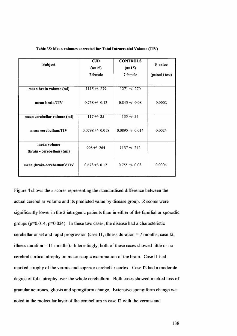
Table 35: Mean volumes corrected for Total Intracranial Volume (TIV)
SubjectCJD
(n=15)
7 female
CONTROLS
(n=15)
7 female
P value
(paired t test)
mean brain volume (ml)
mean brain/TIV
1115+/-279
0.758+/-0.12
1271 +/- 270
0.845 +/- 0.08 0.0002
mean cerebellar volume (ml)
mean cerebellum/TIV
117+/-35
0.0798+/-0.018
135+/-34
0.0895 +/- 0.014 0.0024
mean volume
(brain - cerebellum) (ml)
mean (brain-cerebellum)/TIV
998 +/- 264
0.678+/-0.12
1137+/-242
0.755 +/- 0.08 0.0006
Figure 4 shows the z scores representing the standardised difference between the
actual cerebellar volume and its predicted value by disease group. Z scores were
significantly lower in the 2 iatrogenic patients than in either of the familial or sporadic
groups (p=0.014, p=0.024). In these two cases, the disease had a characteristic
cerebellar onset and rapid progression (case II, illness duration = 7 months; case 12,
illness duration =11 months). Interestingly, both of these cases showed little or no
cerebral cortical atrophy on macroscopic examination of the brain. Case II had
marked atrophy of the vermis and superior cerebellar cortex. Case 12 had a moderate
degree of folia atrophy over the whole cerebellum. Both cases showed marked loss of
granular neurones, gliosis and spongiform change. Extensive spongiform change was
noted in the molecular layer of the cerebellum in case 12 with the vermis and
138

flocculonodular lobes m ost affected. In both cases there was very strong PrP
imm unostaining in both the granular and m olecular layers o f the cerebellar cortex.
Figure 4: Corrected cerebellar volumes adjusted for age and sex
♦ Familial ■ Sporadic A Iatrogenic
Figure 5 shows the z scores representing the standardised difference between the
actual “rest o f brain” volume (brain - cerebellum) and its predicted value by disease
group. Z scores from the two iatrogenic patients were no different from those in the
sporadic and familial groups (z scores -0 .197 and -0 .243 respectively), correlating
with the absence o f cortical atrophy seen at post-m ortem.
13 9

Figure 5: Corrected (brain - cerebellum) adjusted for age and sex
♦ Familial ■ Sporadic A Iatrogenic
Notably, in one sporadic case (case S9), where cerebellar signs were not a prom inent
feature, the m easured corrected cerebellar volume was smaller than expected from the
control data (z score -2 .76 ). At post m ortem, there was widening o f the sulci w ithin
the posterior and m iddle part o f the vermis and the posterior aspects o f the cerebellar
hemispheres. M icroscopically, there was severe depletion o f Purkinje and internal
granule cells and sparse focal spongiosis o f the m olecular layer. PrP imm unostaining
was positive in the granular and m olecular layers.
In other cases (FI 5, FI 9, 0 4 , SI 1 and S12), where little or no difference was seen in
m easured cerebellar volumes from that expected from the control values, 3 out o f 5 o f
the patients did not show prom inent cerebellar features. Post m ortem details are
available for the two sporadic cases, SI 1 and S I2. In case SI 1 (valine hom ozygous at
codon 129, PrP type 1) there was m arked cerebral atrophy with neuronal loss and
gliosis. There was patchy spongiform change but no plaques seen in the cerebellum
and although PrP im m unostaining showed widespread positivity throughout the
140

cerebral cortex, cerebellar positivity was patchy but with reticular focal positivity in
the molecular layer. Macroscopically, case S12 showed no obvious atrophy of the
cerebrum or cerebellum. There was widespread neuronal loss, gliosis and spongiform
change in the frontal, parietal, temporal and occipital lobes with fine vacuolation in
the basal ganglia and thalami, but only mild spongiform degeneration in the molecular
layer of the cerebellum, focal loss of Purkinje cells and gliosis. Immunostaining for
PrP showed extensive positivity throughout the brain.
Although measures of both cerebellar and rest of brain volumes were significantly
smaller for the group of eight patients with known prion mutations, the z scores
calculated for the cerebellar volumes did not distinguish them from sporadic cases.
Post-mortem details are available for cases one and three. Both cases had mild
cerebral and cerebellar atrophy (of the vermis in case F10) visible macroscopically.
Case F20 had abundant large eosinophilic plaques in the molecular layer of the
cerebellum, and also more widespread plaques in the neocortex and subcortical white
matter. In case F10 there was moderate spongiosis within the cerebral cortex and
extensive deposition of PrP in the molecular layer of the cerebellum.
The cerebellar and whole brain volumes of three asymptomatic cases (one had mild
truncal ataxia) with known mutations in the prion gene were not significantly
differently from their matched control (using matched t test p = 0.097 and 0.19
respectively).
Further analysis of the standardised differences between the actual volumes of the rest
of the brain (brain - cerebellum) and the predicted values by disease group showed
141
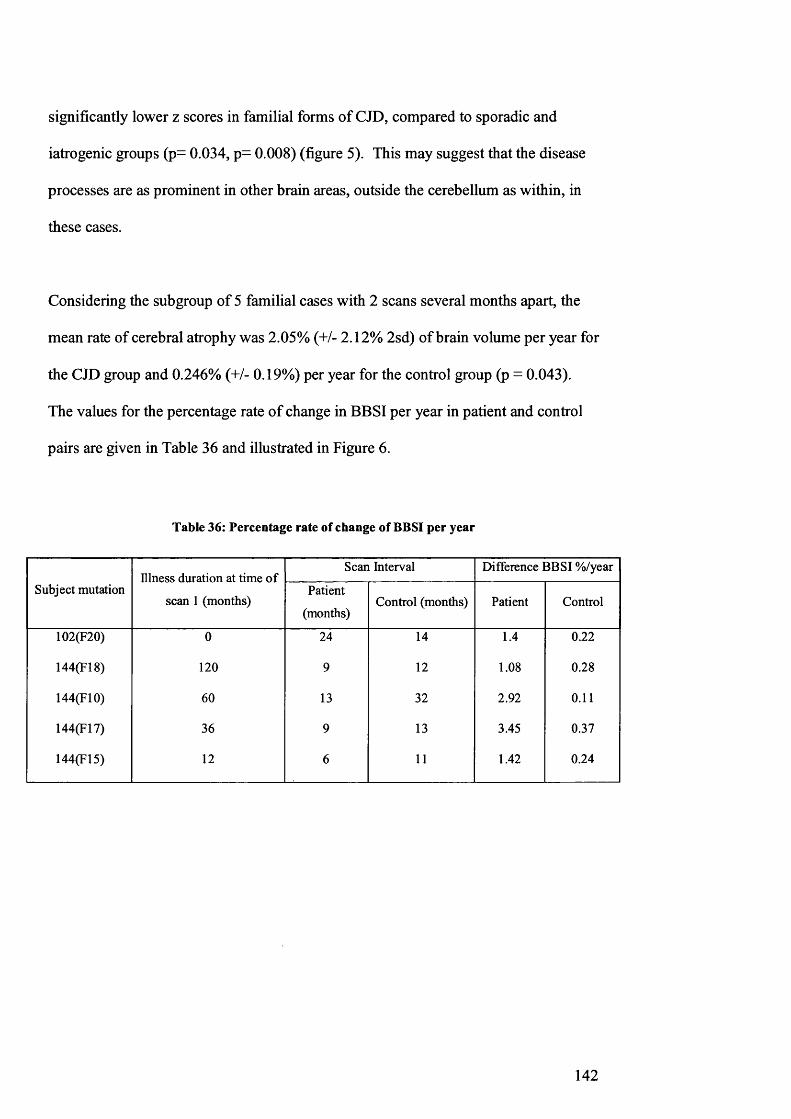
significantly lower z scores in familial forms of CJD, compared to sporadic and
iatrogenic groups (p= 0.034, p - 0.008) (figure 5). This may suggest that the disease
processes are as prominent in other brain areas, outside the cerebellum as within, in
these cases.
Considering the subgroup of 5 familial cases with 2 scans several months apart, the
mean rate of cerebral atrophy was 2.05% (+/- 2.12% 2sd) of brain volume per year for
the CJD group and 0.246% (+/- 0.19%) per year for the control group (p = 0.043).
The values for the percentage rate of change in BBSI per year in patient and control
pairs are given in Table 36 and illustrated in Figure 6.
Table 36: Percentage rate of change of BBSI per year
Subject mutationIllness duration at time of
scan 1 (months)
Scan Interval Difference BBSI %/year
Patient
(months)Control (months) Patient Control
102(F20) 0 24 14 1.4 0.22
144(F18) 120 9 12 1.08 0.28
144(F10) 60 13 32 2.92 0.11
144(F17) 36 9 13 3.45 0.37
144(F15) 12 6 11 1.42 0.24
142

Figure 6: Percentage rate o f change o f BBSI per year
♦ Patients ■ Controls
Discussion
Rates o f atrophy and patterns o f tissue loss w ithin cerebral substructures can be
delineated from serially acquired, registered volum etric MR images. This technique
has been applied in the study o f AD to quantify tissue loss and therefore show disease
progression132 and also to demonstrate progressive cerebral and cerebellar atrophy in
one case o f familial C JD 133. Quantification o f whole brain and medial tem poral lobe
atrophy in AD allows the distinction between those with the disease and those with
atrophy associated with normal aging, and also allows a correlation between the rate
o f volume loss and cognitive decline140’142&143. These techniques m ay be o f sim ilar
value in the diagnosis and m onitoring o f disease progression in CJD.
The first hypothesis was proven. Across the groups o f sporadic, fam ilial and
iatrogenic prion diseases, it is possible to dem onstrate significant cerebral and
cerebellar atrophy with volumetric MR imaging, with scan interval ranging from 6 to
24 months. Further work is underway to assess the volume change in the cerebellum
143

of patients with Alzheimer’s disease (AD). This will allow the evaluation of the
usefulness of this tool in the differential diagnosis of CJD and AD especially in the
elderly.
Volumetric MR image analysis, used cross-sectionally has limitations as a diagnostic
tool as there is overlap in the range of whole brain and cerebellar volumes in CJD and
control groups. Applying the technique longitudinally to registered, serial images
allows calculation of the BBSI, which is a highly reproducible measure of atrophy.
Volumes were corrected for the total intracranial volume which is essential not only
to reduce individual variation when using the data cross-sectionally, but more
importantly to allow for changes in scanner gradient calibration over time in the
longitudinal study. Good quality, serial imaging is difficult to obtain in this patient
group. The patients often tolerate MR scans poorly, they have movement disorders,
for example, myoclonus and chorea, which introduce movement artefact and their
clinical condition may deteriorate over a matter of weeks making repeat scanning
inappropriate. Where serial imaging is possible, in those with familial disease,
atrophy may be quantified only at the time of repeat imaging, six to twelve months
from baseline, thus reducing the feasibility of the technique as an immediate
diagnostic test. However the second hypothesis was proven. It was possible to
quantify annual whole brain atrophy in a subgroup of patients with familial CJD.
In this subgroup of familial cases, there was one case with a P102L mutation (F20) in
the Prion gene and four with 144 base pair insertions. The illness duration at the time
of the first scan ranged from 0-120 months and the scan interval ranged drom 6-24
months. Case F20 was asymptommatic at presentation and over the two-year period
144

between scans; there was no measurable decline in general intellect. However, verbal
and visual memory and frontal executive function became impaired over this time.
Cases F10 and FI 7, with the greatest measured annual rates of cerebral atrophy, both
had severe decline in intellect with global deficits (very impaired memory, naming,
visual perception and frontal executive function). Cases FI 5 and FI 8 had severe
decline in intellect and frontal executive dysfunction but with some preservation in
verbal memory and naming (and visuoperception, FI 5 only) until later in the illness
course. Although there are similar rates of whole brain atrophy compared to those
seen in patients with AD, this suggests that the pattern of cognitive decline in the
familial CJD cases and therefore the distribution of atrophy in the brain may be
distinct in these different diseases.
With the recognition of new prion diseases, such as vCJD, the search for disease
modifying agents has a new impetus. Carefully conducted clinical trials in confirmed
cases of CJD are necessary to test the effectiveness of these therapeutic agents. This
requires well-validated, reproducible markers of clinical outcome, which can detect
changes in disease progression in treated and placebo groups. Although cross
sectional volumetric MR imaging may not be feasible as a diagnostic marker,
quantification of rates of atrophy in the whole brain and cerebellum in CJD over time
using MR may be suitable to monitor disease progression in therapeutic clinical trials.
145
MR spectroscopy in CJD
Introduction
Proton magnetic resonance spectroscopy (1H-MRS) is a non-invasive tool used in the
quantification of neurometabolites in defined regions of the brain. It may be
performed as part of a clinical MRI brain examination, in the same scanner if this is
equipped with appropriate software. Use of this technique in the investigation of the
prion diseases has been limited to date.
The earliest report of HI-MRS in human prion disease, to our knowledge, describes a
53-year-old female with biopsy proven sporadic CJD144. Seven months from illness
onset, MR imaging showed mild cortical atrophy and hyperintense signal in the
lentiform nuclei. Short TE 1H-MRS of the left parietal white matter showed a
reduction in the N-acetylaspartate (NAA) peak by 40% and an increase in myoinositol
(MI) of 30% compared to a normal age and sex matched control. The results were
similar in the right parietal and frontal white matter regions. In the paramedian
frontal and parietal grey matter, NAA alone was reduced by 30%. The authors
correlate the reduction in NAA with the severe neuronal loss found in CJD and note
that the metabolic alterations occurred in regions of the brain which appeared normal
on structural MR imaging.
Subsequent published studies have used long echo time techniques. There is one
report of 1H-MRS performed at an earlier stage in the illness course145. Imaging from
two cases of sporadic CJD performed at three months and less than one month from
symptom onset failed to show any significant change in metabolite concentrations
when compared to results from two non-demented control subjects, using a region of
146

interest (ROI) incorporating the left and right parietal cortex and bilateral subcortical
white matter. In one case, imaging was repeated at ten months revealing a drop in the
NAA concentration in this region by 46%.
There are two further reports of the use of 1H-MRS in familial prion disease. Shyu et
al describe a case with a point mutation at codon 210 (GTT to ATT), presenting with
an unusual panencephalitic form of CJD with an early onset and long illness
duration124. MR imaging at three months from symptom onset demonstrated
increased T2-dependent signal intensity in the basal ganglia and thalami. Imaging
twelve months later showed evidence of cortical thinning, diffuse signal change in the
white matter and hypointense signal in the basal ganglia and thalami. 1H-MRS of the
frontal region showed an absence of NAA, creatine (Cr) and choline. Konaka et al,
2000, report a 22 year old female with Gerstmann-Straussler-Scheinker (GSS) disease
(P102L mutation)146. One year from symptom onset, MR imaging revealed mild
atrophy of the cerebellar hemispheres, vermis and the cerebral cortex. Spectra from
voxels placed in the frontal cortex, putamen, cerebellar vermis and hemispheres
showed reduction in the NAA/Cr ratio in each region, including the frontal lobe,
putamen and cerebellum; correlating with the neuropsychiatric features, and
extrapyramidal and cerebellar signs. SPECT perfusion patterns were normal in the
cerebrum and cerebellum in this case. The authors suggest that subtle changes in GSS
may be discernible with 1 H-MRS even in the early stages of the illness.
Pandya et al report short echo time 1 H-MRS in one case of probable vCJD, twenty
months from illness onset (after 93% of the total illness duration). The patient had
bilateral increased signal in the pulvinar on T2 dependent images and a markedly
147

reduced NAA/Cr ratio ratio in the right pulvinar147. There was no quantification of
MI ratios. Imaging data from two cases with biopsy proven sporadic CJD were also
discussed. In both cases there was increased signal in the putamen and caudate
nucleus on T2 weighted imaging and a reduction in the NAA/Cr ratio in the putamen
using long echo time 1 H-MRS. There was variable thalamic signal change reported
on T2 dependent imaging not further investigated with 1 H-MRS.
To further increase our knowledge of the diagnostic potential of short TE in vivo
spectroscopy and any insight it may give into pathological changes in prion disease,
we studied the spectroscopic features of three cases with probable vCJD, with
comparative data for two cases with familial CJD, one with sporadic disease and eight
normal control subjects.
Methods
A subgroup of five patients enrolled in the project, were recruited for further MR
imaging (V2, V7, V I4, Ol and 04; 2 male, 3 female mean age 30.6 years). Three
cases were given a diagnosis of probable variant CJD with positive tonsillar biopsy.
In the other two cases, a mutation was detected in the prion protein gene and the
clinical symptoms and signs were thought to be compatible with the onset of familial
disease. Data from these subjects were compared to those from a further case with
biopsy proven sporadic CJD (age > 60 years) and one young, asymptomatic subject
with a family history of prion disease and a mutation in the prion gene detected at
presymptomatic testing. A further group of 8 age matched normal controls were
recruited (3 male, 5 female; mean age 26.8 years).
148

Short echo time, single voxel 1 H-MRS was performed using an automated point
resolved spin echo localisation (PRESS) technique on a commercial clinical 1.5 T MR
scanner (PROBE-Q, Signa Horizon EchoSpeed scanner, v 5.7 software, TR 2000ms,
TE 30ms, 192 averages, General Electric Medical Systems, Milwaukee). All of the
cases also had dual echo axial localisation images (axial inversion-recovery fast spin
echo, 3mm thick slices, T1 = 300ms, TR = 3000ms, TE = 15ms). Spectra were
acquired from voxels of interest (1-3 ml volume) selected in the left pulvinar region
(postero-medial thalamus), the left caudate nucleus and the right frontal white matter.
In these areas and with these voxel sizes, infringement on cerebrospinal fluid was not
a problem. There was therefore no need to make corrections for atrophy. Metabolite
ratios were determined using on-line software (PROBE-Q) and concentrations
calculated off-line using validated frequency domain fitting technique, LC Model
[Provencher]148. The T2 and PD-weighted localiser images were reviewed by a
neuroradiologist, together with any existing structural MR imaging, i.e. axial T2 and
proton density and volumetric images, to determine the presence of cerebral atrophy
and pathological signal change in the patient and control groups.
Spectra were obtained using the same protocol in eight normal control subjects.
Localiser images in this group were also reviewed for unsuspected structural
abnormalities. All spectra were inspected visually. Metabolite ratios for the assigned
resonances for N-acetylaspartate (2.01 ppm), Choline (3.2ppm), and myo-inositol
(3.56ppm) were calculated relative to Creatine/phosphocreatine (3ppm) using PROBE
Q. Concentrations of the metabolites, myoinositol [MI] and N-acetylaspartate-
glutamate [NAA (G)] were calculated using LC Model. The repetition times were
shorter than those normally used for quantitative measures in order to minimise
149

acquisition time. T1 effects may potentially lead to an underestimation of the
concentrations calculated and so these were expressed using institutional units (IU).
Metabolite concentrations and ratios were compared between the variant CJD and
normal control groups using the Mann-Whitney U test. The Mann Whitney U test
was the appropriate significance test, for non-parametric data, for two groups with
different (unpaired) subjects.
Results
The clinical characteristics of the cases are given in Table 37. The H l-
MRspectroscopy data for the variant cases and the normal controls are given in tables
38 to 40.
150

Table 37: MR spectroscopy: Clinical characteristics of the cases
Case Illness Clinical symptoms and signs MRIduration
(at time of assessment in months)
personalitychange/
depression
cognitivedecline
myoclonus ataxia pyramidalsigns
extra-pyramidal
signs
sensorydisturbance
atrophy signalchange
thalamus and
V2 14 1 1 0 1 1 0 1 0 midbrain
V7 11 1 1 0 1 0 0 1 0 thalamus
V14 16 1 1 1 1 1 0 0 0 posterior &medial thalamus
01 84 1 1 1 1 1 1 0 1 0
04 23 1 1 0 1 1 0 1 0 mesial frontal
S9 4 1 1 1 0 1 1 0 0 0

There was a significant and dramatic, 2.5 fold (150%) increase in the concentration of
MI, and a significant, 50% reduction in NAA in the left posterior thalamus of the
three cases of probable variant CJD compared to the normal controls. This was
consistent with a rise in the Ml/Cr ratio and a fall in the NAA (G)/Cr ratio in this
region. All three cases were reported to have the pulvinar sign on T2 MR structural
imaging, confirmed on review of the localisation images. The calculated mean
MI/NAA ratio for the vCJD cases was 5.6 times greater than that for the normal
controls (mean MI/NAA vCJD = 2.06, standard deviation (sd) = 0.501; normal
controls = 0.37, sd =0.092).
There was a significant increase in [MI] and the Ml/Cr ratio in the right caudate in the
two cases where this region was examined although only one case showed a
significant reduction in NAA (G). No signal change was reported in this region on T2
MR imaging. Data from the frontal white matter were only available for one of the
three variant cases. These showed a significant decrease in the NAA (G)/Cr level
from the PROBE measurements only. No corresponding white matter signal
abnormality was detectable on T2 weighted images.
The two young familial cases showed different patterns of abnormality with HI-MRS.
Case Ol showed a significant increase in MI and the Ml/Cr ratio in the left thalamus.
0 4 did not show any abnormality in this area. In neither case was any thalamic signal
abnormality apparent on T2 weighted images. Only 0 4 tolerated further imaging in
the caudate and frontal regions. There was a significant reduction in NAA (G) in the
caudate. The value for the concentration of creatine in this region was below the
normal range and so meaningful interpretation of metabolite ratios was not possible.
152
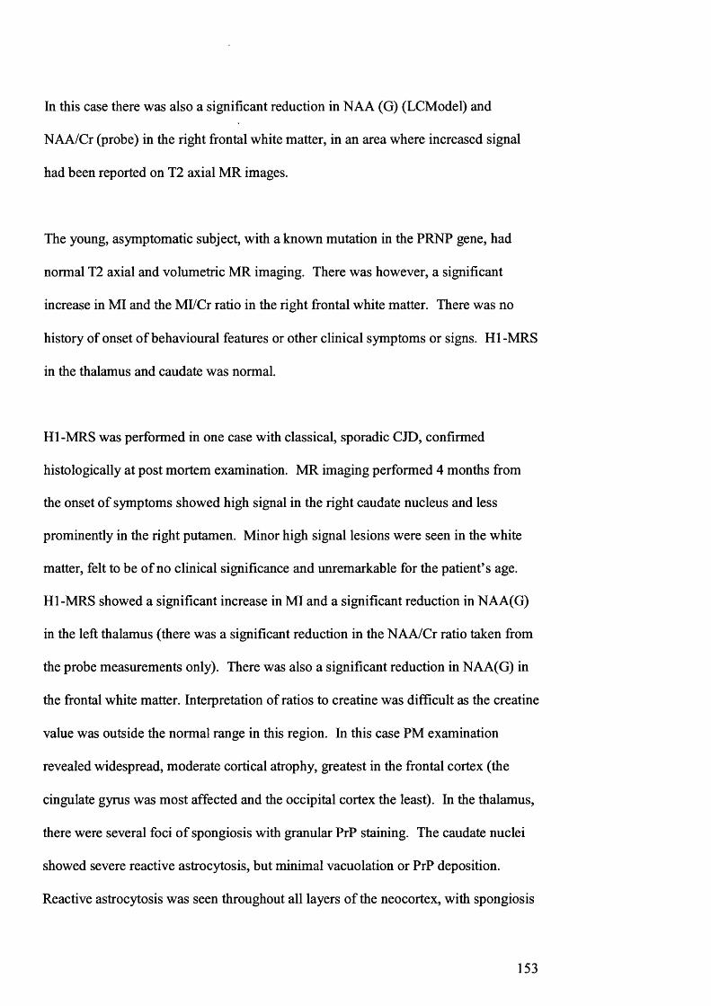
In this case there was also a significant reduction in NAA (G) (LCModel) and
NAA/Cr (probe) in the right frontal white matter, in an area where increased signal
had been reported on T2 axial MR images.
The young, asymptomatic subject, with a known mutation in the PRNP gene, had
normal T2 axial and volumetric MR imaging. There was however, a significant
increase in MI and the Ml/Cr ratio in the right frontal white matter. There was no
history of onset of behavioural features or other clinical symptoms or signs. HI-MRS
in the thalamus and caudate was normal.
HI-MRS was performed in one case with classical, sporadic CJD, confirmed
histologically at post mortem examination. MR imaging performed 4 months from
the onset of symptoms showed high signal in the right caudate nucleus and less
prominently in the right putamen. Minor high signal lesions were seen in the white
matter, felt to be of no clinical significance and unremarkable for the patient’s age.
HI-MRS showed a significant increase in MI and a significant reduction in NAA(G)
in the left thalamus (there was a significant reduction in the NAA/Cr ratio taken from
the probe measurements only). There was also a significant reduction in NAA(G) in
the frontal white matter. Interpretation of ratios to creatine was difficult as the creatine
value was outside the normal range in this region. In this case PM examination
revealed widespread, moderate cortical atrophy, greatest in the frontal cortex (the
cingulate gyrus was most affected and the occipital cortex the least). In the thalamus,
there were several foci of spongiosis with granular PrP staining. The caudate nuclei
showed severe reactive astrocytosis, but minimal vacuolation or PrP deposition.
Reactive astrocytosis was seen throughout all layers of the neocortex, with spongiosis
153
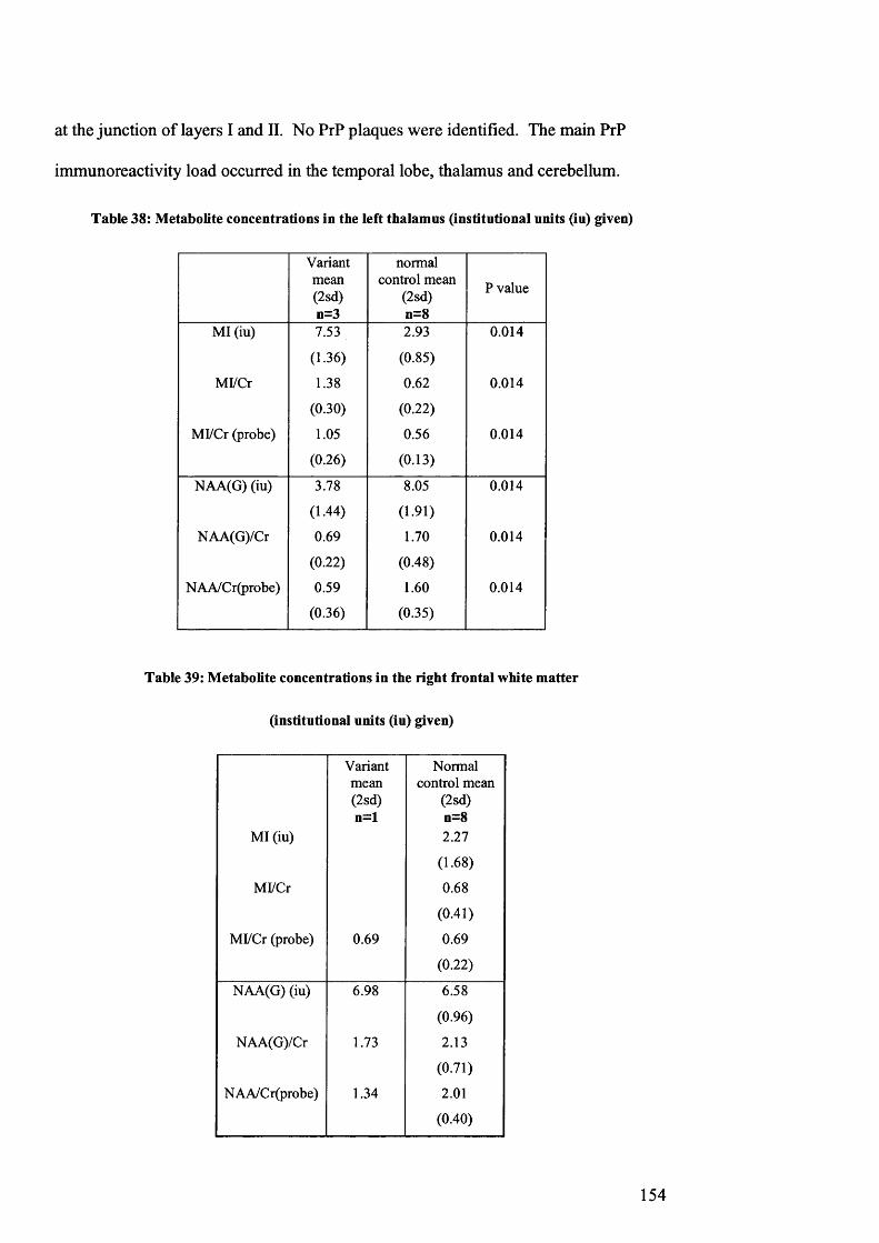
at the junction of layers I and II. No PrP plaques were identified. The main PrP
immunoreactivity load occurred in the temporal lobe, thalamus and cerebellum.
Table 38: Metabolite concentrations in the left thalamus (institutional units (iu) given)
Variantmean(2sd)n=3
normal control mean
(2sd) n=8
P value
MI (iu) 7.53 2.93 0.014
(1.36) (0.85)
Ml/Cr 1.38 0.62 0.014
(0.30) (0.22)
Ml/Cr (probe) 1.05 0.56 0.014
(0.26) (0.13)
NAA(G) (iu) 3.78 8.05 0.014
(1.44) (1.91)
NAA(G)/Cr 0.69 1.70 0.014
(0.22) (0.48)
NAA/Cr(probe) 0.59 1.60 0.014
(0.36) (0.35)
Table 39: Metabolite concentrations in the right frontal white matter
(institutional units (iu) given)
MI (iu)
Ml/Cr
Ml/Cr (probe)
Variantmean(2sd)n=l
0.69
Normal control mean
(2sd) n=8 2.27
(1.68)
0.68
(0.41)
0.69
(0.22)
NAA(G) (iu) 6.98 6.58
(0.96)
NAA(G)/Cr 1.73 2.13
(0.71)
NAA/Cr(probe) 1.34 2.01
(0.40)
154

Table 40: Metabolite concentrations in the right caudate nucleus (institutional units (iu) given)
MI (iu)
Ml/Cr
Ml/Cr (probe)
Variantmean(2sd)n=37.04
(2.44)
1.22
(0.24)
0.99
(0.50)
Normal control mean
(2sd) n=8 3.09
(2.17)
0.54
(0.42)
0.56
(0.22)
P value
0.040
0.040
0.044
NAA(G) (iu) 5.77 6.77 1.000
(4.14) (1.60)
NAA(G)/Cr 1.00 1.19 0.143
(0.56) (0.33)
NAA/Cr(probe) 1.11 1.31 0.242
(0.34) (0.44)
155
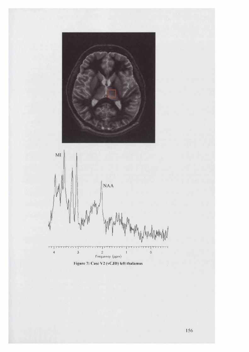
MI I
i i ~| r "i i i i~r r i i ~[~n r r r i i i 'r | i t r i i i i~i i | i n i 1 r r~ i i | t 1 i i i t i i
4 3 2 1 0Frequency (ppm)
F igure 7: C ase V2 (vC JD ) left thalam us
1 5 6

NAA
MI
4 3 2 1 0Frequency (ppm)
Figure 8: N orm al contro l left thalam us
157

Discussion
The pathological significance of the marked increase in myo-inositol and very low
levels of NAA in the thalamus of these cases with vCJD is uncertain but may reflect
the severe astrogliosis and neuronal loss which are seen at post mortem in the pulvinar
nuclei in this condition. These changes are thought to produce the characteristic
bilateral pulvinar hyperintensity that has been described on T2 dependent sequences
in a large number of cases with variant CJD109. The grey matter changes seen with
1H-MRS are similar to but far more marked than those widely reported in other
degenerative dementias. An increase in MI of 15-20% and a decrease of NAA of 10-
15% has been reported in grey matter in AD149. By comparison, in all three cases of
vCJD in this study, there was an approximate 150% increase in the mean MI
concentration and approximate 50% reduction in NAA concentration detected in the
thalamus. The combined effect gives over a 5-fold increase in the mean MI/NAA
ratio for vCJD compared to normal subjects. There was a significant increase in MI
in the right caudate of the two cases with vCJD in whom this region was tested. 1H-
MRS revealed more extensive abnormalities than those seen on structural sequences
and also demonstrated different patterns of metabolic change in the subtypes of prion
disease, possibly reflecting differential involvement of structures within the brain.
The two cases of familial CJD were originally referred with symptoms and signs
suggestive of variant CJD. Both cases were young (ages 28 - 30 years) and fulfilled
the diagnostic criteria for possible vCJD prior to genetic testing. Interestingly, neither
of these cases had the same profile with Hl-MRSpectroscopy. One case did show a
significant increase in MI in the thalamus but with a normal NAA concentration in
this area. The other case had normal spectra in the thalamus but a reduction in NAA
in the frontal white matter and the right caudate nucleus. The study of larger groups
158

is needed to clarify the consistency of this difference in pattern of metabolite change
between subtypes of prion disease.
Many functions have been attributed to MI to account for changes in concentration
with disease150. Glial cells are thought to contain high levels of MI and so this may
act as a marker of glial cell numbers. Other suggested roles include that of an
osmoregulator, an intracellular messenger or as a detoxification agent via glucuronate,
a breakdown product. NAA concentration is thought to act as a marker of neuronal
viability. Mitochondrial phosphorylation and rates of NAA synthesis have been
correlated. Reductions in NAA have been documented in AD in the absence of
detectable cerebral atrophy leading to suggestions that a reduction in NAA may
indicate an alteration in mitochondrial function or neuroinflammation in neurons that
are still viable, rather than a loss of neurons. All three of the cases of vCJD in this
study had the pulvinar sign on T2 weighted images suggesting profound reactive
astrogliosis in this region. It could be postulated that the dramatic increase in MI in
this region could simply be attributed to the high MI content of these glial cells.
One of the limitations of this part of the study was the small number of patients and
their late presentation such that all of the cases had moderately advanced vCJD at the
time of imaging. Single voxel MRS techniques require the patient to remain still for
consecutive, approximate seven minute periods for the acquisition of data for each
region. Patients presenting at an advanced stage of the illness were less able to
tolerate this. However, the magnitude of the metabolic derangement in these cases
suggests that it may be possible to detect abnormalities earlier in the disease course.
Significant metabolic abnormalities were demonstrated in regions where signal was
159

normal on T2 weighted sequences. The presence of regions with an abnormality in
myo-inositol concentration with normal NAA suggest that as with other conditions
e.g. Down’s syndrome, elevation in MI may precede NAA loss associated with
neuronal loss or dysfunction151. Significant MI abnormality was also demonstrated in
one currently asymptomatic gene carrier. All of these findings suggest that short echo
time 1H-MRS may have a role in the early diagnosis of the prion diseases and that
further studies to explore this potential are warranted.
160

STUDY II: THE MANAGEMENT OF YOUNG PEOPLE WITH DEMENTIA
Introduction
Many of the problems faced by young people diagnosed with variant CJD and their
families are not unique. They are shared by others with Alzheimer’s disease,
ffontotemporal degeneration and other causes of early onset dementia. All become
unwell at a time that affects their young families, employment prospects, finances and
generally their life at home. They can undergo difficult behavioural changes,
accompanied by a loss of the cognitive and physical ability to cope with the activities
of daily living and become increasingly reliant on spouses or parents for everyday
care. Carers are unable to envisage or predict the potential difficulties in caring for
someone with this relentlessly progressive disease and education and practical
forward planning may alleviate many problems. Health care professionals have a
duty, not simply to diagnose an illness and alleviate symptoms but also to provide
ongoing advice and support in all aspects of management of the patient and their
family. This is particularly important for the prion diseases where much time has to
be invested in the explanation of this little understood and often misconceived illness.
There needs to be careful explanation of issues surrounding infection control for
example in dental and surgical practice; measures to prevent transmission via blood or
organ donation and genetic screening to exclude a hereditary cause.
One of the major concerns of families is the likelihood of vCJD occurring in siblings
or children of affected family members who may have shared meals in childhood or
possibly been exposed in utero or via breast feeding. Although there is currently no
evidence to date to support an increased risk to siblings or of vertical transmission, the
161
potential risks are not quantifiable and so much counselling time is often spent
dealing with such issues.
Seeing at first hand the difficulties faced throughout the diagnostic and management
phases of this illness by exhausted and distraught families prompted a closer look at
how young people with dementia and their families are managed across the UK.
There have been concerns that whilst young people would naturally be referred to
general adult or paediatric psychiatrists with early onset dementia, it is old age
psychiatrists who have particular experience in the management of patients with
dementia with liaison with professionals in the community. There is a risk that
patients may fall between these and neurology specialities resulting in incomplete
care. As a result of these concerns the Royal College of Psychiatrists has made
recommendations for service provision for young people with Alzheimer ’s disease
(AD) and other dementias152. They propose that each district should have one named
consultant responsible for the service, suggesting old age psychiatrists are best placed
to fulfil this role, whilst acknowledging the need for close collaboration with a range
of specialists.
Although the proposals are welcomed, there were also concerns that patients may be
inadequately investigated if referral to neurologists was incomplete153'156. The task
force initiated by the European Federation of Neurological Societies (EFNS)
recommends, that neurologists should have a clear role in the management of
dementia157.
162

We therefore conducted a survey to assess current stated UK practice in the diagnosis
and management of younger people with dementia. Our main objective was to
determine how patients are investigated and what support and follow up is offered.
Method
The questionnaire (see appendix II) was sent to all consultant neurologists and old age
psychiatrists in the UK, identified from lists supplied by the Royal College of
Psychiatrists and Association of British Neurologists. Reminders were sent after six
weeks to non-responders. Questions required either yes/no or graded (i.e. always,
often, sometimes or never) answers.
Statistical Analysis
Statistical analysis was performed using STATA6 (Stata Statistical Software: Release
6.0 for Windows, Stata Corporation). Where responses were compared between
consultant neurologists and old age psychiatrists, P-values were calculated using the
2-sided Fisher’s exact test. The Wilcoxon rank sum test was applied to compare the
trends of responses given, where replies were graded.
Results
A summary of the main results is given in table 41. Overall, 64% of the population
surveyed responded, comprising 67% (212/318) of consultant neurologists and 62%
(215/346) of consultant old age psychiatrists. All subsequent figures are given as a
percentage of those specialists who responded to each question.
163
The number of new referrals with young onset dementia to neurologists and old age
psychiatrists was comparable. Where it was indicated that a specialist dementia
service was available, 80% (95% confidence interval 71-87%) of neurologists refer to
such a centre compared to 37% (22-53%) of old age psychiatrists (PO.OOOl).
Twenty-one percent of consultant neurologists (16-28%) refer to old age psychiatry
services in comparison to 60% of old age psychiatrists (52-67%) who refer to a
neurologist
Of those neurologists who are not part of and do not refer to a specialist dementia
service and who state they do not receive referrals from old age psychiatrists (n = 75),
23% (14-34%) refer to an old age psychiatrist 77% therefore do not involve an old
age psychiatrist in their patient management. Of the equivalent group of old age
psychiatrists not working as part of or referring to a specialist team and who do not
receive neurological referrals (n = 71), 51% (39-63%) do not refer to a neurologist.
Where no regional centre was available for referral, 87% (77-93%) of neurologists
and 84% (76-90%) of old age psychiatrists stated that they would refer to one if it
became available (P=0.68).
A comprehensive history, including a family history, details of personality change and
somatic symptoms was taken by the majority of neurologists and old age
psychiatrists. In contrast, significantly more old age psychiatrists obtained an
independent history from a carer and assessed symptoms of depression and changes in
driving performance (see table 41).
164

Reported rates of performing physical examinations were statistically significantly
higher amongst neurologists, with 70% (64-77%) and 98% (95-99%) always
performing general physical and neurological examinations respectively, compared to
57% (50-64%) and 50% (43-57%) of old age psychiatrists (P=0.0011 and P< 0.0001).
Comparing the subgroup of old age psychiatrists practicing without liaison with
neurologists and specialist services (n=56), with neurologists, only 54% (40-67%) and
45% (32-59%) of these, always performed general and neurological examinations
respectively (P=0.011 and PO.OOOl).
Although the use of investigations such as, full blood count, vitamin B12, electrolytes,
renal, liver and thyroid function was comprehensive between the groups, other
important tests, for example, measures of plasma viscosity (for example, erythrocyte
sedimentation rate, ESR) and syphilis serology were requested by statistically
significantly more neurologists than old age psychiatrists (ESR: neurologists 92%
(87-95%), old age psychiatrists 78% (72-84%), P = 0.0001). The use of structural
imaging overall was comparable; although only 79% (72-84%) of neurologists and
76% (69-81%) of old age psychiatrists indicated that they always perform either a CT
or VIRI brain scan (P=0.37).
Only 37% (30-44%) of neurologists always and 32% (25-39%) often use EEG and
25% (19-31%) always and 34% (27-41%) often arrange cerebrospinal fluid (CSF)
examination. Significantly fewer old age psychiatrists arrange these investigations
(P<0.0001), and in particular, of the subgroup of old age psychiatrists who practice
witiout liaison with neurologists, 77% (63-87%) never arrange CSF examination.
165
Participants were asked if carers were given an opportunity to see the doctor in the
absence of the patient. Sixty-eight percent (61-74%) of old age psychiatrists always
managed this compared to 50% (43-57%) of neurologists, (PO.OOOl). Forty-four
percent (37-51%) of old age psychiatrists, compared to only 18% (13-24%) of
neurologists have a specialist nurse available in their unit to provide support and
information to patients and carers (P<0.0001).
Significantly more old age psychiatrists, 64% (57-70%), compared to 37% (31-44%)
of neurologists, always provide information about support groups such as the
Alzheimer’s Society (PO.OOOl). Again significantly fewer of the subgroup of
neurologists acting without liaison with old age psychiatry services, give such
information and support, when compared to old age psychiatrists (PO.OOOl). Advice
given regarding informing the driving and vehicle licensing association (DVLA) was
comparable. Significantly more old age psychiatrists involve Community Psychiatric
Nurses (CPN), in managing their patients in the community, with 46% (39-53%)
indicating that they always refer, compared to 6% (3-10%) of neurologists
(PO.OOOl). Those neurologists practicing without involving old age psychiatry
services, in line with neurologists as a whole, were also statistically significantly less
likely to involve CPN than old age psychiatrists (PO.OOOl).
Finally, consultants were asked to grade how often they discussed dmg treatments and
trials with their patients. Significantly more old age psychiatrists always discuss dmg
treatments, 52% (45-59%) compared to 38% (32-45%) of neurologists (PO.OOOl).
Old age psychiatrists were also more likely to discuss therapeutic trials (P=0.0074).
Fifty-two percent (45-59%) of old age psychiatrists compared to only 31% (25-38%)
166

of neurologists prescribe acetylcholinesterase inhibitors as per guidelines (PO.OOOl).
Of the prescribing specialists, only 58% (49-67%) of neurologists, compared to 98%
(94-99%) of old age psychiatrists monitor patients after prescription (PO.OOOl).
167

Table 41: Summary of the key responses from consultant neurologists and old age psychiatrists
N Always
n (%)
Often
n (%)
Sometimes
n (%)
Never
n (%)
P value
HISTORY
CN
COAP
210
211
125 (60)
151 (72)
59 (28)
44 (21)
26 (12)
16 (8)
0 (0)
0 (0)0.0082driving ability
symptoms of depressionCN
COAP
211
211
188 (89)
202 (96)
20 (9)
7 (3)
3 (1)
2 (1)
0 (0)
0 (0)0.011
independent history from carerCN
COAP
211
211
177 (84)
197 (93)
32 (15)
14 (7)
2 (1)
0 (0)
0 (0)
0 (0)0.0021
EXAMINATION
CN
COAP
210
211
148 (70)
120 (57)
36 (17)
43 (20)
26 (12)
36 (17)
0 (0)
12 (6)0.0011general
neurologicalCN
COAP
209
208
204 (98)
103 (50)
5 (2)
55 (26)
0 (0)
36 (17)
0 (0)
14 (7)<0.0001
INVESTIGATION
CN
COAP
196
202
154 (79)
153 (76)
41 (21)
39 (19)
1 (1)
9 (4)
0 (0)
1 (0)0.37imaging
FOLLOW UP
CN
COAP
199
209
12 (6)
96 (46)
45 (23)
102 (49)
90 (45)
10 (5)
52 (26)
1 (0)<0.0001community psychiatric nurse
information on support groupsCN
COAP
210
211
78 (37)
134 (64)
82 (39)
62 (29)
39 (19)
15 (7)
11 (5)
0 (0)<0.0001
C N - consultant neurologist; COAP - consultant old age psychiatrist
Discussion
I devised this survey to investigate my concerns that people with young onset
dementia may experience delays in clinical investigation and diagnosis and inefficient
management due to the lack of a defined age-appropriate specialist service to address
their needs. I was expecting to show that original referral to a general adult (or
paediatric) psychiatrist may mean a delay in referral to a neurologist and a delay or
absence of a full neurological investigation. Simlarly, direct initial referral to a
neurologist may mean a delay or absence of referral to old age psychiatry services and
hence a lack of appropriate community followup. Whilst some areas of the country
have multidisciplinary dementia services with input from neurologists, old age
psychiatrists, CPNs and social workers, working from one centre, this is not the case
everywhere.
There was a good response to the postal survey. Analysis of referral patterns
confirmed that the ideal of full collaboration between consultant neurologists and old
age psychiatrists is not achieved. There are young patients with dementia managed by
consultant neurologists without liaison with old age psychiatry services and vice
versa. This is important because the survey illustrates (as I expected) that consultant
neurologists and old age psychiatrists investigate and manage these patients
differently. More consultant old age psychiatrists take details of symptoms of
depression and changes in driving ability but only half always perform a neurological
examination. Fewer than half of the subgroup of consultant old age psychiatrists, who
do not collaborate with neurologists, perform a neurological examination implying
that this task falls to the general practitioner in many cases. The results also suggest
that fewer consultant old age psychiatrists arrange key serological investigations.
169

EEG and CSF examinations are arranged more frequently by consultant neurologists
but are not routinely used by either specialist. This is expected as the role of these
investigations may be in cases where there is a certain clinical suspicion or unusual
presentation (in accordance with the EFNS guidelines). In the American Academy of
Neurology dementia guidelines, lumbar puncture is not suggested as a routine study,
but is recommended in a person with dementia under 55 years of age .
The Royal College of Psychiatrists have published a consensus statement on the
assessment and investigation of elderly people with cognitive impairment159. This has
been updated and future work will look at broadening the result to treatment,
management and working with voluntary agencies, primary care and other professions
allied to medicine, including social workers and community psychiatric nurses160. In
a retrospective case note study161, all cases of potentially reversible intracranial
pathology were detected if these guidelines for brain imaging were applied. In a
postal survey aimed at comparing the practice styles of US and UK neurologists, 98%
of US and 97.7% of UK neurologists indicated that they would order a neuroimaging
study for an elderly patient presenting with dementia . In our study, neither group
of specialists indicated that they would always perform imaging in a case with young
onset dementia. However this may be because a scan is performed prior to referral.
The recommendation to neurologists of the European task force is that neuroimaging
should be performed once in all cases of dementia referred to a neurologist157.
The survey further reveals that more consultant old age psychiatrists give carers the
opportunity to speak to them in the absence of the patient and a significantly greater
percentage have a specialist nurse available. The former may be due to time
170

constraints in neurology clinics. A greater proportion of consultant old age
psychiatrists give advice on support groups and information on matters such as power
of attorney. Although the practice of informing patients about their responsibilities of
instructing the DVLA was comparable between old age psychiatrists and neurologists,
the number regularly doing so was lower than expected. Significantly more old age
psychiatrists involve community psychiatric nurses in follow up, probably reflecting
their expertise in the management of patients with behavioural problems in the
community. The survey also importantly highlights that a larger proportion of old age
psychiatrists when compared to neurologists, are involved in the prescription and
monitoring of acetylcholinesterase inhibitor therapy.
Recommendations to neurologists from the European task force regarding the
diagnosis and management of people with dementia should assist the development of
local guidelines within neurology, psychiatry and specialist dementia services across
the country. There are some specific areas where UK practice differs. It is
recommended that all patients and carers should be asked about driving and that
neuroimaging should be performed once in all young patients with dementia. More
importantly though, the study shows that the assessment and management of patients
with young onset dementia in the UK would be exemplary if the skills of both
neurologists and old age psychiatrists were employed in the care of each patient.
Clear standards are available for the diagnostic assessment and investigation of
suspected dementia by Specialist Old Age psychiatry services160, including guidelines
from NICE regarding responsible prescribing. Consideration should be given to how
the standards set in the National Service Framework for Older People163, regarding
171

the person centred approach to care, could be extended to younger people with
dementia.
The survey did not investigate the role of palliative care specialists in the care of
patients with variant CJD. This crucial role did become apparent during the study
period and with hindsight it would have been informative to investigate the current
involvement of palliative care teams, by neurologists and old age psychiatrists. The
CJD Surveillance Unit in Edinburgh, have reported the findings of a study of needs,
from the perception of families caring for a relative with vCJD164. In depth interviews
with families of 19 cases illustrated how difficult it was to find a suitable place to
provide terminal care. Where available, specialist palliative care was of a very high
quality and it was recommended that palliative care specialists be involved wherever
possible.
In this project, most of the cases referred with possible vCJD were originally seen by
their GP (mean six months from illness onset), with onward referral to a neurologist
or psychiatrist on average 12 months from disease onset (mean ten and twelve months
respectively). It would seem that to make an impact on early diagnosis, it is vital for
general practitioners to have a high index of suspicion for vCJD to precipitate onward
referral to specialist services with the minimum delay.
172

CONCLUSIONS
This project has involved the detailed study of 21 patients referred to two specialist
centres with suspected vCJD. Fifteen cases were subsequently confirmed to have
definite or probable disease as per diagnostic criteria. Key techniques of
neuropsychology assessment, neuropsychiatric profiling, volumetric MR imaging and
MR spectroscopy were employed to further characterise the illness and to assess their
individual role as tools in the early distinction of vCJD from other diseases.
Detailed neuropsychiatric assessments confirmed the high level of symptoms of
depression in the patient group, albeit at a moderately advanced stage of the illness.
Importantly, whilst carers stated that these symptoms were present at an early stage,
they felt these were uncharacteristic of the patient and often other symptoms were
present to which insufficient importance was attached to prompt a diagnostic
challenge e.g. mild gait disturbance, vague sensory abnormalities, mild cognitive
impairment or even difficultly with micturition. One important lesson therefore, is to
emphasise the need for a high index of suspicion for vCJD in young people with
uncharacteristic symptoms of depression and other mild but unexplained cognitive,
sensory or movement problems.
Other psychiatric features of vCJD were characterised. These included evidence of
thought disorder, most commonly simple delusions of theft or suspicion,
misidentifications, for example, misrepresentation of television images and
behavioural features. The latter comprise uncharacteristic aggression, altered sleep
pattern and emotional lability. However the ultimate distinction from other
173

diagnoses, as illustrated by all but one of the six cases that did not have vCJD, was the
failure to develop characteristic neurological signs and the relentless progression to
increasing disability and eventually akinetic mutism.
Detailed neuropsychology assessment has confirmed that moderate to severe
intellectual decline is characteristic of vCJD and that this may occur as an early
feature of the disease i.e. less than six months from illness onset. Although the
decline generally affected all cognitive domains, with verbal and visual memory
impairment, nominal and frontal executive impairment, only a minority of the vCJD
patients, as a group, presented with perceptual impairment compared to half of those
with sporadic or familial forms of prion disease. The proportion of patients
presenting with nominal impairment was significantly lower in those with familial
disease compared to variant and sporadic forms. These results emphasise again that
evidence of progressive cognitive decline and the pattern of loss should be sought
early, particularly in young people with depressive symptoms as this may prompt
early investigation of an organic cause.
Proton MR Spectroscopy is a simple, non-invasive tool that may be a valuable
adjuvant to MR volumetric imaging in the study of vCJD. In a limited group of
patients with vCJD, a dramatic increase in MI and loss of NAA were detected in the
posterior thalamus. This may reflect the severe astrogliosis and neuronal loss seen at
post mortem in the pulvinar in this condition and the magnitude of the metabolic
derangement suggests that abnormality may be detectable early in disease. The
patterns of abnormality detected with MR spectroscopy differed amongst sporadic,
familial and acquired forms of prion disease, which may reflect the differential
174

involvement of brain structures. There is some evidence that MI elevation may
precede the loss of NAA, which is associated with neuronal dysfunction. MI
abnormality was also detected in an asymptomatic gene carrier, which provides
further hope that short echo time MRS may play a useful diagnostic role in the future.
Although insufficient, good quality, serial registered volumetric MR images were
obtained from the patients with vCJD during the project, to allow the quantification of
cerebral volume change over time, this technique was applied in a group of five
patients with familial disease. The mean annual rate of cerebral atrophy calculated
(2.05% of brain volume) is comparable to that seen in patients with AD. It would
therefore seem appropriate to use this technique to monitor disease progression and to
evaluate the effectiveness of future therapies once problems with patient tolerability
are overcome. Once the effectiveness of a measure is established, the ethical
considerations surrounding the use of sedation and general anaesthetics in people with
cognitive impairment can be fully addressed.
The project was concluded with the analysis of a National survey to assess the broader
picture of the investigation and ongoing management of young people with dementia
across the UK. This highlighted the complexities of the diagnostic process and the
many professional disciplines involved in the management of patients in the
community. A multidisciplinary team approach involving both the expertise of
neurologists and old age psychiatrists is necessary to ensure thorough investigation
and follow up in all cases. Continued vigilance is necessary for unusual presentations
of neurodegenerative diseases as the prion diseases may take different forms in older
age groups and in those with other genetic profiles. Ultimately however, a swift
175
diagnosis lies with all medical practitioners having a high index of suspicion for vCJD
and paying close attention to detail when history taking and during physical
examination. Unexpected findings will then prompt the diagnostic possibility.
The project was limited mainly by the small numbers of cases referred with vCJD,
over the study period. Referral was limited in part due to an understandable
reluctance of patients’ families to allow their spouse or child to travel, sometimes a
fair distance from home for further investigation of the disease at a tertiary referral
centre. This was increasingly the case as the pattern of the disease became more
widely recognised, diagnostic criteria were established and neurologists and
psychiatrists became more ready to make the diagnosis locally. Cases were referred
at a moderately advanced stage of the illness and this limited all aspects of the project.
It was not possible to directly establish the early neuropsychology profile of the
subjects as most were too severely affected to complete complex neuropsychology
tests. The study of psychiatric symptoms was limited by an inability of the patient to
participate in most of the tests and so the information recorded is from in depth
discussion with carers only. It was not possible to perform serial volumetric MR
imaging to quantify cerebral and substructure atrophy in these cases, as all were
unable to remain motionless within the scanner for the time required. The study has
prompted much discussion around informed consent for research procedures from
those with a neurodegenerative disorder. This is particularly important if the debate
considers the use of sedation or anaesthesia to allow for detailed, time consuming
research imaging protocols, which could encompass volumetric imaging, DWI and
1H-MRS. These issues will need to be given much thought to ensure the
176

effectiveness of future studies to look at early diagnostic tools whilst giving maximum
consideration to the wellbeing of the patient and their family.
During the course of this project I have gained experience of, not only the
characterisation of a novel illness, but also the immense political, economic and social
impact of a rare but potentially devastating medical and social problem. The full
impact on medical practice is only slowly being realised as concepts of potential
transmission via medical procedures are considered. We are learning as clinicians to
reconsider the safety of our practice, the true urgency of some operative procedures,
and to rethink our use of many therapeutic products. One positive outcome is the
move towards a more open and questioning society, which challenges practices within
health care, the food and agricultural industries and local and National government.
177

REFERENCES
1. Britton TC, Al-Sarraj S, Shaw C, Campbell T, Collinge J. Sporadic
Creutzfeldt-Jakob disease in a 16-year old in the UK. Lancet 1995; 346:1155.
2. Bateman D, Hilton D, Love S, Zeidler M, Beck J, Collinge J. Sporadic
Creutzfeldt-Jakob disease in an 18-year-old in the UK. Lancet 1995; 346: 1155-1156.
3. Tabrizi SJ, Scaravilli F, Howard RS, Collinge J, Rossor MN. Creutzfeldt-
Jakob disease in a young woman. Lancet 1996; 347: 945-48.
4. Zeidler M, Johnstone EC, Bamber RWK et al. New variant Creutzfeldt-Jakob
disease: psychiatric features. Lancet \ 997; 350: 908-10.
5. Zeidler M, Stewart GE, Barraclough CR et al. New variant Creutzfeldt-Jakob
disease: neurological features and diagnostic tests. Lancet 1997; 350: 903-07.
6. Allroggen H, Dennis G, Abbott RJ and Pye IF. New variant Creutzfeldt-Jakob
disease: three case reports from Leicestershire. Journal o f Neurology, Neurosurgery
and Psychiatry 2000; 68: 375-378.
7. Henry C, Lowman A and Will RG. Creutzfeldt-Jakob disease in elderly
people. Age and Ageing 2002; 31: 7-10.
8. Collinge J. Prion Diseases of Humans and Animals: Their Causes and
Molecular Basis. Annu. Rev. Neurosci. 2001; 24: 519-50.
9. World Health Organisation. WHO Consultation on Public Health and Animal
Transmissible Spongiform Encephalopathies: Epidemiology, Risk and Research
Requirements. December 1999.
10. Wells GAH, Scott AD, Johnson CT et al. A novel progressive spongiform
encephalopathy in cattle. Vet. Rec. 1987; 31: 419-20.
178

11. Bruce ME, Will RG, Ironside JW et al. Transmissions to mice indicate that
“new variant” CJD is caused by the BSE agent Nature 1997; 389: 498-501.
12. Collinge J, Sidle KCL, Meads J, Ironside J and Hill AF. Molecular analysis of
prion strain variation and the aetiology of “new variant” CJD. Nature 1996; 383:
685-690.
13. Spielmeyer W. die histopathologische Forschung in der Psychiatrie. Klin.
Wochenschrift 1922; 2: 1817-19.
14. Cuille J, Chelle PL. La maladie dite tremblante du mouton est-elle inocuable?
C. R. Acad. Sci. 1936; 230 : 1552-54.
15. Gajdusek DC, Gibbs CJ Jr, Alpers MP. Experimental transmission of kuru-
like syndrome to chimpanzees. Nature 1966; 209: 794-96.
16. Gibbs CJ Jr, Gajdusek DC, Asher DM et al. Creutzfeldt-Jakob Disease
(spongiform encephalopathy): transmission to the chimpanzee. Science 1968; 161:
388-89.
17. Masters CL, Gajdusek DC, Gibbs CJ Jr. Creutzfeldt-Jakob disease virus
isolations from the Gerstmann-Straussler syndrome with an analysis of the various
forms of amyloid plaque deposition in the virus induced spongiform
encephalopathies. Brain 1981; 104: 559-88.
18. Alper T, Haig DA, Clarke MC. The exceptionally small size of the scrapie
agent. Biochem. Biophys. Res. Commun. 1966; 22: 278-84.
19. Alper T, Cramp WA, Haig DA, Clarke MC. Does the agent of scrapie
replicate without nucleic acid? Nature 1967;214:764-66.
20. Griffith JS. Self replication and scrapie. Nature 1967; 215: 1043-44.
21. Prusiner SB. Novel proteinaceous infectious particles cause scrapie. Science
1982; 216: 136-44.
179
22. Pan K-M, Baldwin MA, Nguyen J et al. Conversion of a-helices into P-sheets
features in the formation of the scrapie prion proteins. Proc. Natl. Acad. Sci. USA
1993; 90: 10962-66.
23. Meyer RK, McKinley MP, Bowman KA, Braunfeld MB, Barry RA, Prusiner
SB. Separation and properties of cellular and scrapie prion proteins. Proc. Natl.
Acad. Sci. USA 1986; 83: 2310-14.
24. Prusiner SB, Scott M, Foster D et al. Transgenetic studies implicate
interactions between homologous PrP isoforms in scrapie prion replication. Cell
1990; 63:673-686.
25. Brown P, Goldfarb LG, Gajdusek DC. The new biology of spongiform
encephalopathy: infectious amyloidoses with a genetic twist. Lancet 1991; 337:
1019-22.
26. Hill AF, Desbruslais M, Joiner S et al. The same prion strain causes vCJD and
BSE. Nature 1997; 389: 448-450.
27. Parchi P, Giese A, Capellari S et al. Classification of Sporadic Creutzfeldt-
Jakob Disease Based on Molecular and Phenotypic Analysis of 300 Subjects. Ann
Neurol 1999; 46: 224-233.
28. Collinge J, Palmer MS, Dryden AJ. Genetic predisposition to iatrogenic
Creutzfeldt-Jakob disease. Lancet 1991; 337: 1441-42.
29. Hsiao K, Baker HF, Crow TJ et al. Linkage of a prion protein missense
variant to Gerstmann-Straussler syndrome. Nature 1989; 338: 342-45.
30. Collinge J, Owen F, Poulter M et al. Prion dementia without characteristic
pathology. Lancet 1990; 336: 7-9.
180
31. Collinge J, Brown J, Hardy J et al. Inherited Prion Disease With 144 Base
Pair Gene Insertion. 2. Clinical and Pathological Features. Brain 1992; 115: 687-
710.
32. Duffy P, Wolf J, Collins G et al. Possible Person-To-Person Transmission Of
Creutzfeldt-Jakob Disease. New England Journal o f Medicine 1974; 290: 692-93.
33. Will RG, Matthews WB. Evidence for case-to-case transmission of
Creutzfeldt-Jakob disease. Journal o f Neurology, Neurosurgery and Psychiatry 1982;
45:235-38.
34. Bernoulli C, Siegfried J, Baumgartner G. Danger of accidental person-to-
person transmission of Creutzfeldt-Jakob disease by surgery. Lancet 1997; 1: 478-79
(letter).
35. Thadani V, Penar PL, Partington J et al. Creutzfeldt-Jakob disease probably
acquired from a cadaveric dura mater graft. Journal o f Neurosurgery 1988; 69: 766-
69.
36. Powell-Jackson J, Kennedy P, Whitcombe EM et al. Creutzfeldt-Jakob
disease and administration of human growth hormone. Lancet 1985; ii: 244-46.
37. Cochius JI, Bums RJ, Blumberg PC, Mack K, Alderman CP. Creutzfeldt-
Jakob disease in a recipient of human pituitary-derived gonadotrophin. Aust N Z J
Med 1990; 20(4): 592-93.
38. Brown P, Preece MA, Will RG. “Friendly fire” in medicine: hormones,
homografts and Creutzfeldt-Jakob disease. Lancet 1992; 340: 24-27.
39. Brown P, Preece M, Brandel J-P et al. Iatrogenic Creutzfeldt-Jakob disease at
the millennium. Neurology 2000; 55: 1075-81.
40. Will RG, Ironside JW, Zeidler M et al. A new variant of Creutzfeldt-Jakob
disease in the UK. Lancet 1996; 347: 921-25.
181

41. Schiermeier Q. Testing times for BSE. Nature 2001; 409 (6821): 658-9.
42. Monreal J, Collins GH, Masters CL et al. Creutzfeldt-Jakob disease in an
adolescent. Journal o f the Neurological Sciences 1981; 52: 341-350.
43. Brown P, Cathala F, Labauge R, Pages M, Alary JC and Baron H.
Epidemiologic Implications of Creutzfeldt-Jakob Disease In A 19 Year-Old Girl. Eur
J Epidemiol 1985; 1(1): 42-47.
44. Berman PH, Davidson GS and Becker LE. Progressive Neurological
Deterioration in a 14-Year-Old Girl. Pediatr Neurosci 1988; 14: 42-49.
45. Kulczycki J, Jedrzejowska H, Gajkowski K, Tamowska-Dziduszko E and
Lojkowska W. Creutzfeldt-Jakob Disease in Young People. European Journal O f
Epidemiology 1991; 7(5): 501-504.
46. Cousens SN, Zeidler M, Esmonde TF et al. Sporadic Creutzfeldt-Jakob
disease in the United Kingdom: analysis of epidemiological surveillance data for
1970-96. BMJX991; 315: 389-95.
47. Will RG, Alperovitch A, Poser S et al. Descriptive Epidemiology of
Creutzfeldt-Jakob Disease in Six European Countries, 1993-1995. Ann Neurol 1998;
43: 763-67.
48. Cousens S, Smith PG, Ward H et al. Geographical distribution of variant
Creutzfeldt-Jakob disease in Great Britain, 1994-2000. Lancet 2001; 357: 1002-07.
49. Ashraf H. UK investigators put forward theory for vCJD cluster. Lancet
2001; 357: 937.
50. Colchester ACF and Brown PJ. Cluster of vCJD cases in Kent and its
importance. Lancet \ 999; 353: 1357.
51. Williams DJ. Cluster of vCJD cases in Kent and its importance. Lancet 1999;
353: 1357-1358.
182
52. Aylin P, Bunting J, De Stavola B, Coleman MP. Mortality from dementia in
occupations at risk of exposure to bovine spongiform encephalopathy: analysis of
death registrations. BM J1999; 318: 1044-5.
53. Ghani AC, Donnelly CA, Ferguson NM and Anderson RM. Assessment of
the prevalence of vCJD through testing tonsils and appendices for abnormal prion
protein. Proc. R. Soc. Lond. B 2000; 267: 23-29.
54. Bacchetti P. Unexamined assumptions in explorations of upper limit for cases
of variant Creutzfeldt-Jakob disease. Lancet 2001; 357: 3-4.
55. Hilton DA, Fathers E, Edwards P et al. Prion immunoreactivity in the
appendix before the clinical onset of new variant Creutzfeldt-Jakob disease. Lancet
1998; 352: 703-04.
56. Ironside JW, Hilton DA, Ghani A et al. Retrospective study of prion-protein
accumulation in tonsil and appendix tissues. Lancet 2000; 355: 1693-94.
57. Hilton DA, Ghani AC, Conyers L et al. Accumulation of prion protein in
tonsil and appendix: review of tissue samples. BMJ 2002; 325: 633-34.
58. Will RG, Cousens SN, Farrington CP, Smith PG, Knight RSG and Ironside
JW. Deaths from variant Creutzfeldt-Jakob disease. Lancet 1999; 353: 979.
59. Andrews NJ, Farrington CP, Ward HJT et al. Deaths from variant
Creutzfeldt-Jakob disease in the UK. Lancet 2003; 361:751-52.
60. Majeed A, Lehmann P, Kirby L, Knight R and Coleman M. Extent of
misclassification of death from Creutzfeldt-Jakob disease in England 1979-96:
retrospective examination of clinical records. BMJ 2000; 320: 145-7.
61. Hillier CEM, Salmon RL, Neal JW and Hilton DA. Possible
underascertainment of variant Creutzfeldt-Jakob disease: a systematic study. J
Neurol Neurosurg Psychiatry 2002; 72: 304-309.
183

62. Verity CM, Nicoll A, Will RG, Devereux G and Stellitano L. Variant
Creutzfeldt-Jakob disease in UK children: a national surveillance study. Lancet
2000; 356: 1224-27.
63. Will RG, Zeidler M, Stewart GE et al. Diagnosis of New Variant Creutzfeldt-
Jakob Disease. Ann Neurol 2000; 47: 575-582.
64. Silverdale M, Leach JP, Chadwick DW. New variant Creutzfeldt-Jakob
disease presenting as localization-related epilepsy. Neurology 2000; 54: 2188.
65. Reuber M, Al-Din ASN, Baborie A, Chakrabarty A. New variant Creutzfeldt-
Jakob disease presenting with loss of taste and smell. J Neurol Neurosurg Psychiatry
2001;71:412-18.
66. Seipelt M, Zerr I, Nau R et al. Hashimoto’s encephalitis as a differential
diagnosis of Creutzfeldt-Jakob disease. J Neurol Neurosurg Psychiatry 1999; 66:
172-76.
67. Zerr I, Pocchiari M, Collins S et al. Analysis of EEG and CSF 14-3-3 proteins
as aids to the diagnosis of Creutzfeldt-Jakob disease. Neurology 2000; 55: 811-15.
68. Will RG, Zeidler M, Brown P, Harrington M, Lee KH, Kenney KL.
Cerebrospinal-fluid test for new-variant Creutzfeldt-Jakob disease. Lancet 1996; 348:
955.
69. Frantz S. Heartbeat clue to diagnosing vCJD. Nature Medicine 2002; 8(5):
431.
70. Spencer MD, Knight RSG, Will RG. First hundred cases of variant
Creutzfeldt-Jakob disease: retrospective case note review of early psychiatric and
neurological features. BMJ2002; 324: 1479-82.
71. Hill AF, Zeidler M, Ironside J, Collinge J. Diagnosis of new variant
Creutzfeldt-Jakob disease by tonsil biopsy. Lancet 1997; 349(9045): 99-100.
184

72. Hill AF, Butterworth RJ, Joiner S et al. Investigation of variant Creutzfeldt-
Jakob disease and other human prion diseases with tonsil biopsy samples. Lancet
1999; 353(9148): 183-9.
73. Ironside JW and Bell JE. Florid plaques and new variant Creutzfeldt-Jakob
disease. Lancet 1997; 350: 1475.
74. Lantos PL, Bhatia K, Doey LJ et al. Is the neuropathology of new variant CJD
and Kuru similar? [Letter] Lancet 1997; 350(9072): 187-8.
75. Brown P, Cathala F, Sadowsky D, Gajdusek DC. Creutzfeldt-Jakob Disease
in France: II. Clinical Characteristics of 124 Consecutive Verified Cases during the
Decade 1968-1977. Ann Neurol 1979; 6: 430-37.
76. Will RG and Matthews WB. A retrospective study of Creutzfeldt-Jakob
disease in England and Wales 1970-79 I: Clinical features. Journal o f Neurology,
Neurosurgery and Psychiatry 1984; 47: 134-140.
77. Allen NHP, Gordon S, Hope T, Bums A. Manchester and Oxford Universities
Scale for Psychopathological Assessment of Dementia (MOUSEPAD). British
Journal of Psychiatry 1996; 169: 82-3.
78. Alexopoulos GS, Abrams RC, Young RC, Shamoian CA. Cornell Scale for
Depression in Dementia. Biological Psychiatry 1988; 23: 271-84.
79. Snaith RP, Baugh SJ, Clayden AD et al. The Clinical Anxiety Scale: an
instrument derived from the Hamilton Anxiety Scale. BrJPsych 1982; 141: 518-23.
80. Gilewski MJ, Zelinski EM. Memory Functioning Questionnaire (MFQ).
Psychophcrmacology Bulletin 1988; 24: 665-70.
81. Teunisse S, Mayke M and Van Creval H. Assessing the severity of dementia.
Archives of Neurology 1991; 48: 274-77.
185

82. Mann AH, Jenkins R, Cutting JC and Cowen PJ. The development and use of
a standardized assessment of abnormal personality. Psychological Medicine 1981;
11:839-847.
83. Goldberg D, Benjamin S, Creed F. Psychiatry in Medical Practice. Routledge
UK 1989; 22-44.
84. Cummings J. Organic delusions: phenomenology, anatomical correlations
and review. British Journal o f Psychiatry 1985; 146: 184-97.
85. Rubin E, Drevets W and Burke A. The nature of psychotic symptoms in
senile dementia of the Alzheimer type. Journal o f Geriatric Psychiatry and
Neurology 1988; 1: 16-20.
86. Bums A. Psychiatric phenomena in Alzheimer’s Disease I: Disorders of
thought content. British Journal o f Psychiatry 1990; 157: 72-76.
87. Bums A. Psychiatric phenomena in Alzheimer’s Disease II: Disorders of
perception. British Journal o f Psychiatry 1990; 157: 76-81.
88. Bums A. Psychiatric phenomena in Alzheimer’s Disease III: Disorders of
mood. British Journal o f Psychiatry 1990; 157: 81 -86
89. Bums A. Psychiatric phenomena in Alzheimer’s Disease IV: Disorders of
behaviour. British Journal o f Psychiatry 1990; 157: 86-94.
90. Keshavan MS, Lishman WA, Hughes JT. Psychiatric presentation of
Creutzfeldt-Jakob Disease. A case report. British Journal o f Psychiatry 1987; 151:
260-63.
91. Azorin JM, Donnet A, Dassa D, Gambarelli D. Creutzfeldt-Jakob disease
misdiagnosed as depressive pseudodementia. Comprehensive Psychiatry 1993; 34(1):
42-4.
186
92. Stevens EM, Lament R. Psychiatric presentation of Jakob-Creutzfeldt-Jakob
disease. Journal o f Clinical Psychiatry 1979; 40(10): 445-46.
93. Lopez OL, Berthier ML, Bacher JT, Boiler F. Creutzfeldt-Jakob disease with
features of obsessive-compulsive disorder and anorexia nervosa: the role of cortical-
subcortical systems. Neuropsychiatry, neuropsychology and behavioural neurology
1997; 10(2): 120-24.
94. Lacayo A. Mirthless Laughter in a Case of Creutzfeldt-Jakob Disease.
Journal o f Neuropsychiatry 1995; 7(3): 386-87.
95. Gallassi R, Morreale A, Montagna P et al. Fatal familial insomnia:
Behavioral and cognitive features. Neurology 1996; 46: 935-939.
96. Scaravilli F, Cordery RJ, Kretzschmar H et al. Sporadic fatal insomnia: a
case study. Annals o f Neurology 2000; 48(4): 665-8.
97. Cordery RJ, Hall M, Cipolotti L et al. Early cognitive decline in Creutzfeldt-
Jakob disease occurring in recipients of pituitary-derived human growth hormone.
Abstract. Association o f British Neurologists Autumn Meeting September 2001.
98. Alpers MP. Epidemiology and clinical aspects of Kuru, 1987. See Pruisner
SB, McKinley MP eds. 1987. Prions: Novel Infectious Pathogens Causing Scrapie
and Creutzfeldt-Jakob disease. San Diego: Academic.
99. Kapur N, Ironside J, Abbott P, Warner G and Turner A. A
Neuropsychological-Neuropathological Case Study of Variant Creutzfeldt-Jakob
Disease. Neurocase 2001; 7: 261-267.
100. Wechsler, D. Manual for the Wechsler adult intelligence scale revised. New
York: Psychological Corporation, 1981.
101. Raven, J.C Coloured progressive Matrices. Oxford: Oxford Psychologists
Press, 1976.
187

102. Warrington, E.K. Recognition memory tests. Windsor, UK: NFER-Nelson,
1984.
103. McKenna, P. & Warrington, E.K The Graded Naming test. Windsor, UK:
NFER-Nelson, 1984.
104. Oldfield, R.C. & Wingfield, A. Response latencies in naming objects.
Quarterly Journal in Experimental Psychology, 1965, 18:273-281.
105. Nelson, H. The National Adult reading Test (NART): Test Manual. Windsor<
UK: NFER-Nelson, 1982.
106. Warrington, E.K. & James. The visual object and space perception battery.
Bury St Edmunds, UK: Thames Valley Test Company, 1991.
107. Nelson, H. A modified sorting test to frontal lobe deficits. Cortex, 1976, 12,
313-324.
108. Mcfie, J. & Piercy. Weigl Sorting Test. Journal of Mental Science, 1952, 98,
299-308.
109. Zeidler M, Sellar RJ, Collie DA et al. The pulvinar sign on magnetic
resonance imaging in variant Creutzfeldt-Jakob disease. Lancet 2000; 355: 1412-18.
110. Steriade M, Jones EG, McCormick DA. The Primate Pulvinar: Structural,
Functional and Behavioural Components of Visual Salience. In: Thalamus (Vol. II).
Experimental and Clinical Aspects, 1997. Chapter 2, 53-91.
111. Westphal KP and Schachenmayr W. Computed tomography during
Creutzfeldt-Jakob disease. Neuroradiology, 1985; 27: 362-364.
112. Kovanen J, Erkinjuntti T, Iivanaianen M, Ketonen L, Haltia M, Sulkava R,
Sipponen JT. Cerebral MR and CT imaging in Creutzfeldt-Jakob Disease. Journal of
Computer Assisted Tomography, 1985; Vol. 9, No. 1: 125-128.
188
113. Schlenska GK and Walter GF. Serial computed tomography findings in
Creutzfeldt-Jakob disease. Neuroradiology, 1989; 31: 303-306.
114. Galvez S, Cartier L. Computed tomography findings in 15 cases of
Creutzfeldt-Jakob disease with histological verification. Journal o f Neurology,
Neurosurgery and Psychiatry (1984); 47: 1244-1246.
115. Gertz H-J, Henkes H, Cervos-Navarro J. Creutzfeldt-Jakob disease:
Correlation of MRI and neuropathologic findings. Neurology, 1988; 38: 1481-1482.
116. Milton WJ, Atlas SW, Lavi E, Mollman JE. Magnetic Resonance Imaging of
Creutzfeldt-Jakob Disease. Ann Neurol, 1991; 29: 438-440.
117. DiRocco A, Molinari S, Stollman AL, Decker A, Yahr MD. MRI
abnormalities in Creutzfeldt-Jakob disease. Neuroradiology, 1993; 35: 584-585.
118. Finkenstaedt M, Szudra A, Zerr I, Poser S, Hise J, Stoebner JM, Weber T.
MR imaging of Creutzfeldt-Jakob Disease. Radiology, 1996; Vol. 199, No. 3: 793-
798.
119. Samman I, Schulz-Schaeffer WJ, Wohrle JC, Sommer A, Kretzchmar HA,
Hennerici M. Clinical range and MRI in Creutzfeldt-Jakob disease with
heterozygosity at codon 129 and prion protein type 2. J Neurol Neurosurg
Psychiatry, 1999; 67(5): 678-681.
120. Falcone S, Quencer RM, Bowen B, Bruce JH, Naldich TP. Creutzfeldt-Jakob
Disease: Focal Symmetrical Cortical Involvement Demonstrated by MR imaging.
AJNR9 1992; 13: 403-406.
121. Kruger H, Meesmann C, Rohrbach E, Muller J, Mertens HG.
Panencephalopathic Type of Creutzfeldt-Jakob Disease with Primary Extensive
Involvement of White Matter. Eur Neurol, 1990; 30: 115-119.
189
122. Uchino A, Yoshinaga M, Shiokawa O, Hata H, Ohno M. Serial MR imaging
in Creutzfeldt-Jakob disease. Neuroradiology, 1991; 33: 364-367.
123. de Priester JA, Jansen GH, de Kruijk JR, Wilmink JT. New MRI findings in
Creutzfeldt-Jakob disease: high signal in the globus pallidus on T1-weighted images.
Neuroradiology, 1999; 41: 265-268.
124. Shyu W-C, Lee C-C, Hsu Y-D, Lin J-C, Lee J-T, Lee W-H, Tsao W-L.
Panencephalitic Creutzfeldt-Jakob disease. Unusual presentation of magnetic
resonance imaging and proton magnetic resonance spectroscopy. Journal o f the
Neurological Sciences, 1996; 138: 157-160.
125. Tzeng B-C, Chen C-Y, Lee C-C, Chen F-H, Chou T-Y, Zimmerman A. Rapid
Spongiform Degeneration of the Cerebrum and Cerebellum in Creutzfeldt-Jakob
Encephalitis: Serial MR findings. AJNR, 1997; 18: 583-586.
126. Rother J, Schwartz A, Harle M, Wentz KU, Berlit P, Hennerici M. Magnetic
resonance imaging follow-up in Creutzfeldt-Jakob disease. J Neurol, 1992; 239: 404-
406.
127. Ishida S, Sugino M, Koizumi N, Shinoda K, Ohsawa N, Ohta T, Kitamoto T,
Tateishi J. Serial MRI in early Creutzfeldt-Jakob disease with a point mutation of
prion protein at codon 180. Neuroradiology, 1995; 37: 531-534.
128. Schroter A, Zerr I, Henkel K, Tschampa HJ, Finkenstaedt M, Poser S.
Magnetic Resonance Imaging in the Clinical Diagnosis of Creutzfeldt-Jakob Disease.
Archives o f Neurology, 2000; 57: 1751-1757.
129. Coulthard A, Hall K, English PT, Ince PG, Bum DJ, Bates D. Quantitative
analysis of MRI signal intensity in new variant Creutzfeldt-Jakob Disease. British
Journal o f Radiology 1999; 72(860): 742-48.
190

130. Hojjat A, Collie D, Colchester A.C.F. The Putamen Intensity Gradient in CJD
Diagnosis. In: T. Dohi and R. Kikinis (Eds.): Medical Image Computing and
Computer-Assisted Intervention 2002, Berlin: Springer, pp524-532.
131. Colchester ACF, Hojjat SA, Will RG, Collie DA. Quantitative Validation Of
MR Intensity Abnormalities In Variant CJD. Journal o f Neurology, Neurosurgery
and Psychiatry 2002; 73: 216 (Abstract).
132. Fox NC, Freeborough PA, Rossor MN. Visualisation and quantification of
rates of atrophy in Alzheimer’s disease. Lancet, 1996; 348 No. 9020: 94/97.
133. Fox NC, Freeborough PA, Mekkaoui KF, Stevens JS, Rossor MN. Cerebral
and cerebellar atrophy on serial magnetic resonance imaging in an initially symptom
free subject at risk of familial prion disease. BMJ, 1997; 315: 856-857.
134. Pantel J, Schroder J, Essig M, Jauss M, Schneider G, Eysenbach K, von
Kummer R, Baudendistel K, Schad LR, Knopp MV. In vivo Quantification of Brain
Volumes in Subcortical Vascular Dementia and Alzheimer’s Disease. Dement
Geriatr Cogn Disord, 1998; 9: 309-316.
135. Owen F, Poulter M, Lofthouse R, et al. Insertion in prion protein gene in
familial Creutzfeldt-Jakob disease. Lancet, 1989; 1: 51-52.
136. Freeborough PA, Fox NC, Kitney RI. Interactive algorithms for the
segmentation and quantification of 3-D MRI brain scans. Computer Methods and
Programs in Biomedicine, 1997; 53: 15-25.
137. Jackson GD and Duncan JS. MRI Neuroanatomy. 1996; Churchill
Livingstone, USA.
138. Whitwell JL, Crum WR, Fox NC, Watt HC. Intracranial volume
normalisation: Implications for longitudinal quantitative MRI. American Journal o f
Neuroradiology, in press.
191

139. Freeborough PA, Woods RP, Fox NC. Accurate Registration of Serial 3D MR
Brain Images and Its Application to Visualizing Change in Neurodegenerative
Disorders. Journal o f Computer Assisted Tomography, 1996; 20(6): 1012-1022.
140. Fox NC, Freeborough PA. Brain Atrophy Progression Measured from
Registered Serial MRI: Validation and Application to Alzheimer’s Disease. JMRI,
1997; 7: 1069-1075.
141. Bland JM and Altman DG. Statistical Methods For Assessing Agreement
Between Two Methods Of Clinical Measurement. Lancet, 1986; 1(8476): 307-310.
142. Jack CR, Peterson RC, Xu Y, O’Brien PC, Smith GE, Ivnik RJ, Tangalos EG,
Kokmen E. Rate of medial temporal lobe atrophy in typical aging and Alzheimer’s
disease. Neurology, 1998;51:993-999.
143. Fox NC, Scahill RI, Crum WR, Rossor MN. Correlation Between Rates Of
Brain Atrophy And Cognitive Decline In Alzheimer’s Disease. Neurology, 1999; 52:
1687-1689.
144. Bruhn H, Weber T, Thorwirth V, Frahm J. In-vivo monitoring of neuronal
loss in Creutzfeldt-Jakob disease by proton magnetic resonance spectroscopy. Lancet
1991;337:1610-1611.
145. Graham GD, Petroff OAC, Blamire AM, Rajkowska G, Goldman-Rakic P,
Prichard JW. Proton magnetic resonance spectroscopy in Creutzfeldt-Jakob disease.
Neurology 1993; 43: 2065-2068.
146. Konaka K, Kaido M, Okuda Y et al. Proton magnetic resonance spectroscopy
of a patient with Gerstmann-Straussler-Scheinker disease. Neuroradiology 2000; 42:
662-665.
192

147. Pandya HG, Coley SC, Wilkinson ID, Griffiths PD. Magnetic resonance
spectroscopic abnormalities in sporadic and variant Creutzfeldt-Jakob disease Clin
Rad2003; 58: 148-153.
148. Provencher SW. Estimation of metabolic concentrations from localized in
vivo proton NMR spectra. Magn. Reson. Med. 1993; 30(6): 672-9.
149. Valenzuela MJ, Sachdev P. Magnetic resonance spectroscopy in AD.
Neurology 2001; 56: 592-598.
150. Salibi N and Brown MA. Clinical MR Spectroscopy. First Principles.
Chapter 6; 143-202. Wiley-Liss, John Wiley & Sons, Inc. USA.
151. Huang W, Alexander GE, Daly EM et al. High brain myoinsitol levels in the
predementia phase of Alzheimer’s disease in adults with Down’s syndrome. Am. J.
Psychiatry 1996; 156: 1879-86.
152. Royal College of Psychiatrists, London 2000. Services for younger people
with Alzheimer’s disease and other dementias. Council Report CR77.
153. Newens A, Forster D, Kay D. Referral patterns and diagnosis in presenile
Alzheimer’s disease: implications for general practice. British Journal o f General
Practice 1994; 44: 404-407.
154. Baldwin R. Acquired cognitive impairment in the presenium. Psychiatric
Bulletin 1994; 18: 463-465.
155. Allen H, Baldwin B. The referral, investigation and diagnosis of presenile
dementia: two services compared. International Journal o f General Psychiatry 1995;
10:185-190.
156. Ferran J, Wilson K, Doran M, et al. The early onset dementias: A study of
clinical characteristics and service use. International Journal o f Geriatric Psychiatry
1996; 11:863-869.
193

157. Waldemar G, Dubois B, Emre M, et al. Diagnosis and management of
Alzheimer’s disease and other disorders associated with dementia. The role of
neurologists in Europe. European Journal o f Neurology 2000; 7: 133-144.
158. American Academy of Neurology Quality Standards Subcommittee. Practice
parameter for diagnosis and evaluation of dementia. Neurology 1994; 44: 2203-2206.
159. Royal College of Psychiatrists, London 1995. Consensus statement on the
assessment and investigation of an elderly person with suspected cognitive
impairment by a specialist old age psychiatry service. Council Report CR 49.
160. Royal College of Psychiatrists, London 2001. “Forgetful but not forgotten -
assessment by a specialist service”. Diagnostic Assessment and Investigation of
Suspected Dementia by a Specialist Old Age Psychiatry Service. Consensus
Statement, 2000/2001.
161. Branton T. Use of computerised tomography by old age psychiatrists: an
examination of criteria for investigation of cognitive impairment. International
Journal o f Geriatric Psychiatry 1999; 14: 567-571.
162. Vickrey BG, Gifford DR, Belin TR, et al. Practice styles of US compared to
UK neurologists. Neurology 1998; 50: 1661-1668.
163. Department Of Health, UK. National Service Framework for Older People,
March 2001.
164. Douglas M, Campbell H, Will RG. Patients with new variant Creutzfeldt-
Jakob Disease and their families: Care and information needs. CJD Surveillance
Unit, Edinburgh, UK, February 1999.
194

APPENDICES
I DIAGNOSTIC CRITERIA FOR VARIANT CJD
I A) PROGRESSIVE NEUROPSYCHIATRIC DISORDERB) DURATION OF ILLNESS > 6 MONTHSC) ROUTINE INVESTIGATIONS DO NOT SUGGEST AN ALTERNATIVE DIAGNOSISD) NO HISTORY OF POTENTIAL IATROGENIC EXPOSURE
II A) EARLY PSYCHIATRIC SYMPTOMS *B) PERSISTENT PAINFUL SENSORY SYMPTOMS **C) ATAXIAD) MYOCLONUS OR CHOREA OR DYSTONIAE) DEMENTIA
III A) EEG DOES NOT SHOW THE TYPICAL APPEARANCE OF SPORADIC CJD ***(OR NO EEG PERFORMED)B) BILATERAL PULVINAR HIGH SIGNAL ON MRI SCAN
IV A) POSITIVE TONSIL BIOPSY
DEFINITE: IA (PROGRESSIVE NEUROPSYCHIATRIC DISORDER) and NEUROPATHOLOGICAL CONFIRMATION OF vCJD ****
PROBABLE: I and 4/5 OF II and III A and III B or I and IV A
^Depression, anxiety, apathy, withdrawal, delusions.
**This includes both frank pain and/ or unpleasant dysaesthesia
♦♦♦Generalised triphasic periodic complexes at approximately one per second
♦♦♦♦Spongiform change and extensive PrP deposition with florid plaques, throughout the cerebrum and cerebellum.
Issued by the Department of Health, February 2003.
195

II QUESTIONNAIRE
Survey to assess current practices in the diagnosis and management of young
patients with dementia (i.e. patients with onset <65 years)
1. Please state a) your speciality and b) your grade
(tick as appropriate)
a) Neurologist □
Old Age Psychiatrist □
General Adult Psychiatrist □
General Physician □
Other (please specify)___________________
b) Consultant/ Senior Lecturer □
Specialist Registrar/ Senior Registrar □
Clinical Assistant/ Associate Specialist □
Other (please specify)____________________
2. Is there a specialist referral centre for the management of patients with young
onset dementia,
a) in your trust? Yes □ No □
b) in your region? Yes □ No □
If No, go on to Q4
3. Are you part of the specialist team working in such a centre?
Yes □ No □
4. How many new patients with young onset dementia do you estimate that you
see per year?
0 -5 □ 5 - 1 0 □ 10-50 □ >50 □
196
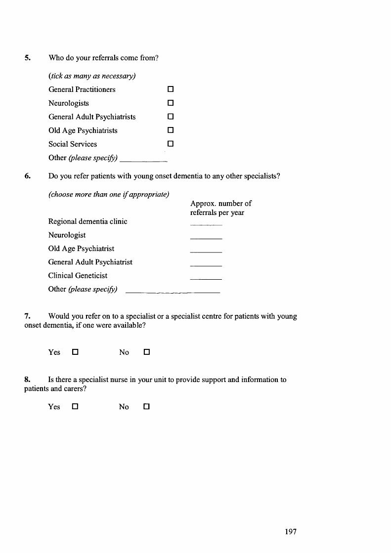
5. Who do your referrals come from?
(tick as many as necessary)
General Practitioners
Neurologists
General Adult Psychiatrists
Old Age Psychiatrists
Social Services
Other (please specify)_______
□
□□□□
6. Do you refer patients with young onset dementia to any other specialists?
(choose more than one i f appropriate)Approx. number of referrals per year
Regional dementia clinic ________
Neurologist ________
Old Age Psychiatrist____________________________
General Adult Psychiatrist_______________________
Clinical Geneticist ________
Other (please specify) ____________________________________________
7. Would you refer on to a specialist or a specialist centre for patients with young onset dementia, if one were available?
Yes □ No □
8. Is there a specialist nurse in your unit to provide support and information to patients and carers?
Yes □ No □
197

9. Are the following points covered in history taking?
Always Often Sometimes Never
Independent history from carer □ □ □ □
Detailed family history □ □ □ □
Changes in personality □ (such as eating habits, sexual behaviour)
□ □ □
Symptoms of depression □ □ □ □
Somatic symptoms □ □ □ □
Driving history □ □ □ □
10. a) Are the following covered on physical examination?
General physical examination
Always
□
Often
□
Sometimes
□
Never
□
General neurological examination □ □ □ □
b) Do you specifically look for?
fasciculation □ □ □ □
primitive reflexes □ □ □ □
visual disorientation □ □ □ □
extrap yramidal features □ □ □ □
11. Which of the following investigations do you perform on your young patientswith cognitive impairment?
Always Often Sometimes Never
Full blood count □ □ □ □
Renal profile □ □ □ □
Liver profile □ □ □ □
Thyroid function □ □ □ □
B12 and folate □ □ □ □
ESR □ □ □ □
Autoantibody screen □ □ □ □
Antithyroid microsomal □ □ □ □
Anticardiolipin Abs □ □ □ □
Treponemal Serology □ □ □ □
198

Always Often Sometimes Never
HIV □ □ □ □
White cell enzymes □ □ □ □
Plasma very long chain fatty acids □ □ □ □
Heavy metal screening □ □ □ □
Blood for genetic screening
Huntington’s □ □ □ □
DRPLA □ □ □ □
Prion protein □ □ □ □
APP, presenilin I,
presenilin 2 □ □ □ □
tau gene □ □ □ □
CT brain scan □ □ □ □
MRI brain scan □ □ □ □
MMSE □ □ □ □
Neuropsychology □ □ □ □
PET, SPECT □ □ □ □
EEG □ □ □ □
CSF □ □ □ □
CSF □ □ □ □(S100, NSE,P 14-3-3) Tonsil biopsy □ □ □ □
Brain biopsy □ □ □ □
12. Are carers given the opportunity to speak to the doctor in the absence of the
patient?
Always Often Sometimes Never
□ □ □ □Please state reasons
199

13. Do you pass on the diagnosis to patients with young onset dementia and their
carers?
Always Often Sometimes Never
□ □ □ □14. Do you give the diagnosis to patients and their carers separately?
Always Often Sometimes Never
□ □ □ □Please state reasons
15. Are any other people present when the diagnosis is given e.g. Nurse Counsellor?
Yes □ _____ please say who____________________________________________
No □
16. After you have made the diagnosis, on average how often will you see the patient with young onset dementia again?
Never □
Only if needed □
Annually □
Biannually □
Three monthly □
Monthly □
More often, please specify _________________________________________________________
17. Who is mainly responsible for follow up of the patient and their family? (tick more than one i f appropriate)
General Practitioner □
Specialist dementia/ memory clinic □
Old Age Psychiatrist □
Consultant Neurologist □
200

Shared care □
Please specify________________________________________________
Other □
Please specify________________________________________________
18. Do you organise or give advice on the following?
19.
Always Often Sometimes Never
referral to a CPN □ □ □ □
contacting the DVLA □ □ □ □
enduring power attorney □ □ □ □
information on support □ □ □ □groups (such as Alzheimer's Disease Society, Pick's Disease Support Group, CJD support network)genetic counselling □ □ □
Do you discuss any of the following?
Always Often Sometimes
Drug treatments □ □ □
Entry into drug trials □ □ □
Post mortem consent □ □ □
□
Never
□□□
20. Are you able to prescribe acetylcholinesterase inhibitors for young onset
dementia patients?
Able to prescribe freely □
Prescribe under certain guidelines □
Very rarely able to prescribe □
Not at all □
Give private prescriptions □
21. Do you undertake the follow up of patients on acetylcholinesterase inhibitors?
Yes □ No □
Thankyoufor taking the time to complete this questionnaire.
201

PUBLICATIONS RELATING TO THIS THESIS
Cordery RJ, Harvey R, Frost C, Rossor MN. National survey to assess current
practices in the diagnosis and management of young people with dementia. In tJ
Geriatr Psychiatry. 2002 Feb; 17(2): 124-7.
Cordery RJ, Hall M, Cipolotti L, Al-Sarraj S, O’Donovan DG, Davidson L, Adlard
P, Rossor MN. Early cognitive decline in Creutzfeldt-Jakob disease occurring in
recipients of pituitary-derived human growth hormone. Journal o f Neurology,
Neurosurgery and Psychiatry 2003; 74(10): 1412-6..
Cordery RJ, Janssen JC, Fox NC, Whitwell JL, Crum WR, Collinge J, Rossor MN.
Cross-sectional Volumetric MRI Measurement of Whole Brain and Cerebellum
Distinguishes Patients with Creutzfeldt-Jakob disease from Normal Controls.
Abstract. (WCN 2001) Journal o f the Neurological Sciences, Vol. 187, Supplement 1,
June 15, 2001, S221-222.
Cordery RJ, Hall M, Cipolotti L, Al-Sarraj S, O’Donovan DG, Davidson L, Adlard
P, Rossor MN. Early cognitive decline in human growth hormone associated
Creutzfeldt-Jakob disease. Abstract. Association o f British Neurologists Autumn
meeting 2001.
Scaravilli F. Cordery RJ. Kretzschmar H. Gambetti P. Brink B. Fritz V. Temlett J.
Kaplan C. Fish D. An SF. Schulz-Schaeffer WJ. Rossor MN. Sporadic fatal insomnia:
a case study. Annals o f Neurology, 48(4): 665-8,2000 Oct.
202

Manuscripts submitted or in preparation
Cordery RJ, Alner K, Cipolotti L, Kennedy A, Ron M, Collinge J, Rossor MN. The
neuropsychology of variant CJD: A comparative study of cases with familial and
sporadic forms of prion disease. Submitted to Journal o f Neurology, Neurosurgery
and Psychiatry.
Cordery RJ, Rossor MN, MacManus D, Collinge J, Waldman AD. Proton magnetic
spectroscopy in variant Creutzfeldt-Jakob disease. Abstract accepted for the ISMRM
2003.
Cordery RJ, Janssen JC, Fox NC, Whitwell JL, Price S, Crum WR, Collinge J,
Rossor MN. Cross-sectional Volumetric MRI Measurement of Whole Brain and
Cerebellum Distinguishes Patients with Creutzfeldt-Jakob Disease from Normal
Controls. In preparation.
Cordery RJ, RossorMN, MacManus D, Godbolt A, Collinge J, Waldman AD.
Proton magnetic spectroscopy in variant Creutzfeldt-Jakob disease. In preparation.
203

ACKNOWLEDGEMENTS
This project was only possible with the assistance and support of a great number of
people.
I am greatly indebted to Martin Rossor whose enthusiasm; direction and sound advice
have been invaluable. I feel very privileged to have worked with the Dementia
Research Group, and I hope that I have learned for the future, the value of leadership,
teamwork and personal worth which he has created within the group. It is a
stimulating and exciting place to work.
Special thanks also go to Maria Ron and John Collinge who have both provided
encouragement and advice whilst allowing me the opportunity and space to develop
the study as the field evolved.
My thanks go to all current and former members of the Dementia Research Group, in
particular to Nick Fox, Richard Harvey, Liz Sampson, John Janssen, Jon Schott,
Dennis Chan, Alison Godbolt, Rachel Scahill, Jenny Whitwell, Shona Price, Bill
Crum, Rhian Gordon, Elizabeth Warrington, Sebastian Crutch, James Friswell, Louise
Fisher, Suzie Barker and Suzanne Tom for their tremendous support along the way. I
would like to acknowledge the help with the statistical analysis given to me by Hiliary
Watt from the London School of Hygiene and Tropical Medicine.
I would also like to thank Kathryn Prout, Clare Morris and Angus Kennedy from the
Prion Unit at St. Mary’s Hospital, whose help and commitment have been invaluable.
204

I would like to thank Lisa Cipolotti, Kizzy Alner and Miriam Hall from the
D epartm ent o f N europsychology at the National Hospital for Neurology and
Neurosurgery, for their hard w ork performing psychology assessm ents and helping to
collect and analyse the data.
Special thanks go to Adam W aldman for his help with the MR im aging and the
interpretation o f the M R spectroscopy data. I would like to thank Dave M acM anus
for his considerable help with perform ing the MR imaging throughout the study
period and to John Stevens for formally reporting the radiological findings.
This project was funded and supported by the M edical Research Council o f G reat
Britain. W ithout this resource, this and many other projects in this important field
would not be possible.
I would like to acknowledge my family, who have not only given m e great
encouragem ent but also the trem endous practical support that has m ade it all possible.
My particular thanks go to Ann Cordery, who has looked after my two daughters with
untiring enthusiasm and energy, to Ted Clary, my sisters and brothers-in-law, Liz and
Nick Gall and Sarah and Richard Starr and finally m y parents Ann and Graham
Hollister.
( L O R D IR .) \ o i W F . y
2 0 5