skull and brain anatomy of late carboniferous sibyrhynchidae...
TRANSCRIPT
595GEODIVERSITAS • 2010 • 32 (4) © Publications Scientifi ques du Muséum national d’Histoire naturelle, Paris. www.geodiversitas.com
KEY WORDS Chondrichthyes,
Inio pterygia,Sibyrhynchidae,
gnathostomes, holocephalans, neuro cranium,
brain, Paleozoic,
Synchrotron, X-ray,USA.
Pradel A. 2010. — Skull and brain anatomy of Late Carboniferous Sibyrhynchidae (Chondrich-thyes, Inio pterygia) from Kansas and Oklahoma (USA). Geodiversitas 32 (4): 595-661.
ABSTRACTRecent gnathostomes are composed of two major clades, the chondrichthyans, or cartilaginous fi shes, and the osteichthyans, or bony fi shes and tetrapods. Recent chondrichthyans include about 1200 elasmobranch (sharks and rays) species, but only about 40 holocephalan (ratfi sh) species. Nevertheless, an important radiation of chondrichthyans took place in the Carboniferous (360-300 Myr), and gave rise to an important diversity of odd and poorly understood taxa, such as the inio pterygians, that are considered as related to extant holocephalans. However, the relationships between these taxa and the two extant chondrichthyan clades remain controversial. Th e material studied here by means of computed microtomography scanning using synchrotron radiation X-ray, consists of neu-rocrania from the Upper Carboniferous of Kansas and Oklahoma (USA), which are remarkably well preserved in three dimensions, and which belong to one of
the two families of inio pterygians, the Sibyrhynchidae Zangerl & Case, 1973. A detailed description of these specimens provides more information about the cranial and brain anatomy of this taxon. No three-dimensionally preserved skull of any stem-holocephalan was known in detail to date, contrary to fossil elasmobranchs. Th e data presented here show the three-dimensionally preserved braincase of a possible stem-holocephalan as these new specimens share with extant chimaeroids some key neurocranial characters. Th is may provide means for a comparative study of skull anatomy in Paleozoic representatives of the main two chondrichthyan clades.
Alan PRADELMuséum national d’Histoire naturelle, Centre de Recherches sur la
Paléobiodiversité et les Paléoenvironnements (CRP2), UMR 7207 du CNRS, Département Histoire de la Terre,
case postale 38, 57 rue Cuvier, F-75231 Paris cedex 05 (France)and Laboratoire Évolution, Génome et Spéciation,
UPR 9034 du CNRS, avenue de la Terrasse, Bâtiment 13, boîte postale 1, F-91198 Gif-sur-Yvette (France)
and Université Paris Diderot – Paris 7, UFR Sciences du Vivant, case 7044, F-75205 Paris cedex 13 (France)
Skull and brain anatomy of Late Carboniferous Sibyrhynchidae (Chondrichthyes, Inio pterygia) from Kansas and Oklahoma (USA)
596 GEODIVERSITAS • 2010 • 32 (4)
Pradel A.
INTRODUCTION
Recent gnathostomes are represented by two major clades, the Chondrichthyes Huxley, 1880, or carti-laginous fi shes, and osteichthyans, or bony fi shes and tetrapods. Th e interrelationships of the two subclasses (Elasmobranchii Bonaparte, 1838, i.e. sharks, skates, rays and Holocephali Bonaparte, 1932, i.e. ratfi sh, chimaera, spookfi sh) of recent chondrichthyans are far from satisfactorily elucidated and this situation is even worse when fossil chondrichthyans are con-sidered, in particular the Paleozoic ones (Woodward 1889; Lund 1977; Zangerl 1981; Maisey 1984; Didier 1995; Janvier 1996; De Carvalho 1996; Grogan & Lund 2004). Th is results from the high degree of morphological disparity in the Paleozoic forms that far exceeds that observed in living forms, as well as
from a generally poor preservation and the diffi culty to observe crucial features.
Crown-group holocephalans could be followed through the fossil record as far back as the Jurassic (e.g., Ischyodus Egerton, 1843; Stahl 1999). Associa-tion of earlier forms with this group is unclear. Ac-cording to Coates & Sequeira (2001a, b), a number of Paleozoic chondrichthyans could be resolved as stem-holocephalans, such as the Symmoriiformes Zangerl, 1981, but there is no consensus about these relationships to date (Maisey 2007). Th e description of new three-dimensional braincases of possible stem-holocephalans, including consideration of the cranial blood vascular system, cranial nerve pattern and endo cranial cavity, may provide information that could support these relationships, as the description of Paleozoic three-dimensionally shark braincases al-
MOTS CLÉSChondrichthyes,
Inio pterygia,Sibyrhynchidae,
gnathostomes, holocéphales,
neurocrâne,cerveau,
Paléozoïque,lumière Synchrotron,
rayons X,États-Unis.
RÉSUMÉAnatomie du crâne et du cerveau des Sibyrhynchidae (Chondrichthyes, Inio pterygia) du Carbonifère supérieur du Kansas et de l’Oklahoma (USA).Les gnathostomes actuels sont représentés par deux clades majeurs, les chon-drichthyens, ou poissons cartilagineux, et les osteichthyens, ou poissons osseux et tétrapodes. Les chondrichthyens modernes comprennent environ 1200 espèces d’élasmobranches (requins et raies), mais seulement une quarantaine d’espèces d’holocéphales (chimères). Cependant, une importante radiation de chondrichthyens a eu lieu durant le Carbonifère (360-300 Ma) et a généré une importante diversité de taxons étranges et mal connus, comme les inioptéry-giens qui sont considérés comme phylogénétiquement proches des holocéphales actuels. Néanmoins, les relations de parenté entre ces taxons et les deux clades actuels de chondrichthyens demeurent controversées. Le matériel, étudié ici grâce à la microtomographie assistée par ordinateur utilisant la lumière Syn-chrotron, consiste en des neurocrânes du Carbonifère supérieur du Kansas et de l’Oklahoma (USA), parfaitement conservés en trois dimensions et qui appartiennent à une des deux familles d’inioptérygiens, les Sibyrhynchidae
Zangerl & Case, 1973. Une description détaillée de ces spécimens fournit des informations supplémentaires quant à l’anatomie du crâne et du cerveau de ce taxon. Contrairement aux élasmobranches, il n’existait jusqu’à ce jour aucun crâne conservé en trois dimensions d’holocéphale fossile. Les spécimens étudiés ici nous fournissent un neurocrâne préservé en trois dimensions d’un possible holocéphale souche, tant ils partagent avec les chimères actuelles des caractères neurocrâniens caractéristiques. Ils permettent donc des études comparatives de l’anatomie crânienne chez les représentants paléozoïques des deux clades majeurs de chondrichthyens.
597
Skull and brain anatomy of Sibyrhynchidae (Chondrichthyes, Iniopterygia)
GEODIVERSITAS • 2010 • 32 (4)
ready did for the total-group elasmobranchs and early gnathostomes phylogeny (Schaeff er 1981; Maisey 2001a, 2005; Maisey & Anderson 2001).
Zangerl & Case (1973) fi rst described a new order of chondrichthyan fi shes from the Pennsylvanian of Indiana (USA), the Inio pterygia Zangerl & Case, 1973, which contain two families: the Iniopterygi-dae Zangerl & Case, 1973, and the Sibyrhynchidae Zangerl & Case, 1973. Other iniopterygids from Indiana and the Mississippian of Montana (USA) were respectively discovered and described by Zangerl (1997) and Grogan & Lund (2009). M. Coates (pers. comm.) recently discovered some elements of an iniopterygian in the Lower Carboniferous of Beardsen (Scotland).
Inio pterygians were assumed to belong to the sub-class Subterbranchialia Zangerl, 1979, as stem-holo-cephalans (Zangerl 1981; Stahl 1999). Nevertheles, recent cladistic analysis on the neuro cranium of the new iniopterygid specimens from Montana, brings them in a stemward position, before the divergence between the elasmobranchs and holocephalans (Gro-gan & Lund 2009), whereas the sibyrhynchids share derived characters with the living chimearoids (e.g., a palatoquadrate that is fused to the neuro cranium). In addition, the inio pterygians also display many characters, which are shared with other Paleozoic chondrichthyans, and a relatively large number of autapomorphies, notably the dorsal position of the pectoral fi ns. Consequently, the phylogenetic status of the Inio pterygia is still in question.
Unfortunately, all the iniopterygian specimens previously described are fl attened (Zangerl & Case 1973; Zangerl 1997; Grogan & Lund 2009). Conse-quently, they do not allow any extensive description, in particular concerning the braincase. Recently, a short paper based on new material from the Penn-sylvanian of Kansas and Oklahoma put an emphasis on a new technique of observation, the quantitative phase contrast tomography that revealed the fi rst three-dimensionally preserved Sibyrhynchidae neurocrania known to date. In addition, one of these new siby-rhynchid braincases houses exceptionally preserved parts of the brain itself (Pradel et al. 2009).
Th ese three dimensionally preserved skulls and the fossilized brain provide new information about the external and internal morpho logy of the braincase.
Th ey allow a comparative study of skull and brain anatomy with other three-dimensionally preserved neurocrania of Paleozoic and extant chrondrichthy-ans as well. According to the importance of these three-dimensionally preserved skulls and this unique case of fossilized brain, a detailed description of the braincases, dentition, and dermal denticles of the head and the preserved brain is provided in this pa-per, prolonging their initial publication with further anatomical details.
MATERIALS AND METHODS
Th e manual preparation of the specimens is very diffi cult because of the extreme fragility of the fossilized calcifi ed cartilage. Consequently, and because it is necessary to investigate the internal anatomy of the braincases, computerized X-ray mi-crotomography (XR-μCT scan), X-ray synchrotron microtomography (SR-μCT scan) and quantitative phase contrast tomography (holotomography) were performed. Th ese techniques provide an ideal non-invasive technology to investigate structures of the fossil that are otherwise inaccessible.
ABBREVIATIONSaa anterior ampulla;ala acoustico-lateral area;alc anterior lobe of the corpus cerebelli;am articular zone of the mandible;amf anterior median fi ssure;antjc anterior opening of the jugular canal;arf articular facet of the palatoquadrate;asc anterior semicircular canal;basa basilar artery;bmc bucco-maxillary complex;br mineralized brain;bso possible branch of a spino-occipital nerve
innervating the epaxial musculature;ca cerebellar auricle;cc crus commune;ccer corpus cerebelli;ceps canal for the eff erent pseudo branchial artery;cerc cerebellar commissure;cerch cerebellar chamber;cha connection between the horizontal ampulla
and the utricular recess;cora canal for the orbital artery;cpcv canal for the posterior cerebral vein;cpitv canal for the pituitary vein;
598 GEODIVERSITAS • 2010 • 32 (4)
Pradel A.
cpr canal for the profoundus ramus of the trigemi-nal nerve;
cso canal for spino-occipital nerve;csoph canal for the superfi cial ophthalmic complex;ctz central tectal zone;cVmx canal for the maxillary ramus of the trigeminal
nerve;cVI canal for the abducens nerve;cVII canal for the main trunk of the facial nerve;cVIIapal canal for the anterior ramule of the palatine
ramus of the facial nerve;cVIIpal canal for the palatine ramus of the facial
nerve;cVIIppal canal for the posterior ramule of the palatine
ramus of the facial nerve;cIX canal for the glossopharyngeus nerve;cX+lcX canal for the vagus nerve and its lateralis
component;dd dermal denticle;dor dorsal otic ridge;dp dorsal part of the post orbital wall;dppr dorsal paroccipital process;d.r. diencephalic region;ds dorsum sellae;end endolymphatic duct;endc endo cranial cavity;eps eff erent pseudo branchial artery;fam fossa for the adductor musculature of the
mandible;fl cX foramen for the lateralis component of the
vagus nerve;fm foramen magnum;fmop foramen situated in the medial otic process;folfcap fl oor of the olfactory capsule;fot foramen for the olfactory tract;fp point of fusion of the parachordals;fpcv foramen for the posterior cerebral vein;fpitv foramen for the pituitary vein;fsoph foramen for the superfi cial ophthalmic com-
plex;fv facial vein;fII foramen for the optic nerve;fIII foramen for the oculomotor nerve;fIV foramen for the trochlear nerve;fVmd foramen for the mandibular ramus of the
trigeminal nerve in the mandible;fVmx foramen for the maxillary ramus of the
trigeminal nerve; fVI foramen for the abducens nerve;fVII foramen for the main trunk of the facial
nerve;fVIIpal foramen for the palatine ramus of facial
nerve;fIX foramen for the glossopharyngeus nerve;fX foramen for the vagus nerve;gas Gasser’s ganglion;
ghr groove for the mandibular-hyoidean ramus of the facial nerve;
gjv groove for the jugular vein;ha horizontal ampulla;hsc horizontal semicircular canal;hyp hypophysis;hypch hypophyseal chamber;ic intertectal commissure;il inner layer of the endocranium;ilh inferior lobe of the hypothalamus;imz intermuscular elevated cranial zone;ing internasal groove;inp internasal plate;inw internasal wall;is isthmus rhombencephali;jc jugular canal;jv jugular vein;lap lateral articular process of the mandible;lcm lateral crest of the mandible probably sup-
porting tooth whorl;lcX lateralis component of the vagus nerve;ld lateral depression housing gills and abductor
musculature;lpm lateral process of the mandible probably
supporting tooth whorl;ls lateral sulcus;map medial articular process of the mandible;mcv middle cerebral vein;med medulla oblongata;mi muscular insertion;mop medial otic process;ntl nucleus tegmentalis lateralis;obmc orbital foramen for the bucco-maxillary
complex;occh occipital chamber;occot occipital cotylus;occr occipital crest;oeps orbital foramen for the eff erent pseudo-
branchial artery;ofp orbital foramen for the ophthalmic profondus
ramus of the trigeminal nerve;ol outer layer of the endocranium;olfc olfactory canal;olfcap olfactory capsule;onc orbitonasal canal;oonc orbital foramen for the orbitonasal canal;oora orbital foramen for the orbital artery;opa optic retinal artery;opch optic chiasm;optec optic tectum;ora orbital artery;P-1 fi rst tooth whorl (“vomerine”);P-2 second tooth whorl (“vomerine”);P-4 fourth tooth whorl or “canine” (“vomerine”);P-5 fi fth tooth whorl (palatine);pa posterior ampulla;
599
Skull and brain anatomy of Sibyrhynchidae (Chondrichthyes, Iniopterygia)
GEODIVERSITAS • 2010 • 32 (4)
palc palatine crest;parc parachordal cartilage;pcv posterior cerebral vein;pdd patch of dermal denticles;pdf posterior dorsal fontanelle;pdob posterior dorsal otic bulges;pf pre occipital fossa;pitv pituitary vein;plc posterior lobe of the corpus cerebelli;pms posterior median sulcus;postc posterior commissure;postjc posterior opening of the jugular canal;pow post orbital wall;pq palatoquadrate;pr ophthalmic profundus ramus of the trigeminal
nerve;prefc prefacial commissure;prefp preorbital foramen for the ophthalmic pro-
fundus ramus of the trigeminal nerve;proo prootic foramen;prp preorbital process;psc posterior semicircular canal;ptf primary transverse fi ssure;ptz periventricular tectal zone;pwo posterior wall of the olfactory capsule;rX root of the vagus nerve;sac saccular chamber;sdp symphysial dentigerous plate;smc somatic motor column;so spino-occipital nerve;sof spino-occipital nerve foramen;soph superfi cial ophthalmic complex;ss sinus superior;ssc somatic sensory column;stz superfi cial tectal zone;sups supraorbital shelf;teg tegmentum;th thalamus;tpf trigemino-pituitary fossa;tr transverse dentigerous ridge on the symphysial
dental plate;ur utricular recess;vc cerebellar ventricle;vbmc ventral foramen for the bucco-maxillary
complex;veps ventral foramen for the eff erent pseudo-
branchial artery;vm ventricular mesencoelia;vmc visceral motor column;vonc ventral opening of the orbitonasal canal;vora ventral foramen for the orbital artery;vppr ventral paroccipital process;vs vascular sac;vsc visceral sensory column;vIII third ventricle;vIV fourth ventricle;
II optic nerve;III oculomotor nerve;IIId dorsal ramus of the trochlear nerve;IIIv ventral ramus of the trochlear nerve;IV trochlear nerve;Vmd mandibular ramus of the trigeminal nerve;Vmx maxillary ramus of the trigeminal nerve;VI abducens nerve;VII main trunk of the facial nerve;VIIapal anterior ramule of the palatine ramus of the
facial nerve;VIIh hyoidean ramule of the mandibular-hyoidean
ramus of the facial nerve;VIIhym mandibular-hyoidean ramus of the facial
nerve;VIImd mandibular ramule of the mandibular-hy-
oidean ramus of the facial nerve;VIIpal palatine ramus of the facial nerve;VIIppal posterior ramule of the palatine ramus of the
facial nerve;IX glossopharyngeus nerve;X vagus nerve.
MATERIALS
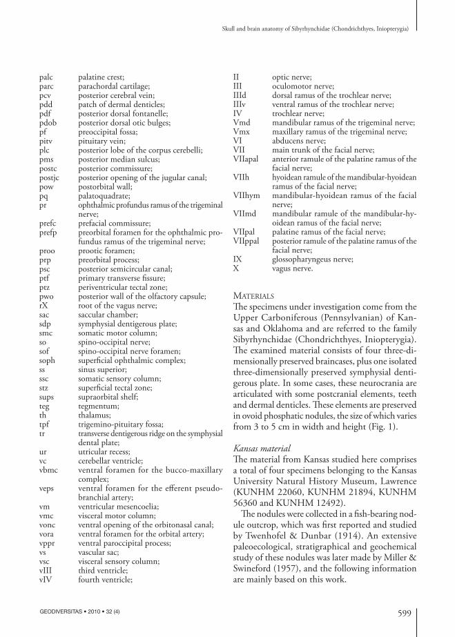
Th e specimens under investigation come from the Upper Carboniferous (Pennsylvanian) of Kan-sas and Oklahoma and are referred to the family Sibyrhynchidae (Chondrichthyes, Inio pterygia). Th e examined material consists of four three-di-mensionally preserved braincases, plus one isolated three-dimensionally preserved symphysial denti-gerous plate. In some cases, these neurocrania are articulated with some postcranial elements, teeth and dermal denticles. Th ese elements are preserved in ovoid phosphatic nodules, the size of which varies from 3 to 5 cm in width and height (Fig. 1).
Kansas materialTh e material from Kansas studied here comprises a total of four specimens belonging to the Kansas University Natural History Museum, Lawrence (KUNHM 22060, KUNHM 21894, KUNHM 56360 and KUNHM 12492).
Th e nodules were collected in a fi sh-bearing nod-ule outcrop, which was fi rst reported and studied by Twenhofel & Dunbar (1914). An extensive paleoecological, stratigraphical and geochemical study of these nodules was later made by Miller & Swineford (1957), and the following information are mainly based on this work.
600 GEODIVERSITAS • 2010 • 32 (4)
Pradel A.
Th e nodules crop out approximately 150 km south-east of Kansas City, between the towns of Lawrence and Baldwin, in about 30 diff erent localities. Th ey occur in a thin bed of yellow-gray shale deposited in a near shore environment, probably within shallow marine basins formed in a semitropical and wet area. Stratigraphically, they occur at the limit between the marine Haskell Limestone Member and the overlying Robbins Shale of the Stranger Forma-tion, deposited in brackish water. Th is sequence is part of the Douglas Group, dated as late Virgilian, Upper Pennsylvanian (305-299 Myr). Similar con-ditions were present in the black shale, where the fi rst described inio pterygians of the Mecca fauna were found (Zangerl & Richardson 1963).
Th e matrix of the nodules is mainly composed of apatite and also displays quartz grains and organic matter. Th e crystalline calcite may fi ll the endo-cranial cavity of the specimen, while the prismatic calcifi ed cartilage is calcium phosphate (see below) as in KUNHM 22060.
Beside the chondrichthyan remains, the fossil record is mostly represented by actinopterygian remains and some cephalopods (goniatites, nau-tiloids, orthocerids), brachiopods (Orbiculoidea d’Orbigny, 1849), arthropods (Idiotheca Girty, 1909), fossil wood and fi sh coprolites.
KUNHM 22060 (Fig. 1A). Th is specimen is a complete neuro cranium articulated with teeth in natural position, the mandible slightly removed from its original position, part of the branchial apparatus almost in its natural position and some dermal denticles. It has been previously prepared superfi cially and mechanically, thereby exposing some features, such as the mandible and the ante-rior part of the braincase.
KUNHM 21894 (Fig. 1B, B’). Th is specimen is an almost complete neuro cranium, only lacking its anterodorsal part, and associated with the pectoral girdle and fi n almost in natural position, as well as other postcranial elements, teeth and dermal denticles.
KUNHM 56360 (Fig. 1C, C’). Th is specimen con-sists of the mid posterior part of the braincase.
KUNHM 12492 (Fig. 1D-D’’). Th is specimen is an isolated ethmoid plate showing the entire sym-physial dentigerous plate.
Oklahoma materialOKM 38 (Fig. 1E, E’). Th is specimen comes from Oklahoma and was loaned by Dr John Maisey (Curator in charge, American Museum of Natural History, New York). It was collected by Dr Royal Mapes (Geology Department, Ohio University, Athens, Ohio, USA) from the Coff eyville Forma-tion (dated as Pennsylvanian, Missourian, c. 307 Myr), at a roadcut in Tulsa County, Oklahoma (Center of NW sec. 2., T. 18 N, R. 12 E, Sapulpa North 7 ½ Quadrangle). Numerous paleoniscoid braincases were also recovered from small nodules at this site.
Th is specimen is an almost complete braincase lacking the ventral part of the orbit, which has been displaced, however, at some distance in the nodule. It is associated with some elements of the pectoral girdle and fi n, teeth and dermal denticles.
RemarksOnly KUNHM 22060 bears the entire internal part of the braincase undoubtedly because of its com-plete matrix fi lling. All the other braincases merely show part of the endo cranial cavity and skeletal labyrinth, and consequently, they only display the external surface of the neuro cranium in the part where the endo cranial cavity is not preserved. Th e diff erent cranial foramina are thus very diffi cult to locate in this region because there are no structural diff erences between a real foramen and a small gap in the cartilage. Only the foramina that are observed in relation with their corresponding internal canals are mentioned in the present work. Th e other ones are speculative and are not described.
All the neurocrania studied in the present work have almost the same proportions and size as the adult specimens studied by Zangerl & Case (1973). Th ere-fore, it is assumed that these are adult individuals.
METHODS
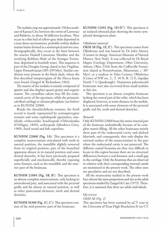
OKM 38 (Fig. 2)Th is specimen has been scanned by μCT scan at the University of Texas High-Resolution X-ray CT
601
Skull and brain anatomy of Sibyrhynchidae (Chondrichthyes, Iniopterygia)
GEODIVERSITAS • 2010 • 32 (4)
Facility, Austin. Scan parameters were as follows: 1024 × 1024 16-bit TIFF images. II, 180 kV, 0.12 mA, no fi lter, air wedge, no off set, slice thick-ness 2 lines (= 0.04872 mm), S.O.D. 70 mm, 1400 views, 2 samples per view, inter-slice spacing 2 lines
(= 0.04872 mm), fi eld of reconstruction 22.8 mm (maximum fi eld of view 23.17 mm), reconstruction off set 8000, reconstruction scale 3200. Acquired with 19 slices per rotation and 15 slices per set. Flash and ring-removal processing done based on correction
FIG. 1. — Photograph of the different nodules studied in the present work: A, KUNHM 22060 in anterior view; B, B’, KUNHM 21894, part (B) and counterpart (B’); C, C’, KUNHM 56360, part (C) and counterpart (C’); D-D’’, KUNHM 12492, part (D), counterpart (D’) and silicone cast (D’’); E, E’, OKM 38, part (E) and counterpart (E’). Scale bars: 5 mm.
C
C’
D
D’
D’’
E
E’
A B B’
C
C’
D
D’
D’’
E
E’
A B B
602 GEODIVERSITAS • 2010 • 32 (4)
Pradel A.
of raw sinogram data by Alison Mote using IDL routines “RK_SinoDeSpike” and “RK_SinoRing-ProcSimul,” both with default parameters. Deleted fi rst four duplicate slices of each rotation except for slices 1-4. Rotation correction processing has done using IDL routine “DoRotationCorrection.” Total fi nal slices = 649.
KUNHM 56360 Th is specimen has been scanned at the University of Poitiers, France by Arnaud Mazurier by means of a Viscom model X8050-16 facility. Scan parameters were as follows: 1004 × 1004 12-bit TIFF images. 125 kV, 0.350 mA, CCD camera, slice thickness 2 lines (= 0.028 mm), 32 integration per projec-tions, 1800/360° projections. Th e reconstruction was performed with DigiCT v1.15 (Digisens) by Arnaud Mazurier. Total fi nal slices = 984 with the format 8-bits, 678 × 678 pixels.
KUNHM 22060 (Fig. 3) and KUNHM 21894Th ey were imaged using X-ray based synchrotron microtomography (SR-μCT) (Taff oreau et al. 2006) on the beamline ID19 of the European Synchrotron Radiation Facility, Grenoble, France, by Paul Taf-foreau. Scan parameters were as follows: monochro-matic X-ray beam of 60 keV energy. Th e detector was a FReLoN (Fast Readout Low Noise) (Labiche et al. 2007) CCD camera coupled with an optical magnifi cation system, yielding an isotropic pixel size of 30.3 μm. 1200/180° projections with 0.4 s
of exposure time. Data were reconstructed using the fi ltered backprojection algorithm (PyHST software, ESRF) by Paul Taff oreau. Reconstructed slices were converted from 32 bits to 8 bits in order to reduce the data size for 3D processing.
Because the posterior part of the orbit of KUNHM 22060 concentrates many important features, such as the fossilized brain and diff erent foramina, it was also imaged using quantitative phase tomography (holotomography) (Cloetens et al. 1999; Pradel et al. 2009), which is more powerful than a syn-chrotron acquisition (Fig. 3B). Scan parameters were as follow: monochromatic X-ray beam of 60 keV energy, isotropic pixel size of 14.92 μm. 1500/180° projections with 0.3 s of exposure time. Two holo-tomographic acquisitions were necessary to cover the whole structure. Th e propagation distances were 50 mm (absorption), 400 mm and 950 mm, respectively. After phase retrieval, the slices were duplicated and reconstructed using the fi ltered backprojection algorithm, then converted into 8-bit TIFF fi les. Finally, the two holo tomographic scans were combined to one volume where the common slices were removed.
KUNHM 12492Th is specimen was studied by means of manual preparation and silicone cast (Fig. 1D’’).
Segmentation and 3D rendering were performed with MIMICS® software (Materialise® Inc. NV, Leu-ven, Belgium) by myself. MIMICS® (Materialise’s Interactive Medical Image Control System) is of great interest to deal with such a large amount of data, as those generated from μCT scan. Th e MIM-ICS® 64 bits version runs on a Dell 690 Windows XP 64 workstation with 16 GB of RAM.
MIMICS® allows diff erent types of measure-ment and segmentation to be performed. Regions of interest can be selected with accuracy using threshold method to create segmentation masks. With this method, selections depend on a range of defi ned grey values, and not on manual outlin-ing operations. 3D models have been calculated from segmentation masks and combined through Boolean operations.
Many of the illustrations in this work represent views captured from MIMICS® surface renderings.
FIG. 2. — Volume and surface rendering generated from XR-μCT scan slices of the specimen OKM 38 partially cleaned virtually, left side. Scale bar: 5 mm.

603
Skull and brain anatomy of Sibyrhynchidae (Chondrichthyes, Iniopterygia)
GEODIVERSITAS • 2010 • 32 (4)
Adobe Photoshop CS3 extended v.10.0.1 was used to increase the contrast of the MIMICS® surface renderings and to make the fi nal illustration.
Th e operation of virtual crushing made on the contour-based rendering of KUNHM 22060 was per-formed with Autodesk Maya 2008 x64 software.
Microprobe analysis was performed to reveal the mineral composition of the fossilized brain, the pre-served skull and the surrounding matrix. Th is provides information that help for understand the mode of fossilization. It was operated by Omar Boudouma at the Electron Microscopy Service of the UFR 928, University Paris 6, by using a fi eld eff ect gun (FEG) scanning electron microscope ZEISS SUPRA55VP, with a nominal resolution of 1.0 nm (at 15 kV and 2 mm distance). Th e analyzer was a detector SDD SAHARA of PGT including the SPIRIT software.
TAPHONOMY
Th e remarkable abundance and exceptional three-dimensional preservation of the fi sh remains from Kansas could be explained by their deposition in a quiet anoxic environment, without strong currents nor overturning condition.
Th e palaeoniscoid braincases found in these concre-tions have been extensively studied by Poplin (1974) and Hamel & Poplin (2008). According to Poplin (1986), the fact that the actinopterygian remains are almost exclusively isolated braincases, suggests that the latter are the result of chondrichthyan regurgitation. It is also possible that, previous to burial, the endocra-nia resulting from scattered carcasses were “sorted” by currents. However the presence of the phosphatic fossilized brain in one specimen (KUNHM 22060) (Pradel et al. 2009) and the fact that several nodules contain, in addition to the neuro cranium, some teeth and postcranial elements almost in natural position, suggests that the diagenesis occured immediately after the death of the animal.
Th e microprobe analysis was performed where the mineralized brain material reaches the surface of KUNHM 22060: the right optic nerve foramen, where the natural cast of the orbital cavity was sepa-rated from the rest of the nodule (Fig. 4A, B), and at the level of an almost transverse break through the nodule, which shows the calcite fi lling of the brain cavity and the rearmost end of the presumably pre-served spinal cord. In both cases, they reveal for the brain a crystalline structure that radically diff ers from that of the surrounding calcite (almost pure calcium
endcendc
br
il
ol
br
il
ol
A B
FIG. 3. — Transverse microtomographic slices of KUNHM 22060 through the posterior end of the optic nerve foramen showing the difference of contrast and precision between a Synchrotron Radiation microtomographic (absorption contrast) acquisition (A) and a Synchrotron Radiation holo tomographic (phase contrast) acquisition (B). Abbreviations: see Materials and methods.
604 GEODIVERSITAS • 2010 • 32 (4)
Pradel A.
carbonate), and proved to be composed of calcium phosphate (Fig. 4C-E).
Among the soft tissues that were present in the head during life, only the brain is fossilized. Th e eyes and muscles, which are external to the skull, are not pre-served. Moreover, the optic nerve, which is preserved inside the endo cranial cavity, is completely absent after reaching the surface of its foramen. Th is suggests that the brain was preserved because of a locally confi ned environment that was favourable to rapid phosphati-zation (Briggs & Kear 1993; Briggs et al. 1993), such as the endo cranial cavity of the braincase. Th is almost completely closed space may have generated an anoxic environment where authigenic mineralization was possible. Such an anoxic environment, plus a possible increase of CO2 and volatile fatty acids (present in the brain) generate a fall in pH that would have shifted the equilibrium of precipitation in favour of calcium phosphate rather than calcium carbonate (Wilby 1993; Briggs et al. 1993; Trinajstic et al. 2007). Th is required a high concentration of phosphorus inside the endo cranial cavity, and the brain itself contains much phosphorus, because the latter participates to the cellular metabolism of the brain. According to Briggs (2003), a microbial fi lm may concentrate and protect the phosphorus. Nevertheless, there is no evidence of preserved microbes inside the iniopterygian specimen here. Th is suggests that if authigenic mineralization has occured, the mineral precipitation is microbially induced rather than the result of autolithifi cation of the microbes proper (Briggs 2003).
Another mode of soft-tissue fossilization is permin-eralization, which is the result of early infi ltration and permeation of tissues by mineral-charged water (Briggs 2003). Th e organic material is subsequently replaced by mineral. Th e Kansas nodules studied here occur in a thin bed of yellow-gray shale deposited in a near shore environment, probably within shallow marine basins formed in a semitropical and wet area. Such an envi-ronment allows a high concentration of phosphorus. Nevertheless, permineralization of soft animal tissues is rare (Briggs 2003) and most permineralizations involve silica (Voigt 1988; Wuttke 1992).
Consequently, it is more probable that the brain un-derwent a microbially induced phosphatization shortly after the beginning of decay (the environment being probably saturated with calcium phosphate, hence the
concretions). It could have been rapidly surrounded by diagenetic calcite after the phosphatization. Some shrinking of the tissues may have occurred before phosphatization (Briggs 2003), which is suggested by the position of the cerebellum far anterior to the otic capsule. Nevertheless, the optic nerve reaches its foramen in a natural position, like the other nerves, which emerge from the brain and reach perfectly to their respective foramina. Consequently, the shrinking of the brain may not have been very signifi cant.
SYSTEMATIC PALEONTOLOGY
Class CHONDRICHTHYES Huxley, 1880Order INIOPTERYGIA Zangerl & Case, 1973
Family SIBYRHYNCHIDAE Zangerl & Case, 1973
TYPE GENUS. — Sibyrhynchus Zangerl & Case, 1973.
INCLUDED GENERA. — Sibyrhynchus, Iniopera Zangerl & Case, 1973, Inioxyele Zangerl & Case, 1973.
ETYMOLOGY. — From sibyne (= hunting spear) and rhynchos (= snout).
DIAGNOSIS. — Th e Sibyrhynchidae are holostylic Inio-pterygia in which the labio-lingual tooth rows (tooth families) are basally fused to form tooth whorls of dif-ferent size and shape.
Sibyrhynchidae gen. et sp. indet.
MATERIAL EXAMINED. — KUNHM 22060, KUNHM 21894, KUNHM 56360, KUNHM 12492 and OKM 38.
REMARKS
Th e braincases described here (KUNHM 22060, KUNHM 21894, KUNHM 56360, KUNHM 12492 and OKM 38) are very close to those in all three genera described by Zangerl & Case (1973) (see discussions). Nevertheless, the diagnosis of the lat-ter is incomplete principally because of the fl atness and the split of the specimens. It is therefore dif-fi cult to assign the new specimens described here to known genera with accuracy. Th e teeth, the overall morpho logy and the diff erent structures

605
Skull and brain anatomy of Sibyrhynchidae (Chondrichthyes, Iniopterygia)
GEODIVERSITAS • 2010 • 32 (4)
2 4 6 8 10
Ca
P
AIO
Fe Fe
E KUNHM22060_3_SE_T003.pgt# hidden files: 3
FS: 9 000
A
CD
E
**
*
B
CCa
P
2 4 6 8 10 2 4 6 8 10
O
AI
Ca
O
Fe FeAI P
D KUNHM22060_3_SE_T002.pgt# hidden files: 3
FS: 11 000KUNHM22060_3_SE_T001.pgt# hidden files: 3
FS: 10 000
a
d
3*
FIG. 4. — SEM micrograph and microprobe analyses of the surface of the calcite-fi lled right optic foramen of KUNHM 22060; the asterisks in A, B indicate the location of the microprobes analyses corresponding to the respective diagrams in C-E: A, general view of the optic foramen and surrounding prismatic calcifi ed cartilage; arrows point anteriorly (a) and dorsally (d); the area delimited by a bold line corresponds to the emergence of the preserved optic nerve; calcium phosphate dominates in the light grey areas and cal-cium carbonate dominates in the dark grey areas; B, detail of the area framed in A, showing the fi nely crystalline structure of calcium phosphate of the preserved optic nerve; C-E, microprobe analyses for P, O, Al, Fe, and Ca at the level of the optic foramen and optic nerve of KUNHM 22060 (C-E in A, B); C, external surface of the prismatic calcifi ed cartilage (mostly calcium phosphate); D, surface of the calcite fi lling the brain cavity and cranial nerve canals (calcium carbonate); E, surface of the emerging preserved optic nerve (mostly calcium phosphate with traces of Al and Fe).

606 GEODIVERSITAS • 2010 • 32 (4)
Pradel A.
sdp
folfcap
fsoph
pf
dor pdob occr pdf mop
dp
dd
sups
prp
B
A
prefp
FIG. 5. — Dorsal view of the braincase of KUNHM 22060: A, surface rendering generated from Synchrotron Radiation microtomographic slices; B, corresponding drawing. Abbreviations: see Materials and methods. Scale bar: 5 mm.

607
Skull and brain anatomy of Sibyrhynchidae (Chondrichthyes, Iniopterygia)
GEODIVERSITAS • 2010 • 32 (4)
of the neuro cranium are smilar in all these new specimens. Th ey are thus considered as belonging to the same genus.
DESCRIPTION OF THE BRAINCASES
GENERAL FEATURES Th e fi ve neurocrania studied here are not complete. Th e most complete one is KUNHM 22060 (Figs 5-13), it will be described in more detail in the present work. Nevertheless, the other specimens provide some sup-plementary data missing in KUNHM 22060.
KUNHM 22060 (Fig. 5) and KUNHM 21894 (Fig. 14A) show in dorsal view the same broad, delta-shaped outline, with the larger part in the posterior end of the braincase.
Th ey have very large orbits, bordered posteriorly and ventrally by an expanded post orbital wall con-fl uent with the supraorbital shelf dorsally and a large suborbital shelf mainly formed by the palatoquadrate (pow, sups, pq, Figs 6; 14B; 16). Despite the large orbits, there is no interorbital septum and the endo-cranial cavity extends between the orbits (Figs 8; 29; 30), suggesting that the neuro cranium is platybasic (sensu Maisey 2007). In the posterior part of the
olfcap
fVmx
sdp
palc
ooncobmc
fII
arf
fpitv
pq
imz
fX+fIcX
vppr
dppr
sof
fpcv
occr
dp
pfpowddantjcproofIIIfIVfsoph
fsoph
supsB
A
FIG. 6. — Lateral view of the braincase of KUNHM 22060, left side: A, surface rendering generated from Synchrotron Radiation micro-tomographic slices; B, corresponding drawing. Abbreviations: see Materials and methods. Scale bar: 5 mm.

608 GEODIVERSITAS • 2010 • 32 (4)
Pradel A.
orbit are gathered various foramina for the blood vessels and cranial nerves (Figs 12; 13).
Th e otic and occipital regions are both short, as in the symmoriiforms elasmobranchs and Pucapampella Janvier & Suarez-Riglos, 1986 (Zangerl & Case 1976; Coates & Sequeira 1998; Maisey & Anderson 2001; Maisey 2007).
Deep post orbital depressions fl ank the ventral part of the occipital region (ld, Fig. 10). Dorsomedial to these depressions are the otic capsules. Th ere is no persistent otico-occipital fi ssure that separates the otic region from the occipital arch.
Anterior to the narrow ventral occipital region, the outline of the ventral part of the braincase is parallel-sided, as far as an anterior rectangular car-tilage plate (Figs 7; 17). Contrary to elasmobranchs, the fl oor of the basi cranium below the otic capsules is not formed by the hypotic lamina. Th e ventral surface of the braincase is fl at and does not display any indication of the basal angle retained in some recent adult elasmobranchs.
ETHMOID REGION AND SYMPHYSIAL DENTIGEROUS PLATE
Nasal regionTh e ethmoid region includes the part of the braincase located anterior to the orbit and surrounding the nasal capsules. Th is region is well preserved in OKM 38 and KUNHM 22060 although the former lacks its right nasal capsule (Fig. 19) and the latter a small por-tion of its dorsal part between the two nasal capsules (Fig. 11). In KUNHM 21894, the anterodorsal part of the braincase is not preserved (Fig. 14A). Th e entire ethmoid region of KUNHM 56360 is lacking.
In the specimens described here, the ethmoid re-gion is broad but very short. Th e ventral internasal plate does not merge anteriorly with a rostral process (Figs 5; 15; inp, Figs 7; 17) and there is no evidence of a precerebral fontanelle at the anterior end of the neuro cranium. In addition, there is no evidence of a separate pineal opening, and the endo cranial cavity is not opened anteriorly. Th ere is also no evidence of a ventral ethmoidal keel.
Th ere is no septum between the nasal capsules and the olfactory capsules are well apart. Th is indicates a broad internasal wall (inw, Figs 11; 19), as in many other Paleozoic chondrichthyans (e.g., Stethacanthus
altonensis St John & Worthen, 1875, S. cf. S. productus Newberry, 1897, Cobelodus aculeatus Cope, 1894 and Akmonistion zangerli Coates & Sequeira, 2001 [Lund 1974: fi g. 3; 1985: fi g. 1; Zangerl & Case 1976: fi gs 2, 3, 7; Coates & Sequeira 1998: fi g. 5]).
Th e nasal capsules are well preserved in KUNHM 22060 and OKM 38, oval in shape, and with well marked borders (olfcap, Figs 11; 19). Th e olfactory canals open posteromedially in the posterior wall of the nasal capsule (fot, pwo, Figs 8; 11; 19). Th e nasal capsules are also pierced by several foramina. One of them is situated dorsal to the foramen for the olfac-tory tract and is connected by canals to a set of dorsal and orbital foramina (prefp, ofp, cpr, Figs 5; 7-9; 11; 15; 16; 19). Th ese foramina were probably for the profundus ramus of the trigeminal nerve. OKM 38 displays other foramina in the nasal region, which were probably also for the branches of the profundus ramus of the trigeminal nerve.
Th e olfactory chambers exhibit a complete carti-laginous fl oor (folfcap, Fig. 5), possibly formed from the cornua trabecularum that are trabeculae-derived embryonic structures (De Beer 1937). Th e incurrent and excurrent narial openings must have been situ-ated in the uncalcifi ed or non-chondrifi ed portion of the capsule and were probably directed frontally, contrary to most other chondrichthyans. Th ere is no indication of basal communicating canals.
In frontal view, between the nasal capsules, a V-shaped groove, only observable in OKM 38 (ing, Fig. 19), probably marks the boundary be-tween the internasal wall and the dorsal surface of the braincase. Two symmetrical large foramina seem to be present between this groove, which is not pre-served in KUNHM 22060, and the dorsal limit of the symphysial dentigerous plate (see below) (fVmx, Figs 11; 19). Each foramen is connected by canals to a foramen situated laterally to the symphysial denti-gerous plate and above the palatine crest (see below) (fVmx, cVmx, Figs 6; 8; 9). All these foramina are thus located near the teeth and may have given passage to maxillary ramules of the trigeminal nerve.
Th e nasal capsules are bordered ventrolaterally by a preorbital process that projects backward and laterally into the supraorbital shelf (prp, sups, Figs 7; 11; 19). Th e preorbital process meets the upper part of the symphysial dentigerous plate

609
Skull and brain anatomy of Sibyrhynchidae (Chondrichthyes, Iniopterygia)
GEODIVERSITAS • 2010 • 32 (4)
vbmc
sups
arf
mi
vora
dpmop
vppr
occr sof fX+fIcX
fpcv fIX
Id
veps
fsoph
ofpfsoph
prp
folfcappalctrinpB
A
vonc
FIG. 7. — Ventral view of the braincase of KUNHM 22060: A, surface rendering generated from Synchrotron Radiation microtomo-graphic slices; B, corresponding drawing. Abbreviations: see Materials and methods. Scale bar: 5 mm.
610 GEODIVERSITAS • 2010 • 32 (4)
Pradel A.
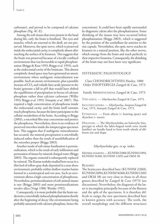
ventromedially to the nasal capsules at the level of their medial wall.
Lateral to the olfactory capsules, two foramina for the superfi cial ophthalmic complex reach the
preorbital process (fsoph, csoph, Figs 5; 8; 9; 11; 15; 19).
At the anteroventral extremity of the orbit is a foramen (oonc, Figs 6; 16), which may have housed
occr
cpcv
pa
cX+IcX
csof
cIX
sac cVI proo
ds
hypch
mi arf
pq
fIIpalc
oncsdp
cVmx
olfc
pwo
csoph
cpr
olilfIV
fIIIcpitvhapdfss ur
B
A
ccr
v
pa
IcX
sof
p
csop
cpr
olilfIV
fIIIcpitvhapdfss ur
B
A
FIG. 8. — Medial view of the braincase of KUNHM 22060, left side: A, sagittal section of surface rendering generated from Synchrotron Radiation microtomographic slices; B, corresponding drawing. Abbreviations: see Materials and methods. Scale bar: 5 mm.

611
Skull and brain anatomy of Sibyrhynchidae (Chondrichthyes, Iniopterygia)
GEODIVERSITAS • 2010 • 32 (4)
the facial vein (an anterior prolongation of the jugular vein) (fv, Fig. 21), since the orbital open-ing of the orbitonasal canal of chondrichthyans is commonly situated in this region. Th e orbitonasal canal runs forward obliquely through the neuro-cranium (onc, Figs 8; 9) and its ventral exit reaches the lateral border of the internasal plate medially to the nasal capsules (vonc, Figs 7; 17). Th e foramina located immediately posterior to the orbital and ventral foramina of the orbitonasal canal prob-ably represent the exit of a canal through which the bucco-maxillary complex passed together with the buccal and possibly the palatine ramus of the facial nerve (obmc, Figs 6; 16; see below). Another foramen for the maxillary ramus of the trigeminal nerve is situated ventral to the nasal capsules (fVmx, Fig. 6; see below).
Symphysial dentigerous plateTh e neuro cranium ends anteriorly with a steep rec-tangular cartilage plate bearing a parallel series of grooves and ridges, which have an anteroposteriorly
elongated axis. Th is plate is continuous ventrally with the internasal plate (inp, Figs 7; 17) and dor-sally with the internasal wall (inw, Figs 11; 19). It is marked ventrally by a transverse ridge from which arises the base of the dental ridges (Fig. 17). It is also marked frontally by a horizontal V-shaped groove (ing, Fig. 19). Th e palatoquadrates are fused to the neuro cranium in the inio pterygians studied here (see below) and are separated from each other by the broad internasal plate. Th e anterior end of the presumed palatoquadrates forms a crest mark-ing of the anteroventral margin of the orbits (palc, Figs 7; 11; 17; 19). Th e crest supports lateral tooth whorls (see below).
Th e symphysial dentigerous plate (sdp, Figs 17; 19) displays three pairs of central ridges plus one pair of lateral ridges (tr, Figs 7; 17; 19). Th e two lateral, medially concave ridges are much larger than the six central ones, in which the two medialmost ridges are much larger than the others.
KUNHM 22060 shows the natural association between the teeth and these ridges (Fig. 32). Each
FIG. 9. — Oblique medial view of the surface rendering generated from Synchrotron Radiation microtomographic slices of the outer layer of the braincase of KUNHM 22060 showing the different canals inside the ethmoid region, right side. Abbreviations: see Materi-als and methods. Scale bar: 5 mm.
csophfsophcprpwofot
cVmx
fVmx
vonc onc oonc
prefp

612 GEODIVERSITAS • 2010 • 32 (4)
Pradel A.
ridge supports a tooth whorl, which diff ers from other ones in the shape of the whole structure, the size and number of teeth, according to their labiolingual position into the mouth (see below for the description of the teeth and dermal den-ticles). Consequently, the dental arcade is con-tinuous despite the absence of palatoquadrate symphysis.
ORBITO-TEMPORAL REGION
OrbitTh e orbits of the inio pterygians described here are remarkably large and represent half the length of the entire braincase (Figs 6; 14B; 16). Th ey are almost circular in shape in lateral view and are separated by a comparatively large cranial cavity.
imz
postjc
mi
fIX
dppr vppr sof
fX+fIcX
fpcv
mopdoroccrpdffmpfld
dp
A
B
A
FIG. 10. — Posterior view of the braincase of KUNHM 22060: A, surface rendering generated from Synchrotron Radiation microtomo-graphic slices; B, corresponding drawing. Abbreviations: see Materials and methods. Scale bar: 5 mm.
613
Skull and brain anatomy of Sibyrhynchidae (Chondrichthyes, Iniopterygia)
GEODIVERSITAS • 2010 • 32 (4)
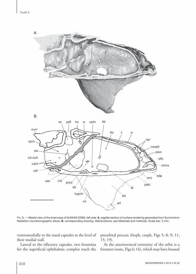
Th ere is no evidence of any ring of sclerotic cartilage or any ring of numerous small presumed bony plates. Th ere is also no evidence for an optic pedicel attachment in the orbit.
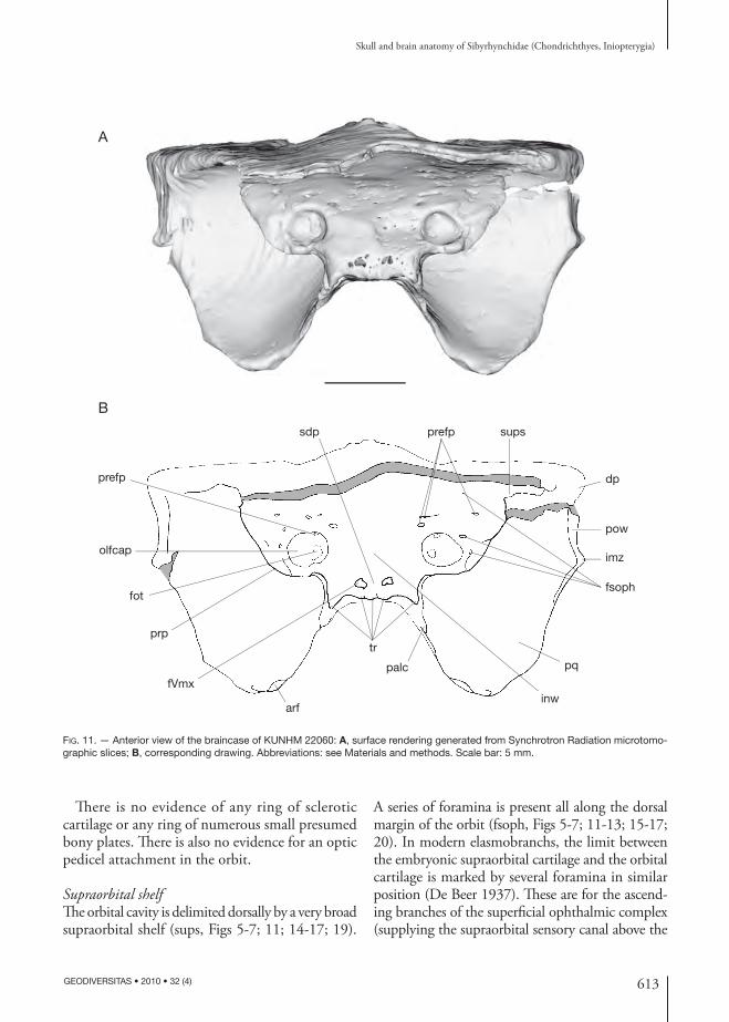
Supraorbital shelfTh e orbital cavity is delimited dorsally by a very broad supraorbital shelf (sups, Figs 5-7; 11; 14-17; 19).
A series of foramina is present all along the dorsal margin of the orbit (fsoph, Figs 5-7; 11-13; 15-17; 20). In modern elasmobranchs, the limit between the embryonic supraorbital cartilage and the orbital cartilage is marked by several foramina in similar position (De Beer 1937). Th ese are for the ascend-ing branches of the superfi cial ophthalmic complex (supplying the supraorbital sensory canal above the
FIG. 11. — Anterior view of the braincase of KUNHM 22060: A, surface rendering generated from Synchrotron Radiation microtomo-graphic slices; B, corresponding drawing. Abbreviations: see Materials and methods. Scale bar: 5 mm.
prefp
olfcap
fot
prp
fVmx
arf
tr
palc
inw
pq
fsoph
imz
pow
dp
supsprefpsdp
A
B
614 GEODIVERSITAS • 2010 • 32 (4)
Pradel A.
braincase). Th is nerve complex includes the superfi cial ophthalmic components of the anterodorsal lateral line (including the superfi cial ophthalmic and buc-cal branches of the facial nerve [Northcutt & Bemis 1993]) and trigeminal nerves (Maisey 2005) when these components are combined.
Consequently, this series of foramina present in the specimens described here may be for the super-fi cial ophthalmic complex and may determine the hypothetical original medial boundary of the su-praorbital shelf.
In dorsal view, the preorbital process fl ares laterally, but tapers slightly to a blunt point when it becomes the supraorbital shelf posteriorly (Figs 5; 14A). Th en, the su-praorbital shelf fl ares laterally as far as the distal extremity of the post orbital wall. Th is morpho logy is comparable to that of Clodoselache Dean, 1894 (Maisey 2007) and Akmonistion Coates & Sequeira, 2001 (Coates & Sequeira 1998) altough the blunt point between the preorbital process and the supraorbital shelf is situated more posteriorly in the sibyrhynchids.
Palatoquadrate, suborbital shelf and extent of the inferred embryonic polar cartilageTh e fi rst description of iniopterygian braincases (Zangerl & Case 1973) showed a holostylic jaw suspension, similar to that of extant holocephalans, where the palatoquadrate is fused to the neuro-cranium. Further investigations demonstrated that not all inio pterygians possess a holostylic jaw sus-pension (Stahl 1980). Indeed, only the Sibyrhynchi-dae possess a palatoquadrate which is fused to the neuro cranium.
Th e two most complete Kansas specimens clearly show a pair of articular facets at the anterior end of what is provisionally referred to as the ventral sub-orbital shelf (arf, Figs 7; 11; 14B). Th e skull OKM 38 is broken at the level of the suborbital shelf and the latter, which shows the same articular facets, is found free in the nodule.
KUNHM 22060 shows the mandible (Fig. 31A, B), which has been slightly displaced during the fossilization (see below for the description of the mandible). Th e mandible possesses at its posterior ends a pair of articular facets complementary to those of the anterior end of the suborbital shelf (am, Fig. 31D). When virtually placed in front of
the suborbital shelf, it perfectly articulates with the latter (Fig. 31B).
As seen previously, the anterior end of the neuro-cranium displays ventrolaterally a pair of pronounced crests (palc, Figs 6; 7; 11; 14B; 16; 17; 19; 32), which form with the symphysial dentigerous plate a continu-ous dental arcade. Th e orbital foramen for the orbito-nasal canal is located immediately above the lateral crest (oonc, palc, Figs 6; 14B; 16) and the ventral foramen is situated at the medial junction of this crest and the internasal plate (vonc, inp, Figs 7; 17). Th is recalls the condition present in extant chimaeroids, in which the anterior part of the palatoquadrate also supports palatine teeth and its dorsal margin is approximately indicated by orbitonasal canal foramina (Grogan et al. 1999). Th is relative disposition in the specimens described here between the palatine crest supporting diff erent tooth whorls and the orbitonasal canal, as well as the presence of articular facets at the anterior extremity of the suborbital shelf in perfect position for the articulation with the lower jaw, suggests that the palatoquadrate is fused to the neuro cranium, as in extant chimaeroids. So considered, the anteroventral extremity of the suborbital shelf displaying articular facets corresponds to the quadrate process, which was in connection with the mandible, whereas the anterior crest corresponds to the palatine part of the palatoquad-rate. Th e anterior margin of the palatoquadrate may be indicated by the foramen for the maxillary ramus of the trigeminal nerve (fVmx, Fig. 6).
If fusion of the palatoquadrate with the neuro cranium in sibyrhynchids was the same as in modern chimae-roids, the palatoquadrate probably extended backward as far as the posterior limit of the orbits, where the orbital foramen for the eff erent pseudo branchial artery is situated (oeps, Figs 12; 13; 16). Indeed, the hind limit of the palatoquadrate of extant chimaeroids is marked by the path of the eff erent pseudo branchial artery and defi ned by the fusion of the palatoquad-rate to the embryonic basitrabecular cartilage, which in extant chimaeroids is said to be derived from the polar cartilage (Grogan et al. 1999). Consequently, the suborbital shelf of the iniopterygian described here is mainly made up by the palatoquadrate, sec-ondarily fused to the neuro cranium and is therefore probably of visceral arch origin and not derived from the primary cranial cartilage.
615
Skull and brain anatomy of Sibyrhynchidae (Chondrichthyes, Iniopterygia)
GEODIVERSITAS • 2010 • 32 (4)
fsoph
proo
gjv
antjc
fIX
ghr
fVII
fVItpffVIIpaloepsoorafpitv
fII
prefc
fIII
fIV
postjc
B
A
d
a p
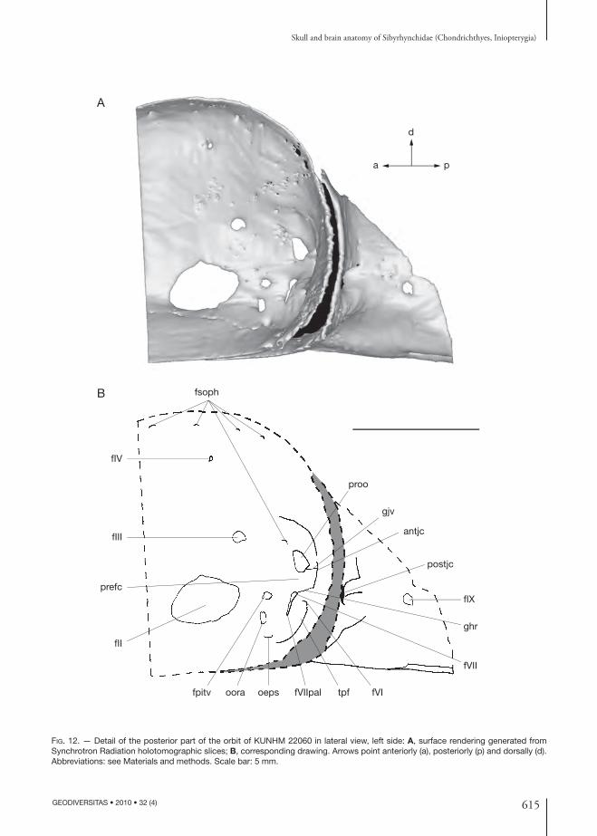
FIG. 12. — Detail of the posterior part of the orbit of KUNHM 22060 in lateral view, left side: A, surface rendering generated from Synchrotron Radiation holo tomographic slices; B, corresponding drawing. Arrows point anteriorly (a), posteriorly (p) and dorsally (d). Abbreviations: see Materials and methods. Scale bar: 5 mm.
616 GEODIVERSITAS • 2010 • 32 (4)
Pradel A.
In the inio pterygians described here, the foramina for the eff erent pseudo branchial artery and the pi-tuitary vein are almost at the same level (oeps, fpitv, Figs 12; 13). Th is may indicate the presence of very small embryonic polar cartilages, since these two foramina generally mark respectively the anterior and the posterior margin of the embryonic polar cartilage in extant elasmobranchs (De Beer 1931, 1937). In addition, the hypophyseal chamber and dorsum sellae are comparatively small (hypch, ds, Figs 8; 26B-D), also suggestive of small embryonic polar cartilages, since the dorsum sellae arise from the polar cartilages during the chondrichthyan ontogeny.
Nevertheless, in extant chimaeroids, the embryonic polar cartilages elaborate structures, which contribute to the suborbital shelf cartilages, and consequently, they extend from the foramen for the eff erent pseudo-branchial artery to the anterior foramen of the jugular canal. Th e suborbital shelf of extant chimaeroids is thereby formed from the fusion of the palatoquad-rate, the basitrabecular process and posteriorly the otic process (Grogan et al. 1999).
By comparison, the polar cartilages in inio pterygians may also extend from the foramen for the eff erent pseudo branchial artery, ventrally, to rather the an-terior foramen of the jugular canal situated more posterodorsally than the foramen for the pituitary vein, thereby indicating a more important polar cartilage, which forms a laterally extended basi-trabecular process. Th e latter may have formed with the palatoquadrate the suborbital shelf.
A lateral extension of the basitrabecular cartilage forming thereby a prominent basitrabecular process, which is connected to the palatoquadrate, is inferred in Paleozoic autodiastylic chondrichthyans, such as cochliodonts (Lund & Grogan 1997a).
Th e orbital artery foramen is present between the eff erent pseudo branchial artery foramen and the pi-tuitary vein foramen (oora, oeps, fpitv, Figs 11; 12). Th is disposition suggests that the orbital artery canal passes through the inferred extent of the embryonic polar cartilage (ora, Fig. 21). In modern holocepha-lans, the orbital artery canal is situated between the foramen for the eff erent pseudo branchial artery and foramen for the pituitary vein, thus running also through the embryonic polar cartilage.
In posterior view, the palatoquadrate displays in its underside a small cavity, which may have housed hy-oidean muscles (mi, Fig. 10), as in extant chimaeroids (e.g., the muscle levator hyoideus; Didier 1995).
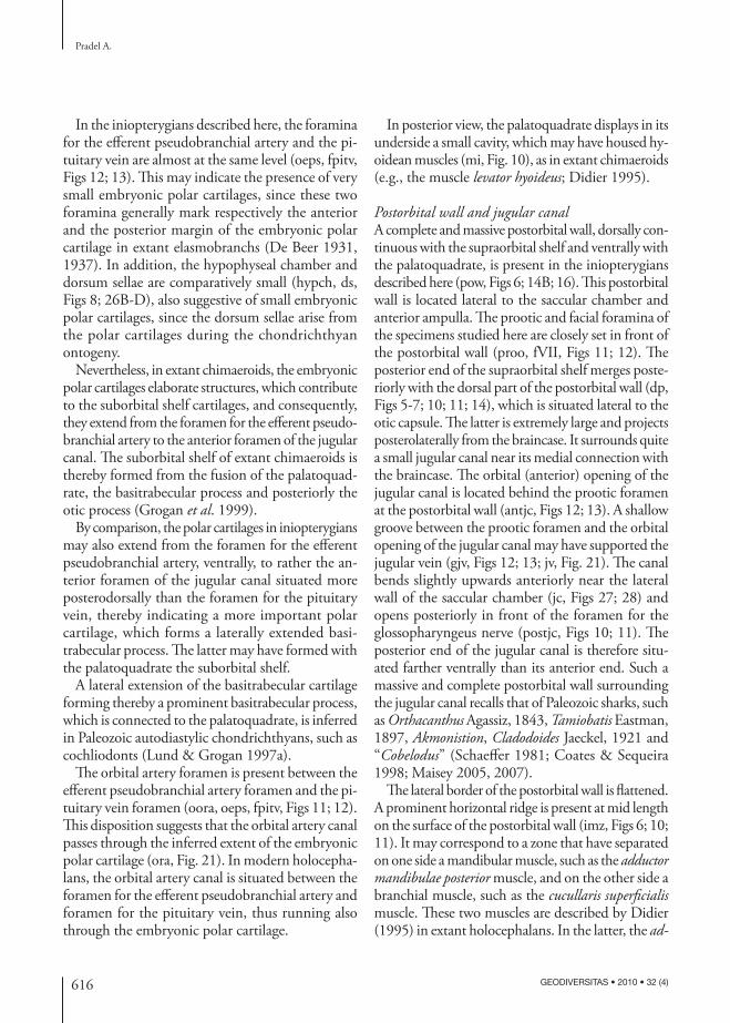
Postorbital wall and jugular canal A complete and massive post orbital wall, dorsally con-tinuous with the supraorbital shelf and ventrally with the palatoquadrate, is present in the inio pterygians described here (pow, Figs 6; 14B; 16). Th is post orbital wall is located lateral to the saccular chamber and anterior ampulla. Th e prootic and facial foramina of the specimens studied here are closely set in front of the post orbital wall (proo, fVII, Figs 11; 12). Th e posterior end of the supraorbital shelf merges poste-riorly with the dorsal part of the post orbital wall (dp, Figs 5-7; 10; 11; 14), which is situated lateral to the otic capsule. Th e latter is extremely large and projects posterolaterally from the braincase. It surrounds quite a small jugular canal near its medial connection with the braincase. Th e orbital (anterior) opening of the jugular canal is located behind the prootic foramen at the post orbital wall (antjc, Figs 12; 13). A shallow groove between the prootic foramen and the orbital opening of the jugular canal may have supported the jugular vein (gjv, Figs 12; 13; jv, Fig. 21). Th e canal bends slightly upwards anteriorly near the lateral wall of the saccular chamber (jc, Figs 27; 28) and opens posteriorly in front of the foramen for the glossopharyngeus nerve (postjc, Figs 10; 11). Th e posterior end of the jugular canal is therefore situ-ated farther ventrally than its anterior end. Such a massive and complete post orbital wall surrounding the jugular canal recalls that of Paleozoic sharks, such as Orthacanthus Agassiz, 1843, Tamiobatis Eastman, 1897, Akmonistion, Cladodoides Jaeckel, 1921 and “Cobelodus” (Schaeff er 1981; Coates & Sequeira 1998; Maisey 2005, 2007).
Th e lateral border of the post orbital wall is fl attened. A prominent horizontal ridge is present at mid length on the surface of the post orbital wall (imz, Figs 6; 10; 11). It may correspond to a zone that have separated on one side a mandibular muscle, such as the adductor mandibulae posterior muscle, and on the other side a branchial muscle, such as the cucullaris superfi cialis muscle. Th ese two muscles are described by Didier (1995) in extant holocephalans. In the latter, the ad-
617
Skull and brain anatomy of Sibyrhynchidae (Chondrichthyes, Iniopterygia)
GEODIVERSITAS • 2010 • 32 (4)
ductor mandibulae posterior muscle originates from the suborbital shelf and the cucullaris superfi cialis muscle originates more posterodorsally from the post orbital
crest. Th e cranial surface between these two muscle insertions remains directly in contact with the over-lying skin (Didier 1995: fi g. 41; amp, cs).
fIV
a
p
d
fIII
prefc
fII
fpitvoora oeps fVIIpal fVII
tpf
fVI
ghr
antjc
gjv
proo
fsoph
fsophB
A
FIG. 13. — Detail of the posterior part of the orbit of KUNHM 22060 in anterolateral view, left side: A, surface rendering generated from Synchrotron Radiation holo tomographic slices; B, corresponding drawing. Arrows point anteriorly (a), posteriorly (p) and dorsally (d). Abbreviations: see Materials and methods. Scale bar: 5 mm.

618 GEODIVERSITAS • 2010 • 32 (4)
Pradel A.
prp
dd
pf
pdob
sdp
palc oonc
arf
pq
flXsof
vppr
occot
dppr
fm
fpcv
occr
mop
flX+flcX
pdob dd
dp
occr
mop
dor
dp
sups
sdpA
B
FIG. 14. — Surface rendering generated from Synchrotron Radiation microtomographic slices of the braincase of KUNHM 21894: A, dorsal view; B, lateral view. Abbreviations: see Materials and methods. Scale bar: 5 mm.
619
Skull and brain anatomy of Sibyrhynchidae (Chondrichthyes, Iniopterygia)
GEODIVERSITAS • 2010 • 32 (4)
Optic nerve (II), oculomotor nerve (III), and pituitary veinTh e orbital wall displays the largest ovoid foramen centrally (fII, Figs 6; 12; 13; 16). According to the relation between this foramen and the brain and nerves preserved in the endo cranial cavity (fII, II, Fig. 26), it contained the optic nerve (Pradel et al. 2009) and probably the optic (retinal) artery (opa, Fig. 21), as in Cladodoides (Maisey 2005) and Chlamydoselachus Garman, 1884 (Allis 1923). It is also through this foramen that the eff erent pseudo branchial artery supplied the blood to the brain (see below).
In chondrichthyans, the oculomotor nerve leaves the brainstem near the midline of the mesen-cephalon (Jollie 1962; Maisey 2005). In the pre-served brain (Pradel et al. 2009) a pair of nerves leaves the ventral part of the mesencephalon at the midline (III, Fig. 26) and passes through a foramen situated dorsal to the optic foramen at the level of its posterior end (fIII, Figs 6; 8; 12; 13; 16; 26), suggesting that it is the oculomotor nerve foramen. As suggested by the shape of the nerve, the oculomotor nerve seems to have been divided into a dorsal ramus, which may have innervated the superior and internal rectus mus-cles, and a ventral ramus, which innervated the inferior rectus and inferior oblique eye muscles (IIId, IIIv, Fig. 21).
Th e pituitary vein foramen is located immediately behind the optic foramen, below the oculomotor nerve foramen (fpitv, Figs 12; 13; pitv, Fig. 21).
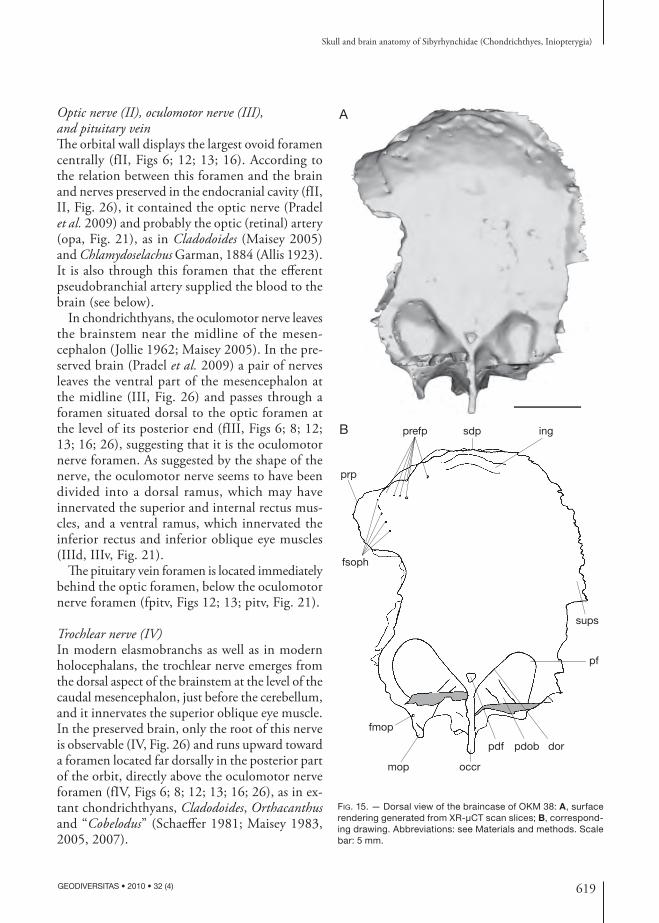
Trochlear nerve (IV)In modern elasmobranchs as well as in modern holocephalans, the trochlear nerve emerges from the dorsal aspect of the brainstem at the level of the caudal mesencephalon, just before the cerebellum, and it innervates the superior oblique eye muscle. In the preserved brain, only the root of this nerve is observable (IV, Fig. 26) and runs upward toward a foramen located far dorsally in the posterior part of the orbit, directly above the oculomotor nerve foramen (fIV, Figs 6; 8; 12; 13; 16; 26), as in ex-tant chondrichthyans, Cladodoides, Orthacanthus and “Cobelodus” (Schaeff er 1981; Maisey 1983, 2005, 2007).
prefp sdp ing
sups
pf
dorpdobpdf
occrmop
fmop
fsoph
prp
B
A
FIG. 15. — Dorsal view of the braincase of OKM 38: A, surface rendering generated from XR-μCT scan slices; B, correspond-ing drawing. Abbreviations: see Materials and methods. Scale bar: 5 mm.

620 GEODIVERSITAS • 2010 • 32 (4)
Pradel A.
Trigeminal (V), profundus ramus and anterodorsal lateral line nerves Th e trigeminal nerve passed through a prootic fo-ramen (proo, Figs 6; 8; 12; 13; 16; 26). Th e middle cerebral vein may also have left the braincase through this foramen (mcv, Fig. 21), as in modern elasmo-branchs and “Cobelodus” (Maisey 2007).
In many modern chondrichthyans, the profun-dus ramus of the trigeminal nerve, the superfi cial ophthalmic ramus of trigeminal and anterodorsal lateral line nerves leave the braincase together. In the inio pterygians studied here, there is no evidence of a separate foramen for the anterodorsal lateral line nerve and superfi cial ophthalmic ramus of the trigeminal nerve in the inner wall of the braincase (proo, Fig. 28). Th is suggests that these nerves left the braincase through the same (prootic) foramen, and formed a dorsal superfi cial ophthalmic complex, as
inferred in other Paleozoic chondrichthyans (Maisey 2005, 2007). Nevertheless, a notch is present in the anterodorsal margin of the prootic foramen in the inner endo cranial wall (Fig. 28), and there is a small foramen anterodorsal to this notch in the external orbital wall of the braincase (fsoph, Figs 12; 13). Th is suggests a separate opening for the dorsal superfi cial ophthalmic complex through the outer wall of the braincase. Th e profundus ramus of the trigeminal nerve (pr, Fig. 21) may have left the braincase sepa-rately from the superfi cial ophthalmic complex (soph, Fig. 21), through the prootic foramen.
A foramen is situated ventrolaterally, immediately posterior to the orbital foramen for the orbitonasal canal and the palatine tooth whorl, at the junction between the palatoquadrate and the ethmoidal plate (obmc, Figs 6; 16). Th is foramen is linked to a ven-tral foramen (vbmc, Figs 7; 17), which is situated
prefp
fsoph sups fIV pow pf ld fmop occr
mop
fpcv
fIcX
fX
fm
dppr
sof
occot
fIXprootpfoepsfIIIfII
obmcooncpalc
prp
sdp
olfcap
vppr
A
B
A
FIG. 16. — Lateral view of the braincase of OKM 38, left side: A, surface rendering generated from XR-μCT scan; B, corresponding drawing. Abbreviations: see Materials and methods. Scale bar: 5 mm.

621
Skull and brain anatomy of Sibyrhynchidae (Chondrichthyes, Iniopterygia)
GEODIVERSITAS • 2010 • 32 (4)
posterior to the ventral foramen of the orbitonasal canal, by a short canal. A ventral bucco-maxillary complex (the maxillary ramus of the trigeminal nerve plus the buccal ramus of the anterodorsal lateral line nerve) is inferred in many Paleozoic chondrichthyans (Maisey 2007) and may be also present in the inio-pterygians studied here. If present, it probably ran through the orbit (bmc, Fig. 21) and probably left the neuro cranium through the foramina described just above.
A more anterior foramen is present ventral to the nasal capsule (fVmx, Figs 6; 9). A foramen in a similar position is present in other holostylic fi shes. In the upper jaw of extant chimaeroids a foramen for a branch of the maxillary ramus is situated “near the dorsal extremity of the lateral set of teeth” (Cole 1896). An anterior foramen for the maxillary ramus of the trigeminal nerve is also present in the posterior part of the preorbital wall and below the foramen for the superfi cial ophthalmic ramus of the facial nerve and the ophthalmic profundus ramus of the trigeminal nerve in holostylic Devonian dipnoans (Pridmore et al. 1994; Campbell & Barwick 2000). Th is suggests that the foramen described above in the specimens studied here was probably for a branch of the maxillary ramus of the trigeminal nerve of the bucco-maxillary complex (Vmx, Fig. 21).
Th e mandibular ramus of the trigeminal nerve may also have run ventrally in the orbit as far as the articulation between the mandible and the pala-toquadrate (Vmd, Fig. 21), as in extant chimaeroids (Cole 1896; De Beer & Moy-Th omas 1935).
Abducens nerve (VI)Th e postero ventral part of the orbit displays a fossa (see below), where a foramen pierces the neuro cranium (fVI, Figs 12; 13). Th is foramen was probably for the exit of the abducens nerve (VI, Fig. 21), which sup-plied the external (= posterior) rectus muscle. Since the dorsum sellae is very short in the inio pterygians described here, the abducens nerve probably left the neuro cranium above and behind the dorsum sellae (cVI, ds, Figs 8; 26), which represents an autapomor-phic condition in chondrichthyans (Gardiner 1984). In the endo cranial cavity, the canal for the abducens nerve is located between the utricular recess and the saccular chamber (cVI, Figs 27; 28).
veps
vora
fIX
vppr occr soffIcX
fpcv mop
fmop
fX
ld
pow
vbmc
fsoph
prp
palcvonctrsdpinpB
A
FIG. 17. — Ventral view of the braincase of OKM 38: A, surface rendering generated from XR-μCT scan slices; B, correspond-ing drawing. Abbreviations: see Materials and methods. Scale bar: 5 mm.
622 GEODIVERSITAS • 2010 • 32 (4)
Pradel A.
Facial nerve (VII) Th e foramen for the main trunk of the facial nerve is located in the surface of the orbital cartilage, between the foramina for the pituitary vein and abducens nerve (fVII, Figs 12; 13). Th is foramen is followed ventrally by another one (fVIIpal, Figs 12; 13), which is connected to the canal for the orbital artery ventrally (cora, Fig. 28) by an almost verti-cal canal (cVIIpal, Fig. 28). Same foramina are present in “Cobelodus” in which the main facial trunk is assumed to divide into anterior (palatine) and posterior (mandibular-hyoidean) rami after leaving the braincase. Th e palatine ramus shared ventrally the same groove as the orbital artery after passing through the fl oor of the post orbital arcade, while the mandibular-hyoidean ramus presumably passed through the jugular canal posteriorly (Maisey 2007). Consequently, in the specimens studied here, the anatomy of the facial nerve may have been the same. Th e palatine and mandibular-hyoidean rami may have given off from the main trunk of the facial nerve when the latter appears to surface (VII, VIIpal, VIIhym, Fig. 21).
Th en, the palatine ramus probably entered the neuro cranium just below the main facial foramen (fVII, fVIIpal, Figs 12; 13) and ran through the vertical canal until the canal for the orbital ar-tery ventrally (cora, Fig. 28). Th e latter is directed anterodorsally and its orbital opening is located immediately below the foramen for the pituitary vein (oora, Figs 12; 13), while its ventral opening is situated behind the ventral foramen (vora, Figs 7; 17) for the eff erent pseudo branchial artery (veps, Figs 7; 17).
Two hypotheses concerning the course of the palatine ramus of the facial nerve (VIIpal, Fig. 21) of sibyrhynchids can be proposed: 1) the palatine ramus entered the orbital artery canal and ran backwards. After exiting from the braincase through the ventral opening of orbital artery canal, it divided into the anterior and posterior ramules; and 2) the palatine ramus entered the orbital artery canal and divided into the anterior and posterior ramules inside it. Th e anterior ramule (VIIapal, Fig. 21) ran forward (cVIIapal, Fig. 28) and exited from the braincase through the orbital opening of the orbital artery canal, whereas the posterior one (VIIppal, Fig. 21)
ran backwards (cVIIppal, Fig. 28) and exited from the braincase through the ventral opening of the orbital artery canal. Th en, the anterior ramule ran in the ventral part of the orbit as far as the foramen for the bucco-maxillary complex.
In extant adult chimaeroids, the palatine ramus divides into the anterior and posterior ramules after exiting from the orbital artery canal through its ventral foramen. Th is pattern corresponds to the fi rst hypothesis proposed here. Nevertheless, in extant chimaeroids the orbital artery canal is more vertical than in sibyrhynchids, in which this canal is oriented anterodorsally (cora, Fig. 28). If the same arrangement was present in inio pterygians, the anterior ramule is far from the palatoquadrate and must have made a loop to innervate the palatine muscles. In addition, the palatine ramus of extant chimaeroids enters the orbital artery canal through its orbital foramen, whereas in inio pterygians, as seen previously, it probably entered the neuro cranium just below this facial foramen and ran through a canal, which meets ventrally the canal for the orbital artery intracranially.
In Cladodoides and “Cobelodus” (Maisey 2005, 2007), the palatine ramus enters a canal, and then divides into the anterior and posterior ramules be-fore exiting from the neuro cranium. Th e division presumably occurs within the lateral commissure, which formed a part of the post orbital wall. Con-sequently, there are two diff erent foramina for the anterior ramule and the posterior ramule respectively. In Cladodoides, the anterior ramule enters the orbit ventrolaterally through the orbital artery groove. In “Cobelodus”, the foramina for the anterior and posterior ramule reach ventrally the braincase in the groove housing the orbital artery. Th is pattern corresponds to the second hypothesis proposed here, which appears to be the most probable.
Th e mandibular-hyoidean ramus presumably passed posteriorly via the jugular canal (VIIhym, Fig. 21), as suggested by the presence of a shallow groove between the foramen for the main trunk of the facial nerve and the orbital opening of the jugular canal (ghr, fVII, antjc, Figs 12; 13), as in extant chimaeroids (De Beer & Moy-Th omas 1935: fi g. 1) and “Cobelodus” (Maisey 2007). Th en, the hyomandibular ramus may have divided into
623
Skull and brain anatomy of Sibyrhynchidae (Chondrichthyes, Iniopterygia)
GEODIVERSITAS • 2010 • 32 (4)
mandibular and hyoidean ramules when it passed lateral to the epihyal (VIImd, VIIh, Fig. 21), again as in extant chimaeroids (De Beer & Moy-Th omas 1935). It is also possible that only the hyoidean ramule passed through the jugular canal and that the mandibular ramule accompanied the maxillary ramus of the trigeminal nerve.
Prefacial commissure and trigemino-pituitary fossa Th e prootic foramen and the foramen for the main trunk of the facial nerve (proo, fVII, Fig. 12) are
separated by a bar of cartilage, which is assumed to be a prefacial commissure (prefc, Figs 12; 13).
In the inio pterygians described here, the postero-ventral corner of the orbit displays a large trigemino-pituitary fossa (sensu Allis 1918, 1923, 1928) (tpf, Figs 12; 13) delimited anteriorly by the foramina for the pituitary vein and for the orbital artery (fpitv, oora, Figs 12; 13) and ventrally by the foramen for the eff erent pseudo branchial artery (oeps, Figs 12; 13). Th is fossa also contains the prootic foramen in its dorsal part (proo, Figs 12; 13), and the fo-
mop
fpcv
fIcX
fX
fIX
sof vppr occot dppr
fm
occr
dorpdfpfB
A
dorpdfpf
FIG. 18. — Posterior view of the braincase of OKM 38: A, surface rendering generated from CT scan slices; B, corresponding drawing. Abbreviations: see Materials and methods. Scale bar: 5 mm.

624 GEODIVERSITAS • 2010 • 32 (4)
Pradel A.
ramen for the facial nerve (fVII, Figs 11; 12). Th e trigemino-pituitary fossa is much more pronounced in its postero ventral part, where the foramen for the abducens nerve pierces the braincase wall (fVI, Figs 12; 13).
Th e palatine and mandibular-hyoidean rami of the facial nerve of the inio pterygians studied here probably diverged within the facial foramen (see above). Consequently, the facial nerve ganglion was probably situated partially inside and partially outside
ing
inw
sdp
fVmxtr
palc
voncolfcap
prp
fot
fsoph
sups
prefpoccrpdf
B
A
fdf
A
FIG. 19. — Anterior view of the braincase of OKM 38: A, surface rendering generated from XR-μCT scan slices; B, corresponding drawing. Abbreviations: see Materials and methods. Scale bar: 5 mm.
625
Skull and brain anatomy of Sibyrhynchidae (Chondrichthyes, Iniopterygia)
GEODIVERSITAS • 2010 • 32 (4)
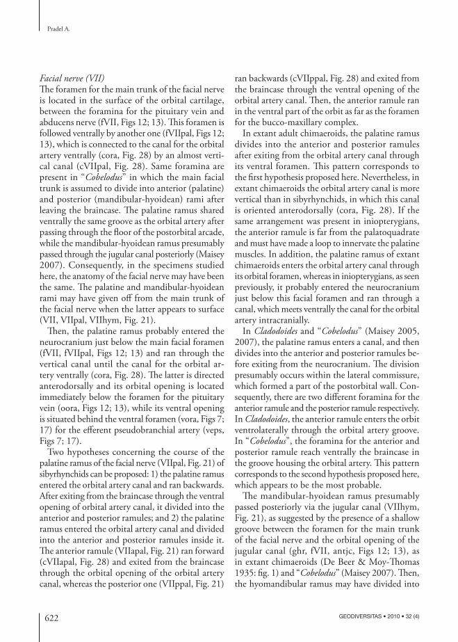
the neuro cranium in the trigemino-pituitary fossa. Th e trigeminal ganglion (Gasser’s ganglion; gas, Fig. 21) was probably situated in part lateral to the posterior part of the orbit, in the trigemino-pituitary
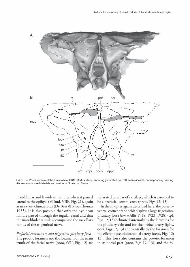
fossa, and partly internally because the superfi cial ophthalmic ramus probably exited the braincase with the anterodorsal lateral line nerve through a foramen that is distinct from the prootic one.
B
A
occr fmop
mop
fpcv
fm
fIcX
dppr
occot
vppr sof fX fIX cora cVIIpal
cpitv
FIG. 20. — Lateral view of the braincase of KUNHM 56360, right side; A, surface rendering generated from XR-μCT scan slices. B, cor-responding drawing. Abbreviations: see Materials and methods. Scale bar: 5 mm.

626 GEODIVERSITAS • 2010 • 32 (4)
Pradel A.
Arterial supply to the headAt the level of the posterior attachment of the post orbital wall of the braincase, the basi cranium displays a pair of foramina (vora, Figs 7; 17), which corresponds to the ventral foramina for the orbital artery canal. Th ere is neither a basicranial fenestra, nor a hypophyseal fenestra. Consequently, there is no evidence of foramina for the internal carotid arteries, which thus probably did not supply blood to the brain.
A small canal (ceps, Fig. 28) passing through the suborbital shelf, with its orbital opening (oeps, Figs 12; 13; 16) situated ventral to the orbital fo-ramen of the orbital artery canal and its ventral opening (voeps, Fig. 17) located in front of the ventral foramen of the orbital artery canal, is present anterior to the internal hypophyseal chamber. It may have housed the eff erent pseudo branchial artery. In such a case, the eff erent pseudo branchial artery (eps, Fig. 21) probably entered the brain cavity through the large optic foramen. Th en, it may have divided into an optical (retinal) artery (opa, Fig. 21), which re-entered into the orbits, and a basilar artery (basa, Fig. 21), which supplied blood to the brain.
Otic regionTh e short otic region of the inio pterygians described here is pierced dorsally by a median posterior dorsal fontanelle (pdf, Figs 7; 14A; 15).
As in many Paleozoic sharks, the otic region shows a posterolaterally directed lateral process on either side of the braincase (dp, Figs 5-7; 10; 11; 14). It extends very far laterally from the loop of the posterior semicircular canal and from the pre occipital fossa (pf, Figs 5; 6; 10; 14A; 15; 16; 18) that probably represents insertion area for the epaxial musculature.
An other pair of processes encloses the loop of each posterior semicircular canal and overlies on a short distance the horizontal semicircular canal (mop, Figs 5; 7; 10; 14-18; 20). Th ey are located more medially, on either sides of the occipital crest, at the lateral margin of the dorsal pre occipital fossae. Th ere is a foramen at the extremity of the medial otic process (fmop, Figs 15-17; 20). Th e attribution of this foramen to either blood vessels or cranial nerves is unclear (see below).
A pair of posterior dorsal otic bulges (pdob, Figs 5; 14; 15) originates from the medial otic processes of either sides and converges anteriorly toward the posterior dorsal fontanelle. Th ey enclose the dorsal part of the posterior semicircular canal. A similar pair of posterior dorsal otic bulges is present in many extinct chondrichthyans, such as Akmonis-tion (Coates & Sequeira 1998: fi g. 5).
Low dorsal otic ridges, which overly the anterior semicircular canals and fl ank the endolymphatic fossa (or posterior dorsal fontanelle), as that of “Cobelodus”, are present in inio pterygians (dor, Figs 5; 10; 14A; 15; 18).
Th e course of the horizontal semicircular canal in inio pterygians is delimited by the lateral margin of the pre occipital fossae and the course of the posterior part of the anterior semicircular canal is delimited by the anteromedial margin of the fossa. Th ese fossae are anteriorly divergent and their position corresponds to the vacuity delimited by the dorsal part of the three semicircular canals (pf, Figs 5; 6; 10; 14A; 15; 16; 18).
Consequently, the dorsal otic ridges seen in some other Paleozoic chondrichthyans (e.g., Tamiobatis, Cladodoides, “Cobelodus”) are represented here by the medial margin of the pre occipital fossae. Th e lateral otic ridge of these Paleozoic chondrichthyans may be also represented in iniopterygian by the lateral margin of the pre occipital fossae. In these Paleozoic sharks, the dorsal and lateral otic ridges delimit a dorsolateral otic fossa, which may be represented in inio pterygians by the pre occipital fossae, even though the latter are completely dorsal in posi-tion. Th is dorsal, rather than dorsolateral position is probably due to the extreme lateral extent of the post orbital wall.
Th e semicircular canals are restricted to the dor-sal part of the posterior half of the neuro cranium, in a region that is very shallow (Fig. 10). Such a condition is otherwise found only in the strongly fl attened braincases of certain arthrodire placo-derms (Stensiö 1969; Goujet 1984) and perhaps Helodus Agassiz, 1838 (Moy-Th omas 1936; Pat-terson 1965). Ventrolateral to the otic region are very deep post orbital depressions (ld, Figs 7; 10; 16; 17) that probably accommodated the jaw and visceral musculature (e.g., the cucullaris profundus,

627
Skull and brain anatomy of Sibyrhynchidae (Chondrichthyes, Iniopterygia)
GEODIVERSITAS • 2010 • 32 (4)
subspinalis, and levator hyoideus muscles, as in extant chimaeroids; Didier 1995) and gills. Th e branchial apparatus was thus situated beneath the braincase, instead of extending behind it. Th is position of the gills relative to the neuro cranium is similar to that of chimaeroids. Th erefore, inio pterygians were fi rst classifi ed among the Subterbranchialia (Zangerl 1981), a taxon including also the extant chimaeroids. In addition, like in extant chimaeroids, there is no evidence of an articular facet for the hyo-mandibula in the otico-occipital region, as well as on the post orbital wall. Nevertheless, more data on the branchial skeleton are needed to know whether a hyomandibula is present or not. “Cobelodus” lacks
a distinct hyomandibular facet on the neuro cranium although the presence of a hyomandibula suggests a weak ligamentous attachment, probably with the periotic process (Maisey 2007).
OCCIPITAL REGION
General featuresPosterior to the post orbital wall, the ventral part of the braincase is narrow (Figs 7; 17). Th e occipital part of the braincase is relatively short and is fl anked by the post orbital depressions (ld, Figs 7; 10; 16; 17). According to Romer (1964) and Coates & Se-queira (1998), a relatively elongated otico-occipital region is presumably a plesiomorphic condition
Vmx
gas mcv
pitv
prIV soph
IIId
VII
VI
IX
XlcX
jv
IIIv
opaII
basa
Vmd VIIapalbmcfv
VIIpal
bso
pcv
soVIIhym
VIIhVIImd
eps VIIppal
ora
FIG. 21. — Hypothetical arrangements of arteries (in red), veins (in blue) and nerves (in yellow) in the chondrocranium of a reconstructed Sibyrhynchidae based on the different 3D reconstructions of the braincase. Abbreviations: see Materials and methods. Not to scale.

628 GEODIVERSITAS • 2010 • 32 (4)
Pradel A.
among gnathostomes. Consequently, a compara-tively short otico-occipital region may represent a derived condition. Nevertheless, a relatively short otico-occipital region commonly occurs in actino-pterygians, sarcopterygians, Acanthodes Agassiz, 1833, Pucapampella, chimaeroids, neoselachians, Symmoriiformes and hybodonts.
Behind the posterior dorsal fontanelle and between the pre occipital fossae is a prominent crest, which extends far posteriorly (occr, Figs 7-8; 10; 14-16; 18). Below this crest, the occipital arch surrounds dorsally the foramen magnum, and ventrally the notochordal canal (fm, occot, Figs 10; 18). Th e notochordal canal forms a deep fovea commonly called occipital cotylus, as in many other Paleozoic chondrichthyans (e.g., Cladodoides, “Cobelodus”). Th is suggests a strong connection with the vertebral column. In life, the occipital cotylus is assumed to have enclosed the anterior end of the notochord, which is usually persistent in the adult in modern cyclostomes, chondrichthyans, sturgeons, lungfi shes and coelacanths. In all inio pterygians studied here, the occipital cotylus merges anteriorly into the rear of the braincase about as far as the level of the anteriormost spino-occipital foramen (sof, Figs 6; 8; 14B; 16; 20) and there is no evidence of a hemicentrum.
A similar cotylus is present in hybodonts and many Paleozoic sharks (Maisey 1983, 2005, 2007). Nevertheless, contrary to many Paleozoic sharks in which the occipital cotylus is larger than the foramen magnum (e.g., Cladodoides), these two structures are about the same width, as in “Cobelodus”. Th e dorsolateral and ventrolateral part of the rim of the occipital cotylus protrudes posteriorly to form the dorsal and ventral paroccipital processes of the occipital cotylus (dppr, vppr, Figs 6; 7; 10; 14B; 16-18; 19). Th ese processes presumably contributed to the craniovertebral joint and are generally absent in Paleozoic sharks with an elongated occipital region (e.g., Cladodoides, Orthacanthus, Tamiobatis). Th ey are present in Cobelodus aculeatus, Stethacanthu-lus Zangerl, 1990, Akmonistion and “Cobelodus” (Zangerl & Case 1976: fi g. 5; Williams 1985: fi gs 3, 6; Zidek 1992: fi g. 4; Coates & Sequeira 1998: fi g. 4; Maisey 2007: fi g. 11).
Th ere is a posterior cartilage ridge that joins the two dorsal paroccipital processes. It separates the
notochordal canal from the foramen magnum at the posterior extremity of the occipital region (occot, fm, Fig. 22A). Th en, the inner wall of the braincase separates the notochordal canal from the endo cranial cavity more anteriorly. Th erefore, the notochord was completely enclosed (presum-ably by the parachordal cartilages), contrary to the condition in extant chimaeroids, where the noto-chord is free above the basal plate. Th e notochord was also enclosed by cartilage (perhaps derived from the parachordals) in placoderms, primitive actino pterygians, and primitive chondrichthyans. Th is suggests that an enclosed notochord in the adult is a plesiomorphic condition among the gnathostomes.
Th e occipital cotylus generally separates posteriorly the paired parachordal cartilages in chondrichthy-ans. In the inio pterygians studied here, between the ventral paroccipital processes is a median posterior notch that extends anteriorly as far as the level of the anteriormost foramen for the spino-occipital nerves (Figs 7; 17). Th is suggests that the two parachordals are incompletely fused posteriorly (parc, Fig. 22A). When the notochordal canal tapers anteriorly, a vertical septum connects the notochordal canal to the basi cranium and marks the beginning of the embryonic fusion of the paired parachordal cartilages. At this level, the basal plate is not yet well defi ned (fp, Fig. 22B). On each side of the septum is an occipital chamber, which is delimited at the level of the spino-occipital nerve by the correspondent canals (occh, sof, Fig. 22B).
Th is septum is thus not primary homologous to the vertical subnotochordal septum, which con-nects the notochordal canal to a well-defi ned basal plate in Cladodoides (Maisey 2005: fi g. 30). In addition, there is no evidence, in inio pterygians, of paired subnotochordal “chambers” present in Cladodoides when the septum disappears anteri-orly at the level of the anterior end of the noto-chordal septum.
Glossopharyngeus nerve (IX) In the inio pterygians described here, there is neither a persistent ventral metotic part of the otico-occipital fi ssure, nor a posterior basicapsular commissure sepa-rating the otic capsules from a hypotic lamina, and
629
Skull and brain anatomy of Sibyrhynchidae (Chondrichthyes, Iniopterygia)
GEODIVERSITAS • 2010 • 32 (4)
the glossopharyngeus nerve does not pass in a canal fl oored by the ventral hypotic lamina (cIX, Figs 27; 28). Th ere is also no evidence of a persistent dorsal otico-occipital fi ssure, which initially separates the occipital pila from the otic capsules in Recent embry-onic elasmobranchs. Consequently, a hypotic lamina seems to be absent in sibyrhynchids
Th e glossopharyngeus nerve (IX, Fig. 21) emerged via a short canal originating in the postero ventral part of the saccular chamber (cIX, Figs 8; 27; 28), and is anterior to the vagus nerve (fIX, fX, Figs 7; 10; 14B; 15; 17; 18; 20), as in extant chimaeroids. It seems to pass through the vestibular chamber, since there is no evidence of a medial wall separating the vestibular chamber from the endo cranial cavity (Fig. 8).
Spino-occipital nervesTh e occipital arch in the inio pterygians described here is relatively short, with three spino-occipital foramina being well visible externally and oriented posterodorsally in KUNHM 21894 (sof, Fig. 14B) and KUNHM 56360 (sof, Fig. 20). It is obscured by a break at the level of the putative anteriormost foramen in KUNHM 22060 (sof, Fig. 6), and by the absence of infi lling matrix in OKM 38 (sof, Fig. 6).
Th e number of spino-occipital foramina is highly variable among chondrichthyans (Maisey 2007).
In addition, there may be a diff erence between the number of externally visible foramina and the real number of spino-occipital nerves (e.g., “Cobelodus”: Maisey 2007). In inio pterygians, there is no diff er-ence between the number of spino-occipital canals showing in the endocast of the endo cranial cavity (cso, Fig. 27) and the externally visible foramina.
Since there may be a relatively large number of spino-occipital nerves (more than three) located in the occipital region even in forms that have a short occipital region (e.g., “Cobelodus”), and conversely, a lower number is sometimes present in forms with a long occipital region (e.g., Orthacanthus), there is no simple correlation between the number of nerves in the occipital region and the length of this region. Nevertheless, three or more canals may represent a ple-siomorphic condition in chondrichthyans, and a lower number is probably a derived condition. According to Bemis & Forey (2001), three foramina may represent the primitive condition for gnathostomes.
As mentioned above, a foramen pierces the pos-terodorsal part of the median otic process (fmop, Figs 15-17; 20). It is possible that a branch of a spino-occipital nerve ran through it medially to innervate the epaxial musculature located in the pre occipital fossae (so, bso, Fig. 21). However, this foramen may have housed a blood vessel as well.
FIG. 22. — Transverse XR-μCT scan slice at the level of the mid occipital cotylus of OKM 38 (A) and transverse Synchrotron Radiation microtomographic slice at the level of the anterior end of the occipital cotylus of KUNHM 21894 (B). Abbreviations: see Materials and methods. Not to scale.
A BA B
occrpf
il
ilol
ol
fp occh
sof
occr
endc
fmoccotparcsof
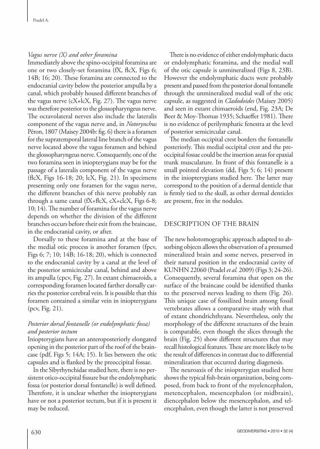
630 GEODIVERSITAS • 2010 • 32 (4)
Pradel A.
Vagus nerve (X) and other foramina Immediately above the spino-occipital foramina are one or two closely-set foramina (fX, fl cX, Figs 6; 14B; 16; 20). Th ese foramina are connected to the endo cranial cavity below the posterior ampulla by a canal, which probably housed diff erent branches of the vagus nerve (cX+lcX, Fig. 27). Th e vagus nerve was therefore posterior to the glossopharyngeus nerve. Th e octavolateral nerves also include the lateralis component of the vagus nerve and, in Notorynchus Péron, 1807 (Maisey 2004b: fi g. 6) there is a foramen for the supratemporal lateral line branch of the vagus nerve located above the vagus foramen and behind the glossopharyngeus nerve. Consequently, one of the two foramina seen in inio pterygians may be for the passage of a lateralis component of the vagus nerve (fl cX, Figs 16-18; 20; lcX, Fig. 21). In specimens presenting only one foramen for the vagus nerve, the diff erent branches of this nerve probably ran through a same canal (fX+fl cX, cX+clcX, Figs 6-8; 10; 14). Th e number of foramina for the vagus nerve depends on whether the division of the diff erent branches occurs before their exit from the braincase, in the endo cranial cavity, or after.
Dorsally to these foramina and at the base of the medial otic process is another foramen (fpcv, Figs 6; 7; 10; 14B; 16-18; 20), which is connected to the endo cranial cavity by a canal at the level of the posterior semicircular canal, behind and above its ampulla (cpcv, Fig. 27). In extant chimaeroids, a corresponding foramen located farther dorsally car-ries the posterior cerebral vein. It is possible that this foramen contained a similar vein in inio pterygians (pcv, Fig. 21).
Posterior dorsal fontanelle (or endolymphatic fossa) and posterior tectumInio pterygians have an anteroposteriorly elongated opening in the posterior part of the roof of the brain-case (pdf, Figs 5; 14A; 15). It lies between the otic capsules and is fl anked by the pre occipital fossae.
In the Sibyrhynchidae studied here, there is no per-sistent otico-occipital fi ssure but the endolymphatic fossa (or posterior dorsal fontanelle) is well defi ned. Th erefore, it is unclear whether the inio pterygians have or not a posterior tectum, but if it is present it may be reduced.
Th ere is no evidence of either endolymphatic ducts or endolymphatic foramina, and the medial wall of the otic capsule is unmineralized (Figs 8, 23B). However the endolymphatic ducts were probably present and passed from the posterior dorsal fontanelle through the unmineralized medial wall of the otic capsule, as suggested in Cladodoides (Maisey 2005) and seen in extant chimaeroids (end, Fig. 23A; De Beer & Moy-Th omas 1935; Schaeff er 1981). Th ere is no evidence of perilymphatic fenestra at the level of posterior semicircular canal.
Th e median occipital crest borders the fontanelle posteriorly. Th is medial occipital crest and the pre-occipital fossae could be the insertion areas for epaxial trunk musculature. In front of this fontanelle is a small pointed elevation (dd, Figs 5; 6; 14) present in the inio pterygians studied here. Th e latter may correspond to the position of a dermal denticle that is fi rmly tied to the skull, as other dermal denticles are present, free in the nodules.
DESCRIPTION OF THE BRAIN
Th e new holo tomographic approach adapted to ab-sorbing objects allows the observation of a presumed mineralized brain and some nerves, preserved in their natural position in the endo cranial cavity of KUNHN 22060 (Pradel et al. 2009) (Figs 3; 24-26). Consequently, several foramina that open on the surface of the braincase could be identifi ed thanks to the preserved nerves leading to them (Fig. 26). Th is unique case of fossilized brain among fossil vertebrates allows a comparative study with that of extant chondrichthyans. Nevertheless, only the morpho logy of the diff erent structures of the brain is comparable, even though the slices through the brain (Fig. 25) show diff erent structures that may recall histological features. Th ese are more likely to be the result of diff erences in contrast due to diff erential mineralization that occurred during diagenesis.
Th e neuroaxis of the iniopterygian studied here shows the typical fi sh-brain organization, being com-posed, from back to front of the myel encephalon, metencephalon, mesencephalon (or midbrain), diencephalon below the mesencephalon, and tel-encephalon, even though the latter is not preserved
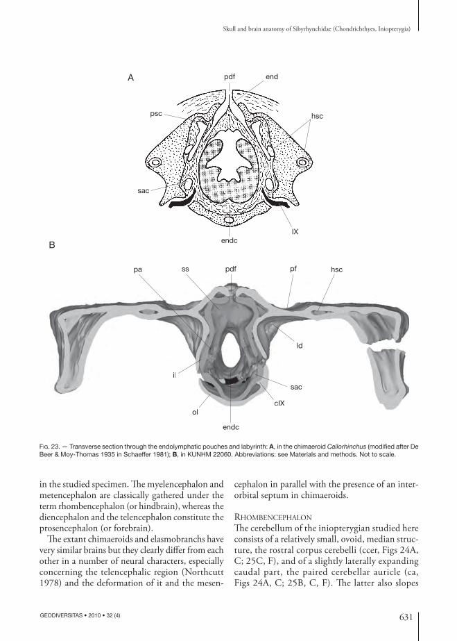
631
Skull and brain anatomy of Sibyrhynchidae (Chondrichthyes, Iniopterygia)
GEODIVERSITAS • 2010 • 32 (4)
in the studied specimen. Th e myel encephalon and metencephalon are classically gathered under the term rhombencephalon (or hindbrain), whereas the diencephalon and the telencephalon constitute the prosencephalon (or forebrain).
Th e extant chimaeroids and elasmobranchs have very similar brains but they clearly diff er from each other in a number of neural characters, especially concerning the telencephalic region (Northcutt 1978) and the deformation of it and the mesen-
cephalon in parallel with the presence of an inter-orbital septum in chimaeroids.
RHOMBENCEPHALON
Th e cerebellum of the iniopterygian studied here consists of a relatively small, ovoid, median struc-ture, the rostral corpus cerebelli (ccer, Figs 24A, C; 25C, F), and of a slightly laterally expanding caudal part, the paired cerebellar auricle (ca, Figs 24A, C; 25B, C, F). Th e latter also slopes
pa ss pdf
endcIX
hscpsc
sac
pf hsc
ld
sac
cIX
endc
ol
il
B
A
ld
sac
cIXl
il
endpdf
FIG. 23. — Transverse section through the endolymphatic pouches and labyrinth: A, in the chimaeroid Callorhinchus (modifi ed after De Beer & Moy-Thomas 1935 in Schaeffer 1981); B, in KUNHM 22060. Abbreviations: see Materials and methods. Not to scale.

632 GEODIVERSITAS • 2010 • 32 (4)
Pradel A.
backward behind the corpus cerebelli and is continuous posteriorly with the acoustico-lateral area (ala, Figs 24A, C; 25F), which is commonly covered by the crista cerebelli in extant chon-drichthyans.
Th e cerebellum appears relatively small and the anterior part of the corpus does not protrude over the optic tectum (ccer, optec, Fig. 24A), contrary to the condition in extant holocephalans (Kuh-lenbeck & Niimi 1969; Yopak et al. 2007; Ari &
II
A B
C
II
IIIIII
ic
teg
X
IV
IV
III
IIhyp
vs
teg
X
25A 25B 25C 25D 25E
optec
IV
vs
ilh
hyp
opchoptec
ccer
ccer
ca
ca
ala
ala
med
med
med
FIG. 24. — Surface rendering generated from Synchrotron Radiation holo tomographic slices of the brain of KUNHM 22060; A, dorsal view; B, ventral view; C, right lateral view; the transverse microtomographic slices of Fig. 25 are indicated. Abbreviations: see Materi-als and methods. Scale bar: 1 mm.
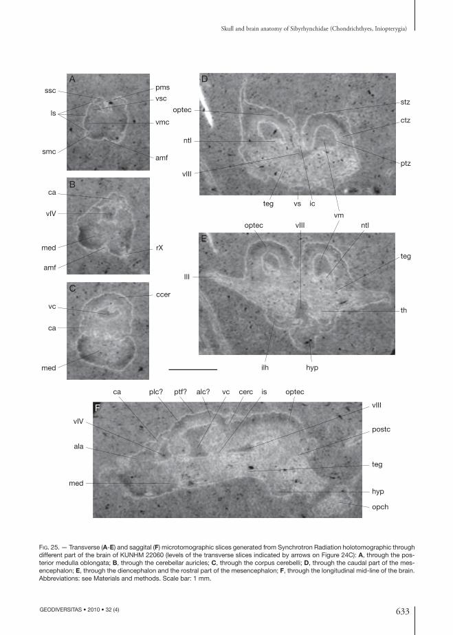
633
Skull and brain anatomy of Sibyrhynchidae (Chondrichthyes, Iniopterygia)
GEODIVERSITAS • 2010 • 32 (4)
FIG. 25. — Transverse (A-E) and saggital (F) microtomographic slices generated from Synchrotron Radiation holo tomographic through different part of the brain of KUNHM 22060 (levels of the transverse slices indicated by arrows on Figure 24C): A, through the pos-terior medulla oblongata; B, through the cerebellar auricles; C, through the corpus cerebelli; D, through the caudal part of the mes-encephalon; E, through the diencephalon and the rostral part of the mesencephalon; F, through the longitudinal mid-line of the brain. Abbreviations: see Materials and methods. Scale bar: 1 mm.
rX
vIV
med
med
amf
ca
vc
ccerC
B
ssc
ls
vsc
vmc
amf
ca
smc
pmsA
optec
ic
stz
ctz
ptz
vm
vsteg
ntl
vIII
D
optec ntlvIII
III
teg
th
hypilh
E
vIV
ala
med
ca plc? ptf? alc? vc cerc is optec
vIII
postc
teg
hyp
opch
F
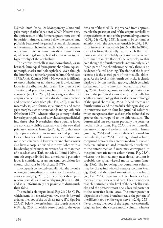
634 GEODIVERSITAS • 2010 • 32 (4)
Pradel A.
Kálmán 2008; Yopak & Montgomery 2008) and galeomorph sharks (Yopak et al. 2007). Nevertheless, the optic tectum of the former appears more ventral in position, situated almost below the cerebellum, probably because of the general ventral displacement of the mesencephalon in parallel with the presence of the interorbital septum immediately anterior to it, whereas in galeomorph sharks, this is due to the hypertrophy of the cerebellum.
Th e corpus cerebelli is non-convoluted, as in hexanchiform, squaliform, pristiophoriform, squati-nomorph sharks and holocephalans, even though the latter have a rather large cerebellum (Northcutt 1978; Ari & Kálmán 2008). However, it is diffi cult to know whether or not the corpus is divided into lobes in the sibyrhynchid brain. Th e presence of anterior and posterior pouches of the cerebellar ventricle (vc, Fig. 25C, F) may suggest that the corpus cerebelli is actually divided into anterior and posterior lobes (alc?, plc?, Fig. 25F), as in chi-maeroids, squatiniforms, squalomorphs and some galeomorphs, such as heterodontids and scyliorhinids (Northcutt 1978), whereas other galeomorph sharks have a hypertrophied and convoluted corpus divided into three lobes. Nevertheless, these putative lobes are not clearly visible externally, and the so-called primary transverse fi ssure (ptf?, Fig. 25F) that usu-ally separates the corpus in anterior and posterior lobes, is barely visible contrary to the condition in most neoselachians. However, extant chimaeroids also have a corpus divided into two lobes with a less developed primary transverse fi ssure than that of neoselachians (Kuhlenbeck & Niimi 1969). A smooth corpus divided into anterior and posterior lobes is considered as an ancestral condition for chondrichthyans by Northcutt (1978).
Th e corpus is connected ventrally with the medulla oblongata immediately anterior to the cerebellar auricles (med, Fig. 25C, F). Th e auricles also appear relatively small, as in osteichthyans (Bertin 1958), and it is unfortunately not possible to distinguish their folds.
Th e medulla oblongata (med, Figs 24; 25A-C, F), which seems to be relatively narrow, extends forward as far as the root of the trochlear nerve (IV, Figs 24; 26A-D) below the cerebellum. Th e fourth ventricle (vIV, Fig. 25B, F), which corresponds to the open
division of the medulla, is preserved from approxi-mately the posterior end of the corpus cerebelli to the posteriormost root of the presumed vagus nerve (X, Fig. 24; rX, Fig. 25B). It seems to be continuous anteriorly with the cerebellar ventricle (vc, Fig. 25C, F), as in extant chimaeroids (Ari & Kálmán 2008). Its roof is formed rostrally by the cerebellum and more caudally by probably a choroid tissue, which is thinner than the fl oor of the ventricle, so that even though the fourth ventricle is commonly called “open part of the rhombencephalon”, there is no evidence of a real opening. Th e fl oor of the fourth ventricle is the closed part of the medulla oblon-gata. At the level of the fourth ventricle, it clearly displays only one median groove, which certainly corresponds to the anterior median fi ssure (amf, Fig. 25B). However, posterior to the posteriormost root of the presumed vagus nerve, the medulla has a diff erent morpho logy that recalls the morpho logy of the spinal chord (Fig. 25A). Indeed, there is no fourth ventricle and the medulla oblongata displays in transverse section four longitudinal columns, which are separated from each other by rather shallow grooves that correspond to the diff erent sulci. Th e dorsomedial one represents probably the posterior median sulcus (pms, Fig. 25A), the ventromedial one may correspond to the anterior median fi ssure (amf, Fig. 25A) and there are three additional lat-eral sulci (ls, Fig. 25A). Th e longitudinal column comprised between the anterior median fi ssure and the lateral sulcus situated immediately dorsolateral to the anteriomedian fi ssure may correspond to the spinal somatic motor column (smc, Fig. 25A), whereas the immediately next dorsal column is probably the spinal visceral motor column (vmc, Fig. 25A). Th e following two dorsalmost column may be the spinal visceral sensory column (vsc, Fig. 25A) and the spinal somatic sensory column (ssc, Fig. 25A), respectively. Th ree branches leave the brainstem in its ventral part. Th e anteriormost branch is situated at the level of the cerebellar auri-cles and the posteriormost one is located posterior to the acoustico-lateral area. Th e anteroposterior disposition of these branches recalls the pattern of the diff erent roots of the vagus nerve (rX, Fig. 25B). Nevertheless, the roots of the vagus nerve normally originate from the visceral motor column, whereas
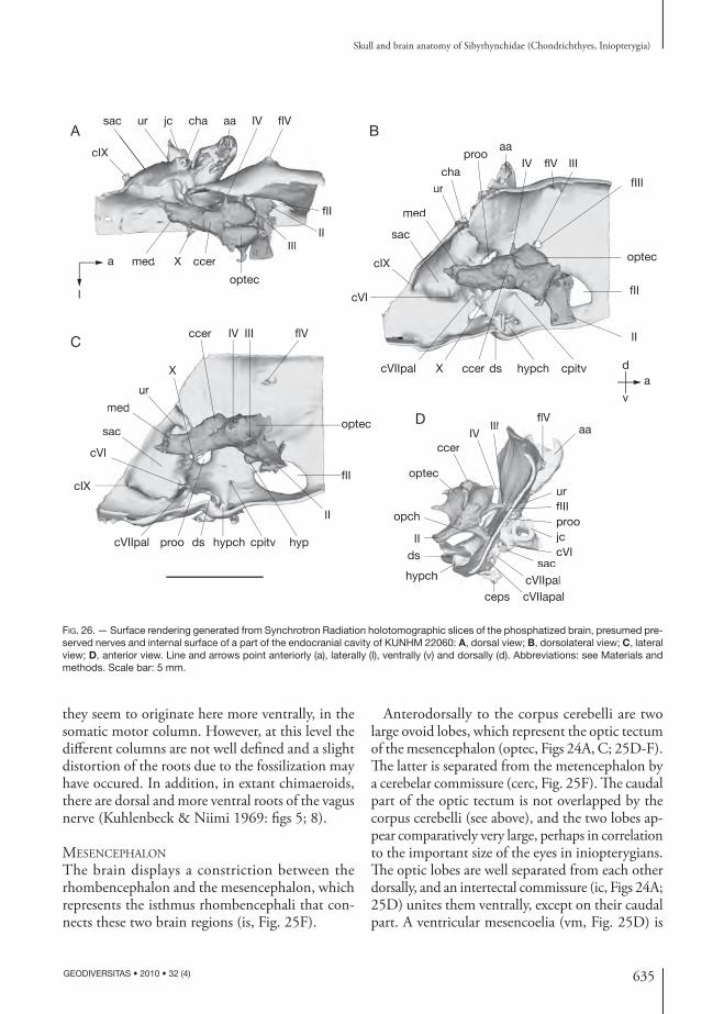
635
Skull and brain anatomy of Sibyrhynchidae (Chondrichthyes, Iniopterygia)
GEODIVERSITAS • 2010 • 32 (4)
they seem to originate here more ventrally, in the somatic motor column. However, at this level the diff erent columns are not well defi ned and a slight distortion of the roots due to the fossilization may have occured. In addition, in extant chimaeroids, there are dorsal and more ventral roots of the vagus nerve (Kuhlenbeck & Niimi 1969: fi gs 5; 8).
MESENCEPHALON
The brain displays a constriction between the rhombencephalon and the mesencephalon, which represents the isthmus rhombencephali that con-nects these two brain regions (is, Fig. 25F).
Anterodorsally to the corpus cerebelli are two large ovoid lobes, which represent the optic tectum of the mesencephalon (optec, Figs 24A, C; 25D-F). Th e latter is separated from the metencephalon by a cerebelar commissure (cerc, Fig. 25F). Th e caudal part of the optic tectum is not overlapped by the corpus cerebelli (see above), and the two lobes ap-pear comparatively very large, perhaps in correlation to the important size of the eyes in inio pterygians. Th e optic lobes are well separated from each other dorsally, and an intertectal commissure (ic, Figs 24A; 25D) unites them ventrally, except on their caudal part. A ventricular mesencoelia (vm, Fig. 25D) is
med
sac
urcha
prooaa
IV flV III
fIII
optec
fII
II
cpitvhypchdsccerXcVIIpal
cVI
cIX
med
sac
urcha
IV flV III
IX
sac ur jc cha aa IV flV
fII
IIIII
optec
ccerXmed
cIX
f
IIIII
ccerXmed
cIX
a
l
A
C
B
med
sac
cVI
cIX
cVIIpal proo ds hypch cpitv hyp
II
ur
X
ccer IV III flV
optec
fII
med
sac
cVI
ur
X
optec
ccerIV
IIIflV
aa
urfIIIproojccVI
saccVIIpalcVIIapalceps
opch
hypch
dsII
ec
ccerIV
III
ufIpjccV
saccVIIpalch
D
av
d
FIG. 26. — Surface rendering generated from Synchrotron Radiation holo tomographic slices of the phosphatized brain, presumed pre-served nerves and internal surface of a part of the endo cranial cavity of KUNHM 22060: A, dorsal view; B, dorsolateral view; C, lateral view; D, anterior view. Line and arrows point anteriorly (a), laterally (l), ventrally (v) and dorsally (d). Abbreviations: see Materials and methods. Scale bar: 5 mm.

636 GEODIVERSITAS • 2010 • 32 (4)
Pradel A.
present inside each lobe and seems to be continuous with the cerebellar ventricle and the third ventricle (vIII, Fig. 25D-F). Moreover, it is open dorsally and communicates with the surrounding endo cranial cavity immediately anterior to the corpus, at the caudal extremity of the mesencephalon (Fig. 24A). Such a condition is unknown among extant chon-drichthyans.
Th e optic tectum of Recent chondrichthyans consists of multiple laminae, which Northcutt (1978) grouped into three zones: periventricular, central and superfi cial. Th ere is considerable varia-tion concerning the histology of the diff erent zones between extant holocephalans and elasmobranchs, especially concerning their relative concentration of cells (Northcutt 1978).
Unfortunately, these zones are not clearly well defi ned in the material studied here, and the dif-ferent contrasts observed in the tectum must be interpreted with great reservations. Th ey could represent indication of fossilized laminae or merely a diagenetic bias. Nevertheless, I suggest here a possible correspondance between these laminae and the zones observed in the fossilized brain (ptz, ctz, stz, Fig. 25D). Unfortunately no compara-tive description of the histology within the extant elasmobranchs and holocephalans is possible. At the level of the caudal part, the lateral wall of the optic tectum displays a lateral projection, which corresponds in position to the root of the trochlear nerve (III, Figs 24; 25E). At mid-length of the mes-encephalon, a dorsally-oriented projection emerges from the dorsal part of the optic tectum (Fig. 25D). Th is position does not correspond to that of any cranial nerve in known brain fi shes. Nevertheless, it could be either a blood vessel or an artefact. Th e rostral part of the mesencephalon shows a rather deep median infolding of the optic tectum (optec, Fig. 25E), as seen in extant chimaeroids.
Th e midbrain fl oor, or tegmentum (teg, Figs 24B, C; 25D-F), is continuous posteriorly with the medulla oblongata. Its external and internal caudal morpho-logy appears regular as far as its mid-region, where the root of oculomotor nerve emerges laterally. At this level, a pair of bulges lays the fl oor of the paired ventricle (ntl, Fig. 25D, E). Th ese may represent the location of the nucleus tegmentalis lateralis, present
at the same position in extant chimaeroids (Ari & Kálmán 2008). Th e rostral end of the tegmentum is prolonged ventrally by the diencephalon.
PROSENCEPHALON
Th e diencephalon is situated below the rostral part of the mesencephalon, posterior to the optic chiasm (opch, Figs 24B; 25F). It is separated from the mesencephalon by the posterior commissure (postc, Fig. 25F).
Th e epithalamus, which corresponds to its dor-sal part, and the thalamus, which forms its lateral walls, are not clearly distinct from each other, but its fl oor is well preserved. Th e thalamus (th, Fig. 25E) seems to be continuous caudally with the tegmen-tum (teg, Fig. 25E) of the mid-brain, as in extant chondrichthyans.
Th e fl oor of the diencephalon comprises, from front to rear, the optic chiasm (opch, Figs 24B; 25F), the inferior lobes of the hypothalamus (ilh, Figs 24B; 25E), the infundibulum containing the hypophysis (hyp, Figs 24B, C; 25E) and the vascular sac (vs, Figs 24B, C; 25D). Th e central third vesicle (vIII, Figs 25E, F) is also well defi ned, fl anked by the inferior lobe of the hypothalamus, and fl oored by the hypophysis. Th e vesicle of each inferior lobe of the hypothalamus is therefore continuous with the third vesicle at the level of the anterior end of the hypophysis.
Th e inferior lobes of the hypothalamus have ex-tensive lateral recesses, as in chimaeroids, sharks and primitive batoids (Northcutt 1978). Between them is a bulb that most probably represents the hypo-physis. Posterior to the hypophysis is a small median process, which may correspond to the vascular sac. Anterior to the inferior lobes of the hypothalamus and the hypophysis is the optic chiasm, which il-lustrates the decussation of very large optic nerves that are a diencephalic derivative.
Th e telencephalon is unfortunately missing in the sibyrhynchid specimen that displays the fossil-ized brain. Nevertheless, the optic nerve is situated very far from the olfactory canals of the endo cranial cavity (olfc, Figs 27; 29; 30), which are assumed to house the olfactory tracts. Th is suggests a very long telencephalon, similar to that of Recent chi-maeroids.
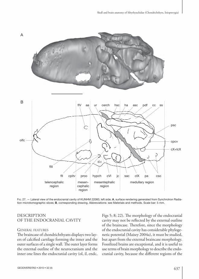
637
Skull and brain anatomy of Sibyrhynchidae (Chondrichthyes, Iniopterygia)
GEODIVERSITAS • 2010 • 32 (4)
DESCRIPTION OF THE ENDO CRANIAL CAVITY
GENERAL FEATURES
Th e braincase of chondrichthyans displays two lay-ers of calcifi ed cartilage forming the inner and the outer surfaces of a single wall. Th e outer layer forms the external outline of the neuro cranium and the inner one lines the endo cranial cavity (ol, il, endc,
Figs 5; 8; 22). Th e morpho logy of the endo cranial cavity may not be refl ected by the external outline of the braincase. Th erefore, since the morpho logy of the endo cranial cavity has considerable phyloge-netic potential (Maisey 2004a), it must be studied, but apart from the external braincase morpho logy. Fossilized brains are exceptional, and it is useful to use terms of brain morpho logy to describe the endo-cranial cavity, because the diff erent regions of the
olfc
fIV aa ur cerch hsc ha asc pdf cc ss
psc
cpcv
cX+IcX
csopacIXsacjccVIhypch
d.r.
proocpitv
fIII
fII
mesentephalicregion
mesen-cephalicregion
B
A
medullary regiontelencephalicregion
FIG. 27. — Lateral view of the endo cranial cavity of KUNHM 22060, left side; A, surface rendering generated from Synchrotron Radia-tion microtomographic slices; B, corresponding drawing. Abbreviations: see Materials and methods. Scale bar: 5 mm.
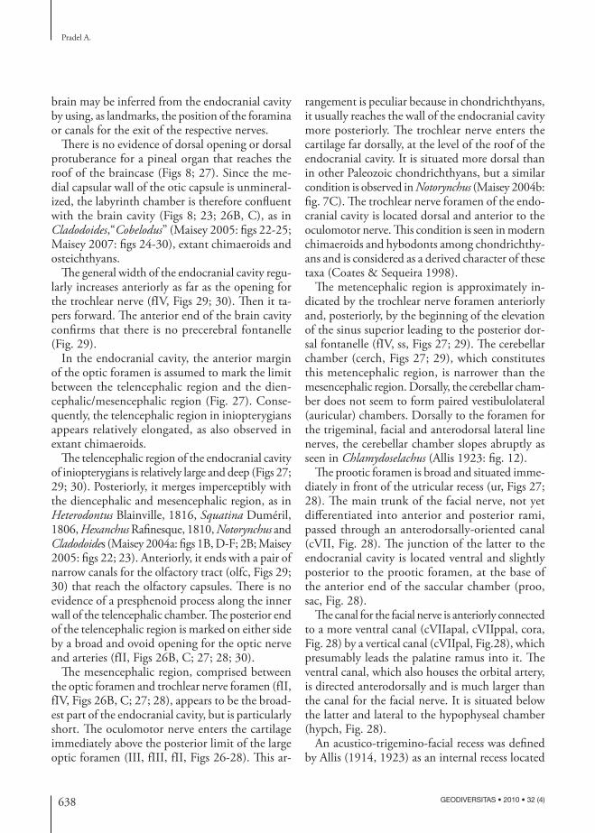
638 GEODIVERSITAS • 2010 • 32 (4)
Pradel A.
brain may be inferred from the endo cranial cavity by using, as landmarks, the position of the foramina or canals for the exit of the respective nerves.
Th ere is no evidence of dorsal opening or dorsal protuberance for a pineal organ that reaches the roof of the braincase (Figs 8; 27). Since the me-dial capsular wall of the otic capsule is unmineral-ized, the labyrinth chamber is therefore confl uent with the brain cavity (Figs 8; 23; 26B, C), as in Cladodoides,“Cobelodus” (Maisey 2005: fi gs 22-25; Maisey 2007: fi gs 24-30), extant chimaeroids and osteichthyans.
Th e general width of the endo cranial cavity regu-larly increases anteriorly as far as the opening for the trochlear nerve (fIV, Figs 29; 30). Th en it ta-pers forward. Th e anterior end of the brain cavity confi rms that there is no precerebral fontanelle (Fig. 29).
In the endo cranial cavity, the anterior margin of the optic foramen is assumed to mark the limit between the telencephalic region and the dien-cephalic/mesencephalic region (Fig. 27). Conse-quently, the telencephalic region in inio pterygians appears relatively elongated, as also observed in extant chimaeroids.
Th e telencephalic region of the endocranial cavity of inio pterygians is relatively large and deep (Figs 27; 29; 30). Posteriorly, it merges imperceptibly with the diencephalic and mesencephalic region, as in Heterodontus Blainville, 1816, Squatina Duméril, 1806, Hexanchus Rafi nesque, 1810, Notorynchus and Cladodoides (Maisey 2004a: fi gs 1B, D-F; 2B; Maisey 2005: fi gs 22; 23). Anteriorly, it ends with a pair of narrow canals for the olfactory tract (olfc, Figs 29; 30) that reach the olfactory capsules. Th ere is no evidence of a presphenoid process along the inner wall of the telencephalic chamber. Th e posterior end of the telencephalic region is marked on either side by a broad and ovoid opening for the optic nerve and arteries (fII, Figs 26B, C; 27; 28; 30).
Th e mesencephalic region, comprised between the optic foramen and trochlear nerve foramen (fII, fIV, Figs 26B, C; 27; 28), appears to be the broad-est part of the endo cranial cavity, but is particularly short. Th e oculomotor nerve enters the cartilage immediately above the posterior limit of the large optic foramen (III, fIII, fII, Figs 26-28). Th is ar-
rangement is peculiar because in chondrichthyans, it usually reaches the wall of the endo cranial cavity more posteriorly. Th e trochlear nerve enters the cartilage far dorsally, at the level of the roof of the endo cranial cavity. It is situated more dorsal than in other Paleozoic chondrichthyans, but a similar condition is observed in Notorynchus (Maisey 2004b: fi g. 7C). Th e trochlear nerve foramen of the endo-cranial cavity is located dorsal and anterior to the oculomotor nerve. Th is condition is seen in modern chimaeroids and hybodonts among chondrichthy-ans and is considered as a derived character of these taxa (Coates & Sequeira 1998).
Th e metencephalic region is approximately in-dicated by the trochlear nerve foramen anteriorly and, posteriorly, by the beginning of the elevation of the sinus superior leading to the posterior dor-sal fontanelle (fIV, ss, Figs 27; 29). Th e cerebellar chamber (cerch, Figs 27; 29), which constitutes this metencephalic region, is narrower than the mesencephalic region. Dorsally, the cerebellar cham-ber does not seem to form paired vestibulolateral (auricular) chambers. Dorsally to the foramen for the trigeminal, facial and anterodorsal lateral line nerves, the cerebellar chamber slopes abruptly as seen in Chlamydoselachus (Allis 1923: fi g. 12).
Th e prootic foramen is broad and situated imme-diately in front of the utricular recess (ur, Figs 27; 28). Th e main trunk of the facial nerve, not yet diff erentiated into anterior and posterior rami, passed through an anterodorsally-oriented canal (cVII, Fig. 28). Th e junction of the latter to the endo cranial cavity is located ventral and slightly posterior to the prootic foramen, at the base of the anterior end of the saccular chamber (proo, sac, Fig. 28).
Th e canal for the facial nerve is anteriorly connected to a more ventral canal (cVIIapal, cVIIppal, cora, Fig. 28) by a vertical canal (cVIIpal, Fig.28), which presumably leads the palatine ramus into it. Th e ventral canal, which also houses the orbital artery, is directed anterodorsally and is much larger than the canal for the facial nerve. It is situated below the latter and lateral to the hypophyseal chamber (hypch, Fig. 28).
An acustico-trigemino-facial recess was defi ned by Allis (1914, 1923) as an internal recess located
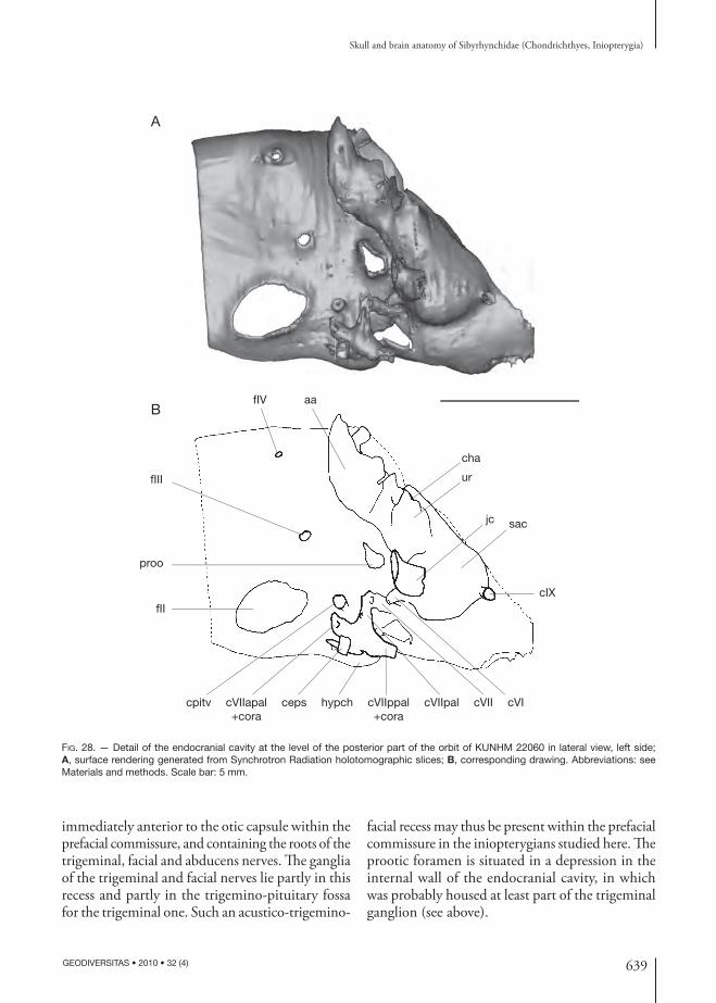
639
Skull and brain anatomy of Sibyrhynchidae (Chondrichthyes, Iniopterygia)
GEODIVERSITAS • 2010 • 32 (4)
immediately anterior to the otic capsule within the prefacial commissure, and containing the roots of the trigeminal, facial and abducens nerves. Th e ganglia of the trigeminal and facial nerves lie partly in this recess and partly in the trigemino-pituitary fossa for the trigeminal one. Such an acustico-trigemino-
facial recess may thus be present within the prefacial commissure in the inio pterygians studied here. Th e prootic foramen is situated in a depression in the internal wall of the endo cranial cavity, in which was probably housed at least part of the trigeminal ganglion (see above).
fIII
fIV aa
cha
ur
sac
cIX
jc
cVIcVIIcVIIpalcVIIppal+cora
hypchcepscVIIapal+cora
cpitv
fII
proo
B
A
FIG. 28. — Detail of the endo cranial cavity at the level of the posterior part of the orbit of KUNHM 22060 in lateral view, left side; A, surface rendering generated from Synchrotron Radiation holo tomographic slices; B, corresponding drawing. Abbreviations: see Materials and methods. Scale bar: 5 mm.
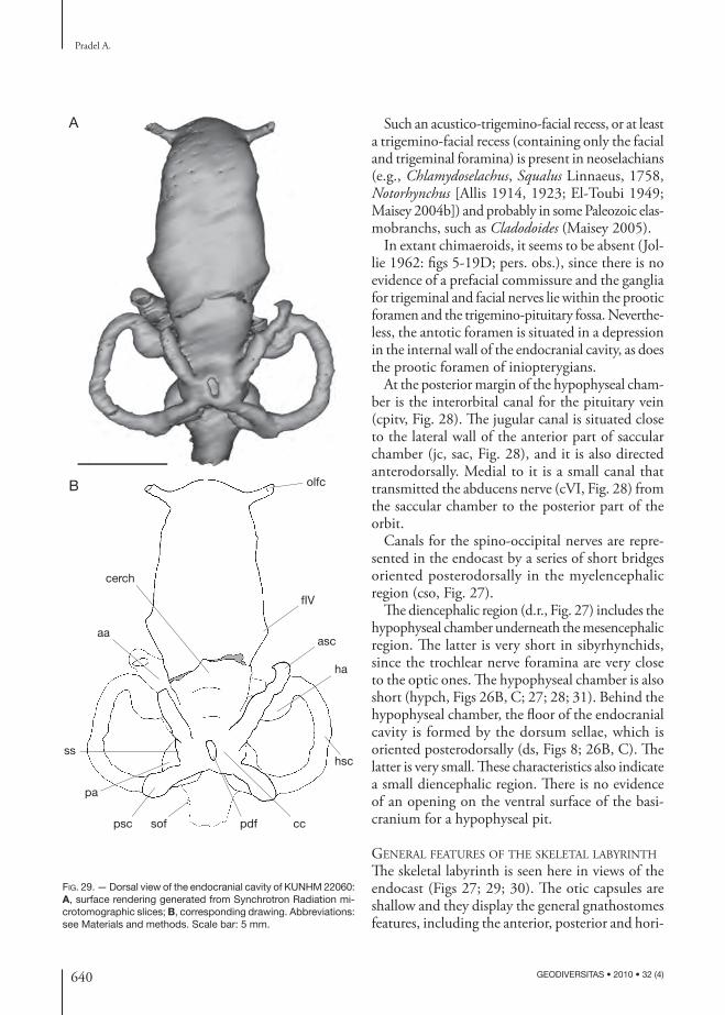
640 GEODIVERSITAS • 2010 • 32 (4)
Pradel A.
Such an acustico-trigemino-facial recess, or at least a trigemino-facial recess (containing only the facial and trigeminal foramina) is present in neoselachians (e.g., Chlamydoselachus, Squalus Linnaeus, 1758, Notorhynchus [Allis 1914, 1923; El-Toubi 1949; Maisey 2004b]) and probably in some Paleozoic elas-mobranchs, such as Cladodoides (Maisey 2005).
In extant chimaeroids, it seems to be absent (Jol-lie 1962: fi gs 5-19D; pers. obs.), since there is no evidence of a prefacial commissure and the ganglia for trigeminal and facial nerves lie within the prootic foramen and the trigemino-pituitary fossa. Neverthe-less, the antotic foramen is situated in a depression in the internal wall of the endo cranial cavity, as does the prootic foramen of inio pterygians.
At the posterior margin of the hypophyseal cham-ber is the interorbital canal for the pituitary vein (cpitv, Fig. 28). Th e jugular canal is situated close to the lateral wall of the anterior part of saccular chamber (jc, sac, Fig. 28), and it is also directed anterodorsally. Medial to it is a small canal that transmitted the abducens nerve (cVI, Fig. 28) from the saccular chamber to the posterior part of the orbit.
Canals for the spino-occipital nerves are repre-sented in the endocast by a series of short bridges oriented posterodorsally in the myelencephalic region (cso, Fig. 27).
Th e diencephalic region (d.r., Fig. 27) includes the hypophyseal chamber underneath the mesencephalic region. Th e latter is very short in sibyrhynchids, since the trochlear nerve foramina are very close to the optic ones. Th e hypophyseal chamber is also short (hypch, Figs 26B, C; 27; 28; 31). Behind the hypophyseal chamber, the fl oor of the endo cranial cavity is formed by the dorsum sellae, which is oriented posterodorsally (ds, Figs 8; 26B, C). Th e latter is very small. Th ese characteristics also indicate a small diencephalic region. Th ere is no evidence of an opening on the ventral surface of the basi-cranium for a hypophyseal pit.
GENERAL FEATURES OF THE SKELETAL LABYRINTH
Th e skeletal labyrinth is seen here in views of the endocast (Figs 27; 29; 30). Th e otic capsules are shallow and they display the general gnathostomes features, including the anterior, posterior and hori-
cerch
aa
ss
pa
psc sof pdf cc
hsc
ha
asc
fIV
olfc
A
B
FIG. 29. — Dorsal view of the endo cranial cavity of KUNHM 22060: A, surface rendering generated from Synchrotron Radiation mi-crotomographic slices; B, corresponding drawing. Abbreviations: see Materials and methods. Scale bar: 5 mm.

641
Skull and brain anatomy of Sibyrhynchidae (Chondrichthyes, Iniopterygia)
GEODIVERSITAS • 2010 • 32 (4)
zontal semicircular canals with their respective ampullae, and the vestibular chamber ventrally. Behind the cerebellar chamber and between the otic capsules, the crus commune rises and merges dorsally with the posterior dorsal fontanelle (cc, pdf, Figs 27; 29). Th is has been considered a primitive gnathostome character.
Th e endo cranial cavity appears more constricted in this region below the sinus superior.
Th e vestibular chamber is almost entirely confl u-ent with the cranial cavity (sac, ur, Figs 8; 23; 26B, C). Th e medial capsular wall is unchondrifi ed as in extant chimaeroids and most of Paleozoic chon-drichthyans. Th e vestibular chamber is subdivided into two regions. Th e dorsal region is the utricular chamber (ur, Figs 27; 28; 30) and the ventral one is the saccular chamber (sac, Figs 27; 28; 30). Th ere is no evidence of a prominent lagenar chamber. Th e utricular and saccular chambers are relatively small and meet medially. Th e utricular recess is represented by a low bulge that is constricted at the level of its junction to the dorsal anterior ampulla and the ventral saccular chamber. Th e anterior ampulla (aa, Figs 27; 28) is connected to the anterodorsal part of the utricular recess, while the horizontal ampulla (ha, Fig. 27) seems to have a separate ventral pro-jection (cha, Fig. 28) connecting the posterodorsal part of the utricular recess. Unfortunately there is a crack just at this level.
Since there is no chondrifi ed medial wall, the octaval nerve passes presumably directly into the utricular recess.
Th e horizontal semicircular canal (hsc, Figs 27; 29) lies just above the deep depressions housing the gills, and lies along the dorsal part of the post orbital wall. Th e anterior (asc, Figs 27; 29) and posterior (psc, Figs 27; 29) semicircular canals meet dorsally at a crus commune (cc, Figs 27; 29), below which is a sinus superior (ss, Figs 27; 29) connecting the crus commune and the saccular chamber. Th e posterior ampulla (pa, Figs 8; 27; 29) is confl uent with the saccular chamber and there is no evidence of a preampullary canal (sensu Maisey 2001b). Th e horizontal canal is strongly curved. It enters the sinus superior just below the crus commune after meeting the posterior canal. It is unclear if the horizontal canal passes inside the loop of the pos-
A
B
fIIfIII
hypch
asc
ha
hsc
psc fpcv sof fX+fIcX
cIX
sac
jc
ur
prooaa
cVI
cpitv
fIV
olfc
FIG. 30. — Ventral view of the endo cranial cavity of KUNHM 22060: A, surface rendering generated from Synchrotron Radiation mi-crotomographic slices; B, corresponding drawing. Abbreviations: see Materials and methods. Scale bar: 5 mm.
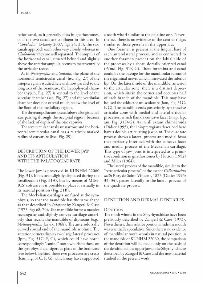
642 GEODIVERSITAS • 2010 • 32 (4)
Pradel A.
a tooth whorl similar to the palatine one. Never-theless, there is no evidence of the central ridges similar to those present in the upper jaw.
One foramen is present at the lingual base of each anterolateral process, and is connected to another foramen present on the labial side of the processes by a short, dorsally oriented canal (fVmd, Fig. 31F, G). Th ese foramina and canal could be the passage for the mandibular ramus of the trigeminal nerve, which innervated the inferior lip. On the lateral side of the mandible, anterior to the articular zone, there is a distinct depres-sion, which sits in the corner and occupies half of each branch of the mandible. Th is may have housed the adductor musculature (fam, Fig. 31C, F, G). Th e mandible ends posteriorly by a massive articular zone with medial and lateral articular processes, which fl ank a concave facet (map, lap, am, Fig. 31D-G). As in all extant chimaeroids (Didier 1995), the inio pterygians described here have a double-articulating jaw joint. Th e quadrate process shows a lateral process and medial fossa that perfectly interlock with the concave facet and medial process of the Meckelian cartilage. Th is type of jaw joint is interpreted as a primi-tive condition in gnathostomes by Hotton (1952) and Miles (1964).
Th e lateral process of the mandible, similar to the “retroarticular process” of the extant Callorhinchus milii Bory de Saint-Vincent, 1823 (Didier 1995: 33, 34), passes laterally to the lateral process of the quadrate process.
DENTITION AND DERMAL DENTICLES
DENTITION
Th e tooth whorls in the Sibyrhynchidae have been previously described by Zangerl & Case (1973). Nevertheless, their relative position inside the mouth was essentially speculative. Since there is no evidence of mandibular tooth whorls in natural position in the mandible of KUNHM 22060, the comparison of the dentition will be made only on the basis of the dentition of the upper jaw of the Sibyrhynchidae described by Zangerl & Case and the new material studied in the present work.
terior canal, as it generally does in gnathostomes, or if the two canals are confl uent in this area. In “Cobelodus” (Maisey 2007: fi gs 24; 25), the two canals approach each other very closely, whereas in Cladodoides they are well separated. Th e ampulla of the horizontal canal, situated behind and slightly above the anterior ampulla, seems to meet ventrally the utricular recess.
As in Notorynchus and Squalus, the plane of the horizontal semicircular canal (hsc, Fig. 27) of the inio pterygians studied here is almost parallel to the long axis of the braincase, the hypophyseal cham-ber (hypch, Fig. 27) is ventral to the level of the saccular chamber (sac, Fig. 27) and the vestibular chamber does not extend much below the level of the fl oor of the medullary region.
Th e three ampullae are located above a longitudinal axis passing through the occipital region, because of the lack of depth of the otic capsules.
Th e semicircular canals are narrow, and the hori-zontal semicircular canal has a relatively marked radius of curvature (hsc, Fig. 29).
DESCRIPTION OF THE LOWER JAW AND ITS ARTICULATION WITH THE PALATOQUADRATE
Th e lower jaw is preserved in KUNHM 22060 (Fig. 31). It has been slightly displaced during the fossilization (Fig. 31A), but by means of MIM-ICS® software it is possible to place it virtually in its natural position (Fig. 31B).
Th e Meckelian cartilages are fused at the sym-physis, so that the mandible has the same shape as that described in Iniopera by Zangerl & Case (1973: fi gs 68; 70). Th e mandible forms a massive rectangular and slightly convex cartilage anteri-orly that recalls the mandible of dipnoans (e.g., Melanognathus Jarvik, 1968). Th e anterodorsally curved rostral end of the mandible is blunt. Th e anterior corners display two large lateral processes (lpm, Fig. 31C, F, G), which could have borne correspondingly “canine” tooth whorls to those on the symphysial dentigerous plate of the braincase (see below). Behind these two processes are crests (lcm, Fig. 31C, F, G), which may have supported

643
Skull and brain anatomy of Sibyrhynchidae (Chondrichthyes, Iniopterygia)
GEODIVERSITAS • 2010 • 32 (4)
lap
lap
mapam
map
fam
map am
lpm
fVmd
fVmd
fam
lcm
lpmlcm
A B
E
G
F
A B
lpm lcm
lap
map am
fam
C D
FIG. 31. — Surface rendering generated from Synchrotron Radiation microtomographic slices of braincase and lower jaw of KUNHM 22060: A, relative position of the lower jaw as found inside the nodule; B, lower jaw virtually placed to its supposed natural position; C-G, lower jaw in anterior (C), posterior (D), ventral (E), dorsal (F), and right lateral (G) views. Abbreviations: see Materials and meth-ods. Scale bar: 5 mm.
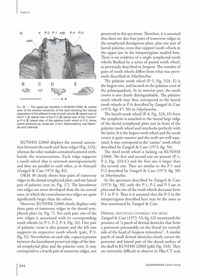
644 GEODIVERSITAS • 2010 • 32 (4)
Pradel A.
KUNHM 22060 displays the natural associa-tion between the teeth and these ridges (Fig. 32A), whereas the other nodules contained scattered teeth, beside the neuro cranium. Each ridge supports a tooth whorl that is oriented anteroposteriorly and they are parallel to each other, as in Inioxyele (Zangerl & Case 1973: fi g. 82).
OKM 38 clearly shows four pairs of transverse ridges in the dental symphysial plate, and one lateral pair of palatine crest (tr, Fig. 17). Th e lateralmost two ridges are more developed than the six central ones, in which the centralmost two ridges are quite signifi cantly larger than the others.
However, KUNHM 22060 clearly displays only three pairs of transverse ridges in the dental sym-physial plate (tr, Fig. 7). For each pair, one of the two ridges is associated with its corresponding tooth whorls (tr, P-1, P-2, P-4, Fig. 32). One pair of palatine crests is also present and the left one supports its respective tooth whorls (palc, P-5, Fig. 32). Nevertheless, on each side, a space is present between the lateralmost preserved ridge of the den-tal symphysial plate and the palatine crest. It may correspond to a fourth pair of transverse ridges, not
preserved in this specimen. Th erefore, it is assumed that there are also four pairs of transverse ridges in the symphysial dentigerous plate, plus one pair of lateral palatine crests that support tooth whorls in the upper jaw in the inio pterygians studied here. Th ere is no evidence of a single symphysial tooth whorls fl anked by a series of paired tooth whorl, as previously described in Iniopera. Th e number of pairs of tooth whorls diff ers from what was previ-ously described in Sibyrhynchus.
Th e palatine tooth whorl (P-5, Fig. 32A, E) is the largest one, and located on the palatine crest of the palatoquadrate. In its anterior part, the tooth crown is not clearly distinguishable. Th e palatine tooth whorls may thus correspond to the lateral tooth whorls or P-6 described by Zangerl & Case (1973: fi gs 47; 50) in Sibyrhynchus.
Th e fourth tooth whorl (P-4, Fig. 32A, D) from the symphysis is attached to the lateral large ridge of the dental symphysial plate just in front of the palatine tooth whorl and interlocks perfectly with the latter. It is the largest tooth whorl and the tooth crown is quite massive and the teeth are well sepa-rated. It may correspond to the “canine” tooth whorl described by Zangerl & Case (1973: fi g. 50).
Th e third tooth whorl is lacking on KUNHM 22060. Th e fi rst and second one are present (P-1, P-2, Fig. 32A-C) and the fi rst one is larger than the second one. Th ey are similar to the P-1 and P-2 described by Zangerl & Case (1973: fi g. 50) in Sibyrhynchus.
In the specimen described by Zangerl & Case (1973: fi g. 50), only the P-1, P-2 and P-3 are in place and the size of the tooth whorls decreases from P-1 to P-3. Th us it is assumed that the P-3 in the inio pterygians described here may be the same as that mentioned by Zangerl & Case.
DERMAL DENTICLES COVERING THE HEAD
Zangerl & Case (1973: 53; fi g. 63) mentioned the presence of “a patch of dermal denticles that form a pavement presumably on the dorsal (or ventral) side of the head of Iniopera richardsoni”. A similar patch of small dermal denticles clearly covers the posterior and lateral part of the dorsal surface of the skull in KUNHM 22060 (pdd, Fig. 33A). Th ey are extremely diffi cult to observe in SRμ-CT scan
A
B C D E
palc
tr P-2 P-1 sdp P-4 P-5
a
FIG. 32. — The upper jaw dentition in KUNHM 22060: A, ventral view of the anterior extremity of the skull showing the natural disposition of the different kinds of tooth whorls; B, lateral view of the P-1; C, lateral view of the P-2; D, lateral view of the “canine” or P-4; E, lateral view of the palatine tooth whorl or P-5. Arrow points anteriorly (a). Scale bar: 5 mm. Abbreviations: see Materi-als and methods.
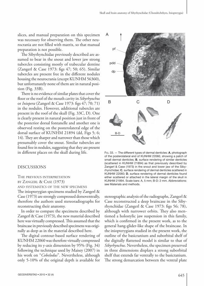
645
Skull and brain anatomy of Sibyrhynchidae (Chondrichthyes, Iniopterygia)
GEODIVERSITAS • 2010 • 32 (4)
slices, and manual preparation on this specimen was necessary for observing them. Th e other neu-rocrania are not fi lled with matrix, so that manual preparation is not possible.
Th e Sibyrhynchidae previously described are as-sumed to bear in the snout and lower jaw strong tubercles consisting mostly of trabecular dentine (Zangerl & Case 1973: fi gs 47; 50; 65). Similar tubercles are present free in the diff erent nodules housing the neurocrania (except KUNHM 56360), but unfortunately none of them are in natural posi-tion (Fig. 33B).
Th ere is no evidence of similar plates that cover the fl oor or the roof of the mouth cavity in Sibyrhynchus or Iniopera (Zangerl & Case 1973: fi gs 47; 70; 71) in the nodules. However, additional tubercles are present in the roof of the skull (Fig. 33C, D). One is clearly present in natural position just in front of the posterior dorsal fontanelle and another one is observed resting on the posterolateral edge of the dorsal surface of KUNHM 21894 (dd, Figs 5; 6; 14). Th ey are sharper and narrower than those which presumably cover the snout. Similar tubercles are found free in nodules, suggesting that they are present in diff erent places on the skull during life.
DISCUSSIONS
THE PREVIOUS INTERPRETATION BY ZANGERL & CASE (1973) AND SYSTEMATICS OF THE NEW SPECIMENS
Th e iniopterygian specimens studied by Zangerl & Case (1973) are strongly compressed dorsoventrally, therefore the authors used stereoradiographs for reconstructing their anatomy.
In order to compare the specimens described by Zangerl & Case (1973), the new material described here was virtually compressed. Th is assumed that the braincase in previously described specimens was origi-nally as deep as in the material described here.
Th e digital contour-based surface rendering of KUNHM 22060 was therefore virtually compressed by reducing its y-axis dimension by 95% (Fig. 34) following the technique used by Maisey (2007) in his work on “Cobelodus”. Nevertheless, although only 5-10% of the original depth is available for
stereographic analysis of the radiographs, Zangerl & Case reconstructed a deep braincase in the Siby-rhynchidae (Zangerl & Case 1973: fi gs 56; 78), although with narrower orbits. Th ey also men-tioned a holostylic jaw suspension in this family, which is confi rmed in the present work, as to the general hang-glider-like shape of the braincase. In the inio pterygians studied in the present work, the outline of the basi cranium and suborbital shelf of the digitally fl attened model is similar to that of Sibyrhynchus. Nevertheless, the specimen preserved in three dimensions displays a strong suborbital shelf that extends far ventrally to the basi cranium. Th e strong demarcation between the ventral plate
FIG. 33. — The different types of dermal denticles: A, photograph of the posterolateral end of KUNHM 22060, showing a patch of small dermal denticles; B, surface rendering of similar denticles (scattered in KUNHM 21894) as that previously described by Zangerl & Case (1973) in the snout and lower jaw of the Siby-rhynchidae; C, surface rendering of dermal denticles scattered in KUNHM 22060; D, surface rendering of dermal denticles found either scattered or attached in the lateral margin of the skull in KUNHM 21894. Scale bars: A, 5 mm; B-D: 2 mm. Abbreviations: see Materials and methods.
A
B
B-D
C D
pf
pdd
dp

646 GEODIVERSITAS • 2010 • 32 (4)
Pradel A.
MATCH BETWEEN THE MORPHO LOGY OF THE ENDO CRANIAL CAVITY AND THE BRAIN
For a long time, the brain morpho logy in fossils was inferred from the shape of the endo cranial cavity. Actually, only few works dealed with a match between the morpho logy of the brain and that of the cavity that houses it (Maisey 2004a). Th e brain fi lls much of the endo cranial cavity in some elasmobranchs (e.g., batoids, lamniformes), but not in others (e.g., squaloids: Maisey 2004a). Th e adult coelacanth Latimeria chalumnae Smith, 1939 also possesses a brain that does not completely fi ll the endo cranial cavity (Anthony & Robineau 1976), although it does so during its early growth stages. Generally, the brain of the juvenile or embryonic individuals of the most inclusive recent vertebrates fi lls out the entire endo cranial cavity (Stensiö 1963), but during development, the brain may grow slower in size than the endocranium. Consequently, the endo cranial cavity is larger (and sometimes much longer) than the brain and may diff er from it in shape.
Th e brain of the iniopterygian studied here is the fi rst fossilized brain (Pradel et al. 2009), and it therefore allows a comparative study between its morpho logy and that of the endo cranial cavity. It appears very small compared to the endo cranial cav-ity (br, Fig. 3; Fig. 26). Nevertheless, the preserved nerves correspond perfectly with their respective foramina, so that a possible post-mortem shrinking of the brain was probably not important.
Maisey (2004a: 158) noted that a correspondence between the length of some parts of the brain and that of their respective regions in the endo cranial cavity may be absent in extant elasmobranchs: “the olfactory passage in the elasmobranch ethmoid re-gion may signifi cantly under-represent the original length of the olfactory tract”. Nevertheless, nothing is said concerning the overall morpho logy.
As mentionned above, the telencephalon of the inio pterygians studied here was probably elongated. Nevertheless, the olfactory canals appear much less developed (olfc, Fig. 29), so that they may not refl ect the original length of the olfactory tracts. It is also possible that the olfactory tracts were as long as the olfactory canals, so that a long telencephalon medium may have extended anteriorly approximately as far as the base of the olfactory canals. Yet according
and the lateral suborbital shelf seen in Iniopera may represent the diff erence in level between the basi-cranium and the suborbital shelf. In addition, the specimens studied here clearly display a preorbital process, as in Iniopera. However the anterior end of the skull of Sibyrhynchus is not well preserved in the specimen described by Zangerl & Case (1973) and it could possibly be present. In sum, based on the braincase alone, it is diffi cult to refer the new material to either Sibyrhynchus or Iniopera.
Th e Meckelian cartilages are fused at the symphy-sis in both genera but that of Iniopera appears more rectangular at its anterior end (Zangerl & Case 1973: fi g. 45 for Sibyrhynchus and fi gs 68 and 70 for Iniop-era). Th e mandible of KUNHM 22060 displays much the same morpho logy as that of Iniopera (Fig. 31). Moreover, the skull of Iniopera bears at its posterior zone the same pavement of dermal denticles as seen in KUNHM 22060 (Fig. 33A).
Regarding the dentition, it is diffi cult to compare the arrangement of the tooth whorls, since the previ-ous material of Sibyrhynchidae does not allow the observation of the morpho logy of the ethmoid part of the braincase, which bears the tooth whorls. In ad-dition, the organization of the tooth whorls remains speculative in Zangerl & Case’s (1973) work since only few specimens show clearly the natural posi-tion of the teeth in the upper jaw. Indeed, only one Sibyrhynchus specimen clearly has the P-1, P-2 and P-3 in place. Moreover, the three genera are supposed to have possessed similar tooth whorls (even though the teeth crowns are sharper in Iniopera).
Th e dentition of KUNHM 22060 is a combina-tion of what was assumed by Zangerl & Case to be the condition in the Sibyrhynchidae. Nevertheless, the arrangement of the teeth in parallel is similar to what is assumed to be the condition in Inioxyele (Fig. 32A). Unfortunately, no braincase of this taxon is fi gured and described.
In conclusion, the specimens described here are very close to those in all three genera described by Zangerl & Case (1973). Th e strong diff erences between them are not at the level of the braincase or teeth (yet the braincase of Inioxyele is still un-known). Nevertheless, the lower jaw and the patch of dermal denticles of Iniopera are similar to those of KUNHM 22060.

647
Skull and brain anatomy of Sibyrhynchidae (Chondrichthyes, Iniopterygia)
GEODIVERSITAS • 2010 • 32 (4)
and near the nerve foramina related to the otic capsule, thereby suggesting a long mesencephalic region. According to Maisey (2007), this condition implies the elongation of the tegmentum of the mesencephalon and the anterior displacement of the hypothalamus in the brain, perhaps in correlation with the hypertrophy of the polar cartilages.
In inio pterygians, since the mesencephalic re-gion of the endo cranial cavity is short, but the mesencephalon of the brain relatively elongated, there is no general match, as suggested by Maisey (2007). A correlation between the outline of the endo cranial cavity and the extent of the embryonic cartilages is probably correct, but a correlation between the outline of the endo cranial cavity and the morpho logy of the brain remains doubtful for some regions.
In modern chondrichthyans, the hypothalamus fi lls most of the hypophyseal chamber. In Ortha-canthus (Schaeff er 1981: fi g. 14C), hybodonts (e.g., Tribodus limae Brito & Ferreira, 1989: Maisey
to the diff erent landmark, the telencephalic region appears also relatively elongated (see above). Actu-ally, in this example, the telencephalic region of the endo cranial cavity may approximate the original length of the telencephalon of the brain, but it does not indicate its general morpho logy.
However, one can notice that some parts of the endo cranial cavity may refl ect the morpho-logy of the corresponding parts of the brain. For instance, the cerebellar chamber does not seem to form paired vestibulolateral (auricular) chambers (cerch, Fig. 29), probably in correlation with the relatively small corpus cerebelli and auricles of the brain (cerch, ca, Fig. 24A).
Conversely, the mesencephalic region of the inio-pterygians studied here appears to be the broadest part of the endo cranial cavity, but is particularly short (Fig. 29), while the mesencephalon proper is comparatively longer (optec, Fig. 24C).
In “Cobelodus” and Cladodoides, the trochlear foramen is situated far behind the optic foramen,
A B
C
FIG. 34. — Digitally fl attened contour-based surface rendering of the braincase of KUNHM 22060 (95 % along the y-axis, unaltered dimensions along x and z axes): A, dorsal view; B, ventral view; C, slightly tilted lateral view.

648 GEODIVERSITAS • 2010 • 32 (4)
Pradel A.
2004a: fi g. 5) and modern elasmobranchs (e.g., Notorynchus: Maisey 2004b: fi g. 7; Squalus: Jollie 1962: fi g. 13-12), the hypothalamus (for extant species) and the hypophyseal chamber (for extinct species) respectively lie ventral to the cerebellum and the cerebellar chamber. In “Cobelodus” and Cladodoides (Maisey 2007: fi g. 31A, B), the hypo-physeal chamber is located completely anterior to the level of the cerebellar chamber. In inio pterygians, the hypo physeal chamber is also anterior to the cerebellar chamber (hypch, cerch, Fig. 27), and the preserved brain displays a hypothalamus that lies below the anterior extremity of the optic lobe, as in extant chimaeroids (hyp, optec, Fig. 26C; Pradel et al. 2009). Maisey (2007) supposed that the relative anterior disposition of the hypophyseal chamber, which is formed by the polar cartilages that fused posteriorly with the parachordals to contribute to the formation of the dorsum sellae, is also due to the hypertrophy of the polar cartilage. Th is suggests that inio pterygians possessed great embryonic polar cartilage, and confi rms that there is an extensive basitrabecular process rather than small polar cartilage (see above).
In sum, the problem of the correspondence be-tween the diff erent regions of the endo cranial cavity and the diff erent regions of the brain is more com-plex than previously supposed. Th e relative length of a region of the endo cranial cavity may refl ect the original length of its corresponding part of the brain, but not its morpho logy (e.g., telencephalic region), or there may be no match (e.g., mesencephalic re-gion). Nevertheless, the relative disposition of the diff erent parts of the endo cranial cavity probably refl ects the relative disposition of the corresponding parts of the brain (e.g., hypophyseal and cerebellar chambers). In all these cases, the overall morpho-logy of the endo cranial cavity does not refl ect that of the brain, but in the case of the cerebellar chamber, the morpho logy of the latter may provide some indication about the overall morpho logy of the cerebellum and cerebellar auricles.
MODE OF LIFE
Inio pterygians are retrieved from localities of North America and western Europe: Th e Mississippian of Montana (USA), the Serpukovian of Bearsden
(Scotland), the Pennsylvanian of Kansas, Oklahoma and Indiana (USA), which all represent marine, epicontinental basins in a semitropical and wet area (Miller & Swineford 1957; Zangerl & Richardson 1963; Clark 1989; Grogan & Lund 2002). Th e gen-eral habitus of inio pterygians is assumed to be similar to that of the modern chimaeroids (Zangerl 1981), i.e. bottom-dwellers. Th e nature of their dentition suggests they were predators (Zangerl & Case 1973), and they were probably durophagous, feeding on hard-shelled prey.
Th e morpho logy of the brain may provide some indications about the life style of these fi shes (North-cutt 1978). Th e sibyrhynchids described here possess a relatively small cerebellum compared to extant preda-tor sharks, such as the galeomorphs. In addition, they display a large mesencephalon. Actually, an increased cerebellar volume is “very likely related to an increase in sensory inputs involved in motor control” (North-cutt 1978), so that the animals move more rapidly (Northcutt 1989; Yopak et al. 2007). Consequently, a small cerebellum indicates slow movement. Moreover, according to Yopak et al. (2007), a relatively average-sized cerebellum and medulla, and enlarged mesen-cephalon characterize sluggish, demersal benthic or benthopelagic extant chondrichthyans. In addition, the horizontal semicircular canal has a relatively marked radius of curvature (hsc, Fig. 29), whereas the highly maneuvrable fi shes have a relative small one (Maisey 2001b). Th is suggests that the Sibyrhynchidae were slow bottom-dweller predators.
COMPARATIVE ANATOMY AND PHYLOGENETIC RELATIONSHIPS
Zangerl & Case (1973) previously pointed out several crown-holocephalan-like characters in inio pterygians, such as a palatoquadrate fused to the neuro cranium, a cartilage ray-supported opercular fl ap, the slender tail, the presence of tenacular hooks (on the basi-pterygium of the pelvic fi n), the general nakedness of the skin, the tendency toward dental fusion, the elaborate clasper mechanisms in males. Zangerl (1979) latter erected the subclass Subterbranchialia to gather inio pterygians and extant holocephalans on the basis of the subcranial position of the gill arches, while Lund & Grogan (1997b) proposed to gather these taxa under the Euchondrocephali.

649
Skull and brain anatomy of Sibyrhynchidae (Chondrichthyes, Iniopterygia)
GEODIVERSITAS • 2010 • 32 (4)
The present description provides additional comparative anatomical data. Th e inio pterygians described here confi rms the previous observation of the holocephalan-like characters relative to the palatoquadrate, position of the gill arches and the teeth. It also provides new characters, which are only shared with extant holocephalans among modern chondrichthyans. Nevertheless, it is necessary to decide whether these characters are apomorphic or plesiomorphic.
Absence of a precerebral fontanelleTh e precerebral fontanelle is absent in placoderms, extant chimaeroids, osteichthyans and jawless fi shes (except perhaps in lampreys) and this probably repre-sents a chondrichthyan plesiomorphy. Th e presence of a precerebral fontanelle has been regarded as an elasmobranch synapomorphy (Maisey 1984; Gaudin 1991). However, some authors have suggested that the ethmoid canal of holocephalans, which encloses the profundus ramus of the trigeminal nerve and the superfi cial ophthalmic complex, is homologous to the precerebral fontanelle (e.g., Holmgren 1942; Schaeff er 1981).
Since the brain is separated from the precerebral fontanelle by the dura mater in modern elasmo-branchs, the fontanelle is considered as an extracra-nial structure. Nevertheless, the boundary between the internal cranial and extracranial spaces may be diffi cult to determine. For instance, in Notorynchus and Chlamydoselachus the pineal organ (which is an intracranial feature) is not enclosed by cartilage and lies instead within the fontanelle (Allis 1923; Maisey 2004b). Th e embryo of the holocephalan Callorhinchus possesses a pineal foramen that De Beer & Moy-Th omas (1935) considered as a rem-nant of a precerebral fontanelle in chimaeroids, obstructed by the formation of the ethmoid canal. In addition, contrary to Holmgren (1942), who considered that the ethmoid canal is intracranial, De Beer & Moy-Th omas (1935) and later Didier (1995) demonstrated that this canal is also extracra-nial, since it is separated, ventrally to the dura mater, by a cartilaginous fl oor. Moreover, the pineal organ, as well as the anterodorsal part of the dura mater, is situated behind the orbital opening of the ethmoid canal (De Beer & Moy-Th omas 1935: fi g. 15).
Nevertheless, the origin of the fl oor of the eth-moid canal and of the precerebral fontanelle dif-fers slightly. Th e fl oor of the former is made up by a posterior extension of the roof of the olfactory capsules (De Beer 1937), whereas the fl oor of the latter is formed from the internasal plate. It is thus diffi cult to elaborate a hypothesis of primary ho-mology between these two structures.
Presence of an internasal plate separating the two palatoquadrates and supporting teethExtant elasmobranchs display both a continuous dental arcade and a palatoquadrate symphysis, which may be well developed between the two palatoquadrates (e.g., Squalus) or more fl exible (e.g., Chlamydoselachus). Th is arrangement has been considered as a primitive feature of crown-group gnathostomes (Jarvik 1980).
In many Paleozoic sharks (i.e. Hybodus Agassiz, 1837, Egertonodus Maisey, 1987 [Maisey 1980, 1983], Cladodoides [Maisey 2005; Gross 1937], Doliodus Woodward, 1892 [Maisey et al. 2009], Pucapampella [Maisey & Anderson 2001], xena-canths [Schaeff er 1981], Tristychius Agassiz, 1837 [Dick 1978], Akmonistion [Coates & Sequeira 2001a, b]), as well as in osteichthyans (Poplin 1974; Gardiner 1984), placoderms (Goujet 1984; Young 1986) and Acanthodes (Miles 1964, 1968, 1973b), there is no palatoquadrate symphysis, and the two palatoquadrates are separated by an inter-nasal plate (or trabecular plate). In taxa where the palatoquadrates are not known, the presence of a lateral process for the orbital articulation of the palatoquadrate situated anteriorly on both sides of the internasal plate indicates that the anterior part of the palatoquadrates were separated by the internasal plate (e.g., Cladoselache, Akmonistion, “Cobelodus”, Cladodoides).
Moreover, the internasal plate that separates the two palatoquadrates may support teeth in stem-elasmobranchs, so that a continuous dental arcade is present (e.g., Egertonodus, Doliodus; Maisey 1983; Maisey et al. 2009).
In modern holocephalans the palatoquadrates are anteriorly fused with the nasal capsules and the internasal plate medially, which separates the two palatoquadrates from each other (Dean 1906; De

650 GEODIVERSITAS • 2010 • 32 (4)
Pradel A.
Beer & Moy-Th omas 1935; Grogan et al. 1999). Th e internasal plate directly supports a pair of “vomerine” tooth plates while a pair of “palatine” more laterally situated tooth plates are supported by the palatoquadrates (De Beer & Moy-Th omas 1935; Didier 1995), thereby forming a continu-ous dental arcade. Th e same condition is present in the stem-holocephalan Helodus (Moy-Th omas 1936).
In placoderms, the anterior superognathals are situated on the ethmoidal plate (e.g., Dicksonosteus Goujet, 1975; Goujet 1984), and in osteichthyans, the anterior teeth of the inner arcade are located on the vomer. In acanthodians, such as Ptomacan-thus Miles, 1973 (Miles 1973; Brazeau 2009), a continous upper dorsal dental arcade is present, and this probably represents a general feature for acanthodians.
A continuous dental arcade seems to be a plesio-morphic character for chondrichthyans, whereas a palatoquadrate symphysis is probably unique to neoselachians.
Absence of a hypotic lamina. Th e glossopharyngeus nerve crosses the otic capsule to reach the ventrolateral side of the braincase.Th e embryonic parachordal plate of modern elas-mobranchs gives rise laterally to the hypotic lamina, which extends entirely beneath the otic capsule. It fuses secondarily with the otic capsule at the ante-rior and posterior basicapsular commissure to close more or less the metotic fi ssure. Th e ontogenetic obliteration of the fi ssure leaves only a canal for the glossopharyngeus nerve in adult neoselachians (De Beer 1931). Th e extent of the metotic fi ssure, and the formation of a distinct glossopharyngeus canal, is therefore apparently governed by the degree to which the hypotic lamina becomes secondarily fused to the otic capsule during the development. In fossils, the presence of a hypotic lamina is inferred when a canal for the glossopharyngeus nerve is bordered ventrally by a lateral expansion of the inferred em-bryonic parachordal cartilage, or when the metotic fi ssure is still open and the glossopharyngeus nerve passes through it. In taxa where there is no hypotic lamina, the glossopharyngeus nerve seems to pass through the posterior part of the saccular chamber
and exits the neuro cranium by a short canal which is more ventrolaterally directed (see Schaeff er 1981: fi g. 10 for an illutration of these conditions).
In most Paleozoic chondrichthyans and Recent elasmobranchs (e.g., Orthacanthus, Tamiobatis, Akmonistion, Cladodoides, “Cobelodus”: Schaef-fer 1981; Cotes & Sequeira 1998; Maisey 2005, 2007), a hypotic lamina is assumed to be present. A hypotic lamina was present in many placoderms (e.g., acanthothoracids, rhenanids, petalichthy-ids: Young 1986). Nevertheless, in Dicksonosteus (Goujet 1984), the condition is uncertain, since it may have possessed no hypotic lamina, and the glossopharyngeus nerve may have passed through the otic capsule.
Th e adult extant chimaeroids have neither per-sistent embryonic fi ssures, nor a hypotic lamina. However, after De Beer (1937), the embryo of extant chimaeroids possesses a fi ssure between the auditory capsule and the occipital arch. De Beer also added “the glossopharyngeus nerve does not pass (or appear to pass) through the cavity of the auditory capsule” (De Beer 1937: 72). Nevertheless, personal observations on Chimaera monstrosa Lin-naeus, 1758 show that the glossopharyngeus nerve passes through a short canal that reaches internally the postero ventral border of the saccular chamber. Its external opening is anterior to that of the vagus nerve. Such a condition is assumed to be present in Helodus (Patterson 1965; Maisey 2001a) and in sibyrhynchids (see above).
A hypotic lamina is also absent in osteichthyans, but the latter sometimes show either a persistent embryonic metotic fi ssure, or a remnant of this fi ssure (such as the vestibular fontanelle; Gardiner 1984; Maisey 2005). In osteichthyans, the glos-sopharyngeus nerve generally exits via a short canal anterior to the vagus, ventrally, and the nerve passes through the otic capsule.
In Acanthodes, the presumed embryonic parachord-al cartilage does not seem to fl are laterally beneath the otic capsule (Miles 1973b). Moreover, in the lateral wall of the otic region is a distinct foramen for the glossopharyngeus nerve, which is situated anterior to the metotic part of the continous cranial fi ssure (otico-occipital fi ssure plus ventral otic fi s-sure). Consequently, the latter does not reach the

651
Skull and brain anatomy of Sibyrhynchidae (Chondrichthyes, Iniopterygia)
GEODIVERSITAS • 2010 • 32 (4)
fi ssure, and is thus not fl oored by the parachordal plate. Th ere is therefore no clear evidence of a hy-potic lamina. Th e condition in Acanthodes is thus similar to that in osteichthyans.
In the osteostracan Norselaspis Janvier, 1981, there is no hypotic lamina and the glossopharyn-geus nerve passed through the saccular chamber of the otic capsule and then ran into a canal situated ventrally to the otic capsule to reach the ventral surface of the braincase anterior to the foramen for the vagus nerve. Nevertheless, according to Janvier (1981), this is probably because of the considerable forward shifting of the gill compartments relative to the brain. Consequently the homology with the condition in gnathostomes is diffi cult to test.
De Beer (1937) and Young (1986) suggested that the glossopharyngeus nerve in gnathostomes is primitively enclosed in a canal beneath the otic capsule. Nevertheless, this view is probably the consequence of the assumption that the modern shark condition was primitive, and the passage of the glossopharyngeus nerve through the otic capsule as in arthrodires, acanthodians, osteichthyans, extant chimaeroids, and now inio pterygians may in fact represents a primitive feature. According to Young (1986), the presence of a hypotic lamina may be a plesiomorphic condition for gnathostomes. Con-versely, Schaeff er (1981) suggested that the hypotic lamina is a derived structure of elasmobranchs and this implies that its embryonic fusion to the fl oor of the otic capsule is a derived feature of neosela-chians and hybodonts.
Presence of a crus commune between the anterior and posterior semicircular canalIn extant lampreys, osteichthyans and holocepha-lans, the anterior and posterior canals meet dor-sally to form a crus commune (Retzius 1881). By contrast, in modern elasmobranchs, the anterior and horizontal semicircular canals are connected together. Th e posterior semicircular canal appears thus separated to form a complete circuit, and is connected only to the vestibular region.
Th e former condition seems to be plesiomorphic for chondrichthyans, since osteostracans, galeaspids (Janvier 1996), Mimia Gardiner & Bartram, 1977 (Gardiner 1984), Acanthodes (Miles 1973) and many
Paleozoic chondrichthyans, such as Pucapampella (Maisey 2001a, 2004a), Cladodoides, Orthacanthus, Tamiobatis sp. (Maisey 2004a; 2005), “Cobelodus” (Maisey 2007) and sibyrhynchidss (see above) dis-play much the same arrangement, while only Eger-tonodus, and apparently Dicksonosteus possess the latter arrangement (Goujet 1984; Maisey 2004b). Stensiö (1963) and Goujet (1984) suggested that there is a crus commune between the anterior and horizontal semicircular canal in Kujdanowiaspis Brotzen, 1934 and Dicksonosteus, as in modern elasmobranchs. Nevertheless, other placoderms (e.g., Jagorina Jaekel, 1921, Brindabellaspis Young, 1980 [Young 1980]) show the dorsal part of the anterior and posterior semicircular canals close to-gether. In addition, the posterior semicircular canal of Kujdanowiaspis is apparently connected to the horizontal semicircular canal, so that the former is not isolated as in neoselachians (Maisey 2005). Th is suggests that, even though some placoderms share with the neoselachians a crus commune between the anterior and horizontal semicircular canals, the arrangement of the skeletal labyrinth is not entirely the same in the two taxa, and some placoderms may show an autapomorphic condition.
Absence of a chondrifi ed medial capsular wallIn Recent elasmobranchs, the endo cranial and laby-rinth cavities are almost completely separated from each other by a medial capsular wall, which is well chondrifi ed, apart from the foramina for the octaval and glossopharyngeus nerves and the perilymphatic fenestra (De Beer 1931; Holmgren 1940; Jollie 1971; Maisey 2001b, 2004a, 2005). Th e medial capsular wall forms mainly in the taenia medialis, which also fl oors the endolymphatic fossa.
Such a condition is also present in Egertonodus (Maisey 2004a) and probably in Tristychius, accord-ing to Maisey (2005).
Th e medial capsular wall is fully ossifi ed in Dick-sonosteus (Goujet 1984) and osteostracans (Janvier 1996).
By contrast, the medial capsular wall is unchon-drifi ed in many Paleozoic chondrichthyans (e.g., Pucapampella, Cladodoides, “Cobelodus”, Ortha-canthus, Tamiobatis sp., Sibyrhynchidae; Maisey 2001a, 2005, 2007; Schaeff er 1981, see above), as

652 GEODIVERSITAS • 2010 • 32 (4)
Pradel A.
well as in osteichthyans and extant chimaeroids. According to Maisey (2005), this is probably the consequence of an absence of chondrifi cation in the taenia medialis. Th is is supported by the fact that taxa which do not possess a chondrifi ed me-dial capsular wall, do not also display a fl oor of the endolymphatic fossa.
Th e condition in Acanthodes, Tamiobatis vetus-tus, Cladoselache, Akmonistion, and Helodus is un-known, since there are no data about their internal anatomy. Consequently, the phylogenetic status of this character is doubtful and needs more data to be as certaines.
Absence of a lagenar chamberA lagenar chamber is a division of the saccular chamber, in which is housed the lagenar macula. It is present in neoselachians, but not in extant chimaeroids (Retzius 1881) and the Paleozoic chondrichthyans in which the saccular chamber is known. It is also absent in osteostracans (e.g., Norselaspis; Janvier 1981), actino pterygians (e.g., Mimia; Gardiner 1984) and Dicksonosteus, and in Acanthodes, most of the vestibular region is unknown. Th is suggests that absence of a lagenar chamber may be a plesiomorphic state for chondrichthyans.
Same pattern of arterial supply to the brainIn crown-holocephalans, the internal carotid arteries are aborted in the adult and do not enter the brain-case (Allis 1912; De Beer & Moy-Th omas 1935), except Rhinochimaera Garman, 1901, in which the internal carotid arteries enter the skull through an unpaired foramen, but do not supply the blood to the brain. In Recent chimaeroids, the eff erent pseudo-branchial artery passed through the basi cranium to ensure the only blood supply to the brain via the orbits (Holmgren 1942). Th e internal carotid arteries never meet the eff erent pseudo branchial arteries, contrary to what happens in neoselachians and other Paleozoic chondrichthyans. Nevertheless, the eff erent pseudo branchial artery ran dorsal to the inferred embryonic trabecula before entering the cranial cavity, as in other chondrichthyans.
Such a condition is present in the sibyrhynchids studied here (see above). Except for an ambiguous case (the Bolivian neuro cranium of Pucapumpella;
Maisey & Anderson 2001), this condition is only seen in extant chimaeroids and the sibyrhynchids among gnathostomes. Th is suggests that it repre-sents a synapomorphy of a putative clade gathering these two taxa.
In Helodus, a possible stem-holocephalan, there is a “ventral short aortic canal, which is continued forward as a shallow groove almost to the hypophysial foramen” (Moy-Th omas 1936). Th is groove is as-sumed to have housed the dorsal aorta. Nevertheless, there is no evidence of grooves for internal carotid arteries. Consequently, the dorsal aorta may have divided into lateral dorsal aortae, but not internal carotid arteries. Th erefore, the bucco-hypophysial foramen may not have housed the internal carotid arteries, as in extant chimaeroids. In addition, an-terior to the basicranial fenestra, there are paired distinct foramina for the eff erent pseudo branchial arteries, which pierce the basi cranium, probably as in extant chimaeroids, either at the lateral margin of inferred embryonic trabeculae, or between them and the palatoquadrate, which is fused to the neuro-cranium (Moy-Th omas 1936; Patterson 1965).
Absence of a hyomandibular attachment on the braincaseSince the rise of segmentalist theories about early head evolution (see review in Janvier 1996; Kim-mel & Eberhart 2008), a possible visceral contribu-tion to the braincase is still under discussion. Extant chimaeroids are the only gnathostomes that possess a presumed “complete” hyoid arch with basi-, cerato-, epi- and pharyngohyal elements, like the branchial arches. Th eir hyoid arch is not involved in jaw sus-pension, since the palatoquadrate of chimaeroids is fused to the neuro cranium (holostylic jaw sus-pension). It is assumed that the palatoquadrate of Helodus and the Sibyrhynchidae is also fused to the neuro cranium (Patterson 1965; see above). Th ere is no evidence of either a fossa, or a process, for an articulation with the hyomandibula on either the otic capsule, or the post orbital process. Th is suggests that their jaw suspension also does not involve an articulation with the hyomandibula.
De Beer & Moy-Th omas (1935) suggested that the “unmodifi ed” hyoid arch of modern holocepha-lans is a plesiomorphic condition for gnathostomes.
653
Skull and brain anatomy of Sibyrhynchidae (Chondrichthyes, Iniopterygia)
GEODIVERSITAS • 2010 • 32 (4)
However, Maisey (1984) argued that the hyoid arch of extant holocephalans is not an unmodi-fi ed plesiomorphic hyoid arch on the basis of fi ve anatomical characteristics: 1) the pharyngohyal of holocephalans lies lateral to the eff erent hyoidean artery, while their pharyngobranchials lie medial to their eff erent arteries (an observation made by Watson [1937: 141] and later reiterated by Patterson [1965: 103, footnote]); 2) the embryonic spiracle is small and does not represent a complete hyoidean gill slit. Th e pseudobranch fails to develop, as in relatively derived members of other taxa, which suggests that holocephalans are specialized in this respect; 3) the epihyal does not bear endoskeletal gill rays, contrary to epibranchials; 4) the hyoid arch possesses no equivalent of the branchial ad-ductor muscles that connect the epibranchials and ceratobranchials; 5) the interpharyngobranchial muscles do not insert into the pharyngohyal. In addition, the “eff erent pseudo branchial artery” of extent chimaeroids has a “reversed” blood fl ow. Nevertheless, as suggested by Didier (1995), the supposed pharyngohyal element of the hyoid arch of extant chimaeroids may be a real pharyngohyal, and the development of a complete hyoid arch may be a secondarily derived condition associated with the evolution of holostyly. Th is hypothesis may be acceptable if one follows Maisey’s (1980, 2001a) conclusion that the hyomandibula primi-tively lends some support to the jaw. However, Lund & Grogan (1997b), Grogan & Lund (2000) and Wilga (2002) concluded that the hyoid arch is not primitively involved in jaw suspension in chondrichthyans (the “autodiastylic” condition) and De Beer & Moy-Th omas (1935), and Patterson (1965) proposed that the holostylic condition is primitive for holocephalans.
Consequently, it is still uncertain whether the presence of a hyomandibular articulation is an apo-morphic condition, or evolved more than once.
Presence of a telencephalon medium?In Recent chimaeroids, the telencephalon is sepa-rated from the diencephalon/mesencephalon by a specialized, elongated “telencephalon medium” (sensu Northcutt 1978), which possesses a membranous roof and lies along the fl oor of the braincase beneath
the entire length of the membranous interorbital septum (Jollie 1962: fi g. 13-13; Kuhlenbeck & Niimi 1969: fi gs 5, 6, 8; Northcutt 1978: fi g. 8). In addition, the extant chimaeroids lack a spe-cialization of the telencephalic roof, termed central nucleus, which is characteristic of all elasmobranch (Northcutt 1978), so that a membranous tissue (te-lencephalon medium) arises immediately anterior to the optic tectum.
As mentioned above, the telencephalon of the sibyrhynchids described here was probably long. Same condition as in extant holocephalans is possible in sibyrhynchids, even though there is no interor-bital septum. It is also possible that the olfactory tracts, which arise from a central nucleus, very long and extend forward through the endo cranial cav-ity. Nevertheless, the region situated dorsal to the optic chiasm and immediately anterior to the optic tectum is missing, whereas the ventral optic chiasm and optic nerves (diencephalon) are preserved. Consequently, one may suppose that membranous tissues, which are not preserved in the fossil, were present at this level, as in extant chimaeroids, rather than a central nucleus, as in extant elasmobranchs. If this hypothesis is confi rmed by further investiga-tions, and if the Sibyrhynchidae are close to extant chimaeroids, then a specialized membranous tel-encephalon medium is not necessarily correlated with the presence of an interorbital septum and may have occurred before the appearance of this structure in chimaeroids.
Absence of a lateral commissure?According to Holmgren (1940), the lateral commis-sure of neoselachians connects the ventral (formed from the otic capsule) and dorsal (the primary post-orbital process, which is formed from a blastemic element) parts of the post orbital process. It covers laterally the main trunk of the facial nerve and the lateral head vein (which later becomes the jugular vein). A lateral commissure, when present, usually covers laterally the ganglia for facial and trigeminal nerves (geniculate and Gasser’s ganglia). It forms the trigemino-facial chamber, whose medial wall is the prefacial commissure, and which is apparently homologous to that of osteichthyans (Gardiner 1984: 236; Maisey 2005).
654 GEODIVERSITAS • 2010 • 32 (4)
Pradel A.
Nevertheless, the lateral commissure is rarely fully chondrifi ed, forming a complete post orbital wall that encloses the jugular canal, and is connected both dorsally and ventrally to the braincase. Among neoselachians, only Squatina and Scymnodon Barbosa du Bocage & Brito Capello, 1864 possess a well-chondrifi ed lateral commissure, thereby forming a complete post orbital wall (Holmgren 1941). In Squalus, the lateral commissure is present, but not totally chondrifi ed, so that there is no complete post orbital wall (Holmgren 1940). According to Holmgren (1940, 1941), the lateral commissure does not persist in hexanchiforms.
Th ere is a general consensus that the lateral com-missure of chondrichthyans is homologous to that of actino pterygians (De Beer 1937; Holmgren 1940; Schaeff er 1981; Gardiner 1984). Th e embryonic origin of the lateral commissure is still debated in both taxa (see discussion in Gardiner 1984), and the hyomandibular does not usually articulate with or near it in chondrichthyans, contrary to what is observed in osteichthyans and placoderms (Schaeff er 1981; Gardiner 1984; Goujet 1984). In Mimia, the lateral commissure is a massive endochondral structure, which is penetrated by a long jugular canal (Gardiner 1984). Nevertheless, contrary to elasmobranchs, the orbital artery ran into the jugular canal. In addition, according to De Beer (1937: 391), the embryonic derivation of the dorsal and ventral processes of the post orbital wall diff ers in osteichthyans. Th e dorsal process (i.e. the prootic process) is formed by a lateral projection from the wall of the otic capsule, rather than from an independent blastemic element. Th e ventral process (i.e. the basitrabecular or basipterygoid and postpalatine processes) is derived from the margins of the trabeculae and parachordal plate, rather than from the ventral part of the otic capsule. Neverthe-less, as in neoselachians, it lies lateral to the jugu-lar vein and forms the side wall of the trigeminal chamber, laterally to the prefacial commissure. In addition, the inferred lateral commissure of Paleo-zoic sharks seems to be attached ventrally to the inferred embryonic polar cartilage in “Cobelodus” (Maisey 2007), or to the trabecular-polar plate in Pucapampella (Maisey 2001a), suggesting that the ventral attachment of the lateral commissure is
more variable in elasmobranchs than previously supposed. Th is arrangement may be correlated with the relative location of the otic capsule and the post orbital process, since in these two taxa, the otic capsules lie entirely posterior to the post orbital process, contrary to the condition in neoselachians, in which the otic capsules usually extend anterioly between the post orbital processes.
In sum, in extant actino pterygians and neo-selachians, the lateral commissure is present and probably primary homologous, but its dorsal and ventral attachments diff er. In fossils, it is diffi cult to decide the presumed embryonic origin of the dorsal attachment (prootic process or primary post orbital process). In addition, the nature of the ventral attachment appears variable in Paleozoic chondrichthyans, and is diffi cult to infer in most cases. Th e nature of ventral attachment is probably a consequence of the position of the otic capsule relative to the post orbital process and/or the pres-ence of a basitrabecular process continuous with the post orbital wall, as in some Paleozoic forms. Consequently, no distinction between the elas-mobranch and actinopterygian pattern is made in the present work.
A lateral commissure is assumed to be present in fossils when a complete post orbital wall, which extends from the posterior part of the supraor-bital shelf to the basi cranium, encloses the jugular vein in a canal. Such a condition is present in the chondrichthyans Akmonistion (Coates & Sequeira 1998), Egertonodus (Maisey 1983), Pucapampella (Maisey 2001a), Cladodoides (Maisey 2005), “Co-belodus”, Cladoselache (Maisey 2007), Tristychius (Maisey 2007), Orthacanthus, Tamiobatis (Schaef-fer 1981).
In the placoderm Dicksonosteus, the anterior post-orbital process forms a complete dorsoventral wall (Goujet 1984). Moreover, it encloses the jugular canal, so that it was probably also derived from the embryonic lateral commissure. Miles (1973) and Maisey (2005) assumed that there is a lateral commissure in Acanthodes.
In extant chimaeroids, there is apparently no lateral commissure, since this feature is correlated with the presence of a spiracular canal in the adult elasmobranchs (Holmgren 1942: 195). Yet a spira-

655
Skull and brain anatomy of Sibyrhynchidae (Chondrichthyes, Iniopterygia)
GEODIVERSITAS • 2010 • 32 (4)
cle is present in the chimaeroids embryos. Th e embryonic cranioquadrate passage through which pass the orbital artery, palatine and mandibular-hyoidean rami of the facial nerve and the jugular vein is reduced to three foramina in the suborbital shelf, containing (from front to rear) the eff erent pseudo branchial artery, the palatine ramus and orbital artery, and the mandibular-hyoidean ramus and the jugular vein (Schauinsland 1903; Goodrich 1930; De Beer 1937; Holmgren 1942; Grogan et al. 1999).Th is cranioquadrate passage is supposed to be formed from the junction of the basitrabecular process, parachordal and hypochordal cartilages, and the otic process (De Beer & Moy-Th omas 1935; Grogan et al. 1999). Th e lateral wall of the jugular canal is therefore formed from the otic proc-ess, which is apparently not a lateral commissure, but an extension of the otic capsule. In addition, in Recent holocephalans there is no evidence of a trigemino-facial chamber, also suggesting the ab-sence of a lateral commissure.
In the sibyrhynchids described here, a complete post orbital wall, which extends from the posterior part of the supraorbital shelf to the basi cranium, encloses the jugular vein in a canal. In addition, a prefacial commissure is probably present (see above). Moreover, the division of the palatine ramus of the facial nerve occurs intracranially, and its anterior and posterior ramules probably ran through the canal for the orbital artery. Th is condition is similar to that of “Cobelodus” and Cladodoides, in which the division of the palatine ramus arises within the lateral commissure (post orbital process), and its anterior and posterior ramules reach the braincases in the groove housing the orbital artery (Maisey 2005, 2007).
Th is suggests the presence of a lateral commis-sure in sibyrhynchids. Th e otic capsules appear in dorsal position relative to the ventral end of the post orbital wall. Th e dorsal part of the post orbital wall may therefore represent a lateral projection from the wall of the otic capsule, rather than the primary post orbital process. Th e ventral part of the post orbital wall cannot be a ventral projection of the otic capsule, because of the dorsal position of the otic capsule. It may rather be a lateral pro-jection of the trabecular/polar cartilages, i.e. the
basitrabecular process. Th is is also suggested by the fact that the palatoquadrate probably fused posteriorly with a basitrabecular process, so that the posterior part of the suborbital shelf of the inio-pterygians described here may have been formed from a basitrabecular process. In actino pterygians, the ventral part of the post orbital wall corresponds to the basitrabecular process. Consequently, the condition in inio pterygians may be similar to that of actino pterygians. Th e dorsal part of the post-orbital wall is formed from a prootic process, while its ventral part is formed from the basitrabecular process, which is fused anteriorly with the pala-toquadrate, and a lateral commissure joins these two parts to form a complete post orbital wall. Th e lateral commissure may have covered laterally the ganglia for the facial and trigeminal nerves and forms the trigemino-facial chamber, whose medial wall is the prefacial commissure.
Nevertheless, the post orbital wall of iniopterygian slightly merges ventroanteriorly with the palatoquad-rate that is fused to the neuro cranium, as in extant chimaeroids. Consequently, it has a diff erent relation to surrounding structures of the neuro cranium and mandibular arch than that of the Paleozoic sharks, the crown-elasmobranchs and the actino pterygians. Considering these observations and the pattern of the formation of the suborbital shelf in extant chimaeroids, the post orbital wall of inio pterygians may have been formed without a lateral commissure. In fact, the process, which forms the dorsal part of the post orbital wall of inio pterygians, may corre-spond to the otic process of extant chimaeroids in a markedly dorsal position, because of the shallow and dorsally place of the otic capsule in the former. As previously mentioned, the extent of the inferred embryonic polar cartilage in inio pterygians may be either very limited in a “shark model”, or more important in a “chimaeroid model”, depending on the embryonic landmarks used for delimiting it. In a “chimaeroid model”, the embryonic polar cartilages may have formed a prominent lateral ba-sitrabecular process between the dorsal otic process and the ventral palatoquadrate. Th e fusion of these structures may have formed a complete suborbital shelf/post orbital wall, since these structures form together a complete suborbital shelf in extant chi-
656 GEODIVERSITAS • 2010 • 32 (4)
Pradel A.
maeroids. Th e extension of the suborbital shelf to a post orbital wall in sibyrhynchids may be explained by the fact that the otic process developed in a dor-sal position rather than in a ventral position, as in extant chimaeroids. In addition, in Callorhinchus, the dorsal post orbital process does not merge with the suborbital shelf (De Beer 1937), but in Chimaera monstrosa, the post orbital process extends ventrally to reach the suborbital shelf slightly anterior to the otic process (pers. obs.).
Th erefore, in such a case, there is no chondrifi ed lateral commissure similar to that of neoselachians, Paleozoic sharks or actino pterygians. Th e lateral wall of the jugular canal is rather formed from the otic process, as in extant chimaeroids.
Th e presence of a lateral commissure is never-theless probably a plesiomorphic condition for chondrichthyans, and there are more data sug-gesting the presence of a lateral commissure in sibyrhynchids, than data suggesting its absence. In addition, the data on the development of the skull of extant chimaeroids are very scarce, so that an absence of a lateral commissure in this taxon is not strongly supported. A cranioquadrate passage is present in Helodus (Moy-Th omas 1936; Pat-terson 1965). As in extant chimaeroids and siby-rhynchids, the palatoquadrate of Helodus is fused to the neuro cranium, and the suborbital shelf, which merges with the post orbital wall, may be formed from the palatoquadrate. As in inio pterygians, the otic capsule appears particularly fl at and dorsal in position. Th e post orbital wall of Helodus extends from the otic capsule to the sub orbital shelf. It is possible that a lateral commissure was present in stem-holocephalans at least.
It appears that, among the cranial characters discussed here, only the characters relative to the blood supply to the brain can be undoubtedly re-garded as synapomorphies for the clade gathering extant holocephalans and the Sibyrhynchidae. Th e Sibyrhynchidae also possess some crown-elasmo-branch-like cranial characters (e.g., presence of an endolymphatic fossa, absence of interorbital septum, presence of foramina for the ascending branches of the superfi cial ophthalmic complex instead of an ethmoid canal). However, these characters are certainly plesiomorphic for chondrichthyans (see
Schaeff er 1981; Maisey 1984, 2005, 2007; Mai-sey & Anderson 2001 for a review of potential elasmobranch synapomorphies).
CONCLUSIONS AND PROSPECTS
Th ese new braincases, which are studied by means of new imaging techniques, belong to sibyrhynchid inio pterygians. Th ey are very closed to that in all three genera previously described by Zangerl & Case (1973), and it is diffi cult to assign the new specimens to known genera with accuracy. Th ey confi rm the characteristics of the Subterbranchialia, i.e. the shoulder girdle located directly posterior to the head (Pradel et al. 2009), and the branchial basket situated beneath the neuro cranium.
Moreover, the new specimens described here pro-vide more information about the cranial anatomy of this taxon. Indeed, besides the previous observations of the fusion of the palatoquadrate to the neuro-cranium, sibyrhynchids and extant holocephalans share the same cranial arterial pattern. Th e internal carotid arteries probably not entered the braincase, and the eff erent pseudo branchial arteries probably supplied blood to the brain. Th ese characters may represent synapomorphies of a taxon gathering the Sibyrhynchidae and the Recent holocephalans. Th e Sibyrhynchidae also possess several cranial characters that are probably plesiomorphic for chondrichthyans (e.g., a continuous dental arcade with “vomerine” teeth, a massive post orbital wall, a crus commune between anterior and posterior semicircular canals). Th is description of a neuro-cranium of a possible stem-holocephalan provides some new characters and informations for further phylogenetic studies.
Th e new techniques allowed the observation of an exceptional case of a fossilized brain. It shows the same axial confi guration as the modern chon-drichthyan brain. Th ere is no general match between the morpho logy of the brain and that of the endo-cranial cavity, and the cast of the endo cranial cavity for inferring the morpho logy of the brain must be considered with caution. Th e discovery of a fossilized brain shows that the fi nding of exceptional preserva-tion of an authigenetically phosphatized brain in key

657
Skull and brain anatomy of Sibyrhynchidae (Chondrichthyes, Iniopterygia)
GEODIVERSITAS • 2010 • 32 (4)
fossil vertebrates may occur, and that SR-μCT and holotomography may reveal it in a larger number of taxa, including in key taxa, such as stem gnath-ostomes or piscine stem tetrapods. Further studies of the mode of formation of the Carboniferous phosphatic concretions from North America will probably provide new hints at such fi ndings.
New holo tomographic scans have been recently performed in order to explore the entire endo cranial cavity of KUNHM 22060, and may provide further information about the brain. In addition to the neuro cranium, some postcranial elements are also preserved inside the nodules. A description based on these new data will be provided later.
We are constrained by the available data on the ontogeny of the neuro cranium of extant holocepha-lans, in order to compare its morpho logy to that of elasmobranchs, especially as to their important diff erences that are probably due to the fusion of the palatoquadrate to the neuro cranium. Some fea-tures may be actually homologous, but in a highly modifi ed state (e.g., the lateral commissure).
AcknowledgementsI thank Philippe Janvier and John Maisey for their discussion on Paleozoic chondrichthyans and their valuable reviews of this work. I also really thank Gilles Cuny (Th e Natural History Museum of Denmark), Annemarie Ohler (MNHN) and an anonymous reviewer for their helpful comments on the original manuscript. Larry Martin (Curator in Charge, KUN-HM), John Maisey (Curator in Charge, AMNH) and Cécile Poplin (MNHN) provided the material studied here. I wish to thank Paul Taff oreau and Arnaud Ma-zurier for their help in scanning the specimens. I also thank Gaël Clément, Didier Geff ard-Kuriyama and Florent Goussard for their technical help for the 3D reconstruction, and Sandrine Ladevèze and Damien Germain for their general comments. Samuel Iglesias and Elena Luchetti provided me with some fresh chimaeroid material. Philippe Loubry and Christian Lemzaouda photographed all the original specimens in the Muséum national d’Histoire naturelle, Paris. I really thank Charlène Letenneur, who drew the reconstruction of the neuro cranium with arterial, veinous and nerve system in Figure 21.
REFERENCES
ALLIS E. P. 1912. — Th e branchial, pseudo branchial and carotid arteries in Chimaera (Hydrolagus) colliei. Anatomischer Anzeiger 42: 10-18.
ALLIS E. P. 1914. — Th e pituitary fossa and trigemino-facialis chamber in selachians. Anatomischer Anzeiger 46: 225-253.
ALLIS E. P. 1918. — Th e myodome and trigemino-facialis chamber of fi shes and the corresponding cavities in higher vertebrates. Proceedings of the National Academy of Sciences 4 (8): 241-246.
ALLIS E. P. 1923. — Th e cranial anatomy of Chlamy-doselachus anguineus. Acta Zoologica 4: 123-221.
ALLIS E. P. 1928. — Concerning the pituitary fossa, the myodome and the trigemino-facialis chamber in recent gnathostome fi shes. Journal of Anatomy 63: 95-141, 185-188.
ANTHONY J. & ROBINEAU D. 1976. — Sur quelques caractères juvéniles de Latimeria chalumnae Smith (Pisces, Crossopterygii Coelacanthidae). Comptes rendus de l’Académie des Sciences de Paris, série D 283: 1739-1742.
ARI C. & KÁLMÁN M. 2008. — Glial architecture of the ghost shark (Callorhinchus milii, Holocephali, Chondrichthyes) as revealed by diff erent immuno-histochemical markers. Journal of Experimental Zo-ology. Part B. Molecular and developmental evolution 310 (6): 504-19.
BEMIS W. E & FOREY P. L. 2001. — Occipital structure and the posterior limit of the skull in actino pterygians, in AHLBERG P. E. (ed.), Major Events in Early Verte-brate Evolution: Paleontology, Phylogeny, Genetics, and Development. Taylor and Francis, for the Systematics Association, New York: 351-369.
BERTIN L. 1958. — Système nerveux, in GRASSÉ P. P. (ed.), Traité de zoologie, volume 13 (1). Masson, Paris: 854-922.
BRAZEAU M. 2009. — Th e braincase and jaws of a Devonian “acanthodian” and the origin of modern jawed vertebrates. Nature 457: 305-308.
BRIGGS D. E. G. 2003. — Th e role of decay and min-eralization in the preservation of soft-bodied fossils. Annual Review of Earth and Planetary Sciences 31: 275-301.
BRIGGS D. E. G. & KEAR A. J. 1993. — Fossilization of soft tissue in the laboratory. Science 259: 1439-1442.
BRIGGS D. E. G., KEAR A. J., MARTILL D. M. & WILBY P. R. 1993. — Phosphatization of soft-tissue in ex-periments and fossils. Journal of the Geological Soci-ety 150: 1035-1038.
CAMPBELL K. & BARWICK R. 2000. — Th e braincase, mandible and dental structures of the Early Devonian lungfi sh Dipnorhynchus kurikae from Wee Jasper, New South Wales. Records of the Australian Museum

658 GEODIVERSITAS • 2010 • 32 (4)
Pradel A.
52: 103-128.CLARK N. D. L. 1989. — A Study of Namurian Crusta-
cean-Bearing Shale from the Western Midland Valley of Scotland. Unpublished thesis. University of Glasgow, UK, 271 p.
CLOETENS P., LUDWIG W., BARUCHEL J., VAN DYCK D., VAN LANDUYT J., GUIGAY J-P. & SCHLENKER M. 1999. — Holotomography: Quantitative phase tomography with micrometer resolution using hard synchrotron radiation X rays. Applied Physics Letters 75: 2912-2914.
COATES M. I. & SEQUEIRA S. E. K. 1998. — Th e braincase of a primitive shark. Transactions of the Royal Society of Edinburgh Earth Sciences 89: 63-85.
COATES M. I. & SEQUEIRA S. E. K. 2001a. — A new stethacanthid chondrichthyan from the Lower Car-boniferous of Bearsden, Scotland. Journal of Vertebrate Paleontology 21: 438-459.
COATES M. I. & SEQUEIRA S. E. K. 2001b. — Early sharks and primitive gnathostome interrelationships, in AHLBERG P. E. (ed), Major Events in Early Verte-brate Evolution: Paleontology, Phylogeny, Genetics and Development. Taylor and Francis for the Systematics Association, New York: 241-262.
COLE F. J. 1896. — On the cranial nerves of Chimaera monstrosa (Linn. 1754); with a discussion of the lateral line system, and of the morpho logy of the chorda tympani. Transactions of the Royal Society of Edinburgh 38: 49-56.
DEAN B. 1906. — Chimaeroid Fishes and their Develop-ment. Carnegie Inst. Washington Publ. 32, Washing-ton, DC, 156 p.
DE BEER G. R. 1931. — Th e development of the skull in Scyliorhinus (Scyliorhinus) canicula L. Quarterly Journal of Microscopical Science, n. ser. 74: 591-652.
DE BEER G. R. 1937. — Th e Development of the Vertebrate Skull. Clarendon Press, Oxford, 552 p.
DE BEER G. R. & MOY-THOMAS J. A. 1935. — On the skull of Holocephali. Philosophical Transactions of the Royal Society of London (B) 224: 287-312.
DE CARVALHO M. R. 1996. — Higher-level elasmo-branch phylogeny, basal squaleans, and paraphyly, in STIASSNY M. L. J., PARENTI L. R. & JOHNSON G. D. (eds), Interrelationships of Fishes. Academic Press, San Diego, CA, 35-62.
DICK J. R. F. 1978. — On the Carboniferous shark Tristy-chius arcuatus Agassiz from Scotland. Transactions of the Royal Society of Edinburgh 70: 63-109.
DIDIER D. 1995. — Phylogenetic systematics of ex-tant chimaeroid fi shes (Holocephali, Chimaeroidei). American Museum Novitates 3119: 1-86.
EL-TOUBI M. R. 1949. — Th e development of the chondrocranium of the spiny dogfi sh, Acanthias vulgaris (Squalus acanthias). Part 1. Neuro cranium, mandibular and hyoid arches. Journal of Morpho logy 84: 227-280.
GARDINER B. G. 1984. — Th e relationships of the pal-aeoniscid fi shes, a review based on new specimens of Mimia and Moythomasia from the Upper Devonian of Western Australia. Bulletin of the British Museum (Natural History) Geology 37 (4): 173-428.
GAUDIN T. 1991. — A re-examination of elasmobranch monophyly and chondrichthyan phylogeny. Neues Jahrbuch für Geologie und Paläontologie Abhandlungen 182: 133-160.
GOODRICH E. S. 1930. — Studies of the Structure and Development of Vertebrates. Macmillan, London, 837 p.
GOUJET D. 1984. — Les poissons placodermes du Spitzberg. Arthrodires Dolichothoraci de la formation de Wood Bay (Dévonien inférieur). Cahiers de Paléontologie, Centre National de la Recherche Scientifi que, Paris, 284 p.
GROGAN E. D, LUND R. & DIDIER D. 1999. — De-scription of the chimaerid jaw and its phylogenetic origins. Journal of Morpho logy 239: 45-59.
GROGAN E. D. & LUND R. 2000. — Debeerius ellefseni (fam. nov., gen. nov., spec. nov.), an autodiastylic chondrichthyan from the Mississippian Bear Gulch Limestone of Montana (USA), the relationships of the Chondrichthyes, and comments on gnathostome evolution. Journal of Morpho logy 243: 219-245.
GROGAN E. D. & LUND R. 2002. — Th e geological and biological environment of the Bear Gulch Limestone (Mississippian of Montana) and a model for its paleo-reconstruction. Geobios 24(2): 295-315.
GROGAN E. D. & LUND R. 2004. — Th e origin and relationships of early Chondrichthyes. (Ch.1), in CARRIER J., MUSICK J. A. & HEITHAUS M. (eds), Th e Biology of Sharks and their Relatives. CRC Press, Boca Raton, FL, 331 p.
GROGAN E. D. & LUND R. 2009. — Two new inio-pterygians (Chondrichthyes) from the Mississipian (Serpukhovian) Bear Gulch Limestone of Montana with evidence of a new form of chondrichthyan neuro cranium. Acta Zoologica 90: 134-151.
GROSS W. 1937. — Das Kopfskelett von Cladodus wildungensis Jaekel. 1. Teil. Endocranium und Pala-toquadratum. Senckenbergiana 19: 80-107.
HAMEL M-H. & POPLIN C. 2008. — Th e braincase anatomy of Lawrenciella schaeff eri, Actinopterygian from the Upper Carboniferous of Kansas (USA). Journal of Vertebrate Paleontology 28 (4): 989-1006.
HOLMGREN N. 1940. — Studies on the head in fi shes. Part I. Development of the skull in sharks and rays. Acta Zoologica (Stockholm) 21: 51-267.
HOLMGREN N. 1941. — Studies on the head in fi shes. Part II. Comparative anatomy of the adult selachian skull with remarks on the dorsal fi ns in sharks. Acta Zoologica (Stockholm) 22: 1-100.
HOLMGREN N. 1942. — Studies on the head in fi shes. Part III. Th e phylogeny of elasmobranch fi shes. Acta Zoologica (Stockholm) 23: 129-261.

659
Skull and brain anatomy of Sibyrhynchidae (Chondrichthyes, Iniopterygia)
GEODIVERSITAS • 2010 • 32 (4)
HOTTON N. 1952. — Jaws and teeth of American xena-canth sharks. Journal of Paleontology 26: 489-500.
HUXLEY T. H. 1880. — On the application of the laws of evolution to the arrangement of the Vertebrata and more particularly of the Mammalia. Proceedings of the Zoological Society of London 1880: 649-662.
JANVIER P. 1981. — Norselaspis glacialis n.g., n.sp. et les relations phylogénétiques entre les Kiaeraspidiens (Osteostraci) du Dévonien inférieur du Spitzberg. Palaeovertebrata 11 (2-3): 19-131.
JANVIER P. 1996. — Early Vertebrates. Clarendon Press, Oxford, 392 p.
JARVIK E. 1980. — Basic Structure and Evolution of Verte-brates. Academic Press, London, volume 1, 575 p.
JOLLIE M. 1962. — Chordate Morpho logy. Chapman and Hall, London, 4789 p.
JOLLIE M. 1971. — Some developmental aspects of the head skeleton of the 35-37 Squalus acanthias foetus. Journal of Morpho logy 133: 17-40.
KIMMEL C. B. & EBERHART J. K. 2008. — Th e midline, oral ectoderm, and the Arch-0 problem. Integrative and Comparative Biology 48 (5): 668-680.
KUHLENBECK H. & NIIMI K. 1969. — Further obser-vations on the morpho logy of the brain in the holo-cephalan elasmobranches Chimaera and Callorhynchus. Journal für Hirnforschung 11: 267-314.
LABICHE J-C., MATHON O., PASCARELLI S., NEWTON M. A., FERRE G. G., CURFS C., VAUGHAN G., HOMS A. & FERNANDEZ CARREIRAS D. 2007. — Th e fast readout low noise camera as a versatile x-ray detector for time resolved dispersive extended x-ray absorp-tion fi ne structure and diff raction studies of dynamic problems in materials science, chemistry, and catalysis. Review of Scientifi c Instruments 78: 091301.
LUND R. 1974. — Stethacanthus altonensis (Elasmo-branchii) from the Bear Gulch Limestone of Montana. Annals of Carnegie Museum 43, art. 8: 161-178.
LUND R. 1977. — New information on the evolution of the bradyodont Chondrichthyes. Fieldiana: Geol-ogy 33: 521-539
LUND R. 1985. — Stethacanthid elasmobranch remains from the Bear Gulch Limestone (Namurian E2b) of Montana. American Museum Novitates 2828: 1-24.
LUND R. & GROGAN E. D. 1997a. — Cochliodonts from the Bear Gulch Limestone (Mississippian, Montana, USA) and the evolution of the Holocephali, in WOL-BERG D. L., STUMP E. & ROSENBERG G. D. (eds), Dinofest International, Proceedings of a Symposium Sponsored by Arizona State University. Academy of Natural Sciences, Philadelphia: 477-492.
LUND R. & GROGAN E. D. 1997b. — Relationships of the Chimaeriformes and the basal radiation of the chondrichthyes. Reviews in Fish Biology and Fisher-ies 7: 65-123.
MAISEY J. G. 1980. — An evaluation of jaw suspension in sharks. American Museum Novitates 2706: 1-17.
MAISEY J. G. 1983. — Cranial anatomy of Hybodus ba-sanus Egerton from the Lower Cretaceous of England. American Museum Novitates 2758: 1-64.
MAISEY J. G. 1984. — Chondrichthyan phylogeny: a look at the evidence. Journal of Vertebrate Paleontol-ogy 4 (3): 359-371.
MAISEY J. G. 2001a. — A primitive chondrichthyan braincase from the Middle Devonian of Bolivia, in AHLBERG P. E. (ed.), Major Events in Early Vertebrate Evolution: Paleontology, Phylogeny, Genetics, and De-velopment. Taylor and Francis, for the Systematics Association, New York: 263-288.
MAISEY J. G. 2001b. — Remarks on the inner ear of elasmobranchs and its interpretation from skeletal labyrinth morpho logy. Journal of Morpho logy 250: 236-264.
MAISEY J. G. 2004a. — Endocranial morpho logy in fossil and Recent chondrichthyans, in ARRATIA G., WILSON M. & CLOUTIER R. (eds), Recent Advances in the Origin and Radiation of Early Vertebrates. Pfeil, Munich: 139-170.
MAISEY J. G. 2004b. — Morpho logy of the braincase in the broadnose sevengill shark Notorynchus (Elasmo-branchii, Hexanchiformes), based on CT scanning. American Museum Novitates 3351: 1-52.
MAISEY J. G. 2005. — Braincase of the Upper Devonian shark Cladodoides wildungensis (Chondrichthyes, Elasmobranchii), with observations on the braincase in early chondrichthyans. Bulletin of the American Museum of Natural History 288: 1-103.
MAISEY J. G. 2007. — Th e braincase in Paleozoic sym-moriiform and cladoselachian sharks. Bulletin of the American Museum of Natural History 307: 1-122.
MAISEY J. G. & ANDERSON M. E. 2001. — A primitive chondrichthyan braincase from the early Devonian of South Africa. Journal of Vertebrate Paleontology 21 (4): 702-713.
MAISEY J. G., MILLER R. & TURNER S. 2009. — Th e brain-case of the chondrichthyan Doliodus from the Lower Devonian Campbellton formation of New Brunswick, Canada. Acta Zoologica 90 (1): 109-122.
MILES R. S. 1964. — A reinterpretation of the visceral skeleton in Acanthodes. Nature 204: 457-459.
MILES R.S. 1968. — Jaw articulation and suspension in Acanthodes and their signifi cance, in ØRVIG T. (ed.), Current Problems of Lower Vertebrate Phylogeny. Almqvist and Wiksell, Stockholm: 109-127.
MILES R. S. 1973. — Relationships of acanthodians, in GREENWOOD P. H., MILES R. S. & PATTERSON C. (eds), Interrelationships of Fishes. Academic Press, London: 63-103.
MILLER H. W. & SWINEFORD A. 1957. — Paleoecology of nodulose zone at top of Haskell Limestone (Upper Pennsylvanian) in Kansas. American Association of Petroleum Geologists Bulletin 41: 2012-2036.
MOY-THOMAS J. A. 1936. — On the structure and affi n-

660 GEODIVERSITAS • 2010 • 32 (4)
Pradel A.
ity of the Carboniferous cochliodont Helodus simplex. Geological Magazine 73: 488-503.
NORTHCUTT R. G. 1978. — Brain organization in the cartilaginous fi shes, in HODGSON E. S. & MATHEW-SON R. F. (eds), Sensory Biology of Sharks, Skates and Rays. Offi ce of the Naval Research, Department of the Navy, Arlington: 117-193.
NORTHCUTT R. G. 1989. — Brain variation and phy-logenetic trends in elasmobranch fi shes. Journal of Experimental Zoology, Th e Supplement 2: 83-100.
NORTHCUTT R. G. & BEMIS W. E. 1993. — Cranial nerves of the coelacanth Latimeria (Osteichthyes: Sarcopterygii: Actinistia) and comparisons with other Craniata. Brain, Behavior and Evolution 42 (suppl. 1): 1-76.
PATTERSON C. 1965. — Th e phylogeny of the chimae-roids. Philosophical Transactions of the Royal Society of London, Series B 249: 101-219.
POPLIN C. 1974. — Étude de quelques paléoniscoidés pennsylvaniens du Kansas. Cahiers de Paléontologie. Centre national de la Recherche scientifi que, Paris, 284 p.
POPLIN C. 1986. — Taphocoenoses et restes alimen-taires de vertébrés carnivores. Bulletin du Muséum national d’Histoire naturelle, 4e série, section C, 8 (2): 257-267.
PRADEL A., LANGER M., MAISEY J. G, GEFFARD-KURI-YAMA D., CLOETENS P., JANVIER P., TAFFOREAU P. 2009. — Skull and brain of a 300 million-year-old chimaeroid fi sh revealed by synchrotron holotomo-graphy. Proceedings of the National Academy of Sciences 106 (13): 5224-5228.
PRIDMORE P. A., CAMPBELL K. S. W. & BARWICK R. E. 1994. — Morpho logy and phylogenetic position of the holodipteran dipnoans of the Upper Devonian Gogo Formation of northwestern Australia. Philosophical Transactions of the Royal Society, B 344: 105-164.
RETZIUS G. 1881. — Das Gehörorgan der Wirbelthiere.Vol. 1. Samson and Wallin, Stockholm, 226 p.
ROMER A. S. 1964. — Th e braincase of the Paleozoic shark Tamiobatis. Bulletin of the Museum of Compara-tive Zoology at Harvard University 131: 89-105.
SCHAEFFER B. 1981. — Th e xenacanth shark neuro-cranium,with comments on elasmobranch mono-phyly. Bulletin of the American Museum of Natural History 169: 3-66.
SCHAUINSLAND H. H. 1903. — Beiträge zur Entwick-elungsgeschichte und Anatomie der Wirbeltiere. I. Sphenodon, Callorhynchus, Chameleo. Zoologica, Heft 39, Stuttgart.
STAHL B. J. 1980. — Non-autostylic Pennsylvanian inio-pterygian fi shes. Palaeontology 23: 315-324.
STAHL B. J. 1999. — Chondrichthyes III. Holocephali, in SCHULTZE H-P. (ed.), Handbook of Paleoichthyology, vol. 4. Gustav Fischer Verlag, Stuttgart, 164 p.
STENSIÖ E. 1963. — Th e brain and the cranial nerves in
fossil, lower craniate vertebrates. Skrifter av det norske Videnskaps-Akademi I Oslo 1, Ny Serie 13: 5-120.
STENSIÖ E. 1969. — Arthrodires, in PIVETEAU J. (ed.), Traité de Paléontologie, T. 4, Vol. 2. Masson, Paris: 71-692.
TAFFOREAU P., BOISTEL R., BOLLER E., BRAVIN A., BRUNET M., CHAIMANEE Y., CLOETENS P., FEIST M., HOSZOWSKA J., JAEGER J-J., KAY R. F., LAZZARI V., MARIVAUX L., NEL A., NEMOZ C., THIBAULT X., VIG-NAUD P. & ZABLER S. 2006. — Applications of X-ray synchrotron microtomography for non-destructive 3D studies of paleontological specimens. Applied Physics A, Materials Science & Processing 83: 195-202.
TRINAJSTIC K., MARSHALL C., LONG J. & BIFIELD K. 2007. — Exceptional preservation of nerve and muscle tissues in Late Devonian placoderm fi sh and their evolution-ary implications. Biology Letters 3: 197-200.
TWENHOFEL W. H. & DUNBAR C. O. 1914. — Nodules with fi shes from the Coal Measures of Kansas. Ameri-can Journal of Sciences, 4th Ser. 14: 157-163.
VOIGT E. 1988. — Preservation of soft tissues in the Eocene lignite of the Geiseltal near Halle/S. Courier Forschungsinstitut Senckenberg 107: 325-343.
WATSON D. M. S. 1937. — Th e acanthodian fi shes. Philosophical Transactions of the Royal Society of Lon-don, B 228 (549): 49-146.
WILBY P. R. 1993. — Th e role of organic matrices in post-mortem phosphatization of soft tissues. Kaupia 2: 99-113.
WILGA C. D. 2002. — A functional analysis of jaw suspension in elasmobranchs. Biological Journal of the Linnean Society 75: 483-502.
WILLIAMS M. E. 1985. — Th e ‘‘cladodont level’’ sharks of the Pennsylvanian black shales of central North America. Palaeontographica Abteilung A, Palaeozoologie-Stratigraphie 190: 83-158.
WOODWARD A. S. 1889. — Catalogue of the fossil fi shes in the British Museum (Natural History). British Museum (Natural History), London.
WUTTKE M. 1992. — Conservation-dissolution-trans-formation. On the behaviour of biogenic materials during fossilization, in SCHAAL S. & ZIEGLER W. (eds), Messel-An Insight into the History of Life and of the Earth. Clarendon, Oxford, 265-275.
YOPAK K. E., LISNEY T. J., COLLIN S. P. & MONTGOMERY J. C. 2007. — Variation in brain organization and cerebellar foliation in chondrichthyans: sharks and holocephalans. Brain Behavior and Evolution 69: 280-300.
YOPAK K. E. & MONTGOMERY J. C. 2008. — Brain organi-zation and specialization in deep-sea chondrichthyans. Brain Behavior and Evolution 71: 287-304.
YOUNG G. C. 1980. — A new early Devonian placoderm from New South Wales, Australia, with a discussion of placoderm phylogeny. Palaeontographica Abteilung A, Palaeozoologie– Stratigraphie 167: 10-76.
YOUNG G. C. 1986. — Th e relationships of placoderm fi shes.
661
Skull and brain anatomy of Sibyrhynchidae (Chondrichthyes, Iniopterygia)
GEODIVERSITAS • 2010 • 32 (4)
Zoological Journal of the Linnean Society 88: 1-57.ZANGERL R. 1979. — New chondrichthyes from the
Mazon Creek fauna (Pennsylvanian) of Illinois, in NITECKI M. H. (ed.), Mazon Creek Fossils. Academic Press, New York: 449-500.
ZANGERL R. 1981. — Chondrichthyes I. Paleozoic Elasmobranchii, in SCHULTZE H-P. (ed.), Handbook of paleoichthyology, vol. 3A. Gustav Fischer Verlag, Stuttgart, 115 p.
ZANGERL R. 1997. — Cervifurca nasuta n. gen. et sp., an interesting member of the Iniopterygidae (Subter-branchialia, Chondrichthyes) from the Pennsylvanian of Indiana, U.S.A. Fieldiana: Geology 35: 1-24.
ZANGERL R. & CASE G. R. 1973. — Inio pterygia, a new
order of chondrichthyan fi shes from the Pennsylvanian of North America. Fieldiana: Geology 6: 1-67.
ZANGERL R. & CASE G. R. 1976. — Cobelodus aculeatus (Cope), an anacanthous shark from Pennsylvanian black shales of North America. Palaeontographica Abteilung A, Palaeozoologie– Stratigraphie 154: 107-157.
ZANGERL R. & RICHARDSON E. S. 1963. — Th e paleo-ecological history of two Pennsylvanian black shales. Fieldiana Geology: Memoirs 4: 1-352.
ZIDEK J. 1992. — Late Pennsylvanian Chondrichthyes, Acanthodii, and deep-bodied Actinopterygii from the Kinney Quarry, Manzanita Mountains, New Mexico. New Mexico Bureau of Mines and Mineral Resources Bulletin 138: 145-182.
Submitted on 29 June 2009;accepted on 3 May 2010.