skin permeation study of liposomes containing...
TRANSCRIPT

i
Skin Permeation Study of Liposomes Containing Nicotinamide for
Cosmeceutical Application
Naruemon Lenglah
A Thesis Submitted in Partial Fulfillment of the Requirements for the Degree of
Master of Pharmacy in Pharmaceutical Sciences
Prince of Songkla University
2012
Copyright of Songkla University

ii
Thesis Title Skin Permeation Study of Liposomes Containing
Nicotinamide for Cosmeceutical Application
Author Miss Naruemon Lenglah
Major Program Pharmaceutical Sciences
The Graduate School, Prince of Songkla University, has approved this
thesis as partial fulfillment of the requirements for the Master of Pharmacy Degree in
Pharmaceutical Sciences
.….…………..……….…………
(Prof. Dr. Amornrat Phongdara)
Dean of Graduate School
Major Advisor :
………………………………………
(Asst. Prof. Dr. Thanaporn Amnuaikit)
Examining Committee :
……………………………Chairperson
(Asst. Prof. Dr. Warisada Sila-on)
……………………………….…......…
(Asst. Prof. Dr. Thanaporn Amnuaikit)
……………………………………..….
(Asst. Prof. Dr. Sirirat Pinsuwan)
……………………………………..….
(Asst. Prof. Dr. Wiwat Pichayakorn)
Co-advisor :
………………………………………
(Asst. Prof. Dr. Sirirat Pinsuwan)

iii
This is to certify that the work here submitted is the result of the candidate’s own
investigations. Due acknowledgement has been made of any assistance received.
………………..………………………Signature
(Asst. Prof. Dr. Thanaporn Amnuaikit)
Major advisor
…………………………....…………Signature
(Miss Nareumon Lenglah)
Candidate

iv
I hereby certify that work has not already been accepted in substance for any degree,
and is not being concurrently submitted in candidature for any degree.
..……………………………Signature
(Miss Nareumon Lenglah)
Candidate
ชื่อวิทยานิพนธ์ การศึกษาการซึมผ่านผิวหนังของนิโคตินามายด์ในรูปแบบลิโปโซมเพื่อการประยุกต์ใช้ทางเวชส าอาง
ผู้เขียน นางสาวนฤมล เล่งล๊ะ สาขาวิชา เภสัชศาสตร์ ปีการศึกษา 2555
บทคัดย่อ
นิโคตินามายด์มีฤทธิ์ในการยับยั้งอาการอักเสบของสิวที่เกิดจากความผิดปกติของต่อมไขมันในรูขุมขน อย่างไรก็ตามการใช้นิโคตินามายด์มีปัญหาคือ การซึมผ่านผิวหนังได้ยากเน่ืองจากนิโคตินามายด์ละลายน้ าได้ดี ท าให้ยากต่อการซึมผ่านชั้นสตราตัม คอร์เนียม ซึ่งประกอบด้วยโครงสร้างของไขมันที่เรียงตัวซ้อนกันสองชั้น เทคโนโลยีลิโปโซมเป็นวิธีการที่ชัดเจนว่าอาจจะเอาชนะปัญหาน้ีได้ ด้วยโครงสร้างของไขมันที่เรียงซ้อนกันของลิโปโซมเหมือนกับโครงสร้างของชั้นสตราตัม คอร์เนียมในผิวหนัง การศึกษาในครั้งนี้ ลิโปโซมที่บรรจุนิโคตินามายด์ได้ถูกพัฒนาเพื่อช่วยเพิ่มการซึมผ่านผิวหนังของนิโคตินามายด์ ลิโปโซมที่ประสิทธิภาพในการกักเก็บสารสูงสุดถูกเลือกเพื่อศึกษาความคงตัวต่อไป รวมทั้งศึกษาการซึมผ่านผิวหนังนอกกาย และ การเตรียมต ารับรูปแบบครีม ต ารับนิโคตินามายด์ลิโปโซมทั้งหมดมีประสิทธิภาพในการกักเก็บสารอยู่ในช่วงร้อยละ 12.04-73.86 และมีขนาดอนุภาคอยู่ในช่วง 110.20-430.59 นาโนเมตร จากการศึกษาพบว่าต ารับที่ดีที่สุดได้แสดงผลของคุณสมบัติทางกายภาพและทางเคมีดีที่สุด ได้แก่ประสิทธิภาพในการกักเก็บสาร (ร้อยละ 73.86±0.09) ขนาดอนุภาค (185±1.25 นาโนเมตร) ศักย์ซีตาร์ (-32±1.87 มิลลิโวลต์) และค่าพีเอช (6.17±0.07) ต ารับนี้ประกอบด้วย ฟอสฟาทิดิลโคลีนจากถั่วเหลือง คอเลสเตอรอล ทวีน80 อัตราส่วนโดยโมล 4:1:1 ปริมาณไขมันทั้งหมด 80 ไมโครโมลต่อมิลลิลิตร และปริมาณนิโคตินามายด์ในต ารับลิโปโซมคิดเป็นร้อยละ 5 ของต ารับ (โดยน้ าหนักต่อปริมาตร) ต ารับนี้ถูกพบว่ามีความคงตัวดีภายใต้การทดสอบความคงตัวที่อุณหภูมิ 4 องศาเซลเซียส เป็นเวลา 2 เดือน การศึกษาการซึมผ่านผิวหนังนอกกายด้วยการใช้เทคนิค โมดิฟายด์ ฟรานซ์ ดิฟฟิวชัน เซลล์ แสดงปริมาณในการซึมผ่านผิวหนังของนิโคตินามายด์ลิโปโซมร้อยละ 1.02±0.02 ในขณะที่สารละลายนิโคตินามายด์คือร้อยละ 0.70±0.03 ยิ่งไปกว่านั้นปริมาณของนิโคตินามายด์จากลิโปโซมที่ถูกสะสมในผิวหนังก็สูงกว่าสารละลายนิโคตินามายด์ (ร้อยละ 1.02±0.03 และร้อยละ 0.80±0.01 ตามล าดับ ) การศึกษาในครั้งนี้นิโคตินามายด์ลิโปโซมในรูปแบบต ารับครีมได้ถูกเตรียมขึ้น และการศึกษาการซึมผ่านผิวหนังนอกกายของต ารับถูกเปรียบเทียบกับ นิโคตินามายด์

ครีม และผลิตภัณฑ์จากท้องตลาด ปริมาณในการซึมผ่านผิวหนังของนิโคตินามายด์จากลิโปโซมครีมคือร้อยละ 7.25±0.05 ซึ่งสูงกว่านิโคตินามายด์ครีม และผลิตภัณฑ์จากท้องตลาด (ร้อยละ 1.03±0.01 และร้อยละ 1.47±0.05 ตามล าดับ ) นอกจากนี้ปริมาณการสะสมของนิโคตินามายด์ในผิวหนังจากนิโคตินามายด์ลิโปโซมครีมคือร้อยละ 1.47±0.05 ซึ่งมากกว่าปริมาณการสะสมของนิโคตินามายด์ครีม และผลิตภัณฑ์จากท้องตลาด จากผลการทดลองดังกล่าวนี้ได้บ่งบอกว่าต ารับลิโปโซมสามารถเพิ่มการซึมผ่านผิวหนังและเพิ่มการสะสมของนิโคตินามายด์ในผิวหนัง ข้อสังเกตดังกล่าวนี้ได้แสดงศักยภาพของลิโปโซมในบทบาทการน าส่งยาทางผิวหนัง อย่างไรก็ตามประสิทธิภาพทางคลินิกของต ารับลิโปโซมควรได้รับการประเมินผล

Thesis Title Skin Permeation Study of Liposomes Containing Nicotinamide
for Cosmeceutical Application
Author Miss Naruemon Lenglah
Major Program Pharmaceutical Sciences
Academic Year 2012
ABSTRACT
Nicotinamide has anti-inflammatory activities of acne vulgaris which
caused from disorders of sebaceous glands in hair follicles. However, the problem of
using nicotinamide is difficult to skin permeation because its hydrofilicity, difficult to
permeate into stratum corneum which composed of lipid bilayer structure. Liposomes
technology is an obvious approach that might overcome this problem with lipid
bilayer structure of liposomes as same as stratum corneum layer in skin structures. In
this study, the liposomes containing nicotinamide were developed to enhance skin
permeation of nicotinamide. The liposome with the highest entrapment efficiency was
selected for further stability studies including in vitro skin permeation, and
formulation of cream dosage form. All nicotinamide liposomal formulations have
percent entrapment efficiency in range of 12.04-73.86% and particle size in range of
110.20-430.59 nm. It was found that, the best formulation exhibited the best result in
physicochemical properties such as entrapment efficiency (73.86±0.09%), particle
size (185±1.25 nm), Zeta potential (-32±1.87 mV), and pH (6.17±0.07). This
formulation was composed of soybean phosphatidylcholine (SPC): Cholesterol
(CHO): Tween80; 4:1:1 molar ratio, total lipid of 80 µmol/ml, and 5% nicotinamide

(weight/volume). This formulation was found to be stable under stability test at 4 °C
for 2 months. The in vitro skin permeation studies, using modified Franz diffusion
cells technique, showed that the percent cumulative amount of nicotinamide
liposomes was 1.02±0.02% while that of the nicotinamide solution was 0.70±0.03%.
Moreover, the amount of nicotinamide from liposomes accumulated in the skin was
higher than nicotinamide solution (1.02±0.03% and 0.80±0.01%, respectively). In this
study, nicotinamide liposome cream was prepared and its in vitro skin permeation
study were performed comparing with nicotinamide cream and the commercial
product. The cumulative amount of nicotinamide from liposome cream was
7.25±0.05% which is higher than the values of nicotinamide cream and the
commercial product (1.03±0.01% and 0.99±0.02%, respectively). In addition, the
accumulation of nicotinamide in the skin, from nicotinamide liposome cream was
1.47±0.05% which is more than those of nicotinamide creams and the commercial
product. From these results, it is indicated that the liposomal formulation could
increase skin permeation and accumulation of nicotinamide in the skin. This
investigation showed potential of liposome as drug skin delivery. However, the
clinical efficacy of the liposomal formulations should be evaluated.

x
CONTENTS
Page
CONTENTS x
LIST OF TABLES xiv
LIST OF FIGURES xvi
LIST OF ABBREVIATIONS AND SYMBOLS xix
CHAPTER
1 INTRODUCTION
1.1 Background and Rationale 1
1.2 Objective of the study 4
2 REVIEWS OF LITERATURE
2.1 Nicotinamide 5
2.1.1 Nicotinamide in cosmeceutical applications 5
2.1.2 Nicotinamide for skin penetration 8
2.1.3 Toxicological data of nicotinamide 9
2.2 Inflammatory of acne vulgaris 9
2.3 Skin structure 10
2.4 Liposomes 12
2.4.1 The benefit of liposomes 13
2.4.2 Classification of liposomes 14
2.4.3 Compositions of liposome formulations 16
2.4.4 Liposomes preparation methods 17

xi
CONTENTS (Continued)
Page
2.4.5 Physical morphology and physicochemical characteristics 18
2.4.6 Mechanisms of liposomes as skin drug delivery systems and
liposomes limitation
19
2.5 Emulsion cream formulations 22
3 MATERIALS AND METHODS
3.1 Materials 25
3.1.1 Active ingredient 25
3.1.2 Chemicals and reagents 25
3.2 Instruments 27
3.3 Methods 28
3.3.1 Quantitative analysis of nicotinamide using High-Perforemance
Liquid Chromatography (HPLC)
28
3.3.1.1 Instruments and chromatography conditions 28
3.3.1.2 Preparation of nicotinamide standard solution 28
3.3.1.3 The validation procedures 29
3.3.2 Formulations and preparation of nicotinamide liposomes 32
3.3.3 Characterizations of nicotinamide liposomes 33
3.3.3.1 Physical appearances 33
3.3.3.2 Particle size and zeta potential 34

xii
CONTENTS (Continued)
Page
3.3.3.3 Entrapment efficiency 34
3.3.3.4 Surface morphology 34
3.3.3.5 pH 35
3.3.4 Stability study of liposomes containing nicotinamide 35
3.3.5 In vitro skin permeation study 35
3.3.5.1 Skin preparation 35
3.3.5.2 In vitro skin permeation procedures 36
3.3.5.3 Determinations of nicotinamide content in pig skin 37
3.3.5.4 Calibration curve of nicotinamide in pig skin 38
3.3.6 Preformulation study of cream containing nicotinamide
liposomes
38
3.3.6.1 Formulation of cream base 38
3.3.6.2 Formulations of nicotinamide cream and nicotinamide
liposome creams
38
3.3.6.3 Quantitative determinations of nicotinamide liposome
creams and nicotinamide cream
40
3.3.6.4 Physical evaluations of cream formulation 40
3.3.6.4.1 Physical appearances 40
3.3.6.4.2 pH measurement 40
3.3.6.4.3 Viscosity measurement 40

xiii
CONTENTS (Continued)
Page
3.3.6.5 Stability study of cream formulations 40
3.3.6.6 In vitro skin permeation study of cream formulation 41
3.3.7 Statistical analysis 41
4 RESULTS AND DISCUSSION
4.1 Quantitative analysis of nicotinamide using HPLC 42
4.2 Formulations and preparation of liposomes containing nicotinamide 50
4.3 Stability study of nicotinamide liposomes 55
4.4 In vitro skin permeation study 63
4.5 Formulations and stability study of nicotinamide cream formulations 66
4.6 Quantitative determination of nicotinamide liposome cream
formulations
68
4.7 In vitro skin permeation study of cream formulations 68
5 CONCLUSION 73
REFERENCES 75
VITAE 86

xiv
LIST OF TABLES
Table Page
2.1 Shows structure of nicotinamide and its derivatives 6
2.2 Physicochemical properties of nicotinamide 7
2.3 Liposome classifications by size and number of lamellae 15
3.1 Experimental instruments 27
3.2 Compositions of nicotinamide liposome formulations 33
3.3 Ingredients of the o/w emulsion cream bases 39
4.1 Accuracy of nicotinamide determination (n=3) 44
4.2 Accuracy of nicotinamide spiked in blank skin determination (n=3) 45
4.3 Intra-day precision of nicotinamide determination (n=3) 45
4.4 Inter-day precision of nicotinamide determination (n=3) 46
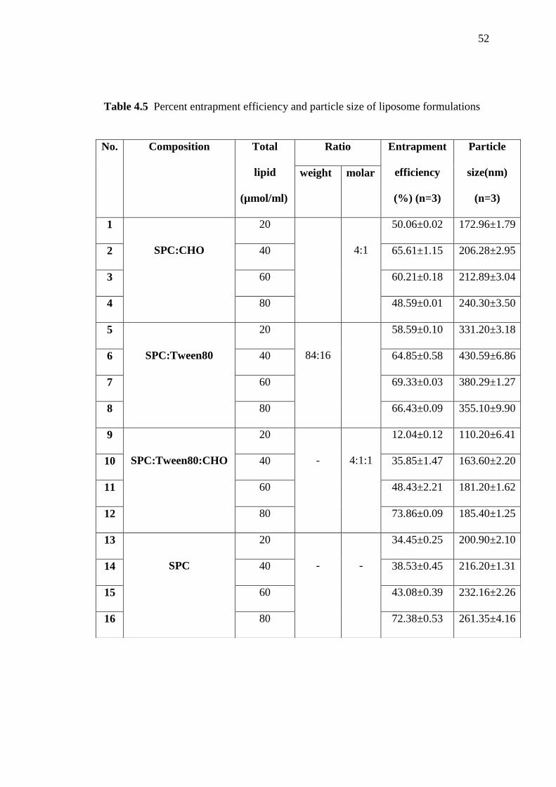
4.5 Percent entrapment efficiency and particle size of liposome
formulations
52
4.6 Percent entrapment efficiency of nicotinamide liposome for
stability study before and after 2 months (n=3)
56
4.7 Particle size and polydispersity index (PI) of nicotinamide
liposome for stability study before and after 2 months
58
4.8 Zeta potential of nicotinamide liposome before and after keeping
the liposome at 4 °C and 25 °C for 2 months
60
4.9 pH of nicotinamide liposome before and after keeping at 4 °C and
25 °C for 2 months
61

xv
LIST OF TABLES (Continued)
Table Page
4.10 In vitro skin permeation parameters of nicotinamide from the
liposome formulation and solution after 24 hours (n=3)
64
4.11 pH and viscosity of cream formulations before and after stability
test by Freeze thaw cycles
67
4.12 Nicotinamide amount in cream formulations (n=3) 68
4.13 In vitro skin permeation parameters of nicotinamide from cream,
liposome cream, and commercial product after 24 hours (n=3)
72
xvi
LIST OF FIGTURES
Figure Page
2.1 Acto pathogenesis, the steps in acne phathogenesis: (A) normal
skin, (B) increased sebum production, (C) perifollicular
hyperkeratinization and follicular obstruction and colonization
with Propionibacterium acnes (D) released of enzymes which
induce humoral and cell mediated inflammations
10
2.2 Normal skin and skin inflammatory acnes 10
2.3 The different layers of the skin 11
2.4 A schematic bilayer membrane of liposomes 13
2.5 Type of liposomes depending on size and number of lamellae 15
2.6 The general structure of a phospholipid and the structure of DOPE,
DSPC, and EPC
17
2.7 (A) Environment scanning electron microscopy (ESEM)
micrographs liposomes 9.0 ºC, 4.32 Torr; (B) Negative staining
transmission electron microscopy (NS-TEM) images; and (C)
Confocal laser scanning microscopy (CLSM) images illustrating
the architecture of liposomes
19
xvii
LIST OF FIGTURES (Continued)
Figure Page
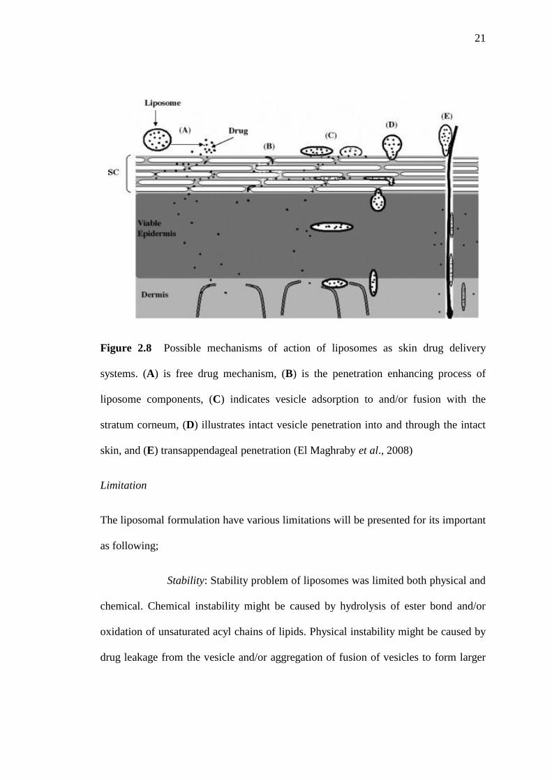
2.8 Possible mechanisms of action of liposomes as skin drug delivery
systems.(A) is free drug mechanism, (B) is the penetration
enhancing process of liposome components, (C) indicates vesicle
adsorption to and/or fusion with the stratum corneum, (D)
illustrates intact vesicle penetration into and through the intact
skin, and (E) transappendageal penetration
21
2.9 Overview about different emulsion types 23
4.1 Typical chromatogram of nicotinamide standard solution (10
µg/ml)
42
4.2 A calibration curve of nicotinamide standard solution 43
4.3 A calibration curve of nicotinamide spiked in blank skin 43
4.4 Chromatograms of (a) blank liposomes and (b) nicotinamide
spiked in liposomes (30 µg/ml)
47
4.5 Chromatograms of (a) blank PBS pH 7.4 and (b) nicotinamide
spiked in PBS pH 7.4 (10 µg/ml)
48
4.6 Chromatograms of (a) blank skin and (b) nicotinamide spiked in
blank skin (20 µg/ml) after extraction with methanol
49
4.7 Physical appearances of nicotinamide liposome formulations 50

xviii
LIST OF FIGTURES (Continued)
Figure Page
4.8 Physical appearances of various nicotinamide liposome for after
storage at 25±2 ºC (A) and 4±2 ºC (B) for 2 months
56
4.9 SEM of nicotinamide liposomes 12 (×50,000 magnification) 62
4.10 In vitro %cumulative amount-time profiles of nicotinamide
permeated across pig skin from the liposome (formulation 12) and
solution at same concentration (50 mg/ml), for 24 hours (p < 0.05)
64
4.11 In vitro amount of nicotinamide (%) accumulated in pig skins and
receptor chamber of diffusion cells from liposome (formulation 12)
and solution
65
4.12 Characteristic appearances of cream base, nicotinamide cream, and
nicotinamide liposome cream before (A1, B1, C1) and after (A2,
B2, C2) stability test under Freeze thaw cycles
67
4.13 In vitro %cumulative amount-time profiles of nicotinamide
permeated across pig skin from nicotinamide cream, nicotinamide
liposome cream, and the commercial product for 24 hours (n=3) (p
< 0.05)
69
4.14 In vitro amount of nicotinamide (%) accumulated in pig skins and
receptor chamber of diffusion cells from nicotinamide cream,
nicotinamide liposome cream, and commercial product (n=3)
71

xix
LIST OF ABBREVIATIONS AND SYMBOLS
AFM Atomic Force Microscopy
BKK Bangkok
bw body weight
ºC degree celsius
DMPC Dimyristoylphosphatidylcholine
DOPE 1, 2-Dioleoyl-sn-glycero-3-phosphoethanolamine
DSPC 1, 2-Distearoyl-sn-glycero phosphocholine
CHO Cholesterol
CLSM Confocal Laser Scanning Microscopy
cm centimeter (s)
cm2 square centimeter (s)
EDTA Ethylene diamine tetra-acetic acetate
EPC Egg phosphocholine
ESEM Environment Scanning Electron Microscopy
et al. and others
FT Freeze thaw cycle
g gram (s)
h hour (s)
HLB Hydrophilic-Lypophilic Balance
HPLC High Performance Liquid Chromatography
Jss steady state flux

xx
LIST OF ABBREVIATIONS AND SYMBOLS (Continued)
Kp permeability coefficient
Log P Logarithm of the partition coefficient
LUV Large Unilamellar Vesicles
mg milligram (s)
µg microgram (s)
µmol micromole (s)
µm micrometer (s)
min minute (s)
ml milliliter (s)
MLV Multilamellar Vesicles
MW Molecular Weight
nm nanometer (s)
NA Nicotinic Acid
NAD Nicotinamide Adenine Dinucleotide
NADP Nicotinamide Adenine Dinucleotide Phosphate
NCM Nicotinamide / Niacinamide
NMR Nuclear Magnetic Resonance
o/w oil in water
p. page
Pa Pascal
PBS Phosphate Buffer Saline

xxi
LIST OF ABBREVIATIONS AND SYMBOLS (Continued)
pH The negative logarithm of the hydrogen ion concentration
PI Polydispersity Index
pKa the negative logarithm of the dissociation constant
Qcum Cumulative amount of drug permeated
R2 coefficients of determination
REV Reverse-phase evaporation
rpm round (s) per minute
RSD Relative Standard Deviation
RT Room temperature
SEM Scanning electron microscope
SPC Soybean Phosphatidylcholine
SUV Small Unilamellar Vesicles
TEM Transmission Electron Microscopy
TEWL Transepidermal water loss
ULV Unilamellar vesicles
USA United Stated of America
v/v volume by volume
vs. versus
w/o water in oil
w/v weigh by volume
% percent

1
CHAPTER 1
INTRODUCTION
1.1 Background and Rationale
Nicotinamide is one of the newest vitamin-based components of
cosmeceutical products. Most of the available studies have focused on its anti-
inflammatory and anti-acne action (Griffiths, 1995). Nicotinamide is also believed
that its anti-inflammatory effect may improve skin appearance by reducing leucocytes
peroxidase systems that may lead to localize tissue damage as well as by ameliorating
the cutaneous barrier (Berson et al., 2003). In a comparative study, the anti-
inflammatory effect of 4% nicotinamide gel in the management of acne vulgaris was
as good as the benefits of 1% clindamycin gel (Shalita et al., 1995). This anti-
inflammatory effect is also useful to reduce cutaneous erythema in various disorders
(Bisset and Oblong, 2005).
Acne vulgaris is a multifactorial disease involving excessive sebum
production by the sebaceous glands related to an increase in the androgen levels in the
onset of puberty, ductal hyperconification of the follicles, and proliferation of
Propionibacterium acnes and other bacterial that activate the toll like receptors,
resulting in attraction of lymphocytes, neutrophils, and macrophages (Monica and
Edileia, 2009). Although, there are many options available to treat the disease, topical
antibiotic therapy is preferred option for the treatment of mild to moderate acne

2
because topical use of antibiotic (such as clindamycin phosphate and benzoyl
peroxide) is generally well tolerated but these drugs may cause skin irritation despite
this therapeutic effectiveness (Srinivasan et al., 2009). Nicotinamide is nonirritating
to facial skin, easily formulated, chemically stable, and compatible with other
formulation components, it has been considered an ideal cosmeceutical agent;
nevertheless, it is from one-third to one-fifth as effective as topical 0.025% tretinoin
(Bisset et al., 2003). So nicotinamide was replaced antibiotic in anti-inflammation of
acnes for reducing skin irritation problem. However, topical use of nicotinamide as
gel or cream is not enough effective to its difficulty in permeating the stratum
corneum layer of the skin (Sara et al., 2008). Skin permeation development and
topical formulation of nicotinamide were interested.
Recently, numerous drug delivery systems have been explored to
achieve optimal drug transport into the skin, and one such promising approach is
entrapment of drug into lipid constructs, commonly, known as liposomes. Because of
physicochemical characteristic and construct diversity, liposomes proved to be an
efficient drug delivery system for topical administration (Kumar et al., 2007).
However, the skin permeation of liposome vesicles significantly varies depending on
lipid composition and particle size (El Maghraby et al., 2006; Choi and Maibach,
2005). Different lipid compositions that constitute the liposomes interact differently
with the skin layer. The most of liposomal formulations are designed for reduction
toxicity, inhibition of rapid clearance of liposome, controlling size, charge, and
surface hydration. Enhanced safety heightened efficacy has been achieved for a wide
range of drug classes (Tianshun and Rodney, 2001).

3
Because the barrier properties of the skin is stratum corneum and the
hydrophilic make nicotinamide difficult to permeate through the skin and reach to its
site of action. Since preparing liposome containing nicotinamide for skin permeation
improved was investigated because the lipid bilayer properties of liposomes which
that likely the layer of stratum corneum in the skin (mostly composed of lipid)
(Tianshun and Rodney, 2001). Though, the delivery of nicotinamide with the aid of
liposome technology has a potential for the prevention inflammation of acnes, until
now a very few work have been performed. Beside, no report is available on the
characterization, formulation, and evaluation of skin permeation behavior of
liposomal vesicles containing nicotinamide.
In this present study, preparation and physicochemical study of
liposomes containing nicotinamide was determined for skin delivery. Nicotinamide
liposome characteristics, skin permeation properties, and stability studies for
conducted suitable liposomes were evaluated. The suitable liposomes were prepared
to cream formulations for future.

4
1.2 Objective of the study
The objectives of this study were to:
1.2.1 Formulate the liposomes containing nicotinamide
1.2.2 Evaluate the physicochemical property and stability of liposomes
containing nicotinamide
1.2.3 Evaluate the in vitro skin penetration retention efficiency of
nicotinamide from the liposomes compared to a nicotinamide solutions
1.2.4 Prepare o/w cream formulations containing nicotinamide liposomes
1.2.5 Evaluate the physicochemical property and stability of o/w cream
formulations containing nicotinamide liposomes
1.2.6 Evaluate the in vitro skin penetration retention efficiency of o/w cream
formulation containing nicotinamide liposomes compared to a
nicotinamide creams and commercial product

5
CHAPTER 2
REVIEWS OF LITERATURE
2.1 Nicotinamide
Nicotinamide is a water-soluble amide of nicotinic acid. It is one of
two principal forms of B-complex vitamin, B3, active form that acts as constituent of
the enzyme cofactors NAD and NADP. These functions were performed in cell
metabolism of carbohydrates, fatty acid, and amino acids. Structure of nicotinamide
and its derivatives showed as in Table 2.1 and the Table 2.2 shows physicochemical
properties of nicotinamide.
2.1.1 Nicotinamide in cosmeceutical applications
The likely usefulness of topical nicotinamide for the improvement of
skin appearance may be related to its action in the synthesis of sphingolipid, free fatty
acid, cholesterol, and ceramide, thus, decreasing transepidermal water loss (Berson et
al., 2003; Bisset et al., 2003; Tanno et al., 2000). Additionally, nicotinamide increases
collagen production in fibroblast culture and this effect may be responsible for the
improvement of skin elasticity and reduction of fine wrinkles (Bisset et al., 2003),
improvement of facial dyspigmentation is also likely mediated by the suppression of
melanosome transfer from melanocyte to keratinocytes (Hakozaki et al., 2002). All
of these effects may help to reverse some of the aging skin signs. Concentration

6
ranging of nicotinamide has been used in cosmetic products from 2-5%, so it has
sufficient (Bisset and Oblong, 2005).
Table 2.1 Shows structure of nicotinamide and its derivatives (Ping and Antony,
2010)
Structure formula Structure formula names
Nicotinic Acid (NA)
Nicotinamide / Niacinamide (NCM)
Nicotinamide adenine dinucleotide
(NAD)
Nicotinamide adenine dinucleotide
Phosphate (NADP)

7
Table 2.2 Physicochemical properties of nicotinamide
Physicochemical properties Forms of nicotinamide
Molecular Formula C6H6N2O
Chemical Name 3-pyridine carboxamide
Molecular Weight (MW) 122.1
Appearance
White crystalline powder or colorless
crystals
Solubility (water solubility) 1g/ml
Partition coefficient
n-octanol/water (log value)
-0.38 (22°C)
(Georg, 2002)
%Use in cosmetic 2-5%
LD50 in rat by injection 1.7 g/kg
Pharmacological dose
500 mg-2 g/day (non toxic), 3 g/day
(toxic)
pKa 0.5 and 3.35 (http:www.drugs.com)
(http://www.scribd.com/doc/30137426/Niacin-Nicotinamide-And-nicotinicotinicacid)

8
2.1.2 Nicotinamide for skin penetration
Sara et al. determined the nicotinamide accumulation in the dermal and
epidermal layers of human in vitro skin after application of topical gels. It was found
the accumulation in the skin in vitro after 20 min application. Most of drug amounts
were accumulated in the epidermis and adsorbed to deeper layer in few amount
because the hydrophilic properties is barrier in skin penetration (Sara (1) et al., no
date).
Sara et al. also study the enhancement effect of nicotinamide in methyl
paraben solutions by Franz diffusion cells with rabbit ear skin. Amount of methyl
paraben with nicotinamide solution (20% w/v) could be permeated to the skin higher
methyl paraben with water for 8 hours. Beneficial of it is enhanced permeation of
substance for permeation skin improving (Sara (2) et al., no date).
Imokawa et al. presented for nicotinamide improves epidermal
ceramide synthesis with concurrent epidermal barrier benefits. Sphingolipids and
other stratum corneum lipids, particularly ceramides, are known to play central roles
in the structural and functional integrity of the epidermal permeability barrier, and
decreased in aged and a topic skin (Imokawa et al., 1997).
Tanno and colleagues shows nicotinamide inducing up to a 5-fold up-
regulation in ceramide synthesis, in a dose dependent fusion, in cultured human
epidermal-keratinocytes. The in vivo clinical significance of these findings was
demonstrated (Tanno et al., 1997). And topically 2% nicotinamide was recovered
ceramide in stratum corneum and free fatty acid lipid fraction. This reduced
transepidermal water loss (TEWL) in the skin human (Tanno et al., 2000).

9
2.1.3 Toxicological data of nicotinamide
Nicotinamide is very low acute toxicity to mammals, both orally and
dermally. For oral, the acute toxicity of nicotinamide after oral exposure was LD50 3-7
g/kg bw in rodents. Its toxicity after dermal application was very low with a dermal
LD50 >2000 mg/kg bw in rabbits. Skin irritation studies indicated that nicotinamide
shown no potential to irritate the skin. In additionally, nicotinamide irritated to the
eyes, application of 0.1 g nicotinamide to the eyes of 3 rabbits induced irritation in
two animals, which was reversible within 7 days (Keri et al., 2005).
2.2 Inflammatory of acne vulgaris
Acne vulgaris is a disorder of the pilosebaceous unit, characterized by
comedones, inflammatory lesion and scars on the face and trunk. The processes in the
pathogenesis of acne were the increase of sebum production in first step; perifollicular
hyperkeratinization and follicular obstruction; colonization with Propionibacterium
acnes; and released of enzymes which induce humoral and cell mediated
inflammations which presented as in Figure 2.1 (Monica and Edileia, 2009; Guy and
Webster, 1995), and inflammation skin appearances of acnes versus normal skin as
shown in Figure 2.2. Nicotinamide was used for anti-inflammatory of acnes by
reducing sebum production. It was reported about anti-inflammatory action affecting
neutrophil chemotaxis by inhibited histamine released and to suppress the lymphocyte
transformation (Yesim and Meltem, 2008).

10
Figure 2.1 Acto pathogenesis, the steps of acne phathogenesis: (A) normal skin, (B)
increased sebum production, (C) perifollicular hyperkeratinization and follicular
obstruction and colonization with Propionibacterium acnes (D) released of enzymes
which induce humoral and cell mediated inflammations (Zoe and Lauren, 2008)
Figure 2.2 Normal skin and skin inflammatory acnes (http://www.healthchrome.com)
2.3 Skin structure
The skin has the primary function to provide a barrier against
environmental influences and protect the body against the loss of endogeneous
substances. Macroscopically, two distinct skin layers are apparent, an unvascularized
outer layer (epidermis) and an inner vascularized layer (dermis and hypodermis) as

11
shown in Figure 2.3, an overview about the dimensions and the stratified appearance
of the skin (Heiko et al., 2006). The several layers of the skin were complained as
following.
Figure 2.3 The different layers of the skin (http://www.1wipe1week.com)
Epidermis
The viable epidermis is divisible into five distinct layers, namely, from
inside to outside: stratum basale, stratum spinosum, stratum granulosum, stratum
lucidum, and stratum corneum. The cells of these layers undergo continuous
differentiation to produce the outermost layer of the skin. The stratum basale is a
single layer of epidermal stem cells which are anchor to a basement membrane that
separates the epidermal tissue from the underlying dermis. Above, these
differentiating cells are forming the stratum spinosum. The cells are now of spiny
appearance, due to abundance of desmosome (Heiko et al., 2006). The stratum
corneum constitutes the outermost layer of the epidermis and represents the main
barrier function, although it is the thinnest and smallest compartment of the skin. It is
about 10 to 20 µm thick and consists of several layers of dead, keratin filled

12
corneocytes that represent the final state of epidermal differentiation. These cells are
embedded in a matrix of lipid lamellas, which gives the stratum corneum a brick and
mortar organization (Menon, 2002). Another specialty of the stratum corneun is the
lipid composition which differs significantly from those of the cells of the lower
epidermal layers. The extracellular lipid matrix of the stratum corneum is finally free
of phospholipids and consists of ceramides, cholesterol, and free fatty acids in roughly
equimolar ratio. The ceramides are crucial for the lipid organization of the stratum
corneum barrier, while the cholesterol is promoting the intermixing of different lipid
species (Heiko et al., 2006).
Dermis and hypodermis
The dermis is connected by the basement membrane to the stratum
basale. Main components of the dermis are collagen and elastin fibers that form a vast
network of filamentous and amorphous connective tissue that prevents strength and
flexibility to the skin (Schaefer and Redelmaier, 1996). Furthermore, the dermis
accommodates cellular residents such as fibroblasts, endothelial cells, mast cells, and
under conditions of inflammation, macrophages, lymphocytes, and leucocytes.
Underneath the dermis is the hypodermis situated that contains, in
contrast to the dermis, loose connective tissue and adipocytes as the main cellular
exponent which represent energy source for the body (Heiko et al., 2006).
2.4 Liposomes
Liposomes are self assemble colloidal particles that occur naturally and
can be prepared artificially, as shown by Bangham and his students in the mid-1960.
At first, they were used to study biological membrane. Several practical applications,
most not ably in drug delivery, emerged in the 1970 (Dan, 1998). The structural of it

13
is spherical self-closed, composes of curved lipid bilayers, which enclose part of the
surrounding solvent into their interior as shown in Figure 2.4. Their size ranges from
some 20 nm up to several µm and liposomes layer may be composed of one or several
concentric membranes, each with a thickness of about 4 nm. Liposomes are
possessing unique properties owing to the amphiphilic character of the lipids, which
make liposomes suitable for drug delivery (Nill, 2003).
Figure 2.4 A schematic bilayer membrane of liposomes
(http://www.2chemistry.msu.edu/faculty/reusch/VirtTxtJml/lipids.htm)
2.4.1 The benefit of liposomes
(i) Improved solubility of lipophilic and amphiphilic drugs. (Lasic,
1992);
(ii) Passive targeting to the cells of the immune system, especially
mononuclear phagocytic cells
(iii) Sustained release system of systermically or locally
administered liposomes.

14
(iv) Site-avoidance mechanism: liposomes do not dispose in certain
organ, such as heart, kidneys, brain, and nervous system, and this reduce cardio-,
nephro-, and neuro-toxicity.
(v) Improved transfer of hydrophilic, changed molecules such as
chelators, antibiotics, plasmids, and genes into cells
(vi) Improved penetration into tissue, especially in the case of
dermally applied liposomal dosage forms.
In general, liposome encapsulation is considered when drugs are very
potent, toxic, and very short life times in the blood circulation or at the sites of local
(subcutaneous, intramuscular, or intrapulmonary) administration.
2.4.2 Classification of liposomes
The liposome size can range from very small (0.025 µm) to large (2.5
µm) vesicles. Furthermore, it may have single or multiple bilayers membranes. The
vesicle size is a critical parameter in determining circulation half-life of liposomes,
and size and number of bilayers influence the extent of drug encapsulation in the
liposomes. On the basis of their size and number of bilayers, liposomes can also be
classified into one of three categories: multilamellar vesicles (MLV); large
unilamellar vesicles (LUV); and small unilamellar vesicles (SUV) as shown in the
Figure 2.5. The size and characteristics of these types of liposomes are listed in Table
2.3 (Amarnath and Uma, 1997).

15
Figure 2.5 Type of liposomes depending on size and number of lamellae (Amarnath
and Uma, 1997)
Table 2.3 Liposome classifications by size and number of lamellae (Amarnath and
Uma, 1997)
Type Usual size Characteristics
MLV > 0.1µm More than one bilayer; greater encapsulation of lipophilic
drugs; prepared by thin film hydration method or hydration of
lipids in presence of an organic solvent
LUV >0.1µm Single bilayer; useful for hydrophilic drug; high capture of
macromolecules; prepared by ether injection, reverse-phase
evaporation or active loading methods

16
Table 2.3 Liposome classifications by size and number of lamellae (continued),
(Amarnath and Uma, 1997)
Type Usual size Characteristics
SUV ≤0.1µm Single bilayer; homogenous in size; thermodynamically
unstable; susceptible to aggregation and fusion at low or no
charge; limited capture of macromolecules; prepared by
reducing the size of MLV or LUV using solvent injection
techniques
2.4.3 Compositions of liposome formulations
The main components of liposomes are phospholipid and cholesterol.
A phospholipid has two acyl chains linked to a head group by means of a glycerol-
backbone. The Figure 2.6 shows the structural formula of a phospholipid, where R1
and R2 are saturated or unsaturated acyl chains and R3 is the polar head group.
Phosphatidylcholines or PC-lipids are the most widely used lipids in
liposome work. PC-lipids are zwitterionic at all relevant pH, therefore, form lamella
structure independently of the pH in the solution. EPC and DSPC are showed
structures in the Figure 2.6. DSPC is a synthetic lipid with only saturated chains,
while EPC is a natural PC-lipid with both saturated and unsaturated fatty acids.
Phosphatidylethanolamines (PE) have pH dependent phase behavior. At physiological
pH, the PE-lipids have zwitterionic head group, they are not capable of forming
lamellar structures (Nill, 2003).

17
Figure 2.6 The general structure of a phospholipid and the structure of DOPE,
DSPC, and EPC (Nill, 2003)
Additionally, also cholesterol is component in liposomal formulation.
Cholesterol incorporated in liposome bilayer brings about major changes in the
preparation of these membranes. It can be incorporated into phospholipid membranes
in very high concentration up to 1:1 or even 2:1 molar ratios of cholesterol to PC.
Cholesterol inserts into the membrane with its hydroxyl groups oriented towards the
aqueous surface and aliphatic chain aligned parallel to the acyl chains in the center of
the bilayer. However, the over concentration of cholesterol was used for condensation
fluidity and increase rigidity of liposomes bilayer membrane (Emnet, 2010;
http://www.pharmaxchange.info).
2.4.4 Liposomes preparation methods
Liposomes with different sizes and characteristics usually require
different methods of preparation. The most simple and widely used method for
preparation of MLV is the thin-film hydration procedure in which a thin film of lipids
is hydrated with an aqueous buffer. The drug to be encapsulated is included either in
the aqueous hydration buffer (for hydrophilic drugs) or in the lipid film (for lipophilic
drugs). Thin-film hydration method produces a heterogeneous population of MLV (1-

18
5 µm diameters) which can be sonicated or extruded through polycarbonate filters to
produce small (up to 0.025 µm) and more uniformly size population of SUV. One of
the major disadvantages of this method is relative poor encapsulation efficiency (5-
15%) of hydrophilic drugs. Reduction of liposome size further decreases the amount
of encapsulated drug. MLV with high entrapment efficiency (up to 40%) can be
prepared by freeze-drying preformed SUV dispersion in an aqueous solution of the
drug to be encapsulated (Ohsawa et al., 1984).
Additionally, several methods have been developed for the preparation
of LUV, including solvent injection of ether and ethanol, detergent dialysis, calcium
induced fusion, and REV techniques. SUV can be prepared from MLV or LUV by
sonication using probe sonicator or extrusion passage through a small orifice under
high pressure (Amarnath and Uma, 1997; Mohammad, 1996).
2.4.5 Physical morphology and physicochemical characteristics
The vesicular shape and surface morphology of liposomes can be
visualized by atomic force microscopy (AFM), environment scanning electron
microscopy (ESEM), transmission electron microscopy (TEM), and confocal laser
scanning microscopy (CLSM; labeling using a fluorochrome marker). Barbara et al.
reported about the improved evaluation of physicochemical and technological
properties of drug delivery by liposome systems with several equipments. Aim in
research was to determine the details of the morphology and structure of conventional
liposomal formulations (Barbara et al., 2011).
The Figure 2.7 shows images morphology of liposome by many
techniques. In summary, microscopic studies improve the characterization of

19
nanoscale structures of liposomes and provide information about shape and
morphology (AFM, TEM), dimensions (AFM, ESEM, TEM, and CLSM), surface
properties (AFM), and internal structure (CLSM).
Figure 2.7 (A) Environment scanning electron microscopy (ESEM) micrographs
liposomes 9.0 ºC, 4.32 Torr; (B) Negative staining transmission electron microscopy
(NS-TEM) images; and (C) Confocal laser scanning microscopy (CLSM) images
illustrating the architecture of liposomes (Barbara et al., 2011).
The behaviors of liposomes in both physical and biological systems are
depended on factors such as physical size, membrane permeability, percentage of
entrapped solutes, and chemical composition.
2.4.6 Mechanisms of liposomes as skin drug delivery systems and
liposomes limitation
Mechanisms of action of liposomes as skin drug delivery systems
The skin barrier is situate in the stratum corneum and consists of the
keratinocytes, proteins, and lipids. Thus, the skin barrier contains hydrophilic and
lipophilic compartments, which act as buffer to retard both water loss and absorption
of water (Imokawa et al., 1989). Alternative mechanisms have been suggested for
liposomes acting as skin drug delivery systems as shown in the Figure 2.8. The drug
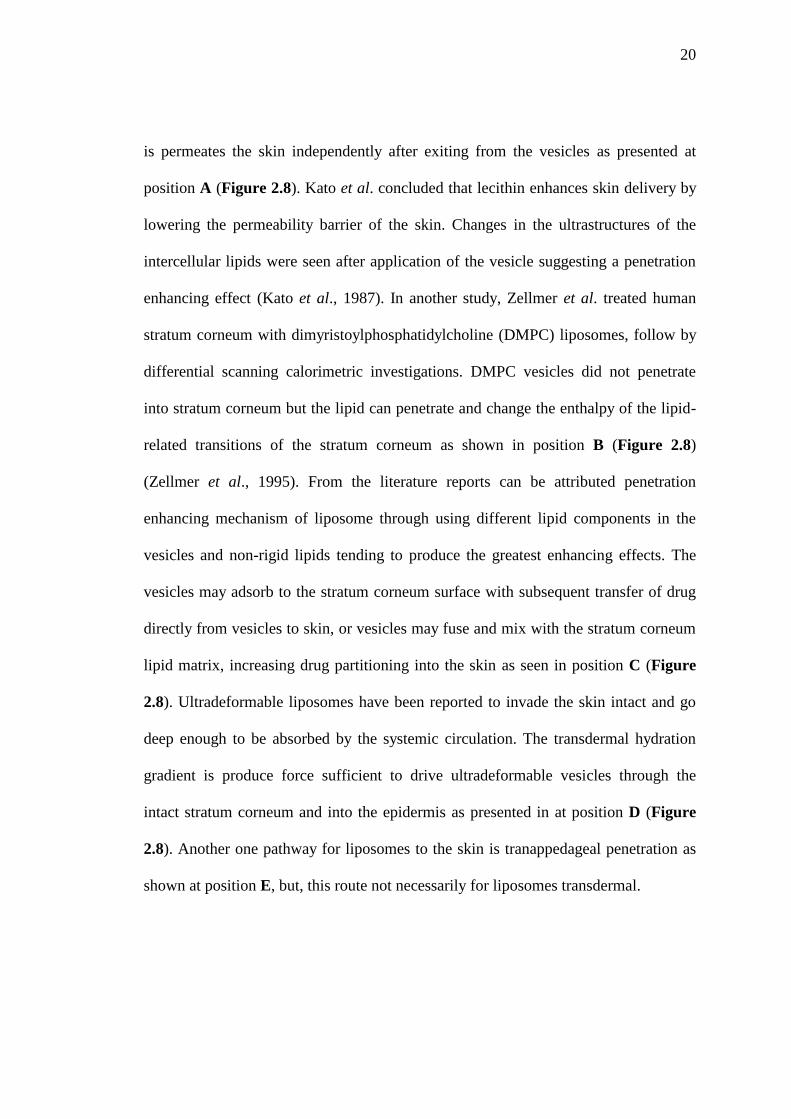
20
is permeates the skin independently after exiting from the vesicles as presented at
position A (Figure 2.8). Kato et al. concluded that lecithin enhances skin delivery by
lowering the permeability barrier of the skin. Changes in the ultrastructures of the
intercellular lipids were seen after application of the vesicle suggesting a penetration
enhancing effect (Kato et al., 1987). In another study, Zellmer et al. treated human
stratum corneum with dimyristoylphosphatidylcholine (DMPC) liposomes, follow by
differential scanning calorimetric investigations. DMPC vesicles did not penetrate
into stratum corneum but the lipid can penetrate and change the enthalpy of the lipid-
related transitions of the stratum corneum as shown in position B (Figure 2.8)
(Zellmer et al., 1995). From the literature reports can be attributed penetration
enhancing mechanism of liposome through using different lipid components in the
vesicles and non-rigid lipids tending to produce the greatest enhancing effects. The
vesicles may adsorb to the stratum corneum surface with subsequent transfer of drug
directly from vesicles to skin, or vesicles may fuse and mix with the stratum corneum
lipid matrix, increasing drug partitioning into the skin as seen in position C (Figure
2.8). Ultradeformable liposomes have been reported to invade the skin intact and go
deep enough to be absorbed by the systemic circulation. The transdermal hydration
gradient is produce force sufficient to drive ultradeformable vesicles through the
intact stratum corneum and into the epidermis as presented in at position D (Figure
2.8). Another one pathway for liposomes to the skin is tranappedageal penetration as
shown at position E, but, this route not necessarily for liposomes transdermal.
21
Figure 2.8 Possible mechanisms of action of liposomes as skin drug delivery
systems. (A) is free drug mechanism, (B) is the penetration enhancing process of
liposome components, (C) indicates vesicle adsorption to and/or fusion with the
stratum corneum, (D) illustrates intact vesicle penetration into and through the intact
skin, and (E) transappendageal penetration (El Maghraby et al., 2008)
Limitation
The liposomal formulation have various limitations will be presented for its important
as following;
Stability: Stability problem of liposomes was limited both physical and
chemical. Chemical instability might be caused by hydrolysis of ester bond and/or
oxidation of unsaturated acyl chains of lipids. Physical instability might be caused by
drug leakage from the vesicle and/or aggregation of fusion of vesicles to form larger

22
particles. Both of these processes, drug leakage and change in liposome size were
effected the vivo performance of the drug formulation.
Encapsulation efficiency: Encapsulation efficiency was depended
amount of lipid. Since lipid in high dose might be toxic and also cause non-linear
(saturable) pharmacokinetics of liposomal drug formulations. Additionally, other
limitations of liposome are not mention such as sterilization method, particle size
control, and short circulation half life (Amarnath and Uma, 1997).
2.5 Emulsion cream formulations
Emulsions are heterogeneous systems containing two immiscible
phases; a hydrophilic liquid phase and a lipophilic or oil phase. For the mixture
consists of hydrophilic droplet dispersed in oil, they refer to it as water in oil (w/o)
emulsion, even when the hydrophilic liquid is not water, they refer to it as oil in water
(o/w) emulsion. It has non polar liquid droplet dispersed in the aqueous phase.
Additionally, multiple emulsions are composed of droplets of one liquid dispersed in
larger droplets of a second liquid, which is dispersed in a final continuous phase. Also
systems may be w/o/w emulsions where the internal and external phases are
hydrophilic; or o/w/o, which have the reverse composition. Figure 2.9 gives an
overview about these emulsion types (Heiko et al., 2006).

23
Figure 2.9 Overview about different emulsion types (Heiko et al., 2006)
Physicochemical properties and preparation of creams
An emulsifier as the additive was added in emulsions for reducing the
interfacial area. It may be divided into four groups as follows: adsorbed non surfactant
ionic materials, colloidal solids, and polymers, which represent the most common
stabilization mechanism. Type of emulsion that is produced with given hydrophilic
and lipophilic compounds, o/w or w/o, depends primarily on the properties of the
surfactant. This characteristic is referred to as hydrophilic-lipophilic balance (HLB)
that is the polar-non polar nature of the emulsifiers (Heiko et al., 2006).
In principal, two common methods are used to prepare cream
formulations. The continental method which the emulsifier, the lipophilic phase, and
parts of the hydrophilic phase forms is primary w/o emulsion. The final o/w emulsion
is obtained due to phase inversion following addition of more hydrophilic solution.
The second method was by dissolving the emulsifier in the hydrophilic phase and the
preparation step involves slowly titration with the oil. This method is referred to as
English method. Each methods, however, requires that energy put into the system in

24
some forms. The energy may supply in a variety of ways, such as triturating, heat,
agitation, or homogenization (Heiko et al., 2006).

25
CHAPTER 3
MATERIALS AND METHODS
3.1 Materials
3.1.1 Active ingredient
Nicotinamide (P.C. Drug center Co., Ltd., BKK, Thailand)
Nicotinamide Standard (Fluka, switzerland)
3.1.2 Chemicals and reagents
Absolute ethanol, AR grade (Merk®, Darmstadt, Germany)
Absolute methanol, AR grade (Merk®,Darmstadt, Germany)
Cetyl alcohol (Vidhyasom, BKK, Thailand)
Cholesterol from lanolin (Fluka®, Buchs, Japan)
Cremophor A 6 (BASF, Ludwigshafen, USA)
Cremophor A 25 (BASF, Ludwigshafen, USA)
Crystal violet (Sponsored by Faculty of Sciences, Department of
Microbiology, Prince of Songkla University, Songkla, Thailand)
Ethanol 95%, HPLC grade (Merk®,Darmstadt, Germany)

26
Glycerin (P.C. Drug Center Co., Ltd., BKK, Thailand)
Glyceryl monostearate (P.C. Drug Center Co., Ltd., BKK, Thailand)
Gram’s iodine (Sponsored by Faculty of Sciences, Department of
Microbiology, Prince of Songkla University, Songkla, Thailand)
Isopropyl myristate (P.C. Drug Center Co., Ltd., BKK, Thailand)
L-∂-Phosphatidylcholine from soybean, Type IV-S, ≥30% (TLC) (Sigma-
Aldrich®, USA)
Mineral oil (P.C. Drug Center Co., Ltd., Bkk, Thailand)
Orthophosphoric acid 85% (Merk®, Darmstadt, Germany)
Polyoxyethylene (20) sorbitan monooleate, Tween80® (Srichand Co., Ltd.,
BKK, Thailand)
Potassium dihydrogen phosphate (KH2PO4) (Merk®,Darmstadt, Germany)
3-sn-Phosphatidylcholine from soybean (Fluka®, Buchs, United States)
Sodium metabisulfite, AR grade (Namsaing international Co., Ltd., BKK,
Thailand)
Stearic acid (Srichand Co., Ltd., BKK, Thailand)

27
3.2 Instruments
Table 3.1 Experimental instruments
Instruments Model Company
Centrifuge
Chromatographic
column
High performance
liquid chromatography
Hot air oven
Magnetic stirrer
Sonicator
Syringe filter
Ultracentrifuge
Viscometer
Zeta potential analyzer
Z323K
Reverse phase BDS
HYPERSIL C18 column,
150×4.6 mm particle size,
5µm
SpectraSystem P1000 pump
SpectraSystem UV1000
detectorChromQuest
software program SN 4000
DIN 12880-KI
MR 3000D
HT Crest
-
Optima L-100XP, equipped
with SW 60 Ti rotor
RVT
Zeta PALS
HermleLabortechnik
GmbH, Germany
Restek Corporation, USA
Thermo Electron
Corporation, USA
Polytron, Switzerland
Heidolph, Germany
S.V. Medico Co.,Ltd.,
Thailand
Vertical chromatography Co.,
Ltd., Thailand
Beckman Coulter, USA
Brook fielded dial reading,
USA
Brookhaven Instrument
Corporation, USA

28
3.3 Methods
3.3.1 Quantitative analysis of nicotinamide using High-Performance
Liquid Chromatography (HPLC)
3.3.1.1 Instruments and chromatography conditions
The HPLC method for quantitative determination of nicotinamide was
modified from the method described by Junaid et al., (2008). The analysis was
performed at room temperature on reverse phase BDS HYPERSIL C18 column (150 ×
4.6 mm, particles size, 5µm). A mixture of methanol and buffer of 0.05 M potassium-
dihydrogen phosphate having pH 3.6±0.1, adjusted with orthophosphoric acid (75:925
ml) was used as mobile phase with the flow rate of 0.6 ml/min. The mobile phase was
filtered through 0.45 µm nylon membrane filter and degassed by sonication before
using. The 20 µl sample solution was injected and absorbance was detected at 254
nm. The concentrations of nicotinamide were quantified from the standard curve by
plotting the peak area of nicotinamide against the nicotinamide concentration.
3.3.1.2 Preparation of nicotinamide standard solution
A 0.10 gram of nicotinamide was accurately weighted, dissolved, and
adjusted volume with distilled water to 100 ml in order to reach the concentration of
1000 µg/ml to form a stock solution. It was diluted with mobile phase which their
concentrations of 1, 5, 10, 20, 30 and 40 µg/ml (Tsang and Zeng, 2007).

29
3.3.1.3 The validation procedures
The HPLC method used for analysis of nicotinamide was validated in
terms of linearity, accuracy, precision, specificity, and limit of detection and
quantification according to International Conference on Harmonization (ICH)
guideline (ICH, 1996) as following;
Linearity
Three sets of nicotinamide standard solution and nicotinamide spiked
in pig skin were analyzed in triplicate. Linear regression analysis of means peak area
versus their concentrations was performed. The coefficient of determination (R2) ≥
0.99 is the acceptable criteria (ICH, 1996).
Accuracy
Nicotinamide stock solution was spiked in blank liposomes to the
concentration of 30, 50, and 70 mg/ml. These three concentrations represent to 60%,
100%, and 140% of nicotinamide concentration in the prepared nicotinamide-loaded
liposomes. Three sets of sample were analyzed in triplicate. Additionally, also the
accuracy for skin permeation studies was determined. Nicotinamide stock solution
was spiked in blank skin to the concentration 20, 30, and 40 µg/ml. To prove the
accuracy of nicotinamide amount determination in the skin permeation study
procedure by Franz diffusion cells. The concentrations of nicotinamide were
calculated from the linear regression equation obtained from linearity test compared
with true value and expressed as percentage of recovery which could be calculated by
the following equation:

30
%Recovery = 𝐂 𝑚𝑒𝑎𝑠𝑢𝑟𝑒𝑑
𝐂 𝑎𝑐𝑡𝑢𝑎𝑙× 100
Where; C measured: concentration of nicotinamide detected by HPLC
C actual: actual concentration of nicotinamide
The percent recovery should be in the range of 98 to 102% (ICH, 1996).
Precision
Precision evaluation was divided to the intra-and inter-day precision.
The intra-day precision
Three concentrations of the nicotinamide standard solution (1, 10 and
30 µg/ml) were analyzed by HPLC in the same day. Each set of sample was analyzed
in triplicate.
The inter-day precision
Three concentrations of nicotinamide standard solution (1, 10, and 30
µg/ml) were analyzed by HPLC in 3 continuously days. Each set of sample was
analyzed in triplicate.
The evaluation of both intra- and inter-day precision expressed as
percentage of relative standard deviation (%RSD) of peak area in every concentration,
both intra-day and inter-day precisions which their values should not be over 2.0%
(ICH, 1996) was calculated by the following equation:
(1)

31
%RSD =𝑆𝐷
𝑋 × 100
Where; SD: Standard deviation of nicotinamide concentration
𝑋 : Average concentration of nicotinamide
Specificity
The specificity of the HPLC method was determined for nicotinamide
in liposome formulations, receptor fluid of skin permeation study, and the pig skin
model. The experiments were performed by spiking nicotinamide in the tested
matrices. The peak of any excipients in liposome formulations, PBS, as well as pig
skin must not interfere with the nicotinamide peak.
Limit of detection (LOD) and quantification (LOQ)
Stock solution of nicotinamide at a concentration of 1 µg/ml was
prepared by dissolving the drug in mobile phase. Through successive dilutions, the
stock solution was diluted to the lowest concentration of 0.1 µg/ml. The LOD and
LOQ were assessed as lowest concentration which provided response of linearity and
precision (RSD < 2%). The LOD and LOQ were calculated based on standard
deviation of response obtained from solution diluted and the slope value of calibration
curve. The solution was injected in three replicates (Thais et al., 2008). The equations
of detection and quantification limits are, thus:
(2)

32
LOD = 3.3 ×S
b
LOQ = 10 ×S
b
Where; S: standard deviation of response
b: the slope of the calibration curve
3.3.2 Formulations and preparation of nicotinamide liposomes
The liposomes containing nicotinamide were formulated using
nicotinamide at the concentration of 50 mg/ml (5% in formulations) and prepared by
modified ethanol injection method. The formulations were optimized in terms of type,
ratio and concentration of lipids and additives as shown in Table 3.2. Firstly, the oil
phase was prepared by dissolving soybean phosphatidylcholine (SPC) and cholesterol
(CHO) in 10 ml ethanol. The water phase was prepared by dissolving nicotinamide
and Tween80 in 10 ml distilled water. Both phases were sonicated at 30 ºC until
homogeneity was obtained. Then, they were repeatedly heated at 60 ºC by swirl
continuously for 1 minute in round bottom flask. Subsequently, the water phase was
poured into the oil phase and ethanol was evaporated by Rotary evaporator at 60 ºC to
obtain the liposome suspensions.
(3)
(4)

33
Table 3.2 Compositions of nicotinamide liposome formulations
Composition Composition ratio of lipid
(*Weight ratio, **Molar ratio)
Total lipid
(µmole/ml)
1. SPC:CHO
2. SPC:CHO
3. SPC:CHO
4. SPC:CHO
4:1**
20
40
60
80
5. SPC:Tween80
6. SPC:Tween80
7. SPC:Tween80
8. SPC:Tween80
84:16*
20
40
60
80
9. SPC:Tween80:CHO
10. SPC:Tween80:CHO
11. SPC:Tween80:CHO
12. SPC:Tween80:CHO
4:1:1**
20
40
60
80
13. SPC
14. SPC
15. SPC
16. SPC
-
20
40
60
80
3.3.3 Characterizations of nicotinamide liposomes
3.3.3.1 Physical appearances
The physical appearances of all liposomes were visually observed for
colloidal appearance, color, phase separation, and precipitation.

34
3.3.3.2 Particle size and zeta potential
The size and zeta potential of liposomes containing nicotinamide were
determined using zeta potential analyzer at 25 ºC. Before determination, the
liposomes containing nicotinamide suspension was diluted about 15 folds with
distilled water.
3.3.3.3 Entrapment efficiency
The entrapment efficiency of liposomes containing nicotinamide was
evaluated by ultracentrifugation technique. The liposome formulations were
centrifuged at 60,000 rpm, 4 ºC for 2 h using ultracentrifuge. The supernatant was
collected to determine the amount of non-entrapped nicotinamide by HPLC. The
liposomes formulation (not centrifuged) was busted with the 20% v/v Triton®X-100
and diluted with mobile phase to determine the total amount of nicotinamide in
liposomes formulation. The entrapment was calculated from the equation;
Entrapment efficiency % =T−F
T× 100
Where; T: total amount of nicotinamide in formulation
F: free nicotinamide (non-entrapped amount)
3.3.3.4 Surface morphology
Surface morphology of liposomes containing nicotinamide was
examined using scanning electron microscopy (SEM). The amount of liposome
formulations 200 µl was diluted with 3 ml MilliQ-water. A drop of diluted liposomes
was allowed to dry on the cover slip and then stained with crystal violet solution for 1
(5)
35
minute. An excess dye was rinsed out with water followed by fixing with Gram’s
iodine solution for 1 minute. This staining method was based on Gram’s Method
where the positive charge of crystal violet interacted with negative charge of the
phospholipids in liposomes and then formed complex with I- and I3
-(Davies et al.,
1983). The sample was then coated with gold in a sputter coater under an argon
atmosphere (50 Pa) at 50 mA for 50 seconds and investigated under SEM at 50,000X
magnification.
3.3.3.5 pH
pH of liposome formulations was evaluated by pH meter. All
determinations were performed in triplicate.
3.3.4 Stability study of liposomes containing nicotinamide
The best liposomes containing nicotinamide were selected for stability
testing based on physical appearance (color, precipitation, and separation) as well as
the optimal particle size, and percent entrapment efficiency. Liposome formulations
were storage at 4±2 ºC and 25±2 °C for 2 months compared to the freshly prepared
liposome formulations (Pinsuwan et al., 2010).
3.3.5 In vitro skin permeation study
3.3.5.1 Skin preparation
The experiments were performed by using the skin of naturally died
newborn pig which the age was not more than three days. The pig skins were cleaned
and removed hair off with clipper without any damage on the skin surface. The

36
subcutaneous fat was carefully removed by surgical scissors. The skin samples were
packed in aluminum foil and stored at -20 ºC until used. Two hours before the
beginning of the experiments, the skin was pre-equilibrated in phosphate buffer
solution (PBS) pH 7.4 at 25 ºC (Pinsuwan et al., 2010).
3.3.5.2 In vitro skin permeation procedures
The skin samples were mounted carefully on Franz diffusion cells
which the effective diffusion area was 1.77 cm2. The stratum corneum of pig skin was
set side up for contacted with sample. The receptor compartment was filled with 11
ml of PBS. Temperature and magnetic stirrer were controlled of 37 ºC and 200 rpm,
respectively. The skin permeation of nicotinamide-loaded liposomes was determined
compared with nicotinamide solution containing the same amount of nicotinamide (50
mg/ml). Moreover, nicotinamide liposome creams was determined compared with
nicotinamide cream and commercial product. The 1 ml of liposome sample and a 1
gram of cream sample were applied on the skin surface in the donor part of the Franz
diffusion cells. The sample of receiver medium in receptor fluid was sampled of 1 ml
at period times of 0.5, 1, 2, 4, 6, 8, 12 and 24 hours and replaced with the same
amount of PBS every sampling (Pinsuwan et al., 2010). All withdrawn samples were
filtered through a 0.45 µm syring filter membrane and analyzed of amount of
nicotinamide permeated by HPLC. The data were plotted between amounts of
nicotinamide permeation skin versus times. All determinations were performed in
triplicate. The cumulative amount was calculated according to the following equation;

37
𝑄𝑐𝑢𝑚 =𝑃𝑛𝑉𝑜 + 𝑃𝑖𝑉𝑛−1
𝑖=1
𝐴
Where; Pn and Pi: the nicotinamide concentration determined at No. n and No. i
(µg/ml)
Vo and V: the volume of a single Franz cell and withdrawn sample (ml)
A: effective diffusion surface area (cm2)
The cumulative amounts were then plotted as a function of time.
Additionally, the flux of nicotinamide at steady state (Jss, µg/cm2/h) through the pig
skin was calculated from the slope of linear portion of the plot and the permeability
coefficient (Kp, cm/h) was calculated from the equation;
𝐾𝑝 =𝐽𝑠𝑠
𝐶𝑜
Where; Co: the initial concentration of nicotinamide in the donor compartment
3.3.5.3 Determinations of nicotinamide content in pig skin
At the end of Franz diffusion cell experiment (24 hours), abundant
nicotinamide was removed from the skin by wiping with cotton balls soaked with
PBS pH 7.4. The part of skin was cut into the small pieces and homogenized in 5 ml
of methanol at 24,000 rpm for 2 minutes. The mixture was centrifuged at 12,000 rpm
4 ºC for 30 minutes to separate supernatant from the skin lipid. The supernatant was
filtered through to 0.45 µm membrane and determined for nicotinamide content by
HPLC. The determination was performed in triplicate (Padula et al., 2008).
(7)
(6)
38
3.3.5.4 Calibration curve of nicotinamide in pig skin
Blank skin was cut to small pieces. Then, the nicotinamide standard
solution was spiked in the six of blank skin samples to obtain nicotinamide
concentration in blank skin of 1, 5, 10, 20, 30, and 40 µg/ml. After 12 hours, the skin
was managed in the same method 3.3.5.3. The calibration curve of nicotinamide in
pig skin was plotted peak area of nicotinamide versus as its concentration. The
determination was performed in triplicate.
3.3.6 Preformulation study of cream containing nicotinamide liposomes
3.3.6.1 Formulation of cream base
Preparation of o/w emulsion creams was conducted by beaker method.
All chemicals used in this experiment were showed in Table 3.3. These chemicals
were divided into 2 phase; oil phase, and water phase. The both oil and water phases
were heated to about 75 ºC using water bath. The water phase was then poured into
the oil phase and continuously stirred until congealed. The formulation was kept
overnight at room temperature.
3.3.6.2 Formulations of nicotinamide cream and nicotinamide
liposome creams
Nicotinamide cream and nicotinamide liposome cream were prepared
in the same method. A 2% nicotinamide (20 mg/g) was added in creams formulation.
Firstly, 40 grams of nicotinamide liposomes or nicotinamide solution were added into
the 60 grams of cream base after its temperature decreased to about 45 ºC. The
mixture was stirred until congealed at room temperature.

39
Table 3.3 Ingredients of the o/w emulsion cream bases (Sukhapat, 2005)
Ingredients Content (g)
Oil phase
Mineral oil 0.8
Cremophor A6 1.8
Cremophor A25 1.8
Glyceryl monostearate 3.8
Isopropyl myristate 2
Stearic acid 2
Water phase
Glycerin 10
Disodium EDTA 0.1
Potassium sorbate 0.2
Sodium metabisulfile 0.1
Purifier water to 100 g
3.3.6.3 Quantitative determinations of nicotinamide liposome
creams and nicotinamide cream
Each 1 gram of the nicotinamide liposome cream or nicotinamide
creams was dissolved in 10 ml of methanol. The mixture was then sonicated for 30

40
minutes, centrifuged at 4 ºC, 4,500 rpm for 10 minutes (Jansuk, 2007). The
supernatant was filtered using 0.45 µm membrane. Quantitative analysis of
nicotinamide was determined by HPLC method. The determination was performed in
triplicate.
3.3.6.4 Physical evaluations of cream formulation
3.3.6.4.1 Physical appearances
The physical appearances of each formulation were
investigated such as color, phase separation, and smoothness or roughness of creams.
3.3.6.4.2 pH measurement
The pH value of each formulation was determined using pH
meter. The measurement was performed in triplicate.
3.3.6.4.3 Viscosity measurement
The viscosity of each formulation was determined by
Brookfield DV-III Ultra Rheometer (Brookfield Rheocale operating software version
3.1-1) controlled the Rheometer. The measurement was performed at 25 ºC. All
measurements were performed in triplicate.
3.3.6.5 Stability study of cream formulations
The stabitlity of cream base, nicotinamide creams, and nicotinamide
liposome creams were tested by Freeze thaw cycle method for 6 cycles (1 cycle; 4±2
ºC for 24 hours and 45±2 ºC 24 hours). pH, viscosity, and physical appearances

41
(phase separation, smoothness, or roughness) were observed before and after the
Freeze thaw testing. Each sample was considered for triplicate.
3.3.6.6 In vitro skin permeation study of cream formulation
The in vitro skin permeation studies of nicotinamide liposome creams
were performed comparing to nicotinamide cream and the commercial product using
modified Franz diffusion cell, as described in section 3.3.5. The experiment was
performed in triplicate.
3.3.7 Statistical analysis
All experiment data were presented as mean±standard deviation (SD).
Analysis of variance (ANOVA) was used to test the statistical significance of
difference among groups. The significance of the difference of the mean was tested
using the Student’s t-test. The differences were considered statically significant when
p < 0.05.

42
CHAPTER 4
RESULTS AND DISCUSSION
4.1 Quantitative analysis of nicotinamide using HPLC
Figure 4.1 shows a typical chromatogram of a nicotinamide standard
solution. The retention time of nicotinamide was about 6 minutes.
Figure 4.1 Typical chromatogram of nicotinamide standard solution (10 µg/ml)
Linearity
The standard curves of nicotinamide standard solution and
nicotinamide spiked in pig skin were constructed as shown in Figure 4.2 and 4.3,

43
respectively. In both cases, the linear relationships were obtained with coefficients of
determination (R2) of more than 0.99.
Figure 4.2 A calibration curve of nicotinamide standard solution
Figure 4.3 A calibration curve of nicotinamide spiked in blank skin
y = 71775x - 67760
R² = 0.999
0
500000
1000000
1500000
2000000
2500000
3000000
0 10 20 30 40 50
Pea
k a
rea
(m
AU
)
Concentration of nicotinamide (µg/ml)
y = 40.85x + 33.07
R² = 0.999
0
200
400
600
800
1000
1200
1400
1600
1800
0 10 20 30 40 50
Pea
k a
rea (
mA
U)
Concentration of nicotinamide in pig skin (µg/ml)

44
Accuracy
As shown in Table 4.1, the average recoveries from nicotinamide
analysis varied between 99.90-100.29%, with RSD in range of 0.17-0.76%. These
values were within acceptable limits of ICH guideline (1996) for both recovery (98-
102%) and RSD (less than 2.0 %).
Table 4.1 Accuracy of nicotinamide determination (n=3)
Spiked conc.
(mg/ml)
Mean of measured
Conc. (mg/ml)
Average recovery
(%)
% RSD
30.05 30.02±0.05 99.90 0.17
50.06 50.04±0.09 100.29 0.43
70.10 70.05±0.04 99.46 0.76
To investigate the possibilities of the liposomes as skin drug delivery system, the in
vitro skin permeation study was carried out using modified Franz diffusion cells. The
average recoveries of nicotinamide were founded in range of 98.30-101.83% as
shown in Table 4.2. These values were within acceptable limits of ICH guideline
(1996).

45
Table 4.2 Accuracy of nicotinamide spiked in blank skin determination (n=3)
Actual concentration of
nicotinamide (µg/ml)
Measured concentration of
nicotinamide (µg/ml)
% Recovery
20 19.66±0.26 98.30
30 30.55±0.12 101.83
40 39.86±0.21 99.65
Precision
Table 4.3 and 4.4 are showing the intra- and inter-day precision of
nicotinamide analysis, respectively. The %RSD was in range of 0.12-1.98% for intra-
day precision and 0.08-1.08% for inter-day precision. Additionally, the average
recoveries of both analyses were between 98-102%. All of these data were acceptable
for the ICH guideline (1996).
Table 4.3 Intra-day precision of nicotinamide determination (n=3)
Spiked conc.
(µg/ml)
Mean of measured
Conc. (µg/ml)
Average recovery
(%)
% RSD
0.99 0.97±0.03 101.28 0.12
10.10 9.99±1.04 100.24 1.98
30.00 29.61±0.29 98.69 0.98

46
Table 4.4 Inter-day precision of nicotinamide determination (n=3)
Spiked conc.
(µg/ml)
Day Mean of measured
Conc. (µg/ml)
Average
recovery (%)
%RSD
0.99 1 0.95±0.03 99.68 0.22
2 1.00±0.01 101.50 0.50
3 0.97±0.01 101.13 0.16
10.10 1 10.08±0.02 100.24 0.08
2 9.88±1.02 98.39 1.08
3 10.04±0.51 98.79 0.17
30.00 1 29.61±0.29 98.69 0.98
2 29.96±0.34 99.90 0.79
3 29.80±0.20 99.76 0.89
Specificity
Since this analytical method was applied for the determination of
nicotinamide in formulations and skin permeation studies, therefore it should be able
to identify the peak of nicotinamide from those of impurity or other components in
formulations, as well as the receptor fluid, and the skin model which used in skin
permeation study. The results were illustrated through the specificity of the
chromatograms obtained from the analysis of nicotinamide in liposomes, PBS pH 7.4,
and the pig skin as given in Figure 4.4, 4.5, and 4.6, respectively. No interfering peak
was observed at the retention times of nicotinamide, indicating the good specificity of
this nicotinamide assay.

47
Figure 4.4 Chromatograms of (a) blank liposomes and (b) nicotinamide spiked in
liposomes (30 µg/ml)
a
b
Nicotinamide

48
Figure 4.5 Chromatograms of (a) blank PBS pH 7.4 and (b) nicotinamide spiked in
PBS pH 7.4 (10 µg/ml)
b
a
Nicotinamide

49
Figure 4.6 Chromatograms of (a) blank skin and (b) nicotinamide spiked in blank
skin (20 µg/ml) after extraction with methanol
Nicotinamide

50
LOD and LOQ
The obtained detection and quantification limits of nicotinamide
analysis were 0.050±0.04 µg/ml and 0.136±0.03 µg/ml, respectively.
4.2 Formulations and preparation of liposomes containing nicotinamide
The nicotinamide liposomes were prepared by modified ethanol
injection method. The advantage of this method is the simplicity and the absence of
many potentially harmful chemical and physical treatments (Minghui et al., 2008). In
this experiment, 5% nicotinamide (w/v) was cooperated in the liposome formulations
with various lipid composition and amount. The physical appearances of all liposome
formulations were shown in the Figure 4.7. All formulations gave the milky to
yellowish colloidal appearance depending on the amount of total lipid content in
formulations. The liposomes with lowest total lipid content exhibited the milky
colloidal appearances as shown in formulations no. 1, 5, 9, and 13, whereas the
formulations with highest total lipid content showed the yellowish colloidal
appearances as shown in formulations no. 4 and 8. In addition, no precipitation and
phase separation were observed in all formulations.
Figure 4.7 Physical appearances of nicotinamide liposome formulations

51
Nicotinamide liposomes consisting of SPC and CHO were formulated
in formulations 1-4, as listed in Table 4.5. It was found that, the total lipid content
affected the degree of entrapment efficiency. The 40 µmol/ml of total lipid content
(formulation 2) gave the highest entrapment efficiency. However, as total lipid
increase led to 60 and 80 µmol/ml, the percent entrapment efficiency decreased from
65% to 60% and 48% (formulations 2-4). CHO could increase the rigidity of
liposomes and affect on their entrapment efficiency (Minghui et al., 2008). It was
inserted into the liposome membrane with its hydroxyl groups oriented toward the
aqueous surface and aliphatic chain aligned parallel to the acyl chains in the center of
bilayer (El Maghraby et al., 2004; Emnet, 2010). In the other hand, CHO regarded as
a crystal-breaker of the gel phase and an inducer of chain-ordering in the fluid phase
without rigidification of the overall phase (Lasic, 1996). But, the CHO level beyond a
certain level might start disrupting the bilayer structure leading to loss of solute
molecules encapsulated (Minghui et al., 2008). Then, it could be predicted that CHO
strongly enhanced the liposomes bilayer but at its certain, the breaking of liposomal
vesicle and the decreasing of percent entrapment efficiency were observed. Therefore,
the appropriate proportions between CHO and total lipid amounts were important to
the percent entrapment efficiency (Minghui et al., 2008). In addition, increasing
amount of the total lipid affected to particle size of nicotinamide liposomes
formulation. The particle size of nicotinamide liposome formulations 1-4 increased
from 172 nm to 240 nm. These might be due to the larger volume of the aqueous
central core in liposome vesicles. This investigation was in agreement with the report
of Prasanth et al. (2012).
52
Table 4.5 Percent entrapment efficiency and particle size of liposome formulations
No. Composition Total
lipid
(µmol/ml)
Ratio Entrapment
efficiency
(%) (n=3)
Particle
size(nm)
(n=3)
weight molar
1
SPC:CHO
20
4:1
50.06±0.02 172.96±1.79
2 40 65.61±1.15 206.28±2.95
3 60 60.21±0.18 212.89±3.04
4 80 48.59±0.01 240.30±3.50
5
SPC:Tween80
20
84:16
58.59±0.10 331.20±3.18
6 40 64.85±0.58 430.59±6.86
7 60 69.33±0.03 380.29±1.27
8 80 66.43±0.09 355.10±9.90
9
SPC:Tween80:CHO
20
-
4:1:1
12.04±0.12 110.20±6.41
10 40 35.85±1.47 163.60±2.20
11 60 48.43±2.21 181.20±1.62
12 80 73.86±0.09 185.40±1.25
13
SPC
20
-
-
34.45±0.25 200.90±2.10
14 40 38.53±0.45 216.20±1.31
15 60 43.08±0.39 232.16±2.26
16 80 72.38±0.53 261.35±4.16

53
In our study, the surfactant added into liposomal formulation is
Tween80. It was added in nicotinamide liposome formulations 5-8 as shown as well
in Table 4.5. The percent entrapment efficiency of liposome was increased from 58%
to 69% as total lipid was increased from 20 to 60 µmol/ml. However, the total lipid
increasing to 80 µmol/ml, percent entrapment efficiency of nicotinamide liposome
was decreased. The resulting of Tween80 in formulations might to weaken the bilayer
membrane of liposomes, easy bilayer leakage. Moreover, Tween80 had influenced on
amount affected particle size of nicotinamide liposomes. As the increasing amount of
the total lipid from 20 to 40 µmol/ml, particle size was increased as shown in
formulations 5-6. However, the increasing total lipid to 60 and 80 µmol/ml, particle
size was decreased as shown in formulations 7 and 8. Since Tween80 is a nonionic
surfactant with a large head group (El Maghraby et al., 2004). El Maghraby et al.
(2004) reported that Tween80 was living between bilayer by the head group of
Tween80 linked to the head group of the phospholipids and the oleate residue
(lipophilic part) parallel to the acyl chains of phospholipid. As Tween80 appearing in
lipophilic region of liposome, the bilayer of liposome vesicle has thicken, lipophilic
region area increased but the aqueous central core volume of liposomes decreased,
whereas, if the Tween80 exposing aqueous central core, the bilayer of liposomes was
thin, and aqueous central core volume of liposome vesicle increased (Minghui et al.,
2008). The high aqueous central core volume, nicotinamide amount was highly
loaded in the liposome. These investigations have influence on the change of percent
entrapment efficiency and particle size of liposome. However, this experiment could
not verify about the position of Tween80 leaved in liposome vesicle, but the change
amount of total lipid might be describe the physicochemical properties of liposome

54
which it composed of SPC and Tween8o in formulation. Our experiment showed that
Tween80 many increase or decrease the particle size of liposome. The effect of
Tween80 on particle size depended on its position inserted in liposome vesicle.
Although Tween80 in liposomal formulation could enhance the flexibility of bilayer,
but its unsuitable quantities could also effect to physicochemical properties of
liposome (El Maghraby et al., 2004).
Formulation 9-12 was prepared with adding both Tween80 and CHO
in formulation. Adding of CHO and Tween80 in formulation affected the percent
entrapment efficiency and particle size as shown in Table 4.5. The increase total lipid
to 80 µmol/ml, formulation 12 showed the highest percent entrapment efficiency. So,
the suitable amount and ratio of Tween80 to CHO could reduce broken or leakage of
liposomes vesicle, leading to increase percent entrapment efficiency. Additionally, as
the total lipid content increased, the particle size of liposome formulations 9-12 were
increased. In this experiment, adding of CHO and Tween80 in liposomal formulations
showed good formulation (high percent entrapment efficiency and small particle size)
when compared to the formulations contained only either CHO or Tween80.
To compare with liposome formulation without any additives,
formulations containing only SPC were prepared as shown in Table 4.5 (formulation
13-16). Increasing the total lipid content, percent entrapment efficiency and particle
size was increased. Theoretically, entrapment efficiency should increase with
increasing lipid concentration both in the lipophilic and hydrophilic drug (Brisaert et
al., 2001). The ultimate lipid concentration in liposomal dispersion depended on
hydration volume used during the preparation, hydration volume higher than lipid

55
concentration, the low liposomal dispersion (Minghui et al., 2008). At the low total
lipid content, lowering nicotinamide was encapsulated in liposomes vesicle, percent
entrapment efficiency was decreased as shown in formulation 13 in Table 4.5. Also,
the enough lipids concentrations, the higher nicotinamide were encapsulated, and the
higher percent entrapment efficiency was exhibited as in formulation 16.
Additionally, the percent entrapment efficiency and particle size of liposomes was
depended on composition of formulations, ratio, and total lipid content (Pinsuwan et
al., 2010). The particle size of all nicotinamide liposomes in this preparation (110-430
nm) might be characterized as the LUV or MLV types.
4.3 Stability study of nicotinamide liposomes
The formulations with percent entrapment efficiency more than 50%
were selected to stability study. Nine formulations were selected including
formulation 1, 2, 3, 5, 6, 7, 8, 12, and 16. Before testing, all formulations were freshly
prepared. It was found that no changed of color, and no precipitation and phase
separation was observed in freshly prepared formulations.
After stability study at 4 °C and 25 °C for 2 months, no precipitation
and phase separation was investigated for both temperatures as shown in Figure 4.8.
However, the physicochemical properties of all formulations were evaluated before
and after stability study. Percent entrapment efficiency of all formulations decreased
after storage of 4 °C and 25 °C for 2 months as shown in Table 4.6. Notice that, all
formulations kept at 4°C had more decreases of entrapment efficiency comparing to
these at 25 °C.

56
Figure 4.8 Physical appearances of various nicotinamide liposome for after storage
at (A) 25±2 ºC (RT) and (B) 4±2 ºC for 2 months
Table 4.6 Percent entrapment efficiency of nicotinamide liposome for stability study
before and after 2 months (n=3)
No.
Entrapment efficiency (%)
Before stability test After stability test
4±2 °C 25±2 °C
1 50.06±0.02 4.93±0.12 3.53±0.10
2 65.61±1.15 46.66±0.04 39.88±0.25
3 60.21±0.18 39.73±0.07 30.77±0.18
5 58.59±0.10 30.09±0.28 25.15±0.18
6 64.85±0.58 42.25±0.29 30.89±0.21
7 69.33±0.03 37.88±0.09 33.70±0.27
8 66.43±0.90 43.96±0.69 37.39±0.14
12 73.86±0.09 57.16±0.15 33.35±0.42
16 72.38±0.53 38.00±0.15 37.89±0.24

57
Omar (2002) reported that liposome was exposure to higher oxidation
at 25 °C than 4 °C, increased leakage or peeling of liposome bilayer and decreased
percent entrapment efficiency. His report was accorded to the result of our
experiment. Additionally, a water soluble drug was incorporated into the aqueous
compartments by associated with the polar head groups of bilayer via electrostatic
interaction and entrapment within the aqueous compartments depend on the aqueous
volume. Then, the aqueous volume was depended on the ratio, total lipid, and
composition in formulation (Kulkarni et al., 1995).
The particle size evaluation was also investigated. The Table 4.7
shows the change of particle size of liposomes kept in both 4 °C and 25 °C for 2
months. Phospholipids are susceptible to oxidation and hydrolysis reactions, effective
lipid bilayer degradation, particle aggregation, fusion and/or vesicle disruption
tendencies, effective increasing or decreasing of particle size (Sriram and Rhodes,
1995; Mitkari et al., 2010). Increasing particle sizes might be occurred by aggregation
or fusions of particles, and decreasing particle size might be occurred with liposome
bilayer degradation. Additionally, a PI value exhibited narrow as shown in Table 4.7.
A PI value has very narrow, liposome particle showed a better uniform in diameter
(Minghui et al., 2008). After 2 months, PI value increased in both 4 °C and 25 °C.
The highest PI of liposomes was 0.38±0.05 for 4 °C and 0.30±0.12 for 25 °C. The
changing of PI values was affected of disrupting the bilayer structure (Minghui et al.,
2008).

Table 4.7 Particle size and polydispersity index (PI) of nicotinamide liposome for stability study before and after 2 months
No.
Particle size (nm) PI
Before stability
After stability test Before stability test After stability test
4±2 °C 25±2 °C 4±2 °C 25±2 °C
1 172.96±1.79 170.20±0.90 167.30±1.70 0.12±0.02 0.27±0.03 0.16±0.02
2 206.28±2.95 263.20±4.00 203.70±1.40 0.15±0.01 0.33±0.03 0.23±0.04
3 212.89±3.04 189.30±1.00 196.90±1.10 0.13±0.09 0.38±0.05 0.16±0.05
5 331.20±3.81 308.20±6.70 289.00±5.20 0.23±0.02 0.32±0.02 0.30±0.12
6 430.59±6.86 406.40±3.40 383.50±7.20 0.16±0.01 0.24±0.08 0.26±0.04
7 380.29±1.27 401.40±0.43 304.20±3.40 0.13±0.02 0.22±0.06 0.06±0.03
8 355.10±9.90 342.10±3.12 310.30±3.30 0.05±0.02 0.29±0.08 0.09±0.16
12 185.40±1.25 197.80±1.62 196.00±1.60 0.08±0.02 0.10±0.07 0.20±0.05
16 261.35±4.16 317.20±3.72 311.70±1.30 0.22±0.06 0.12±0.02 0.14±0.03
58

59
In addition, zeta potentials of liposomes were investigated. Zeta
potential is an important and useful indicator of particle surface charge, which can use
to predict and control the stability of colloidal suspensions or emulsion (Minghui et
al., 2008). All formulations in this study showed negative charges of zeta potentials,
because of zwitterionic nature of phosphatidylcholine monomers and negative charge
of nicotinamide (Table 4.8). It was regarded that the absolute value of zeta potential
under ±30 mV is required for full electrostatic stabilization; zeta potential between ±5
and ±15 mV is in the region of limited flocculation (Minghui et al., 2008). Then, zeta
potential values more and less than 30 were indicated stable and unstable of
formulation, respectively. This rule was in agreement of our results as shown in Table
4.8. It was found that the changes of zeta potential were not significant after stability
test. Additionally, zeta potential values presented increasing trend at 4 °C more than
those at 25 °C. Since, the bilayer of liposomes was damaged by oxidation or
hydrolysis at 25 °C more than at 4 °C, the electrostatic force of particle decreased,
leading to decrease of zeta potential (Omar, 2002).

60
Table 4.8 Zeta potential of nicotinamide liposome before and after keeping the
liposome at 4 °C and 25 °C for 2 months
The change pH values of formulations were subsequently in the
stability test. It was found that the pH values of formulation were initially in the range
of 5.93±0.03- 6.35±0.04. After stability test, no significant change of pH values was
observed in both pHs as shown in Table 4.9. Grasso (1972) reported, the pH of
solution influences percutaneous absorption principally by determining the state of
ionization of a particular compound. A unionized substance penetrated to skin lipid
layer greater than ionized substances. Weak acid or alkaline has ionized less than
strongly acid or alkaline, then, the penetration of weak acid or alkaline more than
No.
Zeta potential (mV)
Before stability test
After stability test
4±2 °C 25±2 °C
1 -16.47±4.65 -20.63±2.96 -11.43±2.47
2 -19.57±5.44 -41.92±1.77 -31.78±2.45
3 -21.56±4.71 -39.80±1.60 -33.83±1.49
5 -31.75±4.94 -30.59±2.88 -34.86±1.50
6 -30.22±2.50 -37.04±3.24 -35.38±1.42
7 -27.90±2.45 -37.78±0.37 -30.73±1.14
8 -41.30±1.40 -41.15±0.42 -36.59±0.02
12 -24.20±1.87 -38.95±0.30 -30.95±0.50
16 -23.49±1.93 -34.38±4.32 -34.30±2.00

61
strongly acid or alkaline. If the substance is strongly acid or alkaline a destructive
effect of skin and skin irritancy occurs which will be decrease skin permeability.
Then, the nicotinamide solution and nicotinamide liposome formulation have pH
value in range of 6.0-7.5 and 5-6, respectively. They are weak acid, which are suitable
for applying to the skin and should not make skin irritation.
Table 4.9 pH of nicotinamide liposome before and after keeping at 4 °C and 25 °C
for 2 months
No.
pH value
Before stability test After stability test
4±2 °C 25±2 °C
1 6.07±0.28 6.08±0.22 6.07±0.06
2 6.35±0.04 6.03±0.33 5.88±0.03
3 6.21±0.08 6.00±0.02 6.01±0.02
5 6.32±0.03 5.94±0.05 5.69±0.01
6 6.27±0.03 5.98±0.02 5.89±0.09
7 6.15±0.03 5.86±0.01 5.82±0.05
8 6.10±0.06 5.91±0.04 5.65±0.05
12 6.17±0.07 5.86±0.02 5.55±0.10
16 5.93±0.03 5.75±0.02 5.63±0.06
The temperature affected on stability of liposomes was reported by
Tiwari Sandip et al. (2000). Liposomes stability is thought to be affected by lipid
composition and type of liposomes. The assessment of leakage degree of drug from
the vesicles and drug decomposition were used as the parameters to ascertain the
stability of the vesicles at various temperatures. In this study, a physicochemical

62
property of nicotinamide liposomes has changed at different temperatures. At the low
temperature (4 °C), nicotinamide liposomal formulations were more stable than those
at high temperature (25 °C). Therefore, the changes in their physicochemical
properties were depended on the ratio, and total amount of composition, as well as
drug behavior in formulation, these results were according to the study of Tiwari
Sandip et al. (2000).
After stability test for 2 months, the appropriate nicotinamide
liposomal formulation was selected for skin permeation studies based on their
physicochemical properties. In this study, the formulation 12 which composed of
SPC: Tween80: CHOL (4:1:1 molar ratio), total lipid content of 80 µmol/ml was
selected. The morphology of nicotinamide liposome formulation 12 was evaluated
using SEM as shown in Figure 4.9. The micrograph could prove the existence of
nanosize closed-vesicles in the colloidal liposomal suspension.
Figure 4.9 SEM of nicotinamide liposomes 12 (×50,000 magnification).

63
4.4 In vitro skin permeation study
The Figure 4.10 shows the skin permeation of nicotinamide from
liposomes and solution. After 24 hours, skin permeation of nicotinamide liposomes
was significantly higher when comparing with nicotinamide solution (0.59±0.02% vs.
0.31±0.06%). Flux values of the nicotinamide liposome and solution were 12.03 and
8.18 µg/cm2/h, respectively as shown in Table 4.10. The Kp values was 2.41 ×10
-4
and 1.64 ×10⁻⁴
cm/h for nicotinamide liposomes and solutions, respectively. The
enhanced skin permeation of nicotinamide liposomes might be the increased fluidity
of the skin barrier, caused by the interaction of phospholipid molecules of the
liposome structure (Valenta et al., 2000; Yarosh, 2001; Sigh et al., 2005; Yu and
Liao, 1996). Likewise, increasing skin permeation affected to increasing flux and Kp
values. These values of nicotinamide liposomes were higher than those values of
nicotinamide solution. A different flux value between nicotinamide liposomes and
nicotinamide solutions was due to the repulsion between charge of liposomes or
solutions and skin membrane. Low repulsion, resulting in high flux value was
presented as shown in Table 4.10. Additionally, Figure 4.10 exhibits sink condition.
A sink condition occurs when the amount of drug that can be dissolving in the
dissolution medium is three times greater than the amount of drug to be dissolved.
The rate of drug dissolution will be showed by the limited solubility of the drug in
that medium (Tejal et al., 2008). The account of nicotinamide dissolved in receptor
fluid (PBS 7.4) was shown three times greater than the amount of nicotinamide to
dissolved. After 12 hours, the nicotinamide solution was constant permeated to skin,
while the nicotinamide liposomes showed higher skin permeation.

64
Figure 4.10 In vitro %cumulative amount-time profiles of nicotinamide permeated
across pig skin from the liposome (formulation 12) and solution at same concentration
(50 mg/ml), for 24 hours (p < 0.05)
Table 4.10 In vitro skin permeation parameters of nicotinamide from the liposome
formulation and solution after 24 hours (n=3)
Moreover, the amount of nicotinamide accumulated in the pig skin and
receptor were investigated as shown in Figure 4.11. After 24 hours, nicotinamide
liposomal formulation was higher accumulated in the skin than that of solution
(1.02±0.03% and 0.80±0.01%, respectively). Similarly in receptor fluid; nicotinamide
0
0.1
0.2
0.3
0.4
0.5
0.6
0.7
0 5 10 15 20 25
%C
um
ula
tive
am
ou
nt
of
nic
oti
nam
ide
Time (h)
Nicotinamide liposomes
Nicotinamide solutions
Formulations Qcum (%) Jss (µg/cm2/h) Kp (×10
-4 cm/h)
Nicotinamide liposomes 12 0.59±0.02 12.03±0.35 2.41±0.21
Nicotinamide solutions 0.31±0.06 8.18±0.54 1.64±0.42

65
from liposome was found higher in the receptor fluid than that from solution
(1.02±0.02% and 0.70±0.03%, respectively). These results showed that liposome
formulation could be suitable for skin delivery and enhance the amount of
nicotinamide penetrated to the skin comparing to nicotinamide. This investigation
could be described that the stratum corneum is the primary barrier for the delivery of
nicotinamide into the dermal layer. Liposome structure is phospholipids as skin
structure, and then the nicotinamide liposome could easily penetrate into the skin
better than nicotinamide solution. Additionally, virtues of structural characteristics of
liposome are proven to deliver and accumulate nicotinamide in the skin by
overcoming the barrier effect of stratum corneum (Srinivasan et al., 2009).
Figure 4.11 In vitro amount of nicotinamide (%) accumulated in pig skins and
receptor chamber of diffusion cells from liposome (formulation 12) and solution
0.00
0.20
0.40
0.60
0.80
1.00
1.20
skin receptor
%N
icoti
nam
ide
am
ou
nt
Nicotinamide liposomes
Nicotinamide solutions

66
4.5 Formulations and stability study of nicotinamide cream formulations
The nicotinamide liposome (formulation 12) was selected to formulate
into dosage form. The characteristics of cream base, nicotinamide cream, and
nicotinamide liposome cream were shown in Figure 4.12 for A1, B1, and C1,
respectively. Each formulation was tested for its stability using Freeze-thaw cycles
method. The appearances of each cream formulation after stability test were
illustrated in Figure 4.12 for A2, B2, and C2. No significance change of the physical
appearances (color, smelt, smoothness, and separation) the freshly prepared was
observed before and after stability test. In addition, the freshly prepared cream
formulation was evaluated for pH and viscosity. All freshly prepared formulations
gave pH values of approximately 5 as shown in Table 4.11, which is similar to the
human skin pH (4-6). After stability test, no change in pH values was observed in all
formulations.
The viscosity of cream formulation was determined before and after
stability testing. Cream base had showed the highest viscosity following by
nicotinamide cream, and liposome cream. The last two formulations had similar
viscosity. No significant change of the viscosity was observed before and after
stability test for all formulations as shown in Table 4.11. The viscosity was affected
by temperature using in Freeze thaw method. Additionally, changes of temperature
affected to inter facial tension changes, viscosity, as well as the vapor pressure of the
liquid phases, and the thermal agitation of the molecules. Thus, an emulsion cream
was very sensitive to temperature changes. Emulsions were more stable when the

67
temperature is near the point of minimum solubility of emulsifying agents. Emulsion
stability was decreased when temperature increased (Abdurahman and Rosli, 2006).
Figure 4.12 Characteristic appearances of cream base, nicotinamide cream, and
nicotinamide liposome cream before (A1, B1, C1) and after (A2, B2, C2) stability test
under Freeze thaw cycles
Table 4.11 pH and viscosity of cream formulations before and after stability test by
Freeze thaw cycles
Formulation
pH Viscosity (Pa)
before FT after FT before FT after FT
Nicotinamide cream 5.65±0.03 5.55±0.02 10706±18.37 9088±119.98
Nicotinamide liposome cream 5.51±0.02 5.45±0.01 10364±14.84 9148±49.99
Commercial product 5.47±0.02 5.40±0.01 Not detected Not detected
Cream base 5.11±0.03 5.20±0.02 16257±419.91 14632±64.29
A1 B1 C1
A2 B2 C2

68
4.6 Quantitative determination of nicotinamide liposome cream formulations
The amount of nicotinamide content in cream formulations was
determined as shown in Table 4.12. The mean recovery assay for each formulation
was in range of 70-130% which is acceptable according to ICH guidline (1996).
Table 4.12 Nicotinamide amount in cream formulations (n=3)
4.7 In vitro skin permeation study of cream formulations
After 24 hours for in vitro skin permeation study, the cumulative
amount of nicotinamide form cream formulations was illustrated in Figure 4.13.
Maximum cumulative amount of nicotinamide liposomes cream was 7.25±0.05%,
whereas those form nicotinamide cream and commercial product were 1.03±0.01%
and 0.99±0.02%, respectively. Nicotinamide liposome cream could significantly
penetrate into the skin better than nicotinamide cream and commercial product for
about 7 folds. Cream formulations could also affect the skin permeation of
nicotinamide. Cream is emulsion of oleaginous substances and water. For this
experiment, type of cream was o/w emulsion where water is incorporated into the
continuous phase of the emulsion. Using this type of cream for skin delivery will
Formulations (1 g)
Actual
concentration
(mg/ml)
Measured
concentration
(mg/ml)
% Recovery
Nicotinamide creams 20 17.55±52.77 87.74±0.26
Nicotinamide liposome creams 20 18.01±56.08 90.07±1.28

69
enhance made to the accumulation of large moisture between the skin and the
ointment layer that caused hydration of the stratum corneum and increasing humidify
of the stratum corneum. Hydration of stratum corneum was allowed „opening up‟ of
intra- and inter- cellular channels and pathways for easier passage of drug molecules.
A drug molecule is able to easily enter the stratum corneum (Ratna, 2004). As shown
in Figure 4.13, the cumulative amount after 24 hours from nicotinamide liposome
cream was found higher than those from nicotinamide cream and commercial product.
That may due to the synergist effect of phospholipids bilayer of liposome and the
emulsion. From this result, nicotinamide liposomes cream seemed to be suitable as
carriers for skin delivering when compared with other formulations.
Figure 4.13 In vitro %cumulative amount-time profiles of nicotinamide permeated
across pig skin from nicotinamide cream, nicotinamide liposome cream, and the
commercial product for 24 hours (n=3) (p < 0.05)
0
1
2
3
4
5
6
7
8
0 5 10 15 20 25
%C
um
ula
tive
am
ou
nt
of
nic
oti
nam
ide
Time (h)
Nicotinamide liposomes cream
Nicotinamide solution cream
Commercial product

70
Additionally, the accumulation of nicotinamide in the skin and receptor
fluid was determined. Figure 4.14 shows that in all cream formulations, nicotinamide
passed into the receptor more than accumulated in the skin. The nicotinamide
liposome cream shows maximum accumulation of nicotinamide in the skin as
1.47±0.05 %, whereas nicotinamide cream and commercial product gave 0.79±0.27%
and 0.40±0.03%, respectively. In receptor fluids, the accumulated amount of
nicotinamide amount from nicotinamide liposome cream, nicotinamide cream, and
commercial product were found to be 11.98±0.13%, 1.38±0.50%, and 0.70±0.03%,
respectively. The liposome cream shows the highest amount of accumulated
nicotinamide in receptor fluids. Cream formulations could enhance skin permeation of
nicotinamide because of the skin permeation enhancer in creams. Isopropyl myristate
could increase skin permeation of nicotinamide by generating disordered bilayers in
the corneocyte-bound lipids (Nava, 2005). Other compositions in creams formulation
could also increase skin permeability. Nicotinamide liposomes cream gave the highest
accumulation of nicotinamide in receptor because phospholipids composition of
liposomes could be mixed with the stratum corneum lipid or penetrated though the
skin by inter cellular pathway mostly when nicotinamide liposomes was contained in
creams formulation. In addition, cell of stratum corneum layer was disrupted, leading
to reversible deformation in the bilayers structure that allowed the creation of various
types of “opening” the bilayers.

71
Figure 4.14 In vitro amount of nicotinamide (%) accumulated in pig skins and
receptor chamber of diffusion cells from nicotinamide cream, nicotinamide liposome
cream, and commercial product (n=3)
Moreover, flux of nicotinamide liposome cream was shown in Table
4.13, the maximum flux and permeation coefficient were 60.45±0.46 µg/cm2/h and
30.22±2.31×10⁻⁴
cm/h, respectively. Flux of nicotinamide creams and commercial
product were 8.62±0.02 and 8.26±0.01 µg/cm2/h. The flux of nicotinamide liposome
cream was increased for about 52% comparing to nicotinamide cream and
commercial product. Nicotinamide liposomes creams exhibited highest permeability
and steady state flux. Liposomes are potential drug carriers for a variety drugs that
includes the traditional small molecular weight drugs, hydrophilic or hydrophobic
drugs, and hydrophilic and lypophilic drugs. The liposomes vesicle consists of simple
lipid bilayers that resemble biological membranes, in the form of a spherical shell can
be enhances skin permeation of drug (Sriram, and Rhodes, 1995). Nicotinamide have
0
2
4
6
8
10
12
Skin Receptor
%N
icoti
nam
ide
am
ou
nt
Nicotinamide creams
Nicotinamide liposome creams
Commercial product

72
Table 4.13 In vitro skin permeation parameters of nicotinamide from cream, liposome
cream, and commercial product after 24 hours (n=3)
Formulations
Qcum (%)
Jss
(µg/cm2/h)
Kp
(×10-4
cm/h)
Nicotinamide creams 0.79±0.27 8.62±0.02 4.31±2.08
Commercial product 0.40±0.03 8.26±0.01 9.20±0.01
Nicotinamide liposome creams 1.47±0.05 60.45±0.46 30.22±2.31
hydrophilicity leading to difficult for skin permeation. Thus, the development of
nicotinamide in liposomal formulations could increase its skin permeation. The
nicotinamide was loaded into vesicle of liposomes, topically applied liposome
vesicles could be mixed with the stratum corneum lipid matrix and penetrate to skin.
So, liposomes proposed the skin penetration and increased substance transport across
the skin (Bouwstra and Honeywell-Nguyen, 2002). Moreover, the objective of this
experiment is to deliver nicotinamide liposome for anti-inflammatory of acne vulgaris
into the dermis layer of skin. Although, the amount of nicotinamide founded in the
receptor fluid was more than that in the skin but this amount has no effect to the
systemic absorption and no toxic because it is not over its pharmacological dose ( 3
g/day). Thus, this research indicates that the nicotinamide liposomal preparations
could improve the skin permeability. The nicotinamide liposome cream was value
added products for nicotinamide. However, this research should be evaluated for in
vivo study for justifying the efficacy of nicotinamide liposome cream.

73
CHAPTER 5
CONCLUSION
To improve the skin permeation efficiency of nicotinamide, the
permeation enhancing substance carrier named liposomes was explained in this
research. For this experiment, sixteen different formulations of nicotinamide
liposomes were prepared with the composition such as SPC, CHO, and Tween80. The
different formulations of nicotinamide liposome were formulated and evaluated in
physicochemical property such as the appearance of formulation (non sedimentation,
non separation), entrapment efficiency, and particle size. This study had purposed to
select the suitable physicochemical properties of formulations for stability study.
Thus, nine suitable formulations were selected such as formulations 1, 2, 3, 5, 6, 7, 8,
12, and 16. These formulations showed the range of entrapment efficiency of 50-74
%, and particle size of 172-430 nm. After stability test for 2 months, nicotinamide
liposomal formulation 12 was stable after storage at 4 ºC with the entrapment
efficiency of 73.86±0.09%, particle size of 185.40±1.25 nm, PI of 0.08±0.02, zeta
potential -32.20±1.87 mV and pH of 6.17±0.07. Thus, nicotinamide liposomal
formulation 12 was selected for cream preparation and skin permeation study
comparing to nicotinamide solution. It was found that, compositions in formulations
affected the physicochemical property of liposomes. In addition, appropriate ratio
caused satisfying entrapment efficiency of the formulation. The best formulation was
then formulated in to cream and evaluated for in vitro skin permeation comparing

74
with nicotinamide cream and commercial product. The in vitro skin permeation of
nicotinamide liposome was found significantly potential when compared with
nicotinamide solution. The skin permeation parameters of nicotinamide liposomes
such as Qcum, Jss, and Kp were 0.59±0.02%, 12.03±0.35 µg/cm2/h, and 2.41±0.21
(×10-4
cm/h), respectively, these all values were higher than those of nicotinamide
solution. Additionally, the in vitro skin permeation of nicotinamide liposome cream
gave significantly amount when compared with nicotinamide cream and commercial
product. The nicotinamide liposome creams shows higher skin permeation parameter
than nicotinamide creams and commercial product, Qcum, Jss, and Kp values of
nicotinamide liposomes cream were 7.25±0.05%, 60.45±0.46 µg/cm2/h, and
30.22±2.31 (×10-4
cm/h), respectively. The accumulation of nicotinamide in the skin
and receptor fluid, from nicotinamide liposomes formulation were 1.02±0.03% and
1.02±0.02%, respectively. Thus, nicotinamide liposomes gave significantly higher
nicotinamide accumulated in the skin and receptor fluid than that of nicotinamide
solution. The nicotinamide liposome creams showed that nicotinamide accumulated
in skin and receptor were 1.47±0.05% and 11.98±0.13%, respectively providing
significantly higher compared with nicotinamide cream and the commercial product.
It is indicated that the liposomes could be enhanced the skin penetration of
nicotinamide, and therefore its concentration could be reduced in cream formulations.
Skin permeation efficacy of nicotinamide liposomes and nicotinamide liposome
cream was higher than those of other formulations. In conclusion, these studies
showed the potential of liposomes as the skin carrier for nicotinamide. However, the
in vivo and clinical evaluation should be achieved to confirm the clinical efficiency.
75
REFFERENCES
Abdurahman, H.N. and Rosli, M.Y. 2006. Stability investigation of water-in-crude
oil emulsion. Journal of Applied Sciences. 6: 2985-2900.
Akhtar, N., Khan, BA., Khan, MS., Mahmood, T., Khan, HMS., Iqbal, M. and Bashir,
S. 2011. Formulation development and moisturizing effects of a topical
cream of Aloe vera extract. World Academy of Sciences, Engineering and
Technology. 7: 172-180.
Amarnath, Sh. and Uma, S.Sh. 1997. Liposomes in drug delivery: Progress and
limitations. International Journal of Pharmaceutics. 154: 123-140.
Barbara, R., Daniela, B., Andrea, T., Giovanni, T., Lucia, B., Flavio, F. and Maria,
A.V. 2011. AFM, ESEM, TEM, and CLSM in liposomal characterization: a
comparative study. International Journal of Nanomedicine. 6: 557-563.
Berson, DS., Chalker, DK., Harper, JC. et al. 2003. Current concepts in the
treatment of acne: report from a clinical roundtable. Cutis. 72: 5-19.
Bisset, DL. and Oblong, JE. 2005. Cosmeceuticals vitamins: vitamin B. In: Draelos,
ZD (ed.). 8-63.
Bisset, DL., Oblong, JE. and Berge, CA. 2003. Niacinamide: a B vitamin that
improve aging facial skin aooearance. Dermatogical Surgery. 31: 6-860.
Bouwstra, J.A. and Honeywell-Nguyen, P.L. 2002. Skin structure and mode of
action of vesicles. Advances Drug Delivery Reviews. 54(1): 41-55.

76
Brisaert, M., Gabriels, M., Matthijs, V. and Plaizier-Vercammen, J. 2001. Liposomes
with tretrinoin: a physical and chemical evaluation. Journal of Pharmaceutical
and Biomedical Analysis. 26: 909-917.
Choi, M. and Maibach, H. 2005. Liposomes and niosomes as topical delivery systems.
Skin Pharmacology and Physiology. 18: 209-219.
Dan, D.L. 1998. Review; Novel applications of liposomes. Elsevier Science. 16: 307-
321.
Davies, J.A., Anderson, G.K., Beverridge, T.J. and Clark, H.C. 1983. Chemical
mechanism of the Gram stain and synthesis of a new electron-opaque marker
for electron microscopy which replaces the iodine mordant of the stain.
Journal of Bacteriology. 156: 837-845.
El Maghraby, G.M.M., Williams, A.C. and Barry, B.W. 2004. Interaction of
surfactants (edge activators) and skin penetration enhancers with liposomes.
International Journal of Pharmaceutics. 276: 143-162.
El Maghraby, G., Williams, A. and Barry, B. 2006. Can drug bearing liposomes
penetrate intact skin? Journal of Pharmaceutical Pharmacology. 58:415-429.
El Maghraby, G.M.M., Williams, A.C. and Barry, B.W. 2008. Liposomes and skin:
From drug delivery to model membranes. European Journal of
Pharmaceutical Sciences. 34(5): 203-222.
Emnet, Y. 2010. Characterization and analytical applications of Dye-encapsulated
Zwitterionic Liposomes, Chemistry, North Carolina State University. 1-241.

77
Georg, K. 2002. 3-Pyridinecarboxamide (nicotinamide). SIDS Initial Assessment
Report for SIAM 15. UNEP Publications, Boston, Massachusetts, Switzerland.
22-25.
Grasso, P. and Lansdown, A.B.G. 1972. Methods of measuring and factors affecting,
percutaneous absorption. Journal of the Society of Cosmetic Chemists. 23:
481-521.
Griffiths, CEM. 1995. Nicotinamide 4% gel for the treatment of inflammatory acne
vulgaris. Journal of Dermatological Treatment. 6: 8-10.
Guy, F., and Webster, MD. 1995. Inflammation in acne vulgaris. Journal of American
Acedemy of Dermatology. 33: 53-247.
Hakozaki, T., Minwalla, L., Zhuang, J. et al. 2002. The effect of niacinamide on
reducing cutaneous pigmentation and suppression of melanosome transfer.
British Journal of Dermatology. 147: 20-31.
Heiko, N. 2006. Dependence of skin drug permeation on microstructure and time
dependent alterations following application of more aphasic dermatological
formulations studies by the continuous phase drug concentration concept.
Doctor of Philosophy. Faculty of Sciences, University of Basel. 1-121.
Imokawa, G., Akasaki, S., Minematsu, Y. and Kawai, M. 1989. Importance of
intercellular lipids in water-retention properties of the stratum corneum:
induction and recovery study of surfactant dry skin. Archives of
dermatological research. 281: 45-51.

78
Imokawa, G., Abe, A., Jin, K. et al. 1997. Decreased level of ceramides in stratum
coeneum of atopic dermatitis: a factor in atopic dry skin? Journal Invest
Dermatology. 96: 523-526.
International Conference on Harmonization (ICH). 1996. Guidance for industry; Q2B
Validation of analytical procedures: Methodology. Maryland: U.S.
Department of Health and Human Services.
Junaid, A., Mohammed, S.M., Saeed, A.K. and Abdul, Q.K. 2008. HPLC analysis of
water-soluble vitamins (B1, B2, B3, B5, B6) in In vitro and Ex vitro germinated
Chickpea (Cicer arietinum L.). Africa Journal of Biotechnology. 7(14): 2310-
2314.
Junsuk, P. 2007. Preparation of punica granutum fruit peel extract and preliminary
formulation study. Master of Pharmacy in Pharmaceutical Sciences. Prince of
Songkla University, Hatyai.
Kato, A., Ishibashi, Y. and Miyake, Y. 1987. Effect of egg yolk lecithin on
transdermal delivery of bunazosin hydrochloride. Journal of Phamacy and
Phamacology. 39: 399-400.
Keri, G. 2005. Reassessment of the one experiment from the requirement of the
tolerance for nicotinamide . United State Environmental Protection Agency
Washington, D.C. 20460. 1-12.
Kulkarni, S.B., Betageri, G.V. and Sight, M. 1995. Factors affecting
microencapsulation of drugs in liposomes. Journal of Microencapsulation.
12(3): 229-246.

79
Kumar, R., Singh, B., Bakshi, G. and Karare, O. 2007. Development liposomal
systems of finasterride for topical applications. Design, characterization, and
in vitro evaluations. Pharmaceutical Development and Technolology. 12: 591-
601.
Lasic, D.D. 1996. Stealth liposomes. In: Benita, S. (ed) Microencapsulation: methods
and industrial application. Marcel Dekker, New York. 299-305.
Lasic, D.D., Frederik, P.M., Stuart, M.C.A., Barenholz, Y. and McIntosh, T.J. 1992.
Gelation of liposome interior. A novel method for drug encapsulation.
Federation of European Biochemical Societies Letters. 312: 255-258.
Layer of the skin picture, available: http://www.1wipe1week.com (Accessed: May
2012).
Menon, G.K. 2002. New insights into skin structure: scratching the surface. Advanced
Drug Delivery Reviews. 54(1): 3-17.
Minghui, F., Shiying, X., Shuqin, X. and Xiaoming, Zh. 2008. Preparation of
salidroside nano-liposomes by ethanol injection method and in vitro release
study. European Food Research and Technology. 227: 167-174.
Mitkari, B.T., Korde, S.A., Mahadik, K.R. and Kokare, C.R. 2010. Formulation and
evaluation of topical liposomal gel for fluconazole. Indain Journal of
Pharmaceutical Education and Research. 44(4): 324-333.
Mohammad, R. 1996. Liposomes preparation methods. Pakistan Journal of
Pharmaceutical Sciences. 19(1): 65-77.

80
Monica, M.A. and Edileia, B. 2009. Cosmeceuticals vitamins. Clinics in
Dermatology. 27: 469-474.
Nava, D. 2005. Pathway of skin penetration. Cosmetics and Toiletries®
magazine.
120(6): 67-76.
Nill, B. 2003. Liposomes for drug delivery from physic-chemical studies to
applications. Acta Universitatis Upsaliensis. UPPSALA. 826:1-72.
Normal skin and skin inflammatory acnes picture, available:
http://www.healthchrome.com (Accessed: April 2012).
Ohsawa, T., Miura, H. and Harada, K. 1984. A novel method for preparing liposome
with a high capacity to encapsulate proteinous drugs: freeze-drying method,
Chemical and Pharmaceutical Bulletin. 32: 2442-2445.
Omar, M.A. 2002. The effect of liposome composition and temperature on the
stability of liposomes and the interaction of liposomes with Human
Neutrophils. Pakistan Journal of Biological Sciences. 5(9): 948-951.
Padula, C., Papani, A. and Santi, P. 2008. In vitro permeation of levothyroxine across
the skin. International Journal of Pharmaceutics. 349: 161-165.
Phamaceutical Sciences and Health Sciences Easier, available:
http://www.Pharmaxchange.info (Accessed March 2012).

81
Physicochemical properties of nicotinamide, available:
http://www.scribd.com/doc/30137426/Niacin-Nicotinamide-And-nicotinicacid
(Accessed March 2012).
Ping, X. and Antony, A.S. 2010. Vitamin B3, the nicotinamide adenine dinucleotides
and aging. Mechanism of Aging and Development. 131: 287-298.
Pinsuwan, S., Thanaporn, A., Suwipa, U. and Arunporn, I. 2010. Liposome-
containing Hibiscus sabdariffa Calyx extract formulations with increased
antioxidant activity, improved dermal penetration and reduced dermal toxicity.
Journal of the Medical Association of Thailand. 93(7): 216-226.
pKa value of nicotinamide, available: http://www.drugs.com (Accesses: June 2012).
Prasanth, VV., Maharshi, S., Sam, T., Mathew, Abin, A. and Kiran, J. 2012.
Formulation and evaluation of doxorubicin liposomes. International Journal
of Pharmacy. 2(2): 294-298.
Ratna, M. 2004. Topical and transdermal drug delivery: What a Pharmacist needs to
know. Drug Application to The Skin. 1-10.
Sara, N., Franca, Z., Stefania, B., Ruggero, B. and Patrizia, S. 2008. Association of
nicotinamide with parabens: effect on solubility, partition and transdermal
permeation. European Journal of Pharmaceutics and Biopharmaceutics.
69(2): 613-621.
Sara (1), N., Sara, M., Vincenzo, D’ A. and Patrizia S. no date. The presence of
niacinamide influences the in vitro skin accumulation of ethyl paraben.
University of Parma, Pharmacy Department. Italian Research Center.

82
Sara (2), N., Padula, C. and Santi. no date. Association of nicotinamide with
parabens: effect on solubility, partition and transdermal permeation. University
of Parma, Phamacy Department. Italy.
Schaefer, H. and Redelmaier, T.E. 1996. Structure and dynamics of the skin barrier.
In: Schaefer, H. and Redelmaier, T.E. (Ed.) Skin barrier. Principles of
Percutaneous Absorption, Karger, Basel, 1-42.
Schematic bilayer membrane of liposomes, available:
http://www.2chemistry.msu.edu/faculty/reusch/VirtTxtJml/lipids.htm
(Accessed: May 2012).
Shalita, AR., Smith, JG., Parish, LC. et al. 1995. Topical nicotinamide compared
with clindamycin gel in the treatment of inflammatory acne vulgaris.
International Journal of dermatology. 34: 7-434.
Sigh, B., Mehta, G., Kumar, R.,Bhatia, A., Ahuja, N. and Katare, O.P. 2005.
Development and optimization of nimesulide-loaded liposomal systems for
topical application. Current Drug Delivery. 2: 143-153.
Srinivasan, Sh., Chung-kil, S., Santhoshkumar N-S., Rengarajan, B., Chul-Soon Y.,
Han-Gon, Ch., Dae-Duk, K., Jong-Soo, W. and Bong-Kyo, Y. 2009.
Physicochemical characterization and skin permeation of liposome
formulations containing clindamycin phosphate. Archives Pharmacological
Research. 32(7): 1067-1075.

83
Sriram, V. and Rhodes, C.T. 1995. Preparation and characterization of liposomes as
therapeutic delivery systems. Phamaceutical Acta Helvetia. 70: 95-111.
Sukhapat, N. 2005. Preformulation studies of Crude extract of Hibiscus Sabdariffa
Linn as an antioxidant in cosmetics. Master of Pharmacy in Pharmaceutical
Sciences. Prince of Songkla University, Hatyai.
Tanno, O., Ota, Y., Kitamura, N. and Inoue, S. 1997. Effects of niacinamide on
ceramide biosynthesis and differentiation of cultured human keratinocytes.
Proceeding 3rd
ASCS Conference, Taipei, Taiwan. 170-176.
Tanno, O., Ota, Y., Kitamura, N. et al. 2000. Nicotinamide increases biosynthesis of
ceramide as well as other stratum corneum lipids to improve the epidermal
permeability barrier. British Journal of Dermatology. 143: 31-524.
Tejal, S., Chirag, N., Tejal, G. and Chotai, N.P. 2008. Development of Discriminating
method for dissolution of aceclofenac marketed formulations. Dissolution
Technologies. 31-35.
Thais, R.B., Bruno, T.S., Edenir, R.P-F. and Juliana, A. L-P. 2008. Folic acid and iron
evaluation in Brazillian enriched corn and wheat flours. Journal Brazilian
Chemical Society. 19(1): 53-59.
Tianshun, L. and Rodney J.Y.H. 2001. Minireview: Trends and developments in
liposome drug delivery systems, Journal of Pharmaceutical Sciences. 90(6):
667-680.

84
Tiwari Sandip, B., Udupa, N., Rao, B.S.S. and Uma devi, P. 2000. Thermosensitive
liposomes and localized hyperthermia-aneffective bimodality approach for
tumor management. Indian Journal of Pharmacology. 32: 214-220.
Tsang, P.K. and Zeng, L. 2007. Quantitative analysis of nicotinic acid, nicotinamide
and 3-cyanopyridine in industrial effluent by high performance liquid
chromatography. Frontiers of Chemistry in China. 2(3): 270-273.
Valenta, C., Wanka, M. and Heidlas, J. 2000. Evaluation of novel soya-lecithin
formulation for dermal use containing ketoprofen as a model drug. Journal
Controlled Release. 63: 165-173.
Yarosh, D.B. 2001. Liposomes in investigative dermatology. Photodermatol
photoimmunol photomed. 17: 203-212.
Yesim, K. and Meltem, O. 2008. An investigation of efficacy of topical niacinamide
for the treatment of mild and moderate acne vulgaris. Journal of the Turkish
Academy of Dermatology. 2(4): 1-4.
Yu, H.Y. and Liao, H.M. 1996. Triamcinolone permeation from different liposome
formulation through rat skin in vitro. International Journal Pharmaceutics.
127: 1-7.
Zellmer, S., Pfeil, W. and Lasch, J. 1995. Interaction of phosphatidylcholine
liposomes with the human stratum corneum. Biochimica et Biophysica Acta.
1237: 176-182.

85
Zoe, D.D. and Lauren, A. 2008. Cosmetic formulation of skin care products.
Cosmetics Science and Technology. 30: 1-451.

86
VITAE
Name Miss Naruemon Lenglah
Student ID 5210720032
Educational Attainment
Degree Name of Institution Year of Graduation
Bachelor of Sciences Prince of Songkla University 2009
(Polymer Sciences)
Scholarship Awards during enrolment
Academic Excellence Program in Pharmaceutical Sciences, Prince of
Songkla University, 2009-2010
List of Publication and Proceedings
N. Lenglah, S. Pinsuwan, T. Amnuaikit. 2012. Preparation and
Physicochemical Study of Liposomes Containing Nicotinamide. Proceeding of 7th
IEEE: International Conference on nano/micro Engineered and Molecular System,
Kyoto University, Kyoto, Japan. p. 726-730.