skin manifestations of chronic kidney disease
TRANSCRIPT

Actas Dermosifiliogr. 2015;106(8):609---622
REVIEW
Skin manifestations of chronic kidney disease
J.C. Robles-Mendez, O. Vazquez-Martinez, J. Ocampo-Candiani ∗
Dermatology Department, University Hospital ‘‘José Eleuterio González’’, Autonomous University of Nuevo León, Monterrey,
Mexico
Received 18 May 2015; accepted 21 May 2015
Available online 17 June 2015
KEYWORDSChronic kidneydisease;Nephrogenic systemicfibrosis;Calciphylaxis;Porphyria cutaneatarda
Abstract Skin manifestations associated with chronic kidney disease are very common. Most
of these conditions present in the end stages and may affect the patient’s quality of life. Knowl-
edge of these entities can contribute to establishing an accurate diagnosis and prognosis. Severe
renal pruritus is associated with increased mortality and a poor prognosis. Nail exploration can
provide clues about albumin and urea levels. Nephrogenic systemic fibrosis is a preventable
disease associated with gadolinium contrast. Comorbidities, such as diabetes mellitus and sec-
ondary hyperparathyroidism, can lead to acquired perforating dermatosis and calciphylaxis,
respectively. Effective and innovative treatments are available for all of these conditions.
© 2015 Elsevier España, S.L.U. and AEDV. All rights reserved.
PALABRAS CLAVEEnfermedad renalcrónica;Fibrosis sistémicanefrogénica;Calcifilaxis;Porfiria cutánea tarda
Manifestaciones Cutáneas de la Enfermedad Renal Crónica Skin manifestations of
Chronic Kidney Disease
Resumen Las manifestaciones cutáneas asociadas a enfermedad renal crónica son muy
comunes. La mayoría de estas enfermedades se presentan en la etapa terminal y pueden
afectar la calidad de vida del paciente. El conocimiento de estas condiciones puede ser útil
para establecer un diagnóstico y pronóstico preciso. El prurito renal severo está asociado a un
incremento en la mortalidad y a un pobre pronóstico. La exploración ungueal puede proveer
datos acerca del nivel plasmático de albumina y urea. La fibrosis sistémica nefrogénica es una
enfermedad prevenible asociada a contrastes con gadolinio. Comorbilidades como la diabetes
mellitus y el hiperparatiroidismo secundario, pueden causar dermatosis perforante adquirida
y calcifilaxis, respectivamente. Existen tratamientos efectivos e innovadores para todos estos
padecimientos.
© 2015 Elsevier España, S.L.U. y AEDV. Todos los derechos reservados.
∗ Corresponding author.E-mail address: [email protected] (J. Ocampo-Candiani).
1578-2190/$ – © 2015 Elsevier España, S.L.U. and AEDV. All rights reserved.
610 J.C. Robles-Mendez et al.
Introduction
Patients with chronic kidney disease (CKD) commonly exhibitcutaneous manifestations associated with impaired renalfunction (Table 1). These skin lesions may affect their qualityof life, and some conditions could even be life threatening.Knowledge of these lesions is important for an accurate diag-nosis and prognosis. This review focuses mainly on classicspecific disorders associated with CKD (Table 2), but othercommon nonspecific conditions are also discussed.
Although the pathogenesis is not clear in the majority ofcases, effective and innovative treatments are available forthese conditions (Table 3).
By definition, CKD comprises a structural renal injury(which may be evident in urine, blood, imaging studiesor tissue biopsies) or a functional impairment (manifestedas a decreased glomerular filtration rate of less than60 ml/min/1.73 m2) over a period of 3 months. Most skinmanifestations of renal impairment occur in patients withend-stage CKD (stage 5) with a glomerular filtration rate lessthan 15 ml/min/1.73 m2.1,2
Renal pruritus
Renal pruritus is observed in 50---90% of patients with end-stage CKD, primarily in individuals who have been onhemodialysis.3 The severity rating scales typically used inrenal pruritus include the visual analogue scale and theYospovitch validated questionnaire.4 Pruritus causes anxi-ety, depression and sleep disorders, and severe pruritus hasbeen described as an independent risk factor for increasedmortality and a poor prognosis.5
The cause of renal pruritus is multifactorial. Risk fac-tors such as male sex and high levels of uraemic nitrogen,calcium, phosphorus, �2 microglobulin, magnesium, alu-minium, vitamin A, histamine and mast cells, have beenreported.6,7
Renal pruritus is considered to be a manifestation ofa chronic inflammatory state, which involves cytokinessuch as TNF, IFN-�, and IL2 and acute phase reactants suchas C-reactive protein. Pruritus is transmitted by C fibres.Opioids stimulate C fibres through � receptors and inhibit Cfibres through � receptors. C fibre stimulation via serotonin,histamine and prostaglandins might also play an importantrole. Abnormal innervation patterns, nerve damage andcentral sensitisation are additional proposed mechanisms.A genetic predisposition particularly associated with HLAB35 has been described.6,7
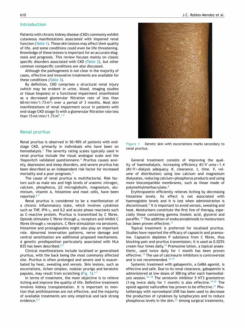
Clinical manifestations include localised or generalisedpruritus, with the back being the most commonly affectedsite. Pruritus is often prolonged and severe and is exacer-bated by heat, sweating and xerosis. Skin lesions, such asexcoriations, lichen simplex, nodular prurigo and keratoticpapules, may result from scratching (Fig. 1).6,7
In terms of treatment, the main objective is to relieveitching and improve the quality of life. Definitive treatmentinvolves kidney transplantation. It is important to men-tion that antihistamines are not effective, and the majorityof available treatments are only empirical and lack strongevidence.6,7
Figure 1 Xerotic skin with excoriations marks secondary to
renal pruritus.
General treatment consists of improving the qual-ity of haemodialysis, increasing efficiency Kt/V urea > 1.4(Kt/V = dialysis adequacy. K, clearance. t, time. V, vol-ume of distribution) using low calcium and magnesiumdialysates, reducing calcium---phosphorus products and usingmore biocompatible membranes, such as those made ofpolymethylmethacrylate.8
Erythropoietin efficiently relieves itching by decreasinghistamine levels. Its effect is not associated withhaemoglobin levels and it is lost when administration isdiscontinued.9 It is important to avoid xerosis, sweating andheat. Moisturisers constitute the first line of therapy, espe-cially those containing gamma linoleic acid, glycerol andparaffin.10 The addition of endocannabinoids to moisturisershas been proven effective.11
Topical treatment is preferred for localised pruritus.Studies have reported the efficacy of capsaicin and pramox-ine. Capsaicin depletes P substance from C fibres, thusblocking pain and pruritus transmission; it is used as 0.025%cream four times daily.12 Pramoxine lotion, a topical anaes-thetic, used twice daily for 1 month has been proveneffective.13 The use of calcineurin inhibitors is controversialand is not recommended.14,15
Systemic treatment with gabapentin, a GABA agonist, iseffective and safe. Due to its renal clearance, gabapentin isadministered at low doses of 300 mg after each haemodial-ysis session.16---18 The serotonin inhibitor 5 HT3 granisetron(1 mg twice daily for 1 month) is also effective.19,20 Theopioid agonist nalfurafine has proven to be effective.21 Pho-totherapy with narrowband UVB has been used to decreasethe production of cytokines by lymphocytes and to reducephosphorus levels in the skin.22 Among surgical treatments,
Skin manifestations of chronic kidney disease 611
Table 1 Classification of skin manifestations of chronic kidney disease and prevalence.
Classic specific disorders Prevalence Nonspecific disorders Prevalence
Renal pruritus 50---90% Xerosis 50---80%
Acquired perforating dermatosis 4.5---10% Pigmentary changes 20---50%
Nephrogenic systemic fibrosis Rare Skin infections 40%
Calciphylaxis 1---4% Uremic frost 1---3%
Porphyria cutanea tarda 1---18% Nail disorders 30---60%
Pseudoporphyria 1---18% Hair disorders 30---50%
Mucosal alterations 50---90%
parathyroidectomy can be effective in cases with sec-ondary hyperparathyroidism and high calcium---phosphorusproducts.23 However, kidney transplantation is the definitivetreatment.
Acquired perforating dermatosis (APD)
APD is commonly observed in patients with end-stage CKD,especially in individuals with diabetes mellitus and individ-uals receiving haemodialysis.24---26
The pathogenesis of APD is unclear. APD is associ-ated with diabetes mellitus, hypothyroidism, liver disease,malignancies and HIV infection. The mechanism involvestransepidermal elimination of dermal components (colla-gen, elastin and cell detritus). Proposed trigger factorsinclude minor trauma, such as scratching, microvascularchanges associated with diabetes mellitus, skin calciumdepositions with foreign body inflammatory response andgenetic predisposition.24,26
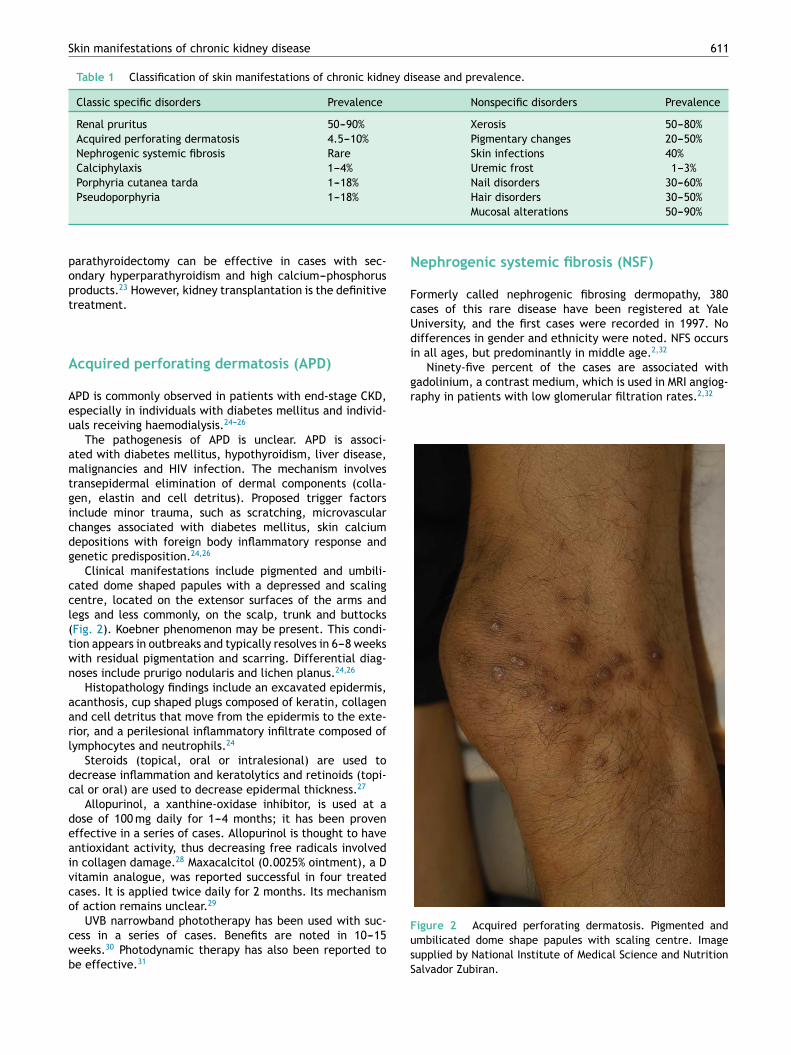
Clinical manifestations include pigmented and umbili-cated dome shaped papules with a depressed and scalingcentre, located on the extensor surfaces of the arms andlegs and less commonly, on the scalp, trunk and buttocks(Fig. 2). Koebner phenomenon may be present. This condi-tion appears in outbreaks and typically resolves in 6---8 weekswith residual pigmentation and scarring. Differential diag-noses include prurigo nodularis and lichen planus.24,26
Histopathology findings include an excavated epidermis,acanthosis, cup shaped plugs composed of keratin, collagenand cell detritus that move from the epidermis to the exte-rior, and a perilesional inflammatory infiltrate composed oflymphocytes and neutrophils.24
Steroids (topical, oral or intralesional) are used todecrease inflammation and keratolytics and retinoids (topi-cal or oral) are used to decrease epidermal thickness.27
Allopurinol, a xanthine-oxidase inhibitor, is used at adose of 100 mg daily for 1---4 months; it has been proveneffective in a series of cases. Allopurinol is thought to haveantioxidant activity, thus decreasing free radicals involvedin collagen damage.28 Maxacalcitol (0.0025% ointment), a Dvitamin analogue, was reported successful in four treatedcases. It is applied twice daily for 2 months. Its mechanismof action remains unclear.29
UVB narrowband phototherapy has been used with suc-cess in a series of cases. Benefits are noted in 10---15weeks.30 Photodynamic therapy has also been reported tobe effective.31
Nephrogenic systemic fibrosis (NSF)
Formerly called nephrogenic fibrosing dermopathy, 380cases of this rare disease have been registered at YaleUniversity, and the first cases were recorded in 1997. Nodifferences in gender and ethnicity were noted. NFS occursin all ages, but predominantly in middle age.2,32
Ninety-five percent of the cases are associated withgadolinium, a contrast medium, which is used in MRI angiog-raphy in patients with low glomerular filtration rates.2,32
Figure 2 Acquired perforating dermatosis. Pigmented and
umbilicated dome shape papules with scaling centre. Image
supplied by National Institute of Medical Science and Nutrition
Salvador Zubiran.
612
J.C.
Roble
s-Mendez
et
al.
Table 2 Summary of classic specific disorders.
Disease Risk factors Pathogenesis Clinical findings Histopathology Differential
diagnosis
Renal pruritus Hemodialysis,
high levels
of uraemic
nitrogen,
calcium and
phosphorus
Chronic inflammatory
state
C fibres stimulation via
opioids, serotonin,
histamine and
prostaglandins
Abnormal innervation
patterns, nerve damage
and central sensitisation
Genetic predisposition,
HLA B35
Localised or generalised
pruritus, with the back
being the most common
affected site
Pruritus is often
prolonged and severe
and is exacerbated by
heat, sweating and
xerosis. Excoriations,
lichen simplex, nodular
prurigo result from
scratching
Biopsy is usually not
required
Other local or
systemic disorders
that cause pruritus
Acquired
perforating
dermatosis
Diabetes
mellitus and
hemodialy-
sis
Hypothyroidism,
liver
disease,
malignan-
cies and HIV
infection
Transepidermal
elimination of dermal
components
Trigger factors include
minor trauma,
microvascular changes
and skin calcium
depositions
Genetic predisposition
Pigmented and
umbilicated dome
shaped papules with a
depressed and scaling
centre, located in the
extensor surfaces of the
arms and legs, and less
commonly, in the scalp,
trunk and buttocks
Acanthosis, cup shaped
plugs composed of
keratin, collagen and
cell detritus that move
from the epidermis to
the exterior, and a
perilesional
inflammatory infiltrate
composed of
lymphocytes and
neutrophils
Prurigo nodularis
and lichen planus
Nephrogenic
systemic
fibrosis
Gadolinium
use as
contrast
medium in
MRI
angiography
Gadodiamide
and
gadopente-
tate are the
most
associated
contrast
media
Genetic predisposition
(HLA A2) and an
inflammatory state,
allow gadolinium
effectively enters the
tissues
Gadolinium is
subsequently
phagocytosed by
macrophages, which
release cytokines that
attract circulating
fibrocytes to the tissues,
then the fibrocytes
differentiate into
fibroblast-like cells in
the dermis leading to
fibrosis
Indurated,
erythematous-
hyperchromic plaques
with an orange peel,
woody appearance. The
lesions are bilateral and
symmetrical on the legs
and forearms
Flexion contractures of
adjacent limbs
5% of patients
experience a rapid
fulminant course in 2
weeks with multiple
organ involvement
Thickened collagen
bundles with clefts,
mucin deposition and
proliferation of spindled
cells that stain positive
for CD34 and procollagen
1. Multinucleated cells
positive for CD68 and
factor VIII are also
present
Scleromyxedema,
systemic sclerosis,
eosinophilia-
myalgia syndrome,
toxic oil syndrome
and eosinophilic
fasciitis

Skin
manife
statio
ns
of
chro
nic
kidney
dise
ase
613
Table 2 (Continuación)
Disease Risk factors Pathogenesis Clinical findings Histopathology Differential
diagnosis
Calciphylaxis Secondary
hyper-
parathy-
roidism and
elevated
calcium---phosphorus
products
Increased
calcium---phosphorus
product
Imbalance between
inhibitors and inducers
of vascular calcification
Painful, pink or purple,
firm nodules or plaques,
surrounded by livedo
reticularis that progress
to central and deep
painful ulcers covered
with black eschar.
Calciphylaxis is bilateral
and symmetrical and
affects thighs, abdomen
and buttocks
Calcification of the
media, intimal
hyperplasia of dermal
and subcutaneous
arterioles, thrombosis of
dermal and
subcutaneous vessels
with ischaemic necrosis
in the epidermis and
lobular panniculitis with
fat necrosis
Systemic
vasculitis,
coagulopathies,
occlusive
vasculopathies and
bacterial and
fungal infections
Porphyria
cutanea
tarda
Hemodialysis
Type 1:
hepatitis B
and C,
HIV-AIDS,
alcohol,
oestrogen
and iron
Type 2 is a
hereditary,
autosomal
dominant
disease
A defect in heme
biosynthesis due to
uroporphyrinogen
decarboxylase deficiency
In CKD, low levels of
uroporphyrins are
excreted; their
accumulation in skin
causes phototoxicity
reactions
Blisters, erosions and
crusts that are located
on exposed areas, such
as the back of hands and
forearms and
occasionally the face and
feet. These lesions heal
with scarring or milia
and may be accompanied
of hypertrichosis and
hyperpigmentation and
esclerodermoid plaques
Subepidermal cleft with
minimal inflammation
and festooned papillary
dermis. Direct
immune-fluorescence
indicates linear and
granular deposition of
immunoglobulin G and
C3 in the
dermal-epidermal
junction and around the
vessels
Pseudoporphyria,
other hereditary
porphyrias
drug-related
phototoxic
reactions,
epidermolysis
bullosa acquisita
Pseudo-
porphyria
Phototoxic
drugs, such
as tetracy-
cline,
furosemide,
naproxen,
amiodarone,
nalidixic
acid and
isotretinoin,
and
exposure to
ultraviolet
light A
Phototoxic metabolites
are formed. Patients
with CKD have decreased
glutathione levels in
erythrocytes, making
them susceptible to
damage by free radicals
Blisters on the back of
hands, forearms and
other exposed areas that
resolve with scarring and
milia. Hypertrichosis on
the face or
sclerodermoid plaques
are rarely observed
Subepidermal cleft with
minimal inflammation.
Direct
immunofluorescence
staining indicates
immunoglobulin G and
C3 deposition at the
dermal-epidermal
junction and around
vessels
Hereditary
porphyrias,
drug-related
phototoxic
reactions,
epidermolysis
bullosa acquisita
MRI: magnetic resonance imaging, CKD: chronic kidney disease.

614 J.C. Robles-Mendez et al.
Table 3 Treatment of classic specific disorders associated to chronic kidney disease.
Disorder Treatment option Dosage Action mechanism
Renal pruritus Improving the quality of
hemodialysis
Increasing efficiency Kt/V
urea > 1.4, using low calcium
and magnesium dialysates and
using more biocompatible
membranes
Reduce calcium---phosphorus
products. Membranes of
polymethylmethacrylate
absorb and eliminate cytokines
Erythropoietin 20---50 U/kg is administered
thrice weekly
Decreasing histamine levels
Moisturiser (gamma linoleic
acid, glycerol and paraffin)
Three times a day Prevent xerosis
Capsaicin 0.025% cream four times daily Depletes P substance from C
fibres, thus blocking pain and
pruritus transmission
Pramoxine Twice daily for 1 month Topical anaesthetic
Gabapentin 300 mg after each hemodialysis
session
GABA agonist
Granisetron 1 mg twice daily for 1 month Serotonin inhibitor 5 HT3
Nalfurafine 5 �g thrice weekly by
intravenous infusion,
immediately after each
hemodialysis for 4 weeks
Opioid agonist. Inhibit C fibres
by � receptor
Narrowband UVB phototherapy Three sessions per week. Initial
dose of 200 mJ/cm2, increasing
100 mJ/cm2 at every session,
until a maximum dose of
1500 mJ/cm
Decrease the production of
cytokines by lymphocytes and
phosphorus levels in the skin
Parathyroidectomy Effective in cases with
secondary hyperparathyroidism
and high calcium-phosphorus
products
Kidney transplant Increased glomerular filtration
Acquired
perforating
dermatosis
Steroids (topical, oral or
intralesional)
Decrease inflammation
Keratolytics and retinoids
(topical or oral)
Decrease epidermal thickness
Allopurinol 100 mg daily for 1---4 months Xanthine-oxidase inhibitor. It
acts by an antioxidant action,
thus decreasing free radicals
involved in collagen damage
Maxacalcitol Twice daily for 2 months D vitamin analogue. Mechanism
of action remains unclear
Narrowband UVB phototherapy It begins at 400 mJ/cm2 until a
therapeutic range of
700---1600 mJ/cm2 is achieved.
Three sessions per week. After
improvement is achieved,
1---2 sessions a week for
maintenance
Anti-proliferative and
anti-inflammatory effects;
relief of pruritus
Photodynamic therapy Single session with
5-aminolevulinic acid
Production of reactive oxygen
species with cell apoptosis and
necrosis
Nephrogenic
Systemic
Fibrosis
Preventive measures Gadoteridol is the safest
option. In hemodialysis
patients, two 4-h sessions
should be performed in the
first 3 h to remove the contrast
Prednisone 1 mg/kg every 24 h Decrease inflammation

Skin manifestations of chronic kidney disease 615
Table 3 (Continuación)
Disorder Treatment option Dosage Action mechanism
Extracorporeal photopheresis Two treatments on consecutive
days every 2 weeks for 6
months, then two treatments
on consecutive days every 4
weeks for 6 months
Enhances the suppression of
signals by inducing
lymphocytes apoptosis and
deflecting the signal of
antigen-presenting cells
Kidney transplant Increased glomerular filtration
and immunosuppression in
transplantation
Imatinib mesylate 400---600 mg/day are
administered, for 4---24 weeks
Tyrosine kinase inhibitor, acts
by inhibiting platelet-derived
growth factor and c-Abl kinase,
thus inhibiting the synthesis of
collagen and fibronectin by
dermal fibroblasts
UV-A 1 phototherapy Three sessions per week, at
60---120 J/cm2 in 22---50 sessions
Alters the cytokine levels
relating to fibrocytes to
promote a less fibrotic
environment
Physical therapy Improves the performance of
daily activities and personal
care
Plasmapheresis,
immunoglobulin G and
photodynamic therapy
Anecdotal success
Calciphylaxis Total or subtotal surgical
parathyroidectomy
Reducing the production of
parathyroid hormone
Cinacalcet 30---60 mg/day orally Calcium mimetic that increases
sensitivity of the calcium
receptor in the parathyroid
gland, thus reducing the
production of parathyroid
hormone
Etindronate 200 mg/day orally for 14 days Inhibit vascular calcification
Pamidronate 30 mg intravenously for five
treatments over a period of 2
weeks
Inhibit vascular calcification
Prednisone 30---50 mg orally every 24 h, for
3---5 weeks
Decrease inflammation
Sodium thiosulfate 25 g is administered
intravenously for 30---60 min,
three times a week after each
hemodialysis session
Displacing calcium from
calcium phosphate, to form
calcium thiosulfate, which is
more soluble
Improving the quality of
hemodialysis
Increasing the frequency and
duration of hemodialysis
through the use of low calcium
dialysate
Reduce calcium---phosphorus
products
Sevelamer Phosphorus chelator that
reduces the intestinal
absorption of phosphorus.
Reduce calcium---phosphorus
products
General wound management Debridement of necrotic
tissue, antibiotics, analgesia,
cures and vacuum dressing
To prevent infection and sepsis
Hyperbaric oxygen therapy Daily sessions for 60---90 min
per week with 100% oxygen at
2.5 atmospheres of pressure
Stimulates angiogenesis and is
toxic to anaerobic bacteria
616 J.C. Robles-Mendez et al.
Table 3 (Continuación)
Disorder Treatment option Dosage Action mechanism
Cultured autologous fibroblasts
and keratinocytes, adding
iloprost
35 consecutive days To coat ulcers. Iloprost is a
vasodilator prostacyclin
analogue
Ulcer debridement, coated
with partial autologous skin
grafts
To prevent infection and
sepsis, and to coat ulcer
Porphyria
cutanea tarda
Preventive measures Avoid sun exposure, use zinc
oxide and titanium oxide-based
sunscreen, avoid alcohol,
estrogens, iron and treat
comorbidities
Avoiding exacerbating factors
Improving the quality of
hemodialysis
To use high-flux membranes
(polysulfone) with high flux
dialysis (300 ml/h), during 4 h
of hemodialysis, three times a
week
Removing high plasma levels of
uroporphyrins
Phlebotomy Low volume of 100 ml, twice
weekly for 8 months, followed
by 50 ml per week thereafter
Decrease in body stores of iron.
Activating uroporphyrinogen
decarboxylase
Erythropoietin 20---50 U/kg is administered
thrice weekly. When
erythropoietin is combined
with phlebotomy, a dose of
150---200 U/kg is used
Decrease in body stores of iron.
Activating uroporphyrinogen
decarboxylase
Deferoxamine 2 g is administered
intravenously every
hemodialysis.
Chelating agent used to
remove iron
Plasmapheresis Two sessions, separated by 48 h Removing high plasma levels of
uroporphyrins
Renal transplant Increasing glomerular
filtration, thus excreting
uroporphyrins
Pseudo-
porphyria
Preventive measures Avoid sun exposure, trauma
and related drugs
Avoiding exacerbating factors
N-acetyl-cysteine 600---1200 mg/day orally for
weeks
Precursor of glutathione with
antioxidant properties
Gadodiamide and gadopentetate are the most commoncauses of contrast media associated with NSF. The poorer theglomerular filtration rate, the longer the half-life of gadolin-ium. In addition, 73.8% and 92.4% of gadolinium is excretedin a single haemodialysis session or two sessions, respec-tively, but gadolinium is not properly excreted in peritonealdialysis.2,32
In patients with a genetic predisposition (HLA A2)or an inflammatory state, a hypercoagulable state orendothelial damage, gadolinium enters the tissues, 35---150-fold more effectively compared with healthy patients.Gadolinium is subsequently phagocytosed by macrophages,which release cytokines that attract circulating fibrocytes(CD34/procollagen I+) to the tissues. Once in the tissue,the fibrocytes differentiate into fibroblast-like cells in thedermis, leading to fibrosis.2,32
Clinical manifestations include indurated, erythematous-hyperchromic plaques with an orange peel, woody orcobblestone appearance. Nodules and bullae may be present
on the hands and feet, or yellowish edematous plaques arenoted on the sclera (Fig. 3). The lesions are bilateral andsymmetrical on the legs and forearms. The condition is occa-sionally observed on the trunk and buttocks and generallyspares the face.2,32
A burning, itching, and stinging pain occurs. Flex-ion contractures of adjacent limbs are noted, daysto weeks after exposure to the contrast, and thecondition tends to be chronic. Remission is achievedin a few cases, leaving atrophic and hypopigmentedlesions; 5% of patients experience a rapid fulminantcourse in 2 weeks with multiple organ involvement(lungs, heart and oesophagus). Differential diagnosesinclude scleromyxedema, systemic sclerosis, eosinophilia-myalgia syndrome, toxic oil syndrome and eosinophilicfasciitis.2,32
The histopathology is characterised by thickened colla-gen bundles with clefts, mucin deposition and proliferationof spindled cells that stain positive for CD34 and procollagen

Skin manifestations of chronic kidney disease 617
Figure 3 Nephrogenic systemic fibrosis. Indurated hyper-
chromic plaques with orange peel and woody appearance.
Image supplied by National Institute of Medical Science and
Nutrition Salvador Zubiran.
1. Multinucleated cells positive for CD68 and factor VIII arealso present.2,24,32
For prevention, if a macrocyclic contrast medium mustbe used, gadoteridol is the safest option. In haemodialy-sis patients, two 4-h sessions are required to remove thecontrast; these sessions should be performed in the first3 h.33
Most treatments demonstrate efficacy in anecdotal andcases reports. Steroids and immunosuppressants are gener-ally ineffective. Oral prednisone may be used with someefficacy, but many adverse effects are observed.33
Extracorporeal photopheresis is available in a few cen-tres, but is prohibitively expensive. It has been proven to beeffective in all patients, increasing softening of injuries andmobility ranges. Extracorporeal photopheresis enhances thesuppression of signals by inducing lymphocyte apoptosis anddeflecting the signal of antigen-presenting cells.34
Kidney transplantation offers improvement in 54% ofpatients and full remission has been achieved in some cases.It improves mobility ranges of flexion contracture, likely dueto increased glomerular filtration and immunosuppressionafter transplantation.35,36
Imatinib mesylate, a tyrosine kinase inhibitor, acts byinhibiting platelet-derived growth factor and c-Abl kinase,thus inhibiting the synthesis of collagen and fibronectin bydermal fibroblasts. Doses of 400---600 mg/day are adminis-tered for 4---24 weeks, achieving mild to moderate success.The lesions are softened, but little improvement is noted inthe range of motion.37
UV-A 1 phototherapy has been proven effective insmoothing skin lesions, with little improvement in range ofmotion. It is used at 60---120 J/cm2 in 22---50 sessions.38
Plasmapheresis and immunoglobulin G exhibit anecdotalsuccess.39 Physical therapy improves the performance ofdaily activities and personal care, with little improvementin the range of motion.40 In a case report, photodynamictherapy was effective along with kidney transplantation.41
Figure 4 Calciphylaxis. Ulcers covered with black eschar.
Calciphylaxis
Calciphylaxis occurs in patients with CKD on hemodialy-sis; it is associated with secondary hyperparathyroidism andelevated calcium-phosphorus products. Calciphylaxis is anobliterative vasculopathy caused by the deposition of cal-cium in the arterioles of the skin, leading to thrombosis andischaemic necrosis with ulceration.2
Calciphylaxis is commonly observed in women, inpatients with diabetes mellitus, obesity, hypoalbuminemia,liver cirrhosis, those who use warfarin, and those with sys-temic inflammation and malignancy. Sepsis can cause up to80% mortality via skin ulcer infection.2
Its ethiopathogenic mechanism is unclear. Predisposi-tion plus a causal factor are necessary. Calcium---phosphorusproducts are increased in the majority of patients, but thisincrease is not sufficient alone to cause the condition. Animbalance between inhibitors and inducers of vascular calci-fication is noted. Osteopontin and bone morphogenic protein2 serve as inducers, whereas GI protein matrix, fetuin andpyrophosphate act as inhibitors. Aluminium deposition mayalso be an inductor.2
Clinical manifestations include painful, pink or purple,firm nodules or plaques, surrounded by livedo reticularisthat progress to central and deep, painful ulcers coveredwith black eschar and a reticulated depigmentation in theperiphery (Fig. 4). Calciphylaxis is bilateral and symmet-rical and affects adipose tissue areas, such as the thighs,abdomen and buttocks.42,43
Histopathologic findings include calcification of themedia, intimal hyperplasia of dermal and subcutaneousarterioles, thrombosis of dermal and subcutaneous ves-sels with ischaemic necrosis in the epidermis and lobularpanniculitis with fat necrosis. On radiography, linear cal-cium deposits are observed. The involvement of smallblood vessels (0.5 mm) is the most specific sign; how-ever, many patients exhibit vascular calcification withoutcalciphylaxis.42,43
For therapy, early total or subtotal surgical parathy-roidectomy increases survival. Cinacalcet is a calciummimetic that increases sensitivity of the calcium receptorin the parathyroid gland, thus reducing the production of

618 J.C. Robles-Mendez et al.
parathyroid hormone. Cinacalcet is used in cases whereparathyroidectomy is delayed or contraindicated. Treat-ment is discontinued once PTH reaches a normal level of1.5 to prevent renal osteodystrophy.44
Among bisphosphonates, etindronate and pamidronatehave proven to be effective. Prednisone is used in earlyunulcerated plaques.44
Sodium thiosulfate is administered either orally daily orintravenously at the end of haemodialysis for 3---5 months.Sodium thiosulfate offers symptomatic and radiologicalrelief of tissue calcification, displacing calcium from calciumphosphate, to form calcium thiosulfate, which is more solu-ble than other salts and is excreted either by haemodialysisor by the kidneys. Sodium thiosulfate (25 g) is administeredintravenously for 30---60 min, three times a week after eachhaemodialysis session. Metabolic acidosis is an importantside effect of this drug.44
Dialysis improves this condition through using low cal-cium dialysate and increasing the frecuency and durationof hemodialysis sessions. Sevelamer is a phosphorus chela-tor that reduces the intestinal absorption of phosphorus.Calcium---phosphorus product levels below 55 mg/dl are theobjective.44
In wound management, debridement of necrotic tissue,antibiotics, analgesia, and vacuum dressing are important.Hyperbaric oxygen therapy stimulates angiogenesis and istoxic to anaerobic bacteria. It is used in daily sessions for60---90 min per week with 100% oxygen at 2.5 atmospheresof pressure. Ulcer debridement is coated with partial autol-ogous skin grafts. Successful attempts have been madeto coat ulcers with cultured autologous fibroblasts andkeratinocytes by adding iloprost infusion (vasodilator pros-tacyclin analogue) for 35 consecutive days.44
Porphyria cutanea tarda
Porphyria cutanea tarda (PCT) occurs in 1.2---18% of patientswith CKD and is more frequently observed in patientson haemodialysis. It is a bullous dermatosis caused byphototoxicity due to uroporphyrinogen decarboxylase defi-ciency, thus causing elevated levels of uroporphyrin andisocoproporphyrin.42,45,46
PCT is classified as Type 1 when it is sporadic andacquired, and is caused by a liver enzyme deficiency. Itis associated with hepatitis B and C, HIV-AIDS, alcoholconsumption, oestrogen and iron. Type 2 is a hereditary,autosomal dominant disease caused by enzyme deficiencyin all tissues. A defect in heme biosynthesis is dueto uroporphyrinogen decarboxylase deficiency. It requiresless than 30% activity of the enzyme to induce clinicalmanifestations.42 In CKD, low levels of uroporphyrins areexcreted and accumulate, causing phototoxicity reactions.Uroporphyrins cannot be removed by haemodialysis, buthigh-flux membranes improve elimination.42,45,46
Clinical manifestations include blisters, erosions andcrusts that are located on exposed areas, such as the backof hands and forearms and occasionally the face and feet(Fig. 5). These lesions heal with scarring or milia and maybe accompanied by hypertrichosis and hyperpigmentationof the face and by esclerodermoid plaques on the hands andface.42,45,46
Figure 5 Porphyria cutanea tarda. Erosions and crusts on the
back of the hands. Image supplied by National Institute of Medi-
cal Science and Nutrition Salvador Zubiran.
Laboratory findings include increased serum iron andferritin, high urine levels of uroporphyrins I and III, and7---8-carboxyl uroporphyrins, high plasma levels of uropor-phyrin and increased levels of isocoproporphyrin III in thefaeces.42,45
Histopathology reveals a subepidermal cleft with minimalinflammation and festooned papillary dermis in the base ofthe cleft. The vessel wall is thickened. Direct immunofluo-rescence staining indicates linear and granular deposition ofimmunoglobulin G and C3 in the dermal-epidermal junctionand around the vessels.42,45
For the treatment of PCT, sun exposure should beavoided; the use of zinc oxide and titanium oxide-based sun-screen is recommended. Treatment of comorbidities and toavoid exacerbating factors (iron and alcohol consumption,and estrogen using) are also important. Furthermore, it isimportant to use high-flux membranes (polysulfone) withhigh-flux dialysis (300 ml/h), during 4 h of haemodialysis,three times a week.47
Reduce body stores of iron in body stores of iron.Iron induces the enzyme 5-aminolevulinate synthase, whichsuppresses uroporphyrinogen decarboxylase. Further, itincreases conversion of uroporphyrinogen into porphyrins.Phlebotomy is used at a low volume of 100 ml, twice weeklyfor 8 months, followed by 50 ml per week thereafter; normalphlebotomy is not tolerated due to anaemia.47 Erythro-poietin (20---50 U/kg) is administered thrice weekly. Whenerythropoietin is combined with phlebotomy, a dose of150---200 U/kg, three times per week is used. Deferoxam-ine (2 g) is administered intravenously every haemodialysissession and is associated with long-term adverse effects.47
In refractory cases, two plasma exchange treatments, sep-arated by 48 h, are applied. Renal transplantation is thedefinitive treatment.47
Pseudoporphyria
Pseudoporphyria is a bullous dermatosis induced by pho-totoxicity that exhibits the clinical and histopathologic
Skin manifestations of chronic kidney disease 619
features of PCT; however, normal uroporphyrin levels areobserved. The condition is associated with the use of pho-totoxic drugs such as tetracycline, furosemide, naproxen,amiodarone, nalidixic acid and isotretinoin, and exposureto ultraviolet light A.42,45
Regarding pathogenesis, phototoxic metabolites areformed. Patients with CKD have decreased glutathione levelsin erythrocytes, making them susceptible to damage by freeradicals. N acetyl-cysteine, a precursor of glutathione withantioxidant properties, is used to effectively treat thesepatients.42,45
Clinical manifestations include blisters on the back ofthe hands, forearms and other exposed areas that resolvewith scarring and milia. Hypertrichosis on the face or scle-rodermoid plaques is rarely observed. Uroporphyrin levels inplasma or urine are normal or slightly elevated.42,45
Histopathology reveals a subepidermal cleft with minimalinflammation and a thick vessel wall. Direct immuno-fluorescence staining indicates immunoglobulin G and C3deposition at the dermal-epidermal junction and aroundvessels.42,45
N-acetyl-cysteine, a precursor of glutathione with antiox-idant properties, has been effective therapy in several casereports. An oral dose of 600---1200 mg/day offers relief in4-8 of weeks. Recurrence has been reported after discontin-uing medication. It is recommendable to avoid exacerbatingfactors such as sun exposure, trauma and related drugs.48
Benign nodular calcification
Firm papules, plaques and nodules appear in the jointsand fingertips due to metastatic calcification affectingskin. Von Kossa stains calcium deposits black. Treatment isbased on reducing calcium---phosphorus products and surgi-cal removal.42
Xerosis
Xerosis is the most common skin manifestation of CKD,occurring in 80% of patients. Etiopathogenic mechanismsinclude: Decreased hydration of the stratum corneum;decreased sweat gland and sebaceous gland size along withabnormal function related to hypervitaminosis A in dialy-sis patients, and the use of diuretics. Treatment is basedon skin hydration and treatment of pruritus. Repetitivebathing must be avoided and the use of soft, neutral soapsis recommended.45
Pigmentary changes
Dark brown pigmentation is caused by increased melaninproduction due to �-melanocyte stimulating hormone accu-mulation, which is caused by decreased excretion due tokidney failure. The yellowish tinge is caused by the accu-mulation of carotenoids and urochrome in the skin. Palloris caused by anaemia due to ineffective erythropoiesis andincreased haemolysis. Treatment is based on avoiding sunexposure and involves sunscreen use for hyperpigmenta-tion as well as erythropoietin for anaemia associated withpallor.49,50
Skin infections
Skin infections are caused by impaired cellular and humouralimmunity. They are more common when diabetic nephropa-thy is the origin. The most common fungal infection isonychomycosis. The most frequent viral infections are com-mon warts, herpes simplex and herpes zoster.49,50
Uremic frost
Uremic frost is currently a rare manifestation. Howeverwhen observed, the condition is often accompanied withincreased BUN, ranging from 250 to 300 mg/dl. It is caused bythe accumulation of urea in sweat. When the sweat evap-orates, a crystal deposit remains on the skin that appearsas a yellowish-white coating on the beard area, neck andtrunk.49,50
Nail disorders
Mees lines. These transverse bands of leukonychia are analteration of the nail plate that occurs during periods ofstress. In addition to its association with CKD, this condi-tion appears in cases of poisoning by arsenic, thallium,and fluorine as well as severe infections, heart disease andmalignancy.51
Muehrcke’s lines. Double white transverse lines arecaused by hypoalbuminemia related to nephrotic syndrome;Muehrcke lines are associated with serum albumin levels lessthan 2.2 g/dl. It originates by vascular compression throughlocal oedema, which causes bleaching of the nail bed. There-fore, no change in position occurs as the nail grows. It hasalso been observed in cases of cirrhosis, malnutrition andchemotherapy use.52
Lindsay nails (half and half). This condition is relatedto azotaemia. A white proximal area and a distal pink red-brown area that does not fade under pressure are observed(Fig. 6). No changes are noted with nail growth because thelesion is located in the nail bed. An increase in the numberof capillaries in the distal nail plate and a deposit of melaninpigmentation in the distal area are observed. This conditionresolves with renal transplantation.53
Other nail changes observed include leukonychia,koilonychia and splinter haemorrhages.49,50
Hair disorders
Diffuse alopecia, the most common manifestation, is causedby telogen effluvium; xerosis, pruritus and medication use(Fig. 7). Sparse body hair is also present. Discolorationand dryness of the hair are attributed to a decreasedsebum production. Treatment for hair loss is based on meet-ing nutritional requirements, which are increased in thesepatients.49,50
Mucosal alterations
Xerostomia is the most common abnormality and is causedby oral breathing and dehydration. Macroglossia is also oftenobserved (Fig. 8). Ulcerative stomatitis is observed with urea

620 J.C. Robles-Mendez et al.
Figure 6 Half and Half nail. A white proximal area and a red-
brown distal area are observed.
Figure 7 Diffuse alopecia.
Figure 8 Macroglossia. An enlarged tongue framed in its edge
by teeth, pallor is also observed in the perioral skin.
serum levels greater 150 mg/dl and is associated with poorhygiene. Uraemic breath is due to increased urea concentra-tions in saliva and its transformation into ammonium. Thecondition is associated with urea levels >200 mg/dl. Treat-ment is based on maintaining good oral hygiene and meetingnutritional requirements.49,50
Conclusions
Skin manifestations are very common in end-stage CKD.Severe renal pruritus is associated with increased mortalityand a poor prognosis. Nephrogenic systemic fibrosis is a rela-tively new preventable disease associated with gadolinium.In a simple exploration of the nails, some findings indicatealterations in albumin and urea levels. Comorbidities suchas diabetes mellitus and secondary hyperparathyroidism canlead to acquired perforating dermatosis and calciphylaxis,respectively. Effective and innovative treatments are avail-able for all of these conditions.
Ethical responsibilities
Protection of human and animal subjects. The authorsdeclare that no experiments were performed on humans oranimals for this investigation.
Skin manifestations of chronic kidney disease 621
Confidentiality of data. The authors declare that they havefollowed the protocols of their work center on the publica-tion of patient data.
Right to privacy and informed consent. The authors musthave obtained the informed consent of the patients and/orsubjects mentioned in the article. The author for correspon-dence must be in possession of this document.
Conflict of interest
The authors declare that they have no conflict of interest.
References
1. Stevens PE, Levin A, Kidney Disease: Improving Global Out-comes Chronic Kidney Disease Guideline Development WorkGroup Members. Evaluation and management of chronic kid-ney disease: synopsis of the kidney disease: improving globaloutcomes 2012 clinical practice guideline. Ann Intern Med.2013;158:825---30.
2. Brewster UC. Dermatological disease in patients with CKD. AmJ Kidney Dis. 2008;51:331---44.
3. Dar NR, Akhter A. Clinical characteristics of uremic pruritus inpatients undergoing haemodialysis. J Coll Physicians Surg Pak.2006;16:94---6.
4. Zucker I, Yosipovitch G, David M, Gafter U, Boner G. Prevalenceand characterization of uremic pruritus in patients undergo-ing hemodialysis: uremic pruritus is still a major problem forpatients with end-stage renal disease. J Am Acad Dermatol.2003;49:842---6.
5. Narita I, Alchi B, Omori K, Sato F, Ajiro J, Saga D, et al. Etiologyand prognostic significance of severe uremic pruritus in chronichemodialysis patients. Kidney Int. 2006;69:1626---32.
6. Kfoury LW, Jurdi MA. Uremic pruritus. J Nephrol. 2012;25:644---52.
7. Lugon JR. Uremic pruritus: a review. Hemodial Int. 2005;9:180---8.
8. Hiroshige K, Kabashima N, Takasugi M, Kuroiwa A. Optimal dial-ysis improves uremic pruritus. Am J Kidney Dis. 1995;25:413---9.
9. Pascual J, Teruel JL, Ortuno J. Erythropoietin therapy for ure-mic pruritus. N Engl J Med. 1992;327:734, author reply 734---735.
10. Chen YC, Chiu WT, Wu MS. Therapeutic effect of topical gamma-linolenic acid on refractory uremic pruritus. Am J Kidney Dis.2006;48:69---76.
11. Szepietowski JC, Reich A, Szepietowski T. Emollients with endo-cannabinoids in the treatment of uremic pruritus: discussion ofthe therapeutic options. Ther Apher Dial. 2005;9:277---9.
12. Tarng DC, Cho YL, Liu HN, Huang TP. Hemodialysis-related pru-ritus: a double-blind, placebo-controlled, crossover study ofcapsaicin 0.025% cream. Nephron. 1996;72:617---22.
13. Young TA, Patel TS, Camacho F, Clark A, Freedman BI, Kaur M,et al. A pramoxine-based anti-itch lotion is more effective thana control lotion for the treatment of uremic pruritus in adulthemodialysis patients. J Dermatolog Treat. 2009;20:76---81.
14. Pauli-Magnus C, Klumpp S, Alscher DM, Kuhlmann U, MettangT. Short-term efficacy of tacrolimus ointment in severe uremicpruritus. Perit Dial Int. 2000;20:802---3.
15. Duque MI, Yosipovitch G, Fleischer AB, Willard J, FreedmanBI. Lack of efficacy of tacrolimus ointment 0.1% for treatmentof hemodialysis-related pruritus: a randomized, double-blind,vehicle-controlled study. J Am Acad Dermatol. 2005;52 3 Pt1:519---21.
16. Vila T, Gommer J, Scates AC. Role of gabapentin in the treat-ment of uremic pruritus. Ann Pharmacother. 2008;42:1080---4.
17. Gunal AI, Ozalp G, Yoldas TK, Gunal SY, Kirciman E, CelikerH. Gabapentin therapy for pruritus in haemodialysis patients:a randomized, placebo-controlled, double-blind trial. NephrolDial Transplant. 2004;19:3137---9.
18. Razeghi E, Eskandari D, Ganji MR, Meysamie AP, Togha M,Khashayar P. Gabapentin and uremic pruritus in hemodialysispatients. Ren Fail. 2009;31:85---90.
19. Ashmore SD, Jones CH, Newstead CG, Daly MJ, ChrystynH. Ondansetron therapy for uremic pruritus in hemodialysispatients. Am J Kidney Dis. 2000;35:827---31.
20. Layegh P, Mojahedi MJ, Malekshah PE, Pezeshkpour F, Vahe-dian M, Nazemian F, et al. Effect of oral granisetron in uremicpruritus. Indian J Dermatol Venereol Leprol. 2007;73:231---4.
21. Wikström B, Gellert R, Ladefoged SD, Danda Y, Akai M, Ide K,et al. Kappa-opioid system in uremic pruritus: multicenter, ran-domized, double-blind, placebo-controlled clinical studies. JAm Soc Nephrol. 2005;16:3742---7.
22. Ada S, Seckin D, Budakoglu I, Ozdemir FN. Treatment of uremicpruritus with narrowband ultraviolet B phototherapy: an openpilot study. J Am Acad Dermatol. 2005;53:149---51.
23. Chou FF, Ho JC, Huang SC, Sheen-Chen SM. A study on pruritusafter parathyroidectomy for secondary hyperparathyroidism. JAm Coll Surg. 2000;190:65---70.
24. Kurban MS, Boueiz A, Kibbi AG. Cutaneous manifestations ofchronic kidney disease. Clin Dermatol. 2008;26:255---64.
25. Hari Kumar KV, Prajapati J, Pavan G, Parthasarathy A, JhaR, Modi KD. Acquired perforating dermatoses in patientswith diabetic kidney disease on hemodialysis. Hemodial Int.2010;14:73---7.
26. González-Lara L, Gómez-Bernal S, Vázquez-López F, Vivanco-Allende B. Acquired perforating dermatosis: a report of 8 cases.Actas Dermosifiliogr. 2014;105:e39---43.
27. Wagner G, Sachse MM. Acquired reactive perforating dermato-sis. J Dtsch Dermatol Ges. 2013;11:723---30, 723---729.
28. Tilz H, Becker JC, Legat F, Schettini AP, Inzinger M, Massone C.Allopurinol in the treatment of acquired reactive perforatingcollagenosis. An Bras Dermatol. 2013;88:94---7.
29. Yoshiya H, Okuyama R, Uhara H. Effectiveness of topical maxa-calcitol for acquired perforating disorder. J Am Acad Dermatol.2013;68:e181---2.
30. Ohe S, Danno K, Sasaki H, Isei T, Okamoto H, Horio T. Treatmentof acquired perforating dermatosis with narrowband ultravioletB. J Am Acad Dermatol. 2004;50:892---4.
31. Sezer E, Erkek E. Acquired perforating dermatosis successfullytreated with photodynamic therapy. Photodermatol Photoim-munol Photomed. 2012;28:50---2.
32. Girardi M, Kay J, Elston DM, Leboit PE, Abu-Alfa A, CowperSE. Nephrogenic systemic fibrosis: clinicopathological defini-tion and workup recommendations. J Am Acad Dermatol.2011;65:1095---106, e1097.
33. Knopp EA, Cowper SE. Nephrogenic systemic fibrosis: earlyrecognition and treatment. Semin Dial. 2008;21:123---8.
34. Mathur K, Morris S, Deighan C, Green R, Douglas KW.Extracorporeal photopheresis improves nephrogenic fibrosingdermopathy/nephrogenic systemic fibrosis: three case reportsand review of literature. J Clin Apher. 2008;23:144---50.
35. Leung N, Shaikh A, Cosio FG, Griffin MD, Textor SC, Gloor JM,et al. The outcome of patients with nephrogenic systemic fibro-sis after successful kidney transplantation. Am J Transplant.2010;10:558---62.
36. Panesar M, Banerjee S, Barone GW. Clinical improvement ofnephrogenic systemic fibrosis after kidney transplantation. ClinTransplant. 2008;22:803---8.
37. Elmholdt TR, Buus NH, Ramsing M, Olesen AB. Antifibrotic effectafter low-dose imatinib mesylate treatment in patients withnephrogenic systemic fibrosis: an open-label non-randomized,uncontrolled clinical trial. J Eur Acad Dermatol Venereol.2013;27:779---84.

622 J.C. Robles-Mendez et al.
38. Tran KT, Prather HB, Cockerell CJ, Jacobe H. UV-A1 therapy fornephrogenic systemic fibrosis. Arch Dermatol. 2009;14:1170---4.
39. Poisson JL, Low A, Park YA. The treatment of nephrogenic sys-temic fibrosis with therapeutic plasma exchange. J Clin Apher.2013;28:317---20.
40. Ramaizel L, Sliwa JA. Rehabilitation in nephrogenic systemicfibrosis. PM&R. 2009;1:684---6.
41. Schmook T, Budde K, Ulrich C, Neumayer HH, Fritsche L,Stockfleth E. Successful treatment of nephrogenic fibrosing der-mopathy in a kidney transplant recipient with photodynamictherapy. Nephrol Dial Transplant. 2005;20:220---2.
42. Robinson-Bostom L, DiGiovanna JJ. Cutaneous manifestations ofend-stage renal disease. J Am Acad Dermatol. 2000;43:975---86,quiz 987---990.
43. Daudén E, Onate MJ. Calciphylaxis. Dermatol Clin. 2008;26:557---68, ix.
44. Ong S, Coulson IH. Diagnosis and treatment of calciphylaxis.Skinmed. 2012;10:166---70.
45. Markova A, Lester J, Wang J, Robinson-Bostom L. Diagnosis ofcommon dermopathies in dialysis patients: a review and update.Semin Dial. 2012;25:408---18.
46. Pérez L, Fernández-Redondo V, Toribio J. Porphyria cutaneatarda in a dialyzed female patient. Actas Dermosifiliogr.2006;97:115---7.
47. Shieh S, Cohen JL, Lim HW. Management of porphyria cutaneatarda in the setting of chronic renal failure: a case report andreview. J Am Acad Dermatol. 2000;42:645---52.
48. Guiotoku MM, Pereira FeP, Miot HA, Marques ME. Pseudopor-phyria induced by dialysis treated with oral N-acetylcysteine.An Bras Dermatol. 2011;86:383---5.
49. Hajheydari Z, Makhlough A. Cutaneous and mucosal manifesta-tions in patients on maintenance hemodialysis: a study of 101patients in Sari, Iran. Iran J Kidney Dis. 2008;2:86---90.
50. Udayakumar P, Balasubramanian S, Ramalingam KS, Lakshmi C,Srinivas CR, Mathew AC. Cutaneous manifestations in patientswith chronic renal failure on hemodialysis. Indian J DermatolVenereol Leprol. 2006;72:119---25.
51. Chauhan S, D’Cruz S, Singh R, Sachdev A. Mees’ lines. Lancet.2008;372:1410.
52. Short N, Shah C. Muehrcke’s lines. Am J Med. 2010;123:991---2.53. Lin CJ, Wu CJ, Chen YC, Chen HH. Half and half nail secondary
to chronic renal failure. South Med J. 2009;102:1189---90.