skin integrity and pressure injuries following spinal cord ...skin integrity and pressure injuries...
TRANSCRIPT

www.scireproject.com Version 7.0
Skin Integrity and Pressure Injuries Following Spinal Cord Injury
Jane Hsieh
Brooke Benton Laura Titus
Sharon Gabison Amanda McIntyre
Dalton Wolfe Robert Teasell

Key Points Introduction The early detection of suspected pressure injuries in individuals with SCI may be improved through the use of a handheld dermal phase meter and ultrasonography. Magnetic resonance imaging may be helpful to anticipate the development of osteomyelitis secondary to non-healing SCI-related pressure injuries. Circulatory biomarkers in people with SCI have not yet proven to be useful or feasible to enhance early detection of suspected pressure injuries. Prevention Electrical stimulation has potential to reduce IT pressures by activating muscles, increasing blood flow and tissue oxygenation to stimulated area, all of which likely helps to prevent pressure injury formation or progression. Fat grafting may have potential as a prevention strategy for those people where other strategies have not been successful; ongoing pressure management strategies are still required post grafting. Pressure mapping studies using able-bodied subjects should not be generalized to the SCI population because pressure differences exist between the two groups. Data generated from pressure mapping studies on seniors should not be generalized to the SCI population because differences exist between the two populations. Early attendance at specialized seating assessment clinics should be part of a comprehensive rehabilitation program. More research is needed to determine if early attendance at a specialized seating assessment clinic (SSA) results in pressure injury prevention over time. Structured pressure injury prevention education, helps individuals post SCI gain and retain knowledge of pressure injury prevention practices, but it is questioned if the same strategies are effective for those with chronic and/or severe pressure injuries. More research is needed to determine the best approaches of pressure injury prevention intervention to reduce pressure injuries post SCI, particularly for chronic and/or severe pressure injuries, to assist with lifestyle and behaviour changes for long term pressure management success. The role of telerehabilitation in engaging individuals with SCI with prevention education and treatment programs has demonstrated potential but to be fully successful, requires a compliment between program content, delivery format and accessibility to that format for all people with an SCI regardless of living situations. Products and surfaces used for prevention should be combined with other preventative measures/strategies to optimize the potential to reduce risk of pressure injury development.

Treatment Electrical stimulation added to standard wound management promotes healing of Stage III and IV pressure injuries post SCI. More research is needed to determine optimum electric current and application protocols to improve healing of pressure injuries post SCI. Laser treatment does not improve pressure injury healing post SCI. US/UVC should be considered as an adjunct treatment when pressure injuries are not healing with standard wound care post SCI. Pulsed electromagnetic energy improves wound healing in Stage II and Stage III pressure injuries post SCI. Wound headling is improved with intermittent negative pressure (INP) devices in combination with standard wound care (SWC) for at-home care of pressure injuries compared to SWC alone. Negative pressure wound therapy (NPWT) has shown to reduce levels of MMP-8, increase the rate of healing, reduce exudate production and enhance the rate of formation of red granulation tissue when compared to conventional wet gauze alone. Pressure injury healing after a SCI is improved when topical negative pressure (TNP) therapy is administered as compared to traditional sodium hypochlorite dressing changes. VAC therapy may be quite a versatile device but has some disadvantages. Only qualified medical/paramedical personnel should use it in order to avoid possible complications that can occur after an improper application. Normothermic dressings may improve healing of pressure injuries post SCI. Recombinant human erythropoietin shows promise in assisting with the healing of stage IV chronic non-healing pressure injuries post SCI. Platelet-rich plasma therapy may be a promising alternative to standard saline dressing for pressure injury healing, however additional study is required to validate PRP therapy as a possible treatment for severe, non-healing pressure injuries in people with SCI. Local application of PRP may reduce bacterial presence and colonization in PIs. The anabolic steroid agent Oxandrolone does not promote healing of serious pressure injuries post SCI. Occlusive hydrocolloid dressings are useful for healing of stage I and II pressure injuries post SCI.

Platelet gel dresings used within the first two weeks of treatment will trigger pressure injury healing post SCI. Pulsatile lavage therapy is an effective, and likely safe, non-surgical management and debridement method for the treatment of grade III and IV pressure injuries secondary to SCI. Maggot therapy is also likely useful in this patient group. Silicone moulding may also be considered as a radical en bloc debridement method for grade IV pressure injuries in people with SCI. Use of topical oxygen therapy may have a positive association with healing of pressure injuries post SCI but more research is needed. Proximal amputations of the lower limbs, in properly selected patients, can reduce the number of hospital stay, improve the quality of life and functional outcome. People with spinal cord injury with persistent grade III and IV pressure injuries in the thigh and buttock region may benefit from surgical reconstruction.
Medihoney may be useful to treat persistent stage III and IV pressure injuries in individuals with SCI. CRFSO may be superior to ARO to promote accelerated healing of pressure injuries in people with SCI. Arginine supplementation in individuals with SCI may be helpful in accelerating pressure injury healing. Pressure point localized cooling is not an effective pressure injury prevention strategy for people with SCI. The use and implemtation of clinical practice guidelines may help individuals stop smoking. Many factors play a role in the development, course and treatment of PIs. It is vital to understand the role of patient risk factors in the development of PIs, to direct subsequent management and reconstruction, and to prevent future recurrences.
www.scireproject.com Version 7.0
Table of Contents
1.0 Chapter Summary ......................................................................................................... 1 2.0 Introduction ........................................................................................................................ 2
2.1 Impact of Pressure injuries ............................................................................................... 2
2.2 Incidence and Prevalence ................................................................................................ 3
2.3 Risk Factors ..................................................................................................................... 7
2.4 Assessment and Diagnosis .............................................................................................11
2.5 Staging ............................................................................................................................14
2.6 Prevention .......................................................................................................................15
2.7 Treatment ........................................................................................................................16
3.0 Prevention .........................................................................................................................17
3.1 Prevention through affecting intrinsic factors ...................................................................17
3.1.1 Electrical Stimulation .................................................................................................18
3.1.2 Fat Grafting ...............................................................................................................24
3.2 Prevention through affecting extrinsic factors ..................................................................26
3.2.1 Considerations ..........................................................................................................26
3.2.2 Education and prevention programs ....................................................................31
3.2.3 Equipment and Products for Pressure Management and Prevention.........................46
4.0 Treatment...........................................................................................................................47
4.1 Electrical Stimulation .......................................................................................................47
4.2 Laser Treatment ..............................................................................................................54
4.3 Ultrasound/Ultraviolet C ...................................................................................................55
4.4 Non-Thermal Pulsed Electromagnetic Energy .................................................................56
4.5 Topical Negative Pressure ...............................................................................................58
4.6 Normothermic Dressings .................................................................................................62
4.7 Alternative Pharmacological Treatments..........................................................................63
4.7.1 Recombinant Human Erythropoietin ..........................................................................63
4.7.2 Sustained-Release Platelet-Rich Plasma Therapy in Grade IV Pressure injuries ......65
4.7.3 Anabolic Steroid Agents ............................................................................................68
4.8 Dressings ........................................................................................................................69
4.9 Non-Surgical Management and Debridement ..................................................................72
4.10 Topical Oxygen ..............................................................................................................76
4.11 Surgical and Other Miscellaneous Topical and Physical Treatments .............................77
4.12 Alternative Organic or Herbal Treatments ......................................................................85

4.12.1 Medihoney ............................................................................................................86
4.12.2 Cured Rot and Flat Sore Ointment ..........................................................................86
4.12.3 Powdered Arginine Supplement ..............................................................................88
4.13 Miscellaneous Physical Treatments ...............................................................................89
4.14 Factors Associated with treatment success ...................................................................90
5.0 Summary ...........................................................................................................................93 References ..............................................................................................................................98 Abbreviations ........................................................................................................................ 117

www.scireproject.com Version 7.0
Skin Integrity and Pressure Injuries Following Spinal Cord Injury 1.0 Chapter Summary
No matter the method used to calculate incidence and prevelance of pressure injuries, the result is always too high for such a preventable complication of SCI. The inordinate cost to quality of life of the individual with an SCI and to healthcare expeditures, necessitates much more focus, understanding and management of pressure injuries, This is particularly true in the acute phase of SCI where care-related causes are the major culprit. That said, the acute phase after initial injury is a period of time with multiple competing priorities. Nevertheless, the healthcare system needs to find balance amongst competing priorities. Thereafter, in the chronic phase post SCI, the most vulnerable population has been identified as poorly educated, unemployed males. Understanding the most vulnerable allows for targeted interventions. It’s also important to note that the complex interactions between the plethora of demographic, medical/clinical, functional and psychosocial risk factors do not discriminate amongst all who live with SCI. The use of risk assessment tools designed for SCI may be helpful for customized prevention strategies. Furthermore, ultrasonography, magnetic resonance imaging and biomarkers are emerging technologies useful for detection and targeted treatment. However, even without high technology, long-standing methods such as pressure mapping, education and self-mangement have proven to be effective preventative and management strategies that have stood the test of time. Despite the best efforts to prevent the onset of a pressure injury, they continue to emerge over the life of the person with SCI and unfortunately development of a pressure injury leads to increased risk of recurrence. Once the injured skin has been identified, early stage injuries can often be managed with pressure relief while later stage injuries may require direct treatment (e.g. electrical stimulation, laser, ultrasonography, non-thermal pulsed electromagnetic energy, topical negative pressure, normothermia, recombinant human erythropoietin, anabolic steroid therapy, effectiveness of various dressings, maggot therapy, topical oxygen, surgery and other herbal remedies) with varying effectiveness. However, level 1 evidence only supports a subset of these treatments (e.g. electrical stimulation, laser, ultrasonography,pulsed electromagnetic energy, topical negative pressure) and only for select grades of pressure injuries. Interestingly, education is supported by level 1b evidence to be effective in empowering individuals in detecting and managing pressure injuries, especially in those where one has not yet developed. This is in keeping with a trend to self-management in chronic diseases, especially with health care provider support, to mitigate for the negative impact on quality of life and healthcare resources as a result of pressure injuries secondary to SCI. Going forward, the SCI research community needs to continue to investigate intervention effectiveness including comparisons between interventions. Below is a discussion regarding gaps in the evidence intended to improve on the body of evidence that already exists for the prevention, detection and management of SCI related pressure injuries. Gaps in the Evidence As identified by the National Institute for Health Research (NIHR) James Lind Alliance (JLA) Priority Setting Partnership (PSP) on complex wounds and with this came the particular challenges of involving people with pressure ulcers in research associated with their age,

multiple morbidities and social isolation. Indeed the service users who participated in this research prioritization exercise were generally younger and fitter than those most at risk of pressure ulcers, nevertheless the PSP succeeded in identifying research priorities which capture the views of patients, carers and healthcare professionals (NIHR JLA PSP, 2019). The following research priorities identified were:
1. How effective is repositioning in the prevention of pressure injuries?
2. How effective at preventing pressure injuries is involving patients, family and lay carers in patient care?
3. Does the education of health and social care staff on prevention lead to a reduction in the incidence of pressure injuries and, if so, which are the most effective education programmes (at organisational and health/social care level)?
4. What is the relative effectiveness of the different types of pressure relieving beds, mattresses, overlays, heel protectors and cushions (including cushions for electric and self-propelling wheelchairs) in preventing pressure injuries?
5. What impact do different service models have on the incidence of pressure injuries including staffing levels, continuity of care [an on-going relationship with same staff members] and the current organisation of nursing care in hospitals?
6. What are the best service models (and are they sufficiently accessible) to ensure that patients with pressure injuries receive the best treatment outcomes (including whether getting people with pressure ulcers and their carers more involved in their own pressure ulcer management improves ulcer healing and if so, the most effective models of engagement)?
7. For wheelchair users sitting on a pressure injuries, how effective is bed rest in promoting pressure ulcer healing?
8. How effective are wound dressings in the promotion of pressure injury healing?
9. Does regular turning of patients in bed promote healing of pressure injuries?
10. Does improving diet (eating) and hydration (drinking) promote pressure injury healing?
11. How effective are surgical operations to close pressure injuries?
12. How effective are topical skin care products and skin care regimes at preventing pressure injuries?
2.0 Introduction
2.1 Impact of Pressure injuries Pressure injuries are a serious, lifelong secondary complication of spinal cord injury (SCI) that have the potential to “interfere with physical, psychological and social well-being and to impact overall quality of life” (Consortium for Spinal Cord Medicine 2000, p. 9). Although preventable in most situations, when they occur, pressure injuries may “disrupt rehabilitation, prevent individuals with SCI from attending work or school, and interfere with community reintegration” (Houghton et al. 2013, p. 6). As well, the occurrence of a pressure injury can lead to rehospitalization often with an extended length of stay (Fuhrer et al. 1993; Krause 1998; Consortium for Spinal Cord Medicine 2000). In fact, pressure injuries are reported to account for

a disproportionate number of rehospitalization days (Dejong et al. 2013; Middleton et al. 2004) that are also typically much longer than length of stays for other conditions such as urinary tract infections (UTI; Dejong et al. 2013; Middleton et al. 2004; New et al. 2004). Rehospitalization secondary to pressure injuries increase in frequency over time since discharge from initial rehabilitation but peaks at year five as seen in the United States SCI Model Systems 20-year database review (Cardenas et al. 2004). It has been estimated that pressure injuries can account for approximately one-fourth of the cost of care for individuals with SCI. In the United States alone, it has been estimated that the cost of care for pressure injuries is about 1.2 to 1.3 billion dollars annually while prevention could cost about one-tenth of this amount (Bogie et al. 2000; Byrne et al. 1996). Because of the costs associated with treating pressure injuries, Krause et al. (2001) state, “they [pressure injuries] have received more attention among rehabilitation and public health professionals than any other type of secondary condition associated with SCI” (p107). Despite the attention given to prevention strategies, pressure injuries are common among individuals with SCI (Krause et al. 2001). The most recent econometric analysis of pressure injury resource utilization for community dwelling people with SCI identified that 62% of the cost of pressure injury treatment was attributable to hospital admission costs (Chan et al. 2013). Nursing costs accounted for the greatest cost amongst non-physician health care providers (Chan et al. 2013). The 2013 Canadian Best Practice Guidelines for the Prevention and Management of Pressure injuries in People with SCI (Houghton et al. 2013) not only provide an updated resource for healthcare professions but also consider the unique challenges of pressure injury management within publicly funded, universally available healthcare. In particular, a comprehensive approach to pressure management as well as self-management and telehealth approaches have been incorporated into these 2013 guidelines, which also serve as a thorough resource handbook for clinicians. There is a growing body of research evidence to augment clinical decision making for Pressure Injuries. While the growth of level 1 and 2 evidence research in the recent years assists to advance this field, it is important to recognize that not all aspects of pressure injuries can be controlled and that level 3, 4 and 5 evidence research continues to be critical for understanding the unique and person-based aspects of this field. This growth of research is exciting and important to the advancement of the field; however, it is resulting in an ever-growing length of this chapter which needs to be managed. For this reason, in sections where there is a mix of levels of evidence, and the level 5 evidence studies do not add novel or compelling evidence, their contribution will be summarized just prior to the discussion section under the subheading of Summarized Level 5 Evidence Studies. This assures the reader of all the studies reviewed and acknowledges the important contribution of all studies to the field of wheelchairs and seating. Please note that the contribution from these studies will not be included in the related discussion or conclusions. 2.2 Incidence and Prevalence Pressure injuries, is the term used in the current document, to acknowledge that pressure related tissue damage includes stages of harm before an ulcer is visible. Pressure injures have also been called pressure ulcers, decubitus ulcers, ischemic ulcers, pressure sores, bed sores or skin sores, have been defined as a “localized injury to the skin and/or underlying tissue usually over a bony prominence as a result of pressure or pressure in combination with shear and/or friction. A number of contributing or confounding factors are also associated with pressure injuries; the significance of these factors is yet to be elucidated.” (National Pressure

Injury Advisory Panel 2007). The NPIAP (2019) identify that the primary cause of pressure injuries is felt to be externally applied pressure over bony prominences such as the sacrum and ischial tuberosities (IT), for a prolonged period of time. Because pressure can be exerted while the body is in different positions, the term “decubitus” is no longer commonly used to describe pressure injuries as it refers only to pressure injuries acquired while “lying down.” Applied pressure leads to decreased blood supply to the overlying soft tissues (i.e., tissue ischemia) and can ultimately cause tissue necrosis (Lamid & Ghatit 1983; Crenshaw & Vistnes 1989; Bogie et al. 1995). DeLisa and Mikulic (1985) have noted that “the visible ulcer represents only the tip of the iceberg or the apex of the lesion” (p. 210). Erba et al. (2010), using 3 dimensional analyses of silicone moulds, confirmed the pyramidal shape of stage IV ischial ulcers in all 10 paraplegic patients included in their study. Deeper tissues, such as muscle, are more sensitive than skin to ischemia caused by pressure (Daniel et al. 1981; Nolan and Vistnes 1980). Deep tissue injuries have been added as a distinct pressure injury in the National Pressure Injury Advisory Panel’s 2019 updated pressure injury staging system (Black et al. 2007). Table 1 reflects the various ways that pressure injury incidence and prevalence is reported: by grade, by location, in paraplegia versus tetraplegia, in people with SCI from traumatic or non-traumatic origin, by time since injury and by jurisdiction (e.g., health-care setting vs. living in community or by geographic region). Table 1 Incidence and Prevalence of Pressure Injuries Post SCI
Author Year Country
Research Design Sample Size
Methods Outcome
Onigbinde et al. 2012 Nigeria
Observational N=318
Population: Mean age: 42.7 yr; Gender: males=204, females=114; Injury etiology: SCI=159, orthopaedic=123, head injury=36. Data Collection: A structured questionnaire was used by hospital staff to gather information on socio-demographic and health data including age, diagnosis, date of admission, the date of skin breakdown (if any) and site of any ulcer.
1. Mean age of participants was 42.7±15.1 yr.
2. 44 inpatients developed nosocomial pressure injuries within the three mo study period.
3. The mean age of those who developed pressure injuries was 41.18±13.98 yr. The incidence rate was 13.84%.
4. Among those who developed pressure injuries, 22 (50%) had spinal cord injuries. Therefore, of 48 people with a SCI, 45.8% developed a pressure injury.
5. Of the 44 inpatients with pressure injuries, 32 (72.7%) were men and 12 (27.3%) women.
6. The period between time of admission and first appearance of pressure injury ranged from 3-90 days, with a median of 25 days.
7. At onset, only four (9.1%) ulcers were classified as stage 2 ulcers, after 90 days, 23 (52.3%) ulcers were at stage 2.
8. Of the 44 patients who developed pressure injuries, 38 developed them at the sacrum, 20 on the heels and two at the occiput.
Taghipoor et al. 2009 Iran
Observational
Population: Median age ranges: 21-30 and 30-40 yr; Gender: males=71.8%,
1. Overall incidence of pressure injury was 39.2% (71.8% traumatic, 28.2% nontraumatic)

Author Year Country
Research Design Sample Size
Methods Outcome
N=5995 females=28.2%; Injury etiology: traumatic=63.2%, non-traumatic=35.2%. Data Collection: Patients who received financial, medico-social, and rehabilitative support provided by the State Welfare Organization of Iran.
2. Age was a factor associated with pressure injury in patients with nontraumatic SCI, but not level of injury, education, and occupational status.
3. Only occupation and education were factors associated with pressure injury in traumatic SCI (p<0.01), but not age.
Nogueria et al. 2006 Brazil
Observational N=47
Population: Age ranges: <20 yr=8, 21-30=17, 31-40=5, 41-50 yr=7, 51-60=3, >60=3; Gender: males=45, females=2; Level of injury: C=19, T=21, L=7. Data Collection: Database on patients who received care at Ribeirão Preto Medical School Hospital das Clínicas.
1. Overall incidence of pressure injury was 42.5% (mean=2.3 pressure injury per patient).
2. Incidence by number pressure injury: 0=27 (57.4%), 1=7 (15.0%), 2=5 (10.6%), 3=4 (8.5%), 4=3 (6.4%), 5=1 (2.1%).
3. Incidence of pressure injury by grade: Grade 1=10.9%, Grade 2=17.4%, Grade 3=6.5%, Grade 4=13.0%, Unknown=52.2%.
4. Most common regions of pressure injury: sacrum=36.9%, heel=17.4%, gluteal=10.8%), ischium=10.8%), coccyx=6.5%.
Raghaven et al. 2003 United Kingdom Observational
N=427
Population: Mean age: 47±14.7 yr; Gender: males=76.0%, females=24.0%; Mean time since SCI: 13.0±10.6 yr; Etiology of injury: traumatic SCI=425, spina bifida=2. Data Collection: Postal survey assessing pressure injury among individuals with SCI in the community who were being followed by the medical centre.
1. Point prevalence was 23%. 2. *Incidence of Grade 1=12.4%, Grade
2=10.3%, and Grade 1 and 2=0.5%. 3. Most common pressure injury sites:
heel=10.8%, sacrum=14%, and gluteal=23.7%.
4. 55% had a Grade 2+ pressure injury at any point since their SCI.
5. Current smoking and regular inspection of skin was associated with the occurrence of pressure injury.
*N=45 patients not included in these results.
Walters et al. 2002 USA
Observational N=99
Population: Most patients were >50 yr and had their SCI >10 yr ago. Data Collection: A database was created to track patients’ self-reported long-term SCI complications following rehabilitation.
1. Overall prevalence was 38%. 2. Pressure injury occurred primarily in
sacral, ischial, and trochanteric areas (71%).
Klotz et al. 2002 France
Observational N=1668
Population: Mean age: 43.6 yr, Gender: males=80%, females=20%; Level of injury: C1-C2=10.5%, C3=13.1%, C4=15.4%, C5=13.9%, C6=13.4%, C7-C8=10.4%; Mean time since injury: 12.9 yr Data Collection: Tetrafigap survey – a self-reported questionnaire given to individuals in rehabilitation.
1. 19.7% of re-hospitalization cases were due to pressure injuries.
Chen et al. 1999 USA
Observational N=1649
Population: Mean age: 36.5 yr, Gender: males=79%, females=21%; Level of injury: incomplete tetraplegia (31%), complete paraplegia (29%), complete tetraplegia (20%), and incomplete paraplegia (19%); Time since SCI: 3 yr=702, 2 yr=716, 1 yr=231. Data Collection: Information was collected from the National SCI Statistical Center
1. Incidence of pressure injury by grade: Grade 1=27.3%, Grade 2=54.5%, Grade 3=11.9%, Grade 4=3.2%, Unknown=2.8%.
2. Participants in rehabilitation; 63.9 had one ulcer, 21.2% had two ulcers, 10.5% had three ulcers, and 4.3% had four or more ulcers.

Author Year Country
Research Design Sample Size
Methods Outcome
(NSCISC) database of patients admitted 1996-1998.
3. Pressure injuries were found most in the sacrum (39%), heels (13%) and ischium (8%).
4. Higher percentage of pressure injuries for participants with complete injuries; 23.1% of complete paraplegia, and 39.5% of complete tetraplegia had at least one ulcer.
Mckinely et al. 1999 USA
Observational N=20354
Population: Time since injury: 1 yr=6,776, 2 yr=5,744, 5 yr=4,100, 10 yr=2,399, 15 yr=1,285, 20 yr=500. Data Collection: Information was collected from the National SCI Statistical Center (NSCISC) database of all patients admitted from 1973 and had a follow-up phone evaluation in 1986-1998.
Prevalence of pressure injury by time since SCI: 1. 1 yr (n=4,978), 2 yr (n=3,421), 5 yr
(n=2,079), 10 yr (n=1,073), 15 yr (n=450), 20 yr (n=102).
Prevalence of pressure injury by time since SCI and level of injury: 2. Incomplete Paraplegia - 1 yr=5.6%, 2
yr=8.3%, 5 yr=10.9%, 10 yr=14.5%, 15 yr=18.4%, 20 yr=12.5%.
3. Complete Paraplegia - 1 yr=22.3%, 2 yr=24.5%, 5 yr=25.5%, 10 yr=28.2%, 15 yr=26.7%, 20 yr=29.8%.
4. Incomplete Tetraplegia - 1 yr=9.3%, 2 yr=10.2%, 5 yr=11.5%, 10 yr=18.4%, 15 yr=20.8%, 20 yr=13.3%.
5. Complete Tetraplegia - 1 yr=25.2%, 2 yr=26.4%, 5 yr=27.2%, 10 yr=25.1%, 15 yr=27.6), 20 yr=40.6%.
6. Individuals who sustained SCI from acts of violence were the most common etiology for pressure injuries.
7. Individuals with paraplegia had the highest prevalence of grade 3 and 4 ulcers (9.1%)
Anson & Shepherd 1996 USA
Observational N=348
Population: Mean age: 37 yr; Gender: males=81.9%, females=18.1%; Level of injury: C0-C4=67, C5-C8=123; T1-T11=100, T12-S5=50; Time since SCI: 1-2 yr=90, 3-5 yr=88, 6-10 yr=10, 11-15 yr=41, >15 yr=27. Data Collection: Information was collected when patients returned to outpatient clinics for routine follow-up examinations.
Incidence of all grades of pressure injury by time since SCI:
Grade 1 or 2=83.3%, Grade 3 or 4=16.6%.
Incidence of Grade 1 or 2 pressure injury by time since SCI: 1. 1-2 yr=92.3%; 3-5 yr=82.4%, 6-10
yr=96.5%, 11-15 yr=94%, >15 yr=68.4%.
Incidence of Grade 3 or 4 pressure injury by time since SCI: 2. 1-2 yr=7.7%, 3-5 yr=17.6%, 6-10
yr=13.5%,11-15 yr=16%, >15 yr=31.6%.
3. The most common locations were foot/heel (27%), sacrum (18.3%), and ischium (18.2%).
4. The most common identified etiology for pressure injuries were lack of weight shifts, postural problems, hot water burns, and improper turning in bed.
Discussion

Annual prevalence rate reports range from 10.2% to 38% (DeLisa & Mikulic 1985; Byrne & Salzberg 1996; Walters et al. 2002). Chen et al. (2005) reported an increasing pressure injury prevalence in recent years not explained by aging, years since injury or varying demographics. Risk of pressure injuries was steady for the first 10 years and increased 15 years post injury. Fuhrer et al. (1993) noted that less severe pressure injuries (stages I and II) comprised about 75% of the total number of ulcers observed, with the 25% as more severe (stage III and IV). When reported overall (no breakdown by grade, location), incidence rates as high as 71.8% have been published (Taghipoor et al. 2009), although these reflect biases in the study population associated with participants limited to having low income, and motor- and sensory-complete injuries. In an Iranian study, overall incidence rates of pressure injuries were reported as 28.2% in patients with non-traumatic SCI and 71.8% in those with SCI secondary to traumatic etiology (Taghipoor et al, 2009). The highest incidence by grade of severity is grade II (Raghaven et al. 2003) and the most common pressure injury site is the sacrum (Nogueria et al. 2006; Raghaven et al. 2003; Chen et al. 1999). Anson and Shepherd (1996) inferred that continuous prevention diligence (e.g., patient education, follow-up and extended medical care) may decline after 15 years post-injury as reflected by the simultaneous increase in grade III and IV ulcers (11-15 yr=16%; >15 yr=31.6%) and decrease in grade I and II ulcers (11-15 yr=94%; >15 yr=68.4%). Although the United States Model Systems report a peak in rehospitalization as a result of pressure injuries at five years post-discharge from initial rehabilitation (Cardenas et al. 2004), pressure injuries were still one of the most common secondary complications at annual follow-ups (McKinley et al, 1999). Prevalence continued to increase up to 20 years post-injury for individuals with a complete injury. Prevalence for those with an incomplete injury peaked at 15 years post-injury and decreased from there when seen at 20 year follow-up. Not surprisingly, pressure injury prevalence was highest in individuals with a complete versus incomplete injury (McKinley et al. 1999). Prevalence continued to increase in both groups over time until 15 years post-injury. Fortunately, people with incomplete injuries saw a slight decrease in prevalence on 20 year follow-up. The difference in prevalence rates was further amplified between those with paraplegia versus tetraplegia, with the latter being more heavily plagued with pressure injuries in general. However, those with either complete paraplegia or tetraplegia continued to reflect increasing pressure injury prevalence at the 20-year follow-up. When a pressure injury is severe and not treated aggressively it can lead to further disability (e.g., reduced mobility, dependence, surgical intervention, amputation, fatal infection; Krause 1998). It has been estimated that 7-8% of those who develop pressure injuries will die from related complications (Richards et al. 2004). Due to the increasing life expectancy for those who sustain an SCI, the risk of developing pressure injuries is even greater; thus, recognition of risk factors and pressure injury prevention is a priority and daily concern for both individuals with SCI and health care providers. 2.3 Risk Factors Pressure injury formation is a complex process that is still not clearly understood despite years of research. While the amount, duration and frequency of the applied pressure, the soft tissue’s response to loading, and the role of shear and/or friction are crucial, individual patient characteristics need to be assessed as well. Intrinsic factors such as diagnosis, history of previous tissue breakdown or surgical repair, body build, posture, muscle atrophy, nutritional status as well as magnitude and distribution of interface pressures must be considered.

Extrinsic factors are also important including number of hours sitting or lying in wheelchair or bed, types of activities performed while sitting, level of functional independence, type of wheelchair, cushion and bed surface used and the support surface microenvironment, environment (climate, continence, temperature), finances; family/caregiver support; living arrangements and ease of follow up (Krouskop et al. 1983; Garber et al. 2007; Fleck & Sprigle 2007; Reger et al. 2007). Observational study is the typical method of identifying risk factors. The analytical methodology used for each study is highly variable and makes for difficult comparisons between studies. Typically risk factors are categorized into demographic (e.g., sex, age, education, occupation, marriage), physical/medical (e.g., SCI factors, nutritional status, co-morbidities, mobility, pressure injury history, bowel/bladder incontinence/moisture, sensory perception, body build), and psychosocial factors such as mental status, social support, living conditions and financial status. Marin et al. (2013) conducted a systematic review and identified that clinical (e.g., spinal lesion characteristics, pre-existing history of pressure injuries) and functional (e.g., independence in pressure injury management) aspects serve as risk factors specific to the SCI population compared the general population. Table 2 Risk Factors for Pressure Injuries Post SCI
Author Year Country
Research Design Score
Sample Size
Method Conclusions
Systematic Reviews
Marin et al. 2013 United Kingdom
Review of published articles between 1980-
2011 AMSTAR=8
N=5
Method: A systematic review including prospective cohort, retrospective record reviews and clinical trials that identified risk factors associated with pressure injury development and recurrence in SCI populations using multivariate analytical techniques. Databases: MEDLINE, EMbase and Cochrane. Level of evidence: Level 2 (prospective cohort study); level 4 (retrospective cohort study and retrospective record review); level 5 (observational study and longitudinal panel cohort) Questions/measures/hypothesis: To identify risk factors predictive of pressure injury development in adults with SCI.
1. 18 risk factors were identified and classified into six themes: sociodemographic, neurological, functional clinical, biological and medical care management.
2. Risk factors for both the general and SCI-specific populations were similar but functional and hospital management emerged as specific risk factor domains for the SCI population.
3. Findings were based on a small number of studies highlighting the need for further confirmatory work to reduce pressure injury development and recurrence and to provide a foundation for SCI risk assessment development.
Gelis 2009 France
Review of published articles between 1966 and
2008 AMSTAR=8 N=6 Studies
Method: Systematic Review of Literature. Databases: Medline (1966), Embase (1980), Pascal (1990), Reedoc (1977). Level of evidence: Moderate Level of Evidence Questions/measures/hypothesis: Determine pressure injury risk factors correlated to the patients with SCI, medical care management during the acute as well as in the rehabilitation and chronic stages. This first part focuses on identifying the risk factors during the acute and rehabilitation stages.
1. Risk factors during the acute stage of an SCI are essentially linked to care management and treatment modalities.
2. There is insufficient evidence to make a recommendation on medical risk factors, however, low blood pressure on admission to the Emergency Room, with a moderate level of evidence.

Interventional Studies
Jan et al. 2011 USA
Prospective Controlled Trial N=23
Population: Mean age: 31.3 yr; Gender: males=11, females=12; Level of injury: paraplegia=4, tetraplegia=7, healthy controls=12. Intervention: Patients underwent thermally induced maximal sacral skin blood flow oscillations (BFO), which were measured by laser Doppler flowmetry. Outcome measures: Multifractal detrended fluctuation analysis (MDFA) was used to characterize nonlinear complexity of metabolic (0.0095 to 0.02 Hz), neurogenic (0.02 to 0.05 Hz), and myogenic (0.05 to 0.15 Hz) BFO.
1. Maximal vasodilation was significantly smaller in people with SCI than in nondisabled controls.
2. Metabolic BFO exhibited less complexity in people with SCI.
3. Neurogenic BFO exhibited less complexity in people with complete SCI.
4. Myogenic BFO did not show significant differences between people with SCI and nondisabled controls.
Li et al. 2011 China
Prospective Controlled Trial N=20
Population: Mean age=36.5 yr; Gender: males=14, females=6; Level of injury: patients with SCI=10, health subjects=10. Intervention: External pressure of 26.6 kPa (200 mmHg) was applied to the sacrum via a specifically designed indentor. The subjects were examined lying face-down. Ultrasound equipment was used to analyze the structure and depth of the tissue on the sacrum area. Outcome measures: Tissue oxygenation signal was monitored for 20 min prior to and after the loading period from the tissue over the sacrum area using near-infrared spectroscopy (NIRS). With spectral analysis based on wavelet transform, five frequency intervals were identified (I, 0.005-0.02 Hz, II, 0.02-0.06 Hz, III, 0.06-0.15 Hz, IV, 0.15-0.40 Hz and V, 0.40-2.0 Hz) corresponding to endothelial related metabolic, neurogenic, myogenic, respiratory and cardiac activities, respectively. Waterlow Scale was used for the pressure injury risk assessment.
1. [HbO2] and [Hb] component significantly lower during rest conditions in SCI vs. healthy subjects.
2. During the post-loading period, the response of [HbO2] and [Hb] oscillatory activities were significantly lower in the tissue over the sacrum for persons with SCI than that for normal subjects.
3. Significant negative correlation between oscillatory activities and Waterlow scale in persons with SCI.
Wilczweski et al. 2012 USA
Case Series N=94
Population: Median age range: 51-65 yr; Gender: males=72, females=21; Level of injury: C1-C4 incomplete injuries=32, unknown=62. Intervention: Retrospective chart review of patients included in study, to identify potential risk factors for pressure injury development. Outcome measures: Primary outcome was the development of a pressure injury. If pressure injury was present, the documented stage was recorded (i.e., stage I, II, III, and IV).
1. Risk factors significantly correlated with development of new pressure injuries:
• Fecal management system
• Incontinent of urine
• Acidosis
• Type of bed surface
• Use of steroids
• Additional equipment
• Prolonged hypotension 2. Prolonged periods of hypotension
were the greatest predictor of pressure injuries.
Rabadi et al. 2011 USA
Case Series N=87
Population: Mean age: 60 yr; Gender: males=85; females=2; Level of injury: cervical=44, thoracic=44, lumbosacral=10; Severity of injury: AISA A=32, B=12, C=21, D=19 and E=3. Intervention: Retrospective chart review of patients included in study, to identify potential risk factors for pressure injury development. Outcome Measures: Basic demographics, presence of modifiable risk factors including:
1. Comparisons between those with and without pressure injuries found no significant differences for the demographic variables of age, gender, age of SCI onset, or SCI duration, but there was a trend for the groups to differ in ethnicity (p=0.05).
2. The presence of modifiable vascular risk factors including hypertension, diabetes mellitus,

Hypertension, diabetes mellitus, hyperlipidemia, current smoking; presence of depression, incontinence and results from blood drawn from hemoglobin level, blood urea nitrogen, creatinine and albumin levels and lipid profile on initial enrolment.
hyperlipidemia, and current smoking did not differ between those with and without pressure injury.
Summarized Level 5 Evidence Studies: Although Saunders et al. (2010) support the notion of race as a risk factor (Guihan 2008), Saunders et al. (2010) found that African Americans with SCI are at higher risk for pressure injury development when they fall in a lower income level. Similar populations in Canada are not subject to this risk factor likely as a result of universal healthcare (Noreau et al. 2009). Gelis et al. (2009) also reported a similar finding and attributed the primary risk factor to the differing social-medical characteristics (e.g., level of education, access to healthcare) as proxy for the race risk factor (e.g., being African American). Garber et al. (2000) suggested that having a pressure injury in the previous three years raises the risk of a subsequent ulcer, especially if the patient is younger at the age of SCI onset and self-reports as being at higher risk. Verschueren et al. (2011) found that the strongest risk factor for pressure injury occurrence was having had a pressure injury during acute rehabilitation; further, they noted that this is not addressed in any of the seven pressure injury assessment scales reviewed, including those widely adopted such as the Braden, Norton and Waterlow. Eslami et al. (2012) identified that lack of an intimate partner predisposed males with lower education and longer post-SCI periods to pressure injuries. Guihan et al. (2008) suggested that difficulties for visual inspection on darkly pigmented skin may be a proxy for race, in general, as a risk factor. Idowu et al. (2011) found that lower nurse-patient ratios was a risk factor for pressure injury development and suggested an optimum ratio of one nurse to three patients. This is in contrast to the ratio of one nurse to seven patients that resulted in 50% of patients developing pressure injuries after admission into a neurosurgical trauma unit. Body build, as reflected by fat infiltration, scar tissue within muscle and fat, and spasticity were considered by Sopher et al. (2011) to be risk factors for pressure injury development in individuals with SCI. Discussion Many studies have found that those most likely to develop pressure injuries fall into a typical demographic population: males who have lower levels of education and are unemployed (Byrne & Salzberg 1996; Schryvers et al. 2000; Ash 2002; Richards et al. 2004 Physical and medical factors include the biggest range of identified factors. Other physical/medical risk factors that have been identified most often include limitation in activity and mobility, injury completeness, moisture from bowel and bladder incontinence, lack of sensation, muscle atrophy, poor nutritional status and being underweight (DeLisa & Mikulic 1985; Salzberg et al. 1996; Krause et al. 2001). Rabadi et al. (2011) found that only ethnicity (p=0.05) was significantly different between those with and without pressure injuries, other than differences due to severity of the lesion. Gelis et al. (2009) also revealed an important difference to risk factors in the acute versus chronic care stages of SCI in that risk factors are mostly care-related in the acute SCI stage. Other physical/medical risk factors include smoking (Lamid & Ghatit 1993; Salzberg et al. 1996; Niazi et al. 1997; Krause et al. 2001), number of comorbidities especially renal, cardiovascular, pulmonary disease and diabetes (Salzberg et al. 1996; Niazi et al. 1997; Ash 2002), residing in a nursing home/hospital (Byrne & Salzberg et al. 1996), autonomic dysreflexia (Salzberg et al.

1996), anemia and hypoalbuminemia (DeLisa & Mikulic 1985; Scivoletto et al. 2004), spasticity, a history of previous ulcers (Vidal & Sarrias 1991; Byrne & Salzberg 1996; Guihan et al. 2008), an increase in tissue temperature (Fisher et al. 1978), and race and ethnicity (Guihan et al. 2008; Saladin & Krause 2009). However, Rabadi et al. (2011) did not find that modifiable vascular risk factors such as hypertension, diabetes mellitus, hyperlipidemia and current smoking, were related to the prevalence of pressure injury presence in a group of 87 veterans with SCI. They further showed that the groups were similar for age, gender, age of SCI onset, or SCI duration. Although some aspects of cardiovascular disease are considered modifiable, the absence of neurogenic control of vascular activity renders blood flow occlusion secondary to unrelieved pressure unmodifiable. This reduced vascular response has been shown to have a negative correlation to the Waterlow scale resulting in early tissue damage (Li 2011). Jan et al. (2011) confirmed this unmodifiable vascular characteristic in people with SCI compared to nondisabled controls. Thomas (2010) further stated that despite pressure relief diligence, tissue perfusion dysfunction in SCI is an unmodifiable intrinsic risk factor that needs special attention for more effective interventions. Wilczweski et al. (2012) identified hypotension as the strongest predictor of pressure injuries. Psychosocial factors are likely the most difficult to monitor but are similarly important to consider for the prevention of pressure injuries. For example, Gelis et al. (2009) point out that behavioural factors have a bigger impact on pressure injury incidence and prevalence in the chronic stage (especially post-discharge) from both the caregiver and patient perspective. This is a concept that follows on the findings from a structured educational initiative to improve pressure injury prevention in veterans with SCI (Garber et al. 2002). Even with the numerous risk factors associated with pressure injuries post SCI, there is limited evidence that, with more understanding of these risk factors, a decrease in pressure injury incidence will occur (Salzberg et al. 1996).
2.4 Assessment and Diagnosis Identifying the significant risk factors associated with pressure injury development and being able to predict which individuals most at risk are considered key elements of prevention. A formal assessment is required as research has shown that clinicians tend to intervene only at the highest levels of risk when an informal risk assessment is completed (Ayello & Braden 2002; AHCRP Executive Summary #3 1992; Keast et al. 2006). Many existing risk assessment tools were designed for the general population and for this reason their predictive value is imprecise in the SCI population (Consortium for Spinal Cord Medicine 2000; Houghton et al. 2013). In fact, the 2013 Canadian Pressure injury Best Practice Guidelines go as far as to say that many existing tools have not been validated for use in the SCI population and “may [in fact] not perform better than clinical judgement” (Houghton et al. 2013). A review of pressure injury risk assessment scales used with the SCI population was conducted by Mortensen and Miller (2008). Findings indicated that the SCIPUS (Salzberg et al. 1996) and SCIPUS-A (Salzberg et al. 1999), while developed specifically for the SCI population, are not yet recommendable for use without further psychometric testing. The Braden scale (Bergstrom et al. 1987) seems to be the best tool available thus far, without being well validated for the SCI population. There is adequate correlation of both the Braden and the SCIPUS scales with determining the stage of the first pressure injury and of the number of pressure injuries

(Salzberg et al. 1996; Salzerg et al. 1999; Wellard et al. 2000; Ash et al. 2002). Individuals with severe and moderate Braden scores are 2.36 and 1.82 more likely to develop pressure injuries respectively than those with mild scores (Fazel et al., 2018). There is no evidence supporting responsiveness for the Braden (Wellard et al. 2000) or SCIPUS over multiple assessments in the SCI population. Furthermore, the Braden scale exhibits a ceiling effect when used in the SCI population (Wellard et al. 2000); ceiling effects have not yet been reported on for SCIPUS. Scovil et al. (2014) reported that perceived non-specificity to the SCI population led to low Braden completion rates (29%) and subsequent piloting of SCIPUS implementation. Psychometric properties of SCIPUS compared to Braden are anticipated from this group. Another review of pressure injury healing assessment instruments was completed by van Lis et al. (2010). Of the eleven instruments reviewed, only two instruments had enough psychometric data to be considered useful and promising for use in the SCI population. The “ruler length and width” method was found to have good intra-rater and inter-rater reliability and concurrent validity. The Sessing scale was found to have moderate concurrent validity (van Lis et al. 2010). The reliability of single wound assessment tools in SCI has also been evaluated by several investigators (Van Asbeck and Post, 2015, Arora et al., 2017). Wound length, width, depth and undermining using a Decu-Stick has shown to have excellent positive and negative predictive values indicative of healing (van Asbeck and Post, 2015). Additionally, the sum of four measurements of undermining using four cardinal points of a clock were shown to have excellent intra-rater and inter-rater reliability (Arora et al., 2017). Detailed analyses of SCI-specific psychometric properties for a variety of skin health assessment tools is available in SCIRE Outcome Measures (e.g., search alphabetically or by clinical area) at http://www.scireproject.com/outcome-measures. Below we will discuss the potential of new tools for diagnosis and assessment of pressure injuries for the SCI population. Simple, reliable tools to regularly and consistently assess a person’s disposition for pressure injury development are much needed. Table 3 Assessment and Diagnosis of Pressure injuries
Author Year Country
Research Design Sample Size
Methods Outcome
Ultrasonography
Kanno et al. 2009 Japan
Prospective Controlled Trial N=43
Population: Mean age=42.6 yr; Gender: males=43; Level of injury: cervical complete=7, cervical incomplete=4, thoracic complete=27, thoracic incomplete=1, lumber complete=3, lumber incomplete=1. Treatment: Pressure injury visual examination and palpation; imaging using high-frequency ultrasonography. Outcome Measures: Examined parts were classified as positive or negative for pressure injury and pattern detected.
1. There were 129 areas examined. 2. Inspection identified the lowest number
of lesions and ultrasound examination detected the highest number.
3. In all examinations112 areas were lesion negative.
4. Ultrasonography alone revealed 9 areas that were abnormal.
5. Palpitation and ultrasonography revealed 6 areas were lesion positive.
6. All three methods detected 2 areas that were abnormal.
7. Ultrasonography always detected a heterogeneous pattern and low echoic areas directly adjacent to the bone.
Magnetic Resonance Imaging
De Heredia et al. 2012 United Kingdom
Case control
Population: N/A Intervention: Magnetic Resonance Imaging.
1. The prevalence of osteomyelitis was highly correlated with cortical bone erosion (r=0.84) and abnormal bone

Author Year Country
Research Design Sample Size
Methods Outcome
N=37
Outcome Measures: Analysis of MRI examinations and clinical records collected over a 4-yr period. Images were independently assessed by 2 experienced radiologists for osteomyelitis based on assigned predictive indicators including cortical bone erosion, soft tissue edema, deep collections, heterotopic new bone, hip effusion, and abnormal signal change of the marrow.
marrow changes on T1-weighted images (r=0.82).
Circulatory Biomarkers
Loerakker et al. 2012 Netherlands
Cohort N=15
Population: SCI patients (n=8): Mean age=56 yr; Gender: males=8; Injury etiology: traumatic=15 SCI. Able-bodied controls (n=7): age- and sex-matched participants without any known comorbidities. Intervention: Blood was drawn for analysis from all participants. Outcome Measures: Circulatory levels of biomarkers for muscle damage were investigated to explore their potential in the early detection of deep pressure injuries. Baseline concentrations of creatine kinase, myoglobin (Mb), heart-type fatty acid binding protein (H-FABP), and C-reactive protein (CRP) were measured in both SCI patients and controls.
1. No significant differences were found in marker concentrations between the two groups, although a trend toward higher CRP levels was observed in the SCI subjects.
2. Because the variations in each of the marker concentrations were smaller than the predicted increases after pressure injuries, this combination of plasma markers may prove appropriate for the early detection of deep pressure injuries.
Summarized Level 5 Evidence Studies: An observational feasibility study to pilot this device was conducted on a group of 34 United States veterans with SCI. The device was found to be feasible but requiring a larger scale study to determine optimal frequency of use and threshold differences for various high risk locations on the body of those with SCI (Guihan et al. 2012). Krishnan et al (2016) found differences in urine and plasma biomarkers for individuals who developed pressure injuries. The expertise and time required for analysis to make use of this method may impose feasibility issues Discussion A new non-invasive and practical handheld dermal phase meter is reported to detect increased sub-epidermal moisture and therefore predict the appearance of stage one pressure injuries in the following week as was shown with a small group of predominantly female nursing home residents prevalent with urinary incontinence issues (Bates-Jensen et al. 2007). In a separate study, subepidermal moisture, captured by a hand-held MoistureMeter-D was measured in 16 veterans with Stages 3 or 4 sacral or ischial pressure injuries (Harrow and Mayrovitz 2014). Increased subepidermal moisture was found in areas of pressure injuries when compared with intact skin (Harrow and Mayrovitz, 2014). Another method that can be used to detect early deep tissue dermal edema is high frequency ultrasonography. Using this technology in a non-randomized study with a blinded assessor, Kanno et al. (2009) demonstrated that ultrasonography was a useful tool for the early detection of deep tissue injuries or pressure injuries. While the presence of low-echoic lesions were

detected under both wounded (e.g., red or free floating) and normal skin detected by inspection and palpation, the absence of low-echoic lesions in the presence of inspection and/or palpation findings never occurred. Magnetic resonance imaging (MRI) has become more common as a tool to visualize soft tissue pathology and therefore more important in the diagnosis and management of pressure injuries in individuals with SCI. When 37 SCI patients with an indication of pressure injury underwent MRI scans (de Heredia et al. 2012), acute cortical bone erosion and abnormal marrow edema accurately predicted osteomyelitis, with strong intra-observer agreement (Hauptfleish et al. 2013). Given that osteomyelitis often follows non-healing pressure injuries, MRIs can be a useful tool to expedite the treatment considerations for pressure injuries and avoid devastating sequelae such as osteomyelitis. Circulatory biomarkers for muscle damage have been proposed as an indicator of deep tissue injury in pressure injury development after SCI and in pressure injury development (Krishnan et al., 2016). Loerakker et al. (2012), in a small study (N=8) comparing muscle damage biomarkers, did not find differences between groups of able-bodied and SCI subjects. Conclusion There is level 2 evidence (from one prospective controlled trial; Kanno et al. 2009) that supports the use of ultrasonography to extend the yield of routine inspection and palpation of suspected or early stage pressure injuries in people with SCI. There is level 3 evidence (from one case control study; de Heredia et al. 2012) that magnetic resonance imaging can predict the development of osteomyelitis in non-healing pelvic pressure injuries in patients. There is level 4 (from one case series study; Loerakker et al. 2012) that reliance on circulatory biomarkers as an indication of muscle damage secondary to deep tissue injury in the SCI population cannot be recommended at this time.
2.5 Staging “The assessment of an individual with a pressure injury is the basis for planning treatments, evaluating treatment effects and communicating with other caregivers” (AHCPR, Executive Summary #15 p 3). One key piece of this assessment is the staging of the pressure injury to classify the degree of tissue damage observed by the clinician (AHCPR, Executive summary # 15 1992). In 1989, a staging system based on the original work of Shea in 1975, was refined
The early detection of suspected pressure injuries in individuals with SCI may be improved through the use of a handheld dermal phase meter and ultrasonography.
Magnetic resonance imaging may be helpful to anticipate the development of osteomyelitis
secondary to non-healing SCI-related pressure injuries.
Circulatory biomarkers in people with SCI have not yet proven to be useful or feasible to enhance early detection of suspected pressure injuries.

and recommended by the National Pressure injury Advisory Panel (NPIAP 1989). In 2016 as knowledge of the many factors associated with pressure injury formation emerged, two additional stages (Deep Tissue Injury [Suspected] Stage and Unstageable) were added to the original four to form the current six descriptive stages (NPIAP 2016). Since 1989, this staging system has been used consistently in the literature and is widely supported (AHCPR 1992; Consortium of Spinal Cord Medicine 2000; Registered Nurses Associated of Ontario 2002; Houghton et al. 2013). However, authors of earlier studies have used numerous ways of documenting the severity of pressure injuries making it challenging to draw parallels between older and newer studies. Table 4 National Pressure injury Advisory Panel’s (NPIAP) updated pressure injury staging system (NPIAP 2016)
Stage Description
Deep Tissue Injury (Suspected) Stage Purple or maroon localized area of discolored intact skin or blood-filled blister due to damage of underlying soft tissue from pressure and/or shear. The area may be preceded by tissue that is painful, firm, mushy, boggy, warmer or cooler as compared to adjacent tissue.
Stage 1 Intact skin with non-blanchable redness of a localized area usually over a bony prominence. Darkly pigmented skin may not have visible blanching; its color may differ from the surrounding area.
Stage 2 Partial-thickness loss of dermis presenting as a shallow open ulcer with a red pink wound bed, without slough. May also present as an intact or open/ruptured serum-filled blister.
Stage 3 Full-thickness tissue loss. Subcutaneous fat may be visible but bone, tendon, or muscles are not exposed. Slough may be present but does not obscure the depth of tissue loss. May include undermining and tunneling.
Stage 4 Full-thickness tissue loss with exposed bone, tendon, or muscle. Slough or eschar may be present on some parts of the wound bed. Often includes undermining and tunneling.
Unstageable Full-thickness tissue loss in which the base of the ulcer is covered by slough (yellow, tan, gray, green, or brown) and/or eschar (tan, brown, or black) in the wound bed.
2.6 Prevention Preventing pressure injuries is ultimately the best approach and begins at the time of injury. Lifelong prevention recommendations include examining skin daily to allow for early detection of a pressure injury, shifting body weight in bed and wheelchair on a regular basis independently or with assistance, keeping moisture accumulation to a minimum and cleaning and drying skin promptly after soiling, having an individually prescribed wheelchair, pressure redistribution seating and power tilt mechanism if manual pressure redistribution is not possible, ensuring all equipment is maintained and functioning properly, decreasing or stopping smoking and limiting alcohol intake (Consortium for Spinal Cord Medicine 2000; Houghton et al. 2013). Krause et al. (2001) note that effective prevention strategies require individuals with SCI to take responsibility for their skin care. Prevention strategies must be individualized to promote sustainable outcomes. Individuals with SCI need assistance from health care professionals to integrate realistic prevention strategies into daily schedules (Clark et al. 2006). King et al. (2008) indicated that the value of preventative behavior needed to be emphasized. While in hospital, individuals with SCI need to practice skin care skills daily, know and direct their skin care program, learn to problem solve potential barriers while getting regular feedback on their performance. Support from both family and the health care team is essential. As well, patients

need to understand how quickly and quietly a pressure injury may appear and how it must be treated promptly. Other strategies suggested for education include training by peers, presenting information in a variety of methods including group learning, simulation exercises and case studies (Dunn et al. 2009). It should be noted that outcome assessment for pressure injury prevention can be measured via either direct or indirect means. That is, the effectiveness of preventative interventions can be determined by direct indicators, such as pressure injury incidence, or by indirect indicators, such as IT pressure mapping or transcutaneous oxygen tension (PTCO2) levels. The former are preferred as they reflect definitive indications of the success (or failure) of preventative interventions. Sheppard et al. (2006) indicated that knowing one’s skin tolerance was related to intention to do pressure relief. Whenever possible, individuals who are at risk for pressure injury development or who are being treated for a pressure injury should be referred to a registered dietitian for assessment and intervention as necessary (Keast et al. 2006). In a study by Houghton and Fraser (2008), individuals with either paraplegia or tetraplegia living in the community with pressure injuries (stage II to unstageable) underwent assessment that included medical and wound characteristics and screening of blood values for the presence of anemia, hydration status, glycemic control and hypoproteinemia. Study subjects with two or fewer abnormal blood values at the time of screening achieved complete wound closure following standard wound care and treatment with adjunctive therapy. Individuals who presented with greater than two abnormal blood values related to nutrition and hydration status did not achieve wound closure. The authors recommended that all individuals with pressure injuries be screened for underlying inadequacies in nutrition and hydration and receive intervention to address these issues to promote optimal wound healing. Alexander et al. (1995) found that patients with paraplegia and a pressure injury had a resting energy expenditure that was hypermetabolic underscoring the need for thorough assessment and adequate nutritional support. Recommendations for prevention or treatment of a pressure injury would include eating a well-balanced, nutritionally complete diet with appropriate calories, proteins, micronutrients (vitamins and minerals) and fluids. The nutrition plan must be individualized based on the assessed needs (Consortium for Spinal Cord Medicine 2000; Keast et al. 2006; Houghton et al. 2013). If a pressure injury is present, the plan would need to be optimized using foods, supplements and/or enteral nutrition, if warranted. The individual’s weight would need to be monitored as an undesirable weight trend has been identified as an early indicator of risk (Keast et al. 2006). There have been numerous recommendations for the prevention of pressure injuries post SCI but it is important to consider the evidence that informs those recommendations. Potential preventative techniques found in the SCI literature that have been reviewed and are outlined in section 2.0 Prevention. 2.7 Treatment Once a pressure injury has begun it is important to prevent it from worsening and ultimately to have it heal quickly but this is challenging. Rappl (2008) examined the metabolic and physiological changes that happen in tissue below the level of a SCI in relation to the events which take place during wound healing. The author examined that every step of wound healing is affected by the physiological changes that occur post SCI explaining why pressure injuries may heal more slowly in individuals with a SCI. As previously stated, severe pressure injuries can lead to further disability, surgery, amputation and death (Krause 1998). According to Chen

et al. (2005) pressure injuries are among the leading cause of unplanned rehospitalization post SCI and can contribute to longer lengths of stay and more costly treatment than other medical conditions. Once an individual has had an ulcer they are at increased risk for recurrence (Krause & Broderick 2004; Verschueren et al. 2011). Pressure injury treatment is more costly than prevention (Bogie et al. 2000; Jones et al. 2003). In addition to standard wound care, many adjunctive therapies are used to accelerate closure of wounds that are hard to heal. It is important to identify appropriate clients, through appropriate and regular assessment, who are likely to benefit for these treatments as they are often time consuming and expensive (Houghton & Fraser 2008; Allen & Houghton 2003). Research has examined the effect of a variety of therapies on pressure injury healing including electrical stimulation, laser, ultrasound, non-thermal pulsed electromagnetic energy, topical negative pressure, normothermia, recombinant human erythropoietin, anabolic steroid therapy, dressings, maggot therapy, topical oxygen, surgery, and herbal remedies; each of these treatments will be discussed in subsequent sections. 3.0 Prevention As noted in section 1.7, the best approach to managing pressure injuries is to prevent them from occurring. Trans et al. (2016), completed a literature review with the purpose of summarizing the innovations and technologies used for pressure injury prevention. In reviewing 353 articles, the authors confirmed that the most common sites for development of pressure injuries are the sacrum, heels and buttocks, but more typically the ischial tuberosities in sitting. They also confirmed that prevention needs to start with risk identification followed by a multidisciplinary approach for support surface assessment and provision, nutritional assessment, and establishment of a repositioning routine delivered through an education-based prevention program tailored to personal needs. As noted in the Incidence and Prevalence section (1.2), a primary cause of pressure injuries is externally applied pressure over bony prominences for a period of time. There are various factors that influence how the body tissue tolerates and responds to this externally applied pressure. These factors have been classified as intrinsic, such as metabolism, age, tissue tolerance, and as extrinsic such as the surfaces a person sits upon. Given the significant differences in the strategies/approaches to the prevention of intrinsic and extrinsic factors, the data presented in this section has been organized into Prevention through Intrinsic Factors including use of electrical stimulation for prevention and fat grafting for prevention and, Prevention Affecting Extrinsic Factors including considerations for affecting extrinsic factors, education and prevention programs, and equipment/products for prevention. It is worth noting here that wheelchair and seating equipment is a separate chapter so is not covered here. 3.1 Prevention through affecting intrinsic factors As noted in the introduction to this section, intrinsic factors are those internal to the body, such as metabolism, tissue tolerance and tissue composition. Until recently, it was felt there was little that could be done to affect the intrinsic factors. Advances have been made in regards to the use of electrical simulation for both prevention and treatment; the latter is addressed in the treatment section of this chapter. More recently, the use of fat grafting as a means to affect pressure risk has been explored.

3.1.1 Electrical Stimulation Electrical stimulation (ES) has been used since the 1960s to enhance healing of various chronic wounds including pressure injuries in both able-bodied and SCI individuals (Kloth & Feeder 1988; Baker et al. 1996; Bogie et al. 2000). More recently, ES has been studied to assess its potential for pressure injury prevention post SCI. Given that the primary cause of pressure injuries is postulated as externally applied pressure over bony prominences such as the IT (Bogie et al. 1995), researchers have studied the role of ES in reducing ischial pressures and redistributing seating interface pressures towards prevention (Bogie et al. 2006). ES-related prevention of pressure injuries in individuals with SCI are directed at skin versus muscle stimulation, dynamic versus long-term effects and surface versus implanted devices (Levine et al. 1990; Bogie et al. 2000; Bogie et al. 2006). ES also has the ability to change blood flow to skin and muscle. Bogie et al. (2006) state that with increasing interface pressures over bony prominences, regional blood flow is adversely affected. By reducing IT pressure, regional blood flow could be improved and in turn, tissue health could be useful in pressure injury prevention (Levine et al. 1990; Bogie et al. 1995; 2000; 2006). Smith et al. (2016) completed a literature review focused on electrical stimulation use for prevention of pressure injuries (n=34). In their review the authors noted variability in the studies with stimulation frequencies, intensities, pulse duration, stimulation parameters and sites. These findings are consistent with the systematic review completed by Liu et al. (2014) related to electrical stimulation in the prevention and treatment of pressure injuries (n=16). (The findings related to treatment from the Liu et al. review are highlighted in the related section of this chapter). Smit et al. report that there wasn’t evidence to support that electrical stimulation prevents pressure injury development, however, they do suggest there is moderate evidence to support positive changes in blood flow and/or oxygenation, muscle volume and ischial tuberosity pressure related to use of electrical stimulation for prevention. Table 5 Effects of Electrical Stimulation on Pressure injury Prevention
Author Year Country
Research Design Sample Size
Methods Outcome
Electrical Stimulation to Reduce Ischial Tuberosity (IT) Pressure
Liu et al. 2015 United Kingdom
Prospective Controlled Trial N=18
Population: 18 participants with suprasacral complete SCI divided into 3 study groups: 1) Functional Magnetic Stimulation (FMS group): Mean age=40.3 yr; Gender: males=5, females=1; Level of injury: C5/6=1, T5=1, T10/11=3; Mean time since injury=8.2 yr. 2) Fintech-Brindley Sacral Anterior Root Stimulator Implant (SARS group): Mean age=44.5yr; Gender: males=5,
1. During optimal FMS, peak pressures and gradient at peak pressures were decreased in all participants, with a 29% average reduction of IT peak pressure (p=0.002) and a 30% average reduction of gradient at peak pressure (p=0.02) compared to baseline.
2. During optimal SARS, peak pressures and gradient at peak pressures were decreased in all participants, with a 30% average reduction of IT peak pressure

Author Year Country
Research Design Sample Size
Methods Outcome
females=1; Level of injury: T3=1, T4=1, T4/T5=1, T7/8=1, T10=1, T10/11=1; Mean time since injury=14.3 yr. 3) Surface Functional Electrical Stimulation (FES group): Mean age=41.5 yr; Gender: males=5, females=1; Level of injury: T4/5=1, T5=1, T10=1, T10/11=3; Mean time since injury=8.3 yr. Intervention: Sacral nerve root stimulation using first 2 modalities and surface stimulation using the last: 1) FMS group (n=6): FMS using a magnetic stimulator over the sacral area with stimulation frequencies in the available range of 15-25 pps. 2) SARS group (n=6): SARS consisting of electrical stimulation bilaterally with only the S2 nerve root stimulated at a frequency of 20 pps. 3) FES group (n=6): FES using a dual-channel neuromuscular stimulator on each side of the gluteus maximus. Outcome Measures: Measured using an Interface Pressure Mapping system: peak pressure, gradient at peak pressure, average pressure under the Ischial Tuberosity (IT) before and during stimulation with participant sitting in a standard w/c with a high density foam cushion.
(p=0.007) and a 35% average reduction of gradient at peak pressure (p=0.03) compared to baseline.
3. During optimal FES, peak pressure and gradient at peak pressure decreased in all participants, with an average peak pressure reduction of 22% (p=0.003) and average reduction of gradient at peak pressure of 25% (p=0.02) compared to baseline. Higher levels of stimulation amplitude resulted in larger pressure reductions in all participants
4. For 4 patients that were part of both the FMS and FES group, the percentage of peak pressure reduction (p=0.03) and percentage of reduction of gradient at peak pressure (p=0.4) were significantly greater in FMS than FES.
Liu et al. 2006b United Kingdom
Prospective Controlled Trial N=5
Population: SCI: Mean age=45 yr; Gender: males=4, females=1; Level of injury: paraplegia=5; Severity of injury: complete=5. Intervention: Sacral anterior root stimulator (SARS) implant applied bilateral electrical stimulation for 10 seconds (frequency=20 pps; pulse width range=8-800 secs; amplitude of “1”). Second sacral nerve root was stimulated (S2). Outcome Measures: Peak Pressure (PP) & Gradient Peak Pressure (GPP); before and during electrical stimulation using an interface pressure mapping system.
1. There was an average 33% decrease in PP during stimulation (at rest=148.6 mmHg; during functional electrical stimulation (FES) =99.8 mmHg; p<0.01).
2. There was also a mean 38% decrease in GPP during stimulation (at rest=54.6 mmHg; during FES=33.8 mmHg; p<0.05).
3. An increase in pulse width resulted in lower PP. Lowest PP was attained at a stimulation pulse width range from 64-600 secs.
4. No complications were reported.
Ferguson et al. 1992 Scotland Cohort
N=9
Population: Mean age=36 yr; Level of injury: tetraplegia=4, paraplegia=5; Intervention: Functional electrical stimulation of quadriceps muscle in sitting position. Outcome Measures: Knee movement, resting and stimulated pressure.
1. Difference between resting and stimulated pressures at the ischia were statistically significant except for in one participant.
2. Pressure reduction occurred at the right ischia of all subjects.
3. Pressure reduction occurred for the left ischia in 7 subjects.
4. Heavier subjects showed relatively small pressure drops.

Author Year Country
Research Design Sample Size
Methods Outcome
5. Average pressure drop at the right buttocks was 44 mmHG and 27 mmHG for the left.
Smit et al. 2013a Netherlands
Pre-post N=10
Population: Mean age=40.6 yr; Gender: males=7, females=3; ASIA Classification: A=6, B=3, C=1. Intervention: Electrical stimulation (ES) using cushion made electrode garment (shorts) with built-in electrodes. Participants took part in two different protocols with differing stimulation-rest intervals (1:1s and 1:4s). Outcome Measures: Usability of shorts and IT pressure.
1. Both protocols resulted in an acute significant decrease of pressure during ES compared to no ES.
2. IT pressure at least 32% in both protocols. 3. IT pressure and pressure gradient during
ES compared with rest were not significantly different between the time within protocol.
4. Overtime, protocol 1:4 had significantly more of an effect than protocol 1:1.
5. Fatigue occurred more in the 1:1 protocol than the 1:4 protocol.
6. Three participants needed help to put on the ES shorts.
7. No participants found the ES shorts to interfere with daily activities but the stimulator did hinder 5 of the participants in daily activities (e.g., hinder the working of a catheter).
8. All participants reported they experienced protocol 1:4 as more comfortable than 1:1.
Smit et al. 2013b Netherlands
Pre-post N=12
Population: Mean age=38.1 yr; Gender: male=12; ASIA Classification: A=6, B=3, C=1. Intervention: Electrical stimulation (ES) induced gluteal and hamstring activation and pressure relief movements (PMRS) – push-ups, bending forward and leaning sideways. Outcome Measures: IT pressure, ischial oxygenation and blood flow (BF) were measured.
1. Compared with rest, IT pressure was significantly lower during all PMRs.
2. ES-induced gluteal and hamstring muscle activation reduced IT pressure.
3. No significant differences between PRM and ES conditions.
4. Nine of the 12 participant’s oxygenation data was collected. PMRs significantly increased mean oxygenation compared to rest but ES did not.
5. PMRs increased BF significantly but ES did not cause a significant change.
Smit et al. 2012 Netherlands
Pre-post N=10
Population: Mean age=33.7 yr; ASIA Classification: A=8, B=1, C=1. Intervention: Electrical stimulation (ES) using cushion made electrode garment (shorts) with built-in electrodes. Just gluteal (g) or gluteal and hamstring (g+h) muscles were activated. Outcome Measures: Ischial tuberosities pressure (ITs pressure).
1. In all participants, both protocols of g and g+h ES-induced activation resulted in a significant decrease of IT pressure.
2. IT pressure after g+h muscles activation was reduced significantly by 34.5% compared with rest pressure.
3. Significant reduction of 10.2% after activation of g muscles only.
4. Pressure gradient reduced significantly only after stimulation of g+h muscles (49.3%).
5. G+h muscle activation showed a decrease in pressure relief over time compared with g muscles.

Author Year Country
Research Design Sample Size
Methods Outcome
Gyawali et al. 2011 Canada Pre-post
N=17
Population: Mean age=37.2 yr; Gender: males=10, females=7; Level of Injury: cervical=13, thoracic=4. Intervention: Intermittent electrical stimulation (IES; 40 Hz) on the gluteus maximus muscles. Two paradigms of IES were used: continuous (7 or 13s) and bursting (3s on, 3s off). Outcome Measures: (1) Surface pressure using a pressure-sensing mattress; (2) T2*-weighted MRI scans to measure oxygenation.
1. Both IES paradigms significantly reduced pressure over the IT (p<0.05), with the mean range of pressure reductions being 10-26%.
2. Both IES paradigms significantly increased signal intensity compared to baseline (p<0.05) showing an increase in tissue oxygenation.
Bogie & Triolo 2003 USA
Pre-post N=8
Population: SCI: Age=27-47 yr; Gender: males=7, females=1; Severity of injury: AIS: A=6, B=2. Intervention: The exercise regimen included 3 different stimulation patterns. Duration of exercise was varied over the 8 wk training period as the muscles became conditioned. Outcome Measures: Mean interface pressure, mean ischial region interface pressure.
1. Overall, with chronic neuromuscular electrical stimulation (NMES), mean interface pressure showed no significant differences between baseline and post exercise levels.
2. Mean ischial region interface pressure had a uniform tendency to decrease post exercise assessment, p<0.01.
Van London et al. 2008 Netherlands Case Series
N=13
Population: Age range: 20-74 yr; Gender: 12 males, 1 female; Cause of injury: SCI; Level of injury: C4-C7 (n=5), T5-T11 (n=8), tetraplegia and paraplegia; Type of injury: 8 complete, 5 incomplete. Intervention: Participants received 2 surface electric stimulation protocols with 15 minutes rest between: 1) left and right gluteal muscles stimulated alternately; 2) left and right gluteal muscles stimulated simultaneously. Outcome Measures: Interface pressure (3x3 sensor area under I.T.s); Maximum pressure (highest pressure in the 3x3 sensor area); ,pressure gradient (pressure difference between points; pressure spread (comparison of 3x3 sensor area to surrounding area within 1 SD), instantaneous effect of stimulation between the 2 protocols (alternating and simultaneous), difference in change between protocols after 30 minutes.
1. No significant difference between left and right for any measure used.
2. Change in pressure under IT (interface pressure) significantly decreased (p<0.001) between rest periods and alternating stimulation (106+/-30 mmHg to 88+/-30 mmHg); and a significant decrease (p<0.001) between rest period and simultaneous stimulation (100+/-30 mmHg to 81+/-33 mmHg.
3. Maximum pressure decreased in both alternating (by 21+/-16 mmHg, p=0.001) and simultaneous (by 25+/-19 mmHg, p=0.001).
4. Pressure spread did not differ significantly for either protocol between stimulation and rest (p=0.123, alternating; p=0.197, simultaneous).
5. Pressure gradient decreased (p=0.002) between rest period and alternating stimulation (65+/-46 mmHg to 53+/-41 mmHg) and decreased (p=0.001) between rest period and simultaneous stimulation (67+/-52 mmHg to 53+/-46 mmHg).
6. No significant change during either alternating or simultaneous protocols between beginning and end of the protocol for interface pressure at IT, pressure distribution, pressure gradient or maximum pressure for the alternating protocol.
7. A significant decrease (p=0.04) in maximum pressure by 2+/-4 mmHg from beginning to end of simultaneous protocol

Author Year Country
Research Design Sample Size
Methods Outcome
8. There were no significant differences between stimulation protocols in the effect between beginning and end.
Liu et al. 2006a United Kingdom
Case Series N=10
Population: Sacral Anterior Root Stimulation (SCI group) (n=5): Gender: 4 males and 1 female; Level of injury: T3-T11 (complete paraplegia); Time since injury: 9-24 yr. Functional Magnetic Stimulation (non-disabled group) (n=5): Age range: 29-60 yr; Gender: 5 males. Intervention: Non-disabled group received Functional Magnetic Stimulation; SCI group received Sacral Anterior Root stimulation; Seat pressures recorded before, during and after stimulations seated in a standard w/c with foam cushion. Outcome Measures: Peak pressure and associated gradient compared before, during and after stimulation.
SCI group: 1. A significant decrease (33%
reduction; p=0.002, paired 2-tailed t-test) in peak pressure from rest to stimulation was observed (148.6+/- 10.0 mmHg to 99.8 +/-6.7 mmHg)
2. A significant decrease (38% reduction; p=0.03, paired 2-tailed t-test) of gradient at peak pressures from rest to stimulation was observed (54.6+/-8.8 mmHg to 33.8+/-7.8 mmHg).
Non-disabled group: 3. A significant decrease (p=0.03,
paired 2 tail t-test) in peak pressure comparing before and during stimulation was observed (123.6+/-8.3 mmHg vs. 98.7 +/-8.2 mmHg)
4. A significant decrease in gradient peak pressure (p<0.01, paired 2 tailed t-test) was observed comparing before and during stimulation (35.0 +/-7.1 mmHg/cm to 27.4 +/-6.6 mmHg/cm).
Electrical Stimulation to Increase Tissue Blood
Liu et al. 2006a United Kingdom
Prospective Controlled Trial N=6
Population: Mean age=35-62 yr; Gender: males=5, females=1; Level of injury: T3-T1; Severity of injury: complete=6; Time since injury=9-24 yr. Intervention: Sacral anterior root stimulator implant applied bilateral electrical stimulation to S2 nerve root for 10 seconds (frequency=20 pps; pulse width range 8-800 seconds; amplitude of “1”). Outcome Measures: Cutaneous Hemagloblin (IHB); Oxygenation (IOX) before and during electrical stimulation.
1. IHB significantly increased during stimulation (before stimulation, M=0.8; during stimulation, M=0.9; p=0.005).
2. IOX also increased (before stimulation, M=1.1; during stimulation, M=3.0; p=0.02).
Mawson et al. 1993 USA
Prospective Controlled Trial N=32
Population: Mean age=18-57 yr; Site of ulcer: sacral=7, heel=2, other=1; Ulcer grade: 1-4. Intervention: Study was carried out on SCI patients lying on egg crate mattresses. Sensor was applied to the skin at approximately the second sacral segment along the midline using a two-sided airtight seal. Two electrodes and conductive sponges, measuring 4 cm in diameter were used for administering electrical stimulation. Outcome Measures: Transcutaneous Oxygen Levels (PTCO2).
1. Experiment 1: Subsequent experiments were performed using 75 volts as no additional effect on PTCO2 was seen when 100 volts was used.
2. Experiment 2: Compared to final baseline PTCO2 reading (mean ± SD) of 49±21mmHg, the level reached at the 30min period of high voltage pulsed galvanic stimulation (HVPGS) was 66±18 mmHg -- 35% higher (p<0.00001).
3. The level fell slightly following the first 15 minutes post stimulation period (p<0.00001).

Author Year Country
Research Design Sample Size
Methods Outcome
4. Experiment 3: No change in PTCO2 with simulated HVPGS.
1. Experiment 4: No significant differences were observed (p=0.66 in all comparisons) when experiment 2 and 4 results were compared.
Bogie & Triolo 2003 USA
Pre-Post N=8
Population: Mean age=27-47 yr; Gender: males=7, females=1; Level of injury: C5/6 to T9; Severity of injury: AIS: A=6, B=2. Intervention: Electrical stimulation delivered via an implanted neuroprosthesis, which included gluteal electrodes, 8 wk of conditioning exercises followed. Outcome Measures: Transcutaneous Oxygen Levels.
2. Baseline mean unloaded tissue oxygen levels increased by 1-36% at post exercise assessment for 5/8 subjects.
3. Differences between baseline and post exercise tissue oxygen levels did not show any statistical significance.
Discussion Several articles examined the effects of ES on ischial pressure. Ferguson et al. (1992) used functional electrical stimulation (FES) on the quadriceps of restrained lower legs in seated SCI subjects and found a significant reduction in ischial pressure. However, the authors were not able to provide recommendations for stimulation frequency and duration parameters based just on this small (N=9) preliminary study. Bogie and Triolo (2003) studied changes in interface pressure distribution at the support/surface interface following eight weeks of gluteal neuromuscular electrical stimulation (NMES) delivered via an implanted neuroprosthesis. With NMES, mean ischial regional interface pressure tended towards a uniform decrease in post-exercise pressures (p<0.01). Liu et al. (2006b) studied the effects of ES delivered via an implanted sacral anterior root stimulator on sitting surface interface pressure distribution. With ES functional magnetic stimulation of the S2 nerve root, sufficient to result in gluteal muscle contraction, there was an average decrease of 33% in peak pressure (p<0.01) and a 38% decrease in gradient peak pressure (p<0.05) at the IT of the seated participants. Liu et al. (2015) compared sitting surface pressure changes across 3 ES modalities; 1) functional magnetic stimulation (FMS), 2) Fintech-Brindley sacral anterior root stimulator implant (SARS), and 3) surface functional electrical stimulation (FES). They found that during stimulation there was a significant decrease in peak pressures and in the gradient at peak pressures at the ischial tuberosities for all modalities Smit et al. (2013a) sought to understand the effect of varying ES parameters applied for three hours to the gluteal and hamstring muscles via a custom-made electrode garment (i.e., shorts) with built-in electrodes to achieve IT pressure reduction. They found that an on-off ES ratio of 1:4 seconds provided better IT pressure reduction (versus 1:1 seconds) (32% reduction; p=0.04) without marked muscle fatigue. Study subjects also provided feedback that the ES shorts were satisfactory for daily use. An earlier study revealed that ES of the gluteal muscles alone and in combination with the hamstring muscles both provided pressure reduction around the IT (Smit et al. 2012); the latter was more effective (p=0.01). When stimulating the gluteal

muscles alone, there was no reported difference between two different stimulation protocols on decreased interface pressure in seated people with SCI (van Londen et al. 2008). Despite the usability of the ES shorts reported by Smit et al. (2013a), Smit et al. (2013b) found that ES-induced muscle activation was not as effective as pressure redistribution movements on IT pressure, and on blood flow and oxygenation of the gluteal and hamstring muscles. However, the frequency of ES is much higher and more reliable than the performance of pressure reduction movements making ES potentially more effective for pressure reduction in the long term. Gyawali et al. (2011) did present evidence to show that intermittent ES resulted in some improved tissue oxygenation in addition to significant pressure redistribution in loaded muscles of individuals with SCI. Mawson et al. (1993; N=29) also found that sacral tissue oxygen levels were 35% higher (p<0.001) after 30 minutes of high voltage pulsed galvanic stimulation. Conclusion There is level 2 evidence (from two prospective controlled trial and one cohort study; Lui et al. 2015; Lui et al. 2006b; Ferguson et al. 1992) supported by level 4 evidence (from five pre-post studies, and two case series studies; Smit et al. 2012, 2013a, 2013b; Gyawali et a. 2011; Bogie & Triolo 2003; Van London et al. 2008; Liu et al. 2006a) that electrical stimulation decreases ischial peak pressures during stimulation. There is level 4 evidence (from one pre-post study; Bogie & Triolo 2003) that electrical stimulation may increase blood flow at sacral and gluteal areas post SCI. There is level 2 evidence (from two prospective controlled trials and one pre-post study; Lui et al. 2006a; Mawson et a 1993; Bogie & Triolo 2003) that electrical stimulation may increase tissue oxygenation post SCI.
3.1.2 Fat Grafting In recent years a few studies have been completed suggesting that several intrinsic factors can be affected through fat grafting to prevent or reverse the effects of sitting surface pressure. Marangi et al. (2014) in his study of 42 people who showed early signs of pressure injury development, found increased skin and subcutaneous thickness, increased vascularization and increased intact continuous fascia superficialis in the high-risk sitting surface areas following fat grafting to these areas and 3 months post. Currently, there are two studies regarding fat grafting which are specific to the SCI population. Table 6 Fat Grafting for Pressure Injury Prevention
Author Year Country
Research Design PEDro Score Sample Size
Methods Outcome
Electrical stimulation has potential to reduce IT pressures by activating muscles, increasing blood flow and tissue oxygenation to stimulated area, all of which likely helps to prevent
pressure injury formation or progression.

Di Caprio et al. 2016 Italy
Pre-post N=7
Population: Mean age=38.7 yr; Gender: males=4, females=3; Level of injury: paraplegia; Pressure injury stage: I or II or had previous reconstructive flap surgery and present with areas of dystrophic and unstable scarring and flap showing thinning, atrophy and scar retractions creating risk for new ulcers. Intervention: Autologous fat injections using fat from participant into areas of risk; avg of 400 cm3 fat was used (range 115-620). Post injection intervention followed standard liposuction practices and preventative antibiotics. Follow up using ultrasound completed at 2 & 4 weeks and 3,6,12 months post injection. Outcomes: Thickness of subcutaneous tissues; Quality and elasticity of tissues;Recurrence of pressure sores.
1. All participants had general improvements in skin characteristics, with improved elasticity in areas of dystrophic and unstable scarring, increased thickness of subcutaneous fat layer facilitated satisfactory restoration of the anatomical profile and the degree of filling of the weight-bearing areas. .
2. Fat grafting resulted in an increase of the subcutaneous layer thickness, which decreased progressively during the postoperative period until stabilizing around 6mo post-intervention.
3. At 12 mo post-intervention, the risk of pressure injury recurrence was reduced in all patients.
Previnaire et al. 2016 France
Pre-post N=10
Population: Mean age=44.1 +/- 6.8 yr; Gender: males=8, females=2; Level of injury: paraplegia=8, tetraplegia=2; Severity of injury: complete=8, incomplete=2; ASIA classification: A=8, B=2; Time since injury=21.1 +/- 9.4 yr; Pressure injury stage: recurring pressure injuries following unsatisfactory previous surgical flap procedures (mean of 3.2, range of 1-6 surgeries). Braden risk scale: mild risk (score of 15-18) =8, no risk (scores of 19 -23) = 2. Intervention: Participants at risk of pressure injury recurrence due to unsatisfactory adipose tissue thickness received the Coleman procedure for fat grafting (water-jet assisted liposuction, decantation, and reinjection of autologous fat) to a thickness of at least 5 cm in both ischial tuberosity regions and as deemed necessary by the surgeon, in the sacral and trochanteric regions. Folow up occurred 14 days, 1, 3 and 6 months post grafting. Outcomes: Pre and post grafting meaures of weight, body mass index, pressure mapping Pressure injury recurrence; Fat wasting; Adipose tissue thickness; Sitting times; self-assessment of Skin quality and Quality of life; Better feeling of positioning; Pelvic Pain.
1. Three participants developed pressure injuries (2 stage I and 1 stage II) due to longer sitting position (patients’ negligence); no recurrence of stage 3 or 4 during follow up period
2. Significant improvement of adipose tissue thickness was seen in 7 patients, with 2 patients having significant fat waste, one of whom purposely lost overall body weight for health reasons..
1. Three participants reported longer sitting times, nine participants reported improved skin quality, six participants reported improved quality of life but none reported a decrease, four participants reported a “better feeling of positioning in their wheelchair”, and 3 participants reported decreased pelvic pain.
2. Severity of lesion (Asia A or B) and type of motor neuron lesion (upper or lower) do not seem to influence the outcome
3. Body mass index seems to play a role in grafting success
Discussion Di Caprio et al. (2016) found that using the participants own body fat and injecting it into the high risk areas noted for that participant, resulted in; 1) a decrease in recurrence of the development of pressure injuries, 2) improvements in the characteristics of the tissue in the area and, 3) the anatomical shaping of the area was restored. The participants were followed for 12 months, so the long term effects were not clear. However, this study suggests that fat injections

into those high risk areas, may be considered as a preventative strategy for some people when all other strategies have been unsuccessful. Similarly, participants who had unsuccessful flap surgeries in the study by Previnaire et al. (2016) had success with reducing the recurrence of stage 3 and 4 pressure injuries following fat grafting. This study also suggests that this preventative strategy is an option for those when all other strategies have not been successful. This study also suggests that pressure management strategies continue to be required to manage sitting pressures, as noted with the 2 participants who did not continue with these strategies and developed stage 1 & 2 pressure injuries. Conclusions There is level 4 evidence (from two pre-post studies; Di Caprio et al. 2016; Previnaire et al. 2016) showing that fat grafting using the participants own body fat may be considered as a prevention strategy for some people when all other prevention strategies have been unsuccessful.
3.2 Prevention through affecting extrinsic factors Extrinsic factors are those factors that are external to the body. These may include factors such as the surfaces a person sits or the knowledge they have related to managing externally applied pressures. The results of a scoping review related to these extrinsic factors, by Tung et al. (2015) identified that no one approach to management was successful and recommended that a multifactorial approach be taken to optimize pressure management. While the data that follow has been separated into sub-sections, the reader is asked to consider it as a multifactorial approach, with considerations for the individual needs.
3.2.1 Considerations This section reviews articles that provide a foundational understanding for pressure management for prevention. The first subsection is related to interface pressure and the generalizability of data between different populations. Further interface pressure mapping data can be found in the wheeled mobility chapter. The second subsection is related to the impact specialized teams can have on pressure management. 3.2.1.1 Differences in interface pressure between SCI and other populations Interface pressure mapping is a tool which can assist clinicians to identify potential high pressures at the interface between a person’s sitting surface and the surface the person is sitting upon (fully discussed in the SCIRE Wheeled Mobility chapter). For the purposes for prevention and pressure management it is essential to understand how interface pressure varies between different populations and that this data cannot be generalized.
Fat grafting may have potential as a prevention strategy for those people where other strategies have not been successful; ongoing pressure management strategies are still
required post grafting.

3.2.1.1.1 SCI vs. Able-Bodied Participants Concern has been raised regarding the use of data from studies where able-bodied subjects are used and results are generalized to a disabled population, particularly in relation to the use of pressure mapping. The use of able-bodied subjects is often seen in the pressure mapping data provided by support surface manufacturers. Several studies have looked at pressure mapping comparisons between disabled and non-disabled subjects to determine if there is a difference in pressure data. Drummond (1985) compared pressure mapping values of 16 subjects with paraplegia (14 with spina bifida cystica; 2 traumatic) with 15 normal subjects .The paraplegia group was divided further into those who developed ulcers (n=10) and those who did not (n=6). In subjects with ulcerations, the posterior distribution of high pressure under the IT and coccyx areas were an average of 60% of the body weight compared to 40% in the normal group. The majority of subjects (8/10) with ulcers showed asymmetrical IT loading with greater than 30% of body weight on one IT, in contrast to 0 subjects in both the non-ulcerated and normal subjects. Further, the majority of subjects (8/10) with ulcers had greater than 11% of the weight distributed to sacral and coccyx regions compared to 2/6 non-ulcerated and 0 normal subjects (Drummond 1985). Results of a study by Stinson et al. (2003), in which the relationship between interface pressure and body mass index, gender and seating positions were evaluated in 63 volunteer students, indicated that there was no significant relationship between average pressures and height, weight or gender. This was confirmed by a study by Karatas et al. (2008) where these same parameters were compared between 16 subjects with SCI and 18 healthy volunteer subjects. Comparing average pressure and body mass index, Stinson et al. (2003) showed significance (p<0.01) whereas the study by Karatas did not (p>0.05). Karatas et al. (2008) also used pressure mapping to examine centre of pressure displacement. Centre of pressure displacement in patients with SCI was significantly smaller in all directions than in healthy volunteers (p<0.05). Table 7 SCI vs. Able-Bodied Participants
Author Year Country
Research Design Sample Size
Methods Outcome
Hobson 1992 USA
Prospective Controlled Trial N=22
Population: SCI group (n=12): Mean Age=40.9 yr; Gender: males=10, females=2; Level of injury: paraplegia=7, tetraplegia=5; Severity of injury: complete; Mean time since injury=19.5 yr. Able-Bodied group (n=10): Mean age=39.3 yr; Gender: males=6, females=4. Intervention: Comparison of Pressure mapping and shear measurements from midline neutral posture to eight typical wheelchair-sitting postures (trunk bending left and right, forward trunk flexion 30 and 50 degrees, back recline 110 and 120 degrees and tilt 10 and 20 degrees). Outcome Measures: Tangentially induced shear (TIS) measuring shear forces; Pressure distribution – Oxford Pressure Monitor Device measuring
5. Mean maximum pressure was on average 26% higher in the SCI group versus the able-bodied group.
6. Forward trunk flexion reduced the average pressure for both groups; however, SCI group encountered a 10% increase in pressure at the initial 30° of forward flex before a reduction occurred.
7. SCI subjects had a mean peak pressure gradient that was 1.5-2.5 greater than able-bodied subjects. Maximum decrease of pressure gradient from a neutral position happened after the backrest reclined to 120°.
8. When a sitting position change occurred, a similar shift to the anterior/posterior midline location of maximum pressure was

Author Year Country
Research Design Sample Size
Methods Outcome
average and maximum pressure and peak pressures gradient.
experienced in both groups. From neutral, a forward trunk flexion at 30° and 50° produced a 2.4 and 2.7 cm posterior shift. When the backrest reclined to 120°, the greatest posterior shift occurred at 6 cm.
Gutierrez et al. 2004 Sweden
Case Control N=33
Population: SCI group: Gender: males=25; Level of injury: paraplegia=25; Severity of injury: AIS A=25. Able-bodied group: Gender: males=8. Intervention: Posture changes as related to pressure, contact area and symmetry of loading, on a standardized hard surface and for SCI, in their wheelchair as well. Outcome Measures: Pressure distribution via Tekscan Pressure Mat.
1. Significant differences were found between the groups. SCI group had increased pressure (p<0.01), decreased contact area (p<0.01) and increased asymmetry (p<0.05).
2. Sitting in their own wheelchair improved pressure distribution, as compared to the hard surface. Although total seating area force increased (p<0.01), the pressure reduced and the contact area increased (p<0.01).
3. No improvements occurred when comparing relaxed and upright position in their own wheelchair.
Discussion Hobson (1992) evaluated the pressure distribution differences between able-bodied and SCI populations. The results indicated that the SCI population had, on average, 26% higher maximum pressure in all nine postures evaluated. Gutierrez et al. (2004) found a significant difference between their SCI group and control group, with the SCI group having increased pressure, decreased contact area and increased asymmetry. Gutierrez et al. (2004) indicated that subjects with SCI were supporting the same weight as the able-bodied subjects, but on a smaller area of surface contact with asymmetries, resulting in a higher maximum pressure; therefore, it is important to assess loading asymmetries for the SCI population. Gutierrez et al. (2004) found no significant differences in sitting configurations for high versus low thoracic SCI.
Conclusions There is level 2 evidence (from one prospective controlled trial; Hobson 1992) and level 4 evidence (from one case control study; Gutierrez et al. 2004) to support not generalizing pressure mapping data from able-bodied subject to SCI subjects.
3.2.1.1.2 SCI vs. Elderly Participants
The elderly have been identified as an at-risk group for skin integrity issues due to the normal changes in skin as it ages. This, in combination with decreased ambulation and more time spent in sitting, results in increased pressure related skin integrity issues. There are many similarities
Pressure mapping studies using able-bodied subjects should not be generalized to the SCI population because pressure differences exist between the two groups.

in potential contributing factors in pressure related skin issues of the elderly population and the SCI population. Brienza and Karg (1998) compared sitting pressures between these two groups. Table 8 SCI vs. Elderly Participants
Author Year Country
Research Design Sample Size
Methods Outcome
Brienza & Karg 1998 USA
Prospective Controlled Trial N=12
Population: Age=21-52 yr; BMI range: 17-32.3 kg/m2. Treatment: Assessed forces for 3 different surfaces (flat foam, the initial contour and final optimized contour) with the force sensing array pad between the cushion and buttocks. Compared SCI to seniors group. Outcome Measures: Electronic Shape Sensor; Computer Automated Seating System.
1. There was no difference in tissue stiffness between SCI and senior group on any of the surfaces.
2. There was a significant difference in pressure for the initial contour condition between SCI and seniors (p=0.027, p=0.017, respectively), but not within other conditions.
3. The mean maximum depth was significantly deeper for the final contour as opposed to the initial contour (p<0.0001). Also, the mean maximum depth was deeper in the SCI group than the senior group within the final contour condition (p=0.016, p=0.052, respectively).
4. Significant differences in interface pressure were found between flat and initial contour (p=0.023) and flat and final contour (p=0.006). No difference was found between the initial and final contour condition.
Discussion Brienza and Karg (1998) found differences in interface pressure mapping between participants with SCI and elderly adults. Elderly adults are also often at risk of skin integrity issues due to pressure but not across as many parameters as people with SCI. This study identified significant differences between these two groups across most parameters measured, suggesting that data from pressure mapping should not be generalized between groups. Conclusion There is level 4 evidence (from one prospective controlled trial; Brienza & Karg 1998) to support not generalizing pressure mapping data from the elderly population to the SCI population.
3.2.1.2 Effect of Specialized Seating Teams on Pressure Management and Prevention Developing the ability to maintain skin integrity and prevent pressure injury formation is an important component of any SCI rehabilitation program. Prevention education includes an emphasis on taking personal responsibility for maintaining healthy skin through personal care, inspection of skin, pressure relief and correct use of prescribed equipment. The incorporation of
Data generated from pressure mapping studies on seniors should not be generalized to the SCI population because differences exist between the two populations.
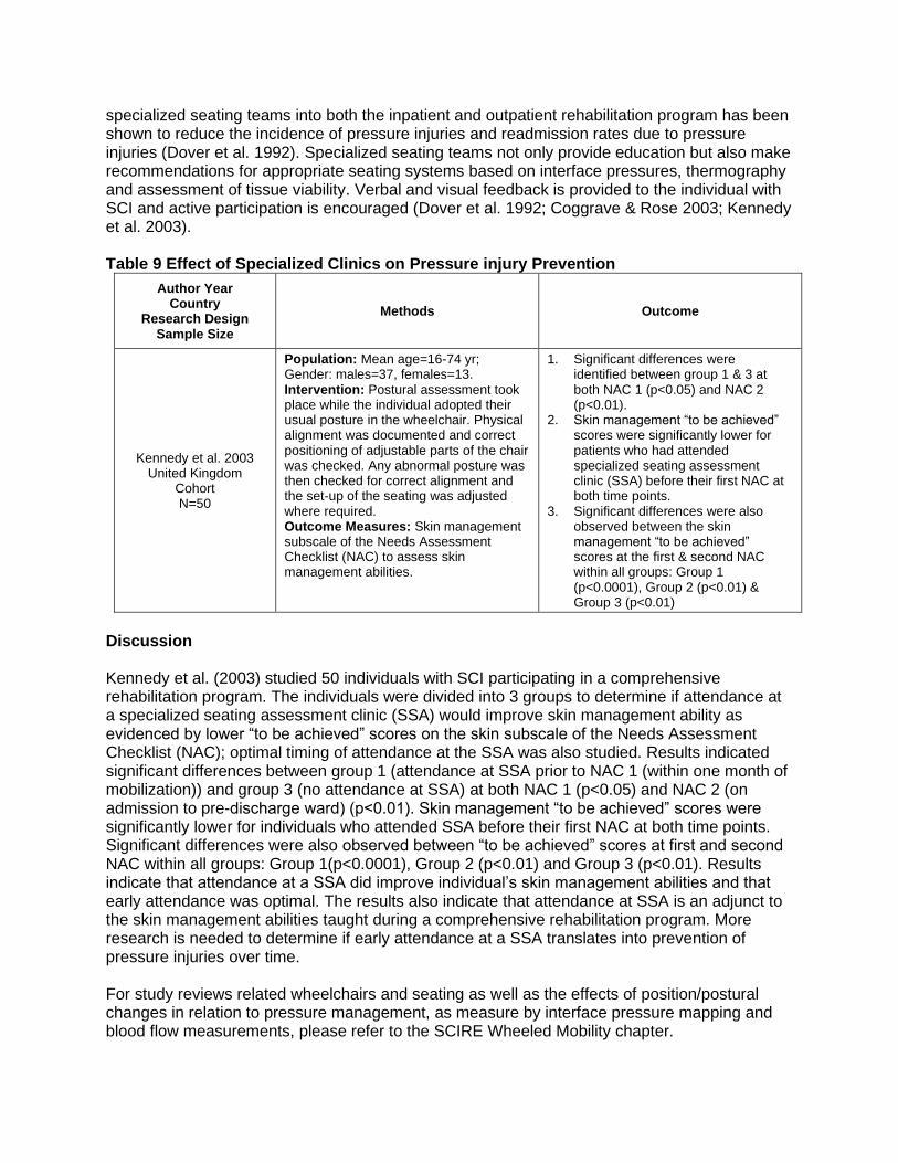
specialized seating teams into both the inpatient and outpatient rehabilitation program has been shown to reduce the incidence of pressure injuries and readmission rates due to pressure injuries (Dover et al. 1992). Specialized seating teams not only provide education but also make recommendations for appropriate seating systems based on interface pressures, thermography and assessment of tissue viability. Verbal and visual feedback is provided to the individual with SCI and active participation is encouraged (Dover et al. 1992; Coggrave & Rose 2003; Kennedy et al. 2003). Table 9 Effect of Specialized Clinics on Pressure injury Prevention
Author Year Country
Research Design Sample Size
Methods Outcome
Kennedy et al. 2003 United Kingdom
Cohort N=50
Population: Mean age=16-74 yr; Gender: males=37, females=13. Intervention: Postural assessment took place while the individual adopted their usual posture in the wheelchair. Physical alignment was documented and correct positioning of adjustable parts of the chair was checked. Any abnormal posture was then checked for correct alignment and the set-up of the seating was adjusted where required. Outcome Measures: Skin management subscale of the Needs Assessment Checklist (NAC) to assess skin management abilities.
1. Significant differences were identified between group 1 & 3 at both NAC 1 (p<0.05) and NAC 2 (p<0.01).
2. Skin management “to be achieved” scores were significantly lower for patients who had attended specialized seating assessment clinic (SSA) before their first NAC at both time points.
3. Significant differences were also observed between the skin management “to be achieved” scores at the first & second NAC within all groups: Group 1 (p<0.0001), Group 2 (p<0.01) & Group 3 (p<0.01)
Discussion Kennedy et al. (2003) studied 50 individuals with SCI participating in a comprehensive rehabilitation program. The individuals were divided into 3 groups to determine if attendance at a specialized seating assessment clinic (SSA) would improve skin management ability as evidenced by lower “to be achieved” scores on the skin subscale of the Needs Assessment Checklist (NAC); optimal timing of attendance at the SSA was also studied. Results indicated significant differences between group 1 (attendance at SSA prior to NAC 1 (within one month of mobilization)) and group 3 (no attendance at SSA) at both NAC 1 (p<0.05) and NAC 2 (on admission to pre-discharge ward) (p<0.01). Skin management “to be achieved” scores were significantly lower for individuals who attended SSA before their first NAC at both time points. Significant differences were also observed between “to be achieved” scores at first and second NAC within all groups: Group 1(p<0.0001), Group 2 (p<0.01) and Group 3 (p<0.01). Results indicate that attendance at a SSA did improve individual’s skin management abilities and that early attendance was optimal. The results also indicate that attendance at SSA is an adjunct to the skin management abilities taught during a comprehensive rehabilitation program. More research is needed to determine if early attendance at a SSA translates into prevention of pressure injuries over time. For study reviews related wheelchairs and seating as well as the effects of position/postural changes in relation to pressure management, as measure by interface pressure mapping and blood flow measurements, please refer to the SCIRE Wheeled Mobility chapter.

Conclusion There is level 2 evidence (from one cohort study; Kennedy et al. 2003) showing that early attendance at specialized seating assessment clinics increases the skin management abilities of individuals post SCI.
3.2.2 Education and prevention programs Education and prevention programs for individuals with SCI have evolved over the past few years, with growing recognition that to have a lasting impact on lifelong pressure management and skin care, the approaches to education and prevention programs must be multifactorial with consideration for situational challenges and individuality of needs (Tung et a. 2015). Education programs provide knowledge and emphasize behaviours intended to reduce the risk of pressure injury occurrence (Bogie 1995; Rodriguez & Garber 1994; Schubart et al. 2008). Although there is much diversity about specific educational programming conducted across various settings, typical approaches in inpatient rehabilitation include structured programs, often delivered in group lecture formats (question and answer), augmented by unstructured, informal “just-in-time” education delivery and content driven educational materials such as pamphlets, information sheets, websites or binders (Lawes et al. 1985; Wolfe et al. 2012, Tung et al. 2015).Typically this education is delivered while the individual is an inpatient at a time when they and their family are adjusting to a diagnosis of SCI and are likely suffering from information overload. Under these circumstances, an individuals’ ability to appreciate the knowledge and behaviours necessary to prevent pressure injuries over their lifetime is likely compromised (Garber et al.1996; Potter et al. 2004; Schubart et al. 2008). With shorter lengths of stay, there is less time to deliver prevention education and fewer opportunities for reinforcement of acquired knowledge. This means that individuals with SCI are being discharged with potentially less information on pressure injury prevention (Garber et al.1996). The focus has shifted from solely inpatient education to ongoing management programs for people in the community living with a SCI (Tung et al. 2015). There continues to be limited data on the specific education needs required by individuals with SCI at risk for pressure injury formation (May et al. 2006; Schubart et al. 2008). Systematic reviews by Cogan et al. (2017) and Baron et al. (2018) also found variability in programs and supports across studies reviewed. Cogan et al. found non-significant outcomes between control and intervention groups in the 5 studies that met their inclusion criteria. In Baron et al.’s review of 15 studies, only 1 was identified as demonstrating significant improvement in skin status; this study examined structured versus standard education, further supporting the need to individualize programs for short and long term pressure management needs. Table 10 Pressure Injury Prevention Education
Early attendance at specialized seating assessment clinics should be part of a comprehensive rehabilitation program.
More research is needed to determine if early attendance at a specialized seating assessment clinic (SSA) results in pressure injury prevention over time.

Author Year Country
Research Design PEDro Score Sample Size
Methods Outcome
Systematic Reviews
Baron et al. 2018 Canada
Review of published articles from 1970-2016
AMSTAR=6 N=15
Method: Conduct a literature review on the content and effective of skin care self-management interventions for people with SCI Databases: MEDLINE, Embase. PsychINFO, CENTRAL, CINAHL, REHABDATA, CIRRIE, PeDro, ERIC and (World Health Organization International Clinical Trials Registry and Meta-Register of Controlled Trials. Level of evidence: 10 RCTs, 5 Non-RCTs. Questions/measures/hypothesis: Aim1: To better understand the content of interventions designed for skin care; Aim2: to focus on the effectiveness of RCTs aimed at skin care management.
1. 15 studies reviewed 17 different Behaviour Change Technique (BCT) interventions, with 5 general intervention types: Structured education programs (2 RCTs, 3 Non-RCTs); Telehealth (3 RCTs, 1 Non-RCT); Wheelchair skills training (3 RCTs); Risk assessment and feedback (1 RCT, 1 Non-RCT); Body positioning skills training (1 RCT).
2. Mediators of skin care measured included: Knowledge (2 studies used the Pressure injury Knowledge Test); Self efficacy (measured in 1 study using a validated scale adapted to PU); Skills relating to skin care (4 studies, three used Wheelchair Skills Training Questionnaire, 1 study measured body-positioning).
1. 7/10 RCTs measured skin status; only one interventions significantly improved skin status compared to controls (structured education versus standard education).
Cogan et al. 2017 USA
Review of published articles from 1999-2014
AMSTAR=5 N=5
Method: Conduct a systematic review of the efficacy of behavioural or educational interventions in preventing pressure injuries in community-dwelling adults with SCI. Databases: Cochrane, Clinical Trials, PubMed, Web of Science. Search combined related terms for pressure injuries, SCI, and behavioural intervention. Level of evidence: 3 RCTs, 2 quasi-experimental. Questions/measures/hypothesis: No specific hypotheses were tested by the authors.
1. 444 records were screened for inclusion, only 5 studies met inclusion criteria with a total of 513 participants.
2. One study used telephone sessions to deliver education, and another study used enhanced education methods. One study supported family members and caregivers. Two studies used relaxation, and stress/mood management for depression and wellness.
The results on pressure injury/skin breakdown outcomes were non-significant between groups in all 5 studies (p>0.05).
Gelis et al. 2012 France
Review of published articles between
2000-2010 AMSTAR=4
N=6
Method: Systematic literature review of clinical trials written in English or French, with a human population. Article must pertain to pressure injuries. Databases: PASCAL, Biomed, PubMed, Cochrane Library. Level of evidence: level 2 evidence (4 RCTs), level 4 (2 pre-post). Questions/measures/hypothesis: 1. Determine the place of therapeutic patient education (TPE) in persons at risk of and/or those who have pressure injuries and make recommendations for clinical practice.
1. Four RCTS and two clinical trials. 2. No studies focused on specifically an
elderly population. 3. TPE had a positive impact on the
occurrence and severity of pressure injuries.
4. Two yr post-intervention showed to have an impact on recurrence rate compared to controls (33% vs. 90%, p=0.007).
3. The impact of TPE on depression and quality of life were conflicting.
Individual Studies

Author Year Country
Research Design PEDro Score Sample Size
Methods Outcome
Kim & Cho (2017)
South Korea RCT
PEDro=4 NInitial=51 NFinal=47
Population: Experimental Group (n=24): Mean age=42yr; Gender: males=17, females=7; Level of Injury: Cervical=4, Thoracic=15, Lumbar=5; ASIA Classification: A=12, B=4, C=6, D=2; Mean time since injury=49.8 mo. Control Group (n=23): Mean age=36.7yr; Gender: males=20, females=4; Level of Injury: Cervical=3, Thoracic=14, Lumbar=6; ASIA Classification: A=15, B=3, C=2, D=3; Mean time since injury=65.8 mo. Intervention: 6 hospitals were randomly allocated to the experimental group or the control group. The experimental group received an 8 wk self-efficacy enhancement program (small group education for 2.25 hrs in the 1st week for education and skill training, face-to-face counselling in the 5th week, telephone counselling for 10-15 minus in 3rd and 7th wks and computer based demonstrations at 3rd, 5th and 7th wks, and maintained a self-management journal). The control group was given a pressure injury prevention information booklet. Outcome Measures: Self-care knowledge tool, self-efficacy tool, self-care behaviors assessment tool, pressure injury incidence
1. Tehre was no significant sifference between the experiemental and controls groups with regards to baseline demographics or clinical characteristics, except for the occupation after injury (P=0.036).
2. Self-care knowledge, self-efficacy and self-care behaviours all improved in both groups but the experimental group showed significantly greater improvements after 8wks (p<0.001, respectively).
1. One participant in the control group developed a pressure injury during the 8wk test period, while none in the experimental group did. This difference was not statistically significant (p=0.49).
Guihan et al. 2014 United States
PEDro=7 RCT N=143
Population: Mean age=59.3 yr; Gender: males=139, females=4; Level of injury: cervical=60, thoracic=76, lumbar=7; ASIA classification: A=101, B=15, C=17, D=10; Mean time since injury=24.0 yr; Pressure injury stage: III or IV. Intervention: Treatment group (n=71): Self-management intervention, consisting of 7 group conference calls lasting 45-60 min and motivational interviewing intervention consisting of 8 one-on-one telephone counselling calls at pre-set times over 24 weeks.. Control group (n=72): Education intervention, equivalent to intervention group in terms of number and timing of sessions and who delivered it. Intervention emphasized teaching and advice giving while not including skills training and motivational interviewing. Outcomes: Skin Care Behavior Checklist; Skin status; Skin-related visits and admissions, Communication with Providers Scale, Self-Efficacy scale and descriptive measures (demographics, SCI factors and pressure injury characteristics); assessed at baseline, 3 & 6 months post discharge.
2. No significant differences between the 2 groups in baseline demographics, medical, SCI or pressure injury characteristics; half had pressure injuries at discharge from hospital and had a high rate of comorbid conditions, (osteomyelitis 19.5%, diabetes 39.5% and depression 40.6%).
3. Study was designed with 80% power to detect a 30% difference between groups, but had only 143 participants so the study has less than 50% power.
4. At 3 mo and 6 mo, greater self-reported improvement in skin care behaviours in the intervention group at 3 and 6 months but it was not statistically significant (P=0.2; P=.04 respectively).
5. no significant differences were observed between groups in terms of Skin Care Behaviour Checklist, Skin status (skin worsening), skin-related visits, or skin-related admissions.
1. More than half of participants (combined groups) (n=75, 52.8%) experienced skin worsening, half of which were reported within the 3

Author Year Country
Research Design PEDro Score Sample Size
Methods Outcome
months post discharge, usually in the first month
Effect Sizes: Forest plot of standardized mean differences (SMD±95%C.I.) as calculated from pre- and post-intervention data.
Rintala et al. 2008 USA RCT
PEDro=6 N=41
Population: Mean age=29-78 yr; Gender: males; Injury etiology: SCI=39, multiple sclerosis=2; Level of injury: cervical=39%, thoracic=56%; Severity of injury: complete=68%. Intervention: SCI and multiple sclerosis patients receiving surgical repair of a stage III or IV pressure injury were randomized into 3 groups: Group 1: received an enhanced education and monthly structured follow-up intervention (via telephone) for 2 yr after discharge; Group 2: received monthly contacts (via mail) for up to 2 yr after discharge to assess skin status, but no education; Group 3: received minimal contact by mail every 3 mo for up to 2 yr after discharge to assess skin status but no education. Outcome Measures: Recurrence of pressure injuries or 2 yr after discharge.
1. Group 1 had a significantly longer time before recurrence of pressure injuries than other groups, p=0.002; while no significant difference was seen between Group 2 and 3.
2. Individuals were ulcer free longer if many yr had passed since their last surgery.
3. Health status had no significant effect on staying ulcer free.
4. For those with no previous ulcer surgery, persons in Group 1 were ulcer free longer than those in Group 2 or 3 (19.6 vs. 10.1 or 10.3 mo).
1. Ulcer recurrence occurred in 1/3 of Group 1 (33.3%) compared to Group 2 (60%) and Group 3 (90%).
Garber et al. 2002 USA RCT
PEDro=5 N=41
Population: Mean age=53 yr; Gender: males=41; Injury etiology: SCI=39, multiple sclerosis (MS)=2; AIS: A=28, B=10, D=1. MS=2; Time since injury=17 yr. Intervention: Intervention group (n=20): four 1-hr enhanced education sessions dealing with management and prevention of pressure injuries and structured follow-up (monthly telephone contact regarding skin status and use of prevention behaviours). Control group (n=21): Standard educational information given with no structured follow-up (periodic telephone contact to address skin status only). All subjects followed for 2 yr after discharge or until recurrence of pelvic pressure injury. Outcome Measures: Demographic and health information questionnaire; Pressure injury knowledge test; Health beliefs questionnaire; Multidimensional health locus of control scale.
1. At discharge, both groups had an improvement on the pressure injury knowledge test, but more pressure injury knowledge was acquired within the intervention group (p<0.03).
2. At discharge, no notable differences were found on the health beliefs questionnaire and the multidimensional health locus of control scale.
1. Even though both groups remembered pressure injury knowledge obtained 2 yr prior, the intervention group maintained a higher level of pressure injury knowledge (68%) than did the control group (60.8%) at 2 yr post-discharge.

Author Year Country
Research Design PEDro Score Sample Size
Methods Outcome
Schubart 2012 USA
Pre-post NInitial=15; Nfinal=14
Population: Median age=37 yr; Gender: males=10, females=5; Level of injury: cervical=8, thoracic=5, lumbar=2. Intervention: Interactive e-learning program about pressure injury prevention and management completed over a two wk timeframe. Follow-up questionnaire. Outcome Measures: (1) Internet evaluation and utility questionnaire; (2) Internet impact and effectiveness questionnaire; (3) Internet adherence questionnaire; (4) Knowledge acquisition using a questionnaire (based on the Needs Assessment Checklist) assessing skin and posture management, mobility and transfers, and wheelchair/equipment.
2. Program rated “mostly” or “very” easy to use, with the information being understandable and useful.
3. The impact of increasing confidence in prevention/detection of pressure injuries was rated “mostly” (n=4) and “very” (n=10).
4. Adherence was rated as “slightly” (n=2), “somewhat” (n=10), and “very” (n=2), with the mean sitting lasting 45 minutes.
1. Mean total knowledge scores increased from 92 to 106. Means by subsection: skin and posture, 39 to 49; mobility, 32 to 34; and equipment, 20 to 23. Greatest improvement was shown for skin checks and prevention of skin problems (p<0.005).
Thietje et al. 2011 Germany Pre-post N=214
Population: Level of injury: paraplegia=122, tetraplegia=92. Intervention: Neurological examinations of patients admitted between January 2005 and May 2008. Outcome Measures: (1) Performance of everyday tasks using the Spinal Cord Injury Measurement (SCIM) II; (2) Knowledge of pressure injuries and bladder management pre and post discharge using the Knowledge Boberg Score; (3) Patients asked source of knowledge. Measures were taken at admission, 1 and 3 mo post-admission, and 6, 18 and 30 mo post discharge.
2. Total SCIM II was higher at discharge compared to admission (p<0.001). Scores increased until 18 mo post-discharge.
3. Mean knowledge scores increased from admission to discharge (5.4 to 11.2, p<0.001). At discharge knowledge was rated as poor, average or good for 22.4%, 30.4% and 47.2%, respectively. Poor knowledge was more common in older adults (65+, p<0.001).
1. Clinical staff and special hospital courses were knowledge resources. Post-discharge, they were general practitioners and physiotherapists.
Brace & Schubart 2010 USA
Pre-post N=20
Population: Mean age=47 yr; Gender: males=13, females=7; Level of Injury: cervical=7, thoracic=6, lumbar=6. Intervention: E-learning Program Learning section. Completion of the Living and Looking section was optional. The focus of the program was pressure injury knowledge. Outcome Measures: A newly developed 20 question test administered pre and post e-learning program.
1. Pressure injury knowledge improved in 16 of 18 individuals, 1 had a decrease in score and 1 had perfect scores at both time points. Median scores pre and post being 65 and 92.5 respectively.
1. A lack of knowledge pertaining to pressure injury prevention was shown before the e-learning program.
May et al, 2006 Canada Pre-post
NInitial=27; Nfinal=23
Population: Mean age=33.7 yr; Gender=18 male, 5 female; Level of injury: Cervical complete=4, Cervical incomplete=7, Thoracic complete=7, Thoracic or Lumbar incomplete=5; Average Intervention: Participants completed inpatient rehabilitation program which included an 8 wk lecture series two times per wk with content including pressure sore prevention techniques among others. Outcome Measures: 29-item Multiple Choice Questionnaire (MCQ), Life Situation
2. 18 of the 23 participants maintained or improved their knowledge from baseline (i.e., admission) with average scores at admission vs discharge vs follow-up of 22.26 vs 24.09 (p=0.041) vs 24.22 (p=0.023)
3. Every participant demonstrated some improvement in problem-solving ability for some of the 12 topics; however individual scores remained unchanged or declined for some as well. For the content topic of skin care there was a

Author Year Country
Research Design PEDro Score Sample Size
Methods Outcome
Scenarios (LSS) (problem-solving ability), Perceived Importance
trend toward improvement (admission to follow-up) (p=0.012; adjusted level of significance=0.004).
1. Topics related to bladder care, bowel care, and skin cares were consistently rated as important at all 3 assessment times.
Jones et al. 2003 USA
Pre-post Study 1: NInitial=8;NFinal=6 Study 2: NInitial=4;NFinal=3
Population: Mean age=25-40 yr; Gender: males=6, females=2; Level of injury paraplegia; Time since injury=12-20 yr. Intervention: Study 1 - Behavioural Intervention: 3 primary components-health plan, clinic visits and financial rewards. Study 2 - Behavioural intervention: 2 treatments components were implemented (Health plan and visits) during the initial phase. Phase 2 - which began after the patient began to experience skin problems (Included visits plus payment). Outcome Measures: Severity of pressure sores were recorded at each level; Ulcer severity - classified using Average Pressure injury Scale for Healing (PUSH) tool.
Study 1: 1. PUSH decreased from baseline by an
average of 10.5 points per participant. 2. Six participants were hospitalized (not
during the intervention) a total of 16 times during baseline for treatment of pressure injuries.
3. Fewer hospitalizations were also noted during the post-intervention phase compared to the baseline phase. Average monthly cost of care decreased from $6262.00/participant to $235.00 (US)
Study 2: 2. Mean PUSH scores decreased from
baseline by 8.3 points (visits only) and a further 3.1 points (visits & payment phase).
2. Total number of hospitalizations decreased from 1.67 (baseline) to 0.33 (intervention and post-intervention phase).
Ghaisas et al. 2015 United States Case series
N=25
Population: Mean age=45.5 yr; Gender: males=23, females=2; Level of injury: paraplegia=18, tetraplegia=6, undetermined=1; Severity of injury: complete=20, incomplete=5; Mean time since injury=20.5 yr. Intervention: Secondary analysis of a subset of participants from the intervention group of a randomized controlled trial (Lifestyle Redesign for Pressure injury Prevention in Spinal Cord Injury 2) were analyzed regarding the relationship between changes in lifestyle and changes in pressure injury status. . Data collected from 1,922 documented notes, an average of 40.9 notes per participant. Outcomes: Qualitative description of patterns behaviour or lifestyle changes relating to pressure injury development and lifestyle changes Behaviour change was conceptualized as eliminating discrete behaviours that increase pressure injury risk or adopting behaviours that reduce risk. Lifestyle change was the altering of one’s daily life routines, adapting the physical and/or social environment, and developing a mindset
2. Of the 47 cases reviewed, only 25 experienced pressure injuries and had clear patterns of lifestyle and behaviour changes.
3. Participants’ characteristics in this secondary analysis closely mirrored the full study.
3. Four patterns of lifestyle changes as they relate to pressure injury development were identified: 1)Positive pressure injury changes accompanied by positive lifestyle/behaviour changes (n=19), 2) Negative or no pressure injury changes accompanied by positive lifestyle/behaviour changes (n=3), 3)Positive pressure injury changes accompanied by minor or no lifestyle/behaviour changes (n=1), 4)Negative or no pressure injury changes accompanied by minor or no lifestyle/behaviour changes (n=2).

Author Year Country
Research Design PEDro Score Sample Size
Methods Outcome
that is cognizant of risk in everyday life situations.
Cobb et al. 2014 Canada
Case Series N=143
Population: Pre-cohort (n=70): Mean age=47.29 yr; Gender: males=61, females=9; Level of injury: C1-C4=23, C5-T1=27, T2-T10=8, T11-L2=11, L3-S3=1; ASIA classification: A=27, B=4, C=15, D=23, Missing=1. Post-cohort (n=73): Mean age=46.90 yr; Gender: males=58, females=15; Level of injury: C1-C4=18, C5-T1=22, T2-T10=12, T11-L2=21, L3-S3=0; ASIA classification: A=26, B=9, C=13, D=25. Intervention: To evaluate the implementation of a Pressure injury Prevention Initiative (PUPI) with a goal of decreasing the incidence of PU’s after admission and severity if they did occur, through a standardized and rigorous assessment and intervention process to ensure optimal care of patients.The protocol developed focused on 1) increased vigilance in skin monitoring by occupational therapist (OT), 2) OT use of a standardized assessment process, and 3) convenient access to necessary equipment. Pre-intervention population (Pre-cohort) (n=70) vs. post-intervention population (post-cohort) (n=73); all data were collected retrospectively. Outcomes: Screening for pressure injuries; Therapeutic Support Service (TSS) upgrade; Pressure injury incidence; Short-Form-36; Functional Independence Measure; Life Satisfaction Test-11.
2. Baseline comparison between Cohort 1 & 2 found they were comparable for distribution of demographic and injury variables.
3. Screening for pressure injuries significantly increased from 31% to 60% (p<0.001) and the percentage of patients with a completed Braden assessments significantly increased from 13% to 55% (p<0.001).
4. No significant difference for number of patients receiving TSS upgrade between cohorts, but the percentage of patients receiving TSS upgrade from OT significantly increased from 31% to 70% (p=0.02).
5. No significant differences for number of patients with pressure injuries based on chart documentation, but the number of patients identified with pressure injuries according to OT skin care assessments significantly increased from 14% (cohort 1) to 33%(cohort 2) (p=0.002).
6. No significant differences for total number of pressure injuries and pressure injury recurrences were observed.
4. No significant differences observed in terms of Short-Form-36, Functional Independence Measure, or Life Satisfaction Test-11.
Summarized Level 5 Evidence Studies: In a secondary analysis of a national cross-sectional survey within the Swiss Spinal Cord Injury Cohort Study, Hug et al. (2018) found that General Self-Efficacy scale results were not association with the data gathered from the 5 PU preventative behaviour items of the Spinal Cord Injury Lifestyle scale. The participants in this survey were community based; positive associations were noted with skin-care prevention items and receiving formal or informal support at home regarding skin-care. The authors suggest that availability of home support may be a factor that can be modified to affect the skin-care preventative behaviour. This is consistent with the suggestions by Guihan et al. (2014). The authors also suggest that the use of a general self-efficacy scale were too general to capture the specific needs and circumstances affecting preventative behaviour in this population. In a secondary analysis of a subset of participants from a larger ethnographic study, Fogelberg et al. (2016) explored the role habits played pre and post injury in relation to pressure injury development. The findings suggest that habits established before injury supported development of pressure management habits in some but not in all participants. However, the study findings do suggest that the integration of pressure management strategies into life habits is important, and that the provision of education related

to the pressure risks needs to be expanded to assisting the person to developing new life habits related to pressure management. Discussion Overall, most investigations reviewed above have demonstrated that specific educational programming can be beneficial for pressure injury prevention in persons with SCI, This aligns with results reported by Gelis et al. (2012) in a systematic review of therapeutic patient education directed toward persons at chronic risk of pressure injury formation, with 5 of 6 studies in this review involving persons with SCI. These authors noted a low level of evidence (Level 2 resulting in Grade B recommendations), acknowledging the relative immaturity of the literature in this area. Additionally, Gelis et al. (2012) noted limitations associated with biomedical thinking resulting in clinicians focusing on “educating” their patients (i.e., dissemination strategies) rather than adopting more behavioural approaches (Jones et al. 2003) or those that are grounded in adult learning principles as noted by May et al. (2006). The scoping review conducted by Tung et al. (2015), who identified that approaches for pressure management found in the literature were moderately successful at educating but found that the effect of these prevention strategies on incidence is not well studied. Although the various and specific educational experiences imparted over an inpatient rehabilitation stay are difficult to characterize, two studies have reported knowledge gains in pressure injury prevention methods associated with general inpatient rehabilitation programming as assessed at admission versus discharge, as well as at a later follow-up (May et al. 2006; Thietje et al. 2011). Thietje et al. (2011) did not specify particular aspects of the educational programming provided, however, they noted that significant knowledge gains were achieved by discharge and maintained at 30 months with patients identifying rehabilitation physicians, physiotherapists and nurses as the most important knowledge resources as well as in-hospital special courses. May et al. (2006) reported similar findings in knowledge gain at discharge and retention at 6 months post discharge as assessed by a customized multiple choice questionnaire developed by a clinical nurse educators and former patients. Notably, May et al. (2006) did characterize a main component of the educational experience provided to patients as involving an 8 week lecture series with classes held twice a week with content including pressure sore prevention techniques along with many other topics relevant to persons with SCI. Skin care, along with bladder and bowel care, was consistently reported by patients as the most important issue in relation to a variety of topics usually deemed relevant within SCI rehabilitation (May et al. 2006). Perhaps the most significant finding associated with this study was that problem-solving ability, as assessed using a qualitative Life Situation Scenario approach, was only marginally improved from admission to discharge and many patients continued to demonstrate poor problem-solving ability (i.e., applying knowledge to behavioural actions) at discharge. May et al. (2006) noted that the lecture series approach was likely not effective in this regard as it did not incorporate adult learning strategies (e.g., focus on perceived learning needs, readiness to learn, active learning). Other studies have tested the effectiveness of more specific educational programming. For example, Brace and Schubart (2010) and Schubart (2012) have conducted pre-post, pilot studies examining the effectiveness of an interactive eLearning program designed to prevent pressure injuries in persons with SCI as applied during inpatient rehabilitation (n=18) or following discharge to home (n=14), respectively. In each case, knowledge as assessed by performance on a customized test about this topic was significantly improved immediately after completing the online module. In addition, Schubart (2012) reported that participants rated aspects of the program’s ease of use and utility very high as well as providing positive self-

reports on perceived knowledge gain and improved self-efficacy about pressure injury prevention. In an RCT conducted by Garber et al. (2002), inpatients awaiting pressure injury surgery were randomly assigned to an intervention group (n=20) that received four 1-hour sessions of enhanced education on the prevention and management of pressure injuries. Information presented at the sessions included education regarding preventative strategies such as skin inspection, weight shifts/turns, nutrition and pressure redistribution surfaces for the bed and wheelchair, as well as pressure injury etiology. The control group (n=21) received standard education regarding preventative practices. After discharge, the groups were followed for two years or until recurrence of pelvic pressure injury. Improvement on the pressure injury knowledge test was noted in both groups upon discharge from hospital; however, it was significantly different between the groups (p<0.03), with those in the intervention group gaining more knowledge about preventing pressure injuries. No significant differences were noted on the multidimensional Health Locus of Control Scale and the Health Beliefs Questionnaire between the two groups at discharge. Two years post treatment, it was noted that both groups had retained most of the knowledge they had gained during their hospitalization, but the level of knowledge retained by the control group was below that of the treatment group: 60.8% versus 68% on the pressure injury knowledge test. In a parallel study, Rintala et al. (2008) randomized similar subjects into three groups: Group 1 (N=20) had received enhanced education sessions. Group 1 was followed through structured monthly telephone contact where they were questioned regarding skin status, pressure injury preventative behaviors and reminded of behaviors they were not using. Group 2 (N=11) were contacted monthly by mail to assess skin status only and group 3 (n=10) were contacted every three months by mail to assess skin status. If those in groups 2 and 3 had not responded in two weeks, they were contacted by telephone. Group 1 had a significantly longer time before recurrence of pressure injuries (19.6 months, p=0.002) while no significant difference was reported between group 2 or 3. For persons who had not had previous pressure injury surgery, the enhanced education and structured follow-ups extended their ulcer free time. As well, less people in group 1 had a recurrence of a pressure injury (33.3%) versus group 2 (60%) and group 3 (90%). In summary, those individuals who received an enhanced education and structured follow-up, showed more improvement on the pressure injury knowledge test at discharge, retained more of this knowledge 2 years post intervention and had fewer recurrences of pressure injuries. For those individuals who went on to have a recurrence, time to recurrence was much longer. To the point in time of this last study, it was the only investigation described in this section to include an assessment of health status as well as to include behavioural aspects to their intervention After this time point, research in this area in general, was strengthened considerably by adopting more fulsome outcome measurement approaches to evaluate the effectiveness of interventions (i.e., assessing behavior change directly in addition to its impact on health) as well as by incorporating theory-based behavior change strategies as parts of an intervention. Early pressure injury prevention was examined by Guihan et al. 2014 through the lens of a Chronic Care Model, focusing on self-management through engagement education, improving motivation and skill building. This multisite, single blind RCT study compared self-management and motivational interviewing interventions to an education only control group over a 6 month period post discharge from 6 VA centres with participants who had been admitted due to chronic and/or severe pressure injuries. This study found that at 3 and 6 months test times that there

were differences between the two groups in relation to skin care behaviours, and skin status but no comparisons were statistically significant. They also found that less than one third of the intervention group participated fully and 15% of the control group. The authors question whether these interventions are effective for people with chronic and/or severe pressure injuries, identifying the factors related to comorbidities as high and potentially confounding as they did not address them. The authors also report their study had less than 50% power instead of the anticipated 80% due to challenges with recruiting amongst this specific subset of the SCI population, despite recruiting from 6 centres. Kim and Cho (2017) based their study on Bandura’s social cognitive theory of self-efficacy. The program focused on promoting self-care behaviours, self- care knowledge, and self-efficacy as a means to prevent pressure injuries. They randomly allotted 3 of the 6 participating hospital into the control or experimental groups. The control group received an education booklet. Their 8 week program included many of the strategies/approaches used in the above studies, combining education using booklets, computer slides, videos, demonstration/observation and practice of skills, computer demonstration, and counselling via phone and face to face. While both groups demonstrated improvements in self-care knowledge, self-care behaviours and self-efficacy, the experimental group demonstrated a significantly greater improvement in these areas, suggesting the mixed methods used was beneficial in improving pressure injury prevention knowledge. However, the effect of this approach on pressure injury incidence longer term was not explored in this study. Cobb et al. (2014) concluded from their retrospective pre-post study of a Pressure injury Prevention Initiative (PUPI) that best practices for assessment and documentation improved but there were no significant changes in pressure injury incidence or in the severity of ulcers if they did occur, over the 20 month study timeframe. However, Cobb cites other similar studies where improvements in pressure injury incidence were found and questioned whether the methodology used in this study was robust enough. Jones et al. (2003) examined the effectiveness of several behavioural strategies (i.e., rewards, counselling and creation of an action plan) in four small pilot studies that examined pressure injury status and health care utilization. Results showed great variability in the average Pressure injury Scale for Healing (PUSH) scores with all behaviour strategies. For some participants PUSH scores were lower by 10.5 points from baseline; no hospitalizations were required and costs declined from $6,263.00 (US) to $235.00 (US). Of these participants only a few maintained the lower PUSH scores post-intervention. Although this was a very small study, results suggest that for some people when behavioural contingencies were introduced, positive behaviours resulted and were sustained. More research is needed to determine if behavioural contingencies (i.e., rewards) and other behavioural strategies offer some benefit for some people in pressure injury prevention post SCI. Ghaisas et al. (2015) completed a secondary analysis of data collected from the intervention group of a randomized control study in which participants’ received the Pressure injury Prevention Program (PUPP); this program is based on the findings from Lifestyle Redesign for Pressure injury Prevention in Spinal Cord Injury study. This secondary analysis focused on the relationship between lifestyle and behaviour changes implemented during the intervention and the development/progression of pressure injuries. The study identified four patterns in the relationship between lifestyle and behaviour changes and pressure injury change. There was a larger proportion of participants who had positive lifestyle or behaviour changes that related to an improvement in pressure injury status (n=19/25).

The 2013 Canadian Best Practice Guideline for Prevention and Management of Pressure injuries in People with SCI provided a Level IV recommendation (based on studies of self-management approaches used within various chronic diseases) to promote self-management for people with SCI by helping them to learn, consistently apply, and incorporate into their daily lives the effective and appropriate pressure injury prevention strategies. Conclusion There is level 1b evidence (from two randomized controlled trials studies; Rintala et al. 2008; Garber et al. 2002); and level 2 evidence (from 1 lower RCT from Kim & Cho, 2017) and level 4 evidence (from four pre-post studies; May et al. 2006; Brace & Schubart 2010; Schubart et al. 2012; Jones et al. 2003 ) that providing enhanced pressure injury prevention education, including behaviour contingencies and strategies, is effective at helping individuals with SCI gain and retain this knowledge, reduce pressure injury severity and decreased health care costs.. There is level 1b evidence (from one randomized control trial; Guihan et al. 2014) suggesting that for the SCI population with chronic and/or severe pressure injuries, an enhanced prevention program using individual motivational intervention and group self-management training does not improve skin protective behaviours or pressure injury outcomes. There is level 4 evidence (from two pre-post studies; Schubart et al. 2012; Brace & Schubart 2010) that online eLearning modules may improve knowledge on prevention of pressure injuries among persons with SCI. There is level 4 evidence (from one case series study; Cobb et al. 2014) suggesting that a formal pressure injury prevention program can improve best practice adherence in an acute care facility. There is level 1b evidence (from one randomized controlled trial; Rintala et al. 2008) that providing enhanced pressure injury education and structured follow-up is effective in reducing recurrence of pressure injuries especially in those individuals with no previous history of pressure injury surgery. There is level 4 evidence (from one case series study; Ghaisas et al. 2015) to suggest that an intervention that focus on reducing risk through lifestyle, particularly habits and behaviour changes are related to improvements in the uptake of pressure management strategies, therefore improvements in pressure injury status.
Structured pressure injury prevention education, helps individuals post SCI gain and retain knowledge of pressure injury prevention practices, but it is questioned if the same strategies
are effective for those with chronic and/or severe pressure injuries.
More research is needed to determine the best approaches of pressure injury prevention intervention to reduce pressure injuries post SCI, particularly for chronic and/or severe pressure injuries, to assist with lifestyle and behaviour changes for long term pressure
management success.

3.2.2.1 Using Telerehabilitation for delivery of prevention or treatment programs Telerehabilitation has been defined as “the use of telecommunication technology to deliver rehabilitation services at a distance” (Vesmarovich et al. 1999; p 264). Telerehabilitation allows for visual and verbal interaction between an individual with SCI and a health care provider. Impaired mobility and great distances to specialized SCI centers often make follow-up care difficult for individuals with SCI (Mathewson et al. 2000; Galea et al. 2006). Telerehabilitation has the potential to deliver medical rehabilitation including education, nutritional and psychosocial elements of health care at a distance thereby facilitating continuity of care (Galea et al. 2006). Shorter lengths of stay have potentially increased the need for education post-discharge and technology can be used to continue education begun during inpatient rehabilitation including education on pressure injury prevention and care of ulcers if they occur. Continuation of pressure injury prevention education and early detection and intervention via technology may reduce the need for hospitalization related to pressure injuries (Phillips et al. 2001). The use of a videophone capable of transmitting high resolution images, and verbal interactions between nurse, patient and caregiver could mean accurate and timely assessment and treatment of wounds and improved healing (Mathewson et al. 1999). In a study conducted at a mock home setting, Hill et al. (2009) found “video conferencing was better overall than the use of the telephone when assessing the detailed clinical characteristics of a pressure injury (p 200).” Both were found to be useful when assessing for the presence of a pressure injury. The 2013 Canadian Best Practice Guideline for Prevention and Management of Pressure injuries in People with SCI provided a Level IV recommendation (based on studies of telerehabilitation for wounds of various etiologies including SCI) telerehabilitation as a promising approach for delivering pressure injury prevention and management to people with SCI. Table 11 Telerehabilitation and Pressure Injury Management
Author Year Country
Research Design PEDro Score Sample Size
Methods Outcome
Telephone and Pressure Injury Management
Arora et al. 2017
Australia RCT
PEDro=8 Ninitial=120 Nfinal=115
Population: Int (n=60): Mean age= 35yr; Gender: males=52, females=8; Level of Injury: ASIA Classification: A=52, B=3, C=2, D=0, Unknown=3. Con (n=60): Mean age= 36yr; Gender: males=54, females=6; Level of Injury: ASIA Classification: A=47, B=2, C=8, D=1, Unknown=2. Intervention: Intervention group (Int) received weekly advice by telephone for 12 wk about the management of their pressure injuries from a trained health-care professional. Advice pertained to seating, bed overlays, cushions, equipment, diet, nutrition, wound dressings, pressure management techniques, moisture management and when to seek medical or nursing consult. Control group (Con) received standard care 12wk follow-up. Outcome Measures: Primary outcome: Pressure injury size, Secondary outcomes:
6. The mean between-group difference for the size of the PU at 12 wks was 2.3 cm2 favouring the intervention group (p=0.08), however the depth was not significantly different after 12 weeks when comparing treatment and intervention groups (p=0.17).
7. A statistically significant between-group decrease in PUSH score was seen in favour of the intervention group (p=0.02).
8. Inconclusive findings for outcomes for pressure injury undermining and depth, participant and clinicians’ subjective impressions about pressure injury improvement
9. Improvements noted on Braden scale, primarily related to nutrition and moisture.
10. Participants’ rating of their confidence in managing their pressure injury

Author Year Country
Research Design PEDro Score Sample Size
Methods Outcome
Pressure injury depth, Pressure injury Scale for Healing (PUSH). Assessments completed at baseline and 12 weeks.
were rated as high (7.3 on 10 point scale
11. Intervention group participants were satisfied with the telephone intervention (8.6 on 10 point scale); control group was 6.5.
Telehealth and Pressure Injury Management
Houlihan et al. 2013 USA RCT
PEDro=6 N=142
Population: Mean age=48.2 yr; Gender: males=51, females=55; Injury etiology: SCI=106, Multiple Sclerosis=36, Level of injury (SCI): paraplegia=54, tetraplegia=46. Medical history: depression=55, pressure injury=66. Intervention: CareCall (telehealth program with interactive voice response) access for 6 mo (unlimited call-in access and received calls 1x/wk) vs. normal care. Outcome Measures: Pressure injury Scale for Healing (PUSH) tool v.3.0, Patient Health Questionnaire -9 (PHQ-9), Cornell Services Index (CSI) and Craig Hospital Inventory of Environmental Factors – Short Form.
1. Overall there was no positive impact on pressure injuries at 6 mo; however, a significant difference in percentage with ≥1 pressure injuries for females in
the intervention group (p=0.04). 2. Among those with depressive
symptoms at baseline, severity at 6 mo differed between groups (p=0.038).
3. There were no between group differences in terms of healthcare utilization.
4. Participants using Carecall self-reported increased health-care availability (p=0.043) although other factors (i.e., availability at baseline, time and age) were more powerful predictors of this.
Phillips et al. 1999 USA
Case Control NInitial=37; NFinal=35
Population: Mean age=35 yr. Intervention: Videoconferencing was used to assist patients in treating and monitoring pressure injuries. Patients were divided into 3 groups: telephone, videophone, and standard care. Outcome Measures: Number of pressure injuries, emergency room (ER) visits, hospitalizations, doctor’s visits annually and employment rate.
1. Overall it was found that the video group reported the largest number of ulcers, followed by the standard care group and the telephone group.
2. The standard care group reported the lowest number of ER visits, hospitalizations, and health care provider visits.
3. The numbers of visits were similar for the other two groups.
Over half the members of each group had no hospitalizations during the study period. It was also noted that 26% of the subjects had returned to work 6 mo after injury.
Vesmarovich et al. 1999 USA
Case Series N=8
Population: Age range= 38-78 yr; Gender: males=8, females=0. Intervention: The outpatient nurse using the Picasso Still Image Videophone conducted 1x/wk telerehabiliatation visits. Subjects and family members received 30 minutes of education; equipment was sent home with subjects. Interviews were conducted to determine level of satisfaction Outcome Measures: Number of ulcers healed, satisfaction.
No statistical results reported 1. Subjects were seen approximately
seven times (range 1-18 visits). 2. Seven wound sites healed completely
and two needed surgery. Subjects and family were highly satisfied.
Internet and Pressure Injury Management
Hilgart et al. 2014 United States
Post-test N=7
Population: Mean age=36.14 yr; Gender: males=2, females=5; Level of injury: tetraplegia=5, paraplegia=2; Mean time since injury=10.43 yr.
1. In terms of program usage over the 6 week intervention period, average use was 14.86 (SD 10.75). All participants may diary entries (avg

Author Year Country
Research Design PEDro Score Sample Size
Methods Outcome
Intervention: Participants had 6 weeks of access to use the iSHIFTup program, an Internet intervention designed to improve skin care behaviour. Assessments were administered after the intervention was completed. Outcomes: Program usage (login, completion of 4 components – cores, modules, diary, and follow up), Internet Evaluation and Utility Questionnaire (IEUQ) which measures participants’ experiences and perceptions of an internet intervention; Internet Impact and Effectiveness Questionnaire (IIEQ) which measures participants’ perceptions of the internet intervention in relation to perceived effectiveness in resolving or preventing the target health condition..
19.57, SD 13.21), all completed at least 1 module (avg 6.86, SD 4.45) and all 3 cores. All 7 completed at least 1 follow up and 1 module.
2. In terms of IEUQ, 100% of participants reported that the program was mostly or very helpful and acceptable, the program was very easy to comprehend, they mostly or very much liked the layout, and they would likely return to the program.
3. In terms of IEUQ, 86% of participants reported that the program was very easy and convenient to use, was mostly or very engaging, was mostly or very useful, credible, mostly or very satisfying and enjoyable, was trustworthy and had no privacy concerns, and was a good mode of delivery.
4. In terms of IIEQ, 100% of participants found the program helpful in improving skin care routines and managing skin care as well as reported knowledge gains in skin care and pressure injury prevention.
5. In terms of IIEQ, 86% of participants found that the program was very helpful in providing behavioural support for skin care activities, was some-what or very effective for long-term use, was easy to follow through with program recommendations, and helpful in being confident in tracking daily skin care activities.
Disucssion Vesmarovich et al. (1999) described the use of telerehabilitation delivered via a videophone system that transmitted still images and audio to treat stage III and IV ulcers. While no statistical results were reported, 7 out of 12 ulcer sites healed. Using the same videophone system, Philips et al. (1999) divided SCI participants into 3 groups. The videophone group had the highest number of identified and/or reported ulcers. The annualized data for emergency room (ER) visits, hospitalizations and health care visits were similar for the video and telephone groups while hospitalizations and visits were less in the standard care group. No differences were significant at p<0.05. However a small non-randomized sample size and several other limitations were identified to inform future investigations. Results of these two small studies fail to support the use of this form of telerehabilitation in delivery of cost effective prevention strategies and early pressure injury identification and treatment. However, Houlihan et al. (2013) did achieve some positive results by employing interactive voice response (IVR) telephone called “CareCall” to enable virtual health care to

monitor and assess patients’ health with respect to pressure injuries and depression and to increase appropriate use of health resources such as preventative outpatient clinics and to reduce ER visits. Participants (N=142) were randomized into either a control (i.e., usual care) or intervention (i.e., “CareCall”) group and received service over a 6 month period. Those receiving “CareCall” received weekly automated calls and could call into the service at any time to receive algorithm-based, branched-logic modules (scripts of content deemed relevant to their health concerns). The scripts were delivered by both clinicians and persons with spinal cord disease and were developed through consideration of health behavior change theory (i.e., Social Cognitive Theory and Transtheoretical Model) to promote healthy behaviours. Using this approach, women were found to have reduced pressure injury incidence over the study period (p<0.0001) for the “CareCall” group versus control, whereas men did not. There was no difference in healthcare utilization between the two groups although the intervention group did self-report perceived increases in health-care availability. Arora et al. (2017) explored effectiveness of intervention at a distance using weekly telephone contact as the higher tech options are not feasible in low-and middle-income countries. This study was a multi-site RCT in India and Bangladesh. The intervention group received weekly telephone consultations from an experienced health care professional, on a wide variety of factors. The control group received standard care. The size of the pressure injury was the primary outcome measure, which showed greater improvement in the intervention group (P=0.08). The intervention group also felt more confident in being able to manage their pressure injury, had improvements on their Braden scale scores and their PUSH scores. The authors question whether the size of the treatment effect was meaningful. The authors also suggested that there was some indication that there was a greater benefit to health and wellness from the regular telephone contact that just for the pressure injuries based on the World Health Organization Disability Assessment Schedule score between group difference (95% CI 0.8-3.8), but again they questioned the treatment effect meaningfulness. Hilgart et al. (2014) explored the effectiveness of intervention using an internet format called iSHIFTup. This program was developed and tested previously, with this study focusing on the participants’ perceived effectiveness of the program in relation to prevention. The authors report that the majority of participants found the program easy to use, effective and useful to enable them to implement the strategies recommended from the program and to independently manage their skin care. It is suggested that this type of intervention holds promise for ongoing education and intervention regardless of the distance from the facility. Conclusion There is level 1b evidence (from one randomized controlled trial; Houlihan et al. 2013) that telerehabilitation using an automated call-in system with built-in theory-based behavior change strategies may make a significant difference for women but not men in preventing pressure injuries post SCI. There is level 4 evidence (from one case series; Vesmarovich et al. 1999) that telerehabilitation via videophone to support clinical interactions and digital photography does not make a significant difference in the prevention and treatment of pressure injuries post SCI. There is level 2 evidence (from one randomized control trial; Arora et al. 2017) that treatment intervention provided by telephone has potential to provide a low cost means of treatment intervention in low-and middle-income countries.

There is level 4 evidence (from one post-test study; Hilgart et al. 2014) that a comprehensive prevention program provided using an internet format has potential to meet ongoing needs for pressure management beyond the hospital/rehabilitation facility.
3.2.3 Equipment and Products for Pressure Management and Prevention Support surfaces play a significant role in the prevention of pressure injuries, addressing some of the extrinsic factors of pressure, friction, shear, moisture and heat. Support surfaces used in a wheelchair are covered in the wheeled mobility chapter. The one article in this new subsection suggests that there is research supporting the use of a multi-layer foam dressing to protect high risk people against developing sacral ulcers; however the research is not specific to the SCsI population. This study explores a multi-layer foam dressing versus a gel mattress for prevention of pressure injuries in the SCI population while waiting for spinal stabilization surgery. Table 12 Using Equipment and/or Products for Pressure Injury Prevention
Author Year Country
Research Design PEDro Score Sample Size
Methods Outcome
Richard-Denis et al. (2017)
Canada Cohort N=315
Population: Gel Mattress Group (n=226): Mean age=47.8yr; Gender: males=81, females=19; Level of Injury: Tetraplegic=56.2, Paraplegic=43.8; ASIA Classification: A=38.1, B=10.8, C=15.7, D=35. Multi-layer Foam Dressing Group (n=89): Mean age=50.7yr; Gender: males=73, females=27; Level of Injury: Tetraplegic=60.7, Paraplegic=39.3; ASIA Classification: A=24.7, B=11.2, C=16.9, D=47.2. Intervention: Patients were given a preventative multi-layer foam dressing to their sacral-coccygeal area upon arrival to emergency room, compared to those who used a gel mattress pre-operatively. Outcome Measures: Occurrence of sacral PU, Severity of PU, and comparators of age, gender, smoking status, obesity, co-morbidities and injury severity score.
1. 17.7% of participants in the gel mattress group developed PU, while 19.1% of participants with the Multi-layer dressing developed PU (p=0.77).
2. There were no statistically significant differences between the gel mattress group or multi-layer dressing group for severity of PU (p=0.71) a. Grade 1: gel mattress=30%
versus multi-layer dressing 29.4%
b. Grade 2: gel mattress=62.5% versus multi-layer dressing 70.6%
c. Grade 1: gel mattress=2.5% versus multi-layer dressing 0%
Grade 1: gel mattress=5% versus multi-layer dressing 0%
Potential predictors of sacral pressure injury development during acute hospitalization: complete tetraplegia, older age, higher injury severity score.
Discussion
The role of telerehabilitation in engaging individuals with SCI with prevention education and treatment programs has demonstrated potential but to be fully successful, requires a
compliment between program content, delivery format and accessibility to that format for all people with an SCI regardless of living situations.

Richard-Denis et al. (2017) study reports that research suggest the pre-operative use of multi-layer dressings can improve the protection of the skin and decrease the occurrence of pressure injuries pre-operatively in critically ill patients, Richard-Denis’ study suggests otherwise. His findings indicate that for patients sustaining acute SCI, this preventive measure is not superior to the conventional use of a gel mattress pre-operatively. The study authors suggest that if used, the multi-layer dressings should be used with caution in SCI patients, particularly with people who sustained a complete tetraplegia. Conclusions There is level 2 evidence (from one cohort study; Richard-Denis et al. 2017) that indicated, for pre-operative prevention for the SCI population, a multi-layer foam dressing is not superior in preventing sacral pressure injuries compared to viscoelastic polymer gel mattress over a foam stretcher pad.
4.0 Treatment Once a pressure injury has begun it is important to prevent it from worsening and is challenging to have it heal quickly. Rappl et al. (2008) examined the metabolic and physiological changes that occur in tissue below the level of a SCI in relation to the events which take place during wound healing to explain why pressure injuries may heal more slowly in individuals with a SCI. It is widely known that severe pressure injuries can lead to further disability, surgery, amputation and death (Krause 1998); further, pressure injuries are among the leading cause of unplanned rehospitalization post SCI that can also contribute to longer lengths of stay with more costly treatment than other medical conditions (Chen et al. 2005). Pressure injury treatment is more costly than prevention (Bogie et al. 2000; Jones et al. 2003) and once an individual has had an ulcer they are at increased risk for recurrence (Krause & Broderick 2004; Verschueren et al. 2011). Furthermore, in addition to standard wound care, many adjunctive therapies are required to accelerate closure of hard to heal wounds. As such, it is important to identify appropriate clients, through appropriate and regular assessment, who are likely to benefit from these often time consuming and expensive treatments (Houghton & Fraser 2008; Allen & Houghton 2003).Research in this field covers examines electrical stimulation, laser, ultrasonography, non-thermal pulsed electromagnetic energy, topical negative pressure, normothermia, recombinant human erythropoietin, anabolic steroid therapy, effectiveness of various dressings, maggot therapy, topical oxygen, surgery and other herbal remedies for healing of pressure injuries post SCI. Each of these treatments will be discussed in subsequent sections. 4.1 Electrical Stimulation The use of various forms of electrical current in augmenting tissue repair was reported as early as the 1600s when charged gold leaf was used to prevent scarring in smallpox survivors (Kloth & Feedar 1988). The therapeutic effects of electrical stimulation for wound healing have been well documented since the 1960s, especially for wounds not responding to standard forms of treatment (Kloth & Feeder 1988; Bogie et al. 2000). Galvanotaxis is the process by which electrical stimulation directs cell movement and it is thought to be a process that can impact wound healing through the migration of cells such as
Products and surfaces used for prevention should be combined with other preventative measures/strategies to optimize the potential to reduce risk of pressure injury development.

epithelium, macrophages, neutrophils and fibroblasts (Feedar et al. 1991; Bogie et al. 2000). Under normal circumstances there is a flow of charged particles from an uninjured area to an injured area triggering a biological repair system. The belief is that application of exogenous electrical current should be able to enhance healing in non-healing wounds by mimicking the body’s own healing system (Carley & Wainapel 1985). A second theory purports that the application of electric current activates cutaneous nerves and creates a centrally mediated increase in circulation to the wound to indirectly promote healing (Kaada 1982). Despite the increasing use of electrical stimulation to promote wound healing, there remains a lack of clear understanding as to how it works to repair tissue (Bogie et al. 2000). Some of the documented effects of electrical stimulation on wound healing include decreased healing time, increased collagen synthesis, increased wound tensile strength, increased rate of wound epithelialization and increased bactericidal and bacteriostatic effects (as cited in Kloth & Feedar 1988). Electrical stimulation has also been shown to indirectly improve healing by improving tissue perfusion and reducing edema formation (Houghton & Campbell 2007). The studies on electrical stimulation for wound healing have examined low-intensity direct current, high voltage pulsed direct current, and alternating current. The literature shows a high variability as to which protocols are the most effective for a specific patient or ulcer (Bogie et al. 2000). The use of electrical stimulation to promote closure of pressure injuries, when combined with standard wound interventions, has been recommended in both the able bodied and individuals with SCI. Most studies discuss the adjunctive role of electrical stimulation in pressure injuries which have failed to respond to standard treatments (Houghton et al. 2013; Consortium of Spinal Cord Medicine 2000; Keast et al. 2006; AHCPR, Executive Summary # 15 1992). Table 13 Electrical Stimulation for Pressure injury Healing
Author Year
Country Research
Design Score
Sample Size
Methods Results
Systematic Reviews
Lala et al. 2016
Canada Review of published
articles until Jan2014
AMSTAR=6 N=15
Method: Systematic review of literature including randomized controlled trials (RCTs) and clinical non-controlled trials assessing electrical stimulation therapy (EST) for pressure injury (PU) treatment. Databases: CINAHL, The Cochrane Library, Dissertation & Theses, EMBASE, ProQuest – Nursing & Allied Health, PubMed, SCOPUS. Level of evidence: Level 1a (2 RCTs), Level 2 (4 RCTs, 3 PCTs), Level 3 (2 retrospective controlled studies), Level 5 (4 case series). Questions/measures/hypothesis: To determine the effectiveness of EST on the healing of PUs in individuals with spinal cord injury in comparison with control groups.
1. A meta-analysis of three studies found that EST resulted in a significantly larger decrease in PU size compared to standard wound care or sham EST (p<0.001).
2. One retrospective control study and one RCT also found that those treated with biphasic pulsed current healed significantly faster than those treated with low intensity direct current, sham, or conservative therapy.
3. A meta-analysis of four RCTs found that healing of a PU with EST was 1.55 more likely than with standard wound care or sham EST (p=0.01).
4. Three RCTs found that PUs receiving high-voltage pulsed current had a larger percent

Author Year
Country Research
Design Score
Sample Size
Methods Results
decrease in wound surface area compared to a sham group.
1. Only one study reported minor adverse events related to EST treatment and none reported on the potential of EST to alleviate pain or improve quality of life.
Liu et al. 2016
United Kingdom Review of published
articles from 1985-
Jul2014 AMSTAR=6
N=8
Method: Systematic review of literature including randomized controlled trials (RCTs) and non-randomized clinical controlled trials (CCTs) assessing electrical stimulation (ES) for pressure injuries (PUs) in spinal cord injury (SCI) patients. Databases: Medline, EMBASE, CINAHL, PsycInfo, Cochrane Central Register of Controlled Trials. Level of evidence: Jadad: Low risk of bias (2 RCTs), Moderate risk of bias (4 RCTs), High risk of bias (2 CCTs). Questions/measures/hypothesis: To assess the effect of ES as an adjunctive therapy to improve healing rates for PU in people with SCI; to explore whether different types of ES currents and electrode placement have any influence; to examine whether ES treatment worsens PU in SCI compared to no treatment.
1. Pooled analyses of seven trials showed that ES resulted in a significantly higher weekly healing rate than sham/no ES (p=0.001).
2. Pooled analysis of six trials showed that pulsed current ES resulted in a significantly higher weekly healing rate than those without ES treatment (p=0.0005).
3. One CCT found that pulsed current ES resulted in a significantly higher weekly healing rate compared to direct current ES (p=0.03).
4. Meta-analysis of four trials found that both placing electrodes directly on the wound (p=0.01) and placing on intact skin (p=0.01) significantly increased the weekly healing rate compared to those that did not receive ES.
5. Two trials showed that ES resulted in significantly higher numbers of completely healed ulcers (p=0.02) but non-significantly lower numbers of ulcers worsening compared to no ES.
2. Only one study reported minor adverse events related to ES.
Individual Studies
Karsli et al. 2017
Germany RCT
PEDro=4 Ninitial=35 Nfinal=27
Population: Mean age=32.6 yr; Gender: males=22, females=5; HVES Group (n=15): Injury etiology: SCI=8, TBI=1, Stroke=1, Myelitis=1, SCI+TBI=4; Mean time with pressure injury: 2.76mo; Ulcer location: Sacral=7, Ischial=5, Trochanter=6, Heel=5, Lateral malleolus=1, Head of fubula=1; Pressure injury stage: II=5; III=13, IV=7. US Group (n=12): Mean age=38.2 yr; Gender: males=22, females=5; Injury etiology: SCI=6, TBI=4, Stroke=2; Mean time with pressure injury: 2.30mo; Ulcer location: Sacral=5, Ischial=8, Heel=6, Lateral malleolus=3; Pressure injury stage: II=9, III=13. Intervention: Patients were randomized to receive either high-voltage electrical stimulation (HVES), applied for 60min, 3x/wk, versus ultrasound (US),
3. The WSA improved significantly after treatment in both groups for stages I, II and III (p<0.05).

Author Year
Country Research
Design Score
Sample Size
Methods Results
applied 3x/wk. All patients received standard wound care in addition to treatment. Outcomes: Wound Surface Area (WSA).
Effect Sizes: Forest plot of standardized mean differences (SMD±95%C.I.) as calculated from pre- and post-intervention data.
Houghton et al. 2010 Canada
RCT PEDro=9
N=34
Population: Mean age=23-74 yr; Gender: male=20, female=14; Time since injury=1-51 yr; Severity of injury: complete and incomplete; Ulcer location: IT, sacrum, coccyx, hip, leg; Duration of ulcer=0.3-20 yr. Intervention: Stimulation with monophasic high voltage pulsed current (HVPC) 19,200 min/day 7 days/wk with standard wound care (interdisciplinary team assessment) or standard wound care alone (SWC). Outcome Measure: Percent decrease in wound surface area.
1. Percent decrease in wound surface area was significantly greater (p=0.048) in those treated with HVPC+SWC (70 ± 25%); versus those with only SWC (36 ± 61%).
2. Proportion of Stage III, IV, X pressure injuries improving by at least 50% was significantly greater in the HVPC+SWC than in the SWC (p=0.20)
Cukjati et al. 2001
Slovenia RCT
PEDro=5 N=217
Population: Mean age: 28-59 yr; Injury etiology: 71.7% SCI; Time since injury: 2-38 mo; Wound area >1cm2 and at least 4 wk duration; Ulcer location: trochanter, sacrum, gluteus, other; Ulcer duration: 3-18 wk. Intervention: Biphasic-current stimulation (AC group) (N=136) received biphasic current by placing electrodes on intact skin across the wound. Direct-current stimulation (DC group) (N=35) received direct current (0.6mA) through positive electrode placed over wound and 4 negative electrodes placed on intact skin around the wound. Stimulation was applied 0.5hrs, 1hr, or 2 hours/day 7 days/wk. Comparisons were made to the Conservative treatment group (N=54) and sham group (N=23). Outcome Measure: Wound healing rate.
1. AC group healed significantly faster than the sham group (p=0.018) and at the same rate as the DC group (p=0.170) with the 2-hour wound treatment.
2. AC group healed significantly faster than DC group with 1-hour treatment (p=0.001).
3. Wound healing rate depend upon wound area, grade, shape, patient’s age, elapsed time from SCI to wound appearance, and elapsed time from wound appearance to beginning of treatment.
Adegoke & Badmos
2001 Nigeria
RCT PEDro=6
N=7
Population: Mean age=21-60 yr; Mean ulcer surface area=15.8 mm; Ulcer location: greater trochanter and sacrum. Intervention: Stimulation with interrupted direct current (IDC) and nursing care or placebo IDC and nursing care; 3-45 minute treatments 1x/wk for 4 wk. Outcome Measures: Percent decrease in wound surface area.
1. Surface area of pressure injuries of IDC group decreased by 22.2% versus 2.6% in placebo IDC group.
2. Most of the decrease in surface area occurred during the first two wk of the study (IDC group 15.4 to 13.3 mm2, % change 15.8%;
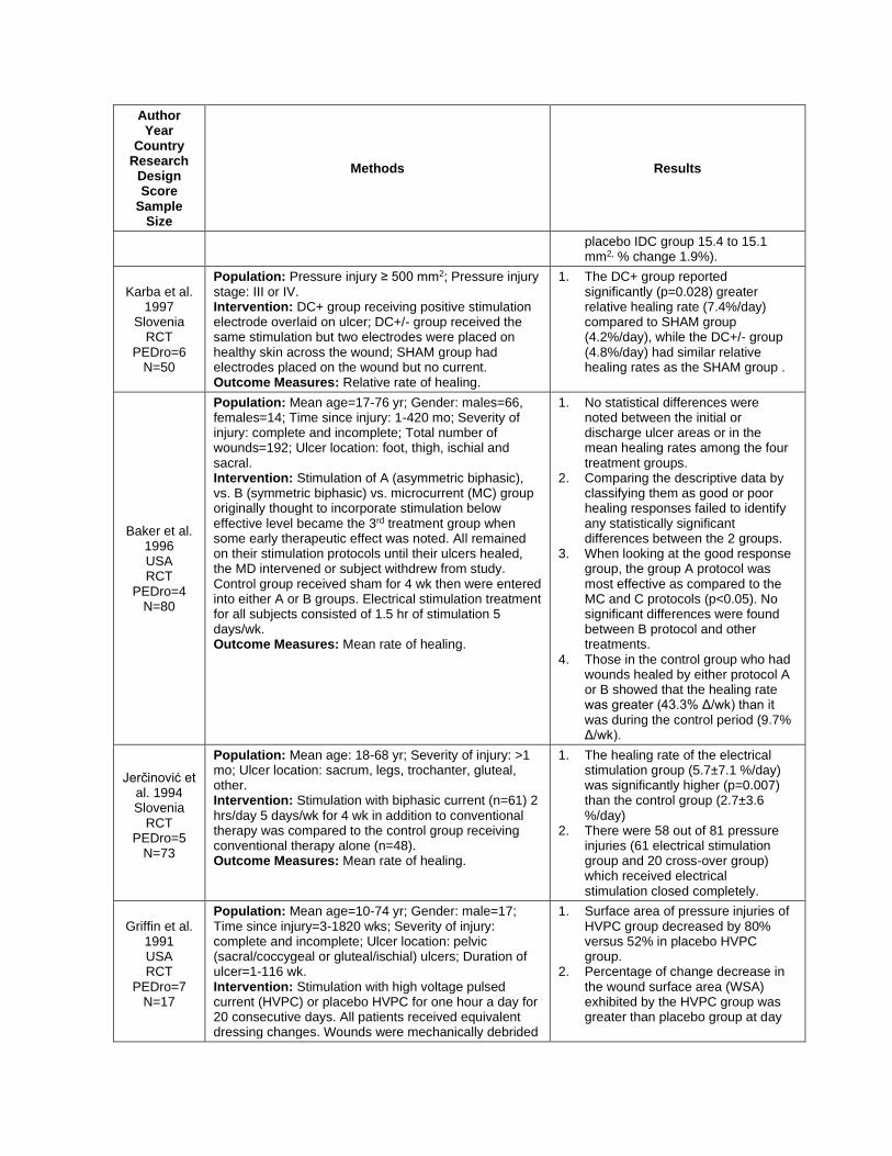
Author Year
Country Research
Design Score
Sample Size
Methods Results
placebo IDC group 15.4 to 15.1 mm2, % change 1.9%).
Karba et al. 1997
Slovenia RCT
PEDro=6 N=50
Population: Pressure injury ≥ 500 mm2; Pressure injury stage: III or IV. Intervention: DC+ group receiving positive stimulation electrode overlaid on ulcer; DC+/- group received the same stimulation but two electrodes were placed on healthy skin across the wound; SHAM group had electrodes placed on the wound but no current. Outcome Measures: Relative rate of healing.
1. The DC+ group reported significantly (p=0.028) greater relative healing rate (7.4%/day) compared to SHAM group (4.2%/day), while the DC+/- group (4.8%/day) had similar relative healing rates as the SHAM group .
Baker et al. 1996 USA RCT
PEDro=4 N=80
Population: Mean age=17-76 yr; Gender: males=66, females=14; Time since injury: 1-420 mo; Severity of injury: complete and incomplete; Total number of wounds=192; Ulcer location: foot, thigh, ischial and sacral. Intervention: Stimulation of A (asymmetric biphasic), vs. B (symmetric biphasic) vs. microcurrent (MC) group originally thought to incorporate stimulation below effective level became the 3rd treatment group when some early therapeutic effect was noted. All remained on their stimulation protocols until their ulcers healed, the MD intervened or subject withdrew from study. Control group received sham for 4 wk then were entered into either A or B groups. Electrical stimulation treatment for all subjects consisted of 1.5 hr of stimulation 5 days/wk. Outcome Measures: Mean rate of healing.
1. No statistical differences were noted between the initial or discharge ulcer areas or in the mean healing rates among the four treatment groups.
2. Comparing the descriptive data by classifying them as good or poor healing responses failed to identify any statistically significant differences between the 2 groups.
3. When looking at the good response group, the group A protocol was most effective as compared to the MC and C protocols (p<0.05). No significant differences were found between B protocol and other treatments.
4. Those in the control group who had wounds healed by either protocol A or B showed that the healing rate was greater (43.3% Δ/wk) than it was during the control period (9.7% Δ/wk).
Jerčinović et al. 1994 Slovenia
RCT PEDro=5
N=73
Population: Mean age: 18-68 yr; Severity of injury: >1 mo; Ulcer location: sacrum, legs, trochanter, gluteal, other. Intervention: Stimulation with biphasic current (n=61) 2 hrs/day 5 days/wk for 4 wk in addition to conventional therapy was compared to the control group receiving conventional therapy alone (n=48). Outcome Measures: Mean rate of healing.
1. The healing rate of the electrical stimulation group (5.7±7.1 %/day) was significantly higher (p=0.007) than the control group (2.7±3.6 %/day)
2. There were 58 out of 81 pressure injuries (61 electrical stimulation group and 20 cross-over group) which received electrical stimulation closed completely.
Griffin et al. 1991 USA RCT
PEDro=7 N=17
Population: Mean age=10-74 yr; Gender: male=17; Time since injury=3-1820 wks; Severity of injury: complete and incomplete; Ulcer location: pelvic (sacral/coccygeal or gluteal/ischial) ulcers; Duration of ulcer=1-116 wk. Intervention: Stimulation with high voltage pulsed current (HVPC) or placebo HVPC for one hour a day for 20 consecutive days. All patients received equivalent dressing changes. Wounds were mechanically debrided
1. Surface area of pressure injuries of HVPC group decreased by 80% versus 52% in placebo HVPC group.
2. Percentage of change decrease in the wound surface area (WSA) exhibited by the HVPC group was greater than placebo group at day
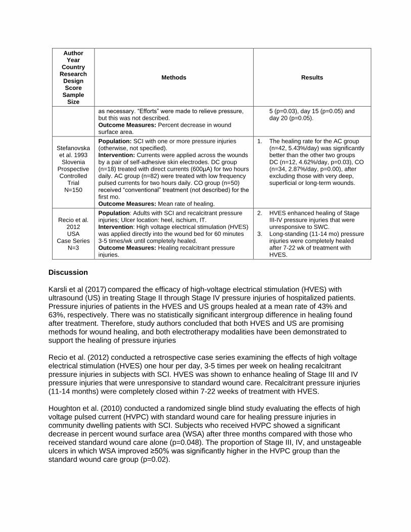
Author Year
Country Research
Design Score
Sample Size
Methods Results
as necessary. “Efforts” were made to relieve pressure, but this was not described. Outcome Measures: Percent decrease in wound surface area.
5 (p=0.03), day 15 (p=0.05) and day 20 (p=0.05).
Stefanovska et al. 1993 Slovenia
Prospective Controlled
Trial N=150
Population: SCI with one or more pressure injuries (otherwise, not specified). Intervention: Currents were applied across the wounds by a pair of self-adhesive skin electrodes. DC group (n=18) treated with direct currents (600µA) for two hours daily. AC group (n=82) were treated with low frequency pulsed currents for two hours daily. CO group (n=50) received “conventional” treatment (not described) for the first mo. Outcome Measures: Mean rate of healing.
1. The healing rate for the AC group (n=42, 5.43%/day) was significantly better than the other two groups DC (n=12, 4.62%/day, p=0.03), CO (n=34, 2.87%/day, p=0.00), after excluding those with very deep, superficial or long-term wounds.
Recio et al. 2012 USA
Case Series N=3
Population: Adults with SCI and recalcitrant pressure injuries; Ulcer location: heel, ischium, IT. Intervention: High voltage electrical stimulation (HVES) was applied directly into the wound bed for 60 minutes 3-5 times/wk until completely healed. Outcome Measures: Healing recalcitrant pressure injuries.
2. HVES enhanced healing of Stage III-IV pressure injuries that were unresponsive to SWC.
3. Long-standing (11-14 mo) pressure injuries were completely healed after 7-22 wk of treatment with HVES.
Discussion Karsli et al (2017) compared the efficacy of high-voltage electrical stimulation (HVES) with ultrasound (US) in treating Stage II through Stage IV pressure injuries of hospitalized patients. Pressure injuries of patients in the HVES and US groups healed at a mean rate of 43% and 63%, respectively. There was no statistically significant intergroup difference in healing found after treatment. Therefore, study authors concluded that both HVES and US are promising methods for wound healing, and both electrotherapy modalities have been demonstrated to support the healing of pressure injuries Recio et al. (2012) conducted a retrospective case series examining the effects of high voltage electrical stimulation (HVES) one hour per day, 3-5 times per week on healing recalcitrant pressure injuries in subjects with SCI. HVES was shown to enhance healing of Stage III and IV pressure injuries that were unresponsive to standard wound care. Recalcitrant pressure injuries (11-14 months) were completely closed within 7-22 weeks of treatment with HVES. Houghton et al. (2010) conducted a randomized single blind study evaluating the effects of high voltage pulsed current (HVPC) with standard wound care for healing pressure injuries in community dwelling patients with SCI. Subjects who received HVPC showed a significant decrease in percent wound surface area (WSA) after three months compared with those who received standard wound care alone (p=0.048). The proportion of Stage III, IV, and unstageable ulcers in which WSA improved ≥50% was significantly higher in the HVPC group than the standard wound care group (p=0.02).

Adegoke and Badmos (2001) randomly treated six stage IV pelvic pressure injuries with standard nursing care augmented with interrupted direct current or with placebo IDC. Subjects treated with IDC and nursing care showed a decrease in WSA by 22.2% versus 2.6% in the placebo group. Cukjati et al. (2001) randomly divided participants into four treatment groups: biphasic current, direct current, sham treatment, and conservative treatment. Wounds treated for two hours with biphasic current healed significantly faster than sham-treated wounds (p=0.018) and conservative therapy, but healed at similar rates as direct current (p=0.170). Although wounds treated with direct current healed faster than sham treated wounds, the difference was not statistically significant. (p=0.085). Karba et al. (1997) demonstrated that when using direct current, placement of the positive stimulation electrode covering the pressure injury and the negative electrodes on intact skin resulted in a greater relative healing rate per day (7.4%, p=0.028) compared to when the positive and negative electrodes were both placed on intact skin on opposite sides across the wound (4.8%). Baker et al. (1996) showed that for ulcers that responded to any form of electrical simulation (“good responses”), asymmetric biphasic stimulation (group A) was most effective for enhanced wound healing. Wounds that were already showing healing in the control group, with the addition of either protocol A or B (symmetrical Biphasic) showed that healing rate was greater (43.3% Δ/week) when compared to control period (9.7% Δ/week) Jerčinović et al.(1994) demonstrated that pressure injuries in patients with SCI treated with low frequency pulsed current and conventional therapy for four weeks had a significantly (p=0.006) higher healing rate than those treated with conventional therapy alone. Subjects in the conventional group who crossed over to the electrical stimulation group after four s had improved healing rates in 19 out of the 20 subjects. Stefanovska et al. (1993) treated 150 pressure injuries in individuals with SCI with conventional therapy alone, or in combination with direct or alternating current. Wounds treated with low frequency pulsed current (alternating current) showed significantly better healing rates than those treated with direct current or conventional treatment alone after the exclusion of deep, superficial and long-term wounds. Griffin et al. (1991) also performed a randomized controlled trial showing the efficacy of HVPC for healing pelvic pressure injuries in subjects with SCI. When compared to the placebo group, subjects treated with HVPC showed a greater percentage decrease in WSA at day 5 (p=0.03), day 15 (p=0.05) and day 20 (p=0.05). While there were differences in the type and duration of electric current applied in the nine studies, and in some cases electrode placement, all of the studies demonstrated that when used in conjunction with standard wound management electrical stimulation accelerates the healing rate of pressure injuries in patients with SCI. More study is needed to determine optimum electric current and application protocols to enhance healing of pressure injuries post SCI. Mittman et al. (2011) reported that in additional to standard wound care, electrical stimulation results in a cost savings of $224 over a one-year time frame for treating stage III and IV pressure injuries in individuals with SCI. The cost-savings associated with improved healing rates offset the cost of adding electrical stimulation to standard practice.

Conclusion There is level 1 evidence (from seven randomized controlled trials; Karsli et al. 2017; Houghton et al. 2010; Cukjati et al. 2001; Adegoke & Badmos 2001; Karba 1997; Jercinovic 1994; Griffin 1991) that electrical stimulation accelerates the healing rate of stage III and IV pressure injuries when combined with standard wound management.
4.2 Laser Treatment Lasers have been used in the treatment of wounds since the 1970s because of the belief that fibroblast activity and tissue granulation in the proliferative phase of non-healing, chronic wounds are enhanced. Currently the use of laser to promote wound closure in chronic wounds is not supported by evidence (Houghton et al. 2013; Houghton & Campbell 2007; Consortium of Spinal Cord Medicine 2000) and the two relevant studies are presented and discussed below. Table 14 Laser Treatment for Pressure injury Healing Post SCI
Author Year Country
PEDro Score Research Design
Sample Size
Methods Outcome
Taly et al. 2004 India RCT
PEDro=10 N=35
Population: Mean age=8-65 yr; Gender: males=27, females=8; Stage of ulcers: Stage 2=55, Stage 3=8, Stage 4=3. Intervention: Treatment for the experimental group consisted of multi-wavelength light therapy (gallium-aluminum arsenide laser source) in addition to conventional treatment. For the control group, the light therapy source was held over the ulcer after switching off the beam. 14 treatments were given, 1 every other day, three times per wk. Treatments ended when the ulcer healed or after the 14 treatment exposures. Outcome Measures: Number of ulcers that healed.
1. Overall no significant differences were found between the control and treatment groups. 14 ulcers healed completely in the control group compared to 18 in the treatment group (p<0.802).
2. The mean time taken to heal was 2.45 wk in the treatment group and 1.78 in the control group (p<0.330).
3. Multi-wavelength light therapy reduced the time taken by a small subgroup of stage three and four ulcers to reach stage two, treatment group (n=4) 2.25 wk; control group (n=5) 4.33 wk (p=0.047).
Nussbaum et al. 1994 Canada
RCT PEDro=6
NInitial=20; NFinal=16
Population: Mean age=15-61 yr. Intervention: Control group received “standard” wound care consisting of twice daily cleansing with Hygeol (1:20) and Jelonet dressing, and avoidance of pressure on existing ulcers; Laser group received standard wound care plus laser treatment 3x/wk; Ultrasound/Ultraviolet C (US/UVC) group received standard wound care with US and UVC treatments alternating over a 5 day period.
1. US/UVC and laser treatment with US/UVC showing greater effect on wound healing than laser or control. Mean percentage of change per wk in ulcer size from day 0 to complete healing for control (32.4%), US/UVC (53.5%), and laser (23.7%).
2. Several subjects showed deterioration over the study. Ulcers increased in size; (laser=3, 62-167% change; control=1, 58% change; US/UVC=1, 1% change).
Electrical stimulation added to standard wound management promotes healing of Stage III and IV pressure injuries post SCI.
More research is needed to determine optimum electric current and application protocols to improve healing of pressure injuries post SCI.

Author Year Country
PEDro Score Research Design
Sample Size
Methods Outcome
Outcome Measures: Healing rate, ulcer size.
All ulcers healed by end of study with last ulcer healed in laser group by wk 20 as opposed to US/UVC group at wk 6.
Discussion Taly et al. (2004) studied 35 subjects (64 ulcers) using multi-wavelength light therapy compared to “standard” wound care alone. Overall, no significant differences were found between the two groups with regard to the number of ulcers healed and time taken to heal. Nussbaum et al. (1994) studied 16 patients and compared standard wound care alone or combined with either laser or Ultrasound/Ultraviolet C (US/UVC). Results showed that laser treatment combined with standard wound care had the least effect on wound healing compared to the control group and US/UVC group. A significant difference was found between the groups with the US/UVC treatment demonstrating a greater effect on wound healing. Both of these studies demonstrated that laser treatment was no more effective in promoting wound healing than standard wound care alone, post SCI. Conclusion There is level 1 evidence (from two randomized controlled trials; Taly et al. 2004; Nussbaum et al. 1994) that laser treatment has no added benefit in pressure injury healing post SCI than standard wound care alone.
4.3 Ultrasound/Ultraviolet C Houghton and Campbell (2007) note that both ultrasound (US) and ultraviolet light C (UVC) have been used in the treatment of chronic wounds. Ultrasound acts mainly at the “inflammatory stage of the wound healing cascade to stimulate the release of chemical mediators of cells which in turn produces changes in the amount and strength or integrity of the scar tissue” (Houghton et al. 2001; p 464). The bactericidal effects of UVC suggest that it is indicated for the treatment of chronic infected wounds where there is much surface bacteria or where bacteria have become resistant to antibiotic therapy. As well, research supports the use of UVC in the treatment of chronic infected wounds, while therapeutic US was not shown to have added benefit when used to treat pressure injuries. The Consortium of Spinal Cord Medicine (2000) and Houghton et al. (2013) found minimal data specific to the use of US or UVC to treat pressure injuries in SCI. Schmuckler (2008) in a case series of 5 SCI patients with sacral pressure injuries used low frequency, noncontact, nonthermal ultrasound (Acoustic Pressure Wound Therapy, MIST Therapy Systems) to prepare the wound bed for subsequent treatments. The author demonstrated that in 80% of wounds the therapy was effective in reducing slough and eschar (e.g., shedding dead tissue), promoting granulation tissue and reducing wound area and volume. One small RCT will be discussed that combined US/UVC and compared its effects to laser and standard wound care.
Laser treatment does not improve pressure injury healing post SCI.

Table 15 Ultrasound/Ultraviolet C for Pressure injury Healing
Author Year Country
PEDro Score Research Design
Sample Size
Methods Outcome
Nussbaum et al. 1994 Canada
RCT PEDro=6
N=16
Population: Mean age=15-61 yr Intervention: Subjects were assigned into one of three groups: control group received “standard” wound care consisting of twice daily cleansing with Hygeol (1:20) and Jelonet dressing, and avoidance of pressure on existing ulcers; Laser group received standard wound care plus laser treatment 3x/wk; Ultrasound/Utraviolet C (US/UVC) group received standard wound care with US and UVC treatments alternating over a 5 day period. Outcome Measures: Healing rate, ulcer size.
1. Healing rate was not equal under all treatment conditions.
2. Paired comparisons showed the significant difference was between US/UVC and laser treatment with US/UVC showing greater effect on wound healing.
3. Mean percentage of change per wk in ulcer size from day 0 to complete healing for control (32.4%), US/UVC (53.5%), and laser (23.7%)
4. Several subjects showed deterioration over the study. Ulcers increased in size; (laser=3, 62-167% change; control=1, 58% change; US/UVC=1, 1% change). All ulcers healed by end of study with last ulcer healed in laser group by wk 20 as opposed to US/UVC group at wk 6.
Discussion In one small RCT (n=16) Nussbaum et al. (1994) demonstrated that between standard wound care with or without US/UVC, the combined treatment showed a greater effect on wound healing in a shorter period of time. Since US and UVC alternated over 5 days was considered a single treatment, efficacy conclusions do not cover the individual effects of US or UVC. Understanding the effects of US or UVC individually or in different combinations would require further study.
Conclusion There is level 1b evidence (from one randomized controlled trial; Nussbaum et al. 1994) that the combination of US/UVC with standard wound care decreases wound healing time of pressure injuries post SCI; there is no evidence to support the benefit of UVC or US, used individually.
4.4 Non-Thermal Pulsed Electromagnetic Energy Keast et al. (2006), in updating best practices recommendations for the prevention and treatment of pressure injuries, recommends considering electromagnetic fields as one adjunctive modality for stimulating closure of chronic non-healing pressure injuries even though results from more than one RCT are not consistent. Electromagnetic energy is believed to act at
US/UVC should be considered as an adjunct treatment when pressure injuries are not healing with standard wound care post SCI.

the proliferative stage of wound healing to promote production of granulation tissue formation (Houghton & Campbell 2007). Table 16 Non-Thermal Pulsed Electromagnetic Energy for Healing of Pressure injuries
Author Year Country
PEDro Score Research Design
Sample Size
Methods Outcome
Salzberg et al. 1995 USA RCT
PEDro=8 N=30
Population: Stage II group: Age=24-69 yr. No data for Stage III group. Intervention: A non-thermal pulsed high frequency, high peak power, electromagnetic energy (PEE), was delivered through a treatment head placed in light contact with the wound site and tuned to resonance in the area of the wound. Treatment was non-invasive and delivered through wound dressings for 30 minutes, twice daily for 12 wk or until healed. The control group received 12 wk of sham treatment. Outcome Measures: Pressure injury Status.
1. Stage II group: Treatment (n=10), control (n=10). After 1 wk, the treatment group had a greater percentage of ulcer healed (84%) compared to the control group (40%), p=0.01 and the median size of ulcer was also smaller at one wk (2.7 versus 16.5 cm2, p=0.015).
2. For complete healing, the treatment group healed in a median of 13 days versus 31.5 days for the controls (p<0.001).
3. Given that there were more large ulcers (>60 cm2) in the sham group, data was reanalyzed for 15 subjects with ulcers <60 cm2 with similar results as found initially.
4. Stage III group: Treatment n=5, control n=5. 3/5 healed with an average of 43 days of treatment. 0/5 healed in control group. Ulcer area decreased by average 70.6% versus 20.7% in control group.
Discussion An RCT studying the effects of electromagnetic energy on pressure injury healing in patients with SCI (Salzberg et al. 1995) evaluated the effects of non-thermal pulsed electromagnetic energy for healing of stage II and III ulcers in patients with SCI. In the stage II ulcer group (n=10), a greater proportion of ulcers healed (84%) after one week of treatment compared to the control group (40%; p=0.01). For complete healing, the treatment group healed in a median 13 days versus 31.5 days for controls (p<0.001). In the stage III ulcer group, healing was also associated with non-thermal pulsed electromagnetic energy treatment where three of five ulcers healed, on average, within 43 days. In the control group none of the ulcers healed. Ulcer area decreased 70.6% in the treatment group versus 20.7% in the control group. In a Cochrane systematic review, Ravaghi et al. (2006) examined two articles and found no statistically significant difference in the healing rates of people treated with electromagnetic therapy compared to controls. More research is needed to further our understanding of the mechanism of action of non-thermal pulsed electromagnetic energy and its role in pressure injury healing in individuals post SCI. Conclusion There is level 1b evidence (from one randomized controlled trial; Salzberg et al. 1995)

that pulsed electromagnetic energy accelerates healing of stage II and III pressure injuries post SCI.
4.5 Topical Negative Pressure Topical negative pressure therapy (TNP) distributes negative pressure (i.e., sub-atmospheric pressure) across an ulcer WSA via continuous or intermittent application of vacuum through a sealed dressing. This therapy to promote wound healing has been used to treat a variety of acute and chronic wounds including pressure injuries (Smith et al. 2007; Argenta & Morykwas 1997). An airtight system is created using special foam, sterile tubing and canister, and an adhesive film drape (Houghton & Campbell 2007). Vacuum is applied via a suction bottle or pump (Műllner et al.1997). The negative pressure in the wound bed removes local edema, increases blood flow, decreases local tissue edema, decreases bacterial colonization and increases granulation tissue formation and mechanical wound closure (Smith et al.2007; Houghton & Campbell 2007; Argenta & Morykwas 1997). Table 17 Topical Negative Pressure Therapy for Pressure injury Healing
Author Year Country
PEDro Score Research Design
Sample Size
Methods Outcome
Sundby et al. 2018 Norway
RCT Crossover PEDro=6 Ninitial=9 Nfinal=7
Population: Mean age=57.3yr; Gender: males=8, females=1; ASIA Class: A=8, C=1; Mean time with pressure injuries: 52wk; Pressure injury stage: III=6, IV=3. Intervention: Patients were randomized to receive either at-home intermittent negative pressure (INP) plus standard wound care (SWC) versus SWC alone. INP protocol was used 120min/day. A crossover design as used, with the first 8 wks using INP to avoid potential carryover, and then crossed over. Outcomes: Ulcer healing (Photographic Wound Assessment Tool (PWAT)), wound surface area (WSA)
1. There was greater improvement in the treatment group for WSA compared to control, but it was not statistically significant (p=0.72).
2. Improvements in PWAT were seen in all four INP+SWC patients, compared to 2/5 SWC alone, however it was not statistically significant (p=0.13).
Dwivedi et al. 2017 India RCT
PEDro=6 N=44
Population: NPWT Group (n=22): Mean age=53.5yr; Gender: males=17, females=5; Pressure injury stage: III=8, IV=14; Control Group (n=22): Mean age=54.3yr; Gender: males=16, females=6; Pressure injury stage: III=9, IV=13. Intervention: Participants were randomized to receive either a novel negative pressure wound therapy (NPWT) device, or to conventional wound care with wet to moist gauze dressings. Measurements were taken at weeks 0, 3, 6 and 9.
1. Length of PU reduced significantly in the NPWT group compared to controls in weeks 6 and 9 (p=0.04, p=0.001 respectively).
2. Width of PU reduced significantly in the NPWT group compared to controls in week 9 (p=0.006).
3. Depth of PU reduced significantly in the NPWT group compared to controls in week 9 (p=0.01).
4. The NPWT had significantly less exudate compared to controls in weeks 3,6, and 9 (p=0.001 for all time points).
Pulsed electromagnetic energy improves wound healing in Stage II and Stage III pressure injuries post SCI.

Outcomes: Matrix Metalloproteinase-8 (MMP-8) level, Wound healing parameters (PU length, PU width, PU depth, exudate amount, tissue type)
5. Tissue parameters improved (less sloughing, improved formation of red granulation tissue) in the NPWT group when compared to controls at weeks 6 and 9 (p=0.001).
6. Between group comparison showed a significantly significant MMP-8 level decrease in the NPWT device group compared to controls at weeks 6 and 9 (p=0.006, p<0.0001 respectively).
Effect Sizes: Forest plot of standardized mean differences (SMD±95%C.I.) as calculated from pre- and post-intervention data.
De Laat et al. 2011 Netherlands
RCT PEDro=7
N=24
Population: Patients 18 yr who were admitted to the study hospital with difficult-to-heal surgical wounds, or paraplegic and tetraplegia patients with pressure injuries grade IV according to the European Pressure injury Advisory Panel grading system 19. Intervention: Topical negative pressure therapy or treatment with conventional dressing therapy with sodium hypochlorite Outcome Measures: 50% wound volume reduction, with a maximum follow-up time of 6 wk, measuring the difference between the weekly measured wound volume and the initial wound volume before treatment.
1. Topical negative pressure resulted in almost 2 times faster wound healing than treatment with sodium hypochlorite, and is safe to use in patients with difficult-to-heal wounds.
Srivastava et al. 2016 India
Prospective Controlled Trial N=48
Population: Negative Pressure Wound Therapy (NPWT group): Mean age=53.5 yr; Gender: males=19, females=5; Level of injury: paraplegia; Pressure injury stage: III=9, IV=15. Standard care (Control group): Mean age=54.34 yr; Gender: males=18, females=6; Level of injury: paraplegia; Pressure injury stage: III=10, IV=14. Intervention: NPWT group (n=24): Negative pressure wound therapy using sterilized foam and negative pressure in addition to standard care. Control group (n=24): Standard care, where the pressure injury (PU) was cleaned with normal saline and packed with sterilized gauze, changed once or twice daily depending on dressing soakage. Outcomes: Wound surface area; Depth of wound; Discharge; Conversion of slough into red granulating tissue.
1. At 3 wk, 6 wk, and 9 wk, NPWT group had a significantly smaller wound surface area (p=0.0001) and wound depth (p=0.0001) compared to control group. The wound surface area and wound depth decreases at each time point were significant in NPWT group (p=0.0001) but not in the control group.
2. In NPWT group, wound discharge became minimal at 3-6 wk and negligible at 9 wk, but in control group, wound discharge continued until 9 wk.
3. At 3 wk, wound bed slough converted to granulation tissue in 33.3% of NPWT participants and 0% of control participants. At 9 wk, conversion was at 100% for NPWT group and 41.7% for control group.

Coggrave et al. 2002 United Kingdom
Pre-post N=7
Population: Mean age=44.4 yr; Gender: males=5, females=2; Level of injury: paraplegia=4, tetraplegia=3; Location of pressure injury: trochanter=3, sacrum=4; Stage of ulcer: IV=6. Intervention: Topical negative pressure (TNP) applied continuously (125 mmHg), dressing changed every 4-7 days. All patients seen and assessed by dietitian; nursed on a pressure redistribution surface; turned frequently; wound debrided as necessary pre-treatment. Outcome Measures: Picture and wound swabs (every dressing change); Pressure injury volume (beginning and end of treatment).
1. Within 1-2 days of treatment initiation, granulation tissue developed in all wounds.
2. Wound volume and grade decreased (33-96%) in all subjects, but rate and extent varied. Bacterial colonization was also reduced in each wound.
3. Limited dressing problems were described, although rashes and pain were reported in some.
4. Seal preservation in certain areas, overlapping foam on healthy skin and pressure application on bony protrusions, were reported as practical problems.
Dessy et al. 2015 Italy
Case Series N=11
Population: Mean age=30 yr; Gender: males=10, females=1; Level of injury: paraplegia; Pressure injury stage: III or IV. Intervention: Vacuum-assisted closure (VAC), consisting of polyurethane foams and negative pressure. Outcomes: Presence of foram fragments.
1. 11 cases of foam fragment retention within the wound were described, resulting in progressive wound worsening that consisted of symptomatic bad smelling, discharge with positive germ culture, and progressive wound enlargement.
Summarized Level 5 Evidence Studies: Ho et al. (2010) conducted a retrospective analysis of negative pressure wound therapy versus traditional best practice standard care on stage III or IV pelvic pressure injuries in patients with SCI. No significant difference in WSA was found between groups. Treated patients registered as having significantly poor nutritional status as measured by lowered serum albumin concentrations (p<0.05) during the four-week study. This was not apparent in the control group and therefore suggests that the treatment may have partially contributed to the lower serum albumin concentrations in malnourished participants who are less able to compensate for wound-related protein loss. A case study (N=1) described increased TNP performance when used in combination with super-oxidised solution (SOS-Dermacyn) for infection control (Angelis et al. 2012). Another potential contraindication for TNP is described in Mhatre et al. (2013) where a case study of two individuals with SCI described TNP triggered episodes of autonomic dysreflexia. Since only three TNP studies for people with SCI were found, these two case studies are included only as additional information, but they do not impact the evidentiary conclusions. Discussion Sundby et al. (2018) randomized 9 participants in a lower powered cross-over trial to explored the use of an intermittent negative pressure (INP) device for home use in addition to standard wound care (SWC) for SCI patients with chronic leg and foot ulcers. Seven of nine study participants adhered to a median of 90% of the prescribed 8-week INP-protocol and completed the study without side effects. PWAT improvement was observed in 4/4 patients for INP + SWC vs. 2/5 patients for SWC alone (P=0.13). Wound surfact area (WSA) improved in 3/4 patients allocated to INP + SWC vs. 3/5 patients in SWC alone (P=0.72). INP can be used as a home-based treatment for parients with SCI, and its efficiacy showed statistical significance compared to the control group, but should be tested in anadequately sized, preferably multicenter randomized trial. Dwivedi et al. (2017) randomized 44 SCI patients to receive negative pressure wound therapy (NPWT) using a novel negative pressure device (n=22) or PI treated with a traditional wet to

moist gauze (n=22). The authors investiaged the level of matrix metalloproteinase-8 (MMP-8) and wound-healing outcome measures (length, width, and depth, exudate amount, and tissue type) of PIs. At the end of 9 week, significantly lower levels of MMP-8 were observed and showed a positive correlation with reduction in the length, width, and depth of PIs in the NPWT group (P=0.04, P=0.001, P<0.05 respectively), while in the control group, negative correlation was observed in association with MMP-8 and the length, width, and depth of PIs. Exudate levels were significantly lower in the NPWT group compared with the conventional dressing group which lasted from week 3 through week 9. Red granulation tissue formation was significantly higher in the NPWT group after week 6 (P =0.001). Similarly, a prospective controlled trial by Srivastava et al. (2002) compared pressure injury wound healing with conventional dressing and by an innovative negative pressure device (NPD). 48 SCI patients with PIs of stages 3 and 4 were recruited. Patients were divided into two groups: group A (n=24) received NPWT using NPD, and group B (n=24) received conventional methods of dressing. At week 9, all patients on NPD showed a statistically significant improvement in PI healing in terms of slough clearance, granulation tissue formation, wound discharge and culture. A significant reduction in wound size and ulcer depth was observed in NPD as compared with conventional methods at all follow-up time points (P=0.0001). NPWT by the innovative device heals PIs at a significantly higher rate than conventional treatment. These devices have been safe, easy to apply and cost-effective. De Laat et al. (2011) randomized 12 inpatients with SCI to TNP or conventional sodium hypochlorite dressing (control) and yielded an almost two-times faster TNP healing time to 50% wound volume reduction (p<0.001) with minimal adverse events. Similar results were achieved for a parallel group of 12 inpatients of mixed disease etiology who also suffered with difficult-to-heal wounds. Combined results including both groups of patients did not alter the efficacy or safety conclusions. Another advantage of TNP is the reduced workload required of caregivers. The TNP sealed sponges are changed every 48 hours in contrast to the thrice daily sodium hypochlorite-soaked dressing changes. Coggrave et al. (2002) applied TNP continuously to pressure injuries of seven individuals with SCI to prepare the wound for surgical closure. Treatment time varied from 11 to 73 days with percent decrease in wound volume varying from 33% to 96%. Granulation tissue was seen to develop and bacterial colonization decreased in five cases. Given the small sample size and variable responses, this study provides limited evidence. A case series by Dessy et al. (2015) invesitaged the complications of a closed-loop, non-invasive vacuum-assisted closure (VAC) therapy. The intended use of this intervention is to use localised negative pressure applied on porous polyurethane absorbent foams to promote healing of acute and chronic wounds. Study authors reported 11 cases of a rare complication of foam-fragment retention within the wound. Thus, this therapy did not promote healing, but further hindered it. Conclusion There is level 1b evidence (from one RCT; Sundby et al. 2018) that intermittent negative pressure (INP) device plus standard wound care (SWC) is effective for wound healing compared to SWC alone in SCI patients home care. There is level 1b and level 2 evidence (from one RCT and one prospective controlled study; Dwivedi et al. 2017; Srivastava) that negative pressure wound therapy (NPWT) has shown to reduce levels of MMP-8, increase the rate of healing, reduce exudate

production and enhance the rate of formation of red granulation tissue when compared to conventional wet gauze alone. There is level 1b evidence (from one RCT and one pre-post study; De Laat et al. 2011; Coggrove et al. 2012) that topical negative pressure facilitates wound healing for pressure injuries in people with SCI and other patient etiologies. There is level 4 evidence (from one case series; Dessy et al. 2015) that vacuum-assisted closure (VAC) therapy does not promote wound healing and may cause rare complications including foam-fragment retention within the wound.
4.6 Normothermic Dressings Heat has been used for centuries because of its positive effects on wound healing (Kloth et al. 2000). Heat when applied to healthy skin causes vasodilation resulting in an increase in blood flow and oxygen delivery to tissues (Rund & Sussman 2007). This has led to a belief by some, that these effects may be beneficial for wounds such as pressure injuries where perfusion is compromised by pressure (Kloth et al. 2000). Normothermia is the application of controlled levels of radiant-heat energy to a wound (Consortium of Spinal Cord Medicine 2000; Kloth et al. 2000). Table 18 Effects of Normothermic Dressing in Pressure injury Healing
Wound headling is improved with intermittent negative pressure (INP) devices in combination with standard wound care (SWC) for at-home care of pressure injuries
compared to SWC alone.
Negative pressure wound therapy (NPWT) has shown to reduce levels of MMP-8, increase the rate of healing, reduce exudate production and enhance the rate of formation of red
granulation tissue when compared to conventional wet gauze alone.
Pressure injury healing after a SCI is improved when topical negative pressure (TNP) therapy is administered as compared to traditional sodium hypochlorite dressing changes.
VAC therapy may be quite a versatile device but has some disadvantages. Only qualified medical/paramedical personnel should use it in order to avoid possible complications that
can occur after an improper application.

Author Year Country
Research Design Sample Size
Methods Outcome
Kloth et al. 2000 USA
Prospective Controlled Trial N=20
Population: Treatment group: Mean age=65.4 yr; Pressure injury stage: III=9, IV=7; Number of pressure injuries: SCI=7, Geriatrics=8. Control group: Mean age=59 yr; Pressure injury stage: III=3, IV=3; Number of pressure injuries: SCI=3, Geriatrics=3. Intervention: A semiocclusive heated dressing applied (38°C) to treatment groups’ ulcers 4.5 hrs/day, Monday to Friday, for 4 wks. Heating element delivered two 60 minute periods of warmth with 1 hr of no heat between and at the end of treatment session. Standard wound care was received by both groups. Outcome Measures: Peak daily skin temperature; change in wound surface area.
1. Treatment group’s skin temperature increased 1.88°C inside and 1.86°C outside the pressure injury on average, between baseline and end of a session (p<0.05).
2. Treatment group’s pressure injury surface area improved significantly, when compared to the control group (60.73% and 19.24% respectively, p<0.05).
Discussion In a four week controlled trial of fifteen stage III and IV pressure injuries where almost 50% of the wounds were post SCI, Kloth et al. (2000) reported a 61% reduction in WSA for wounds treated with a normothermic dressing. In the six control wounds treated with standard wound care, there was a 19% reduction in WSA. Conclusion There is level 2 evidence (from one prospective controlled trial; Kloth et al. 2001) that normothermic dressings may improve healing of pressure injuries post SCI.
4.7 Alternative Pharmacological Treatments There are many common treatment options for pressure injury healing (e.g., topical antimicrobials) and other less common pharmacological treatments including exudate and odour absorbents, protective barriers, healing stimulants, dressings with varying characteristics, non-traditional, holistic and/or naturopathic remedies, physical treatment modalities, etc. It is not uncommon for those with SCI to have multiple co-morbidities that contribute to non-healing pressure injuries and therefore some adjunctive alternative pharmacological treatments have specifically been trialed for individuals with SCI suffering with pressure injuries. Recombinant Human Erythropoietin (rHuEPO), Platelet-Rich Plasma (PRP) and anabolic steroids are three such specialized treatments that show promise for pressure injury healing in people with SCI.
4.7.1 Recombinant Human Erythropoietin Chronic ulcers experienced by individuals, such as those with SCI, with hemoglobin values less than 100 g/L may be difficult to heal because of impaired tissue oxygenation. It is important to
Normothermic dressings may improve healing of pressure injuries post SCI.

distinguish between iron deficiency anemia and anemia of chronic disease. This condition occurs in individuals with chronic inflammatory and/or infectious processes; a chronic non-healing pressure injury is a chronic inflammatory condition. Anemia of chronic disease is thought to be the result of impaired red blood cell production because of persistent elevated levels of circulating inflammatory cytokines (Spivak 2002). The endogenous hormone erythropoietin and recombinant human erythropoietin (rHuEPO) play crucial roles in the regulation of hematopoiesis and induce red blood cell production. It has direct hemodynamic and vasoactive effects and modulates the inflammatory process, thereby potentially reversing the conditions believed to underlie chronic pressure injuries. Treatment with rHuEPO has been shown to be effective in increasing hemoglobin values in five individuals with stage IV pressure injuries related to anemia of chronic disease (Turba et al. 1992) and in the complete healing of a chronic leg ulcer in a single case report (Al-Momen 1991). One study has been performed that investigated the value of rHuEPO in the healing of chronic wounds among those with SCI. Table 19 Recombinant Human Erythropoietin for Healing of Pressure injuries
Author Year Country
Research Design Sample Size
Methods Outcome
Vair et al. 2015 Canada Pre-post
N=4
Population: Mean age=57 yr; Gender: males=2, females=2; Pressure injury stage: III=3, IV=1. Intervention: Participants with non-healing ulcers and low hemoglobin levels received recombinant human erythropoietin (rhuEPO) injections of 75 IU/kg, 3 times/wk, for 6 wk in addition to regular care. Outcomes: Wound surface area; Wound depth.
1. Wound healing results were variable among participants, with no significant differences from baseline to 8 and 20 wk follow-up in terms of wound surface area and wound depth.
Keast & Fraser 2004 Canada Pre Post
N=4
Population: Mean age: 59 yr; Gender: males=4, females=0. Intervention: Six wk of recombinant human erythropoietin (75 IU/kg, three times per wk), in addition to regular rehabilitation practice (inpatient and outpatient). Outcome Measures: Hemoglobin count; Pressure injury quantity, area and depth.
1. Day 1: Mean hemoglobin=88g/L; Mean ulcer surface area=42.3cm2; Mean ulcer depth=2.3cm.
2. End of wk 6: Mean hemoglobin=110g/L. Mean ulcer surface=38.4cm2; Mean ulcer depth=1.2cm.
Discussion A prospective pilot study was designed to estimate the prevalence of anemia of chronic disease (ACD) in outpatients with spinal cord injury (SCI) and chronic PIs and examine the impact of rhuEPO on PI healing Vair et al. (2015). Four patients had wound area, depth and cytokines measured before, during, and after 6 weeks of treatment with rhuEPO, with a 3-month follow-up. Wound surface area and depth had mean decreases of 1.35 cm² and 0.58 cm, respectively, immediately post-treatment. Participants’ elevated C-reactive protein levels (91.1–14.2 mg/L) decreased with rhuEPO treatment, but returned to baseline levels post-treatment (83.2–14.3 mg/L). This research indicates rhuEPO treatment may improve some outcomes for ACD-SCI PU patients, but larger randomized controlled trials are required. The results of this study suggest the prevalence of ACD in the SCI outpatient population with PIs is at least 35%, and increased vigilance of patient nutrition is recommended.

A retrospective chart review of four individuals with SCI and stage IV chronic pressure injuries was performed by Keast and Fraser (2004). Following treatment with 75 IU/kg of rHuEPO subcutaneously thrice weekly for six weeks, the mean baseline hemoglobin for the subjects increased from 88±7.4 g/L to 110±3.7 g/L. Mean ulcer surface area decreased from 42.3±40.2 cm2 to 38.4±44.3 cm2 over six weeks of treatment despite the fact that one of the subjects showed a significant increase in WSA as a result of surgical de-roofing performed to eliminate all undermining. All subjects showed a decrease in the depth of the target ulcer from 2.3±1.2 cm to 1.2±1.0 cm. Observations suggested that some of the subjects demonstrated increased ability to fight recurrent infections; all subjects reported that they felt more energetic and better able to participate in their rehabilitation activities. No adverse effects were observed. rHuEPO shows promise not only in resolving the anemia of chronic disease associated with stage IV pressure injuries but also in the healing of these wounds in persons with SCI although further study is warranted. Conclusion There is level 4 evidence (from two pre-post tests; Keast & Fraser 2004; Vair et al. 2015) that recombinant human erythropoietin aids in the healing of stage IV chronic non-healing pressure injuries post SCI.
4.7.2 Sustained-Release Platelet-Rich Plasma Therapy in Grade IV Pressure injuries Platelet-rich plasma is an enriched source of growth factors and cytokines that are critical to tissue regeneration and resolution of inflammation. PRP therapy has been used successfully in orthopedic medicine for the repair of cartilaginous and ligamentous damage (Alsousou et al. 2009). People with SCI often have multiple co-morbidities that contribute to chronic inflammation and non-healing ulcers. PRP therapy has also been used for the treatment of chronic skin ulcers (Anitua et al. 2008). Table 20 Platelet-Rich Plasma for Treatment of Pressure Injuries
Author Year Country
Research Design Sample Size
Methods Outcome
Singh et al. 2015 India
Prospective Controlled Trial N=25
Population: Mean age=36.84 yr; Gender: males=19, females=6; Level of injury: cervical=6, D1-D11=6, D12-L1=11, lumbar=2; ASIA classification: A=13, B=4, C=7, D=1; Pressure injury stage: Treatment ulcers: IV=25; Control ulcers: II=11, III=4, IV=10. Intervention: Secondary analysis of Singh et al., 2014 to evaluate antimicrobial properties of platelet-rich plasma (PRP) in pressure injuries. Participants received treatment (PRP dressing) for one ulcer with the other ulcer serving as a control (saline dressing). Outcomes: Bacterial colonization.
1. Colonization rate decreased from 92% at enrollment to 24% at 5 wk for treatment ulcers and from 84% at enrollment to 76% at 5 wk for control ulcers.
2. At 5 wk, treatment ulcers had significantly fewer positive bacteria cultures than control ulcers (p=0.007).
Recombinant human erythropoietin shows promise in assisting with the healing of stage IV chronic non-healing pressure injuries post SCI.

Singh et al. 2014 India
Prospective Controlled Trial N=25
Population: Mean age=36.84 yr; Gender: males=19, females=6; Level of injury: cervical=6, D1-D11=6, D12-L1=11, lumbar=2; ASIA classification: A=13, B=4, C=7, D=1; Pressure injury stage: Treatment ulcers: IV=25; Control ulcers: II=11, III=4, IV=10. Intervention: Each participant had one pressure injury receive treatment and one pressure injury serve as a control (twice-weekly dressings with a minimum of 10 dressings). Treatment group: Wound dressed with about 6 mL of platelet-rich plasma (PRP) and calcium chloride in addition to usual care (normal saline cleaning and debridement). Control group: Wound dressed with normal saline and usual care. Outcomes: Wound-site measurement; Pressure injury Scale for Healing (PUSH); Clinical examination.
1. At 5 wk post-intervention, both treatment and control ulcers showed statistically significant decreases in PUSH (p<0.001) but no significant differences between groups.
2. From baseline to final follow-up at 6 wk, wound surface area decreased significantly more in the treatment than the control ulcers (p=0.002). Mean percentage of surface area healed was 57.94% in treatment ulcers and 2.36% in control ulcers.
3. 96% of treatment ulcers improved compared to 68% of control ulcers.
Biglari et al. 2015 Germany Post-test
N=15
Population: Mean age=38.2 yr; Gender: males=7, females=8; Level of injury: paraplegia=13, tetraplegia=2; ASIA classification: A=15; Pressure injury stage: III=12, IV=3. Intervention: Participants with non-healing fistulas following multiple surgical interventions for pressure injuries received 7-9 mL of platelet-rich plasma (PRP) and thrombin from autologous blood after debridement of the fistula. Outcomes: Secretion; Closure of fistulas; Recurrence of pressure injuries.
1. At 3 d post-intervention, there was minimal secretion from suture lines in all participants and at 1 wk, there was no secretion around the bandages.
2. At 3 wk post-intervention, fistulas from all participants were closed with no allergic reactions or incompatibilities.
3. At 12 mo follow-up, no participants returned for treatment of their pressure injuries.
Sell et al. 2011
USA Case Series
N=3
Population: Mean age: 50 yr; Gender: male=3, female=0; Level of injury: paraplegic=1, tetraplegic=2; Wound status: grade IV ulcer=3. Intervention: Treated with a sustained release platelet-rich plasma (PRP) therapy to stimulate wound healing. Outcome Measures: Formation of tissue, vascularity, ulcer area and volume.
1. Treatment resulted in the formation of granulation tissue.
2. Improved vascularity for each patient treated.
3. Overall ulcer area and volume decreased.
Discussion A prospective controlled trial by Singh et al. (2014) evaluated the local application of platelet-rich plasma (PRP) in relation to pressure injury (PI) healing on one PI case versus saline
dressing on another PI (control) in the same patient. Twenty-five SCI patients with 2 PIs were included. Statistically significant decrease in mean PUSH scores and wound surfact area of PIs (case) (P=0.000 respectively) was observed after five weeks. Mean PUSH scores decreased significantly in controls (P=0.000), however the decrease in wound surface area of PI (control) was not significant (P=0.924). Advanced wound therapy using local applications of PRP seems to be a promising alternative to standard saline dressings in PI healing. With the advantages of simple preparation, biocompatible safety, low cost and significant clinical effectiveness, it may be beneficial to study the effects of PRP in large-scale trials to validate it as an ideal therapy for enhanced wound healing in PIs.

Singh et al. (2015) conducted a follow-up to their previous prospective controlled trial to study the source of microbial colonization and antimicrobial properties of autologous platelet rich plasma (PRP) in controlling it in PIs. Colonization rate of PIs (case) decreased from 92%-24% from enrollment to thethe 5th week but did not significantly decrease in PIs (control). Association between PI (case) and perineal cultures was observed for Staphylococcus aureus at enrollment 41% (P< 0.01) and at the 2nd week 47% (P< 0.05). 47% association between PI (control) and perineal cultures at enrollment (P< 0.05) and 29% association at the 2nd week (P< 0.01) were observed for Staphylococcus aureus. Thus, study authors concluded that there is a significant association between PIs colonization and bacteria present in local environment (urine and feces). Local application of autologous PRP changes the “biological milieu” of the PIs through its antimicrobial properties leading to reduction in bacterial colonization. A post-test by Biglari et al. (2015) aimed to determine the effectiveness of platelet-rich plasma (PRP) in the treatment of nonhealing fistulas in 15 SCI patients. After one week of treatment with PRP, the authors observed low levels of secretion from the fistulas. After two weeks, they noted no further secretion from the fistulas. After three weeks, MRI showed the complete disappearance of the fistulas. No negative effects and no allergic reactions were noted in the use of PRP. These results suggest that the application of PRP combined with debridement is an effective therapy option and good alternative to recurrent surgical interventions for treating nonhealing fistulas resulting from the surgical closure of PIs. Sell et al. (2011) sought to use immediate and sustained-release CaCl2-activated PRP therapy for the purpose of improving stage IV (sacral or greater trochanter) pressure injury healing in three veterans with SCI. Each patient underwent a different course of treatment (e.g., one patient underwent vacuum therapy and skin allograft) before healing rates stalled or plateaued. PRP therapy accelerated pressure injury healing in all three patients but more so in the patient whose pressure injury was not undermined. The two patients with severely undermined pressure injuries did improve with respect to increased granulation and vascularity of tissue in-growth. These latter two patients’ pressure injuries were also in more pressure sensitive locations which may have additionally contributed to slower improvement. This small case study of reactivated pressure injury healing in three patients is a promising indicator for a larger scale study to investigate the invigoration of healing in severe, non-healing pressure injuries. Although SCIRE criteria do not allow for single case studies, the single case study by De Angelis et al. (2012) increases the total sample size of patients by 33% and therefore is added here until future studies involving a larger sample size is available. In total, all four severe, non-healing ulcers reflected in these two studies showed improvement in healing as a result of PRP therapy. Conclusion There is level 2 evidence (from one prospective controlled trial; Singh et al. 2014) that advanced wound therapy using local applications of PRP seems to be a promising alternative to standard saline dressings in PI healing. There is leve 2 evidence (from one prospective controlled trial; Singh et al. 2015) that local application of PRP may reduce bacterial presence and colonization in PIs. There is level 4 evidence (from one case series and one post-test; Sell et al. 2011; Biglari et al. 2015) that supports the possibility of platelet-rich plasma therapy facilitation of reactivated healing in severe, non-healing pressure injuries, post SCI.

4.7.3 Anabolic Steroid Agents Impaired nutritional status and decreased nutritional intake are common in those with SCI and significantly associated with development and timely healing of pressure injuries (Consortium for Spinal Cord Medicine 2000; Houghton et al. 2013). Spungen et al. (2001) stated that use of anabolic steroids and increased protein intake have been associated with promoting anabolism, weight gain and in turn wound closure in burn patients. The United Stated Food and Drug Administration approved oxandrolone for the treatment of involuntary weight loss and for chronic infections. Since a “hypermetabolic, potentially catabolic state also is associated with pressure injuries” (Spungen et al. 2001, p. 140), the use of an anabolic steroid agent may also promote closure of nonhealing, pressure injuries in the SCI population. Table 21 Anabolic Steroid Agents for Healing of Pressure injuries
Author Year Country
Research Design PEDro Score Sample Size
Methods Outcome
Bauman et al. 2013 USA RCT
PEDro=5 N=212
Population: Inpatients with SCI and stage III or IV target pressure injuries (TPUs). Intervention: Oxandrolone, 20 mg/d (n=108), or placebo (n=104) until the TPU healed or 24 wk. Outcome Measures: The primary outcome was healed TPUs. The secondary outcome was the percentage of TPUs that remained healed at 8-wk follow-up.
1. Oxandrolone showed no benefit over placebo for improving healing or the percentage of TPUs that remained closed after 8 wk of treatment.
Spungen et al. 2001 USA
Case Series N=9
Population: Mean age=24-73 yr; Gender: males=9; Total number of pressure injuries since SCI=1 to 7. Intervention: Subjects with stage III and IV pressure injuries were treated with 20 mg of oxandrolone daily with 20 g of glutamine dissolved in orange juice. Pressure injury care and support surfaces remained consistent. Outcome Measures: Number of pressure injuries healed.
1. After oxandrolone and glutamine treatment, 8/9 subjects were completely healed, the majority within 3-6 mo. Two subjects required 12 mo of treatment for complete healing.
Discussion In a case series of nine subjects with stage III and IV pressure injuries, Spungen et al. (2001) demonstrated complete healing in the majority of subjects (8/9), 3-12 months after daily administration of 20mg of oxandrolone. Bauman et al. (2013; N=212) initiated a RCT in a similar
Platelet-rich plasma therapy may be a promising alternative to standard saline dressing for pressure injury healing, however additional study is required to validate PRP therapy as a
possible treatment for severe, non-healing pressure injuries in people with SCI.
Local application of PRP may reduce bacterial presence and colonization in PIs.

patient group suffering from stage III and IV pressure injuries. Unfortunately, this ambitious study was terminated early due to feasibility issues. The most significant of these issues included the heterogeneity of confounding co-morbidities across the study cohort, non-uniformity of clinical care across contributing sites, and high withdrawal rates in both groups likely due to the propensity of people with SCI to develop complications independent of the study. Available results did not show improvement in wound healing or closure between the active and control groups. The results did, however, reveal markedly increased serum pre-albumin levels that reflected improved better nutritional status in general. Ultimately, this RCT did not produce results to support that oxandrolone was more useful than placebo to improve chronic pressure injury healing. Conclusion There is level 2 evidence (from one flawed randomized controlled trial; Bauman et al. 2013) that does not support the use of oxandrolone (anabolic steroid) to facilitate healing of serious pressure injuries post SCI. However, very limited, earlier level 4 evidence (from one case series; Spungeon et al. 2001) did lend some support for the use of oxandrolone to promote healing of stage III and IV pressure injuries post SCI.
4.8 Dressings Dressings are one of several interventions required to treat a wound. The appropriate choice of a dressing aids the body’s ability to heal a wound. Dressings are intended to keep the wound bed moist, remove excess exudate, provide a barrier against contamination, and to promote gas exchange. An appropriate dressing increases healing rate, reduces pain, and decreases infection rates while being cost effective and affordable (Broussard 2007). Due to the estimated costs associated with pressure injuries and their treatment, various dressings used with the SCI population have been investigated. There is little evidence that dressing protocols in pressure injuries in people with SCI are different than the general population (Houghton et al. 2013). The Registered Nurses Association of Ontario (2007), as cited by Houghton et al. (2013), recommends that the dressing selected:
• Keeps the ulcer bed continuously moist and the surrounding skin dry
• Controls exudate but does not dry out the wound bed or macerate the peri-wound
• Provides thermal insulation and wound temperature stability
• Protects the wound from microbial contamination
• Maintains its integrity and does not leave fibres or foreign substances in the wound
• Does not cause wound bed trauma on removal
• Meets the following criteria: simple handling, economical in cost and time, promotes (or does not slow) wound healing, acceptable to the person with the pressure injury
Although many dressing products are available, only three specific dressing materials (i.e., hydrocolloid, hydrogel, platelet gel and phenytoin) have some evidence for use in the SCI population.
The anabolic steroid agent Oxandrolone does not promote healing of serious pressure injuries post SCI.

When hydrocolloid dressings are placed over a wound, the dressing absorbs the exudate and changes into a gel. The outside of the dressing allows for gas exchange and protects against outside contamination. Hydrocolloid dressings maintain a moist wound environment and support autolytic debridement. Dressings can be left in place for 3-7 days, decreasing time and costs (Heynemen et al. 2008; Consortium for Spinal Cord Medicine 2000; Houghton & Campbell 2007; Houghton et al. 2013). Hydrocolloid dressings are typically used for stage II and III pressure injuries (Heynemen et al. 2008). Hydrogel dressings act to retain moisture and rehydrate wounds, provide autolytic debridement and fill dead space. They provide minimal absorption of exudates. Hydrogel is available as a sheet or in an amorphous viscous form which requires a secondary dressing (Broussard 2007; Consortium for Spinal Cord Medicine 2000). Hydrogel dressings can be left in place for 48-72 hours depending on the type of hydrogel in use (Broussard 2007). Platelet gels are rich in growth factors that are thought to aid wound healing and are stored in the frozen state until ready for use. It is commonly used in conjunction with a polyurethane sponge/semi-permeable film dressing system (Biatain Coloplast®). Phenytoin is most commonly known as an oral anti-epileptic medication but the healing properties of topical phenytoin were first reported over 50 years ago. Over the years, various topical preparations of phenytoin have been studied and, while its exact mechanism of action is unknown, it is thought to enhance healing by stimulation of fibroblast proliferation, promotion of collagen deposition, antibacterial activity and decreased collagenase activity (Anstead et al. 1996; Kelin & Gorling 1961; Subbanna et al. 2007). It has not been widely used because its efficacy has not been sufficiently established through controlled clinical trials (Ovington 1999; Subbanna et al. 2007). Table 22 Effectiveness of Dressings for Treatment of Pressure injuries
Author Year Country
Research Design PEDro Score Sample Size
Methods Outcome
Scevola et al. 2010 Italy RCT
PEDro=4 N=13
Population: Patients affected by SCI with 16 pressure sores over a period of 20 mo. Intervention: Allogenic platelet gel or with current best practice approach to chronic wounds dressing protocol. Outcome Measures: Volume reduction of pressure injuries, reduction in amount of ulcers.
1. No statistically significant difference was demonstrated in volume reduction between the two groups, although a statistically significant difference could be demonstrated in the onset time of granulation tissue proliferation as in the wounds treated with platelet gel the healing process was triggered earlier.
2. Platelet gel is mostly effective within the first 2 wk of treatment while a prolonged treatment does not provide any significant advantage.
Subbanna et al. 2007 India RCT
PEDro=9 N=28
Population: Patients with stage II ulcers. Treatment group (n=14): Mean age=34.25 yr; Gender: males=13, females=1; Control group (n=14): Mean age=31.64 yr; Gender: males=12, females=2.All subjects had stage II ulcers.
1. Improvement in PUSH 3.0 and ulcer size was seen in the topical phenytoin group however this difference did not reach statistical significance, p=0.261 and 0.132 respectively.

Author Year Country
Research Design PEDro Score Sample Size
Methods Outcome
Intervention: Patients were randomly placed (double blind manner) in the treatment group which received 5mg/ml phenytoin solution soaked sterile gauge for their pressure injury dressing once daily for 15 days and the control group received saline soaked gauge. Outcome Measures: Pressure injury scale for healing (PUSH) 3.0, ulcer size, ulcer volume
2. Reduction in ulcer volume was seen in the control group however it was not significant, p=0.777
Hollisaz et al. 2004 Iran RCT
PEDro=7 N=83
Population: Gender: males=83; Level of injury: paraplegia; Stage of pressure injuries: I=33, II=58. Intervention: 1 of 3 dressing groups: 1) Simple dressing (SD) – changed twice daily; 2) Hydrocolloid dressing (HD) – changed 2x/wk; 3) Adhesive plus phenytoin cream (PC) – changed once daily. Outcome Measures: Healing status of pressure injury.
1. HD group had the most healing (74%) as compared to the PC group (40%, p<0.01) and the SD group (27%, p<0.005).
2. Of all the groups, the HD group also had higher rating of healing of stage I ulcers (p<0.05).
3. For stage II ulcers, HD treated ulcers healed better than SD treated ulcers (67% vs. 16%) p<0.005 but no better than PC (48%, p>0.05) treated ulcers.
4. HD also healed gluteal ulcers more completely than other groups (p<0.001); however, groups did not differ on healing of sacral ulcers.
Kaya et al. 2005 Turkey RCT
PEDro=4 N=27
Population: Mean age=16-56 yr; Gender: males=24, females=3; Ulcer stage: I=12, II=34, III=3. Intervention: Treatment group (n=15, 25 PU): an occlusive hydrogel dressing, changed every 4 days (unless contaminated). Control group (n=12, 24 PU): povidone-iodine soaked gauze changed daily. Outcome Measures: Healing rate (cm2/day).
1. Healing rates did not differ between groups.
2. More wounds healed in the treatment group (n=21) than in the control group (n=13), p<0.04.
Whittle et al. 1996 Canada
Case Series N=4
Population: Mean age=55.75 yr; Gender: males=3, females=1; Level of injury: paraplegia=2, tetraplegia=2; Pressure injury (5 ulcers) stage: II=1, III=3, IV=1. Intervention: Hydrogel dressings, for different time periods depending on the case. Outcome Measures: Pressure injury improvement, measured 1x/wk.
1. Hydrogel dressing application varied from 4 to 6 wks.
2. In all cases, pressure injuries improved drastically, with 3 cases being completely healed.
Discussion In a RCT, Scevola et al. (2010) found healing was triggered earlier as indicated through onset time of granulation tissue proliferation when platelet gels were used to treat pressure injuries in individuals with SCI. Platelet gel use did not make a difference in wound volume reduction and was only effective within the first two weeks of treatment.
In a RCT involving 83 subjects, Hollisaz et al. (2004) found that those in the hydrocolloid dressing group (n=28) had the greatest completion of healing regardless of ulcer location and stage (74%; p<0.005), compared to those in the phenytoin cream group (40%; n=28) or simple dressing group (27%; n=27). For stage I ulcers, those in the hydrocolloid group healed faster than those in the other two groups; however, for stage II ulcers, there was no difference in healing between the hydrocolloid and phenytoin cream groups (67% vs 48%; p>0.05). In examining the area of injury, gluteal ulcers also healed more completely in the hydrocolloid group than in the other two, whereas the healing of sacral ulcers did not differ between the three groups. Using a phenytoin solution (5mg/ml), Subbanna et al. (2007) found improvements in PUSH 3.0 and ulcer size when compared to normal saline but the differences did not reach statistical significance (p=0.261 and p=0.132, respectively). Whittle et al. (1996) treated five pressure injuries (stage II-IV) with hydrogel dressings. After approximately 4-6 weeks of treatment, three ulcers healed completely while the two others showed a large improvement. Kaya et al. (2005) compared the effectiveness of applying an occlusive hydrogel type dressing to a poviodine-iodine soaked gauge dressing. There were no statistically significant differences in rate of healing but significantly more ulcers healed with the hydrogel dressing. Conclusion There is Level 1 evidence (from one randomized controlled trial; Hollisaz et al. 2004) that completion of healing for stage I and II pressure injuries is greater with an occlusive hydrocolloid dressing compared to phenytoin cream or simple dressing post SCI. There is Level 2 evidence (from one randomized controlled trial; Kaya et al. 2005) that occlusive hydrogel-type dressings heal more pressure injuries than conservative treatment post SCI. There is level 1 evidence (from one randomized controlled trial; Subbanna et al. 2007) that topical phenytoin shows a trend towards healing of stage I and II pressure injuries post SCI. There is level 2 evidence (from one randomized controlled trial; Scevola et al. 2010) that platelet gel dressings, when used within the first 2 weeks of treatment can trigger earlier granulation tissue proliferation towards pressure injury healing, post SCI.
4.9 Non-Surgical Management and Debridement A long-standing pressure injury management procedure is to excise diseased tissue in and around the wound to reduce the risk of wound progression and infection (Conway & Griffith
Occlusive hydrocolloid dressings are useful for healing of stage I and II pressure injuries post SCI.
Platelet gel dressings used within the first two weeks of treatment will trigger pressure injury
healing post SCI.

1956). Although the most common method of debridement is surgical, non-surgical debridement methods are also beneficial to the healing of pressure sores in people with SCI. For people with SCI, three types of non-surgical debridement methods have been studied that are also useful in regular pressure injury management: pulsatile lavage, maggot therapy and silicone moulding. Autolytic debridement using occlusive dressings is discussed in section 3.8 (Effectiveness of Dressings for Treatment of Pressure injuries Post SCI) above since dressings are used predominantly for routine care of pressure injuries and contraindicated in the presence of infection where debridement is often required. Table 23 Maggot Therapy for Healing of Pressure injuries Post SCI
Author Year Country
Research Design PEDro Score Sample Size
Methods Outcome
Pulsed Lavage
Ho et al. 2012 USA RCT
PEDro=5 N=28
Population: People with SCI and stage III and IV pelvic pressure injuries. Intervention: Daily low-pressure pulsatile lavage treatment with 1 L of normal saline at 11 psi of pressure was applied to the treatment group along with standard dressing changes. The control group received only sham treatment and standard dressing changes. Outcome Measures: Linear and volume measurements of pressure injury dimensions were obtained 1x/wk for 3 wk.
1. Pulsatile lavage enhanced stage III and IV pelvic pressure injury healing rates in people with SCI relative to standard pressure injury treatment alone.
Bogie et al. 2013 USA
Observational N=28
Population: Mean age=55 yr; Gender: males=28, females=0; Level of Injury: paraplegic=12, tetraplegic=12; Wound status: grade III ulcers=8, grade IV ulcers=IV. Intervention: Chart reviews of patients who were treated by pulsatile lavage therapy. Outcome Measures: Adverse effects, treatment discontinuation and injuries to clinical care providers.
1. No adverse events for patients or care providers (mean therapy duration 46 days).
2. Treatment was temporarily discontinued in one patient due to mild bleeding from wound and resumed six days later.
3. Treatment discontinued for two patients due to a fever in one patient and rapid improvement in wound size in another.
Maggot Therapy
Sherman et al. 1995 USA
Prospective Control Trial N=8
Population: Mean age=44-68 yr; Gender: males=81; Level of injury: paraplegia=7, tetraplegia=1; Ulcer stages: III and IV. Treatment: 3-4 wks of conventional therapy preceded maggot placement under porous sterile dressings, for 48-72 hr cycles. Sodium hypochorite, normal saline or wet-to-dry gauze dressings were applied every 8 hours in between maggot cycles. Outcome Measures: Healing of pressure injury; wound area size.
1. Maggot therapy decreased pressure injury surface area by 22% per wk (p<0.001).
2. No adverse consequences of treatment were noted.
Wang et al.2010 China
Observational N=25
Population: Patients with diabetic foot ulcers and 18 patients with pressure injuries after SCI. Intervention: Maggot therapy or traditional dressing Outcome Measures: Changes in the lesions were observed and bacterial cultures tested.
1. Maggot therapy is a safe and effective method for treating chronically infected lesions
2. All ulcers healed completely. The times taken to achieve bacterial negativity, granulation and healing of lesions were all significantly shorter in

Author Year Country
Research Design PEDro Score Sample Size
Methods Outcome
Pulsed Lavage
the maggot therapy group than in the control group, both for diabetic foot ulcers (p< 0.05) and pressure injuries (p< 0.05).
Silicone Moulding
Erba et al. 2010
Switzerland Post-Test
N=10
Population: Mean age=42 yr; Gender: males=6, females=4; Level of injury: paraplegic=10; Wound status: grade IV ulcer=10. Intervention: Injection of fluid silicone. Silicone moulding to facilitate debridgement. Outcome Measures: Radical en bloc debridgement achievement, complications and recurrences.
1. No complications or recurrences occurred (mean follow-up 25 mo).
2. In all patients debridgement was performed en blooc without perforation into the decubital cavity and without additional excisions needed.
3. No postoperative complications occurred.
Discussion Hydrotherapy is considered a form of mechanical debridement that is gentle and almost free of pain (Krasner D 1990). However, hydrotherapy via daily whirlpool (Burke et al.1998) can be costly in terms of personnel and facility resources and potentially can be a source of cross contamination, especially when wounds are draining heavily (Maragakis et al.2004). Conversely, Ho et al. (2012) found that pressurized saline hydrotherapy directed on the wound at the bedside is an effective form of hydrotherapy that is less resource intensive and reduces the potential for cross-contamination. This form of pulsatile lavage therapy has been successfully used in intra-operative joint cleansing during orthopaedic surgery. As such, pulsatile lavage therapy can also be used for non-debridement related cleansing of wounds at the bedside. The findings by Bogie et al. (2013) on cleansing and debridement for grade III and IV align with those by Ho et al. (2012) in that it is beneficial. In both studies, the wound dimensions decreased significantly in the pulsatile lavage group with a significantly faster rate of healing. In the retrospective chart review undertaken by Bogie et al. (2013), adverse events were reported as a result of pulsatile lavage therapy used to promote pressure injury healing. One adverse event was reported Bogie (2013) but none by Ho et al. (2012). However, an outbreak of multidrug-resistant Acinetobacter baumannii associated with pulsatile lavage wound treatment was reported at Johns Hopkins Hospital as a result of extensive environmental contamination during pulsatile lavage (Maragakis et al.2004). In addition to multidrug-resistant Acinetobacter baumannii transmission between patients, and caregiver splash injuries, the authors postulated additional potential adverse events such as pain, bleeding, and autonomic dysreflexia with subsequent hypertensive crisis, myocardial infarction and cerebrovascular accident. The one adverse event reported for one of 28 patients in Bogie et al. (2013) was described as minor wound bleeding leading to interruption of treatment. Although environmental contamination could not be evaluated in the 2013 retrospective chart review, after examination of the circumstances of the Johns Hopkins outbreak, Bogie et al. (2013) suggested that standard infection control practices would eliminate the potential for clinical safety concerns. These standard infection control practices include low-pressure pulsatile lavage, avoidance of common wound care treatment areas (e.g., bedside lavage is preferred), and wound area draping.

The beneficial effects of fly larvae have been known for centuries but the intentional use of fly larvae (maggot therapy) for the treatment of wounds was used extensively the 1930s and 1940s. It then fell out of favour when antimicrobials and other forms of debridement (including surgical) were introduced. Maggot therapy, using sterilized larvae of Phaenicia sericata species, was reintroduced to treat intractable wounds in the 1990s (Mumcuoglu et al. 1999; Sherman 1995, 2002). Maggot therapy is believed to work through three processes: debridement of necrotic tissue, disinfection of the wound and promotion of tissue growth (as cited by Sherman 2002). In a non-RCT, Sherman et al. (1995) reported that 8 of 20 patients diagnosed with stage III and IV pressure injuries were treated with maggot therapy. All eight patients underwent three weeks of conventional treatment, followed by maggot therapy. All necrotic wounds were debrided within one week of maggot treatment and wound healing was faster among those who had received maggot therapy than those who had not. Another maggot study that included only 42% SCI patients (Wang et al.2010, SCIN/N=18/43) was included in this discussion given the dearth of studies on this intervention. Wang et al. (2010) examined maggot bio-debridement and reported that all SCI related pressure injuries healed more quickly as evidenced by bacterial negativity, granulation (p<0.05) and no recurrence after an average follow-up of 3.5 months. Effective mechanical and sharp debridement requires clear visualization of the necrotic tissue in order to be thorough and also to not cause additional damage by perforating healthy and/or regenerating tissue. Methylene blue diluted with hydrogen peroxide instilled in the wound can leave a visual guide for tissue excision. However, the wound cavity shape itself can be a barrier to effective visualization even if the necrotic tissue deeper in the cavity has been stained. To overcome this barrier, Erba et al. (2010), with a convenience sample of 10 paraplegic patients presenting with grade IV ischial ulcers, injected silicone to fill the ulcer cavity. Within six minutes, the silicone vulcanized into a tumor-like mass that was an imprint of the ulcer cavity. Under palpatory control, the imprint was then expelled and examined for completeness of excised necrotic tissue. Wheelchair mobilization at six weeks after the procedure, discharge after ten weeks and four consecutive 6-month follow-up assessments (including clinical assessment for potential adverse events (e.g., hematoma, infection, seroma) and photographic documentation), revealed no observation of early or late postoperative consequences including recurrence. Conclusion Level 1 evidence (from one randomized controlled trial; Ho et al. 2012) underpins the use of pulsatile lavage hydrotherapy debridement for Stage III and IV pressure injuries secondary to SCI. There is level 5 evidence (from one observational study; Bogie et al. 2013) that pulsatile lavage therapy, used in conjunction with standard infection control standards, is likely a safe debridement method for Stage III and IV pressure injuries in people with SCI. There is level 2 evidence (from one prospective controlled trial and one observational study; Wang et al. 2010; Sherman et al. 1995) that supports the use of maggot therapy as an adjunctive therapy for pressure injury debridement post SCI. There is level 4 evidence (from one post-test study; Erba et al. 2010) that supports the use of silicone moulding as a radical en bloc debridement method for pyramidal shaped grade IV pressure injury cavities in people with SCI.

4.10 Topical Oxygen Chronic hypoxia of a wound and periwound tissues is known to impede wound healing by impairing collagen formation, angiogenesis and epithelialization. Hypoxia also lowers a wounds resistance to infection (Stotts et al. 2007). Oxygen supply to chronic wounds has been augmented by treatment with systemic (hyperbaric) oxygen therapy or through a less studied modality, topical oxygen therapy (Stotts et al. 2007; Kalliainen et al. 2003). No controlled studies have examined the efficacy of hyperbaric oxygen on the healing of pressure injuries (Houghton & Campbell 2007; Consortium of Spinal Cord Medicine 2000; Houghton et al. 2013). Kalliainen et al. (2003) studied topical oxygen and its effects on the healing of chronic wounds, some of which were noted to be pressure injuries but the exact number was not reported. Among 58 wounds, 38 (65.5%) healed during treatment with topical oxygen alone but pressure injuries were included in wounds found to be least responsive to topical oxygen. Table 24 Topical Oxygen for Treatment of Pressure injuries
Author Year Country
Research Design Sample Size
Methods Outcome
Banks & Ho 2008 USA
Pre-post N=3
Population: Mean age=61 yr; Gender: males; Level of injury: C7, T12, T10; Severity of injury: AIS A. Treatment: SCI patients with stage IV pressure injuries in their pelvic region were treated with EpiFLO device (extracts oxygen from air, concentrates to near 100% and delivers oxygen via cannula to saturate wound) daily during rehabilitation. Outcome Measures: Wounds linear measure and volume.
1. First patient linearly had 49% improvement 9 wk post treatment; pre-treatment the patient's wound measured 20.5cm linearly and volume was 252cm3, while 9 wk later, the linear measure reduced to 10.2cm linearly with a volume of 24 cm3.
2. The second patient's wound reduced from 10.5 cm linearly to 5.5cm after 5 wk treatment and volume decreased from 30cm3 to 4 cm3, linearly this was a 48% improvement from baseline.
3. The last patient's wound decreased by 31% from baseline after 5 wk treatment.
Discussion In one very small, pre-post study of three patients, Banks and Ho (2008) demonstrated that when topical oxygen (EpiFLO device) was applied to stage IV pelvic pressure injuries, comparison of pre- and post-treatment linear measurements showed 49%, 48% and 31% improvement, respectively, for each patient. While a positive effect was shown, more research is needed to determine the role of topical oxygen therapy as an adjunctive therapy for the healing of pressure injuries post SCI.
Pulsatile lavage therapy is an effective, and likely safe, non-surgical management and debridement method for the treatment of grade III and IV pressure injuries secondary to SCI.
Maggot therapy is also likely useful in this patient group. Silicone moulding may also be
considered as a radical en bloc debridement method for grade IV pressure injuries in people with SCI.

Conclusion There is level 4 evidence (from one pre-post study; Banks & Ho 2008) that topical oxygen therapy may improve healing of pressure injuries post SCI.
4.11 Surgical and Other Miscellaneous Topical and Physical Treatments The introductory information on incidence, prevalence and impact of pressure injuries solidify the importance of understanding the spectrum of prevention and management of pressure injuries, especially in people with SCI given their neurologically compromised sensation, mobility and cardiovascular functions. Previous sections of this chapter have discussed assessment of risk factors and assessment. The most important factor in the management of all stages of ulcers (i.e. SDTI, stage I, II, III, IV) is a comprehensive assessment of risk factors and co-morbidities to choose the most effective treatment methods. Regular reassessment is also necessary, especially in more severe ulcers (i.e. stage III and IV) that are often persistent and/or recur. There are many non-surgical methods of management that facilitate healing of all pressure injuries but stage IV ulcers almost always require surgery. Before surgery is elected, removal of unhealthy tissue using surgical (and non-surgical) methods is a standard procedure that can also expedite the healing of persistent stage III and IV ulcers. The studies reviewed in this section provide evidence that surgery can reduce rehabilitation costs and time by preventing protein loss from the wound, development of sepsis or osteomyelitis and development of additional skin conditions, such as Fournier’s gangrene (Backhaus et al. 2011) and necrotizing fasciitis (Citak et al. 2011), secondary to the pressure injury. Some studies also provide data to support surgical choices that improve quality of life of people with severe ulcers by decreasing limitations of daily functioning and improving the hygiene and appearance of the skin. With the many surgical reconstruction options available for wound repair, this section attempts to summarize the potential benefits of surgical repair for pressure injuries in people with SCI. Table 25 Surgical and Other Miscellaneous Topical and Physical Treatments
Author Year Country
Research Design Sample Size
Methods Outcome
Velasco et al. 2015 Spain Cohort N=98
Population: Study group: Mean age=46 yr; Gender: males=20, females=7; Level of injury: paraplegia; Pressure injury stage: III or IV. Control group: Mean age=46 yr; Gender: males=59, females=12; Pressure injury stage: III or IV. Intervention: Study group (n=27): In addition to standard care, received preclosure application of 2 mL of the fibrin sealant Tissucol Duo in combination with an antibiotic. Control group (n=71): Historical control received standard care, consisting of debridement plus closure with a local muscular flap.
1. Study group had significantly lower rates of hematoma-seroma (p=0.005), fewer mean days until drain removal (p<0.05), lower mean drainage volume (p<0.05), shorter mean lengths of stay (p<0.05), and fewer mean days of antibiotic treatment (p<0.05).
2. 19.7% of the control group experienced surgical failure compared to 3.7% in the study group, though this was not statistically significant.
3. 8.5% of the control group experienced relapse after 6mo compared to 3.7% in the study group,
Use of topical oxygen therapy may have a positive association with healing of pressure injuries post SCI but more research is needed.

Outcomes: Hematoma-seroma rate; Days until drain removal; Drainage volume; Length of hospital stay; Days of antibiotic treatment; Surgical failure and relapse after 6 mo.
though this was not statistically significant.
Kuo et al. 2014 Taiwan Cohort N=99
Population: Mean age=59.7 yr; Gender: males=54, females=45; Pressure injury stage: III or IV. Intervention: Patients received complete debridement, including bursectomy and partial ostectomy of the bony prominence where required, followed by immediate regional flap reconstruction. Free-style perforator-based flap (Group A) (n=35) vs. Gluteal rotation fasciocutaneous flap (Group B) (n=37) vs. Musculocutaneous flap or combination of muscle and fasciocutaneous flaps (Group C) (n=27). Outcomes: Complication rate; Flap necrosis rate.
1. Group A had a mean follow-up of 24.2 mo, with a 22.9% complication rate and an 11.4% flap necrosis rate.
2. Group B had a mean follow-up of 20.8 mo, with a 32.4% complication rate and a 13.5% flap necrosis rate.
3. Group C had a mean follow-up of 19 mo, with a 22.2% complication rate and a 0% flap necrosis rate.
4. Differences in complication rates and flap necrosis rates between groups were not statistically significant.
Ljung et al. 2017 Post Test Germany Nintial=51 Nfinal=34
Population: Mean age= 43yrs; Gender: males=41, females=10; Level of injury: paraplegia=34, tetraplegia=17; ASIA classification: A=268, B/C=84; Number of pressure injuries: One=43, Two=7, Three=1; Pressure injury classification: IV=51; Ulcer location: Tuber ischia=45, Hip=8, Sacral=7; Type of flap: Gluteal=42, Biceps=12, Tensor=6. Intervention: Patients were examined 10 yrs after receiving pressure injury wound flap/excision surgery. Outcomes: Ulcer status(recurrent ulcers, new ulcers), Healing Results, Visual Analog Scale (VAS).
1. After 10 yrs, 27% (n=9) patients had recurrent ulces and 18% (n=6) had new ulcers.
2. 36% (n=12) had PU that healed, 3 of which had been re-operated.
3. Median VAS score was 70 at 10-year follow-up.
Singh et al. 2010 India
Pre-post N=30
Population: Mean age: 33.2 yr; Gender: male=23, female=7; Level of injury (ASIA): grade A=21, grade B=6, grade C=2, grade D=1, grade E=0; Wound Status: grade III or IV ulcer=30. Intervention: Various types of flap surgery were performed. Outcome Measures: Improvement in health (hemoglobin, serum proteins, and general well-being), patient satisfaction and global quality of life scores according to the visual analog scale.
1. There was a statistically significant increase in mean values of quality of life, hemoglobin and total serum proteins at 6-mo follow-up.
2. Improvements in subjective well-being were reported in 76.7% of patients.
1. Satisfaction was reported in 83.3% of patients with the ultimate outcome of the surgery.
He et al. 2012 China
Post-test N=11
Population: Mean age=47 yr; Gender: males=9, females=2; Level of injury: paraplegic=11; Wound status: grade III or IV ischial ulcer=11. Intervention: Surgery using free partial lateral latissimus dorsi musculocutaneous flap. Outcome Measures: Recurrence and success rates of surgery.
1. All surgeries were successful. 2. No recurrence occurred during follow-
up (mean 60 mo). 3. All patients experienced various
degrees of back tightness, shoulder weakness and limited shoulder motion since surgery.
4. Within 9 mo adverse side effects had stopped in all but 3 patients.
Unal et al. 2012 Japan
Post-test N=11
Population: Mean age: 37.8 yr; Gender: males=9, females=2; Level of injury: paraplegic=11; Wound status: recurrent lesions or multiple ischial sores=11.
1. Nine IGA and five posterior thigh perforator flaps were used and in two patients a combination of both was done.
2. Six patients presented with recurrent lesions and five were operated for

Intervention: Use of inferior gluteal artery (IGA) and posterior thigh perforators in ischial pressure sore management. Outcome Measures: Perforator of flap, recurrences, complications and postoperative follow-up.
sacral and contralateral ischial pressure sores previously.
3. Two patients had suture detachments and their wounds were resutured (mean, follow-up 34.3 mo).
4. During follow-up, two patients had recurrences.
Kim et al. 2010 Korea
Post-test N=75
Population: Mean age=54 yr; Gender: males=43, females=32; Level of injury: paraplegia=34, tetraplegia=5, ambulatory=36. Intervention: Gluteus maximus perforator-based island flap for coverage of buttocks defects. Outcome Measures: Recurrence and complication rates of gluteus maximus perforator-based island flap surgery.
1. No recurrence at follow-up (mean, 15 mo).
2. 3 complications (distal flap necrosis, wound dehiscence, infected sacral sore).
1.
Borgognone et al. 2010 Italy
Post-test N=12
Population: Mean age=52 yr; Gender: males=10, females=2; Level of injury: paraplegic=12; Wound status: grade III ulcers or grade IV ulcers=12. Intervention: ‘Criss-cross’ surgical treatment of ischial pressure sores. Outcome Measures: Recurrence of ulcer.
1. Recurrence of ulcer occurred in one patient (mean follow-up 45 mo).
2. All flaps survived however two distal cutaneous partial necroses of the flaps occurred.
3. Suture dehiscence at the wound edge occurred in one patient.
4. A second procedure was necessary for one patient with a hematoma under the flap.
Lin et al. 2010 China
Post-test N=12
Population: Mean age=40 yr; Gender: males=9, females=3; Level of injury: paraplegic=10, tetraplegic=2; Wound status: grade IV ulcer=12. Intervention: Surgical reconstruction with a laterally based posterior-thigh fasciocutaneous flap. Outcome Measures: Flap survival, recurrence of pressure sore and primary wound healing.
1. In two patients pressure sores recurred (mean follow-up, 62 mo).
2. All the flaps survived. 3. Primary wound healing occurred in all
patients.
Srivastava et al. 2009 India
Post-test N=25
Population: Mean age=27.6 yr; Gender: female=6, male=19; Level of injury: paraplegic=22, tetraplegic=3; Level of ulcer: stage III=13, stage IV=23, unknown=3. Intervention: Surgery (debridement, split skin grafting, flap mobilization and closure). Outcome Measures: Ulcer healing rate, postoperative complications, ulcer recurrence rate and neurologic (ASIA grade) and functional recovery (Barthel index).
1. Four participants had initial complications: wound dehiscence and delayed graft healing (mean follow-up, 15.4 mo).
2. Four participants had ulcer recurrence. 3. The majority of participants (56.5%)
significantly improved neurologically on ASIA grade and functional evaluation on Barthel Index.
Relander et al. 1988 Sweden
Post-Test N=39
Population: Mean age=38 yr; Gender: males=24, females=15; Localization of primary pressure sores: sacral ulcers=21, ischial ulcers=29, trochanteric ulcers=16. Intervention: Patients underwent surgical treatment of primary pressure sores. Outcome Measures: Pressure injury recurrences, unhealed vs. healed sores and reasons for discontinuing surgical treatment.
1. Average time of hospitalization was 51 days.
2. The greatest amount of time was spent in the hospital for those with trochanteric ulcers staying for a mean of 79 days.
3. There was recurrence in 48% of sores during follow-up which lasted from 2 to 12 yr.
4. In 9 cases, 2 operations were performed.
5. Eighteen sores remained unhealed.

6. 91% of sacral sores healed, 62% of ischial sorts and 69% of trochanteric sores.
7. Discontinuation of surgical treatment occurred in 10 patients with 4 of these patients demonstrating poor general health, 4 opposing the operation, in one case the surgeon opposed the operation and in the final case the patient had died.
Tadiparthi et al. 2016 United Kingdom
Case Series N=45
Population: Mean age=47 yr; Gender: males=23, females=22; Level of injury: paraplegia=35, tetraplegia=10; Number of pressure injuries=60. Intervention: Data was retrospectively analyzed for those with pressure sores managed under the new multidisciplinary protocol with joint management by plastic surgery and spinal injury teams. Outcomes: Treatment method; Complication rate; Recurrence rate; Patient readmission rate; Readmission length of stay.
1. 29% were treated with conservative methods (debridement and dressings alone or in combination with negative pressure wound therapy) and 71% underwent flap reconstruction surgery.
2. After a mean follow-up of 33 mo, the major complication rate was 9%, the minor complication rate was 22%, and the recurrence rate was 6%.
3. Comparison of the admission database before and after intervention using a 5 yr follow-up period showed a reduction in readmissions for complications from 14% to 5.5% and a reduction in average readmission length of stay from 65 d to 45 d.
Wettstein et al. 2015 Switzerland Case Series
N=119
Population: Mean age=51 yr; Gender: males=91, females=28; Level of injury: paraplegia=74, tetraplegia=27, other=18; Number of pressure injuries=170; Pressure injury stage: III=50, IV=116, V=4. Intervention: Data was retrospectively analyzed for those with their pressure sores managed under the new interdisciplinary treatment protocol with cooperation between conservative and surgical disciplines. Outcomes: Time from debridement to defect closure; Length of hospitalised bed rest after defect closure; Complication rate; Recurrence rate.
1. The mean time from debridement to defect closure was 40 d and the mean length of hospitalised bed rest after defect closure was 58 d.
2. 23 patients died during the study period.
3. The rate of major complications was 16%, the rate of minor complications was 10%, and the rate of recurrences was 11%.
4. Higher complication rate was significantly associated with osteomyelitis (p=0.002) and increased hospitalisation days (p<0.0001).
5. Higher recurrence rate was significantly associated with increased hospitalisation days (p<0.0001).
Yusmido et al. 2014 Malaysia
Case Series N=3
Population: Mean age=40 yr; Gender: males; Level of injury: T3=1, T10=2; Etiology of injury: traumatic=2, transverse myelitis=1. Intervention: Elective proximal lower limb amputation to treat chronic complicated pressure injuries. Outcomes: Length of hospital stay; World Health Organization Quality of Life (WHO-QOL); Spinal Cord Independence Measure (SCIM).
1. In the 3 patients, the number of hospital stays was reduced by 203, 130, and 290 d after intervention.
2. After intervention, the WHO-QOL score was increased by 11%, 5.9%, and 53%.
3. After intervention, the SCIM score was increased by 12%, 2.89%, and 1.39%.
Biglari et al. 2014 Case Series
Germany N=352
Population: Gender: males=285, females=67; Level of injury: cervical=89, thoracic=227, lumbar=36; ASIA classification: A=268, B/C=84; Number of
1. Of the 421 flaps used, there were 87 complications, consisting of 27 cases of suture line dehiscence, 22 cases of infection, 17 cases of hematoma, 12

pressure injuries=657; Pressure injury classification: I=84, II=152, III=254, IV=167. Intervention: Patient records were reviewed for those receiving flap surgery. Outcomes: Rate and type of complications.
cases of partial necrosis, and 9 cases of total flap necrosis.
2. Tensor fascia lata flaps had the highest rate of complications (48.6%) and rotation flaps had the lowest rate of complications (11.5%).
Grassetti et al. 2014 Case Series
Italy N=143
Population: Median age=51 yr; Gender: males=87, females=56; Level of injury: traumatic paraplegia=58, traumatic tetraplegia=49, non-traumatic paraplegia=21, non-traumatic tetraplegia=14; Pressure injury stage: IV. Intervention: Patient charts were reviewed for those receiving perforator flap surgery. Outcomes: Rate and type of complications.
1. 5.6% of patients had major complications requiring reoperation, 4.2% of patients developed a new occurrence, and 22.4% had a recurrence at 2 yr.
2. Overall complication percentage was 22.4%, resulting in an average of 5.8 additional days of bed rest.
3. When developing a risk estimation model for developing recurrence, the type of perforator flap used (P-FAP vs I-GAP, p=0.035) and coronary artery disease (p=0.017) were significantly associated with higher risk and renal disease was significantly associated with lower risk (p=0.017).
Kim et al. 2014 Korea
Case Series N=14
Population: Mean age=52.8 yr; Gender: males=11, females=3; Level of injury: paraplegia=11, tetraplegia=2, ambulant=1; Injury etiology: SCI=12, spinal stenosis=1, spinal cord metastases=1; Number of pressure injuries=16. Intervention: Data was retrospectively analyzed for those with ischial pressure sores reconstructed using both a profunda femoris artery perforator flap and a muscle flap. Outcomes: Recurrence rate; Complication rate.
1. In the mean follow-up period of 27.9 mo, wound dehiscence occurred in 25% of ulcers and 1 case recurred after 34 mo.
2. Post-operative follow-up at 18 mo revealed no evidence of recurrence or complications.
Summarized Level 5 Evidence Studies: Consistent across the studies included in this review, was the confirmation of the most severe pressure injuries occurring in the buttock and hip areas in individuals with SCI due to being primary wheelchair users. The most common types of severe pressure injuries in people with SCI were confirmed, in descending order: ischial, sacral and trochanteric (Tavakoli et al. 1999 Kierney et al.1998, Ahluwalia et al. 2010, Chen et al. 2011, Mehta et al. 2012, Bertheuil et al. 2013, Josvay et al. 2015, Biglari et al. 2013, Grassetii et al. 2013). Caregiver collaboration (Kierney et al. 1998) and flap type selection (Ahluwalia et al. 2010; Borgognone et al. 2010; Bertheuil et al. 2013) when considering surgical reconstruction of severe pressure injuries, facilitates healing (Grassetti et al. 2013) without undue risk of complications (Ahluwalia et al. 2010; Borgognone et al. 2010) and improves the well-being and satisfaction with the rehabilitation experience (Singh et al. 2010) of people with SCI. Chen et al. 2011 and Grassetti et al. 2013 similar in study size, and pressure injury grade and location suggest an average length of hospitalization of approximately 45 and 16 days, respectively. The study by Chen et al. (2011) was primarily a description and observation of a novel traction closure method that should not be compared with traditional surgical closure. Use of combination flaps (Ahluwalia et al. 2010) or flaps novel in design (Borgognone et al. 2010) have reported even further decreases in recurrence rates. However, use of the biceps femoris flap on its own may be prone to a high complication rate (38.4% for grade IV ischial sores; Bertheuil et al. 2013) which may be reduced if used in conjunction with a posterior medial thigh fascio-cutaneous flap (15% for
grade III and IV Ischial sores; Ahluwalia et al. 2010). Tavakoli et al. (1999) reported that 33% and 47.8% of patients had recurrence of ulcers at an average follow-up period of 20 and 62 months, respectively. Discussion Before surgical reconstruction is elected as a treatment option, a long-standing preparatory procedure is to excise diseased tissue in and around the wound to reduce the risk of wound progression and infection (Conway & Griffith, 1956). Although the most common method of debridement is also surgical, non-surgical debridement methods have also been used to treat pressure sores in people with SCI and are discussed in section 3.9 (Non-Surgical Management and Debridement for Healing of Pressure injuries Post SCI). Erba et al. (2010) confirmed the pyramidal shape of severe pressure injuries (i.e. grade III and IV) by analyzing silicone imprints of the ulcer cavity and provided evidence of more pronounced deep muscle tissue pressure sensitivity relative to resilience to pressure in the superficial layers of ectodermal tissue. Therefore, the surface opening of the wound is quite small relative to base of the pyramidal cavity. Even with active pressure relief strategies counteracted by the almost sole reliance on the wheelchair for mobility, grade IV ulcers are at constant risk for persistence and progression. It is not uncommon that grade IV ulcers in the buttock and hip regions are large and require careful considerations of surgical flaps to provide enough coverage and vascularization for effective closure. Diaz et al. (2013) provide a listing of the types of flaps best suited for different types of pressure injuries and although the authors providing evidence in this current review, echo the matching of flap type to defect type, some flaps appear to be more commonly chosen for defect correction in people with SCI. Specifically, the subset of flaps reported in this current review include thigh (posterior, lateral, medial) and buttocks (gluteus maximus, tensor fascia latae), fascio-cutaneous, myofascio-cutaneous, bi-planer, and flaps perforated with inferior and superior gluteal artery and profunda femoris artery. An informed comparison of all the types of flaps used to close the various types of pressure injuries (e.g., severity grade and location) is beyond the scope of this chapter. Instead, a table detailing the flap type and pressure injury characteristics is provided for those who wish to look into further comparisons. Velasco et al. (2015) performed a comparative study to evaluate the effectiveness and costs of fibrin sealant to improve postoperative outcomes including hematoma-seroma rate, days until drain removal, drainage volume, length of hospital stay, and detertmine surgical failure and relapse rates after 6 mo. Lower rates of hematoma-seroma were observed in the study group (3.7% vs 33.8%; P<0.05), drain removal occurred earlier (10 vs 15 days; P<0.05), and the average drain volume was also lower (155 vs 360 mL; P<0 .05) for this group. The mean length of hospital stay was significantly lower in the study group and was the main contributing factor to the overall costs. Kuo et al. (2014) investigated three cohorts through pressure sore reconstruction using free-style perforator flap (group A), fasciocutaeous flap (group B), and musculocutaneous flaps (group C). Wound complications such as wound infection, dehiscence, seroma formation of the donor site, partial or complete flap loss, and recurrence were reviewed. The overall complication rate was 22.9%, 32.4%, and 22.2% in groups A, B, and C, respectively. The flap necrosis rate was 11.4%, 13.5%, and 0% in groups A, B, and C, respectively. There was no statistical significance regarding complication rate and flap necrosis rate among different groups. A case series by Tadiparthi et al. (2016) assessed the surgical outcomes following reconstruction using the team approach and to compare inpatient stay and readmissions for

complications before and after the multidisciplinary protocol was introduced. In total, 45 patients with 60 pressure sores (grade 3 or 4) were reviewed. Flap reconstruction was required in 32 patients (71%); after a mean follow-up time of 33 months (range 25-72 months), there were three (9%) major complications (two recurrences of pressure sores and one sinus) and seven (22%) minor complications. Similiarly, Wettstein et al. (2015) analysed the effectiveness of an interdisciplinary cooperation between conservative and surgical disciplines for the treatment of pressure sores. Defect size, grade, method of reconstruction, complication and recurrence rate as well as average length of hospitalisation were studied. The overall complication and recurrence rate was 26% and 11%, respectively. If no complication occurred, the average duration of hospitalisation stay after the first debridement was 98±62days. The study authors concluded the treatment concept to be reliable, effective and results in a low recurrence rate. The complication rate, even though favourable when compared with the literature, still needs to be improved. Kim et al. (2014) found that the combination of a profunda femoris artery perforator fasciocutaneous flap and muscle flap favorable for the treatment of ischial pressure sores provided pliability, adequate bulkiness and few long-term complications. Thus, it may be used as an alternative treatment method for ischial pressure sores. With the aim to improve the outcomes for spinal cord-injured patients undergoing surgery for pressure injuries, Ljung et al. (2017) investigated a structured treatment programme regulating pre- and postoperative care and rehabilitation was introduced in 2002 in Stockholm. At one month postoperatively, 49 out of 51 (96%) patients were completely healed. Five patients (5/44, 11%) developed recurrent or new ulcers within 3 years of surgery. Two patients were re-operated on (2/44, 5%). Between 3 and 10 years after surgery, 9 patients (9/33, 27%) had a history of recurrent ulcers, and 6 (6/33, 18%) had a history of new ulcers, a total of 15 patients (15/33, 45%). Of these, three needed re-operation (3/33, 9 %). The health status values using a visual analogue rating scale were 70 (median) at 3 and 10 years compared with 30 (median) preoperatively. The good initial healing, the low ulcer recurrence rate and the raise in health status indicate the value of a structured treatment programme, especially for the first few postoperative years. A case study of 3 invididuals who underwent elective proximal lower limb amputations was studied by Yusmindo et al. (2014. The clinical impact and functional outcome of the patients were reviewed by comparing the length of hospital stay, the short version of the World Health Organiza- tion Quality of Life (WHOQOL- BREF) score and the Spinal Cord Independence Measures (SCIM) score be- fore and after amputation. After amputation, all patients have marked reduction in hospital stay (mean reduction of 208 days), improvement in WHOQOL-BREF scores (mean increment of 14.68 scores) and minimal improvement in SCIM scores (mean increment of 3 scores) compared to before amputation. Several case series (Bilagri et al. 2014; Grassetti et al. 2014; ) described flap complications after pressure injury surgery in SCI patients. The common theme of these studies recognizes that pressure injuries in SCI patients are very common and difficult and expensive to treat. The high rate of complications and the associated costs suggest the importance of evaluating the efficacy of treatment options. Conservative procedures have been standardized, but there still has been limited success in establishing guidelines on how to manage complications arising from flap surgery. Table 26 Studies Assessing Pressure Injury Surgical Flaps
Study Population
Grade Location
Flap type Compl† (%) Other

Kim et al. 2010
SCIN/N=39/75 “varying” “gluteal”
GMP 4 R=N/A
Srivastava et al. 2009
SCIN/N=25 III, IV Is, S, T, gluteal, LE
Split skin grafts, local flaps
10.2 R=17.3
LoS=66.47 d (16-269) Improved Barthel Index Scores (p<0.03)
Singh et al. 2010
SCIN/N=30 III, IV Not specified
“various types” 13 R=10
At 6 mo post-op 76.6% improved well-being, 83.3% satisfied
Ahluwalia et al. 2010
SCIN/N=78 III, IV Is
PMTFc + BF
15 R=7
Borgognone et al. 2010
SCIN/N=11 III, IV Is
PTCCGM 16 R=8
Lin et al. 2010
SCIN/N=12 IV Ischial
PTFc 17 R=17
Biglari et al. 2013
SCIN/N=352 III (254/657) IV (98/657 Is, S, T, LE, pelvic
9 types listed including BF, gluteal
21 R= N/A
Unal 2012
SCIN/N=11 N/A Is
IGAP 36.4 R=18
Bertheuil et al. 2013
SCIN/N=23 IV Is
BF 38.4 R= N/A
He et al. 2012
SCIN/N=11 III, IV Is
FPLLDM N/A R=0
Chen et al. 2011
SCIN/N=141/160 IV Is, S, T
Not specified N/A R=6
LoS=45±21 days
Grassetti et al. 2013
SCIN/N=107/143 IV Is, S, T
I-GAP, S-GAP, PFAP-1, PFAP-am
N/A R=22.4 N=4.2
LoS=16 days
Kierney et al. 1998
SCIN/N=121/158 III, IV Is, S, T
Cutaneous, Limberg, Fc, Mc
N/A R=23 (SCI)
Mehta et al. 2012
SCIN/N=90 III, IV Is, S, T
Biplanar Fc
N/A Rb=25 RFc=53
Tavakoli et al. 1999 N/A Hamstring Mc Island flap (V-Y advancement)
N/A R=41.4 (by ulcer) R=47.8 (by pt)
89.5% patients had intact flaps at average 62 mo follow-up
Relander & Palmer 1988
SCIN/N=~56-72% SCI Persistent and requiring surgery Is, S, T
Cutaneous, Mc R=48 Rc=43 RMc=33
LoSIs=48 days LoSS=37 days LoST=79 days
Note. †Complications: dehiscence, infection, flap necrosis (partial or total), hematoma, R=recurrence, N=new occurrence; Is=Ischial; S=Sacral; T=Trochanteric; LE=lower extremity; PMTFc=posterior medial thigh fasciocutaneous; Mc=Musculocutaneous; BF=biceps femoris; PTCCGM=partial thickness criss cross gluteus maximum; I- and S-GAP=inferior and superior gluteal artery perforator flap; PFAP-1=first perforator of the profunda femoris artery flap; PFAP-am=profunda femoris artery adductor magnus flap; FPLLDM=free partial lateral latissimus dorsi musculocutaneous flap, GMP=gluteus maximum perforator-based island; N/A=not reported.
This table is intended to help identify the flap types used for the most common severe pressure injuries (i.e., grade III and IV, ischial, sacral and trochanteric) in people with SCI and echos a subset of a much longer list of flap types presented by Diaz et al. (2013). Specifically, the subset of flaps reported in this current review include thigh (posterior, lateral, medial), buttocks (gluteus maximus, tensor fascia latae), fascio-cutaneous, myofascio-cutaneous, bi-planer, and flaps perforated with inferior and superior gluteal artery and profunda femoris artery. In all studies considered here, reported recurrence rates continue to decline as surgical reconstruction methods evolved over the last 75 years (Davies 1938). The use of flaps with varying degrees of vascularization and tissue layers chosen to match the type of wound requiring repair has been of benefit to recurrence rates. Relander and Palmer (1988) reported
no difference in recurrence rate between pressure injuries treated with cutaneous and musculo-cutaneous flaps despite better initial healing response to musculo-cutaneous flaps (e.g., muscle tissue eventually undergoes atrophy). However the high recurrence rate for these flap types reported 25 years ago (48%; Relander & Palmer 1988) has fortunately continued to decrease (17%, Lin et al. 2010), likely due to the improvement in collaboration between caregivers and also in patient education. The study by Grassetti et al. (2013) can be favourably compared to length of stay data for general pressure injury reconstruction surgery patients reported at 20 days (Larson et al. 2012; Foster et al. 1997) and even up to 79 days (Marriott & Rubayi 2008; Isik et al. 1997; Srivastava et al. 2009; Relander & Palmer 1988). Considering the significant costs associated with pressure injury healing in the hospital (Zoutman et al.1998) and in the community (Chan et al. 2013) a reduction in healing time is also of benefit to health care systems. Surgical reconstruction of pressure injuries in people with SCI can also improve Barthel Index scores (Srivastava et al. 2009), feelings of well-being (76.6%) and satisfaction with rehabilitation (83.3%) as reported after patients underwent surgical repair of severe pressure injuries (Singh et al. 2010). Conclusion There is level 2 evidence (from one cohort study; Velasco et al. 2015) that the application of Tissucol Duo during surgical treatment of PrUs in patients with SCI has been shown to be effective in reducing postoperative complications and in shortening the duration of the hospital stay with a consequent savings in costs. There is level 3 evidence (from one post-test; Ljung et al. 2017) that a structured treatment programme regulating pre- and postoperative care and rehabilitation can help raise health status of SCI pressure injury patients. There is level 4 evidence (from one case series; Yusmindo et al. 2014) that proximal amputations of the lower limbs are procedures that can be considered as part of the treatment for complicated pressure injuries. In properly selected patients, it can reduce the number of hospital stay, improve the quality of life and functional outcome. There is level 5 evidence (multiple studies; Table 27) that supports various surgical repair methods for persistent, severe thigh and buttock pressure injuries secondary to SCI, as a beneficial treatment option.
4.12 Alternative Organic or Herbal Treatments For a variety of reasons, including the critical concern of antibiotic resistance, availability of alternative organic or herbal treatments will be a welcome addition to the toolkit to manage pressure injuries. As with other disease populations that are susceptible to pressure injuries,
Proximal amputations of the lower limbs, in properly selected patients, can reduce the number of hospital stay, improve the quality of life and functional outcome.
People with spinal cord injury with persistent grade III and IV pressure injuries in the thigh
and buttock region may benefit from surgical reconstruction.
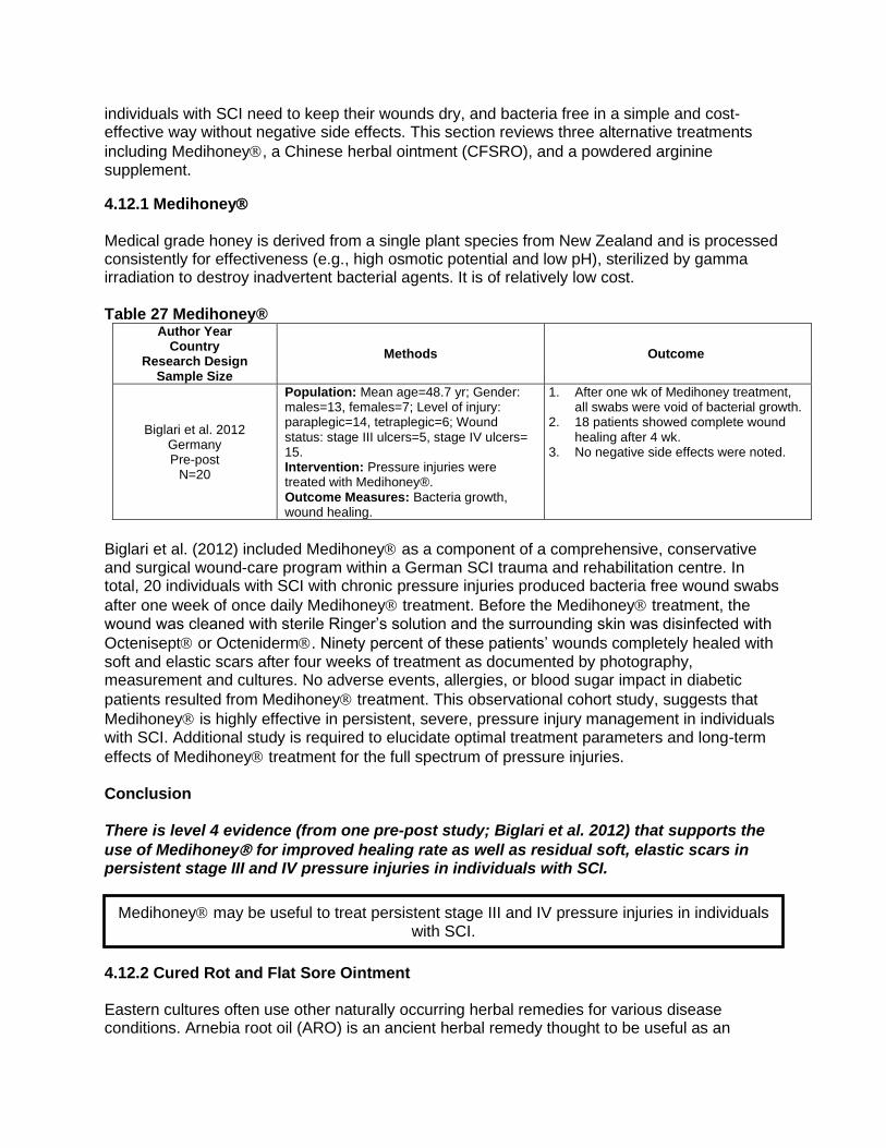
individuals with SCI need to keep their wounds dry, and bacteria free in a simple and cost-effective way without negative side effects. This section reviews three alternative treatments
including Medihoney, a Chinese herbal ointment (CFSRO), and a powdered arginine supplement.
4.12.1 Medihoney Medical grade honey is derived from a single plant species from New Zealand and is processed consistently for effectiveness (e.g., high osmotic potential and low pH), sterilized by gamma irradiation to destroy inadvertent bacterial agents. It is of relatively low cost. Table 27 Medihoney®
Author Year Country
Research Design Sample Size
Methods Outcome
Biglari et al. 2012 Germany Pre-post
N=20
Population: Mean age=48.7 yr; Gender: males=13, females=7; Level of injury: paraplegic=14, tetraplegic=6; Wound status: stage III ulcers=5, stage IV ulcers= 15. Intervention: Pressure injuries were treated with Medihoney®. Outcome Measures: Bacteria growth, wound healing.
1. After one wk of Medihoney treatment, all swabs were void of bacterial growth.
2. 18 patients showed complete wound healing after 4 wk.
3. No negative side effects were noted.
Biglari et al. (2012) included Medihoney as a component of a comprehensive, conservative and surgical wound-care program within a German SCI trauma and rehabilitation centre. In total, 20 individuals with SCI with chronic pressure injuries produced bacteria free wound swabs
after one week of once daily Medihoney treatment. Before the Medihoney treatment, the wound was cleaned with sterile Ringer’s solution and the surrounding skin was disinfected with
Octenisept or Octeniderm. Ninety percent of these patients’ wounds completely healed with soft and elastic scars after four weeks of treatment as documented by photography, measurement and cultures. No adverse events, allergies, or blood sugar impact in diabetic
patients resulted from Medihoney treatment. This observational cohort study, suggests that
Medihoney is highly effective in persistent, severe, pressure injury management in individuals with SCI. Additional study is required to elucidate optimal treatment parameters and long-term
effects of Medihoney treatment for the full spectrum of pressure injuries. Conclusion There is level 4 evidence (from one pre-post study; Biglari et al. 2012) that supports the
use of Medihoney for improved healing rate as well as residual soft, elastic scars in persistent stage III and IV pressure injuries in individuals with SCI.
4.12.2 Cured Rot and Flat Sore Ointment Eastern cultures often use other naturally occurring herbal remedies for various disease conditions. Arnebia root oil (ARO) is an ancient herbal remedy thought to be useful as an
Medihoney may be useful to treat persistent stage III and IV pressure injuries in individuals with SCI.

antipyretic, antiseptic and analgesic. It is postulated to promote basic fibroblast growth factor in healing wound tissue in a Chinese study (Pei et al. 2005) but has not been replicated in the SCI population. However, practitioners of traditional Chinese medicine in a Chinese Military Hospital called upon their years of experience with various traditional Chinese medicine remedies when they were not satisfied with the analgesic and curative effect of ARO. They produced a traditional Chinese medicine ointment called cured rot and flat sore ointment (CRFSO) comprised of hydrargyrum oxydatum crudum, red orpiment, borneol and gypsum fibrosum as a dilutant and treatment of “heat syndrome” according to “Yin-yang” theory. Anecdotal observations of superior effects led to a randomized, parallel-group, retrospective trial comparing ARO and CRFSO to treat stage IV pressure injuries in paraplegic patients (Liu et al. 2013). Table 28 Cured Rot and Flat Sore Ointment CRFSO
Author Year Country
Research Design Sample Size
Methods Outcome
Liu et al. 2013
China Case Control
N=35
Population: Median age: 59.1 yr; Gender: male=19, female= 16; Level of injury: paraplegic=10; Wound status: grade IV ulcer= 35. Intervention: Patients underwent 28 days of arnebia root oil (ARO) or Chinese herbal formula (CRFSO) treatment after undergoing surgery. Outcome Measures: Therapeutic effect (healing, effective, improved, no response) and therapeutic duration of treatment.
1. Statistically significant difference (p<0.005) between the two groups (follow-up occurred after 28 days of treatment).
2. No patients had adverse reaction during treatment.
3. Healed: 17 out of 20 patients in the CRFSO group.
4. Healed: 2/11 patients in ARO group. 5. Effective: 2/20 patients in CRFSO
group. 6. Effective: 3/11 patients in ARO group. 7. Improved: 1/11 ulcers in ARO group. 8. Therapeutic duration of treatment with
CRFSO averaged 19.47 days. 9. Therapeutic duration of treatment with
ARO averaged 29.18 days.
Discussion Liu et al. (2013) reported that there was a significantly shorter treatment time recorded for
CRFSO (19.55.0 days) compared to those receiving ARO (29.23.2 days; p<0.05). Other aspects of improved outcome included presence of a scab, contractibility, granulation tissue, reduced secretions and alleviation of pain upon day 28 evaluation. ARO or CRFSO treatments were used in conjunction with sodium chloride cleansing, hydrogen peroxide and saline removal of vesicular, ulcerated and necrotic tissue, 30 minute infrared irradiation of disinfected pressure injuries and surrounding 2-3 cm area, and routine care (e.g., aseptic dressing/bandage changes every 1-2 days) and pain treatment until the ulcers had healed. The ARO group also received 240,000 units of gentamicin gauze wetted with 100ml 9% sodium chloride. CRFSO responses were reported to be better in all categories of outcome assessment versus ARO responses with time to healing as the primary outcome. However, no mention of NPIAP pressure injury documentation guidelines use was mentioned (e.g., drainage amount-scant, moderate, copious), colour/consistency (e.g., serous, serosanguineous, purulent), odor). As well, no stratification for comparison of pressure injury healing by location was provided.

Conclusion There is level 3 evidence (from one case control study; Liu et al. 2013) that supports the use of CRFSO over ARO to accelerate pressure injury healing but it needs to be noted that objective outcome assessment was not clearly outlined.
4.12.3 Powdered Arginine Supplement Alternative medicine often relies on nutritional supplements to augment deficiencies in disease states. Because the exudate of pressure injuries are a source of bodily protein loss, daily intake of up to 2.0 g/kg of protein for individuals at risk of pressure injury development is advocated by Van Anholt et al. (2010). Similarly, supplementation with amino acids that act as substrates for protein synthesis and subsequent nitrogen balancing may also promote pressure injury healing. Brewer et al. (2010) undertook to test the effectiveness in supplementing individuals with SCI with the semi-essential amino acid, arginine, for improvement in pressure injury healing. Table 29 Powdered Arginine Supplement
Author Year Country
Research Design Sample Size
Methods Outcome
Brewer et al. 2010
Australia Pre-post
N=35
Population: Mean age= 50.5 yr; Gender: males=34, females=1; Level of injury: paraplegic=25, quadriplegic= 10; Wound status: grade II ulcer= 10, grade III ulcer=14, grade IV ulcer= 6. Intervention: 9 g commercial powdered arginine supplement per day. Outcome Measures: Healing rates.
1. Time to healing took an average of 21.1 wk in the historical control group.
2. Time to healing took an average of 10.5 wk in the intervention (powdered arginine supplement) group.
Discussion Brewer et al. (2010) compared a cohort of 18 community dwelling individuals with SCI and pressure injuries, who were supplemented with 9 g/day powdered arginine, against audits of a matched historical control group. Arginine treatment continued until pressure injuries were healed but it should be noted that the supplemental powder also contained carbohydrates and vitamins C and E, the latter vitamins of which are controversial in their effects over pressure injury healing in other patient populations (Ehrlich et al.1972; Vilter 1980; Albina 1994; Dorner et al. 2012). However, the authors did take care to exclude participants who presented with other healing comorbidities such as renal insufficiency, metabolic diseases and other co-morbidities. The resulting healing time was significantly improved by half the number of weeks required for healing by the historical controls across all categories of pressure injuries (p<0.006). Conclusion There is level 4 evidence (from one pre-post study; Brewer et al. 2010) for arginine supplementation for pressure injury healing.
CRFSO may be superior to ARO to promote accelerated healing of pressure injuries in people with SCI.

4.13 Miscellaneous Physical Treatments Although pressure redistribution to reduce ischemia is the primary method for pressure injury prevention and management, it is also understood that decreased skin temperature leads to reduced tissue metabolic demand that might translate to reduced reactive hyperemia post ischemia. To test this theory, Tzen et al. (2013) compared the effects of controlled local cooling on reactive hyperemia resulting from pressure induced ischemia for people with and without SCI. Table 30 Localized Cooling for Treatment of Pressure injuries
Author Year Country
Research Design Sample Size
Methods Outcome
Tzen et al. 2013
USA Prospective Controlled
Trial N=28
Population: Mean age: 35.79 yr; Gender: males=20, females= 8; Level of injury (ASIA): grade A or B=14, control group=14. Intervention: All subjects were tested under three conditions: pressure with fast cooling (-4 degrees Celsius/min), pressure with slow cooling (-33 degrees Celsius) and pressure with no cooling. Outcome Measures: Reactive hyperemia and its spectral densities in the metabolic, neurogenic and myogenic frequency ranges.
1. Reactive hyperemia was greater in pressure only when compared to the cooling conditions in the control group.
2. No change in spectral densities for both cooling conditions in SCI or control group.
3. Neurogenic spectral density increased without cooling for control group.
4. In SCI patients, no difference noted in reactive hyperemia in any conditions.
5. Metabolic and myogenic spectral densities increased without cooling and all spectral densities increased with slow cooling for the SCI group.
6. No change in all spectral densities with fast cooling.
Discussion Tzen et al. (2013) were able to confirm that microclimate control was an effective pressure injury prevention strategy in at-risk individuals without SCI; unfortunately, this was not the case for individuals with neurologically compromised vasoconstriction and capillary smooth muscle contraction as in people with SCI. Conclusion There is level 2 evidence (from one prospective controlled trial; Tzen et al. 2013) that localized cooling is not a viable pressure injury prevention strategy that is effective for individuals with SCI. Conversely, with neurological control of vasoconstriction and capillary smooth muscle contraction, those without SCI may benefit from microclimate controlled surfaces as a pressure injury prevention strategy.
Arginine supplementation in individuals with SCI may be helpful in accelerating pressure injury healing.
Pressure point localized cooling is not an effective pressure injury prevention strategy for people with SCI.

4.14 Factors Associated with Pressure Injury Treatment Success There are a wide range of recognized risk factors for the development of pressure injuries; poor nutritional status, dehydration, being under- or overweight, stroke, recent bone fractures, anaemia, diabetes, vitamin deficiency, and age >70 years old (Kenneweg et al. 2015). Infected pressure injuries are difficult to treat. Underlying causes often cannot be corrected, and are associated with a high risk of clinical recurrence. Long-term surveys report an ulcer recurrence risk from 12% to 82%, with a total complication rate of 17% to 46% (Jugun et al. 2016). The table below outlines different factors that are associated with various treatment successes. Table 32 Factors Associated with Treatment Success of Pressure Injuries
Author Year Country
Research Design Score
Sample Size
Methods Outcome
Systematic Reviews
Baron et al., 2018 Canada
Not specified AMSTAR=9
N=15
Method: A systematic search was conducted using several databases to identify articles that reported on skin care self-management. A MEDLINE search strategy was designed to include search terms on SCI, self-management, and skin care. Studies were included if they were RCTs or non-randomized trials with a control group receiving the standard of care, population ≥ 50% with SCI, published in English and addressed at least one of the following outcomes: mediators of skin care behaviour, skin care behaviours, or pressure ulcer related clinical outcomes. Type of intervention utilized, and effectiveness was extracted from each study. Databases: Embase, PsycINFO, CENTRAL, CINAHL, REHABDATA, CIRRIE, PeDro, and ERIC Level of evidence: I, II Questions:
1. What skin care self-management interventions are utilized in people with SCI?
2. How effective are the interventions?
1. The most common interventions used were “instructions on how to perform behaviour” (16 interventions), “information from a credible source” (12 interventions), and “social support (unspecified)” (9 interventions).
2. Evidence to support the effectiveness of interventions improving knowledge, self-efficacy, skills relating to skin care/pressure ulcer prevention, skin care behaviours, skin status and health-care utilization for skin problems was limited, particularly for clinical outcomes.
Indivdual Studies
Lane et al. 2016 United States
Cohort NIntial=158, NFinal=133
Population: Intervention group: Mean age=44 yr; Gender: males=60, females=15; Level of injury: paraplegia=55, tetraplegia=20. Control group: Mean age=44 yr; Gender: males=69, females=14; Level of injury: paraplegia=53, tetraplegia=30. Intervention: Intervention group (n=75): 6 wk period after implementation of smoking cessation guidelines (Ask, Advise, Assess, Assist, Arrange). Control group (n=83): Historical control consisting of 6 wk period prior to intervention initiation.
1. Intervention group had a significantly higher percentage of participants who stopped smoking (p=0.03).
2. No significant differences between groups in terms of percentage of participants who desired and underwent surgery.
3. In terms of percentage of participants with a decreased number of wounds, the smoker groups was 33.3%, the non-smoker group was 51.6%, and the smokers who stopped group was 65.2% (p=0.03).
Outcomes: Smoking cessation at 6 mo; Change in wound size; Change in number of wounds.
4. Smokers had an increase in total wound size while non-smokers and smokers who stopped had a decrease in total wound size (p=0.004).
Jugun et al. 2016 Switzerland Case Series
N=31
Population: Median age=60 yr; Number of pressure injuries=70. Intervention: Patient records were reviewed for those that underwent a complete surgical debridement or excision for an infected pressure injury. Outcomes: Clinical recurrence.
1. There were 44 pressure injury recurrences and 26 remissions after a median of 1 yr post-treatment. In 86% of these recurrences, cultures yielded a different organism than the preceding episode.
2. Clinical recurrence was not significantly associated with sex, age, number of prior pressure infection episodes, immune suppression, osteomyelitis, bacteremia, serum C-reactive protein level, involvement of Staphylococcus aureus, number of surgical debridements, use of vacuum-assisted closure devices, flap use, duration of antibiotic use, or duration of parenteral antibiotics.
Kenneweg et al. 2015 United States Case Series
N=49
Population: Mean age=45.4 yr; Gender: males=90.20%, females=9.80%; Level of injury: paraplegia=71.57%, tetraplegia=19.61%, no SCI=7.8%; Mean time since injury=214.49 mo; Number of pressure injuries=102; Pressure injury stage: II=2, III=8, IV=92. Intervention: Patient records were reviewed for those with pressure injury reconstructions. Outcomes: Differences between primary and recurrent ulcers; Pressure injury closure.
1. Primary ulcers had significantly longer ulcer duration (p=0.02), larger surface area (p=0.009), and higher number of debridements (p=0.006) compared to recurrent ulcers.
2. Primary ulcers had significantly lower albumin levels (p=0.002), higher erythrocyte sedimentation rate (p=0.02), lower glucose (p=0.03), and higher platelet count (p=0.002) compared to recurrent ulcers.
3. Pressure injury closure was significantly correlated with lower BMI (p=0.033), smaller surface area (p=0.049), and fewer debridements (p=0.049).
1. In 83.6% of cases, closure outcome could be successfully predicted using a multivariable model consisting of prealbumin (β=0.151), haematocrit (β=0.705), haemoglobin (β=-1.419), and creatinine (β=-4.622).
Discussion In a retrospective chart review, Lane et al. (2016) evaluated the impact of implementing evidence-based guidelines on smoking cessation in persons with spinal cord injuries and pressure injuries, as well as the impact of smoking on pressure injury healing in this population. 48% percent of the control group participants and 57% of the intervention group participants smoked cigarettes at baseline. Smoking cessation doubled with the use of the clinical practice guidelines (P=0.03). Smokers presented with a greater number of pressure injuries than nonsmokers. They experienced a mean increase rather than reduction in wound size. Nearly half (45.5%) of the intervention group participants who desired to have surgery had it performed, compared with only 34.9% of the control group participants (P=0.35).

Jugun et al. (2016) assessed interdisciplinary surgical and medical parameters associated with recurrences of infected pressure injuries. Authors found that in patients with infected pressure injuries, clinical recurrence occurs in almost two-thirds of lesions, but in only 14% with the same pathogens. The number of surgical debridements, flap use, or duration of antibiotic therapy was not associated with recurrence of pressure injury infection. Similarily, Kenneweg et al (2015) aimed to identify perioperative risk factors that may predict improved outcomes and reduced complications in primary and recurrent pressure injury reconstructions. A total of 49 patients with 102 reconstructions were reviewed. Numerous differences between primary and recurrent pressure injuries were identified, including ulcer location, patient nutritional status, wound infection, postoperative course and recurrence. Multivariate analysis revealed a flap reconstruction prediction model using creatinine, haematocrit, haemoglobin, and prealbumin that is able to successfully predict closure outcome in 83.6% of cases. Conclusion There is level 2 evidence (from one cohort study; Lane et al. 2017) that implementing evidence-based guidelines on smoking cessation in persons with spinal cord injuries can improve pressure injury healing. There is level 4 evidence (from two series; Jugun et al. 2016, Kenneweg et al. 2015) that recurrences of pressure injury infection may be caused by reinfections from extrahospital factors other than of surgical debridements, flap use, or duration of antibiotic therapy.
The use and implemtation of clinical practice guidelines may help individuals stop smoking.
Many factors play a role in the development, course and treatment of PIs. It is vital to understand the role of patient risk factors in the development of PIs, to direct subsequent
management and reconstruction, and to prevent future recurrences.

5.0 Summary There is level 2 evidence (from one prospective controlled trial; Kanno et al. 2009) that supports the use of ultrasonography to extend the yield of routine inspection and palpation of suspected or early stage pressure injuries in people with SCI. There is level 3 evidence (from one case control study; de Heredia et al. 2012) that magnetic resonance imaging can predict the development of osteomyelitis in non-healing pelvic pressure injuries in patients. There is level 4 (from one case series study; Loerakker et al. 2012) that reliance on circulatory biomarkers as an indication of muscle damage secondary to deep tissue injury in the SCI population cannot be recommended at this time. There is level 2 evidence (from two prospective controlled trial and one cohort study; Lui et al. 2015; Lui et al. 2006b; Ferguson et al. 1992) supported by level 4 evidence (from five pre-post studies, and two case series studies; Smit et al. 2012, 2013a, 2013b; Gyawali et a. 2011; Bogie & Triolo 2003; Van London et al. 2008; Liu et al. 2006a) that electrical stimulation decreases ischial peak pressures during stimulation. There is level 4 evidence (from one pre-post study; Bogie & Triolo 2003) that electrical stimulation may increase blood flow at sacral and gluteal areas post SCI. There is level 2 evidence (from two prospective controlled trials and one pre-post study; Lui et al. 2006a; Mawson et a 1993; Bogie & Triolo 2003) that electrical stimulation may increase tissue oxygenation post SCI. There is level 4 evidence (from two pre-post studies; Di Caprio et al. 2016; Previnaire et al. 2016) showing that fat grafting using the participants own body fat may be considered as a prevention strategy for some people when all other prevention strategies have been unsuccessful. There is level 2 evidence (from one prospective controlled trial; Hobson 1992) and level 4 evidence (from one case control study; Gutierrez et al. 2004) to support not generalizing pressure mapping data from able-bodied subject to SCI subjects. There is level 4 evidence (from one prospective controlled trial; Brienza & Karg 1998) to support not generalizing pressure mapping data from the elderly population to the SCI population. There is level 2 evidence (from one cohort study; Kennedy et al. 2003) showing that early attendance at specialized seating assessment clinics increases the skin management abilities of individuals post SCI. There is level 1b evidence (from two randomized controlled trials studies; Rintala et al. 2008; Garber et al. 2002); and level 2 evidence (from 1 lower RCT from Kim & Cho, 2017) and level 4 evidence (from four pre-post studies; May et al. 2006; Brace & Schubart 2010; Schubart et al. 2012; Jones et al. 2003 ) that providing enhanced pressure injury prevention education, including behaviour contingencies and strategies, is effective at

helping individuals with SCI gain and retain this knowledge, reduce pressure injury severity and decreased health care costs.. There is level 1b evidence (from one randomized control trial; Guihan et al. 2014) suggesting that for the SCI population with chronic and/or severe pressure injuries, an enhanced prevention program using individual motivational intervention and group self-management training does not improve skin protective behaviours or pressure injury outcomes. There is level 4 evidence (from two pre-post studies; Schubart et al. 2012; Brace & Schubart 2010) that online eLearning modules may improve knowledge on prevention of pressure injuries among persons with SCI. There is level 4 evidence (from one case series study; Cobb et al. 2014) suggesting that a formal pressure injury prevention program can improve best practice adherence in an acute care facility. There is level 1b evidence (from one randomized controlled trial; Rintala et al. 2008) that providing enhanced pressure injury education and structured follow-up is effective in reducing recurrence of pressure injuries especially in those individuals with no previous history of pressure injury surgery. There is level 4 evidence (from one case series study; Ghaisas et al. 2015) to suggest that an intervention that focus on reducing risk through lifestyle, particularly habits and behaviour changes are related to improvements in the uptake of pressure management strategies, therefore improvements in pressure injury status. There is level 1b evidence (from one randomized controlled trial; Houlihan et al. 2013) that telerehabilitation using an automated call-in system with built-in theory-based behavior change strategies may make a significant difference for women but not men in preventing pressure injuries post SCI. There is level 4 evidence (from one case series; Vesmarovich et al. 1999) that telerehabilitation via videophone to support clinical interactions and digital photography does not make a significant difference in the prevention and treatment of pressure injuries post SCI. There is level 2 evidence (from one randomized control trial; Arora et al. 2017) that treatment intervention provided by telephone has potential to provide a low cost means of treatment intervention in low-and middle-income countries. There is level 4 evidence (from one post-test study; Hilgart et al. 2014) that a comprehensive prevention program provided using an internet format has potential to meet ongoing needs for pressure management beyond the hospital/rehabilitation facility. There is level 2 evidence (from one cohort study; Richard-Denis et al. 2017) that indicated, for pre-operative prevention for the SCI population, a multi-layer foam dressing is not superior in preventing sacral pressure injuries compared to viscoelastic polymer gel mattress over a foam stretcher pad.

There is level 1 evidence (from seven randomized controlled trials; Karsli et al. 2017; Houghton et al. 2010; Cukjati et al. 2001; Adegoke & Badmos 2001; Karba 1997; Jercinovic 1994; Griffin 1991) that electrical stimulation accelerates the healing rate of stage III and IV pressure injuries when combined with standard wound management.
There is level 1 evidence (from two randomized controlled trials; Taly et al. 2004; Nussbaum et al. 1994) that laser treatment has no added benefit in pressure injury healing post SCI than standard wound care alone. There is level 1b evidence (from one randomized controlled trial; Nussbaum et al. 1994) that the combination of US/UVC with standard wound care decreases wound healing time of pressure injuries post SCI; there is no evidence to support the benefit of UVC or US, used individually. There is level 1b evidence (from one randomized controlled trial; Salzberg et al. 1995) that pulsed electromagnetic energy accelerates healing of stage II and III pressure injuries post SCI. There is level 1b evidence (from one RCT; Sundby et al. 2018) that intermittent negative pressure (INP) device plus standard wound care (SWC) is effective for wound healing compared to SWC alone in SCI patients home care. There is level 1b and level 2 evidence (from one RCT and one prospective controlled study; Dwivedi et al. 2017; Srivastava) that negative pressure wound therapy (NPWT) has shown to reduce levels of MMP-8, increase the rate of healing, reduce exudate production and enhance the rate of formation of red granulation tissue when compared to conventional wet gauze alone. There is level 1b evidence (from one RCT and one pre-post study; De Laat et al. 2011; Coggrove et al. 2012) that topical negative pressure facilitates wound healing for pressure injuries in people with SCI and other patient etiologies. There is level 4 evidence (from one case series; Dessy et al. 2015) that vacuum-assisted closure (VAC) therapy does not promote wound healing and may cause rare complications including foam-fragment retention within the wound. There is level 2 evidence (from one prospective controlled trial; Kloth et al. 2001) that normothermic dressings may improve healing of pressure injuries post SCI. There is level 4 evidence (from two pre-post tests; Keast & Fraser 2004; Vair et al. 2015) that recombinant human erythropoietin aids in the healing of stage IV chronic non-healing pressure injuries post SCI. There is level 2 evidence (from one prospective controlled trial; Singh et al. 2014) that advanced wound therapy using local applications of PRP seems to be a promising alternative to standard saline dressings in PI healing. There is leve 2 evidence (from one prospective controlled trial; Singh et al. 2015) that local application of PRP may reduce bacterial presence and colonization in PIs. There is level 4 evidence (from one case series and one post-test; Sell et al. 2011; Biglari

et al. 2015) that supports the possibility of platelet-rich plasma therapy facilitation of reactivated healing in severe, non-healing pressure injuries, post SCI. There is level 2 evidence (from one flawed randomized controlled trial; Bauman et al. 2013) that does not support the use of oxandrolone (anabolic steroid) to facilitate healing of serious pressure injuries post SCI. However, very limited, earlier level 4 evidence (from one case series; Spungeon et al. 2001) did lend some support for the use of oxandrolone to promote healing of stage III and IV pressure injuries post SCI. There is Level 1 evidence (from one randomized controlled trial; Hollisaz et al. 2004) that completion of healing for stage I and II pressure injuries is greater with an occlusive hydrocolloid dressing compared to phenytoin cream or simple dressing post SCI. There is Level 2 evidence (from one randomized controlled trial; Kaya et al. 2005) that occlusive hydrogel-type dressings heal more pressure injuries than conservative treatment post SCI. There is level 1 evidence (from one randomized controlled trial; Subbanna et al. 2007) that topical phenytoin shows a trend towards healing of stage I and II pressure injuries post SCI. There is level 2 evidence (from one randomized controlled trial; Scevola et al. 2010) that platelet gel dressings, when used within the first 2 weeks of treatment can trigger earlier granulation tissue proliferation towards pressure injury healing, post SCI. Level 1 evidence (from one randomized controlled trial; Ho et al. 2012) underpins the use of pulsatile lavage hydrotherapy debridement for Stage III and IV pressure injuries secondary to SCI. There is level 5 evidence (from one observational study; Bogie et al. 2013) that pulsatile lavage therapy, used in conjunction with standard infection control standards, is likely a safe debridement method for Stage III and IV pressure injuries in people with SCI. There is level 2 evidence (from one prospective controlled trial and one observational study; Wang et al. 2010; Sherman et al. 1995) that supports the use of maggot therapy as an adjunctive therapy for pressure injury debridement post SCI. There is level 4 evidence (from one post-test study; Erba et al. 2010) that supports the use of silicone moulding as a radical en bloc debridement method for pyramidal shaped grade IV pressure injury cavities in people with SCI. There is level 4 evidence (from one pre-post study; Banks & Ho 2008) that topical oxygen therapy may improve healing of pressure injuries post SCI. There is level 2 evidence (from one cohort study; Velasco et al. 2015) that the application of Tissucol Duo during surgical treatment of PrUs in patients with SCI has been shown to be effective in reducing postoperative complications and in shortening the duration of the hospital stay with a consequent savings in costs.

There is level 3 evidence (from one post-test; Ljung et al. 2017) that a structured treatment programme regulating pre- and postoperative care and rehabilitation can help raise health status of SCI pressure injury patients. There is level 4 evidence (from one case series; Yusmindo et al. 2014) that proximal amputations of the lower limbs are procedures that can be considered as part of the treatment for complicated pressure injuries. In properly selected patients, it can reduce the number of hospital stay, improve the quality of life and functional outcome. There is level 5 evidence (multiple studies; Table 27) that supports various surgical repair methods for persistent, severe thigh and buttock pressure injuries secondary to SCI, as a beneficial treatment option. There is level 4 evidence (from one pre-post study; Biglari et al. 2012) that supports the
use of Medihoney for improved healing rate as well as residual soft, elastic scars in persistent stage III and IV pressure injuries in individuals with SCI. There is level 3 evidence (from one case control study; Liu et al. 2013) that supports the use of CRFSO over ARO to accelerate pressure injury healing but it needs to be noted that objective outcome assessment was not clearly outlined. There is level 4 evidence (from one pre-post study; Brewer et al. 2010) for arginine supplementation for pressure injury healing. There is level 2 evidence (from one prospective controlled trial; Tzen et al. 2013) that localized cooling is not a viable pressure injury prevention strategy that is effective for individuals with SCI. Conversely, with neurological control of vasoconstriction and capillary smooth muscle contraction, those without SCI may benefit from microclimate controlled surfaces as a pressure injury prevention strategy. There is level 2 evidence (from one cohort study; Lane et al. 2017) that implementing evidence-based guidelines on smoking cessation in persons with spinal cord injuries can improve pressure injury healing. There is level 4 evidence (from one series; Jugun et al. 2016) that recurrences of pressure injury infection may be caused by reinfections from extrahospital factors other than of surgical debridements, flap use, or duration of antibiotic therapy.

References Adegoke BO, Badmos KA. Acceleration of pressure injury healing in spinal cord injured patients
using interrupted direct current. Afr J Med Med Sci 2001;30(3):195-197.
Agency for Health Care Quality and Research (AHCPR). Guideline 3: Pressure injuries in
adults: Prediction and Prevention; 1992. www.guidelines.gov
Agency for Health Care Quality and Research. (AHCPR) Guideline 15: Treatment of Pressure
injuries; 1992. www.guidelines.gov.
Ahluwalia R, Martin D, Mahoney JL. The operative treatment of pressure wounds. Int Wound J
2010;7(2):103-106.
Albina JE. Nutrition and wound healing. Parental and Enteral Nut 1994;18(4):367-376.
Alexander LR, Spungen AM, Liu MH, Losada M, Bauman WA. Resting metabolic rate in
subjects with paraplegia: the effect of pressure sores. Arch of Phys Med and Rehab
1995;76(9):819-822.
Allen J, Houghton PE. A case study for electrical stimulation on a stage III pressure injury.
Wound Care Canada 2003;2(1):34-36.
Al-Momen AK. Recombinant human erythropoietin induced rapid healing of a chronic leg ulcer
in a patient with sickle cell disease. Acta Haematol 1991;86(1):46-48.
Alsousou J, Thompson M, Hulley P, Noble A, Willett K. The biology of platelet-rich plasma and
its application in trauma and orthopaedic surgery: a review of literature. Journ of Bone Joint
Surg 2009;91(8):987-996.
Anitua E, Aguirre JJ, Algorta J, Ayerdi E, Cabezas AI, Orive G, et al. Effectiveness of
autologous preparation rich in growth factors for the treatment of chronic cutaneous ulers. Journ
of Biomed Materials Research Part B: Applied Biomaterials 2008;84b(2):415-421.
Anson CA, Shepherd C. Incidence of secondary complications in spinal cord injury. Inter Journ
of Rehab Research 1996;19(1):55-66.
Anstead GM, Hart LM, Fong Sunahara J, Liter ME. Phenytoin in wound healing. Annals of
Pharm 1996;30(7-8):768-775.
Arora M, Harvey LA, Glinsky JV, Chhabra HS, Hossain S, Arumugam N, et al. Telephone-based management of pressure ulcers in people with spinal cord injury in low- and middle-income countries: a randomised controlled trial. Spinal cord. 2017;55(2):141-7. Argenta LC, Morykwas MJ. Vacuum-assisted closure: a new method for wound control and
treatment: clinical experience. Ann Plast Surg 1997:38(6):563-576
Ash D. An exploration of the occurrence of pressure injuries in a British spinal injuries unit.
Journ Clin Nurs 2002:11(4):470-478.
Ayello EA, Braden B. How and why to do pressure injury risk assessment. Adv Skin Wound
Care 2002;15(3):125-131.

Backhaus M, Citak M, Tilkorn DJ, Meindl R, Schildhauer TA, Fehmer T. Pressure sores
significantly increase the risk of developing a Fournier's gangrene in patients with spinal cord
injury. Spinal Cord 2011;49(11):1143-1146.
Baker LL, Rubayi S, Villar F, Demuth SK. Effect of electrical stimulation waveform on healing of
ulcers in human beings with spinal cord injury. Wound Repair Regen 1996;4(1):421-28.
Banks PG, Ho CH. A novel topical oxygen treatment for chronic and difficult-to-heal wounds:
case studies. Journ of Spinal Cord Med 2008;31:297-301.
Baron JS, Sullivan KJ, Swaine JM, Aspinall A, Jaglal S, Presseau J, et al. Self-management interventions for skin care in people with a spinal cord injury: part 1-a systematic review of intervention content and effectiveness. Spinal cord. 2018. Baron, J.S., Sullivan, K.J., Swaine, J.M. et al. Self-management interventions for skin care in people with a spinal cord injury: part 1—a systematic review of intervention content and effectiveness. Spinal Cord 2018; 56: 823–836. Bates-Jensen BM, McCreath HE, Pongquan V, Apeles NC.Subepidermal moisture differentiates
erythema and stage I pressure injuries in nursing home residents. Wound Repair and Regen
2008;16(2):189-197.
Bauman WA, Spungen AM, Collins JF, Raisch DW, Ho C, Deitrick GA, et al. The effect of
oxandrolone on the healing of chronic pressure injuries in persons with spinal cord injury: a
randomized trial. Ann Int Med 2013;158(10):718-726.
Bergstrom N, Braden BJ, Laguzza A, Holman V. The Braden Scale for Predicting Pressure Sore
Risk. Nurs Res 1987;36:205-210.
Bertheuil N, Huguier V, Aillet S, Beuzeboc M, Watier E. Biceps femoris flap for closure of ischial
pressure injuries. European Journal of Plastic Surgery 2013;36(10):639-644.
Biglari B, Büchler A, Reitzel T, Swing T, Gerner H J, Ferbert T, et al. Moghaddamspective
study on flap complications after pressure injury surgery in spinal cord-injured patients. Spinal
cord 2013;52(1):80-83.
Biglari B, Buchler A, Reitzel T, Swing T, Gerner HJ, Ferbert T, et al. A retrospective study on flap complications after pressure ulcer surgery in spinal cord-injured patients. Spinal cord. 2014;52(1):80-3. Biglari B, Reitzel T, Swing T, Buchler A, Gerner HJ, Schmidmaier G, et al. A pilot study on the effectiveness of platelet-rich plasma and debridement for the treatment of nonhealing fistulas in spinal cord-injured patients. Advances in skin & wound care. 2015;28(3):123-8. Black J, Baharestani M, Cuddigan J, Dorner B, Edsberg L, Langemo D, et al. Posthauerl
pressure injury advisory panel’s updated pressure injury staging system. Urol Nurs
2007;27:144-150.
Bogie KM, Nuseibeh I, Bader DL. Early progressive changes in tissue viability in the seated
spinal cord injured subject. Paraplegia 1995;33:141-147.

Bogie KM, Reger SI, Levine SP, Sahgal V. Electrical stimulation for pressure sore prevention
and wound healing. Assist Technol 2000;12:50-66.
Bogie KM, Triolo RJ. Effects of regular use of neuromuscular electrical stimulation on tissue
health. J Rehabil Res Dev 2003;40:469-475.
Bogie KM, Wang X, Triolo RJ. Long-term prevention of pressure injuries in high-risk patients: a
single case study of the use of gluteal neuromuscular electric stimulation. Arch Phys Med
Rehabil 2006;87:585-591.
Bogie KM, Ho CH. Pulsatile lavage for pressure injury management in spinal cord injury: a
retrospective clinical safety review. Spinal Cord 2013;59(3):35-38.
Borgognone A, Anniboletti T, De Vita F, Schirosi M, Palombo P. Ischiatic pressure sores: our
experience in coupling a split-muscle flap and a fasciocutaneous flap in a ‘criss-cross’ way.
Spinal cord 2010;48(10):770-773.
Brace JA, Schubart JR. A prospective evaluation of a pressure injury prevention and
management E-learning program for adults with spinal cord injury. Ostomy Wound Management
2010;56(8):40-50.
Brewer S, Desneves K, Pearce L, Mills K, Dunn L, Brown D, et al. Effect of an arginine-
containing nutritional supplement on pressure injury healing in community spinal patients. J
Wound care 2010;19:7:311-6.
Brienza DM, Karg PE. Seat cushion optimization: a comparison of interface pressure and tissue
stiffness characteristics for spinal cord injured and elderly patients. Arch Phys Med Rehabil
1998;79:388-394.
Broussard CL. Dressing decisions. In: Krasner DL, Rodeheaver GT, Sibbald RG, editors.
Chronic Wound Care: A Clinical Source Book for Healthcare Professionals. Malvern, PA: HMP
Communications 2007; 22:249-262.
Burke DT, Ho CH, Saucier MA, Stewart G. Effects of Hydrotherapy on Pressure injury Healing.
Amer Journ of Phys Med and Rehab 1998;77(5):394-398.
Byrne DW, Salzberg CA. Major risk factors for pressure injuries in the spinal cord disabled: a
literature review. Spinal Cord 1996;34:255-263.
Cardenas DD, Hoffman JM, Kirshblum S, McKinley W. Etiology and incidence of
rehospitalization after traumatic spinal cord injury: a multicenter analysis. Arch of Phys Med and
Rehab 2004;85(11):1757-1763.
Carley PJ, Wainapel SF. Electrotherapy for acceleration of wound healing: low intensity direct
current. Arch Phys Med Rehabil 1985;66:443-446.
Chan BC, Nanwa N, Mittmann N, Bryant D, Coyte PC, Houghton PE. The average cost of
pressure injury management in a community dwelling spinal cord injury population. Int Wound
Journ 2013;10(4):431-440.
Chen Y, DeVivo MJ, Jackson AB. Pressure injury prevalence in people with spinal cord injury:
age-period-duration effects. Arch Phys Med Rehabil 2005;86:1208-1213.

Chen D, Apple Jr DF, Hudson LM, Bode R. Medical complications during acute rehabilitation
following spinal cord injury—current experience of the Model Systems. Arch of Phys Med and
Rehab 1999;80(11):1397-1401.
Chen, X., Jiang, Z., Chen, Z., & Wang, D. (2011). Application of skin traction for surgical
treatment of grade IV pressure sore: a clinical report of 160 cases. Spinal cord, 49(1), 76-80.
Citak M, Backhaus M, Tilkorn DJ, O'Loughlin PF, Meindl R, Muhr G, et al. Necrotizing fasciitis in
patients with spinal cord injury: an analysis of 25 patients. Spine 2011;36(18):1225-1229.
Clark FA, Jackson JM, Scott MD, Carlson ME, Atkins MS, Uhles-Tanaka D, et al. Data-based
models of how pressure injuries develop in daily-living contexts of adults with spinal cord injury.
Arch Phys Med Rehabil 2006;87:1516-1525.
Cobb JE, Belanger LM, Park SE, Shen T, Rivers CS, Dvorak MF, et al. Evaluation of a pilot Pressure Ulcer Prevention Initiative (PUPI) for patients with traumatic spinal cord injury. Journal of wound care. 2014;23(5):211-2, 4, 6-8 passim. Coggrave M, West H, Leonard B. Topical negative pressure for pressure injury management. Br
J Nurs 2002;11(6):29-36.
Cogan AM, Blanchard J, Garber SL, Vigen CL, Carlson M, Clark FA. Systematic review of behavioral and educational interventions to prevent pressure ulcers in adults with spinal cord injury. Clinical rehabilitation. 2017;31(7):871-80. Coggrave MJ, Rose LS. A specialist seating assessment clinic: changing pressure relief
practice. Spinal Cord 2003;41:692-695.
Consortium for Spinal Cord Medicine. Pressure injury prevention and treatment following spinal
cord injury: A clinical practice guideline for health care professionals (pp. 1-77). 2000.
Conway H, Griffith BH. Plastic surgery for closure of decubitus ulcers in patients with
paraplegia: based on experience with 1,000 cases. The Amer Journ of Surg 1956;91(6):946-
975.
Crenshaw RP, Vistnes LM. A decade of pressure sore research: 1977-1987. J Rehabil Res Dev
1989;26:63-74.
Cukjati D, Robnik-Šikonja M, Reberšek S, Kononenko I, Miklavčič D. Prognostic factors in the
prediction of chronic wound healing by electrical stimulation. Med and Bio Eng and Comp
2001;39(5):542-550.
Daniel RK, Priest DL, Wheatley DC. Etiologic factors in pressure sores: An experimental model.
Arch Phys Med Rehabil 1981;62(10):492-498.
Davis JJ. Operative treatment of scar following bed sores. Surgery 1938;3:1.
De Angelis B, Lucarini L, Agovino A, Migner A, Orlandi F, Floris M, et al. Combined use of
super-oxidised solutions with negative pressure for the treatment of pressure injuries: case
report. Int Wound Jorn 2013;10(3):336-339.
DeJong G, Tian WF, Hsieh CH, Junn C, Karam C, Ballard PH, et al. Rehospitalization in the first
year of traumatic spinal cord injury after discharge from medical rehabilitation. Arch Phys Med
Rehab 2013;94(4):87-97.

De Laat EH, van den Boogaard MH, Spauwen PH, van Kuppevelt DH, van Goor H,
Schoonhoven L. Faster wound healing with topical negative pressure therapy in difficult-to-heal
wounds: a prospective randomized controlled trial. Annals of Plastic Surgery 2011;67(6):626-
631.
DeLisa JA, Mikulic MA. Pressure injuries. What do we do if preventive management fails.
Pressure injuries 1985;77(6):209-212.
Dessy LA, Serratore F, Corrias F, Parisi P, Mazzocchi M, Carlesimo B. Retention of polyurethane foam fragments during VAC therapy: a complication to be considered. International wound journal. 2015;12(2):132-6. Diaz S, Li X, Rodriquez L, Salgado J. Update in the surgical management of decubitus ulcers.
Anaplastology 2013;2(3):1-10.
Di Caprio G, Larocca F, Scioli M, Ziccardi P, Nicoletti G, Iervolino S, et al. Fat grafting for the prevention of pressure ulcers: a case series. European Journal of Plastic Surgery. 2016;39(2):113-8. Dorner, B. The complete guide to nutrition care for pressure injuries: Prevention and treatment.
Akron, OH: Becky Dorner and Associates. 2012.
Dover H, Pickard W, Swain I, Grundy D. The effectiveness of a pressure clinic in preventing
pressure sores. Paraplegia 1992;30(4):267-272.
Dunn CA, Carlson M, Jackson JM, Clark FA. Response factors surrounding progression of
pressure injuries in community-residing adults with spinal cord injury. Amer Journ of Occup
Therapy 2009;63(3):301-309.
Drummond D, Breed AL, Narechania R. Relationship of spine deformity and pelvic obliquity on
sitting pressure distributions and decubitus ulceration. Journ of Pediatric Orthopaedics
1985;5(4):396-402.
Dwivedi MK, Bhagat AK, Srivastava RN, Jain A, Baghel K, Raj S. Expression of MMP-8 in Pressure Injuries in Spinal Cord Injury Patients Managed by Negative Pressure Wound Therapy or Conventional Wound Care: A Randomized Controlled Trial. Journal of wound, ostomy, and continence nursing: official publication of The Wound, Ostomy and Continence Nurses Society. 2017;44(4):343-9. Ehrlich HP, Tarver H, Hunt TK. Inhibitory effects of vitamin E on collagen synthesis and wound
repair. Annals of surgery 1972;175(2):235.
Erba P, Wettstein R, Schumacher R, Schwenzer-Zimmerer K, Pierer G, Kalbermatten DF.
Silicone moulding for pressure sore debridement. Journ of Plast Reconstr Aesthet Surg
2010;63(3):550-553.
Feedar JA, Kloth LC, Gentzkow GD. Chronic dermal ulcer healing enhanced with monophasic
pulsed electrical stimulation. Phys Ther 1991;71(9):639-649.
Ferguson AC, Keating JF, Delargy MA, Andrews BJ. Reduction of seating pressure using FES
in patients with spinal cord injury: a preliminary report. Paraplegia 1992;30(7):474-478.

Fisher SV, Szymke TE, Apte SY, Kosiak M. Wheelchair cushion effect on skin temperature.
Arch Phys Med Rehab 1978;59(2):68-72.
Fleck CA, Sprigle S. Support surfaces: Tissue integrity, terms, principles, and choice. In:
Krasner DL, Rodeheaver GT, Sibbald RG, editors. Chronic wound care: A clinical source book
for healthcare professionals. Malvern, PA: HMP Communications, 2007:629-640.
Foster RD, Anthony JP, Mathes S, Hoffman WY, Young D, Eshima I. Flap selection as a
determinant of success in pressure sore coverage. Arch of Surg 1997;132(8):868-873.
Fuhrer MJ, Garber SL, Rintala DH, Clearman R, Hart KA. Pressure injuries in community-
resident persons with spinal cord injury: prevalence and risk factors. Arch Phys Med Rehabil
1993;74(11):1172-1177.
Galea M, Tumminia J, Garback LM. Telerehabilitation in spinal cord injury persons: a novel
approach. Telemed J E Health 2006;12(2):160-162.
Garber SL, Reddy NP, McLane KM, Krouskop TA. Role of technology in pressure injury
prevention. In: Krasner DL, Rodeheaver GT, Sibbald RG, editors. Chronic wound care: A clinical
source book for healthcare professionals (pp. 653-660). Malvern, PA: HMP Communications.
2007.
Garber SL, Rintala DH, Holmes SA, Rodriguez GP, Friedman J. A structured educational model
to improve pressure injury prevention knowledge in veterans with spinal cord dysfunction. J
Rehab Res Dev 2002;39(5):575-588.
Garber SL, Rintala DH, Rossi CD, Hart KA, Fuhrer MJ. Reported pressure injury prevention and
management techniques by persons with spinal cord injury. Arch Phys Med Rehab
1996;77(8):744-749.
Garber SL, Rintala DH, Hart K A, Fuhrer MJ. Pressure injury risk in spinal cord injury:
predictors of ulcer status over 3 years. Arch of Phys Med and Rehab 2000;81(4):465-471.
Gelis A, Pariel S, Colin D, Barrois B, Passadori Y, Ribinik P, et al. What is the role of TPE in
management of patients at risk or with pressure injury as of 2012? Towards development of
French guidelines for clinical practice. Annals of Physical and Rehabilitation Medicine
2012;55(7):517-529.
Ghaisas S, Pyatak EA, Blanche E, Blanchard J, Clark F. Lifestyle changes and pressure ulcer prevention in adults with spinal cord injury in the pressure ulcer prevention study lifestyle intervention. The American journal of occupational therapy : official publication of the American Occupational Therapy Association. 2015;69(1):6901290020p1-10. Grassetti L, Scalise A, Lazzeri D, Carle F, Agostini T, Gesuita R, et al. Perforator flaps in late-stage pressure sores treatment: Outcome analysis of 11-year-long experience with 143 patients. Ann Plast Surg 2013. Grassetti L, Scalise A, Lazzeri D, Carle F, Agostini T, Gesuita R, et al. Perforator flaps in late-stage pressure sore treatment: outcome analysis of 11-year-long experience with 143 patients. Ann Plast Surg. 2014;73(6):679-85.

Griffin JW, Tooms RE, Mendius RA, Clifft JK, Vander ZR, El Zeky F et al. Efficacy of high
voltage pulsed current for healing of pressure injuries in patients with spinal cord injury. Phys
Ther 1991;71(6):433-444.
Guihan M, Smith BM, LaVela SL, Garber SL. Factors predicting pressure injuries in veterans
with spinal cord injuries. Amer Journ of Phys Med & Rehab 2008;87(9):750-7.
Guihan M, Bates-Jenson BM, Chun S, Parachuri R, Chin AS, McCreath H. Assessing the
feasibility of subepidermal moisture to predict erythema and stage 1 pressure injuries in persons
with spinal cord injury: a pilot study. The journal of spinal cord medicine 2012;35(1):46-52.
Guihan M, Bombardier CH, Ehde DM, Rapacki LM, Rogers TJ, Bates-Jensen B, et al. Comparing multicomponent interventions to improve skin care behaviors and prevent recurrence in veterans hospitalized for severe pressure ulcers. Archives of physical medicine and rehabilitation. 2014;95(7):1246-53.e3. Guihan M, Sohn MW, Bauman WA, Spungen AM, Powell-Cope GM, Thomason SS, et al. Difficulty in Identifying Factors Responsible for Pressure Ulcer Healing in Veterans With Spinal Cord Injury. Archives of physical medicine and rehabilitation. 2016;97(12):2085-94.e1. Gutierrez EM, Alm M, Hultling C, Saraste H. Measuring seating pressure, area, and asymmetry
in persons with spinal cord injury. Euro Spine Journ 2004;13(4)374-379.
Gyawali S, Solis L, Chong SL, Curtis C, Seres P, Kornelsen I, et al. Intermittent electrical
stimulation redistributes pressure and promotes tissue oxygenation in loaded muscles of
individuals with spinal cord injury. Journ Appl Physiol (2011);110(1):246-255.
Harrow JJ, Mayrovitz HN. Subepidermal moisture surrounding pressure ulcers in persons with a spinal cord injury: a pilot study. The journal of spinal cord medicine. 2014;37(6):719-28. Hauptfleisch J, Meagher TM, Hughes R.J, Singh JP, Graham A, López de Heredia L (2013);
Interobserver Agreement of Magnetic Resonance Imaging Signs of Osteomyelitis in Pelvic
Pressure injuries in Patients With Spinal Cord Injury. Archives of physical medicine and
rehabilitation, 94(6),1107-1111.
He J, Xu H, Wang T, Ma S, Dong J. Treatment of complex ischial pressure sores with free
partial lateral latissimus dorsi musculocutaneous flaps in paraplegic patients. Journ of Plastic,
Reconstructive & Aesthetic Surgery 2012;65(5);634-639.
Heyneman A, Beele H, Vanderwee K, Defloor T. A systematic review of the use of hydrocolloids
in the treatment of pressure injuries. Journ Clin Nurs 2008;17:1164-1173.
Hilgart M, Ritterband L, Baxter K, Alfano A, Ratliff C, Kinzie M, et al. Development and perceived utility and impact of a skin care Internet intervention. Internet Interventions. 2014;1(3):149-57. Hill ML, Cronkite RC, Ota DT, Yao EC, Kirat BJ. Validation of home telehealth for pressure
injury assessment: a study in patients with spinal cord injury. Journ of Telemed and Telecare
2012;15:196-202.
Ho CH, Bensitel T, Wang X, Bogie KM. Pulsatile lavage for the enhancement of pressure injury
healing: a randomized controlled trial. Physt Therapy 2012;92(1):38-48.
Ho CH, Powell HL, Collins JF, Bauman WA, Spungen AM. Poor nutrition is a relative
contraindication to negative pressure wound therapy for pressure injuries: preliminary
observations in patients with spinal cord injury. Advances in Skin and Wound Care
2010;23(11):508-516.
Hobson DA. Comparative effects of posture on pressure and shear at the body-seat interface. J
Rehabil Res Dev 1992;29(4):21-31.
Hollisaz MT, Khedmat H, Yari F. A randomized clinical trial comparing hydrocolloid, phenytoin
and simple dressings for the treatment of pressure injuries. BMC Dermatol 2004;4(1):18.
Houghton PE, Campbell KE and CPG Panel. Canadian best practice guidelines for the
prevention and management of pressure injuries in people with spinal cord injury: A resource
handbook for clinicians. Accessed at http://www.onf.org. 2013.
Houghton PE, Campbell KE. Therapeutic modalities in the treatment of chronic recalcitrant
wounds. In: Krasner DL, Rodeheaver GT, Sibbald RG, editors. Chronic wound care: A clinical
source book for healthcare professionals (pp. 403-415). Malvern, PA: HMP Communications.
2007.
Houghton PE, Fraser CH. Biomarkers that predict response to adjunctive therapies in people
with spinal cord injury. Proceedings of World Union of Wound Healing Societies. 2008.
Houlihan BV, Jette A, Friedman AH, Paasche-Orlow M, Ni P, Wierbicky J, et al. A pilot study of
a telehealth intervention for persons with spinal cord dysfunction. Spinal Cord 2013;51(9):715–
720.
Hug K, Stumm C, Debecker I, Fellinghauer CS, Peter C, Hund-Georgiadis M. Self-Efficacy and Pressure Ulcer Prevention After Spinal Cord Injury—Results From a Nationwide Community Survey in Switzerland (SwiSCI). PM and R. 2018;10(6):573-86. Idowu OK, Yinusa W, Gbadegesin SA, Adebule GT. Risk factors for pressure injuryation in a
resource constrained spinal injury service. Spinal Cord 2011;49(5):643-67.
Isik FF, Engrav LH, Rand RP, Kierney P, Cardenas DD. Reducing the period of immobilization
following pressure sore surgery: a prospective, randomized trial. Plastic and reconstructive
surgery 1997;100(2):350-354.
Jan YK, Liao F, Burns S. Effect of spinal cord injury on non-linear complexity of skin blood flow
oscillations. In C. Stephanidis, Universal access in human-computer interaction. Applications
and services (pp. 345-355). Berlin, Germany: Springer Berlin Heidelberg. 2011.
Jercinovic A, Karba R, Vodovnik L, Stefanovska A, Kroselj P, Turk R, et al. Low frequency
pulsed current and pressure injury healing. Rehab Eng IEEE Transactions on 1994;2(4):225-
233.
Jones ML, Mathewson CS, Adkins VK, Ayllon T. Use of behavioral contingencies to promote
prevention of recurrent pressure injuries. Arch Phys Med Rehabil 2003;84(6):796-802.
Josvay J, Klauber A, Both B, Kelemen PB, Varga ZZ, Pesthy PC. The operative treatment of pressure sores in the pelvic region: A 10-year period overview. The journal of spinal cord medicine. 2015;38(4):432-8.

Jugun K, Richard JC, Lipsky BA, Kressmann B, Pittet-Cuenod B, Suva D, et al. Factors Associated With Treatment Failure of Infected Pressure Sores. Annals of surgery. 2016;264(2):399-403. Kaada B. Vasodilation induced by transcutaneous nerve stimulation in peripheral ischemia
(Raynaud’s phenomenon and diabetic polyneuropathy). Eur Heart J 1982;3(4):303-14.
Kalliainen LK, Gordillo GM, Schlanger R, Sen CK. Topical oxygen as an adjunct to wound
healing: a clinical case series. Pathophysiology 2003;9(2):81-87.
Kanno N, Nakamura T, Yamanaka M, Kouda K, Nakamura T, Tajima F. Low-echoic lesions
underneath the skin in subjects with spinal-cord injury. Spinal cord 2008;47(3):225-229.
Karatas GK, Tosun AK, Kanatl U. Center-of-pressure displacement during postural changes in
relation to pressure injuries in spinal cord-injured patients. Amer Journ of Phys Med and Rehab
2008;87(3):177-182.
Karba R, Semrov D, Vodovnik L, Benko H, Savrin R. DC electrical stimulation for chronic wound
healing enhancement. Part 1. Clinical study and determination of electrical field distribution in
the numerical wound model. Bioelectrochemistry and Bioenergetics.1997;43(2):265-270.
Karsli PB, Gurcay E, Karaahmet OZ, Cakci A. High-Voltage Electrical Stimulation Versus Ultrasound in the Treatment of Pressure Ulcers. Advances in Skin and Wound Care. 2017;30(12):565-70. Kaya AZ, Turani N, Akyuz M. The effectiveness of a hydrogel dressing compared with standard
management of pressure injuries. Journ of Wound Care 2005;14(1):42-44.
Keast DH, Fraser C. Treatment of chronic skin ulcers in individuals with anemia of chronic
disease using recombinant human erythropoietin (EPO): a review of four cases. Ostomy Wound
Manage 2004;50(10):64-70.
Keast DH, Parslow N, Houghton PE, Norton L, Fraser C. Best practice recommendations for the
prevention and treatment of pressure injuries: Update 2006. Wound Care Canada
2006;20(8):22-32.
Kelin EE, Gorling J: Healing qualities of an epilepsy drug. Dental Prog 1961;1:126-129.
Kennedy P, Berry C, Coggrave M, Rose L, Hamilton L. The effect of a specialist seating
assessment clinic on the skin management of individuals with spinal cord injury. J Tissue
Viability 2003;13(3):122-125.
Kenneweg KA, Welch MC, Welch PJ. A 9-year retrospective evaluation of 102 pressure ulcer reconstructions. Journal of wound care. 2015;24 Suppl 4a:S12-21. Kierney PC, Engrav LH, Isik FF, Esselman PC, Cardenas DD, Rand RP. Results of 268
pressure sores in 158 patients managed jointly by plastic surgery and rehabilitation medicine.
Plast and Reconstr Surg 1998;102(3):765-772.
Kim J, Ho CH, Wang X, Bogie K. The use of sensory electrical stimulation for pressure injury
prevention. Physiotherapy Theory and Practice 2010;26(8):528-536.

Kim CM, Yun IS, Lee DW, Lew DH, Rah DK, Lee WJ. Treatment of ischial pressure sores with both profunda femoris artery perforator flaps and muscle flaps. Archives of plastic surgery. 2014;41(4):387-93. Kim JY, Cho E. Evaluation of a self-efficacy enhancement program to prevent pressure ulcers in patients with a spinal cord injury. Japan journal of nursing science : JJNS. 2017;14(1):76-86. King RB, Porter SL, Vertiz KB. Preventive skin care beliefs of people with spinal cord injury.
Rehab Nurs 2008;33(4):154-62.
Kloth LC, Berman JE, Dumit-Minkel S, Sutton CH, Papanek PE, Wurzel J. Effects of a
normothermic dressing on pressure injury healing. Adv Skin Wound Care 2000;13(2):69-74.
Kloth LC, Feedar JA. Acceleration of wound healing with high voltage, monophasic, pulsed
current. Phys Ther 1988;68(4):503-508.
Klotz R, Joseph PA, Ravaud JF, Wiart L, Barat M. The Tetrafigap Survey on the long-term
outcome of tetraplegic spinal cord injured persons: Part III. Medical complications and
associated factors. Spinal cord 2002;40(9):457-467.
Krasner D. Chronic wound care: a clinical source book for healthcare professionals. Health
Management Publication Communications. 1990.
Krause JS, Broderick L. Patterns of recurrent pressure injuries after spinal cord injury:
identification of risk and protective factors 5 or more years after onset. Arch Phys Med Rehabil
2004;85(8):1257-1264.
Krause JS, Vines CL, Farley TL, Sniezek J, Coker J. An exploratory study of pressure injuries
after spinal cord injury: relationship to protective behaviors and risk factors. Arch Phys Med
Rehabil 2001;82(1):107-113.
Krause JS. Skin sores after spinal cord injury: relationship to life adjustment. Spinal Cord
1998;36(1):51-56.
Krishnan S, Vodovotz Y, Karg PE, Constantine G, Sowa GA, Constantine FJ, et al. Inflammatory Mediators Associated With Pressure Ulcer Development in Individuals With Pneumonia After Traumatic Spinal Cord Injury: A Pilot Study. Archives of physical medicine and rehabilitation. 2017;98(9):1792-9. Krouskop TA, Nobel PC, Garber SL, et al. The effectiveness of preventive management in
reducing the occurrence of pressure sores. Journ Rehabil Res Dev 1983;20(1):74-83.
Kuo PJ, Chew KY, Kuo YR, Lin PY. Comparison of outcomes of pressure sore reconstructions among perforator flaps, perforator-based rotation fasciocutaneous flaps, and musculocutaneous flaps. Microsurgery. 2014;34(7):547-53. Lala D, Spaulding SJ, Burke SM, Houghton PE. Electrical stimulation therapy for the treatment of pressure ulcers in individuals with spinal cord injury: a systematic review and meta-analysis. International wound journal. 2016;13(6):1214-26. Lamid S, El Ghatit AZ. Smoking, spasticity and pressure sores in spinal cord injured patients.
Amer Journ Phys Med 1983;62(6):300-306.

Lane CA, Selleck C, Chen Y, Tang Y. The Impact of Smoking and Smoking Cessation on Wound Healing in Spinal Cord-Injured Patients With Pressure Injuries: A Retrospective Comparison Cohort Study. Journal of wound, ostomy, and continence nursing : official publication of The Wound, Ostomy and Continence Nurses Society. 2016;43(5):483-7. Lawes CJ. Patient education in practice: an international survey of spinal injury units. Int Journ
Rehabil Res 1985;8(3):321–329.
Levi R, Hultling C, Nash MS, Seiger Å. The Stockholm spinal cord injury study: 1. Medical
problems in a regional SCI population. Spinal Cord 1995;33(6), 308-315.
Levine SP, Kett RL, Cederna PS, Brooks SV. Electric muscle stimulation for pressure sore
prevention: tissue shape variation. Arch Phys Med Rehabil 1990;71(3):210-215.
Li Z, Zhang M, Wang Y, Wang Y, Xin Q, Li J, et al. Wavelet analysis of sacral tissue
oxygenation oscillations by near-infrared spectroscopy in persons with spinal cord injury.
Microvascular Research 2011;81(1):81-87.
Lin H, Hou C, Chen A, Xu Z. Long-term outcome of using posterior-thigh fasciocutaneous flaps
for the treatment of ischial pressure sores. Journ of Reconstr Microsurgery 2010; 26(06):355-
358.
Liu X, Meng Q, Song H, Zhao T. A traditional Chinese herbal formula improves pressure
injuries in paraplegic patients: A randomized, parallel-group, retrospective trial. Experimental
and Therapeutic Med 2013;5(6):1693-1696.
Liu L, Moody J, Gall A. A Quantitative, Pooled Analysis and Systematic Review of Controlled Trials on the Impact of Electrical Stimulation Settings and Placement on Pressure Ulcer Healing Rates in Persons With Spinal Cord Injuries. Ostomy/wound management. 2016;62(7):16-34. Ljung AC, Stenius MC, Bjelak S, Lagergren JF. Surgery for pressure ulcers in spinal cord-injured patients following a structured treatment programme: a 10-year follow-up. International wound journal. 2017;14(2):355-9. Loerakker S, Huisman ES, Seelen HA, Glatz JF, Baaijens FP, Oomens CW, et al. Plasma
variations of biomarkers for muscle damage in male nondisabled and spinal cord injured
subjects. Journ of Rehab Research and Develop 2012;49(3):361-372.
López de Heredia L, Hauptfleisch J, Hughes R, Graham, A, Meagher TM. Magnetic Resonance
Imaging of Pressure Sores in Spinal Cord Injured Patients: Accuracy in Predicting
Osteomyelitis. Topics in Spinal Cord Injury Rehab 2012;18(2):146-148.
Lui QL, Nicholson GP, Knight SL, Chelvarajah R, Gall A, Middleton FRI, et al. Pressure changes
under the ischial tuberosities of seated individuals during sacral nerve root stimulation. J Rehab
Res Dev 2006b;43(2):209-218.
Lui QL, Nicholson GP, Knight SL, Chelvarajah R, Gall A, Middleton FRI, et al. Interface pressure
and cutaneous hemoglobin and oxygenation changes under ischial tuberosities during sacral
nerve root stimulation in spinal cord injury. Journ Rehabil Res Dev 2006a;43(4):553-564.
Maragakis LL, Cosgrove SE, Song X, Kim D, Rosenbaum P, Ciesla, N, et al. An outbreak of
multidrug-resistant Acinetobacter baumannii associated with pulsatile lavage wound treatment.
Jama 2004;292(24):3006-3011.

Marin J, Nixon J, Gorecki C. A systematic review of risk factors for the development and
recurrence of pressure injuries in people with spinal cord injuries.Spinal Cord 2013;51(7):522-
527.
Marriott R, Rubayi S. Successful truncated osteomyelitis treatment for chronic osteomyelitis
secondary to pressure injuries in spinal cord injury patients. Annals of Plast Surg
2008;61(4):425-429.
Mathewson C, Adkins VK, Jones ML. Initial experiences with telerehabilitation and contingency
management programs for the prevention and management of pressure injuryation in patients
with spinal cord injuries. Journ Wound Ostomy Continence Nurs 2000;27(5):269-271.
Mathewson C, Adkins VK, Lenyoun MA, Schmidt AM, Jones ML. Using telemedicine in the
treatment of pressure injuries. Ostomy Wound Manage 1999;45(11):58-62.
Mawson AR, Siddiqui FH, Connolly BJ, Sharp CJ, Stewart GW, Summer WR, et al. Effect of
high voltage pulsed galvanic stimulation on sacral transcutaneous oxygen tension levels in the
spinal cord injured. Paraplegia 1993;31(5):311-319.
May L, Day R, Warren S. Evaluation of patient education in spinal cord injury rehabilitation:
Knowledge, problem-solving and perceived importance. Disabil Rehabil, 2006;28(7):405–413.
McKinley WO, Jackson AB, Cardenas DD, De Vivo MJ. Long-term medical complications after
traumatic spinal cord injury: a regional model systems analysis. Arch of Phys Med and Rehab
1999;80(11):1402-1410.
Mehta A, Baker TA, Shoup M, Brownson K, Amde S, Doren E, et al. Biplanar flap reconstruction
for pressure injuries: experience in patients with immobility from chronic spinal cord injuries.
Amer Journ Surg 2012;203(3):303-306.
Middleton JW, Lim K, Taylor L, Soden R, Rutkowski S. Patterns of morbidity and
rehospitalisation following spinal cord injury. Spinal Cord 2004;42(6):359-367.
Mittmann N, Chan BC, Craven BC, Isogai PK, Houghton P. Evaluation of the cost-effectiveness
of electrical stimulation therapy for pressure injuries in spinal cord injury. Arch of Phys Med and
Rehab 2011;92(6):866-872.
Mhatre P, Huang M, Cohler M. Autonomic Dysreflexia With Negative-Pressure Wound Therapy:
A Report of Two Cases. PMR 2013;5(3):238-241.
Mortenson WB, Miller WC. A review of scales for assessing the risk of developing a pressure
injury in individuals with SCI. Spinal Cord 2008;46(3):168-175.
Müllner T, Mrkonjic L, Kwasny O, Vécsei V. The use of negative pressure to promote the
healing of tissue defects: a clinical trial using the vacuum sealing technique. Br J Plast Surg
1997;50(3):194-199.
Mumcuoglu KY, Ingber A, Gilead L, Stessman J, Friedmann R, Schulman H et al. Maggot
therapy for the treatment of intractable wounds. Int Journ Dermatol 1999;38(8):623-627.
National Institute for Health Research (NIHR) James Lind Alliance (JLA) Priority Setting Partnerships (PSP). Pressure Ulcers Top 10. 2019. Available at

http://www.jla.nihr.ac.uk/priority-setting-partnerships/pressure-ulcers/top-10-priorities/priority-1-from-the-pressure-ulcer-psp.htm National Pressure Ulcer Advisory Panel. Pressure injury stages revised by NPIAP. 2007.
Available at http://www.NPIAP.org/resources/educational-and-clinical-resources/NPUAP-
pressure-ulcer-stagescategories/.
National Pressure Ulcer Advisory Panel, European Pressure Ulcer Advisory Panel and Pan Pacific Pressure Injury Alliance. Prevention and Treatment of Pressure Ulcers: Quick Reference Guide. Emily Haesler (Ed.). Cambridge Media: Osborne Park, Australia; 2014. New PW, Rawicki HB, Bailey MJ. Nontraumatic spinal cord injury rehabilitation: pressure injury
patterns, prediction, and impact. Arch of Phys Med and Rehab 2004;85(1):87-93.
Niazi ZB, Salzberg CA, Byrne DW, Viehbeck M. Recurrence of initial pressure injury in persons
with spinal cord injuries. Adv Wound Care 1997;10(3):38-42.
Nogueira PC, Caliri MH, Haas VJ. Profile of patients with spinal cord injuries and occurrence of
pressure injury at a university hospital. Revista Latino-Americana de Enfermagem 2006;14(3):
372-377.
Nola GT, Vistnes LM. Differential response of skin and muscle in the experimental production of
pressure sores. Plast Reconstr Surg 1980;66(5):723-733.
Noreau L, Proulx P, Gagnon L, Drolet M, Laramée MT. Secondary impairments after spinal
cord injury: A population-based study. Amer Journ Phys Med Rehabil. 2009;79(6):526-535.
Nussbaum EL, Biemann I, Mustard B. Comparison of ultrasound/ultraviolet-C and laser for
treatment of pressure injuries in patients with spinal cord injury. Phys Ther 1994;74(9):812-823.
Onigbinde A, Ogunsanya GI, Oniyangi SO. Pressure injury incidence among high-risk inpatients
in Nigeria. British Journ of Nurs 2012;21(8):4-10.
Ovington LG. Dressings and ajunctive therapies: AHCPR guidelines revisited. Ostomy Wound
Manage 1999;45(1):94-106.
Phillips VL, Temkin A, Vesmarovich S, Burns R, Idleman L. Using telehealth interventions to
prevent pressure injuries in newly injured spinal cord injury patients post-discharge. Results
from a pilot study. Int Journ Technol Assess Health Care 1999;15:749-755.
Phillips VL, Vesmarovich S, Hauber R, Wiggers E, Egner A. Telehealth: reaching out to newly
injured spinal cord patients. Public Health Rep 2001;116(1):94-102.
Potter PJ, Wolfe DL, Burkell JA, Hayes KC. Challenges in educating individuals with SCI to
reduce secondary conditions. Top Spinal Cord Inj Rehab. 2004;10(1):30-40.
Previnaire JG, Fontet P, Opsomer C, Simon M, Ducrocq T. Lipofilling (fat grafting) in the secondary prevention of ischial tuberosity and pelvic pressure ulcers. Spinal cord. 2016;54(1):39-45. Rabadi MH, Vincent AS. Do vascular risk factors contribute to the prevalence of pressure injury
in veterans with spinal cord injury?. The Journ of Spinal Cord Med 2011;34(1):46-51.

Raghavan P, Raza WA, Ahmed YS, Chamberlain MA. Prevalence of pressure sores in a
community sample of spinal injury patients. Clinical Rehab 2003;17(8):879-884.
Rappl LM. Physiological changes in tissues denervated by spinal cord injury tissues and
possible effects on wound healing. International Wound Journal 2008;5(3):435-44.
Ravaghi H, Flemming K, Cullum N, Olyaee Manesh A. Electromagnetic therapy for treating
venous leg ulcers. Cochrane Database Syst Rev 2006;2.
Recio AC, Felter CE, Schneider AC, McDonald JW. High-voltage electrical stimulation for the
management of Stage III and IV pressure injuries among adults with spinal cord injury:
Demonstration of its utility for recalcitrant wounds below the level of injury. The Journ of Spinal
Cord Medicine 2012;35(1):58.
Reger SI, Ranganathan VK, Sahgal V. Support surface interface pressure, microenvironment,
and the prevalence of pressure injuries: an analysis of the literature. Ostomy Wound
Management 2007;53(10):50-8.
Relander M, Palmer B. Recurrence of surgically treated pressure sores.Scandinavian Journ of
Plast and Reconstr Surg and Hand Surg 1998;22(1):89-92.
Richard‐Denis A, Thompson C, Mac‐Thiong JM. Effectiveness of a multi-layer foam dressing in preventing sacral pressure ulcers for the early acute care of patients with a traumatic spinal cord injury: comparison with the use of a gel mattress. International wound journal. 2017;14(5):874-81. Richards JS, Waites K, Chen YY, Kogos K, Schumitt MM. The epidemiology of secondary
conditions following spinal cord injury. Top Spinal Cord Inj Rehab 2004;10(1):15-29.
Rick Hansen Institute (2009). Rick Hansen Institute. Available at
http://www.rickhanseninstitute.org/en/publications/news-archive/47-maintaining-health-at-home.
Rintala DH, Garber SL, Friedman JD, Holmes SA. Preventing recurrent pressure injuries in
veterans with spinal cord injury: impact of a structured education and follow-up intervention.
Arch of Phys Med and Rehab 2008;89(8):1429-1441.
Rodriguez GP, Garber SL. Prospective study of pressure injury risk in spinal cord injury
patients. Paraplegia 1994;32(3):150-158.
Rund C & Sussman G. Nontraditional or alternative topical therapies for wound care. In: Krasner
DL, Rodeheaver GT, Sibbald RG, editors. Chronic wound care: A clinical source book for
healthcare professionals (pp. 287-295). Malvern, PA: HMP Communications. 2007.
Saladin LK, Krause JS. Pressure injury prevalence and barriers to treatment after spinal cord
injury: comparisons of four groups based on race-ethnicity. Neurorehab 2009;24(1):57-66.
Salzberg CA, Byrne DW, Cayten CG, van Niewerburgh P, Murphy JG, Viehbeck M. A new
pressure injury risk assessment scale for individuals with spinal cord injury. Amer Journ Phys
Med Rehab 1996;75(2):96-104.
Salzberg CA, Cooper-Vastola SA, Perez F, Viehbeck MG, Byrne DW. The effects of non-
thermal pulsed electromagnetic energy on wound healing of pressure injuries in spinal cord-
injured patients: a randomized, double-blind study. Ostomy Wound Manage 1995;41(3):42-8.

Saunders LL, Krause JS, Peters BA, Reed KS. The relationship of pressure injuries, race, and
socioeconomic conditions after spinal cord injury. The Journ of Spinal Cord Med
2010;33(4):387.
Scevola S, Nicoletti G, Brenta F, Isernia P, Maestri M, Faga A. Allogenic platelet gel in the
treatment of pressure sores: a pilot study. Int Wound Journ 2010;7(3):184-190.
Schmuckler J. Acoustic pressure wound therapy to facilitate granulation tissue in sacral
pressure injuries in patients with compromised mobility: A case series. Ostomy Wound
Management 2008;54(8):50-53.
Schryvers OI, Stranc MF, Nance PW. Surgical treatment of pressure injuries: 20-year
experience. Arch Phys Med Rehab 2000;81(12):1556-1562.
Schubart J. An E-learning program to prevent pressure injuries in adults with spinal cord injury:
A pre- and post- pilot test among rehabilitation patients following discharge to home. Ostomy
Wound Management 2012;58(10):38-49.
Schubart JR, Hilgart M, Lyder C. Pressure injury prevention and management in spinal cord-
injured adults: analysis of educational needs. Adv Skin Wound Care 2008;21(7):322-329.
Scivoletto G, Fuoco U, Morganti B, Cosentino E, Molinari M. Pressure sores and blood and
serum dysmetabolism in spinal cord injury patients. Spinal Cord 2004;42(8):473-476.
Scovil CY, Flett HM, McMillan LT, Delparte JJ, Leber DJ, Brown J, et al. The application of
implementation science for pressure injury prevention best practices in an inpatient spinal cord
injury rehabilitation program. J Spinal Cord Med 2014.
Sell SA, Ericksen JJ, Reis TW, Droste LR, Bhuiyan MB, Gater DR. A case report on the use of
sustained release platelet-rich plasma for the treatment of chronic pressure injuries. The Journ
of Spinal Cord Med 2011;34(1):122.
Shea JD. Pressure sores classification and management. Clin Ortho and Related Research
1975;112(1):89-100.
Sheppard R, Kennedy P, Mackey CA. Theory of planned behaviour, skin care and pressure
sores following spinal cord injury. Journ Clin Psychol Med Settings. 2006;13(4):359-367.
Sherman RA, Wyle F, Vulpe M. Maggot therapy for treating pressure injuries in spinal cord
injury patients. Journ Spinal Cord Med 1995;18(2):71-74.
Sherman RA. Maggot versus conservative debridement therapy for the treatment of pressure
injuries. Wound Repair Regen 2002;10(4):208-214.
Singh R., Singh R, Rohilla RK, Siwach R, Verma V, Kaur K. Surgery for pressure injuries
improves general health and quality of life in patients with spinal cord injury. The Jour of Spinal
Cord Med 2010;33(4):396.
Singh R, Rohilla RK, Dhayal RK, Sen R, Sehgal PK. Role of local application of autologous platelet-rich plasma in the management of pressure ulcers in spinal cord injury patients. Spinal cord. 2014;52(11):809-16.

Singh R, Dhayal RJ, Sehgal PK, Rohilla RK. To Evaluate Antimicrobial Properties of Platelet Rich Plasma and Source of Colonization in Pressure Ulcers in Spinal Injury Patients. Ulcers. 2015:1-7. Smit CA, Haverkamp GL, de Groot S, Stolwijk-Swuste JM, Janssen TW. Effects of electrical
stimulation-induced gluteal versus gluteal and hamstring muscles activation on sitting pressure
distribution in persons with a spinal cord injury. Spinal cord 2012;50(8):590-594.
Smit CA, Zwinkels M, van Dijk T, de Groot S, Stolwijk-Swuste JM, Janssen TW. Gluteal blood
flow and oxygenation during electrical stimulation-induced muscle activation versus pressure
relief movements in wheelchair users with a spinal cord injury. Spinal cord 2013;51(9):694-699.
Smit CA, Legemate KJ, de Koning A, de Groot S, Stolwijk-Swuste JM, Janssen,TW. Prolonged
electrical stimulation-induced gluteal and hamstring muscle activation and sitting pressure in
spinal cord injury: Effect of duty cycle. Journ of Rehab Res Dev 2013;50(7):1035-1046.
Smith APS, Kieswetter K, Goodwin AL, McNulty AK. Negative pressure wound therapy. In:
Krasner DL, Rodeheaver GT, Sibbald RG, editors. Chronic wound care: A clinical source book
for healthcare professionals (pp. 271-286). Malvern, PA: HMP Communications. 2007.
Smith BM, Guihan M, LaVela SL, Garber SL. Factors predicting pressure injuries in veterans
with spinal cord injuries. Amer Journ of Phys Med and Rehab 2008;87(9):750-757.
Sopher R, Nixon J, Gorecki C, Gefen A. Effects of intramuscular fat infiltration, scarring, and
spasticity on the risk for sitting-acquired deep tissue injury in spinal cord injury patients. Journ
Biomechan Eng 2011;133(2):021011.
Spivak JL. Iron and the anemia of chronic disease. Oncology (Williston Park) 2002;16(9):25-33.
Spungen AM, Koehler KM, Modeste-Duncan R, Rasul M, Cytryn AS, Bauman WA. 9 clinical
cases of nonhealing pressure injuries in patients with spinal cord injury treated with an anabolic
agent: a therapeutic trial. Adv Skin Wound Care 2001;14:139-144.
Srivastava A, Gupta A, Taly AB, Murali T. Surgical management of pressure injuries during
inpatient neurologic rehabilitation: outcomes for patients with spinal cord disease. Journ of
Spinal Cord Med 2009;32(2):125.
Srivastava RN, Dwivedi MK, Bhagat AK, Raj S, Agarwal R, Chandra A. A non-randomised, controlled clinical trial of an innovative device for negative pressure wound therapy of pressure ulcers in traumatic paraplegia patients. International wound journal. 2016;13(3):343-8. Stefanovska A, Vodovnik L, Benko H, Turk R. Treatment of chronic wounds by means of electric
and electromagnetic fields. Part 2. Value of FES parameters for pressure sore treatment. Med
Biol Eng Comput 1993;31(3):213-220.
Stinson M, Schofield R, Gillan C, Morton J, Gardner E, Sprigle S, et al. Spinal cord injury and
pressure injury prevention: using functional activity in pressure relief. Nurs Res Prac
2013;860396.
Stotts NA, Wipke-Tevis DD, Hopf HW. Cofactors in impaired wound healing. In: Krasner DL,
Rodeheaver GT, Sibbald RG, editors. Chronic wound care: A clinical source book for healthcare
professionals (pp. 215-220). Malvern, PA: HMP Communications. 2007.

Subbanna PK, Margaret Shanti FX, George J, Tharion G, Neelakantan N, Durai S, et al. Topical
phenytoin solution for treating pressure injuries: A prospective, randomized, double-blind clinical
trial. Spinal Cord 2007;45(11):739-743.
Sundby ØH, Irgens I, Høiseth LØ, Mathiesen I, Lundgaard E, Haugland H, et al. Intermittent mild negative pressure applied to the lower limb in patients with spinal cord injury and chronic lower limb ulcers: a crossover pilot study. Spinal cord. 2018;56(4):372-81. Tadiparthi S, Hartley A, Alzweri L, Mecci M, Siddiqui H. Improving outcomes following reconstruction of pressure sores in spinal injury patients: A multidisciplinary approach. Journal of plastic, reconstructive & aesthetic surgery : JPRAS. 2016;69(7):994-1002. Taghipoor KD, Arejan RH, Rasouli MR, Saadat S, Moghadam M, Vaccaro AR, et al. Factors
associated with pressure injuries in patients with complete or sensory-only preserved spinal
cord injury: is there any difference between traumatic and nontraumatic causes? Clinical article.
Journal of Neurosurgery: Spine 2009;11(4):438-444.
Takechi H, Tokuhiro A. Evaluation of wheelchair cushions by means of pressure distribution
mapping. Acta Medica Okayama 1998;52(5):245-254.
Taly AB, Sivaraman Nair KP, Murali T, John A. Efficacy of multiwavelength light therapy in the
treatment of pressure injuries in subjects with disorders of the spinal cord: A randomized
double-blind controlled trial. Arch Phys Med Rehabil 2004;85(10):1657-1661.
Tavakoli K, Rutkowski S, Cope C, Hassall M, Barnett R, Richards M, Vandervord J. Recurrence
rates of ischial sores in para-and tetraplegics treated with hamstring flaps: an 8-year study.
British Journ of Plast Surg 1999;52(6):476-479.
Thietje R, Giese R, Pouw M, Kaphengst C, Hosman A, Kienast B, et al. How does knowledge
about spinal cord injury-related complications develop in subjects with spinal cord injury? A
descriptive analysis in 214 patients. Spinal Cord 2011;49(1):43–48.
Thomas DR. Does pressure cause pressure injuries? An inquiry into the etiology of pressure
injuries. Journ of the Amer Med Directors Association 2010;11(6):397-405.
Turba RM, Lewis VL, Green D. Pressure sore anemia: response to erythropoietin. Arch Phys
Med Rehabil 1992;73(5):498-500.
Tzen YT, Brienza DM, Karg PE, Loughlin PJ. Effectiveness of local cooling for enhancing tissue
ischemia tolerance in people with spinal cord injury. The journal of spinal cord medicine,
2013;36(4):357-364.
Unal C, Ozdemir J, Yirmibesoglu O, Yucel E, Agir H. Use of inferior gluteal artery and posterior
thigh perforators in management of ischial pressure sores with limited donor sites for flap
coverage. Annals of Plast Surg 2012;69(1)-67-72.
Vair A, Keast D, LeMesurier A. The Prevalence of Anemia of Chronic Disease in Patients With Spinal Cord Injuries and Pressure Ulcers and the Impact of Erythropoietin Supplementation on Wound Healing: A Descriptive Pilot Study. Ostomy/wound management. 2015;61(6):16-26.

Van Anholt RD, Sobotka L, Meijer EP, Heyman H, Groen HW, Topinková E, et al. Specific
nutritional support accelerates pressure injury healing and reduces wound care intensity in non-
malnourished patients. Nutrition 2013;26(9):867-872.
Van Lis MS, van Asbeck FW, Post MW. Monitoring healing of pressure injuries: a review of
assessment instruments for use in the spinal cord unit. Spinal Cord 2009;48(2):92-99.
van Londen A, Herwegh M, van der Zee CH, Daffertshofer A, Smit CA, Niezen A, Janssen TW.
The effect of surface electric stimulation of the gluteal muscles on the interface pressure in
seated people with spinal cord injury.Arch of Phys Med and Rehab 2008;89(9): 1724-1732.
Verschueren JH, Post MW, De Groot S, Van der Woude LH, Van Asbeck FW, Rol M.
Occurrence and predictors of pressure injuries during primary in-patient spinal cord injury
rehabilitation. Spinal Cord 2010;49(1):106-112.
Verveld CJ, Fuchs SP, Buncamper ME, Winters HA. The tunnelled lower leg fillet flap, a reconstructive salvage option in patients with severe pressure ulcers. Journal of plastic, reconstructive & aesthetic surgery : JPRAS. 2014;67(3):427-8. Vesmarovich S, Walker T, Hauber RP, Temkin A, Burns R. Use of telerehabilitation to manage
pressure injuries in persons with spinal cord injuries. Adv Wound Care 1999;12(5):264-269.
Vidal J, Sarrias M. An analysis of the diverse factors concerned with the development of
pressure sores in spinal cord injured patients. Paraplegia 1991;29(4):261-267.
Vilter RW. Nutritional aspects of ascorbic acid: uses and abuses.Western Journal of Medicine
1980;133(6):485.
Walter JS, Sacks J, Othman R, Rankin AZ, Nemchausky B, Chintam R, & Wheeler JS. A
database of self-reported secondary medical problems among VA spinal cord injury patients: its
role in clinical care and management.Journ of Rehab Research and Develop 2002;39(1):53-62.
Wang SY, Wang JN, Lv DC, Diao YP, Zhang Z. Clinical research on the bio-debridement effect
of maggot therapy for treatment of chronically infected lesions. Orthop Surg 2010;2(3):201-206.
Wellard S, Lo SK. Comparing Norton, Braden and Waterlow risk assessment scales for
pressure injuries in spinal cord injuries. Contemporary Nurs 2000;9(2):155-160.
Wettstein R, Tremp M, Baumberger M, Schaefer DJ, Kalbermatten DF. Local flap therapy for the treatment of pressure sore wounds. International wound journal. 2015;12(5):572-6. Whittle H, Fletcher C, Hoskin A, Campbell K. Nursing management of pressure injuries using a
hydrogel dressing protocol: four case studies. Rehabil Nurs 1996;21(5):239-242.
Wilczweski P, Grimm D, Gianakis A, Gill B, Sarver W, McNett M. Risk factors associated with
pressure injury development in critically ill traumatic spinal cord injury patients. Journ of Trauma
Nurs 2012;19(1):5-10.
Wolfe D, Mills S, Shepherd J, Craven C and the E-Scan Investigative Team. Informed self-
management. In C. Craven, M Verrier, Balioussis, D Wolfe, J Hsieh, V Noonan, A Rasheed, E
Cherban, editors, Rehabilitation environmental scan atlas: Capturing capacity in Canadian SCI
rehabilitation (pp. 159-166). Vancouver, BC: Rick Hansen Institute. 2012.

Yusmido YA, Hisamud-Din N, Mazlan M. Elective proximal lower limb amputation in spinal cord injury patients with chronic pressure ulcers: improve quality of life, function, and shorten hospital stay. Case report. European journal of physical and rehabilitation medicine. 2014;50(5):557-60. Zoutman D, McDonald S, Vethanayagan D. Total and attributable costs of surgical wound
infections at a Canadian tertiary care centre. Infect Control Hosp Epidemiol 1998;19(4):254-259.

Abbreviations
ARO Arnebia Root Oil CRFSO Cured Rot and Flat Sore Ointment ES Electrical Stimulation FES Functional Electrical Stimulation HVES High Votage Electrical Stimulation HVPC High Voltage Pulsed Current IT Ischial Tuberosities MRI Magnetic Resonance Imaging NMES Neuromuscular Electrical Stimulation NPIAP National Pressure Injury Advisory Panel PI Pressure Injury PRP Platelet-Rich Plasma PUSH Pressure Ulcer Scale for Healing RCT Randomized Controlled Trial rHuEPO Recombinant Human Erythropoietin SCI Spinal Cord Injury SSA Specialized Seating Assessment TNP Topical Negative Pressure US Ultrasound UTI Urinary Tract Infection UVC Ultraviolet C WSA Wound Surface Area