skeletal muscle cav1.1 channelopathies · 2020-03-28 · v1.1 is specifically expressed in skeletal...
TRANSCRIPT

INVITED REVIEW
Skeletal muscle CaV1.1 channelopathies
Bernhard E. Flucher1
Received: 12 February 2020 /Revised: 6 March 2020 /Accepted: 17 March 2020# The Author(s) 2020
AbstractCaV1.1 is specifically expressed in skeletal muscle where it functions as voltage sensor of skeletal muscle excitation-contraction(EC) coupling independently of its functions as L-type calcium channel. Consequently, all known CaV1.1-related diseases aremuscle diseases and the molecular and cellular disease mechanisms relate to the dual functions of CaV1.1 in this tissue. To date,four types of muscle diseases are known that can be linked to mutations in the CACNA1S gene or to splicing defects. These arehypo- and normokalemic periodic paralysis, malignant hyperthermia susceptibility, CaV1.1-related myopathies, and myotonicdystrophy type 1. In addition, the CaV1.1 function in EC coupling is perturbed in Native American myopathy, arising frommutations in the CaV1.1-associated protein STAC3. Here, we first address general considerations concerning the possible roles ofCaV1.1 in disease and then discuss the state of the art regarding the pathophysiology of the CaV1.1-related skeletal musclediseases with an emphasis on molecular disease mechanisms.
Keywords Voltage-gated calcium channel . Skeletal muscle . Hypokalemic periodic paralysis . Myotonic dystrophy . Malignanthyperthermia susceptibility . CaV1.1-myopathy . Native Americanmyopathy
Introduction
The skeletal muscle calcium channel (CaV1.1) is the prototyp-ical voltage-gated calcium channel. CaV1.1 was the first of theten voltage-gated calcium channel (CaV) isoforms to be iso-lated from rabbit skeletal muscle [17], the first to be clonedand sequenced [87], and the first for which a high-resolutioncryo-EM structure has been solved [105, 106]. Based on se-quence homology, its gating properties, and its pharmacolog-ical profile, CaV1.1 belongs to the class of L-type calciumchannels [13]. Due to its binding and sensitivity to thedihydropyridine class of channel blockers, CaV1.1 is com-monly referred to as dihydropyridine (DHP) receptor.
CaV1.1 is specifically expressed in skeletal muscle, and atleast in adult skeletal muscle, it is the only CaV isoform
expressed at relevant levels. In light of the preponderance ofmuscle tissue—it is the largest organ of our body—and ofCaV1.1’s central role in controlling muscle contraction [56,88], any defects in its function are likely to result in disease.Nevertheless, the tally of known CaV1.1 channelopathies israther terse. Why is this so? There may be a number of rea-sons: First its central role in excitation-contraction (EC) cou-pling does not allow major defects or even loss of function.Consequently, sequence variants with severe effects onCaV1.1 function would not be inheritable. Secondly, skeletalmuscle is an amazingly plastic tissue, capable of compensat-ing lesser functional aberrations [4]. Therefore, minor func-tional defects on the molecular or cellular level may not resultin noticeably reduced motor function at all, or present pathol-ogy only in combination with additional stressors like physi-cal exertion or during age-related sarcopenia. Thirdly, in ma-ture skeletal muscle, CaV1.1 does not function as calciumchannel. Therefore, any defects resulting primarily in alter-ations or even loss of channel function might have little tono effect on its primary physiological function in skeletalmuscle EC coupling. Nevertheless, the existing CaV1.1-relat-ed diseases are of exceptional interest in that they provideexciting insights in molecular mechanisms of CaV1.1 functionas well as in diverse and unusual patho-mechanisms.
This article is part of the special issue onChannelopathies: frommutationto diseases in Pflügers Archiv—European Journal of Physiology
* Bernhard E. [email protected]
1 Department of Physiology and Medical Biophysics, MedicalUniversity Innsbruck, Schöpfstraße 41, A6020 Innsbruck, Austria
Pflügers Archiv - European Journal of Physiologyhttps://doi.org/10.1007/s00424-020-02368-3
CaV1.1’s role in skeletal muscleexcitation-contraction coupling
CaV1.1 is the voltage sensor for skeletal muscle EC coupling[35]. Similar to the role of CaV1.2 in heart and smoothmuscle,CaV1.1 senses the depolarization of the muscle cell duringbursts of action potentials and leads to a rapid increase ofthe myoplasmic free calcium concentration, which in turntriggers shortening of the contractile elements and force pro-duction. Also, like CaV1.2, it does so by a functional interac-tion with the calcium release channel in the sarcoplasmic re-ticulum (SR), called the ryanodine receptor (RyR), which is amajor source of the calcium transient in all muscles. However,unlike the interaction between CaV1.2 and RyR2 in cardiacmyocytes that depends on calcium-induced calcium release, inskeletal muscle, CaV1.1 and RyR1 interact physically witheach other and this interaction is independent of the influx ofcalcium through the CaV1.1 channel [27]. In fact, in a land-mark experiment frog muscle was shown to continuetwitching for about 20 min in a bath solution containing highconcentrations of calcium chelators, thus establishing the in-dependence of skeletal muscle EC coupling from extracellularcalcium [2]. Therefore, whereas in the heart EC coupling andconsequently the force of myocardial contraction strongly de-pends on the magnitude and properties of the L-type calciumcurrent, in skeletal muscle, this is not the case. Consequently,deficiencies or even the loss of CaV1.1 channel functions willnot affect its primary function in skeletal muscle EC coupling.
While the channel function of CaV1.1 is dispensable, itsvoltage-sensing function is essential for skeletal muscle contrac-tion. The spontaneous null-mutant of Cacna1S, the geneencoding CaV1.1, in dysgenic mice results in completely para-lyzed muscles and in the death of the mice at birth from respi-ratory failure [88]. Dysgenic myotubes lack depolarization-induced calcium transients, even though they are electricallyactive and the RyR1 is expressed and functional, as demonstrat-ed by the presence of caffeine-induced calcium transients [74].Depolarization-induced calcium transients and contractility canbe restored in dysgenicmyotubes by reconstitution with recom-binant CaV1.1 [88]. Thus, CaV1.1 couples membrane depolari-zation to calcium release from the intracellular stores,distinguishing it as the voltage sensor for skeletal muscle ECcoupling.
This physiological role requires functional voltage-sensingdomains (although not necessarily all four of them) and a mech-anism to physically couple the voltage sensor motion to activa-tion of the RyR1 calcium release channel. Seminal freeze-fracture electron microscopy studies have demonstrated that injunctional T-tubules and plasma membrane-SR junctions,CaV1.1s are organized in groups of four (called tetrads) directlyopposite the cytoplasmic “foot”-domains of the RyR1 homo-tetramer and that this striking organization is isoform-specificfor both CaV1.1 and RyR1 [5, 27]. Moreover, sequences in
CaV1.1 have been identified that are essential for both its orga-nization in tetrads and its functional interaction with the RyR1[31, 45, 86]. Together, these findings strongly support a mechan-ical EC coupling model via protein-protein interactions (Fig. 1).
Allosteric interactions between CaV1.1 and RyR1 activatethe opening of the SR release channel in response to CaV1.1voltage sensor activation. However, to date, it is still debatedwhether this interaction is direct or indirectly mediated byadditional components of the macromolecular EC couplingcomplex [75]. With regard to CaV1.1-related pathology, thisfunctional interaction with the RyR1 and possibly further pro-teins suggests that any mutations in CaV1.1 occluding theinteraction with RyR1 will result in failure of EC couplingand consequently in death. On the other hand, mutations thatmodulate CaV1.1’s interaction with RyR1 will likely present aphenotype like that of RyR1 mutations itself. If additionalproteins significantly participate in the functional CaV1.1-RyR1 coupling, the prediction is that these too are candidatesfor EC coupling disease genes with similar phenotypes as inRyR1 or CaV1.1 mutations.
High-voltage activated CaV channels typically exist as multi-subunit complexes comprising the pore-forming α1 subunit andan auxiliary extracellular α2δ and a cytoplasmic β subunit [12].In skeletal muscle, the complex specifically contains CaV1.1(α1S), α2δ-1, β1a, and the γ1 subunit. The α2δ-1 subunit shapesthe typical slow activation kinetics of skeletal muscle L-typecalcium currents but has no known effects on EC coupling[66, 67]. α2δ-1 knockout mice are viable and show no apparentmotor defects [28]. In contrast, the β1a subunit is essential forskeletal muscle EC coupling; its knockout in mice results inparalyzed muscles and perinatal death, a phenotype similar tothat of the dysgenic (CaV1.1-null) mice [32]. Studies inmyotubes from mice and zebrafish have shown that β1a is im-portant for the organization of CaV1.1 in tetrads opposite RyR1and for the voltage-sensing function of CaV1.1 [18, 77, 78].Thus, β1a is the third essential component of the EC couplingcomplex and a role in coupling the voltage sensor to the releasechannel has been proposed [16]. The transmembrane γ1 subunitis not essential for muscle function, as γ1 knockout mice areviable and normal [99]. However, the γ1 subunit modulates thevoltage dependence of inactivation of both CaV1.1 L-type cur-rents and EC coupling [1, 100]. Thus, of the classical auxiliarysubunits of the calcium channel complex, only the β1a subunitwould be a candidate for an EC coupling disease gene.Nevertheless, to our knowledge, no CACNB1 variants havebeen associated with skeletal muscle disease.
Recently, another essential EC coupling protein has been dis-covered. STAC3 (SH3 and cysteine-rich domain 3) is one ofthree members of a family of scaffold proteins containing a C1and tandem SH3 domains. The STAC3 isoform is specificallyand exclusively expressed in skeletal muscle where it colocalizeswith CaV1.1 and RyR1 in the triad junctions [23]. Knockout ofSTAC3 inmice and fish results in a failure of EC coupling, while
Pflugers Arch - Eur J Physiol
muscle excitability and caffeine-induced SR calcium release re-main intact [36, 63]. In heterologous cells, STAC3 is critical forefficient functional expression of CaV1.1 channels, suggesting achaperone function for membrane expression [71].
Two distinct interactions of STAC3 with CaV1.1 have beendemonstrated. A specific interaction with the C-terminus ofCaV1 channels promotes the incorporation of STAC3 into skel-etal muscle triads and possibly interferes with the calmodulin-mediated calcium-dependent inactivation of L-type calcium cur-rents [8, 9]. A specific interaction with the critical EC couplingsequence in the CaV1.1 II–III loop is important for proper ECcoupling [73, 103]. Thus, in the skeletal muscle calcium channelcomplex, STAC3 may play multiple roles as chaperone forCaV1.1, as modulator of current properties, and as essential com-ponent in the mechanism coupling CaV1.1 to RyR1, or even thecoupling protein proper [23]. Considering these properties,STAC3 can be rightly viewed as another auxiliary subunit ofCaV1.1 and it represents the fourth essential component of skel-etal muscle EC coupling. In fact, RyR1, CaV1.1, β1a, and
STAC3 (plus junctophilin2, which is important for the formationof plasma membrane-SR junctions but not essential for EC cou-pling) were sufficient to reconstruct skeletal muscle-like depo-larization-induced calcium release in heterologous cells [69], in-dicating that these four proteins represent the full complement ofessential EC coupling proteins. Considering the status of STAC3as a factual auxiliary subunit of CaV1.1 channels and its essentialrole in skeletalmuscle EC coupling, it too needs to be included inthe consideration of CaV1.1-related channelopathies. In fact, amutation in STAC3 causes a rare form of myopathy initiallydescribed in Native Americans, and therefore named NativeAmerican myopathy (NAM) [36].
Calcium channel function of CaV1.1 in skeletalmuscle
So far, we considered that CaV1.1 and its associated proteinsare essential for skeletal muscle EC coupling independently of
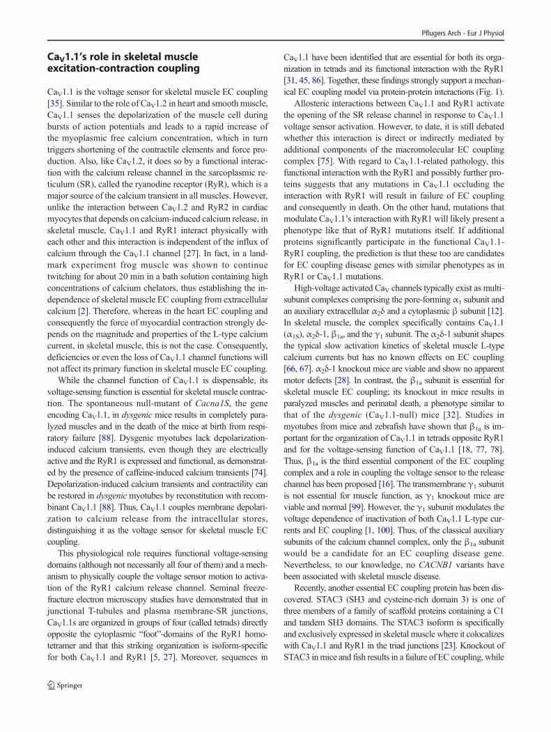
Fig. 1 Functions of CaV1.1 as L-type calcium channel and voltage sensorin EC coupling in skeletal muscle. a In skeletal muscle, EC couplingCaV1.1 functions as voltage sensor and activates SR calcium release byinteracting with RyR1 (directly or mediated by associated proteins likeSTAC3). b Domain structure of CaV1.1 highlighting the alternativelyspliced exon 29. c The two splice variants differ in their function ascalcium channel but not in EC coupling. d Comparison of calcium
currents of CaV1.1a (with exon 29; blue) and CaV1.1e (without exon29; red) and CaV1.2 (gray). The voltage-dependence curves show thatinclusion of exon 29 right-shifts V½ of current activation but not that ofEC coupling. Note that the augmented calcium influx in CaV1.1e adds anextra (Cd/La-dependent) component to the depolarization-dependent cal-cium signals [96]
Pflugers Arch - Eur J Physiol
CaV1.1’s role as calcium channel and therefore that perturbedCaV1.1 function inevitably will result in EC coupling pathol-ogy but not in a channelopathy in the classical sense. This isimpressively demonstrated in mouse models in which thechannel function of CaV1.1 has been perturbed or occludedwithout compromising its function as voltage sensor for ECcoupling [19, 48]. These mice lack skeletal muscle L-typecalcium currents but show no muscle pathology whatsoever.Therefore, CaV1.1 L-type calcium currents are dispensable fornormal growth and function of skeletal muscle. Consequently,calcium channel loss-of-function mutations in CaV1.1 are notexpected to result in disease. Does this imply that CaV1.1currents are irrelevant for disease? By no means. Apparently,it is important for normal muscle function to abolish or curtailthe current function [24]. Consequently, any gain of channelfunction might have pathological effects.
Before we will turn to such diseases, let us briefly considerhow and when calcium currents are essentially abolished un-der physiological conditions in skeletal muscle. Of all thevoltage-gated calcium channels, the classical skeletal muscleL-type calcium currents are the slowest in activation, the oneswith the most right-shifted voltage dependence of activation,and they exhibit small current amplitude. Therefore, the 1–2 ms depolarization of a skeletal muscle action potential willhardly activate opening of this channel, not even when deliv-ered as high-frequency bursts. The action potential barelyreaches the voltages necessary for CaV1.1 activation, and ifso, within this brief pulse, the slow activation kinetics will notallow a substantial response. Finally, should a small number ofchannels still activate under these unfavorable conditions,these channels are characterized by an extremely low openprobability. Interestingly, in mammalian CaV1.1 channels, amultitude of mechanisms contribute to this downregulation ofcalcium currents. The slow activation kinetics is encoded inthe specific amino acid sequence of the voltage-sensing do-main (VSD) of the first repeat [62, 97]. The right-shifted volt-age dependence of activation and the low open probability aredetermined by structures in the VSD of the fourth repeat [96,97]. Specifically, the insertion of an alternatively spliced exoninto the extracellular linker of transmembrane helices VIS3and VIS4 causes a 30-mV right shift of V½ and a > 5-foldreduction of current density (Fig. 1b–d). Importantly, the ac-tivation of channel opening is uncoupled from activation ofEC coupling in that depolarization-induced calcium release isactivated with fast kinetics and at 30 mV less depolarizedpotentials relative to current activation. Accordingly, inclusionof the 19 amino acids encoded by exon 29 into the IVS3-S4linker right-shifts the voltage dependence of current activa-tion, but not of EC coupling [96]. Finally, the auxiliary α2δ-1 subunit further slows down activation kinetics [68].Interestingly, this effect of α2δ-1 is specific to CaV1.1, as withCaV1.2 the α2δ-1 subunit has exactly the opposite effect [95].Altogether, having four distinct mechanisms at work to curtail
CaV1.1 currents suggests that in mature skeletal muscle L-type calcium currents are not only dispensable but also prob-ably disadvantageous.
If this is the case, why not abolish calcium conductancecompletely? Actually, teleost fish utilize this strategy by ex-pressing non-conducting CaV1.1 channels [79]. Perhaps, inmammals, CaV1.1 currents are needed in a different context.Indeed, the predominant splice variant in embryonic skeletalmuscle lacks exon 29 and thus has substantially different cur-rent properties [85, 94]. The embryonic CaV1.1e channel var-iant has a voltage dependence of activation and current densitycomparable to the cardiac/neuronal CaV1.2 (Fig. 1d). In otherwords, during development, skeletal muscle expresses a nor-mal calcium channel allowing calcium entry in response tospontaneous and motor nerve-induced electrical activity.Nevertheless, if depolarization-induced calcium influx in de-veloping muscle cells were essential, loss-of-function muta-tions affecting current properties would be expected to resultin developmental pathology of the motor system. At least inmice, this is highly unlikely, because mice expressing non-conducting CaV1.1 do not display muscle pathology [19].This does, however, not exclude the possibility that duringearly development the calcium-conducting CaV1.1e is in-volved in physiologically relevant functions. For example,recently, we and others discovered a crucial role of CaV1.1-dependent calcium signals in the earliest stages of neuromus-cular junction development [15, 43]. Patterning of postsynap-tic acetylcholine receptors in the synaptic target zone in thecenter of the muscle fibers was highly sensitive to the size ofthe muscle calcium signals. Importantly, in the absence ofRyR1, calcium influx through the CaV1.1e channel was suffi-cient for normal neuromuscular junction development, indic-ative of its physiological role during development. Althoughcalcium influx was not essential, as in mice with non-conducting channels the subsequent onset of EC couplingcompensated for the loss of channel function [43].
Whereas loss of CaV1.1 channel function is inconsequen-tial, would a gain of channel function be harmful? The obser-vation that curtailing calcium currents is a process activelyregulated during development in itself indicates that calciuminflux during normal EC coupling might be disadvantageous.Indeed, a mouse model in which the developmentally regulat-ed inclusion of exon 29 (resulting in the poorly conductingCaV1.1a) was occluded showed severe effects on muscle dif-ferentiation and health [85]. First, the extra calcium influxcaused an aberrant fiber type specification. Both in predomi-nantly fast extensor digitorum longus (EDL) and in slow so-leus muscle, the fiber type composition was substantiallyshifted towards slower myosin isoforms, with the expectedeffects on muscle strength and fatigability. Furthermore, withincreasing age, the continuing calcium influx caused severedamage and loss of mitochondria, resembling a disease phe-notype characteristic for mouse models with increased
Pflugers Arch - Eur J Physiol

calcium load in skeletal muscles [6]. Thus, even though thechannel function of CaV1.1 is dispensable for normal skeletalmuscle physiology, it is still possible that gain-of-functionmutations causing calcium influx in adult muscle will giverise to a CaV1.1 channelopathy.
Potential CaV1.1-related pathologiesin tissues other than skeletal muscle
Now let us briefly consider the possibility that CaV1.1 chan-nels might contribute to body functions in other tissues andthat genetic variants resulting in loss or gain of function mightcause non-muscle CaV1.1 channelopathies. Using whole ex-ome sequencing, a missense mutation CACNA1S (I289V) hasbeen linked to aberrant tooth morphogenesis in several indi-viduals of 5 Thai families [47]; however, the mechanistic linkto a CaV1.1 function has not been explored. Several reportsindicate the expression of CaV1.1 in activated T-lymphocytes[3] where it contributes to the calcium signal in response to Tcell receptor stimulation. Intriguingly, this channel lacks exon29 [55], suggesting that it may possess the improved gatingproperties of the splice variant expressed in embryonic skele-tal muscle. Nevertheless, how this voltage-dependent channelis activated upon T cell receptor activation in non-excitablecells remains a mystery. Several studies show immunostainingof CaV1.1 in synapses of retinal bipolar cells [82, 98].However, confirmation of CaV1.1 expression with indepen-dent methods and a characterization of its possible role inretinal function are still lacking and indeed this finding mayreflect antibody cross-reactivity [34]. At the time of this pub-lication, CaV1.1 channelopathies with deficiencies in the im-mune system or vision have not been reported, indicative ofmerely non-essential functions of CaV1.1 channels in thesetissues. This notion is further supported by the observationsthat the mouse models expressing non-conducting CaV1.1(loss-of-function) [19] or CaV1.1 excluding exon 29 (gain-of-function) [85] did not reveal deficiencies in vision nor inthe immune response. Thus, for the time being, CaV1.1 chan-nelopathies principally represent skeletal muscle diseases.
Skeletal muscle channelopathies
Clinically, skeletal muscle channelopathies manifest as recur-ring episodes of muscle weakness or muscle stiffness trig-gered by exercise, cold stress, excessive potassium uptake,or volatile anesthetics. Episodic muscle weakness (periodicparalysis) is caused by the transiently reduced excitability ofthe muscle. Usually, it comes in two forms distinguished bythe potassium levels during an attack: hypokalemic periodicparalysis (HypoPP) and normokalemic periodic paralysis(NormoPP). Muscle stiffness, called myotonia, is caused by
uncontrolled repetitive firing of action potentials. Malignanthyperthermia (MH) susceptibility represents another chan-nelopathy affecting skeletal muscle. Typically, it occurs as acrisis during application of volatile anesthetics or depolarizingmuscle relaxants and manifests as an attack of extreme musclecontractures accompanied by increased metabolism, increasedbody temperature (therefore its name), and damage to themusculature. Congenital myopathies represent a clinicallyand genetically heterogenous group of early-onset muscle dis-eases characterized by pronounced muscle weakness and dis-tinctive histological abnormalities. Myotonic dystrophy com-bines the symptoms of transient hyperexcitability and chronicmuscle wasting. For all these muscle diseases, an involvementof ion channel genes including CACNA1S has been demon-strated (Fig. 2). In addition, recently, a rare but debilitatingmuscle disease (Native American myopathy, NAM) has beenlinked to mutations in STAC3, a skeletal muscle-specific scaf-folding protein intimately linked to the function of CaV1.1 inEC coupling.
Hypokalemic periodic paralysis (HypoPP)
HypoPP is a dominantly inherited autosomal disease charac-terized by episodes of flaccid generalized muscle weaknessaccompanied by low serum potassium levels (< 3.5 mM)[10]. In the majority of patients, HypoPP is accompanied bypermanent progressive muscle weakness and muscle degener-ation. Episodes of muscle weakness are triggered by rest afterexercise, hypokalemia following intake of carbohydrates orinsulin administration, and cold stress. During an attack,voltage-gated sodium channels become inactivated by long-lasting membrane depolarization (from − 90 to − 60 mV),which, paradoxically, is associated with a reduction in extra-cellular potassium concentrations [42]. The genetic cause ofHypoPP is mutations in two voltage-gated cation channels,CaV1.1 (in approximately 60% of the cases) and NaV1.4 (inapproximately 20%). Importantly, almost all known causativemutations in both channels neutralize gating charges of the S4helices of the voltage-sensing domains (VSDs) (Fig. 2). Thesepositive gating charges (usually between 4 and 6 arginines andlysines in every third position of S4) serve multiple importantfunctions in the voltage-sensing process. They are the positivecharges attracted by the force of the transmembrane electricpotential, thus pulling the S4 helix inward and outward at restand depolarization, respectively. They form transient ion-pairinteractions with negative countercharges in other parts of theVSD to facilitate the state transitions and to stabilize the rest-ing and activated states. And the sequential positioning ofgating charges in the hydrophobic constriction site (HCS)seals the otherwise hydrophilic gating pore through whichS4 helix slides upon activation and deactivation. Therefore,it is highly plausible that neutralizing mutations of the gating
Pflugers Arch - Eur J Physiol
charges will cause defects in channel gating, and initially, suchdefects were suspected to be the cause of HypoPP.
The well-known HypoPP mutations in CaV1.1 are R528G/H,the outermost arginine (R1) in the VSD of the second repeat(VSD II); R897S and R900S/G, corresponding to R1 and R2in VSD III; and R1239H/G, corresponding to R2 in VSD IV(Figs. 2 and 3). Early biophysical characterization of calciumcurrent properties of CaV1.1(R528H/G) and CaV1.1(R1239H/G) in heterologous cells andmyotubes indicated a loss of channelfunction [40, 46, 60, 61]. The mutant channels showed slowedactivation, a reduction of open probability and current amplitude,and a left-shift in the voltage dependence of the steady-stateinactivation. These current defects were accompanied by actionpotential broadening and a reduction of its amplitude. However,none of these changes in the current properties of the channelvariants could explain the long-lasting depolarizations observedin HypoPP muscles. Let alone the fact that L-type calcium cur-rents are dispensable for normal muscle function, and thereforeloss of channel function hardly could cause muscle disease (seeabove). Similarly, the loss-of-function effects of HypoPP muta-tions observed in NaV1.4 were equally inconsistent with the ob-served depolarization phenotype. So what do these mutations inCaV1.1 and NaV1.4 have in common that could cause long-lasting membrane depolarizations and HypoPP?
The answer is “omega currents.” Omega currents (alsocalled gating pore currents) are leak currents through VSDsof voltage-gated cation channels with mutations of one of the
gating charges [14]. Gating pore currents are unrelated to theionic currents through the channel’s pore domain (PD) andmuch smaller than these (< 1%). The anomalous ion conduc-tion pathway is established by the misalignment of the mutat-ed gating charge (R1 or R2 in CaV1.1) and the hydrophobicconstriction site (HCS), thus abolishing the seal between theouter hydrophilic vestibule and the cytoplasmic compartment[59]. Omega currents are state-dependent in that, dependingon the position of the mutated gating charge along the trans-membrane S4 helix, they typically occur at hyperpolarized(resting) or depolarized (activated) conditions. In the case ofR1 or R2 mutations, the omega pore opens at hyperpolarizedpotentials when these gating charges reside in the HCS. Incontrast, depolarization pulls the S4 helix outward so thatthe intact inner gating charges occupy the HCS and seal theleak. Contingent on the channel type and the nature of theamino acid substitution (bulky or small), omega pores conductprotons and/or monovalent cations. Today, such leak currentsare generally accepted to be the initial cause of the long-lastingmembrane depolarization observed in HypoPP muscle fibers.
Omega currents were first described in voltage-gated po-tassium channels [83] and later identified as the cause ofhyperkalemic and normokalemic periodic paralysis inNaV1.4 mutants [80, 84]. For the longest time, direct biophys-ical identification and characterization of omega currents inthe CaV1.1 HypoPP mutants were hampered by the inabilityto functionally express CaV1.1 channels in non-muscle
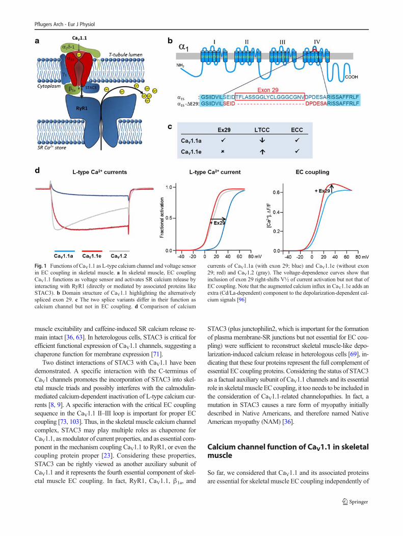
Fig. 2 Positions of disease mutations in the domain topology model ofCaV1.1. CACNA1S variants and splicing defects linked to muscle diseaseprimarily concern functionally important residues of the VSDs and of theP-loop involved in ion conduction and selectivity. In addition, substitu-tions and truncations of intracellular loop sequences directly or indirectlyaffect interactions with associated proteins. Accordingly, some of themutations exert their effects by directly altering the properties ofCaV1.1 (calcium currents, omega currents), whereas other mutationscause disease by altering the interactions with, or the function of, the
RyR1 (EC coupling, calcium leak). Cylinders S1–S4 shaded in blue rep-resent the voltage-sensing domains (VSDs), and cylinders S5–S6 shadedin light green represent the pore domains (PDs) of repeats I, II, III, and IV.HypoPP, hypokalemic periodic paralysis; NormoPP, normokalemic peri-odic paralysis; MHS, malignant hyperthermia susceptibility; myotonicdystrophy type 1, DM1. Red X, truncations probably resulting in non-functional channel fragments; green X, truncations compatible with func-tional expression of the channel
Pflugers Arch - Eur J Physiol

expression systems. However, the observation that musclefibers from patients and mice carrying the R528H mutationexhibit inward cation leak currents at hyperpolarized poten-tials with omega current characteristics strongly supported thenotion that omega currents underlie HypoPP in patients withCACNA1Smutations [41, 104].More recently, expression of atypical (R1239H) and an atypical (V876E) HyppoPP mutantinmouse muscle demonstrated omega pore currents carried byprotons and sodium leak, respectively [29, 30]. Finally, a re-cent advance in heterologous expression of CaV1.1 by co-expressing STAC3 made it possible to directly record andcharacterize the omega leak currents in the CaV1.1-R528Hand -R528GHypoPPmutants in the oocyte expression system[107]. The improved recording conditions compared to re-cordings in muscle fibers or myotubes allowed the analysisof the permeation properties of the two variants. Surprisingly,the leak current in CaV1.1-R528H (the most frequent HypoPPmutation) was carried primarily by sodium and not by protons
as is the case in the corresponding mutations in NaV and KV
channels. Together, these experiments unambiguously estab-lish omega pore leak current as the patho-mechanism ofCaV1.1-linked HypoPP. Furthermore, the new expression sys-tem now affords the opportunity to systematically characterizeall known CaV1.1 HypoPP variants.
How does this omega current lead to the long-lasting depo-larizations during attacks of HypoPP and why is it triggered bylow external potassium concentrations? Remarkably, skeletalmuscle cells possess a bistable resting potential, with one stablestate near the K+-equilibrium potential (at − 80 mV) and anotherat about − 50 to − 60mV [42]. The latter arises from the balanceof an outward current through an inward rectifying K+ channeland a linear inward leak current. Therefore, both lowering theextracellular potassium concentration and/or increasing the in-ward leak current can shift the membrane potential into the lesspolarized stable state. Both these conditions occur during attacksin muscles expressing HypoPP mutant CaV1.1 channels.
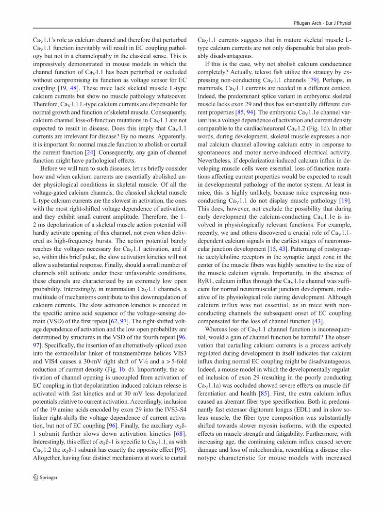
Fig. 3 CaV1.1 voltage-sensing domains are hot spots for disease muta-tions. a Schematic drawing of a generic VSD with the positions of cur-rently known CACN1S disease mutations. Charge neutralizations of thegating charges R1 to R3 in the S4 helix cause state-dependent omega leakcurrents leading to HypoPP or NormoPP, respectively. Adding a negativecharge (V876E) in the neighboring S3 helix may have the same effect.Changing the charge of the innermost gating charge R4 or its ion-pairpartner E2 perturbs the function of the VSD. b The structure model of aVSD shows how specifically in the resting state the extra negative charge
in the non-canonical HypoPP mutant V876E allows the gating charge R2to form an additional ion pair above the hydrophobic constriction site(HCS). c The NormoPP mutation R1242G removes the gating chargeR3, which is positioned above the HCS in the activated state and justbelow in the resting state, consistent with the reported state-dependent bi-directional omega currents. d The loss of a counter charge in the myop-athy mutant E100K weakens the stabilizing ion-pair interactions of thegating charge located just below the HCS. Structure models, courtesy ofM. Fernandez-Quintero
Pflugers Arch - Eur J Physiol
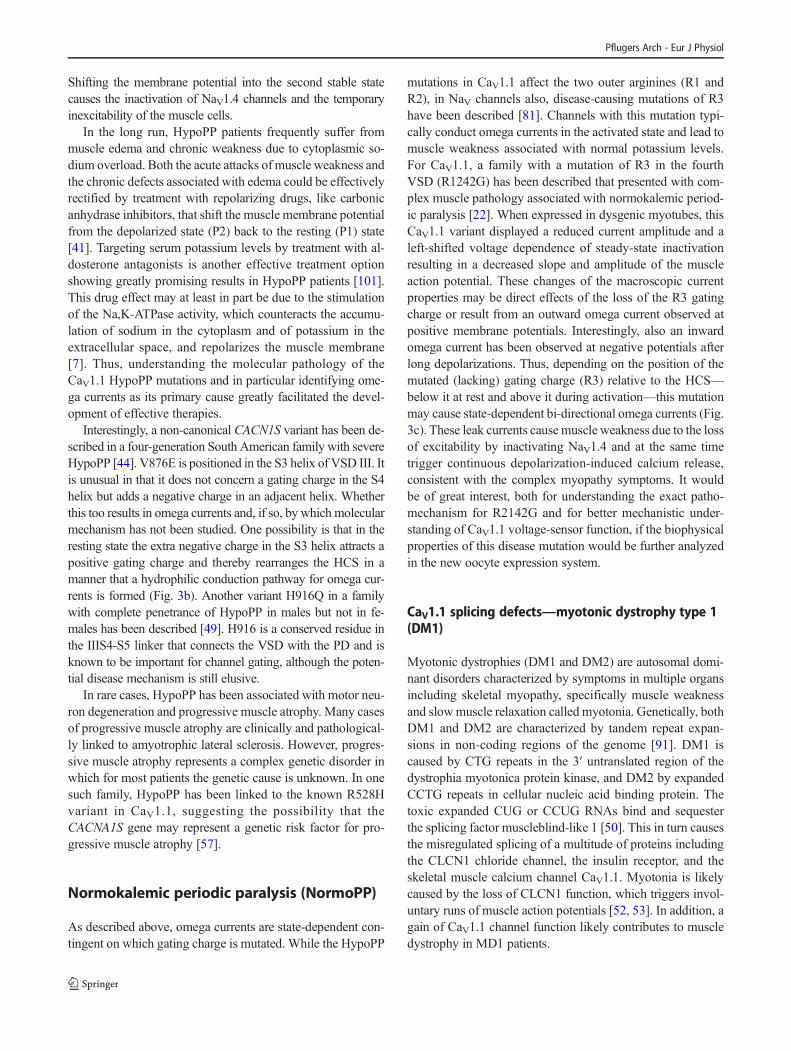
Shifting the membrane potential into the second stable statecauses the inactivation of NaV1.4 channels and the temporaryinexcitability of the muscle cells.
In the long run, HypoPP patients frequently suffer frommuscle edema and chronic weakness due to cytoplasmic so-dium overload. Both the acute attacks ofmuscle weakness andthe chronic defects associated with edema could be effectivelyrectified by treatment with repolarizing drugs, like carbonicanhydrase inhibitors, that shift the muscle membrane potentialfrom the depolarized state (P2) back to the resting (P1) state[41]. Targeting serum potassium levels by treatment with al-dosterone antagonists is another effective treatment optionshowing greatly promising results in HypoPP patients [101].This drug effect may at least in part be due to the stimulationof the Na,K-ATPase activity, which counteracts the accumu-lation of sodium in the cytoplasm and of potassium in theextracellular space, and repolarizes the muscle membrane[7]. Thus, understanding the molecular pathology of theCaV1.1 HypoPP mutations and in particular identifying ome-ga currents as its primary cause greatly facilitated the devel-opment of effective therapies.
Interestingly, a non-canonical CACN1S variant has been de-scribed in a four-generation South American family with severeHypoPP [44]. V876E is positioned in the S3 helix of VSD III. Itis unusual in that it does not concern a gating charge in the S4helix but adds a negative charge in an adjacent helix. Whetherthis too results in omega currents and, if so, by which molecularmechanism has not been studied. One possibility is that in theresting state the extra negative charge in the S3 helix attracts apositive gating charge and thereby rearranges the HCS in amanner that a hydrophilic conduction pathway for omega cur-rents is formed (Fig. 3b). Another variant H916Q in a familywith complete penetrance of HypoPP in males but not in fe-males has been described [49]. H916 is a conserved residue inthe IIIS4-S5 linker that connects the VSD with the PD and isknown to be important for channel gating, although the poten-tial disease mechanism is still elusive.
In rare cases, HypoPP has been associated with motor neu-ron degeneration and progressive muscle atrophy. Many casesof progressive muscle atrophy are clinically and pathological-ly linked to amyotrophic lateral sclerosis. However, progres-sive muscle atrophy represents a complex genetic disorder inwhich for most patients the genetic cause is unknown. In onesuch family, HypoPP has been linked to the known R528Hvariant in CaV1.1, suggesting the possibility that theCACNA1S gene may represent a genetic risk factor for pro-gressive muscle atrophy [57].
Normokalemic periodic paralysis (NormoPP)
As described above, omega currents are state-dependent con-tingent on which gating charge is mutated. While the HypoPP
mutations in CaV1.1 affect the two outer arginines (R1 andR2), in NaV channels also, disease-causing mutations of R3have been described [81]. Channels with this mutation typi-cally conduct omega currents in the activated state and lead tomuscle weakness associated with normal potassium levels.For CaV1.1, a family with a mutation of R3 in the fourthVSD (R1242G) has been described that presented with com-plex muscle pathology associated with normokalemic period-ic paralysis [22]. When expressed in dysgenic myotubes, thisCaV1.1 variant displayed a reduced current amplitude and aleft-shifted voltage dependence of steady-state inactivationresulting in a decreased slope and amplitude of the muscleaction potential. These changes of the macroscopic currentproperties may be direct effects of the loss of the R3 gatingcharge or result from an outward omega current observed atpositive membrane potentials. Interestingly, also an inwardomega current has been observed at negative potentials afterlong depolarizations. Thus, depending on the position of themutated (lacking) gating charge (R3) relative to the HCS—below it at rest and above it during activation—this mutationmay cause state-dependent bi-directional omega currents (Fig.3c). These leak currents causemuscle weakness due to the lossof excitability by inactivating NaV1.4 and at the same timetrigger continuous depolarization-induced calcium release,consistent with the complex myopathy symptoms. It wouldbe of great interest, both for understanding the exact patho-mechanism for R2142G and for better mechanistic under-standing of CaV1.1 voltage-sensor function, if the biophysicalproperties of this disease mutation would be further analyzedin the new oocyte expression system.
CaV1.1 splicing defects—myotonic dystrophy type 1(DM1)
Myotonic dystrophies (DM1 and DM2) are autosomal domi-nant disorders characterized by symptoms in multiple organsincluding skeletal myopathy, specifically muscle weaknessand slowmuscle relaxation called myotonia. Genetically, bothDM1 and DM2 are characterized by tandem repeat expan-sions in non-coding regions of the genome [91]. DM1 iscaused by CTG repeats in the 3′ untranslated region of thedystrophia myotonica protein kinase, and DM2 by expandedCCTG repeats in cellular nucleic acid binding protein. Thetoxic expanded CUG or CCUG RNAs bind and sequesterthe splicing factor muscleblind-like 1 [50]. This in turn causesthe misregulated splicing of a multitude of proteins includingthe CLCN1 chloride channel, the insulin receptor, and theskeletal muscle calcium channel CaV1.1. Myotonia is likelycaused by the loss of CLCN1 function, which triggers invol-untary runs of muscle action potentials [52, 53]. In addition, again of CaV1.1 channel function likely contributes to muscledystrophy in MD1 patients.
Pflugers Arch - Eur J Physiol

The splicing defect in CaV1.1 concerns the inclusion ofexon 29 [89]. During normal development, embryonicCaV1.1e channels, lacking exon 29, are completely replacedby the adult CaV1.1a channel variant, containing exon 29 [85,96]. Inclusion of exon 29 dramatically right-shifts the voltagedependence of activation by 30 mV and reduces the currentamplitude greater than fivefold. Thus, alternative splicing ofexon 29 renders the skeletal muscle calcium current small andless responsive to depolarization, while fully maintaining itsactivity as the voltage sensor of EC coupling. In DM1 pa-tients, this splicing event is reversed, leading to the aberrantexpression of the embryonic CaV1.1e splice variant in adults.Noticeably, the fraction of embryonic CaV1.1e expressed inDM1 patients correlated with the degree of clinically assessedmuscle weakness [89]. Thus, it is expected that DM1 patientskeletal muscles with every movement experience a patholog-ically enhanced calcium influx.
Likewise, in muscles of a DM1mouse model, the addition-al knockdown of muscle blind resulted in the upregulation ofCaV1.1e and aggravated muscle pathology evidenced by anincreased frequency of centrally located myonuclei [89].Thus, missplicing of CaV1.1 exon 29 contributes to theDM1 pathology in mice. On the other hand, a mouse modelin which insertion of exon 29 had been abolished and there-fore the embryonic CaV1.1e splice variant is exclusivelyexpressed throughout life did not display DM-like muscleweakness [85]. This is consistent with the notion that myoto-nia primarily arises from the deficiency in chloride channelfunction. However, progressively, the muscles of this mousemodel showed mitochondrial damage and ultimately a severeloss of mitochondria that was accompanied by reduced endur-ance [85]. Such a phenotype is commonly observed in mus-cular dystrophy mouse models, the muscles of which experi-ence chronic calcium overload, thus supporting the conclusionthat the increased calcium influx in muscles aberrantly ex-pressing the embryonic CaV1.1e splice variant causes myop-athy. In human DM1 patients, these slowly progressing de-fects may be exacerbated by the combined defects of multiplemisspliced genes as well as with increasing age. In conclusion,the gain of the CaV1.1 channel function likely contributes tothe pathology of DM1.
Currently, no disease-modifying treatments for DM1 andDM2 exist and therapy is limited to symptomatic treatmentsand preserving motor function. As DM involves multiplemisspliced and dysregulated proteins, the affected ion channels,CLCCN1 and CaV1.1, are no viable targets. Rather the mostpromising experimental treatment strategies target the commonupstream cause the toxic expanded CUG or CCUG RNAs [91].
Malignant hyperthermia susceptibility
MH susceptibility is an autosomal dominantly transmittedpredisposition to respond with uncontrollable calcium release
and consequently massive muscle contractions to volatile an-esthetics and depolarizing muscle relaxants [20]. The symp-toms of an MH reaction include muscle rigidity, acidosis, rap-idly raising body temperature and usually lead to musclebreakdown and the death of the patient if left untreated.Otherwise individuals affected by this pharmaco-genetic con-dition are clinically inconspicuous. In the majority of MHsusceptible individuals (> 70%) the massive calcium releaseis linked to functional missense mutations within the RyR1release channel itself [93]. Close to 200 such mutations havebeen identified in all parts of the RyR1. The commonality ofMHS mutations in RyR1 is that these destabilize the closedstate of the calcium release channel and make it hypersensitiveto activation by triggering agents like volatile anesthetics orcaffeine. MS susceptibility mutations also cause leaky RyR1channels, depletion of the sarcoplasmic reticulum calciumstores and chronically elevated myoplasmic calcium concen-trations at rest. These symptoms are frequently associatedwitha myopathy called central core disease (CCD), because of anabundance of central cores in type 1 muscle fibers, and clini-cally manifests as congenital muscle hypotonia with delayedmotor development [39].
In addition to the MHS mutations in RyR1, several MHsusceptibility mutations (representing about 1% of the genet-ically solved cases) have been identified in the CACNA1Sgene [11, 21, 58, 70, 92]. The R1086H/C/S mutations affecta highly conserved arginine residue located in the cytoplasmicloop connecting repeats III and IV of CaV1.1 (Fig. 2).Biophysical analysis in dysgenic myotubes reconstituted withCaV1.1 carrying the R1086H substitution demonstrated thatthis mutation somewhat reduced the current density and in-creased the sensitivity of calcium release to activation by de-polarization and by caffeine [102]. The T1354S MHS muta-tion located in the outer pore region of CaV1.1 acceleratedactivation kinetics of the L-type calcium current and againleft-shifted the sensitivity of calcium release to caffeine [70].Evidently, this mutation in the voltage sensor of EC coupling(CaV1.1) caused the hypersensitivity of the calcium releasechannel (RyR1) to physiological and pharmacological activa-tion, and thus functionally mimicked the disease-causing ef-fects of mutations in the RyR1 itself. Interestingly, the muta-tion in the third MHS site in CaV1.1, R174W, affects theinnermost gating charge of the first VSD and displayed some-what different effects. Unlike the other CaV1.1 MHS muta-tions, R174W did not alter EC coupling but essentially ablatedthe L-type calcium currents [21]. Nevertheless, the sensitivityof calcium release to caffeine and volatile anesthetics wasincreased, sarcoplasmic reticulum calcium stores were partial-ly depleted, and conversely, the resting myoplasmic calciumconcentration was increased. These differential effects indicat-ed that the R174W mutation stabilized the affected VSD I ofCaV1.1 in the resting state, ablating current activation, andindependently interfered with the ability of CaV1.1 to stabilize
Pflugers Arch - Eur J Physiol

the RyR1 in a closed conformation, resulting in a leaky calci-um release channel. While the loss of channel function may beinconsequential for normal muscle function and the health ofaffected patients, the chain of events initiated by the leakyRyR1 probably gives rise to the hypersensitivity of the calci-um release channel characteristic of MHS. Notably, neither ofthe two defects in the function of CaV1.1 affected its ability toactivate EC coupling. The molecular mechanism by whichthese three HMS mutations in structurally and functionallydistinct domains of CaV1.1 sensitize RyR1 calcium releaseto activation by caffeine and volatile anesthetics is unknown.The affected CaV1.1 domains have not been implicated ininteractions with the RyR1 or in a functional role in EC cou-pling. Nevertheless, their common effect on RyR1 functionand pharmacology impressively demonstrates the complexinteractions between the voltage sensor and the effector re-lease channel within the macromolecular EC couplingapparatus.
As MH crises are mostly limited to the clinical setting,therapeutic efforts concentrate on their avoidance by testingpatients potentially at risk either functionally, with a caffeine-sensitivity test, or genetically for known disease-causing mu-tations. MH crisis management is accomplished by whole-body cooling and the rapid administration of the MH antidotedantrolene [51].
CaV1.1-related myopathy
Congenital myopathies represent a genetically heterogenousgroup of early-onset, non-dystrophic muscle diseases charac-terized by varying degrees of muscle weakness and distinctivehistopathological abnormalities [39]. The severity of muscledysfunctions ranges from severe fetal akinesia to milder formsof hypotonia and muscle weakness. The disease-characterizing histopathological features include central cores,multi-minicores, central nuclei, and nemaline rods.Congenital myopathies are mostly disorders of EC couplingand altered calcium handling, and numerous mutations in theRyR1 gene have been identified as the cause of myopathy[93]. Therefore, it was not unexpected that recent whole exonsequencing studies also identified several (altogether 12) pu-tative myopathy mutations in the CACNA1S gene in familiespresenting with perinatal hypotonia, severe axial and general-ized weakness, and, in several cases, ophthalmoplegia [38,76]. Genetically, the identified cases include compound reces-sive mutations, dominant mutations, and de novomutations invarious domains of CaV1.1. All recessive cases described inthese studies carried at least one nonsense mutation causing aframe shift resulting in the premature stop and consequentlythe dysfunction and/or loss of the CaV1.1 protein. Although atpresent functional characterizations of the missense myopathymutations are lacking, their positions in domains with known
functions allow prediction as to the effects of the mutationsand to possible patho-mechanisms.
For example, the recessive missense mutation E100K re-places a highly conserved negative counter charge within thecharge transfer center of the first VSD with a positivelycharged residue. According to the sliding helix model of volt-age sensing, this residue is critical for the translocation of theS4 helix through the membrane electrical field upon depolar-ization [14]. The reversal of the charge in the E100K mutantwill prohibit the sequential formation of ion pairs between thegating charges and E100, and thus is expected to severelyimpede voltage sensing and activation of L-type calcium cur-rents. Yet, this defect is not necessarily expected to interferewith EC coupling, which is likely independent of a functionalfirst VSD [24]. Remember that also the MHS mutationR174W, representing the corresponding gating charge ofVSD I, ablated calcium currents but not EC coupling (seeabove) [21]. In contrast, the dominant de novo missense mu-tation P742Q resides in the sequence of the cytoplasmic loopbetween repeats II and III that is critical for skeletal muscle-specific EC coupling. Previously we demonstrated that sub-stitution of this residue with threonine (P742T), the residuefound in the corresponding position of CaV1.2, diminishesdepolarization-induced calcium release by the RyR1 withoutaffecting CaV1.1 calcium currents [45]. Therefore, the diseasemutation in this position is expected to specifically perturb thefunctionally important interaction of CaV1.1 with the RyR1,and possibly with STAC3 [103], and thus obstruct EC cou-pling. Together, these two missense mutations suggest theintriguing possibility that mutations expected to specificallyperturb either the channel function or the EC coupling func-tion of CaV1.1 both result in a similar disease phenotype. Thisis unexpected in light of the evidence demonstrating that L-type calcium currents are expandable for normal developmentand function of skeletal muscles in mice [19].
Another missense mutation, F275L, affects a conserved phe-nylalanine in the extracellular loop between S5 and S6 of thefirst repeat. This P-loop contributes to the channel ion selectiv-ity filter [106] although the mutated residue is not part of theselectivity filter proper. Nevertheless, the mutation of a con-served residue in this domain may compromise the calciuminflux through the channel pore. Again, this raises the questionas to the possible role of CaV1.1 calcium currents in causingmuscle disease. However, also the MHS mutation T1354S inthe P-loop of the fourth repeat altered channel gating andcaused MHS (see above) [70]. Finally, several of the CaV1.1myopathy frame shift mutations cause truncation of the C-terminus of CaV1.1 that still might allow expression of func-tional channels. Overall, the C-terminus of CaV1.1 can be di-vided into two sections: a structurally conserved proximal part,containing multiple calcium/calmodulin regulatory elements,an interaction site for STAC3, and the triad targeting signal[26, 65, 103]. And the non-conserved distal C-terminus of
Pflugers Arch - Eur J Physiol
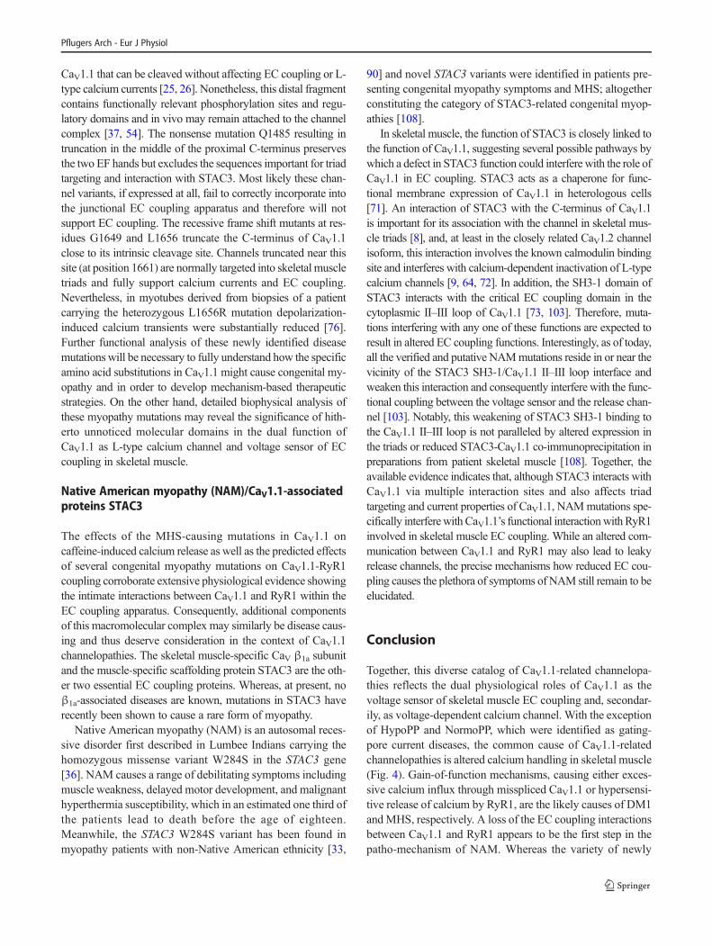
CaV1.1 that can be cleaved without affecting EC coupling or L-type calcium currents [25, 26]. Nonetheless, this distal fragmentcontains functionally relevant phosphorylation sites and regu-latory domains and in vivo may remain attached to the channelcomplex [37, 54]. The nonsense mutation Q1485 resulting intruncation in the middle of the proximal C-terminus preservesthe two EF hands but excludes the sequences important for triadtargeting and interaction with STAC3. Most likely these chan-nel variants, if expressed at all, fail to correctly incorporate intothe junctional EC coupling apparatus and therefore will notsupport EC coupling. The recessive frame shift mutants at res-idues G1649 and L1656 truncate the C-terminus of CaV1.1close to its intrinsic cleavage site. Channels truncated near thissite (at position 1661) are normally targeted into skeletal muscletriads and fully support calcium currents and EC coupling.Nevertheless, in myotubes derived from biopsies of a patientcarrying the heterozygous L1656R mutation depolarization-induced calcium transients were substantially reduced [76].Further functional analysis of these newly identified diseasemutations will be necessary to fully understand how the specificamino acid substitutions in CaV1.1 might cause congenital my-opathy and in order to develop mechanism-based therapeuticstrategies. On the other hand, detailed biophysical analysis ofthese myopathy mutations may reveal the significance of hith-erto unnoticed molecular domains in the dual function ofCaV1.1 as L-type calcium channel and voltage sensor of ECcoupling in skeletal muscle.
Native American myopathy (NAM)/CaV1.1-associatedproteins STAC3
The effects of the MHS-causing mutations in CaV1.1 oncaffeine-induced calcium release as well as the predicted effectsof several congenital myopathy mutations on CaV1.1-RyR1coupling corroborate extensive physiological evidence showingthe intimate interactions between CaV1.1 and RyR1 within theEC coupling apparatus. Consequently, additional componentsof this macromolecular complex may similarly be disease caus-ing and thus deserve consideration in the context of CaV1.1channelopathies. The skeletal muscle-specific CaV β1a subunitand the muscle-specific scaffolding protein STAC3 are the oth-er two essential EC coupling proteins. Whereas, at present, noβ1a-associated diseases are known, mutations in STAC3 haverecently been shown to cause a rare form of myopathy.
Native American myopathy (NAM) is an autosomal reces-sive disorder first described in Lumbee Indians carrying thehomozygous missense variant W284S in the STAC3 gene[36]. NAM causes a range of debilitating symptoms includingmuscle weakness, delayed motor development, and malignanthyperthermia susceptibility, which in an estimated one third ofthe patients lead to death before the age of eighteen.Meanwhile, the STAC3 W284S variant has been found inmyopathy patients with non-Native American ethnicity [33,
90] and novel STAC3 variants were identified in patients pre-senting congenital myopathy symptoms and MHS; altogetherconstituting the category of STAC3-related congenital myop-athies [108].
In skeletal muscle, the function of STAC3 is closely linked tothe function of CaV1.1, suggesting several possible pathways bywhich a defect in STAC3 function could interfere with the role ofCaV1.1 in EC coupling. STAC3 acts as a chaperone for func-tional membrane expression of CaV1.1 in heterologous cells[71]. An interaction of STAC3 with the C-terminus of CaV1.1is important for its association with the channel in skeletal mus-cle triads [8], and, at least in the closely related CaV1.2 channelisoform, this interaction involves the known calmodulin bindingsite and interferes with calcium-dependent inactivation of L-typecalcium channels [9, 64, 72]. In addition, the SH3-1 domain ofSTAC3 interacts with the critical EC coupling domain in thecytoplasmic II–III loop of CaV1.1 [73, 103]. Therefore, muta-tions interfering with any one of these functions are expected toresult in altered EC coupling functions. Interestingly, as of today,all the verified and putative NAMmutations reside in or near thevicinity of the STAC3 SH3-1/CaV1.1 II–III loop interface andweaken this interaction and consequently interfere with the func-tional coupling between the voltage sensor and the release chan-nel [103]. Notably, this weakening of STAC3 SH3-1 binding tothe CaV1.1 II–III loop is not paralleled by altered expression inthe triads or reduced STAC3-CaV1.1 co-immunoprecipitation inpreparations from patient skeletal muscle [108]. Together, theavailable evidence indicates that, although STAC3 interacts withCaV1.1 via multiple interaction sites and also affects triadtargeting and current properties of CaV1.1, NAMmutations spe-cifically interferewith CaV1.1’s functional interactionwithRyR1involved in skeletal muscle EC coupling. While an altered com-munication between CaV1.1 and RyR1 may also lead to leakyrelease channels, the precise mechanisms how reduced EC cou-pling causes the plethora of symptoms of NAM still remain to beelucidated.
Conclusion
Together, this diverse catalog of CaV1.1-related channelopa-thies reflects the dual physiological roles of CaV1.1 as thevoltage sensor of skeletal muscle EC coupling and, secondar-ily, as voltage-dependent calcium channel. With the exceptionof HypoPP and NormoPP, which were identified as gating-pore current diseases, the common cause of CaV1.1-relatedchannelopathies is altered calcium handling in skeletal muscle(Fig. 4). Gain-of-function mechanisms, causing either exces-sive calcium influx through misspliced CaV1.1 or hypersensi-tive release of calcium by RyR1, are the likely causes of DM1andMHS, respectively. A loss of the EC coupling interactionsbetween CaV1.1 and RyR1 appears to be the first step in thepatho-mechanism of NAM. Whereas the variety of newly
Pflugers Arch - Eur J Physiol
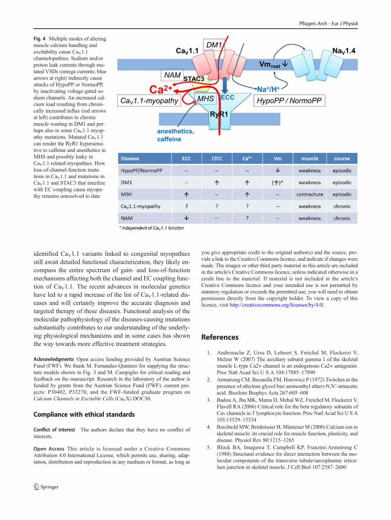
identified CaV1.1 variants linked to congenital myopathiesstill await detailed functional characterization, they likely en-compass the entire spectrum of gain- and loss-of-functionmechanisms affecting both the channel and EC coupling func-tion of CaV1.1. The recent advances in molecular geneticshave led to a rapid increase of the list of CaV1.1-related dis-eases and will certainly improve the accurate diagnosis andtargeted therapy of these diseases. Functional analysis of themolecular pathophysiology of the diseases-causing mutationssubstantially contributes to our understanding of the underly-ing physiological mechanisms and in some cases has shownthe way towards more effective treatment strategies.
Acknowledgments Open access funding provided by Austrian ScienceFund (FWF). We thank M. Fernandez-Quintero for supplying the struc-ture models shown in Fig. 3 and M. Campiglio for critical reading andfeedback on the manuscript. Research in the laboratory of the author isfunded by grants from the Austrian Science Fund (FWF); current pro-jects: P30402, P33270, and the FWF-funded graduate program onCalcium Channels in Excitable Cells (CaVX) DOC30.
Compliance with ethical standards
Conflict of interest The authors declare that they have no conflict ofinterests.
Open Access This article is licensed under a Creative CommonsAttribution 4.0 International License, which permits use, sharing, adap-tation, distribution and reproduction in any medium or format, as long as
you give appropriate credit to the original author(s) and the source, pro-vide a link to the Creative Commons licence, and indicate if changes weremade. The images or other third party material in this article are includedin the article's Creative Commons licence, unless indicated otherwise in acredit line to the material. If material is not included in the article'sCreative Commons licence and your intended use is not permitted bystatutory regulation or exceeds the permitted use, you will need to obtainpermission directly from the copyright holder. To view a copy of thislicence, visit http://creativecommons.org/licenses/by/4.0/.
References
1. Andronache Z, Ursu D, Lehnert S, Freichel M, Flockerzi V,Melzer W (2007) The auxiliary subunit gamma 1 of the skeletalmuscle L-type Ca2+ channel is an endogenous Ca2+ antagonist.Proc Natl Acad Sci U S A 104:17885–17890
2. ArmstrongCM, Bezanilla FM, Horowicz P (1972) Twitches in thepresence of ethylene glycol bis(-aminoethyl ether)-N,N’-tetraceticacid. Biochim Biophys Acta 267:605–608
3. Badou A, Jha MK, Matza D, Mehal WZ, Freichel M, Flockerzi V,Flavell RA (2006) Critical role for the beta regulatory subunits ofCav channels in T lymphocyte function. Proc Natl Acad Sci U S A103:15529–15534
4. BerchtoldMW, Brinkmeier H,MüntenerM (2000) Calcium ion inskeletal muscle: its crucial role for muscle function, plasticity, anddisease. Physiol Rev 80:1215–1265
5. Block BA, Imagawa T, Campbell KP, Franzini-Armstrong C(1988) Structural evidence for direct interaction between the mo-lecular components of the transverse tubule/sarcoplasmic reticu-lum junction in skeletal muscle. J Cell Biol 107:2587–2600
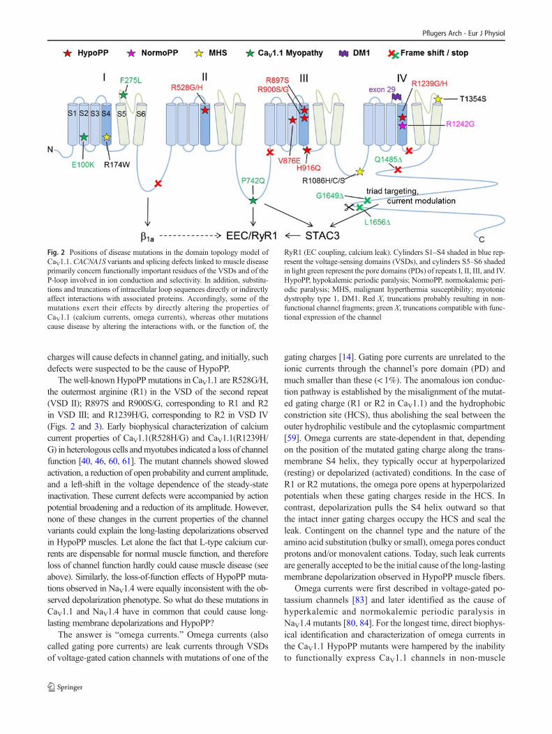
Fig. 4 Multiple modes of alteringmuscle calcium handling andexcitability cause CaV1.1channelopathies. Sodium and/orproton leak currents through mu-tated VSDs (omega currents; bluearrows at right) indirectly causeattacks of HypoPP or NormoPP,by inactivating voltage-gated so-dium channels. An increased cal-cium load resulting from chroni-cally increased influx (red arrowsat left) contributes to chronicmuscle wasting in DM1 and per-haps also in some CaV1.1 myop-athy mutations. Mutated CaV1.1can render the RyR1 hypersensi-tive to caffeine and anesthetics inMHS and possibly leaky inCaV1.1-related myopathies. Howloss-of-channel-function muta-tions in CaV1.1 and mutations inCaV1.1 and STAC3 that interferewith EC coupling cause myopa-thy remains unresolved to date
Pflugers Arch - Eur J Physiol

6. Boncompagni S, Rossi AE, Micaroni M, Hamilton SL, DirksenRT, Franzini-Armstrong C, Protasi F (2009) Characterization andtemporal development of cores in a mouse model of malignanthyperthermia. Proc Natl Acad Sci U S A 106:21996–22001
7. Breitenbach S, Lehmann-Horn F, Jurkat-Rott K (2016)Eplerenone repolarizes muscle membrane through Na,K-ATPaseactivation by Tyr10 dephosphorylation. Acta Myol MyopathiesCardiomyopathies Off J Mediterr Soc Myol 35:86–89
8. Campiglio M, Flucher BE (2017) STAC3 stably interacts throughits C1 domain with CaV1.1 in skeletal muscle triads. Sci Rep 7:1–12
9. Campiglio M, Costé de Bagneaux P, Ortner NJ, Tuluc P, VanPetegem F, Flucher BE (2018) STAC proteins associate to theIQ domain of Ca V 1.2 and inhibit calcium-dependent inactiva-tion. Proc Natl Acad Sci 115:1376–1381
10. Cannon SC (2015) Channelopathies of skeletal muscle excitabil-ity. Compr Physiol 5:761–790
11. Carpenter D, Ringrose C, Leo V, Morris A, Robinson RL, HalsallPJ, Hopkins PM, Shaw MA (2009) The role of CACNA1S inpredisposition to malignant hyperthermia. BMC Med Genet 10:104
12. Catterall WA (2011) Voltage-gated calcium channels. Cold SpringHarb Perspect Biol 3:a003947
13. Catterall WA, Lenaeus MJ, El-Din TMG (2020) Structure andpharmacology of voltage-gated sodium and calcium channels.Annu Rev Pharmacol Toxicol 60:1–22
14. Chanda B, Bezanilla F (2008) A common pathway for chargetransport through voltage-sensing domains. Neuron. 57:345–351
15. Chen F, Liu Y, Sugiura Y, Allen PD, Gregg RG, Lin W (2011)Neuromuscular synaptic patterning requires the function of skele-tal muscle dihydropyridine receptors. Nat Neurosci 14:570–577
16. Coronado R, Ahern CA, Sheridan DC, Cheng W, Carbonneau L,Bhattacharya D (2004) Functional equivalence of dihydropyridinereceptor a1S and b1a subunits in triggering excitation-contractioncoupling in skeletal muscle. Biol Res 37:1–15
17. Curtis BM, Catterall WA (1984) Purification of the calcium antag-onist receptor of the voltage-sensitive calcium channel from skel-etal muscle transverse tubules. Biochemistry. 23:2113–2118
18. Dayal A, Bhat V, Franzini-Armstrong C, Grabner M (2013)Domain cooperativity in the β1a subunit is essential fordihydropyridine receptor voltage sensing in skeletal muscle.Pnas. 110:7488–7493
19. Dayal A, Schrötter K, Pan Y, Föhr K, Melzer W, Grabner M(2017) The Ca2+influx through the mammalian skeletal muscledihydropyridine receptor is irrelevant for muscle performance. NatCommun 8:1–14
20. Dowling JJ, Lawlor MW, Dirksen RT (2014) Triadopathies: anemerging class of skeletal muscle diseases. Neurotherapeutics.11:773–785
21. Eltit JM, Bannister RA,MouaO, Altamirano F, Hopkins PM, PessahIN, Molinski TF, López JR, Beam KG, Allen PD (2012) Malignanthyperthermia susceptibility arising from altered resting coupling be-tween the skeletal muscle L-type Ca2+ channel and the type 1ryanodine receptor. Proc Natl Acad Sci U S A 109:7923–7928
22. Fan C, Lehmann-Horn F, Weber MA, Bednarz M, Groome JR,Jonsson MKB, Jurkat-Rott K (2013) Transient compartment-likesyndrome and normokalaemic periodic paralysis due to a Cav1.1mutation. Brain 136:3775–3786
23. Flucher BE, CampiglioM (2019) STAC proteins: the missing linkin skeletal muscle EC coupling and new regulators of calciumchannel function. Biochim Biophys Acta Mol Cell Res 1866:1101–1110
24. Flucher BE, Tuluc P (2017) How and why are calcium currentscurtailed in the skeletal muscle voltage-gated calcium channels? JPhysiol 595:1451–1463
25. Flucher BE, Kasielke N, Gerster U, Neuhuber B, Grabner M(2000) Insertion of the full-length calcium channel alpha(1S) sub-unit into triads of skeletal muscle in vitro. FEBS Lett 474:93–98
26. Flucher BE, Kasielke N, Grabner M (2000) The triad targetingsignal of the skeletal muscle calcium channel is localized in theCOOH terminus of the ??(1S) subunit. J Cell Biol 151:467–477
27. Franzini-Armstrong C (2018) The relationship between form andfunction throughout the history of excitation–contraction cou-pling. J Gen Physiol 150:189–210
28. Fuller-Bicer GA, Varadi G, Koch SE, Ishii M, Bodi I, Kadeer N,Muth JN, Mikala G, Petrashevskaya NN, Jordan MA, Zhang S-P,Qin N, Flores CM, Isaacsohn I, Varadi M, Mori Y, Jones WK,Schwartz A (2009) Targeted disruption of the voltage-dependentcalcium channel alpha2/delta-1-subunit. Am J Physiol Heart CircPhysiol 297:H117–H124
29. Fuster C, Perrot J, Berthier C, Jacquemond V, Allard B (2017)Elevated resting H+ current in the R1239H type 1 hypokalaemicperiodic paralysis mutated Ca2+ channel. J Physiol 595:6417–6428
30. Fuster C, Perrot J, Berthier C, Jacquemond V, Charnet P, Allard B(2017) Na leak with gating pore properties in hypokalemic peri-odic paralysis V876E mutant muscle Ca channel. J Gen Physiol149:1139–1148
31. Grabner M, Dirksen RT, Suda N, BeamKG (1999) The II-III loopof the skeletal muscle dihydropyridine receptor is responsible forthe bi-directional coupling with the ryanodine receptor. J BiolChem 274:21913–21919
32. Gregg RG, Messing A, Strube C, Beurg M, Moss R, Behan M,Sukhareva M, Haynes S, Powell JA, Coronado R, Powers PA(1996) Absence of the beta subunit (cchb1) of the skeletal muscledihydropyridine receptor alters expression of the alpha 1 subunitand eliminates excitation-contraction coupling. Proc Natl AcadSci U S A 93:13961–13966
33. Grzybowski M, Schänzer A, Pepler A, Heller C, Neubauer B,Hahn A (2017) Novel STAC3 mutations in the first non-Amerindian patient with Native American myopathy.Neuropediatrics. 48:451–455
34. Hasan N, Ray TA, Gregg RG (2016) CACNA1S expression inmouse retina: novel isoforms and antibody cross-reactivity withGPR179. Vis Neurosci 33:E009
35. Hernández-Ochoa EO, Schneider MF (2018) Voltage sensingmechanism in skeletal muscle excitation-contraction coupling:coming of age or midlife crisis? Skelet Muscle 8:1–20
36. Horstick EJ, Linsley JW, Dowling JJ, HauserMA,McDonald KK,Ashley-Koch A, Saint-Amant L, Satish A, Cui WW, Zhou W,Sprague SM, Stamm DS, Powell CM, Speer MC, Franzini-Armstrong C, Hirata H, Kuwada JY (2013) Stac3 is a componentof the excitation-contraction coupling machinery and mutated inNative American myopathy. Nat Commun 4:1952
37. Hulme JT, Konoki K, Lin TW-C, Gritsenko MA, Camp DG,Bigelow DJ, Catterall WA (2005) Sites of proteolytic processingand noncovalent association of the distal C-terminal domain ofCaV1.1 channels in skeletal muscle. Proc Natl Acad Sci U S A102:5274–5279
38. Hunter JM, Ahearn ME, Balak CD, Liang WS, Kurdoglu A,Corneveaux JJ, Russell M, Huentelman MJ, Craig DW, CarptenJ, Coons SW, Demello DE, Hall JG, Bernes SM, Baumbach-Reardon L (2015) Novel pathogenic variants and genes for my-opathies identified by whole exome sequencing. Mol GenetGenomic Med 3:283–301
39. Jungbluth H, Treves S, Zorzato F, Sarkozy A, Ochala J, Sewry C,Phadke R, Gautel M, Muntoni F (2018) Congenital myopathies:disorders of excitation-contraction coupling and muscle contrac-tion. Nat Rev Neurol 14:151–167
40. Jurkat-Rott K, Uetz U, Pika-Hartlaub U, Powell J, Fontaine B,Melzer W, Lehmann-Horn F (1998) Calcium currents and
Pflugers Arch - Eur J Physiol

transients of native and heterologously expressed mutant skeletalmuscle DHP receptor 1/2 subunits (R528H). FEBS Lett 423:198–204
41. Jurkat-Rott K, Weber M-A, Fauler M, Guo X-H, Holzherr BD,Paczulla A, Nordsborg N, Joechle W, Lehmann-Horn F (2009)K+-dependent paradoxical membrane depolarization and Na+overload, major and reversible contributors to weakness by ionchannel leaks. Proc Natl Acad Sci U S A 106:4036–4041
42. Jurkat-Rott K, Groome J, Lehmann-Horn F (2012)Pathophysiological role of omega pore current in channelopathies.Front Pharmacol 3:1–19
43. Kaplan MM, Sultana N, Benedetti A, Obermair GJ, Linde NF,Papadopoulos S, Dayal A, Grabner M, Flucher BE (2018)Calcium influx and release cooperatively regulate AChR pattern-ing and motor axon outgrowth during neuromuscular junctionformation. Cell Rep 23:3891–3904
44. Ke T, Gomez CR, Mateus HE, Castano JA, Wang QK (2009)Novel CACNA1S mutation causes autosomal dominant hypoka-lemic periodic paralysis in a South American family. J HumGenet54:660–664
45. Kugler G, Weiss RG, Flucher BE, Grabner M (2004) Structuralrequirements of the dihydropyridine receptor α 1S II-III loop forskeletal-type excitation-contraction coupling. J Biol Chem 279:4721–4728
46. Lapie P, Goudet C, Nargeot J, Fontaine B, Lory P (1996)Electrophysiological properties of the hypokalaemic periodic pa-ralysis mutation (R528H) of the skeletal muscle alpha 1s subunitas expressed in mouse L cells. FEBS Lett 382:244–248
47. Laugel-Haushalter V, Morkmued S, Stoetzel C, Geoffroy V,Muller J, Boland A, Deleuze J-F, Chennen K, Pitiphat W,Dollfus H, Niederreither K, Bloch-Zupan A, Pungchanchaikul P(2018) Genetic evidence supporting the role of the calcium chan-nel, CACNA1S, in tooth cusp and root patterning. Front Physiol9:1–14
48. Lee CS, Dagnino-Acosta A, Yarotskyy V, Hanna A, Lyfenko A,Knoblauch M, Georgiou DK, Poché RA, Swank MW, Long C,Ismailov II, Lanner J, Tran T, Dong K, Rodney GG, DickinsonME, Beeton C, Zhang P, Dirksen RT, Hamilton SL (2015) Ca(2+)permeation and/or binding to CaV1.1 fine-tunes skeletal muscleCa(2+) signaling to sustain muscle function. Skelet Muscle 5:4
49. Li F-F, Li Q-Q, Tan Z-X, Zhang S-Y, Liu J, Zhao E, Yu G-C, ZhouJ, Zhang L-M, Liu S-L (2012) A novel mutation in CACNA1Sgene associated with hypokalemic periodic paralysis which has agender difference in the penetrance. J Mol Neurosci 46:378–383
50. Lin X, Miller JW, Mankodi A, Kanadia RN, Yuan Y, Moxley RT,SwansonMS, Thornton CA (2006) Failure of MBNL1-dependentpost-natal splicing transitions in myotonic dystrophy. Hum MolGenet 15:2087–2097
51. Litman RS, Smith VI, Larach MG, Mayes L, Shukry M, TherouxMC, Watt S, Wong CA (2019) Consensus statement of theMalignant Hyperthermia Association of the United States on un-resolved clinical questions concerning the management of patientswith malignant hyperthermia. Anesth Analg 128:652–659
52. Lueck JD, Mankodi A, Swanson MS, Thornton CA, Dirksen RT(2007) Muscle chloride channel dysfunction in two mouse modelsof myotonic dystrophy. J Gen Physiol 129:79–94
53. Mankodi A, Takahashi MP, Jiang H, Beck CL, Bowers WJ,Moxley RT, Cannon SC, Thornton CA (2002) Expanded CUGrepeats trigger aberrant splicing of ClC-1 chloride channel pre-mRNA and hyperexcitability of skeletal muscle in myotonic dys-trophy. Mol Cell 10:35–44
54. Marshall MR, Clark JP, Westenbroek R, Yu FH, Scheuer T,Catterall WA (2011) Functional roles of a C-terminal signaling
complex of Cav1 channels and A-kinase anchoring protein 15 inbrain neurons. J Biol Chem 286:12627–12639
55. Matza D, Badou A, Klemic KG, Stein J, Govindarajulu U, NadlerMJ, Kinet J-P, Peled A, Shapira OM, Kaczmarek LK, Flavell RA(2016) T cell receptor mediated calcium entry requires alternative-ly spliced Cav1.1 channels. PLoS One 11:e0147379
56. MelzerW, Herrmann-Frank A, Lüttgau H-C (1995) The role of Ca2++ ions in excitation-contraction coupling of skeletal musclefibres. Biochim Biophys Acta 1241:59–116
57. Meyer T, Jurkat-Rott K, Huebner A, Lehmann-Horn F, Linke P,Van Landeghem F, Dullinger JS, Spuler S (2008) Progressivemuscle atrophy with hypokalemic periodic paralysis and calciumchannel mutation. Muscle Nerve 37:120–124
58. Monnier N, Procaccio V, Stieglitz P, Lunardi J (1997) Malignant-hyperthermia susceptibility is associated with a mutation of thealpha 1-subunit of the human dihydropyridine-sensitive L-typevoltage-dependent calcium-channel receptor in skeletal muscle.Am J Hum Genet 60:1316–1325
59. Moreau A, Gosselin-Badaroudine P, Chahine M (2014)Biophysics, pathophysiology, and pharmacology of ion channelgating pores. Front Pharmacol 5:53
60. Morrill JA, Cannon SC (1999) Effects of mutations causinghypokalaemic periodic paralysis on the skeletal muscle L-typeCa2+ channel expressed in Xenopus laevis oocytes. J Physiol520 Pt 2:321–336
61. Morrill JA, Brown RH, Cannon SC (1998) Gating of the L-typeCa channel in human skeletal myotubes: an activation defectcaused by the hypokalemic periodic paralysis mutation R528H.J Neurosci 18:10320–10334
62. Nakai J, Adams BA, Imoto K, Beam KG (1994) Critical roles ofthe S3 segment and S3-S4 linker of repeat I in activation of L-typecalcium channels. Proc Natl Acad Sci 91:1014–1018
63. NelsonBR,WuF, LiuY,AndersonDM,McAnally J, LinW,CannonSC, Bassel-Duby R, Olson EN (2013) Skeletal muscle-specific T-tubule protein STAC3 mediates voltage-induced Ca2+ release andcontractility. Proc Natl Acad Sci U S A 110:11881–11886
64. Niu J, Dick IE, Yang W, Bamgboye MA, Yue DT, Tomaselli G,Inoue T, Ben-Johny M (2018) Allosteric regulators selectivelyprevent Ca2+-feedback of CaV and NaV channels. Elife 7.https://doi.org/10.7554/eLife.35222
65. Niu J, Yang W, Yue DT, Inoue T, Ben-Johny M (2018) Duplexsignaling by CaM and Stac3 enhances CaV1.1 function and pro-vides insights into congenital myopathy. J Gen Physiol 150:1145–1161
66. Obermair GJ, Kugler G, Flucher BE (2004) The role of the calci-um channel alpha 2 delta-1 subunit in skeletal muscle. J MuscleRes Cell Motil 25:239–240
67. Obermair GJ, Kugler G, Baumgartner S, Tuluc P, Grabner M,Flucher BE (2005) The Ca2+ channel alpha2delta-1 subunit de-termines Ca2+ current kinetics in skeletal muscle but not targetingof alpha1S or excitation-contraction coupling. J Biol Chem 280:2229–2237
68. Obermair GJ, Kugler G, Baumgartner S, Tuluc P, Grabner M,Flucher BE (2005) The Ca2+ channel α2δ-1 subunit determinesCa2+ current kinetics in skeletal muscle but not targeting ofα1S orexcitation-contraction coupling. J Biol Chem 280:2229–2237
69. Perni S, Lavorato M, Beam KG (2017) De novo reconstitutionreveals the proteins required for skeletal muscle voltage-inducedCa2+ release. Proc Natl Acad Sci 114:13822–13827
70. Pirone A, Schredelseker J, Tuluc P, Gravino E, Fortunato G,Flucher BE, Carsana A, Salvatore F, Grabner M (2010)Identification and functional characterization of malignant
Pflugers Arch - Eur J Physiol
hyperthermia mutation T1354S in the outer pore of theCavalpha1S-subunit. Am J Physiol Cell Physiol 299:C1345–C1354
71. Polster A, Perni S, Bichraoui H, Beam KG (2015) Stac adaptorproteins regulate trafficking and function of muscle and neuronalL-type Ca2+ channels. Proc Natl Acad Sci U S A 112:602–606
72. Polster A, Dittmer PJ, Perni S, Bichraoui H, SatherWA, BeamKG(2018) Stac proteins suppress Ca2+-dependent inactivation ofneuronal L-type Ca2+ channels. J Neurosci 38:9215–9227
73. Polster A, Nelson BR, Papadopoulos S, Olson EN, Beam KG(2018) Stac proteins associate with the critical domain forexcitation–contraction coupling in the II–III loop of Ca V 1.1. JGen Physiol 150:613–624
74. Powell JA, Petherbridge L, Flucher BE (1996) Formation of triadswithout the dihydropyridine receptor subunits in cell lines fromdysgenic skeletal muscle. J Cell Biol 134:375–387
75. Rebbeck RT, Karunasekara Y, Board PG, Beard NA, CasarottoMG, Dulhunty AF (2014) Skeletal muscle excitation-contractioncoupling: who are the dancing partners? Int J Biochem Cell Biol48:28–38
76. Schartner V, Romero NB, Donkervoort S, Treves S, Munot P,Pierson TM, Dabaj I, Malfatti E, Zaharieva IT, Zorzato F, NetoOA, Brochier G, Lornage X, Eymard B, Taratuto AL, Böhm J,Gonorazky H, Ramos-Platt L, Feng L, Phadke R, Bharucha-Goebel DX, Sumner CJ, Bui MT, Lacene E, Beuvin M, LabasseC, Dondaine N, Schneider R, Thompson J, Boland A, Deleuze JF,Matthews E, Pakleza AN, Sewry CA, Biancalana V, Quijano-RoyS, Muntoni F, Fardeau M, Bönnemann CG, Laporte J (2017)Dihydropyridine receptor (DHPR, CACNA1S) congenital myop-athy. Acta Neuropathol 133:517–533
77. Schredelseker J, Di Biase V, Obermair GJ, Felder ET, Flucher BE,Franzini-Armstrong C, Grabner M (2005) The beta 1a subunit isessential for the assembly of dihydropyridine-receptor arrays inskeletal muscle. Proc Natl Acad Sci U S A 102:17219–17224
78. Schredelseker J, Dayal A, Schwerte T, Franzini-Armstrong C,Grabner M (2009) Proper restoration of excitation-contractioncoupling in the dihydropyridine receptor ?? 1-null zebrafish re-laxed is an exclusive function of the ?? 1a subunit. J Biol Chem284:1242–1251
79. Schredelseker J, Shrivastav M, Dayal A, Grabner M (2010) Non-Ca2+−conducting Ca2+ channels in fish skeletal muscleexcitation-contraction coupling. Proc Natl Acad Sci U S A 107:5658–5663
80. Sokolov S, Scheuer T, Catterall WA (2007) Gating pore current inan inherited ion channelopathy. Nature. 446:76–78
81. Sokolov S, Scheuer T, Catterall WA (2008) Depolarization-activated gating pore current conducted by mutant sodium chan-nels in potassium-sensitive normokalemic periodic paralysis. ProcNatl Acad Sci U S A 105:19980–19985
82. Specht D, Wu SB, Turner P, Dearden P, Koentgen F, Wolfrum U,Maw M, Brandstätter JH, Dieck ST (2009) Effects of presynapticmutations on a postsynaptic Cacna1s calcium channel colocalizedwith mGluR6 at mouse photoreceptor ribbon synapses. InvestigOphthalmol Vis Sci 50:505–515
83. Starace DM, Bezanilla F (2001) Histidine scanning mutagenesisof basic residues of the S4 segment of the shaker k+ channel. JGen Physiol 117:469–490
84. Struyk AF, Cannon SC (2007) A Na+ channel mutation linked tohypokalemic periodic paralysis exposes a proton-selective gatingpore. J Gen Physiol 130:11–20
85. Sultana N, Dienes B, Benedetti A, Tuluc P, Szentesi P, SztretyeM,Rainer J, Hess MW, Schwarzer C, Obermair GJ, Csernoch L,Flucher BE (2016) Restricting calcium currents is required for
correct fiber type specification in skeletal muscle. Development.143:1547–1559
86. Takekura H, Paolini C, Franzini-Armstrong C, Kugler G, GrabnerM, Flucher BE (2004) Differential contribution of skeletal andcardiac II-III loop sequences to the assembly of dihydropyridine-receptor arrays in skeletal muscle. Mol Biol Cell 15:5408–5419
87. Tanabe T, Takeshima H, Mikami A, Flockerzi V, Takahashi H,Kangawa K, Kojima M, Matsuo H, Hirose T, Numa S (1987)Primary structure of the receptor for calcium channel blockersfrom skeletal muscle. Nature. 328:313–318
88. Tanabe T, Beam KG, Powell JA, Numa S (1988) Restoration ofexcitation-contraction coupling and slow calcium current in dys-genic muscle by dihydropyridine receptor complementary DNA.Nature. 336:134–139
89. Tang ZZ, YarotskyyV,Wei L, Sobczak K,NakamoriM, EichingerK, Moxley RT, Dirksen RT, Thornton CA (2012) Muscle weak-ness in myotonic dystrophy associated with misregulated splicingand altered gating of Ca v1.1 calcium channel. Hum Mol Genet21:1312–1324
90. Telegrafi A, Webb BD, Robbins SM, Speck-Martins CE,FitzPatrick D, Fleming L, Redett R, Dufke A, Houge G, vanHarssel JJT, Verloes A, Robles A, Manoli I, Engle EC, J.Moebius Syndrome Research Consortium, Jabs EW, Valle D,Carey J, Hoover-Fong JE, Sobreira NLM (2017) Identificationof STAC3 variants in non-Native American families with over-lapping features of Carey-Fineman-Ziter syndrome and Moebiussyndrome. Am J Med Genet A 173:2763–2771
91. Thornton CA, Wang E, Carrell EM (2017) Myotonic dystrophy:approach to therapy. Curr Opin Genet Dev 44:135–140
92. Toppin PJ, Chandy TT, Ghanekar A, Kraeva N, Beattie WS, RiaziS (2010) A report of fulminant malignant hyperthermia in a patientwith a novel mutation of the CACNA1S gene. Can J Anaesth 57:689–693
93. Treves S, Jungbluth H, Voermans N, Muntoni F, Zorzato F (2017)Ca2+ handling abnormalities in early-onset muscle diseases: novelconcepts and perspectives. Semin Cell Dev Biol 64:201–212
94. Tuluc P, Flucher BE (2011) Divergent biophysical properties, gat-ing mechanisms, and possible functions of the two skeletal muscleCaV1.1 calcium channel splice variants. J Muscle Res Cell Motil32:249–256
95. Tuluc P, Kern G, Obermair GJ, Flucher BE (2007) Computermodeling of siRNA knockdown effects indicates an essential roleof the Ca2+ channel alpha2delta-1 subunit in cardiac excitation-contraction coupling. Proc Natl Acad Sci U S A 104:11091–11096
96. Tuluc P, Molenda N, Schlick B, Obermair GJ, Flucher BE, Jurkat-Rott K (2009) A Cav1.1 Ca2+ channel splice variant with highconductance and voltage-sensitivity alters EC coupling in devel-oping skeletal muscle. Biophys J 96:35–44
97. Tuluc P, Benedetti B, Coste De Bagneaux P, Grabner M, FlucherBE (2016) Two distinct voltage-sensing domains control voltagesensitivity and kinetics of current activation in CaV1.1 calciumchannels. J Gen Physiol 147:437–449
98. Tummala SR, Neinstein A, Fina ME, Dhingra A, Vardi N (2014)Localization of cacna1s to ON bipolar dendritic tips requiresmGluR6-related cascade elements. Investig Ophthalmol Vis Sci55:1483–1492
99. Ursu D, Sebille S, Dietze B, Freise B, Flockerzi V, Melzer W,Freise D, Flockerzi V, Melzer W (2001) Excitation-contractioncoupling in skeletal muscle of a mouse lacking thedihydropyridine receptor subunit gamma1. J Physiol 533:367–377
100. Ursu D, Schuhmeier RP, Freichel M, Flockerzi V, Melzer W(2004) Altered inactivation of Ca2+ current and Ca2+ release in
Pflugers Arch - Eur J Physiol

mouse muscle fibers deficient in the DHP receptor gamma1 sub-unit. J Gen Physiol 124:605–618
101. Weber MA, Jurkat-Rott K, Lerche H, Lehmann-Horn F (2019)Strength and muscle structure preserved during long-term therapyin a patient with hypokalemic periodic paralysis (Cav1.1-R1239G). J Neurol 266:1623–1632
102. Weiss RG, O’Connell KMS, Flucher BE, Allen PD, Grabner M,Dirksen RT (2004) Functional analysis of the R1086H malignanthyperthermia mutation in the DHPR reveals an unexpected influ-ence of the III-IV loop on skeletal muscle EC coupling. Am JPhysiol Cell Physiol 287:C1094–C1102
103. Wong King Yuen SM, Campiglio M, Tung CC, Flucher BE, VanPetegem F (2017) Structural insights into binding of STAC pro-teins to voltage-gated calcium channels. Proc Natl Acad Sci 114:E9520–E9528
104. Wu F, Mi W, Hernández-ochoa EO, Burns DK, Fu Y, Gray HF,Struyk AF, Schneider MF, Cannon SC (2012) A calcium channelmutant mouse model of hypokalemic periodic paralysis. J ClinInvest 122:4580–4591
105. Wu J, Yan Z, Li Z, Yan C, Lu S, DongM, Yan N (2015) Structureof the voltage-gated calcium channel Cav1.1 complex. Science350(80):aad2395–1–9
106. Wu J, Yan Z, Li Z, Qian X, Lu S, Dong M, Zhou Q, Yan N (2016)Structure of the voltage-gated calcium channel Cav1.1 at 3.6 Åresolution. Nature 537:191–196
107. Wu F, Quinonez M, DiFranco M, Cannon SC (2018) Stac3 en-hances expression of human CaV1.1 in Xenopus oocytes andreveals gating pore currents in HypoPP mutant channels. J GenPhysiol 150:475–489
108. Zaharieva IT, Sarkozy A, Munot P, Manzur A, O’Grady G, RenduJ, Malfatti E, Amthor H, Servais L, Urtizberea JA, Neto OA,Zanoteli E, Donkervoort S, Taylor J, Dixon J, Poke G, FoleyAR, Holmes C, Williams G, Holder M, Yum S, Medne L,Quijano-Roy S, Romero NB, Fauré J, Feng L, Bastaki L, DavisMR, Phadke R, Sewry CA, Bönnemann CG, Jungbluth H,Bachmann C, Treves S, Muntoni F (2018) STAC3 variants causea congenital myopathy with distinctive dysmorphic features andmalignant hyperthermia susceptibility. Hum Mutat 39:1980–1994
Publisher’s note Springer Nature remains neutral with regard to jurisdic-tional claims in published maps and institutional affiliations.
Pflugers Arch - Eur J Physiol