skeletal manifestations of infantile scurvy in a late
TRANSCRIPT
173© 2013 The Anthropological Society of Nippon
ANTHROPOLOGICAL SCIENCEVol. 121(3), 173–185, 2013
Skeletal manifestations of infantile scurvy in a late medieval anthropological series from Hungary
Gabriella LOVÁSZ1,2*, Michael SCHULTZ3, Johanna GÖDDE
3, Zsolt BERECZKI1, György PÁLFI1,
Antónia MARCSIK1, Erika MOLNÁR
1
1Department of Biological Anthropology, University of Szeged, Szeged H-6726, Hungary2Municipal Museum of Subotica, Subotica RS-24000, Serbia
3Department of Anatomy, University Medical School Göttingen (UMG), Göttingen D-37075, Germany
Received 22 March 2013; accepted 5 September 2013
Abstract This study provides a review of the skeletal manifestations of infantile scurvy and presentsfour cases observed in the Bácsalmás–Óalmás series (247 subadults and 234 adults; 126 males, 113females, 242 unknown sex) dating from the 16–17th centuries AD. In the case of these four infants(aged 6 months–3 years) bilateral porous bone lesions were found on the external surface of the cranialbones. In three cases, these features were associated with porous new bone formations and/or abnormalblood vessel impressions in the internal surface of the skull. Moreover, porotic alterations of the longbones occurred in all cases and were mostly symmetrically developed. In order to confirm the originof the observed features, the lesions were investigated using microscopic techniques. Analyses of thecross sections provided similar results in each case: newly built bone formations were visible on theexternal bone surfaces. These new bone layers are only found externally to the original bone surfaces.Thus, the original bone substance was not affected. On the basis of the characteristics of the observedlesions and their topographic distribution in the skeleton, and, additionally, by the use of microscopicanalyses, we can state that the most likely diagnosis is scurvy. Furthermore, the co-occurrence ofanaemia was also confirmed in one case. Up to now, there has been no archaeological evidence ofscurvy in Central Europe in this time period, although on the basis of historical sources, the occurrenceof this disease was expected. Additionally, besides these four cases, there are numerous other possiblecases of infantile scurvy in this population, which suggests that the diet of the examined individualswas probably poor in vegetables and fruits.
Key words: scurvy, palaeopathology, palaeohistology, Hungary
Introduction
The observation of scurvy in the Bácsalmás–Óalmás se-ries is an important part of our research project dealing withthe palaeopathological reconstruction of the southern Slavpopulations from 16–17th century AD Hungary. In the cur-rent study, numerous cases were found with similar alter-ations: bilateral porous lesions on the skull and thepostcranial skeleton that might be caused by scurvy. Theauthors present four of the 47 possible cases that best fittedthe macroscopic descriptions of scurvy and exhibit the beststate of preservation of all potential cases. Thus, these caseswere chosen for more detailed investigation and to confirmthe diagnosis via microscopic examinations. The aim of thisstudy is to provide a review and to enhance our understand-ing of the skeletal manifestation of scurvy at the macro-scopic and microscopic levels, and also to provide insight
into the nature of this disease in archaeological materials.Scurvy is a nutritional disease caused by prolonged vita-
min C (L-ascorbic acid) deficiency in the diet. During evo-lution, humans lost the ability to produce ascorbic acid, thusthey need to obtain this vitamin from dietary sources(Chatterjee, 1973; Nishikimi and Yagi, 1996). Ascorbic acidis found in a wide range of foods, especially in fruits andvegetables. However, access to these sources usually de-pends on climatic, geographic, social, or occupational cir-cumstances, e.g. fruits (and thus ascorbic acid) are easilyavailable in tropical areas, but in regions with cooler cli-mates these sources are limited (Aufderheide and Rodríguez-Martín, 1998; Brickley, 2000; Brickley and Ives, 2008).
Vitamin C has diverse functions in the human body, in-cluding a role in the hydroxylation of lysine and proline inprotocollagen, which is necessary for the formation of thehelical structure of collagen. Therefore, vitamin C deficien-cy leads to weak and quickly destroyable molecules, result-ing in defective connective tissues (Mussini et al., 1967;Barnes and Kodicek, 1972; Barnes, 1975). The consequencesare the various features of scurvy, in particular weakenedwalls of blood vessels, bleeding and reduced osteoblasticactivity with insufficient osteoid synthesis, and skeletalgrowth retardation (Aufderheide and Rodríguez-Martín,
* Correspondence to: Gabriella Lovász, Department of BiologicalAnthropology, University of Szeged, Szeged H-6726, Közép fasor52, Hungary.E-mail: [email protected]
Published online 29 November 2013 in J-STAGE (www.jstage.jst.go.jp) DOI: 10.1537/ase.130905

174 G. LOVÁSZ ET AL. ANTHROPOLOGICAL SCIENCE
1998; Ortner, 2003; Brickley and Ives, 2006, 2008; Vigoritaet al., 2008).
The skeletal manifestations of scurvy in archaeologicalsamples are more recognizable in infants than in adults. Be-cause of the rapid growth of bone formations in infants andyoung children, the symptoms of scurvy develop quickly,while in adults this process is slow (Brickley and Ives, 2006;Stuart-Macadam, 1989). Concerning these bony changes,some possible diagnostic criteria have already been estab-lished for infantile scurvy (also called Barlow’s disease) inarchaeological materials. On the one hand, radiographic ex-amination of impaired bone formation has a possible diag-nostic value: infractions (partial fractures) can be observedat the long bones’ metaphyses due to the weak bone struc-ture and thin diaphyseal cortex of the metaphyses (Resnickand Niwayama, 1988; Aufderheide and Rodríguez-Martín,1998; Brickley, 2000; Ortner, 2003). On the other hand,bleeding due to the fragile blood vessel walls can also causebony changes. In infants, the widening of the metaphyses atthe wrists, ankles and knees can be caused by haemorrhagesinto the interior of the bone. Similar widening can also de-velop due to bleeding at the costochondral junction of ribs(Aufderheide and Rodríguez-Martín, 1998; Ortner, 2003).However, the most remarkable alterations are the signs ofsubperiosteal bleeding. In children, the haematoma can eas-ily lift the loosely attached periosteum from the bone surfacetriggering a bony reaction. Recent palaeopathological stud-ies have focused on the latter phenomenon as a possible cri-terion (e.g. Ortner and Ericksen, 1997; Ortner et al., 2001;Schultz, 2001, 2012; Brickley and Ives, 2006).
Pioneering work has been done by Ortner and his co-workers (Ortner and Ericksen, 1997; Ortner et al., 1999,2001) in an attempt to diagnose infantile scurvy palaeo-pathologically. Studying archaeological materials, they sug-gested that abnormal porosity on the bone surface expressedby fine holes typically less than 1 mm in diameter that pene-trate the compact bone surface are signs of scurvy. This po-rosity is suggested to appear due to chronic bleeding causedby minor mechanical stress (e.g. physiological muscle con-tractions) that traumatize the defective blood vessels. Theextravasated blood lifts the loosely attached periosteum andmay trigger chronic inflammatory changes in the underlyingbone as well as the formation of additional blood vessels, re-sulting in holes that penetrate the cortex. In serious cases, theabnormal porosity is accompanied by hypertrophic newbone formations. The most frequently affected site of theskeleton is the skull, especially the greater wings of thesphenoid, but the orbits, the maxillae, the zygomatic bones,the hard palate and the mandible may also show abnormalporosity. On the postcranial skeleton, the scapula as well aslong-bone metaphyses can be affected. These criteria havebeen used in numerous studies (e.g. Ortner et al., 1999,2001; Melikan and Waldron, 2003; Brickley and Ives, 2006;Brown and Ortner, 2011; Geber and Murphy, 2012).
However, Melikan and Waldron (2003) stated that the dis-tribution and appearance of lesions in the clinical cases ofscurvy they examined was completely different from any ofthe archaeological series where the diagnosis of scurvy wasbased on Ortner’s criteria. In the clinical cases, they ob-served large areas of new bone depositions on the frontal,
parietal, and occipital bone, as well as on the roof of the or-bits and the greater wings of the sphenoid bone. On the basisof their observations, Melikan and Waldron considered thecriteria developed by Ortner et al. (Ortner and Ericksen,1997; Ortner et al., 1999, 2001) ambiguous. They also pointout that the cases reported by Ortner and co-workers werenot clinically documented, and palaeopathological diagnos-tic criteria can only be derived from cases with known histo-ry (Melikan and Waldron, 2003). However, Brickley andIves (2006), as well as Mays (2008), drew attention to aproblem: the clinically documented cases are usually moresevere cases of scurvy with well-developed bone lesions,while the less severe cases probably will not be present insuch collections. In their interpretation, the contradictionbetween Ortner’s criteria and the results of Melikan andWaldron can be resolved by considering Ortner’s criteria asbeing indicative of early-stage scurvy, while more recentclinical cases (e.g. the samples of Melikan and Waldron)represent an advanced stage of the disease.
Schultz in his study dealing with histological methods inpalaeopathology stated that the most characteristic scurvylesions are due to subperiosteal bleeding, and these alter-ations are proliferative reactions that represent organizedand then secondarily mineralized subperiosteal haematomas(Schultz, 2001). These changes are usually flat, porous le-sions, but these proliferative reactions may reach enormoussizes in serious cases. They might appear bilaterally and arefrequently found in the skull, particularly on the ectocranialsurfaces, but sometimes also on the endocranial surface aswell as in the area of the jaw. The shafts of the long bonesare also often affected. The distribution of the lesions moreor less corresponds to the opinion of Ortner and his co-workers, who, however, had not taken endocranial alter-ations into consideration. The latter location was also men-tioned by Lewis (2004) and Carli-Thiele (1996).
The main feature of these lesions is that the proliferationof new bone formation is found only externally to the origi-nal bone surface which is not affected by the pathologicalprocess (except for the endocranial surface, where pressureatrophy may affect the internal lamina). This feature hadbeen already described in an experimental study in an ani-mal model by Murray and Kodicek (1949). However, thesecharacteristics are only visible using light microscopy andthin ground sections (Murray and Kodicek, 1949; Carli-Thiele, 1996; Schultz, 1990, 2001, 2012).
Materials and Methods
This study was performed on the skeletal series ofBácsalmás–Óalmás from Hungary. The village of Bácsalmásis located in the southern part of the Danube–Tisza interfluve.There is a sandmine near the village in an area called Óalmás,where a 16–17th AD century graveyard was excavated. Theexcavation was carried out in three phases: the first phasewas led by Erika Wicker in 1993, the second phase by ZoltánPolgár and László Pintér in 1995–1996, and the third phaseby Erika Wicker in 2001–2003 (Wicker, 2008). During thethree periods of excavation, 481 skeletons were unearthed,which are now stored and curated at the Department of Bio-logical Anthropology, University of Szeged, Hungary.

SKELETAL MANIFESTATIONS OF INFANTILE SCURVY 175Vol. 121, 2013
On the basis of the archaeological findings, this popula-tion is supposed to have immigrated from the southern partof Serbia or Montenegro during the Turkish occupation ofHungary between 1526 and 1686 AD (Kovács and Tomka,2003; Laszlovszky, 2003; Vaday, 2003; Wicker, 2008). Thedata show that the population of the cemetery subsisted onagriculture and animal husbandry. According to old Turkishdefters (tax registries of the Ottoman Empire), these peopleraised mainly sheep and pigs, and grew cereals and verysmall amounts of vegetables (Basbakanlik Osmanli Arsivi,Tapu defteri 554, 175 and 570, 135 in Wicker, 2008). Thesesources also suggest that this population was a poor and so-cially, economically, and geographically isolated group. Be-cause of the sandy soil of the excavation site, most of theskeletons are in very good state of preservation, except theyoungest (neonate and infant I) age groups (Wicker, 2008).
General osteological and palaeopathological analyses ofthe cemetery inhabitants was performed previously in theDepartment of Biological Anthropology, University ofSzeged. The Bácsalmás–Óalmás series has been the subjectof numerous publications dealing with general palaeopatho-logical investigation (Lovász et al., 2005; Lovász, 2008),various palaeopathological cases (Horváth et al., 1994; Pálfiet al., 1996; Lovász et al., 2009), tuberculosis (Molnár andPálfi, 1994; Pálfi et al., 1997; Pálfi and Ardagna, 2002;Maczel, 2003; Molnár et al., 2005; Marcsik et al., 2006; Pálfiand Molnár, 2009; Bereczki et al., 2009), as well as palaeo-microbiology (Haas et al., 1999, 2000; Zink et al., 2007;Neparáczki et al., 2011; Pósa et al., 2013).
In the course of the palaeodemographic analysis, data offormer studies have been revised and the series re-evaluated.This work yielded 234 adults and 247 subadults inBácsalmás–Óalmás; thus the subadult : adult ratio is51.4% : 48.6% (Table 1). Infant mortality accounted for43.5% of all deaths, with the highest mortality rate duringthe first stage of infancy (infant I) (26.6%). Only 12.3% ofthe individuals died during infant II stage and 4.2% as neo-nates. 7.9% of the individuals died during the juvenile stage.Adults represent nearly the half of the dead population withsimilar percentage in each age classes except the senile stagewhich shows a somewhat smaller percentage. Themale : female ratio is 52.3% : 47.7%.
The results of the palaeopathological investigationshowed that pathological alterations occurred with high fre-
quency. The abundance of minor developmental defects canrefer to endogamy, which may indicate that this immigratedpopulation probably did not have contact with the autochth-onous Hungarian population (Merbs, 2004; Barnes, 2008;Kiss, 2008). The low number and the distribution of trau-matic lesions show that this population presumably be-longed to an agropastoralist group. In addition, thefrequency of non-specific stress indicators (e.g. porotic hy-perostosis, periostitis) and specific infections such as tuber-culosis, were also high, which might indicate a poor state ofhealth in the examined population (Lovász et al., 2005;Lovász, 2008).
The examinations concerning the detection of scurvy inthe Bácsalmás–Óalmás series were carried out using macro-morphological methods based on the observations of Schultz(2001) and also considering other papers, including Ortnerand Ericksen (1997), Ortner et al. (1999, 2001), Brickley(2000), Ortner (2003), Melikan and Waldron (2003), Lewis(2004), and Brickley and Ives (2006, 2008). Therefore, wesearched for bilateral porous lesions on the skeletons of in-fants. In order to confirm the aetiology, microscopic exami-nations were carried out in the four most probable cases.Samples were taken from bones showing suspicious alter-ations which are characteristic of scurvy. The undecalcifiedsamples were embedded in special epoxy resin (von Hagens,1979) and ground down to 50 and 70 µm thin sections(Schultz, 1988, 2001). The thin ground sections were analy-sed with plain and polarized transmission light using hilf-sobject red first order (quartz) as compensator (Schultz,1988, 2001).
Results
Grave no. 381The individual was identified as an infant aged 1.5–2
years. In the almost complete skeleton, extreme pathologicalalterations were discovered bilaterally on the skull and onthe postcranial bones.
The external surface of the skull shows flat, porous, andalso thick new bone formations in several areas. The alter-ation is most remarkable on the glabella (Figure 1A). Fur-thermore, the orbital regions of the frontal, the sphenoid andthe maxillae all show bilateral and symmetrical fine poroticnew bone formations. Similar alterations are seen on all
Table 1. Age and sex distribution of the Bácsalmás–Óalmás series
Age class Age cohort Male Female UnknownTotal
n %
Subadults, n = 247 (51.40%)
Neonatus 20 20 4.2Infant I 0–6 years 128 128 26.6Infant II 7–13 years 59 59 12.3Infant ? 0–13 years 2 2 0.4Juvenile 14–22 years 11 8 19 38 7.9
Adults, n = 234 (48.60%)
Adult 23–40 years 33 51 1 85 17.7Mature 41–60 years 43 30 73 15.2Senile + 61 years 33 16 1 50 10.4Unknown 6 8 12 26 5.4Total 126 113 242 481 100

176 G. LOVÁSZ ET AL. ANTHROPOLOGICAL SCIENCE
surfaces of the zygomatic bones. Porous lesions are also ap-parent on the anterior surfaces of the maxillae surroundingthe infraorbital foramina and below the anterior nasal spine(Figure 1B). The palatal surfaces of the maxillae also exhibitdense porous changes. In addition, both the external and theinternal surfaces of the coronoid processes of the mandibledisplay flat porous lesions which extend posteriorly into themandibular notch and then to the base of the mandibular fo-ramen. Fine porotic new bone formations are visible at vari-ous locations on the mandibular body and the mentaleminences.
Moreover, bilateral dense porous lesions can be seen onthe temporal surfaces of the greater wings of the sphenoid,which continues on the temporal bones’ zygomatic process-es and the adjacent region of the squamous part. The petrousparts of the temporal bones are also affected. Small areas ofthin porotic new bone layers can be seen on the parietalbones surrounding all sutures and on the occipital bone mostpronounced at the superior and inferior nuchal lines. There isno evidence of expanded diploë.
The internal surface of the skull also reveals severe le-sions. Porous new bone forming a thick layer on top of theoriginal bone surface can be detected over almost the entireendocranial surface of the frontal bone. Moreover, the tem-poral, parietal, and occipital bones show large areas of fineporous new bone plaques. On the upper parts of the parietalbones, the newly built bone formations are associated withabnormal, branching vessel impressions (Figure 1C). In ad-
dition, fine porotic lesions can be observed on the lesserwings of the sphenoid bone too.
The postcranial skeleton is also affected. Except both ra-dii, each preserved long bone exhibits fine porous lesions.On the upper limb, the sternal ends and the anterior part ofthe shafts of the claviculae, the distal shafts of both humeriabove the olecranon fossa, and the proximal ends of both ul-nae below the radial notch are affected. Irregular porositycan also be detected in the supraspinous fossae of both scap-ulae. In addition, at the sternal ends of the ribs both the ex-ternal and the visceral surfaces show porous new boneformation and widening.
On the lower limb, the dorsal and the medial surfaces ofthe shafts and the anterior surface of the distal metaphyses ofboth femora and the right shaft of the fibula (the left fibula ismissing post mortem) display porous new bone. The porouslesions reach enormous size on the medial surfaces of theshaft of both tibia (Figure 1D). Widened metaphyses of thefemora and tibiae are also observed.
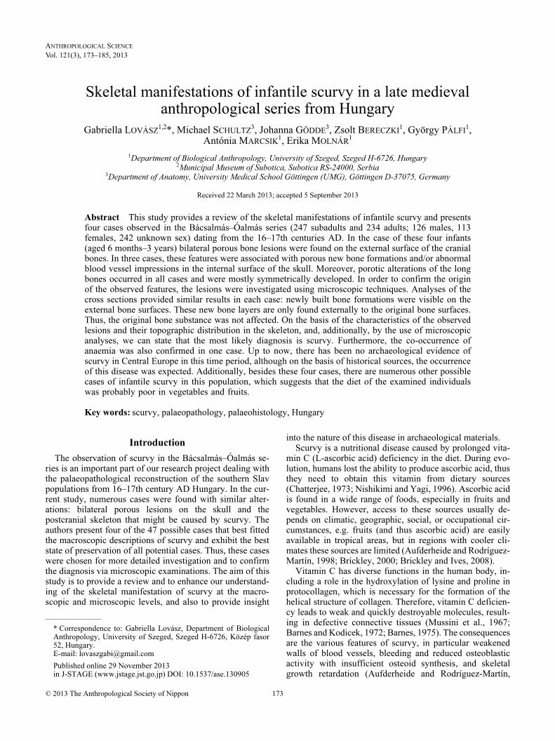
For microscopic examinations, six samples were takenfrom the following bones: second left rib, mandible (rightdeciduous molar region), left orbit and right greater wing ofthe sphenoid bone. Samples from the rib exhibit vestiges ofthin but well-organized new bone layers. Moreover, a slip-like newly built bone layer is visible on the orbital roof. Onthe sample of the sphenoid bone, short trabeculae are visiblewith extensive bridging (Figure 2A). Similar lesions are visi-ble on each samples of the mandible; however, multiple
Figure 1. Pathological alterations in the individual from grave no. 381 (1.5–2 year old infant). (A) Ectocranial view of the frontal bone dis-playing porous new bone layers in the glabellar region. (B) Anterior surface of the maxillae showing porous lesions surrounding the infraorbialforamina and below the anterior nasal spine. (C) Endocranial view of the left parietal bone displaying newly built bone formations associated withabnormal blood vessel impressions. (D) Medial surface of the shaft of the left tibia showing dense porosity and post mortem damage.
SKELETAL MANIFESTATIONS OF INFANTILE SCURVY 177Vol. 121, 2013
layers are also observed (Figure 2B). The original surfacesare not affected in any examined bones.
Grave no. 436On the well-preserved skeleton (identified as an infant
aged 6 months) both the skull and the postcranial elementsshow porous lesions. There is a sharply demarcated porousnew bone formation on the frontal bone at the glabellar re-gion (Figure 3A). The orbits are also severely affected: theorbital surfaces of the frontal and the sphenoid bones, theright maxilla and the right zygomatic bone show dense po-rous lesions (the left maxilla and zygomatic bone are miss-ing post mortem). Moreover, the temporal and the palatalsurfaces of the right maxilla display flat porous alterations.On the internal and external surfaces of the coronoid pro-cesses of the mandible fine porotic new bone formations arevisible, which externally continue to the mandibular notchand the posterior edges of the rami (Figure 3B). Small areasof flat porous lesions are seen on the external surface of themandibular body, especially on the mental eminences.
Extreme porous lesions can be observed on the temporalsurfaces of the greater wings of the sphenoid bone(Figure 3C). These lesions spread over the external surfacesof the temporal bones’ entire squamous and petrous parts aswell as the zygomatic processes. The porous alteration ex-tends also to the parietal bones covering approximately one-third of the surface of the bones around the squamous andlambdoid suture and also to the squamous part of the occip-ital bone. In addition, the bregmatic region of the parietalbones also exhibits porous lesions.
Endocranial lesions were also revealed in this case. Smallareas of thin porous new bone apposition were found all overthe endocranial surface of the frontal bone and lesser wingsof the sphenoid. Similar lesions were found on both parietalbones, mostly located along the sagittal suture. Smallplaques of new bone are also apparent on the internal surfaceof the occipital bone, although post mortem damage limitsthe observability of pathological processes. It is worth men-
tioning that the endocranial alterations in this case are lesspronounced than in the previous one.
Regarding the postcranial skeleton of this individual, al-terations were recognized on several bones. All preservedlong bones (both humeri, left radius and ulna, right femur,both tibiae) show pathological lesions. In the case of the up-per limb, the dorsomedial parts of the humeral shafts(Figure 3D), the anteromedial parts of the diaphysis of theleft radius, and the anterolateral part of the shaft of the leftulna display dense porous lesions. Flat porous alterations arealso seen in the supraspinous fossae of both scapulae. More-over, on the external surfaces of the ribs, porous lesions wereobserved mostly at the angular parts and the sternal ends.Widened sternal ends of the ribs were also discernible. Thelower limbs are also affected: the dorsal part of the diaphysisof the right femur and the dorsomedial parts of the shafts ofthe tibiae display flat porous lesions.
Samples for microscopic analyses were taken from theoccipital, frontal, and left temporal bones. Ribs, the leftscapula, the right clavicle, and the mandible were alsosampled. The examinations revealed a thin slip-like layer ofnew bone on the internal surface of the frontal bone, the ex-ternal surface of the occipital and temporal bones, the man-dible and also the dorsal surface of the scapula (Figure 4A).Short, bridged trabeculae of newly built bone were found onthe external surface of the frontal (Figure 4B) and the inter-nal surface of the occipital bones and the ribs. Thick, well-organized bone formations were observed in the sample ofthe clavicle. There is also no evidence of pathological alter-ations on the original surfaces of the examined bones.
Grave no. 439The poorly preserved skeleton of an infant aged 1–1.5
years also displays porous alterations on the skull and thepostcranial bones. Only a few parts of the skull are pre-served: the glabellar region and small pieces of the orbitalsurfaces of the frontal bone, a few parts of the left parietalbone, the base and the left lateral part of the occipital bone,
Figure 2. Microscopic images of the pathological alterations in the individual from grave no. 381 (1.5–2 year old infant). (A) Transverse sec-tion through the right greater wing of the sphenoid bone: (a) reorganized new bone layer; (b) original external lamina. Magnification × 100. (B)Frontal section through an alveolus of the mandible (right 4th tooth): (a) secondary built woven bone; (b) original surface of the alveolus.Magnification × 16. Thin ground sections viewed in polarized transmission light using a hilfsobject red 1st order (quartz) as compensator.

178 G. LOVÁSZ ET AL. ANTHROPOLOGICAL SCIENCE
Figure 3. Pathological alterations in the individual from grave no. 436 (6 month old infant). (A) Distinctly demarcated porous new bone for-mation on the frontal bone in the glabellar region. (B) Medial surface of the right coronoid process of the mandible exhibiting fine porotic newbone continuing to the mandibular notch and the posterior edge of the ramus. (C) Extreme porous lesions on the temporal surface of the rightgreater wing of the sphenoid. (D) Dense porous new bone on the dorsomedial face of the shaft of the right humerus.
Figure 4. Microscopic images of the pathological alterations in the individual from grave no. 436 (6 month old infant). (A) Sagittal sectionthrough the left scapula: (a) slip-like layer of new bone on the dorsal surface; (b) original bone surface. Magnification × 16. (B) Sagittal sectionthrough frontal bone: (a) new bone formation on the external surface; (b) external lamina; (c) diploë; (d) internal lamina. Magnification × 16. Thinground sections viewed in polarized transmission light using a hilfsobject red 1st order (quartz) as compensator.

SKELETAL MANIFESTATIONS OF INFANTILE SCURVY 179Vol. 121, 2013
and the mandibular body. The pieces of the left parietal boneand the lateral part of the occipital bone show slight, flat po-rous lesions on their external surfaces. The mandibular bodyalso displays flat porosity on the mental eminences. Nopathological alterations of the glabella and the orbital partsof the frontal bone were found. Endocranial lesions were notobserved on the preserved bones.
Besides the lesions of the skull, fine porotic alterations onthe surfaces of each preserved long bone and some otherpostcranial elements were detected. On the upper limb, thelesions are located on the anterior surface of the sternal endof the left clavicle, the dorsal shafts of the left humerus(Figure 5A), and both radii (the right clavicle, the right hu-
merus and both ulnae are missing post mortem). On the ribs,flat porous lesions were found on the external surfaces of theangular parts (Figure 5B). Widened sternal ends with in-creased porosity were observed as well (Figure 5C). In addi-tion, slight porous lesions are visible on the supraspinousarea of the right scapula (the left is missing post mortem).
On the lower limb, all surfaces of the femoral shafts(Figure 5D), medial surfaces of the tibial shafts and lateralsurfaces of the fibular shafts show slight, flat porosity.Moreover, the distal metaphyses of the femora and the prox-imal metaphyses of the tibiae are widened.
Microscopic analyses were carried out on the samples ofthe left humerus, the right ulna, and the left tibia. Each
Figure 5. Pathological alterations in the individual from grave no. 439 (1–1.5 year old infant). (A) Fine porotic alteration on the dorsal face ofthe shaft of the left humerus. (B) Flat porous lesions on the external surface of the angular part of the 9th right rib. (C) Widened sternal end withincreased porosity of the 9th right rib; external view. (D) Flat porous lesions on the dorsal face of the shaft of the right femur.
Figure 6. Microscopic images of the pathological alterations in the individual from grave no. 439 (1–1.5 year old infant). (A) Cross sectionthrough the right ulna: (a) newly built bone layers; (b) original cortical bone. Magnification × 16. (B) Cross section through the left tibia: (a) newlybuilt bone layers; (b) original compact bone. Magnification × 25. Thin ground sections viewed in polarized transmission light using a hilfsobjectred 1st order (quartz) as compensator.
180 G. LOVÁSZ ET AL. ANTHROPOLOGICAL SCIENCE
sample showed similar results: short bone trabeculae with ex-tensive bridging (Figure 6A, B). In the case of tibia and ulna,several layers of newly built bone were revealed. The origi-nal bone substance was not affected in any of the samples.
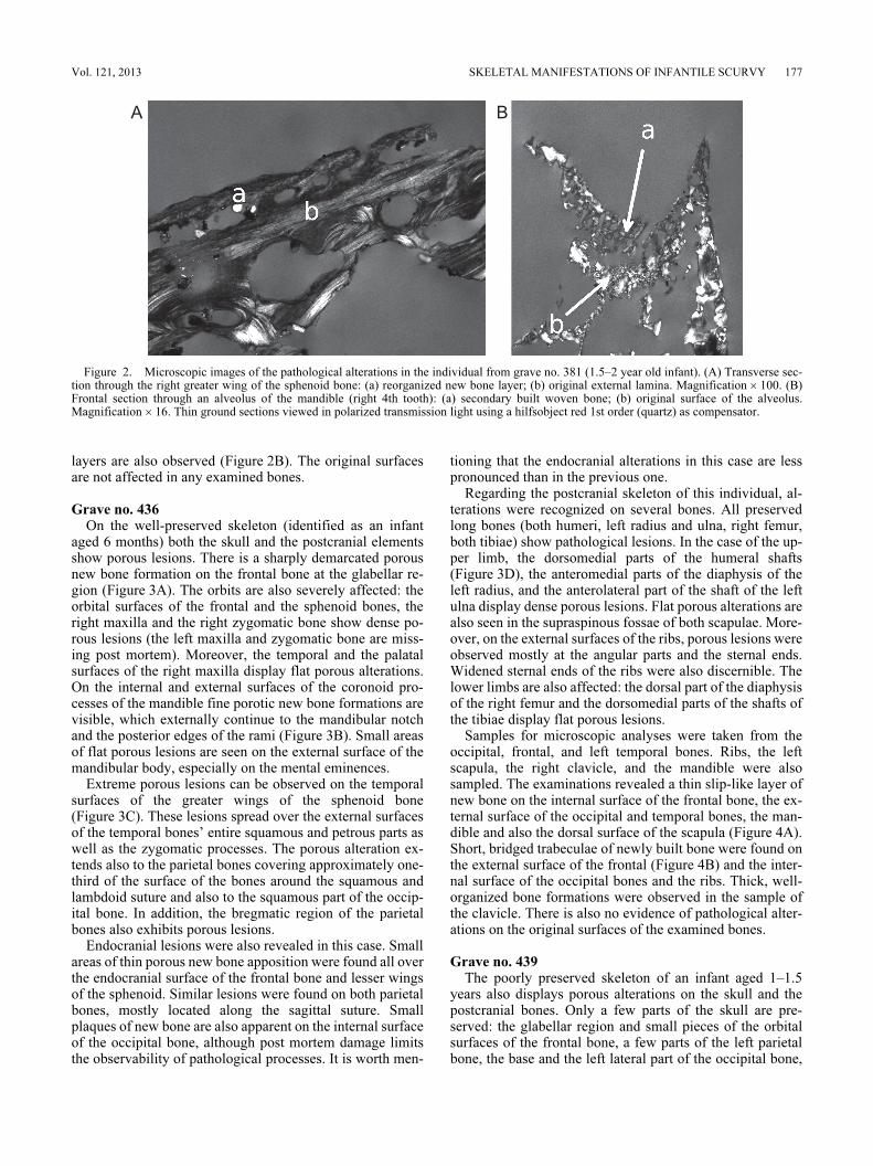
Grave no. 451The skeleton of an infant aged 2.5–3 years is almost com-
plete and shows multiple symmetric areas of porous lesionson the skull and the postcranial bones.
Abnormal flat porous lesions can be detected on the gla-bella. Moreover, both orbits are severely affected: the orbitalroofs display dense, porous, abnormal bone growth with as-sociated vessel impressions (Figure 7A). This bony over-growth is approximately 1.5 mm at the thickest point. Theorbital surfaces of the sphenoid bone and both maxillae alsoshow flat porous lesions. Besides these alterations, the pala-tal, temporal, and anterior surfaces around the infraorbitalforamina of both maxillae also show porosity. Slight porouslesions are also visible on the anterior surfaces of the zygo-matic bones. Moreover, the internal surfaces of the coronoid
processes of the mandible also display fine porotic lesionsthat extend to the bases of the mandibular foramina. On themandible, the oblique lines are covered with small patchesof porous new bone.
Bilateral abnormal porous lesions can be observed on theexternal surface of the greater wing of the sphenoid(Figure 7B). These alterations also continue on the temporalbones’ zygomatic processes and on the whole squamous part.Abnormal porosity can be found on the surrounding area ofthe lambdoid suture on both parietals and the occipital.
Endocranial alterations are also discernible in this case.Fine porotic new bone formations are visible on the upperpart of the frontal bone, accompanied by slight vascular im-pressions. Moreover, highly pronounced branching bloodvessel impressions are apparent on the upper part of both pa-rietal bones (Figure 7C). Thickening of the affected regionsis also observable. Fine porosity can be seen in the sinus ofthe sagittal suture as well.
The postcranial skeleton of this individual also displaysporous alterations. The upper limb bones show slight, fine
Figure 7. Pathological alterations in the individual from grave no. 451 (2.5–3 year old infant). (A) Ectocranial view of the right orbital roofdisplaying dense, porous, abnormal bone growth with associated blood vessel impressions. (B) External surface of the right greater wing of thesphenoid exhibiting abnormal porous lesions. (C) Endocranial view of the right parietal bone showing well pronounced branching blood vesselimpressions. (D) Fine porous lesions and slight and short blood vessel impressions on the dorsal face of the shaft of the right ulna.
SKELETAL MANIFESTATIONS OF INFANTILE SCURVY 181Vol. 121, 2013
porous changes on the anterior surfaces of the sternal ends ofclavicles, lateral parts of the humeral shafts, the dorsal sur-faces of the shafts of both ulnae (Figure 7D), and lateral sur-faces of the shafts of both radii. Moreover, flat porouslesions can be detected on the supraspinous areas of bothscapulae. Extended porosity and widening of the sternalends of the ribs were observed as well. On the lower limb,the anterior and posterior surfaces of both femoral shafts, themedial and lateral surfaces of both tibial shafts, and the lat-eral surfaces of the shafts of both fibulae are covered withfine porotic lesions. However, the changes in the tibiae andfibulae can are barely visible to the naked eye. In addition,the distal ends of the femora as well as the proximal ends ofthe tibiae are widened.
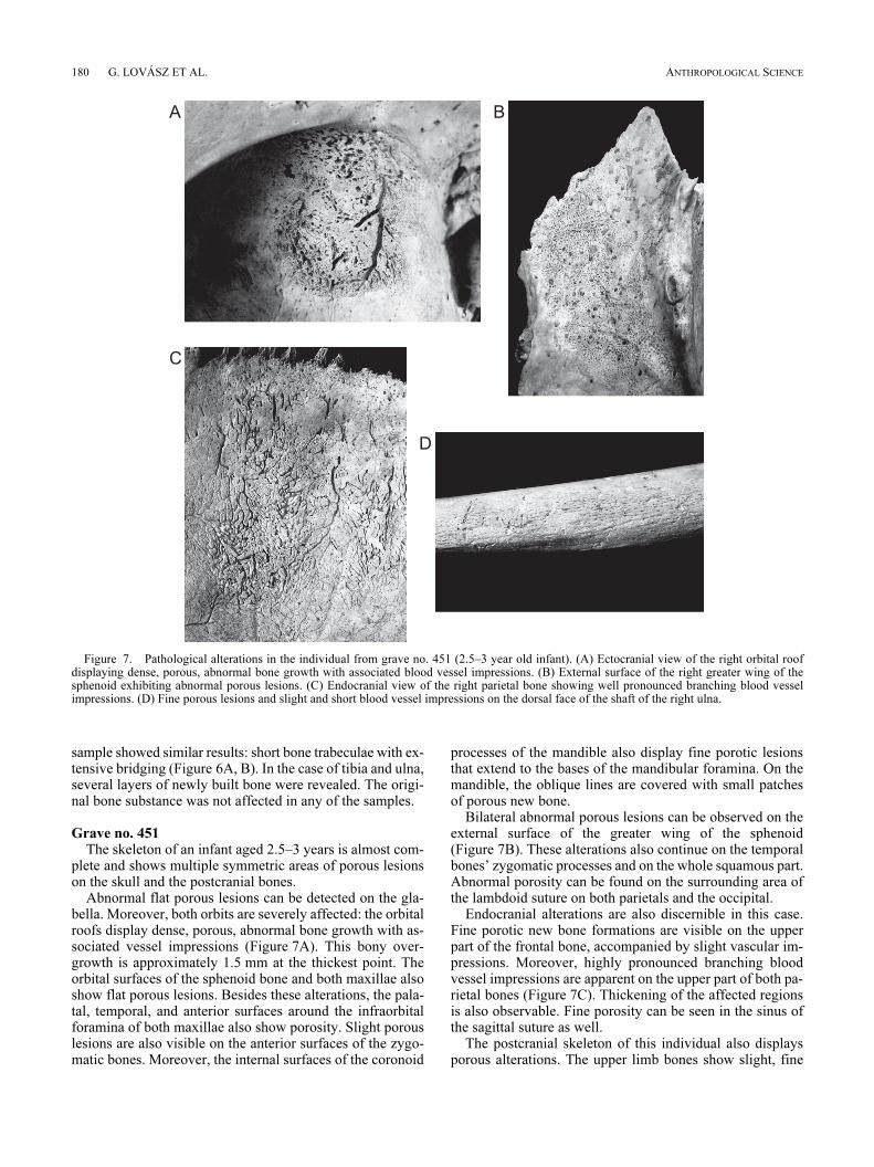
Microscopic examinations were carried out on samplestaken from the left orbit, the left parietal bone, and the righttibia. The sections through the parietal bone including theexternal and the internal lamina as well as the diploë and thecross section of the tibia show remains of thick, well-organized new bone formation (Figure 8A). There are nopathological changes in the original substance of the bones.On the external surface of the sample taken from the roof ofthe left orbit, very fine and thin layers of newly built wovenbone and the onset of the orbital lamina’s reduction are visi-ble (Figure 8B). Furthermore, there are abnormal trabecularformations in some regions of the orbital lamina and theexternal lamina of the left parietal where the original laminais slightly reduced, though only in some parts. Such changesare sometimes found in the case of chronic anaemia. In theleft orbital roof, a relatively pronounced change of the diplo-ic trabeculae is observable which is characterized by a par-allel packing of the trabeculae (Figure 8B, c). Thismorphological change of the diploë comes along with theformation of some small blood vessel canals which are radi-ally oriented to the internal lamina and open out into the or-bit, perforating the flat bone apposition on the orbital surface(Figure 8B, d). All these changes concede the case that wemight have here an organized haemorrhagic process in a rel-
atively advanced stage of healing. The locality inside thediploë is seemingly unusual and difficult to explain. Suchchanges on external bone surfaces are characteristic ofchronic vitamin-C-deficiency in infants and young children.
Differential Diagnoses
On the basis of the macromorphological characteristics ofthe observed lesions, in all of the four cases the most likelydiagnosis was scurvy. In the cases of grave no. 381, 436 and451, advanced stages of scurvy were presumed because ofthe abundance of porous lesions. In the case of grave no.439, the lesions are only slightly visible in the macroscopicassessment. Thus, an early stage of this disease was sup-posed. In the case of grave no. 451, the thickening of the pa-rietal bones at the site of the endocranial lesions and theslightly visible porosity of the long bones indicate a possiblehealing process. In the latter case, the co-occurrence of anadvanced stage of anaemia was also assumed because of se-vere porotic hyperostosis in the orbits.
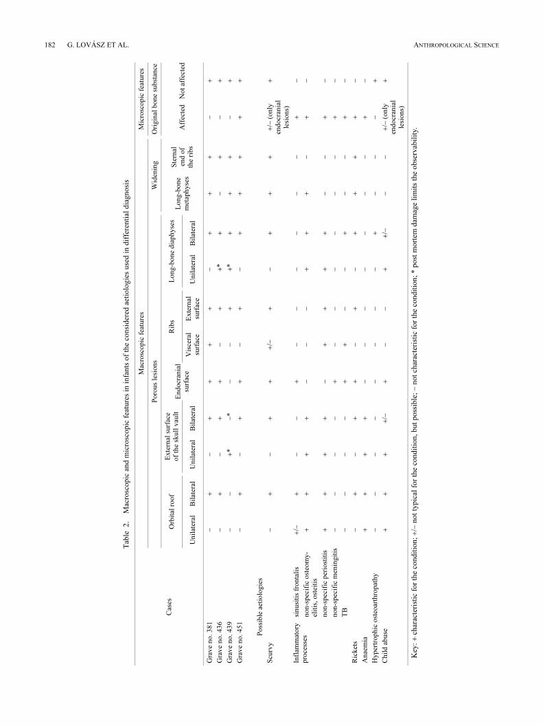
However, several other diseases must be taken into con-sideration, since similar periosteal reactions can be causedby inflammatory processes of various origin (e.g. infectiousconditions, such as sinusitis frontalis, non-specific periostitisand osteomyelitis or TB), rickets, anaemia, hypertrophic os-teoarthropathy, or child abuse (Aufderheide and Rodríguez-Martín, 1998; Schultz, 2001, 2012; Ortner, 2003; Lewis,2004; Wapler et al., 2004; Brickley and Ives, 2006). To ex-clude these aetiologies, we have to consider not only themacroscopic appearance of the lesions but also the distri-bution and the microscopic features of the alterations(Table 2).
The analyses of the cross sections yielded similar resultsin each case: newly built bone formations and/or remodellednew bone was/were visible on the bone surfaces. These newbone layers are only found externally to the original bonesurfaces, and the original bone substance was not affectedexcept for the sample of the left orbit of the case from grave
Figure 8. Microscopic images of the pathological alterations in the individual from grave no. 451 (2.5–3 year old infant). (A) Cross sectionthrough the right tibia: (a) well-organized new bone formation; (b) original compact bone. Magnification × 25. (B) Sagittal section through the leftorbital roof: (a) secondary bone layers; (b) original lamella of the orbital roof; (c) parallel diploic woven bone trabeculae which are tangentially ori-ented to the external and internal laminae; (d) small blood vessel canal which perforates the original orbital lamina and has its opening in the newlybuilt bone formation. Magnification × 16. Thin ground sections viewed in polarized transmission light using a hilfsobject red 1st order (quartz) ascompensator.
182 G. LOVÁSZ ET AL. ANTHROPOLOGICAL SCIENCE
Tab
le2.
Mac
rosc
opic
and m
icro
scopic
fea
ture
s in
infa
nts
of
the
consi
der
ed a
etio
logie
s use
d i
n d
iffe
renti
al d
iagnosi
s
Cas
es
Mac
rosc
opic
fea
ture
sM
icro
scopic
fea
ture
s
Poro
us
lesi
ons
Wid
enin
gO
rigin
al b
one
subst
ance
Orb
ital
roof
Exte
rnal
surf
ace
of
the
skull
vau
ltE
ndocr
ania
l su
rfac
e
Rib
sL
ong-b
one
dia
physe
sL
ong-b
one
met
aphyse
s
Ste
rnal
en
d o
f th
e ri
bs
Aff
ecte
dN
ot
affe
cted
Unil
ater
alB
ilat
eral
Unil
ater
alB
ilat
eral
Vis
cera
l su
rfac
eE
xte
rnal
su
rfac
eU
nil
ater
alB
ilat
eral
Gra
ve
no. 381
−+
−+
++
+−
++
+−
+
Gra
ve
no. 436
−+
−+
+−
++
*+
−+
−+
Gra
ve
no. 439
−−
+*
−*
−−
++
*+
++
−+
Gra
ve
no. 451
−+
−+
+−
+−
++
++
+
Poss
ible
aet
iolo
gie
s
Scu
rvy
−+
−+
++
/−+
−+
++
+/−
(only
en
docr
ania
l le
sions)
+
Infl
amm
atory
pro
cess
essi
nusi
tis
fronta
lis
+/−
+−
−+
−−
−−
−−
+−
non-s
pec
ific
ost
eom
y-
elit
is, ost
eiti
s+
++
+−
−−
++
+−
+−
non-s
pec
ific
per
iost
itis
++
++
−+
++
+−
−+
−
non-s
pec
ific
men
ingit
is−
−−
−+
−−
−−
−−
+−
TB
−−
−−
++
−−
+−
−+
−
Ric
ket
s−
+−
++
−+
−+
++
+−
Anae
mia
++
++
−−
−−
−−
−+
−
Hyper
trophic
ost
eoar
thro
pat
hy
−−
−−
−−
−−
+−
−−
+
Chil
d a
buse
++
++
/−+
−−
++
/−−
−+
/− (
only
en
docr
ania
l le
sions)
+
Key
: +
char
acte
rist
ic f
or
the
condit
ion; +
/− n
ot
typic
al f
or
the
condit
ion, but
poss
ible
; −
not
char
acte
rist
ic f
or
the
condit
ion;
* p
ost
mort
em d
amag
e li
mit
s th
e obse
rvab
ilit
y.

SKELETAL MANIFESTATIONS OF INFANTILE SCURVY 183Vol. 121, 2013
no. 451. In this case we have also morphological changeswhich, apparently, were caused by anaemia. All these alter-ations are characteristics of subperiosteal haematomas(Schultz, 2001, 2012).
In the cases of grave no. 381 and 436, the microscopic im-ages of the lesions revealed different morphological condi-tions of the haemorrhages: newly built bone layers and well-organized, remodelled bone tissue were detected in the samesection showing different stages of a healing process. Thiswas found in various elements of the skeleton of those twoindividuals. In the cases of grave no. 381 and 439, somesamples displayed multiple layers of newly built bone whichrefer to recidivation (Schultz, 2001). Even though the indi-vidual from grave no. 439 seemed to represent a case of anearly stage of scurvy on the basis of the macroscopic analy-sis, the microscopic examination revealed a far more severestage. In the case of grave no. 451, each sample of the micro-scopic analysis showed only remodelled bone appositions,confirming our presumption of a healing process (Schultz,2001).
In contrast to haemorrhagic processes, inflammatory pro-cesses always affect the original bone surface, and frequent-ly also the deeper structures. Rickets also causes changes inthe original bone substance, usually changing its microstruc-ture completely (Schultz, 2001, 2012). Therefore, on the ba-sis of the results of the microscopic examination, we caneliminate in these four cases the possibility of an occurrenceof inflammatory processes of infectious origin or caused byrickets.
Microscopic images of porotic hyperostosis in chronicanaemia display a reduction of the external lamina of theskull vault and the orbital roof and/or radial orientation ofthe diploic trabeculae (Schultz, 1986, 2001, 2012; Wapler etal., 2004). Thus, anaemia can also be excluded in three cases(grave no. 381, 438 and 439). In the case of grave no. 451,the onset of the external lamina’s destruction in the orbitalroof as well as the abnormal structure of the newly builtbone formation are indicative of the coexistence of chronicanaemia and scurvy (Stuart-Macadam, 1989; Schultz, 2001,2012; Wapler et al., 2004). Interestingly, the macroscopicobservations suggested advanced stage of anaemia, since theouter morphology of the multiple layers of new bone mimicswell-pronounced expanded diploë.
The microscopic images of the remaining possible aetiol-ogies such as hypertrophic osteoarthropathy or child abuseare similar or the same, since subperiosteal bleedings arecharacteristic for all of these possibilities (Schultz, 2001,2012). Therefore we have to consider the distribution patternof the lesions.
In each of the presented cases, the skull and the postcrani-al skeleton (usually each long bone of the limbs and the ribs)were bilaterally affected. In the case of hypertrophic osteoar-thropathy (caused by, for example, chronic pulmonary orheart disease), diffuse, bilaterally symmetrical, subperi-osteal bone deposition on the bone shafts usually occurs. Asa rule, these lesions affect the forearms and the lower legs,while involvement of the skull is not characteristic. There-fore, on the basis of the localization of the observed lesions,this diagnosis is unlikely (Aufderheide and Rodríguez-Martín, 1998; Ortner, 2003). Child abuse, such as “shaken
baby syndrome” or other forms of abuse, might cause bleed-ing all over the body, including subperiosteal haemorrhages.However, bilaterally symmetrical appearance of the alter-ations is uncommon, and therefore the possibility of thisaetiology is very low (Caffey, 1974; Kempe, 1985).
Considering the macromorphological and microscopicfeatures of the lesions, the distribution pattern of the changesand their comparison with the features of the other possibleaetiologies, we can establish the diagnosis of scurvy in eachexamined case. Moreover, the co-occurrence of anaemiawas only confirmed in one case.
Discussion and Conclusions
Besides these four cases, there are 43 other possible casesof infantile scurvy in the Bácsalmás–Óalmás series. How-ever, we have to take into consideration that among thesepossible cases the macromorphological characteristics andthe pattern of the lesions do not exclude the possibility ofother aetiologies. The uncertainty of the diagnosis is alsoincreased by the poor state of preservation in most of thesecases. Thus, in the evaluation of the palaeoepidemiologicaldata of scurvy we have to be very cautious. The four provedand 43 potential cases of scurvy suggest a remarkable 19%frequency among subadults. Among these cases 44.7% wereyounger than 1 year old, 31.9% were 1–2 years of age, and23.4% were older than 2 years. These data correspond to thepathological literature that scurvy commonly occurs in in-fants between the ages 5 and 24 months and reaches its max-imum prevalence between 8 and 10 months (Stuart-Macadam, 1989; Ortner, 2003; Brickley and Ives, 2006).Considering the age distribution of the affected children inthis population, the possibility of false nutrition during or af-ter weaning should be taken into account.
According to the Turkish tax registries (defters) the diet ofthis population was poor in vegetables and fruits (Wicker,2008). Therefore the population of Bácsalmás–Óalmás hadinsufficient access to natural vitamin C resources which in-creased the risk of scurvy.
Chronic vitamin C deficiency could occur alongside otherdiseases, such as infections, which are also frequent in thispopulation—many of these infectious cases might be due toTB infection. The large number of pathological alterations(including non-specific stress indicators such as porotic hy-perostosis and periostitis) might refer to the poor state ofhealth in this population. The very high infant mortalitycompared to neighbouring series also supports this finding(e.g. the palaeodemographical data of the Zombor–Repülőtér series, the closest known cemetery from the samearchaeological period and same ethnic group, where the ratioof subadults : adults is 33.7% : 66.3% (Lovász, 2009a, b)).In the background to this phenomenon there might be sever-al possible factors. This population immigrated most proba-bly from the southern region of Serbia or Montenegro(Wicker, 2008), and had to adapt to a new environmentwhich definitely induced stress phenomena. This stressmight have been increased by the social, economical, andgeographical isolation of this population. In addition, justlike in the rest of Europe, the climate of the Carpathian Basinin the 16–17th centuries became extremely cold (the so-

184 G. LOVÁSZ ET AL. ANTHROPOLOGICAL SCIENCE
called “Little Ice Age”). In this period, the average tempera-ture was significantly lower than in the years before or eventoday, and the winters were often quite hard and long. Thisclimate provided adverse conditions for agriculture. There-fore hunger was frequent in this period (Glaser et al., 1999;Rácz, 2001). The political and military crisis during theTurkish occupation must have also contributed to frequentstarvation of the people in this region. All these conditionsmight have affected this population too, causing decreasedresistance to disease.
Up to now, no archaeological evidence of scurvy in Cen-tral Europe in this time has existed, although on the basis ofhistorical sources, occurrence of the disease was expected(Réthly, 1962 in Rácz, 2001). Additionally, the scurvy casesdescribed above give insights into the nature of this diseasein archaeological materials, since not only the macroscopic,but also the microscopic features of the lesions were alsopresented. Our study provides an excellent example of thecomplementarity of macromorphological and micromorpho-logical observations in palaeopathological practice and howmicroscopic techniques can reorient a diagnostic process ina case otherwise seemingly simple and easy to diagnose.
Acknowledgments
The study was supported by the Hungarian Scientific Re-search Fund, OTKA no. 78555 and 78696. Special thanksare due to the staff of the Department of Biological Anthro-pology, University of Szeged for their continuous help andsupport during the course of the study.
References
Aufderheide A.C. and Rodríguez-Martín C. (1998) The Cam-bridge Encyclopedia of Human Paleopathology. CambridgeUniversity Press, Cambridge.
Barnes E. (2008) Congenital Anomalies. In: Pinhasi R. and MaysS. (eds.), Advances in Human Paleopathology. John Wiley &Sons, Chichester, pp. 329–362.
Barnes M.J. (1975) Function of ascorbic acid in collagen metabo-lism. Annals of the New York Academy of Sciences, 258:264–277.
Barnes M.J. and Kodicek E. (1972) Biological hydroxylations andascorbic acid with special regard to collagen metabolism.Vitamins and Hormones, 30: 1–43.
Bereczki Zs., Paja L., Lovász G., Maczel M., Molnár E., MarcsikA., and Pálfi Gy. (2009) New infectious conditions fromBácsalmás-Óalmás, Hungary—possible cases of skeletal TB.In: Pálfi Gy., Molnár E., Bereczki Zs., and Pap I. (eds.),Des Lésions du Passé aux Diagnostics Modernes. UniversityPress, Szeged, pp. 46–47.
Brickley M. (2000) The diagnosis of metabolic disease in archaeo-logical bone. In: Cox M. and Mays S. (eds.), Human Osteol-ogy: In Archaeology and Forensic Science. GreenwichMedical Media, London, pp. 183–198.
Brickley M. and Ives R. (2006) Skeletal manifestations of infantilescurvy. American Journal of Physical Anthropology, 129:163–172.
Brickley M. and Ives R. (2008) The Bioarchaeology of MetabolicBone Disease. Academic Press, San Diego.
Brown M. and Ortner D.J. (2011) Childhood scurvy in a medievalburial from Macvanska Mitrovica, Serbia. International Jour-nal of Osteoarchaeology, 21: 197–207.
Caffey J. (1974) The Whiplash Shaken Infant Syndrome: manual
shaking by the extremities with whiplash-induced intracranialand intraocular bleedings, linked with residual permanentbrain damage and mental retardation. Pediatrics, 54: 396–403.
Carli-Thiele P. (1996) Spuren von Mangelerkrankungen an stein-zeitlichen Kinderskeleten. Verlag Erich Goltze, Göttingen.
Chatterjee I.B. (1973) Evolution and biosynthesis of ascorbic acid.Science, 182: 1271–1272.
Geber J. and Murphy E. (2012) Scurvy in the great Irish famine:evidence of vitamin C deficiency from a mid-19th centuryskeletal population. American Journal of Physical Anthropol-ogy, 148: 512–524.
Glaser R., Brázdil R., Pfister C., Dobrovolný P., Vallvé M.B.,Bokwa A., Camuffo D., Kotyza O., Limanówka D., Rácz L.,and Rodrigo F.S. (1999) Seasonal temperature and precipita-tion fluctuations in selected parts of Europe during the six-teenth century. Journal of Climatic Change, 43: 169–200.
Haas C.J., Zink A., Molnár E., Marcsik A., Dutour O., and PálfiGy. (1999) Molecular evidence for tuberculosis in Hungarianskeletal samples. In: Pálfi Gy., Dutour O., Deák J., and HutásI. (eds.), Tuberculosis: Past and Present. Golden BookPublisher and Tuberculosis Foundation, Budapest-Szeged,pp. 383–391.
Haas C.J., Zink A., Molnár E., Szeimies U., Reischl U., MarcsikA., Ardagna Y., Dutour O., Pálfi Gy., and Nerlich A.G.(2000) Molecular evidence for different stages of tuberculosisin Hungarian skeletal samples. American Journal of PhysicalAnthropology, 113: 293–304.
Horváth G., Molnár E., Kovács J., Wicker E., Bérato J., and PálfiGy. (1994) Paleopathological diagnosis and interpretation ofseronegative spondylarthropathies from the 17th century.Acta Biologica Szegediensis, 40: 103–115.
Kempe C.H., Silverman F.N., Steele B.F., Droegemueller W., andSilver H.K. (1985) The battered-child syndrome. Child Abuseand Neglect, 9: 143–154.
Kiss T. (2008) A koponya fejlődési rendellenességeinek tanul-mányozása a Bácsalmás-homokbánya temető embertanianyagán. Master’s thesis, Department of Anthropology,University of Szeged.
Kovács Gy. and Tomka G. (2003) The archaeology of the OttomanPeriod. In: Visy Zs., Nagy M., and B. Kiss Zs. (eds.), Hungar-ian Archaeology at the Turn of the Millennium. Ministry ofNational Cultural Heritage, Budapest, pp. 405–413.
Laszlovszky J. (2003) Ottoman Period archaeology—post medi-eval archaeology. In: Visy Zs., Nagy M., and B. Kiss Zs.(eds.), Hungarian Archaeology at the Turn of the Millennium.Ministry of National Cultural Heritage, Budapest, pp. 405.
Lewis M.E. (2004) Endocranial lesions in non-adult skeletons:understanding their aetiology. International Journal ofOsteoarchaeology, 14: 82–97.
Lovász G. (2008) Comparative anthropological analysis of non-Hungarian skeletal populations from the 16–17th centuries.Acta Biologica Szegediensis, 52: 343–344.
Lovász G. (2009a) Antropološka obrada srednjovekovne nekro-pole iz Sombora. Godišnjak Gradskog muzeja u Somboru, 2–3: 141–168.
Lovász G. (2009b) A zombor-repülőtéri temető általánosantropológiai vizsgálata. Museion—A szabadkai városimúzeum évkönyve, 8: 143–157.
Lovász G., Molnár E., and Marcsik A. (2005) Bácsalmás-Óalmás16–17. századi temető paleopatológiai vizsgálatának ered-ményei (2001–2003-as feltárás). In: Korsós Z. (ed.),Előadáskötet, IV. Kárpát-medencei Biológiai Szimpózium,17–19th Oct 2005. Fővárosi Állat- és Növénykert, Budapest,pp. 153–158.
Lovász G., Bereczki Zs., Pálfi Gy., and Molnár E. (2009) A possi-ble case of echinococcosis from a late medieval series inHungary. In: Pálfi Gy., Molnár E., Bereczki Zs., and Pap I.(eds.), Des Lésions du Passé aux Diagnostics Modernes.University Press, Szeged, pp. 70–71.
SKELETAL MANIFESTATIONS OF INFANTILE SCURVY 185Vol. 121, 2013
Maczel M. (2003) On the Traces of Tuberculosis. Diagnostic Cri-teria of Tuberculous Affection of the Human Skeleton andTheir Application in Hungarian and French AnthropologicalSeries. Ph.D. thesis, University of La Méditerranée, Marseilleand Department of Anthropology, University of Szeged.
Marcsik A., Molnár E., and Szathmáry L. (2006) The antiquity oftuberculosis in Hungary: the skeletal evidence. Memórias doInstituto Oswaldo Cruz, 101: 67–71.
Mays S. (2008) Metabolic bone disease. In: Pinhasi R. and MaysS. (eds.), Advances in Human Paleopathology. John Wiley &Sons, Chichester, pp. 215–252.
Melikan M. and Waldron T. (2003) An examination of skulls fromtwo British sites for possible evidence of scurvy. InternationalJournal of Osteoarchaeology, 13: 207–212.
Merbs C.F. (2004) Sagittal clefting of the body and other vertebraldevelopmental errors in Canadian Inuit skeletons. AmericanJournal of Physical Anthropology, 123: 236–249.
Molnár E. and Pálfi Gy. (1994) Probable cases of skeletal infec-tions in the 17th century anthropological series fromBácsalmás (Hungary). Acta Biologica Szegediensis, 40: 117–132.
Molnár E., Maczel M., Marcsik A., Pálfi Gy., Nerlich G.A., andZink A. (2005) A csont-ízületi tuberkulózis molekulárisbiológiai vizsgálata egy középkori temető embertanianyagában. Folia Anthropologica, 3: 41–51.
Murray P.D.F. and Kodicek E. (1949) Bones, muscles and vitaminC. II. Partial deficiencies of vitamin C and mid-diaphysealthickenings of the tibia and fibula in guinea-pigs. Journal ofAnatomy, 83: 205–223.
Mussini E., Hutton J.J., and Udenfriend S. (1967) Collagen prolinehydroxylase in wound healing, granuloma formation, scurvy,and growth. Science, 157: 927–929.
Neparáczki E., Török T., Pósa A., Molnár E., Lovász G., MaixnerF., Zink A., Dutour O., and Pálfi Gy. (2011) Preliminaryresults from the paleomicrobiological studies of Mycobacte-rium tuberculosis infection in the Bácsalmás-Óalmás anthro-pological series. Acta Biologica Szegediensis, 55: 41–45.
Nishikimi M. and Yagi K. (1996) Biochemistry and molecularbiology of ascorbic acid biosynthesis. In: Harris J.R. (ed.),Subcellular Biochemistry, Volume 25: Ascorbic Acid: Bio-chemistry and Biomedical Cell Biology. Plenum Press, NewYork, pp. 17–40.
Ortner D.J. (2003) Identification of Pathological Conditions inHuman Skeletal Remains. Academic Press, San Diego.
Ortner D.J. and Ericksen M.F. (1997) Bone changes in the humanskull probably resulting from scurvy in infancy and child-hood. International Journal of Osteoarchaeology, 7: 212–220.
Ortner D.J., Kimmerle E.H., and Diez M. (1999) Probable evi-dence of scurvy in subadults from archeological sites in Peru.American Journal of Physical Anthropology, 108: 321–331.
Ortner D.J., Butler W., Cafarella J., and Milligan L. (2001) Evi-dence of probable scurvy in subadults from archeologicalsites in North America. American Journal of Physical Anthro-pology, 114: 343–351.
Pálfi Gy. and Ardagna Y. (2002) Gerincbetegségek és tuberkulózisa török hódoltság korából. A Bácsalmás-Óalmás (Bácsalmás-Homokbánya) 16–17. századi antropológiai leletegyüttes fon-tosabb paleopatológiai adatai. In: Gerelyes I. and Kovács Gy.(eds.), Opuscula Hungarica, 3: A hódoltság régészetikutatása. Magyar Nemzeti Múzeum, Budapest, pp. 237–244.
Pálfi Gy. and Molnár E. (2009) The paleopathology of specificinfectious diseases from southeastern Hungary: a brief over-view. Acta Biologica Szegediensis, 53: 111–116.
Pálfi Gy., Molnár E., Bérato J., Wicker E., and Dutour O. (1996)
Deux observations ostéoarchéologiques de spondylarthriteankylosante et leur possible relation familiale. Revue duRhumatisme, 63: 160–161.
Pálfi Gy., Molnár E., Bérato J., and Dutour O. (1997) Cinq cas depathologie infectieuse ou inflammatoire dans une sérieanthropologique hongroise. Anthropologie et Préhistoire,108: 165–176.
Pósa A., Maixner F., Lovász G., Molnár E., Bereczki Zs., Zink A.,and Pálfi Gy. (2013) Revision of tuberculous lesions in theBácsalmás-Óalmás series—preliminary morphological andbiomolecular studies. Anthropologischer Anzeiger, 70: 83–100.
Rácz L. (2001) Magyarország éghajlattörténete az újkor idején.Juhász Gyula Felsőoktatási Kiadó, Szeged.
Resnick D. and Niwayama G. (1988) Diagnosis of Bone and JointDisorders. Saunders, Philadelphia.
Réthly A. (1962) Időjárási események és elemi csapások Mag-yarországon 1700-ig [Weather events and natural disasters inHungary until 1700]. Akadémiai Kiadó, Budapest.
Schultz M. (1986) Die mikroskopische Untersuchung prähis-torischer Skeletfunde. Anwendung und Aussagemöglich-keiten der differentialdiagnostischen Untersuchung in derPaläopathologie. Archäologie und Museum 6. Amtes fürMuseen und Archäologie des Kantons Baselland, Liestal.
Schultz M. (1988) Methoden der Licht- und Elektronenmikrosko-pie. In: Knußmann R. (ed.), Anthropologie. Handbuch dervergleichenden Biologie des Menschen. Band 1, Teil 1. G.Fischer Verlag, Stuttgart, New York, pp. 698–730.
Schultz M. (1990) Erkrankungen des Kindesalters bei der früh-bronzezeitlichen Population vom Ikiztepe (Türkei). In:Andraschko F.M. and Teegen W-R. (eds.), Gedenkschrift fürJürgen Driehaus. Zabern Verlag, Mainz, pp. 83–90.
Schultz M. (2001) Paleohistopathology of bone: a new approach tothe study of ancient diseases. American Journal of PhysicalAnthropology, 116: 106–147.
Schultz M. (2012) Light microscopic analyses of macerated patho-logically changed bones. In: Crowder Ch. and Stout S.D.(eds.), Bone Histology: An Anthropological Perspective.CRC Press, Boca Raton, FL, pp. 253–296.
Stuart-Macadam P.L. (1989) Nutritional deficiency diseases. In:Isçan M.Y and Kennedy K.A.R. (eds.), Reconstruction ofLife From the Skeleton. Alan R. Liss, New York, pp. 201–222.
Vaday A. (2003) Chronological charts. In: Visy Zs., Nagy M., andB. Kiss Zs. (eds.), Hungarian Archaeology At the Turn of theMillennium. Ministry of National Cultural Heritage, Budap-est, pp. 483–486.
Vigorita V.J., Ghelman B., and Mintz D. (2008) OrthopaedicPathology. Lippincott Williams & Wilkins, Philadelphia.
von Hagens G. (1979) Impregnation of soft biological specimenswith thermosetting resins and elastomers. The AnatomicalRecord, 194: 247–255.
Wapler U., Crubézy E., and Schultz M. (2004) Is cribra orbitaliasynonymous with anemia? Analysis and interpretation of cra-nial pathology in Sudan. American Journal of PhysicalAnthropology, 123: 333–339.
Wicker E. (2008) Rácok és vlahok a hódoltság kori Észak-Bácskában. Bács-Kiskun Megyei Önkormányzat MúzeumiSzervezete, Kecskemét.
Zink A.R., Molnár E., Motamedi N., Pálfi Gy., Marcsik A., andNerlich A.G. (2007) Molecular history of tuberculosis fromancient mummies and skeletons. International Journal ofOsteoarchaeology, 17: 380–391.