six rna viruses and forty-one hosts: viral small rnas and...
TRANSCRIPT
Six RNA Viruses and Forty-One Hosts: Viral Small RNAsand Modulation of Small RNA Repertoires in Vertebrateand Invertebrate SystemsPoornima Parameswaran1, Ella Sklan2¤a, Courtney Wilkins3¤b, Trever Burgon1¤c, Melanie A. Samuel4¤d,
Rui Lu5¤e, K. Mark Ansel6, Vigo Heissmeyer7, Shirit Einav2, William Jackson1¤f, Tammy Doukas1, Suman
Paranjape8¤g, Charlotta Polacek8¤h, Flavia Barreto dos Santos8¤i, Roxana Jalili9, Farbod Babrzadeh9,
Baback Gharizadeh9, Dirk Grimm10¤j, Mark Kay10, Satoshi Koike11, Peter Sarnow1, Mostafa Ronaghi 9¤k,
Shou-Wei Ding5, Eva Harris8, Marie Chow3, Michael S. Diamond12, Karla Kirkegaard1, Jeffrey S. Glenn2,
Andrew Z. Fire13*
1 Department of Microbiology & Immunology, Stanford University School of Medicine, Stanford, California, United States of America, 2 Department of Gastroenterology &
Hepatology, Stanford University School of Medicine, Stanford, California, United States of America, 3 Department of Microbiology & Immunology, University of Arkansas
for Medical Sciences, Little Rock, Arkansas, United States of America, 4 Department of Molecular Microbiology, Washington University School of Medicine, St. Louis,
Missouri, United States of America, 5 Department of Plant Pathology & Microbiology, University of California at Riverside, Riverside, California, United States of America,
6 Strategic Asthma Basic Research Center and the Department of Microbiology & Immunology, University of California at San Francisco, San Francisco, California, United
States of America, 7 Institute of Molecular Immunology, Helmholtz Center Munich, German Research Center for Environmental Health, Munich, Germany, 8 Division of
Infectious Diseases and Vaccinology, School of Public Health, University of California at Berkeley, Berkeley, California, United States of America, 9 Stanford Genome
Technology Center, Stanford University School of Medicine, Stanford, California, United States of America, 10 Departments of Pediatrics & Genetics, Stanford University
School of Medicine, Stanford, California, United States of America, 11 Tokyo Metropolitan Organization for Medical Research, Tokyo Metropolitan Institute of Medical
Science, Tokyo, Japan, 12 Departments of Medicine, Molecular Microbiology, Pathology & Immunology, Washington University School of Medicine, St. Louis, Missouri,
United States of America, 13 Departments of Pathology & Genetics, Stanford University School of Medicine, Stanford, California, United States of America
Abstract
We have used multiplexed high-throughput sequencing to characterize changes in small RNA populations that occur duringviral infection in animal cells. Small RNA-based mechanisms such as RNA interference (RNAi) have been shown in plant andinvertebrate systems to play a key role in host responses to viral infection. Although homologs of the key RNAi effectorpathways are present in mammalian cells, and can launch an RNAi-mediated degradation of experimentally targetedmRNAs, any role for such responses in mammalian host-virus interactions remains to be characterized. Six different viruseswere examined in 41 experimentally susceptible and resistant host systems. We identified virus-derived small RNAs (vsRNAs)from all six viruses, with total abundance varying from ‘‘vanishingly rare’’ (less than 0.1% of cellular small RNA) to highlyabundant (comparable to abundant micro-RNAs ‘‘miRNAs’’). In addition to the appearance of vsRNAs during infection, wesaw a number of specific changes in host miRNA profiles. For several infection models investigated in more detail, the RNAiand Interferon pathways modulated the abundance of vsRNAs. We also found evidence for populations of vsRNAs that existas duplexed siRNAs with zero to three nucleotide 39 overhangs. Using populations of cells carrying a Hepatitis C replicon, weobserved strand-selective loading of siRNAs onto Argonaute complexes. These experiments define vsRNAs as one possiblecomponent of the interplay between animal viruses and their hosts.
Citation: Parameswaran P, Sklan E, Wilkins C, Burgon T, Samuel MA, et al. (2010) Six RNA Viruses and Forty-One Hosts: Viral Small RNAs and Modulation of SmallRNA Repertoires in Vertebrate and Invertebrate Systems. PLoS Pathog 6(2): e1000764. doi:10.1371/journal.ppat.1000764
Editor: Charles M. Rice, The Rockefeller University, United States of America
Received September 18, 2009; Accepted January 13, 2010; Published February 12, 2010
Copyright: � 2010 Parameswaran et al. This is an open-access article distributed under the terms of the Creative Commons Attribution License, which permitsunrestricted use, distribution, and reproduction in any medium, provided the original author and source are credited.
Funding: Funding sources: Burroughs Wellcome Fund (CABS1006173; to K. Mark Ansel), Deutsche Forschungsgemeinschaft SFB571 and the Fritz ThyssenFoundation (to Vigo Heissmeyer), NIH AI071068 (to Mark Kay), NIH K08-AI079406-01 (to Shirit Einav), 1S10RR022982-01 (to Mostafa Ronaghi), NIH AI069000 (toPeter Sarnow), NIH R01 AI052447 (to Shou-Wei Ding), NIH AI052324 (to Eva Harris), UAMS Foundation Chow research endowment fund (to Marie Chow), TheMidwest Regional Center of Excellence for Biodefense and Emerging Infectious Diseases Research (NIH U54 AI057160; to Michael S. Diamond), NIH Director’sPioneer Award (to Karla Kirkegaard), Burroughs Wellcome Fund Clinical Scientist Award in Translational Research, RO1 DK066793, and RO1 DK064223 (to Jeffrey S.Glenn), NIH ROI GM37706, Stanford Dept. of Pathology funds, Bill & Melinda Gates Foundation (to Andrew Z. Fire), Stanford Graduate Fellowship (to PoornimaParameswaran), and for work in the Fire Lab from NIAID-U54065359 (Pacific Southwest Regional Center of Excellence, PI Alan Barbour). The funders had no role instudy design, data collection and analysis, decision to publish, or preparation of the manuscript.
Competing Interests: The authors have declared that no competing interests exist.
* E-mail: [email protected]
PLoS Pathogens | www.plospathogens.org 1 February 2010 | Volume 6 | Issue 2 | e1000764

¤a Current address: Department of Clinical Microbiology and Immunology, Tel-Aviv University, Ramat-Aviv, Tel-Aviv, Israel¤b Current address: Department of Immunology, University of Washington School of Medicine, Seattle, Washington, United States of America¤c Current address: Sg2, Evanston, Illinois, United States of America¤d Current address: Harvard University, Cambridge, Massachusetts, United States of America¤e Current address: Department of Biological Sciences, Louisiana State University, Baton Rouge, Louisiana, United States of America¤f Current address: Department of Microbiology and Molecular Genetics, Center for Biopreparedness and Infectious Diseases, Medical College of Wisconsin,Milwaukee, Wisconsin, United States of America¤g Current address: Science and Technology Policy Fellowship Program, American Association for the Advancement of Sciences, Washington, D.C., United States ofAmerica¤h Current address: National Veterinary Institute, Lindholm, Denmark¤i Current address: Laboratorio de Flavivirus, Pav. Helio e Peggy Pereira, sala B 102, Instituto Oswaldo Cruz/Fundacao Oswaldo Cruz, Rio de Janeiro, Brazil¤j Current address: Dept. of Virology, University of Heidelberg, Heidelberg, Germany¤k Current address: Illumina, San Diego, California, United States of America
Introduction
Biological systems are protected by innate immune mechanisms
initiated by host sensors called pattern recognition receptors
(‘PRRs’) that recognize specific ‘‘foreign’’ features of invading
pathogens to initiate multiple downstream anti-pathogen cascades.
PRRs that detect nucleic acid structures characteristic of viral
infection (such as single- or double-stranded RNA or DNA) are
among the innate responders that protect diverse cell types from
viral pathogenesis (for review, see [1,2]). How the cell handles viral
double-stranded RNA (dsRNA) is of special interest because
dsRNA is a necessary intermediate in the replication of RNA
viruses. In addition to dsRNA that forms during replication of the
virus genome, RNA duplexes can form due to self-complemen-
tarity in the virus genome, and in some instances, from sense-
antisense transcription of overlapping genes.
Four of the most studied families of PRRs for dsRNA are: (a)
cytoplasmic RNA helicases like Retinoic acid-inducible gene I &
Melanoma differentiation-associated gene-5 (‘‘RIG-I’’ & ‘‘Mda-
5,’’ which trigger mitochondrial-localized antiviral pathways); (b)
Protein Kinase R (‘‘PKR,’’ which induces a translational arrest
state in cells after sensing dsRNA); (c) 29–59 oligoadenylate
synthetase (‘‘OAS,’’ which stimulates the ssRNase activity of
RNase L in response to dsRNA); and (d) Toll-like receptors
(‘‘TLRs,’’ which bind various forms of RNA or DNA). All of these
PRRs trigger the Interferon (IFN) responses, and activate IFN-
stimulated genes (ISGs) that establish an antiviral state in the
infected cell (for review, see [3]). The IFN signaling pathway is
central to the detection of, and response to, virus infections in cells.
Type I IFNs (IFN-a and IFN-b) make up one of the first lines of
defense in the innate immune response to viruses by inducing
antiviral ISGs, modulating the levels of specific host-encoded
miRNAs [4], and in a feedback loop, that of PKR and OAS.
Many viruses are also susceptible to treatment with Type I IFNs,
and conversely, cells that have higher basal activity of ISGs seem
to mount a more successful antiviral response, and are not targeted
by viruses [5].
Dicer is another PRR that recognizes dsRNA, chopping it into
smaller duplexes called siRNAs that are 19–27 nucleotides (nt)
long [6,7]. These siRNAs have a terminal 59 mono-phosphate and
a terminal 39 hydroxyl on both strands, generally have 2 nt 39
overhangs, and are fed into an RNA-induced silencing complex
‘‘RISC’’ (for review on Dicer and Argonautes, see [8,9]). siRNA
duplexes are unwound, and only one strand remains associated
with RISC (the mechanism of unwinding and choice of strand is
poorly understood; for review, see [10]). One of the key
components of RISC is a protein called Argonaute-2 (Ago-2),
which belongs to the Argonaute family of proteins. Ago-2 is the
only member of the family that has cleavage activity, and is the
designated ‘slicer’ protein in RISC that mediates cleavage of
mRNA in a sequence-directed manner by a process termed RNA
interference, or ‘RNAi’ [11,12,13,14].
There is strong evidence for an antiviral role for RNAi in plant
and invertebrate systems (for review, see [15,16,17]). Viruses
replicate most effectively in these systems in the absence of key
elements of the RNAi pathway: either in cells lacking components
of the RNAi machinery, or in the presence of virus-encoded
suppressors of the silencing pathway (for review, see [18,19]). As
expected, virus-derived siRNAs (vsRNAs) can be detected in some
plant and invertebrate systems that are capable of mounting a
successful/partially successful RNAi response [15,16,17]. A
population of vsRNAs would be an expected component of any
viral defense pathway that acted through an RNAi mechanism.
In mammalian cells, short duplex RNAs can effectively enter
the RNAi pathway and function in sequence-specific silencing,
while duplexes longer than 30 nt generally produce a more
complex response including the induction of multiple non-specific
pathways including the IFN response (for review, see [20,21]).
Indeed, RNA and DNA viruses have evolved a host of defense
mechanisms to counteract the nonspecific signaling effects of
dsRNA. For example, Adenovirus VA RNA sequesters PKR [22],
while proteins from Vaccinia virus (E3L), Porcine Rotaviruses
(NSP3), and Influenza A virus (NS1) sequester dsRNA and prevent
stimulation of the IFN response [23,24,25,26]. Viral proteins can
also inhibit signaling downstream of dsRNA binding, as in the case
of the HCV protease NS3/4A, which cleaves IPS-1 (the RIG-I/
MDA-5 signaling partner) to consequently disrupt induction of
IFN responses [27]. Several of these dsRNA-binding proteins may
also facilitate viral evasion of host immune responses by inhibiting
RNAi [28]. Additionally, some viruses make their genomes
Author Summary
Short RNAs derived from invading viruses with RNAgenomes are important components of antiviral immunityin plants, worms and flies. The regulated generation ofthese short RNAs, and their engagement by the immuneapparatus, is essential for inhibiting viral growth in theseorganisms. Mammals have the necessary protein compo-nents to generate these viral-derived short RNAs(‘‘vsRNAs’’), raising the question of whether vsRNAs inmammals are a general feature of infections with RNAviruses. Our work with Hepatitis C, Polio, Dengue, VesicularStomatitis, and West Nile viruses in a broad host repertoiredemonstrates the generality of RNA virus-derived vsRNAproduction, and the ability of the cellular short RNAapparatus to engage these vsRNAs in mammalian cells.Detailed analyses of vsRNA and host-derived short RNApopulations demonstrate both common and virus-specificfeatures of the interplay between viral infection and shortRNA populations. The vsRNA populations described in thiswork represent a novel dimension in both viral pathogen-esis and host response.
Viral and Host Small RNAs in Animal Systems
PLoS Pathogens | www.plospathogens.org 2 February 2010 | Volume 6 | Issue 2 | e1000764

inaccessible to PRRs of various types including IFN effectors and
the siRNA-programmed RISC complex (e.g. [29]).
Viruses may also perturb another class of effectors involved in
RNAi called micro-RNAs (miRNAs), which are a class of cellular
small RNAs generated by Dicer from hairpin structures. Cellular
miRNA profiles are frequently modulated upon infection by
viruses, and this may contribute in some cases to infectivity and
pathogenesis [30]. Conversely, some viruses usurp the host
miRNA machinery for processing miRNA-like structures encoded
in the viral genome, potentially using these molecules for
regulation of virus/host gene expression [31].
With so much potential for RNA-mediated cross talk between
the IFN response, the RNAi pathway, and the virus itself, it has
been difficult to demonstrate a precise role for the RNAi pathway
in vertebrate antiviral defense. The difficulties in segregating IFN
and RNAi functions have given rise to speculations that the
antiviral role of RNAi may have been lost during evolution, or
alternatively, that RNAi-based defense may only be harnessed by
triggers such as short hairpins and siRNAs that do not stimulate
the IFN pathway. There has been some attempt at demonstrating
recognition of viral RNA by the RNAi machinery. For instance, in
Vero cells (which lack IFNa/b), inhibition of RNAi by Dicer
knockdown increases replication of an RNA virus, the Influenza A
Virus [32]. Additionally, there are cases where short virus-derived
RNAs can be detected in vertebrate systems (e.g. from HDV [33]
by high-throughput sequencing, and the HCV replicon [34], by
bulk analysis methods). However, it is still not clear how general
the presence of such RNAs is, and whether these RNAs can
participate in host defense mechanisms. To complicate this issue,
many of the classically-studied virus-host systems have been
chosen based on the ability of the virus to rapidly replicate and kill
host cells; these experimental infection systems may artificially
under-represent the capacity of vertebrate cells to protect
themselves, hence biasing against systems where RNAi might
have a significant role in host-virus interactions.
Here, we sought a broader survey of potential RNA-derived
defenses in viral infection systems. Given no knowledge of which
virus type might engage the RNAi machinery, and which cell types
might efficiently use this machinery in defense, we cast a wide net in
terms of both virus families and host cells. In this study, we describe
small RNA populations from six different RNA viral pathogens,
each in a variety of animal cell infection systems (including both
immune-competent and immune-compromised hosts). Upon ex-
amining small RNA populations from ,150 samples with sample-
specific DNA barcodes, we found viral-derived small RNAs
(vsRNAs) from each virus, with vsRNA populations sensitive to
both viral and host characteristics. A more detailed analysis of
vsRNAs in two viral infection models (Hepatitis C Virus and
Poliovirus) in various host types revealed that multiple distinct pools
of vsRNAs may co-exist during infection: as single strands, as part of
duplexes, and in complexes that may contain Argonautes. We also
observed specific changes in cell-derived miRNA populations,
providing a clear indication of host perturbation by the virus. The
characterization of small RNA populations during RNA virus
infections provides both an experimental entry point, and an
indication of the complexity that will need to be addressed in
understanding roles for small RNAs in host and viral processes.
Results
Detection and analysis of small RNA populations duringviral infection
In the following sections, we will describe small RNA
populations present during infection of animal cells with six
different viruses. In each case, we have taken infected cells,
extracted small RNA populations, and characterized these
populations using high-throughput sequencing methods. Two
high-throughput sequencing platforms were used: Roche/454
pyrosequencing (http://www.454.com/), to obtain several hun-
dred thousand sequences from pools of appropriately linkered
amplicon templates; and Solexa/Illumina technology, which yields
larger datasets of shorter reads (http://www.illumina.com/). Due
to the large number of samples to be analyzed, we used DNA
barcodes to ‘tag’ RNA samples from individual experiments,
which facilitated sequencing in parallel from multiple samples.
This allowed us to work with samples from different viral systems
and diverse experimental conditions in a cost-effective manner,
with a small number of instrument runs. Viral-derived sequences
were identified in sequence datasets through pattern matching
using standard software (BLAT [35] and BLAST [36]). We use the
term ‘vsRNA’ to refer to small RNA segments whose sequences
show perfect complementarity to the infecting viral genome at
every base position (reference genomes listed in Table S1).
vsRNAs are distinct from host-derived miRNAs that may show
partial complementarity to sites in the viral genome (e.g.
[37,38,39], Fig. S1). We detected 77,609 vsRNAs out of
19,425,777 sequences from 151 datasets (Fig. 1, Figure S2:length distributions). The most abundant vsRNAs from each
virus are listed in Table S2.
For a small number of vsRNAs (0.033%) we observed a perfect
match to both the host and viral genomes (Table S3, Table S4).
The fractions of vsRNAs that matched host genomes were
approximately as expected by random sequence coincidence (for
example: the human genome, with a unique genome complexity of
26109 bp, would match approximatly 1 in 4000 arbitrary 22-mer
sequences). The perfect nature of the homology makes it difficult
to determine whether this minor class of sRNAs was derived from
the host or from the virus.
Furthermore, to validate the specificity of the barcoding and
sequencing assays, we carried out sequence comparisons to the full
set of viruses for each experimental sample. We identified 13
vsRNAs that were ‘rogue’ hits i.e. mapped to one of the other 5
viruses not used in that particular experiment. In no sample were
the ‘rogue’ matches present at more than 0.008% of all parsed
sequences (Table S3).
For certain purposes, it will be of interest to compare vsRNA
incidences in different samples. Such comparisons require some
normalization for total depth of RNA sequencing. In Table S3,
we provide two distinct normalizations for each sample:
normalization to total small RNAs recovered and sequenced (v/
sRNA), and normalization to the population of cellular miRNAs
that are expected to represent a large proportion of bona-fide
small RNA effectors (v/miR; miRNAs are defined as documented
in miRBase ver9.2 [40,41,42]). There is a substantial challenge in
choosing and interpreting appropriate normalization schemes: any
change in sample character that results in increased levels of non-
specific degradation of RNA will increase the levels of non-specific
decay products (which may include decay products of both cellular
and viral long RNAs), and impact both v/miR and v/sRNA
ratios. Another important consideration is whether the difference
in v/miR (or v/sRNA) ratios between two samples is above the
variance in ratios observed between technical replicates. For all
relevant technical replicates in our analysis, the variances in v/
miR and v/sRNA ratios were ,3-fold and ,1.5-fold, respectively.
These notes provide caution in interpreting small differences in
normalization values between samples.
In describing the results of this work, we have taken care to
avoid any a-priori assumption that small RNAs identified by
Viral and Host Small RNAs in Animal Systems
PLoS Pathogens | www.plospathogens.org 3 February 2010 | Volume 6 | Issue 2 | e1000764

Figure 1. Virus-derived vsRNA abundance varies as a function of virus type & strain, host type & genotype, time post-infection, andcloning method used. Abundance of vsRNAs in various host systems infected with (1A) Dengue Virus, Vesicular Stomatitis Virus, or Polio Virus, and(1B) Hepatitis C Virus, West Nile Virus, or Flock House Virus. Samples sequenced on the Solexa platform are prefixed with ‘S-,’ while samplessequenced on the GS-20/GS-FLX are pre-fixed with ‘4-.’ The asterisks indicate vsRNAs from RNA pools captured using the 59-P-INDependent cloningprotocol. Samples that had no detectable vsRNAs were not plotted. Levels of vsRNAs in these samples (sense ‘BLUE’ or antisense ‘RED’ relative to themRNA of the virus) are represented as a ratio relative to the count of all miRNAs (i.e. v/miR). miRNA sequences are defined in species-specific miRNAdatabases obtained from miRBase ver9.2. Note: v/miR values are represented on a logarithmic scale.doi:10.1371/journal.ppat.1000764.g001
Viral and Host Small RNAs in Animal Systems
PLoS Pathogens | www.plospathogens.org 4 February 2010 | Volume 6 | Issue 2 | e1000764

sequencing play a functional role in gene silencing, viral
pathogenesis, or host response. In the Discussion section, we will
summarize arguments pertaining to this question.
vsRNAs in an invertebrate infection model (C. elegans)Components of the worm RNAi machinery such as the
argonaute, rde-1 [43], the dsRNA binding protein, rde-4 [44,45],
and the RNA-dependent RNA Polymerase or RdRP, rrf-1 [46] are
essential for protection against Vesicular Stomatitis Virus ‘VSV’
[47], and Flock House Virus ‘FHV’ replication [48]. To
characterize small RNA populations in an animal system known
to utilize the RNAi machinery in antiviral defense, we used C.
elegans experimentally infected with FHV RNA1DB2 (FHV RNA1
that expresses a mutant version of the RNAi suppressor protein,
B2; [48]).
Two different vsRNA capture and library production schemes
were used to enrich for Dicer products or for RdRP products, both
of which have structures distinct from those of RNA fragments
generated by alkali-induced degradation. The first (59-phosphate-
dependent cloning) requires a single phosphate at the 59 end of the
RNA, and allows for the capture of Dicer products (which have a
mono-Phosphate and a hydroxyl moiety at their 59 and 39
termini). The second (59-phosphate-independent cloning; [49]) is
designed to capture RNA populations with any number of 59
phosphates (zero, mono, di, tri), including both RdRP products
(which have a tri-Phosphate and a hydroxyl moiety at their 59 and
39 termini) and Dicer products. Both procedures require a 39 end
that can ligate to a pre-adenylated linker, and allow for the capture
of 39-OH and 29-O-Methyl structures but not 39 phosphate
termini, thus minimizing the extent of capture of degradation
products (many of which have 39 mono-phosphate termini).
59 mono-phosphorylated (59-P) vsRNAs were present during
abortive FHV RNA1DB2 replication in wild-type animals (v/
miR = 0.007; Fig. 2B). vsRNAs were absent in two RNAi-
defective mutants, rrf-1(pk1417)I and rde-4(ne299)III (Table S3),
while as predicted, genomic viral RNA replicated to high levels in
these mutants (Parameswaran P, unpublished). Similarly,
vsRNAs were much reduced (19-fold; P-value = 2.3E-227) in rde-
1(ne300)V mutants (Fig. 2C). We also observed a difference in
strand ratios of vsRNAs (Positive:Negative) between strains: 1:2.4
in wild-type, versus 1:1.1 in the mutant, rde-1 (P-value = 0.0016).
The population of RNAs captured with no requirement for a 59-
P terminus (i.e. 59-xP RNAs) yielded a stronger signature for
vsRNAs in wild-type worms with replicating RNA1DB2 (v/
miR = 0.019; Fig. 2F). Fewer vsRNAs mapped to the positive
strand of FHV than to the negative strand, with a Positive:Ne-
gative vsRNA strand ratio of 1:3.5 (P-value = 1.1E-48). rde-42/2
was the only RNAi-defective mutant that yielded a detectable
signature for 59-xP vsRNAs (v/miR = 0.0014), with a strand ratio
(Positive:Negative) of 1.3:1 (Fig. 2G). Interestingly, in wild-type
worms, both 59-P and 59-xP vsRNAs were distributed throughout
the length of the genome, with increased frequencies of positive-
strand vsRNAs detected in the 39 region that also encodes the
subgenomic RNA species RNA3 (Fig. 2B, 2F).
vsRNAs in various mammalian host-virus infectionmodels
To identify virus-host systems in which RNAi might participate
as an antiviral defense mechanism, we sequenced small RNAs
from diverse populations of cells (of human or mouse origin)
infected with one of five viruses: Vesicular Stomatitis Virus (VSV),
Poliovirus, West Nile Virus (WNV), Dengue Virus, or Hepatitis C
Virus (HCV). These viruses were purposefully chosen as token
members of diverse families (Fig. 1), and are mostly positive-
stranded (except for Vesicular Stomatitis Virus, which is negative-
stranded). We identified vsRNAs from all six surveyed viruses
(Fig. 1; Table S3), albeit in only a fraction of all infected samples
investigated. From this initial survey, we made a choice of a single
host-virus system in which to further investigate vsRNA biogenesis.
The viral system chosen for this purpose was HCV infection of
human Hepatoma cells. While the remainder of the Results section
will focus primarily on HCV, we will briefly summarize our
observations in the four other virus systems. For the Polio, VSV,
West Nile and Dengue (Fig. S3) systems (Table S3), the
abundance and molecular features of vsRNAs were dependent
on the nature of the host and/or the virus, with some notable
trends:
(a) vsRNA abundance was generally low (for samples in which
v/miR was greater than 0, median v/miR = 0.012; TableS3).
(b) vsRNA strand ratios (Positive:Negative) were divergent
from the strand ratios observed for full-length viral RNAs.
For Polio, VSV, and West Nile, experimentally-
determined strand ratios of full-length viral RNAs in infected
cells range from 10:1 to .100:1 (Positive:Negative;
[50,51,52,53,54,55,56]). Each of these viruses showed a
more equivalent vsRNA strand ratio, particularly seen in
59P-dependent capture. Two VSV-infected, one WNV-
infected and ten Poliovirus-infected samples each demon-
strated a Positive:Negative ratio of ,5:1 (Table S3). The
substantially less skewed +/2 vsRNA strand balance argues
against a major fraction of vsRNAs deriving from simple
random degradation of viral long RNA pools.
(c) In the absence of Dicer, the observed vsRNA abundance in
MEFs only dropped about 2.1-fold (relative to all sequences;
P-value = 1.3E-106; Table S3), while the miRNA abun-
dance dropped by over 100-fold. Relative to miRNA counts,
the vsRNA abundance increased 175-fold in the dcr-12/2
MEFs (P-value = 0), indicating that unlike miRNAs, there
were substantial populations of vsRNAs that did not require
Dcr-1 for their biogenesis.
(d) In the absence of host Argonaute-2 (tested for VSV and
Polio in MEFs; Fig. 3, Fig. S4, Fig. S5; cell lines described
in [11]), the population of vsRNAs may have increased
relative to miRNAs [P-values: 1.7E-06 (VSV; 4.4-fold
increase), 8.7E-89 (Polio; .8-fold increase)]. This cannot
be attributed to increased viral load, as there is no significant
change in the levels of Poliovirus (Fig. S6), or in VSV full-
length RNAs (Courtney Wilkins, Marie Chow, per-sonal communication) between ago-22/2 and ago-2+/+cells. One intriguing possibility is that the increased vsRNA
abundance could reflect a consequence of enhanced vsRNA
duplex stability in the absence of unwinding or ‘‘recycling’’
by Argonaute-2.
(e) In the absence of a functional IFN-a/b receptor in the host
(tested for WNV and Polio; Fig. 4), vsRNAs were more
abundant relative to miRNAs [P-values: 5.8E-25 (WNV;
.30-fold), 0.021 (Polio; 1.7- to 5.5-fold)].
(f) In addition to the production of vsRNAs, viral infection may
be expected to lead to perturbations in levels of endogenous
small RNAs (e.g. miRNAs). Although a much more
extensive experimental dataset will be required for definitive
assessment of individual miRNA changes, several changes in
miRNA patterns that were consistently observed in diverse
infection conditions illustrate the potential for host miRNA
influences during viral infection (Fig. S7). One example of
Viral and Host Small RNAs in Animal Systems
PLoS Pathogens | www.plospathogens.org 5 February 2010 | Volume 6 | Issue 2 | e1000764

Figure 2. Flock House Virus-derived vsRNAs are more abundant in RNAi-competent worms, and exist as both 59-monopho-sphorylated, and 59-triphosphorylated species. The incidence, strandedness and lengths of vsRNAs are drawn as a function of their positionalong the viral genome. Each filled box represents one instance of a captured vsRNA, with the lengths of the boxes proportional to the lengths of thevsRNAs. vsRNAs from the positive and negative strands are shaded black and red respectively. All samples were sequenced on Illumina’s platform.(2A) Sequence counts for all small RNAs, miRNAs, vsRNAs (Y-axis: log scale). 59-P vsRNAs from wild-type Bristol N2 (2B; Sol-73), rde-1 (2C; Sol-72), rde-4(2D; Sol-71), and rrf-1 (2E; Sol-74) worms, 24 hours post-heat-shock. 59-xP vsRNAs from wild-type Bristol N2 (2F; Sol-52), rde-4 (2G; Sol-50), rde-1 (2H;Sol-51), and rrf-1 (2I; Sol-53) worms, 24 hours post-heat-shock.doi:10.1371/journal.ppat.1000764.g002
Viral and Host Small RNAs in Animal Systems
PLoS Pathogens | www.plospathogens.org 6 February 2010 | Volume 6 | Issue 2 | e1000764
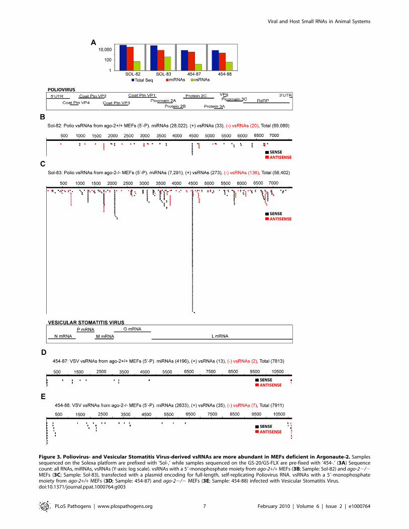
Figure 3. Poliovirus- and Vesicular Stomatitis Virus-derived vsRNAs are more abundant in MEFs deficient in Argonaute-2. Samplessequenced on the Solexa platform are prefixed with ‘Sol-,’ while samples sequenced on the GS-20/GS-FLX are pre-fixed with ‘454-.’ (3A) Sequencecount: all RNAs, miRNAs, vsRNAs (Y-axis: log scale). vsRNAs with a 59-monophosphate moiety from ago-2+/+ MEFs (3B; Sample: Sol-82) and ago-22/2MEFs (3C; Sample: Sol-83), transfected with a plasmid encoding for full-length, self-replicating Poliovirus RNA. vsRNAs with a 59-monophosphatemoiety from ago-2+/+ MEFs (3D; Sample: 454-87) and ago-22/2 MEFs (3E; Sample: 454-88) infected with Vesicular Stomatitis Virus.doi:10.1371/journal.ppat.1000764.g003
Viral and Host Small RNAs in Animal Systems
PLoS Pathogens | www.plospathogens.org 7 February 2010 | Volume 6 | Issue 2 | e1000764

Figure 4. vsRNAs are abundant in infected hosts that do not have a functional Interferon-ab Receptor. (4A) Sequence count: all RNAs,miRNAs, vsRNAs (Y-axis: log scale). vsRNAs from leg muscle of an IFNabR+/+; PVR+/+ (4B; 454-163), or IFNabR2/2; PVR+/+ (4C; Sol-1) mouse infectedwith poliovirus (4 d.p.i; 59-Phosphate-dependent capture). vsRNAs with a 59 monophosphate from the spleen of an IFNabR+/+ (4D; 454-131), orIFNabR2/2 (4E; 454-143) mouse infected with West Nile Virus (3 d.p.i).doi:10.1371/journal.ppat.1000764.g004
Viral and Host Small RNAs in Animal Systems
PLoS Pathogens | www.plospathogens.org 8 February 2010 | Volume 6 | Issue 2 | e1000764
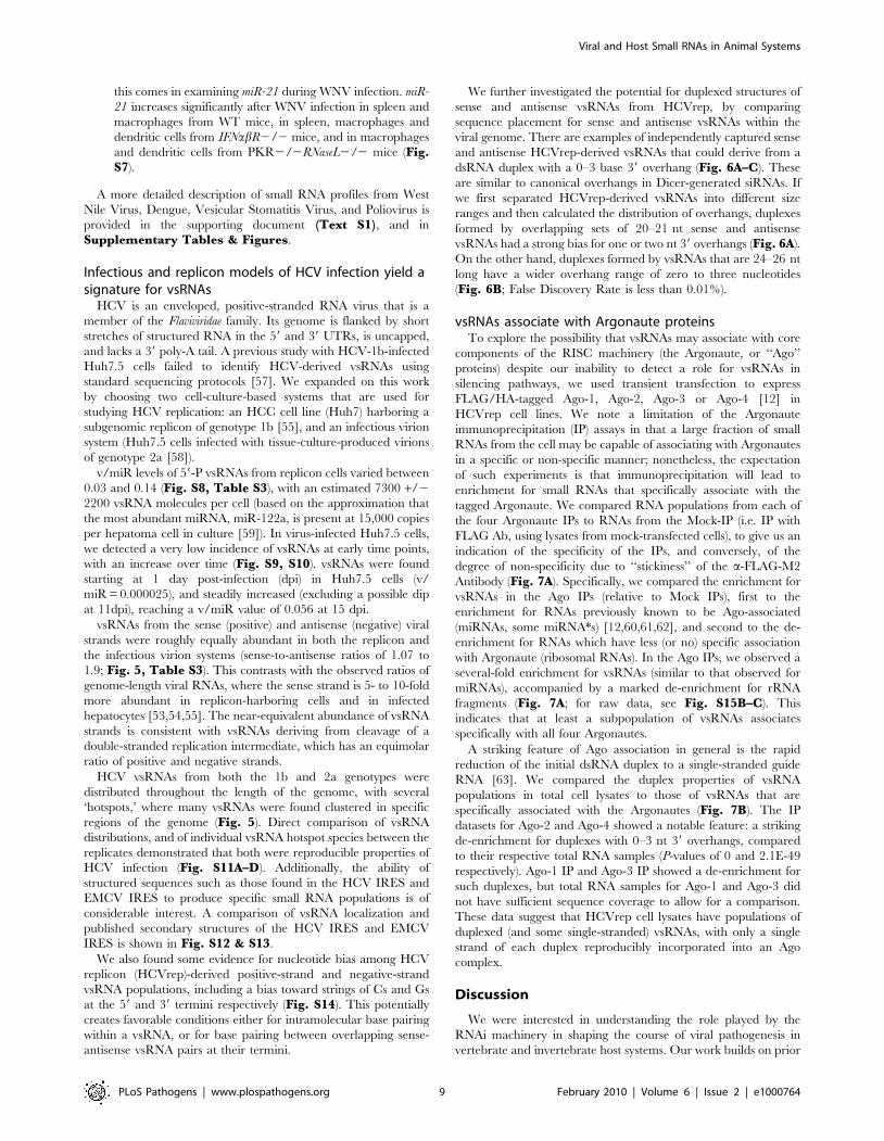
this comes in examining miR-21 during WNV infection. miR-
21 increases significantly after WNV infection in spleen and
macrophages from WT mice, in spleen, macrophages and
dendritic cells from IFNabR2/2 mice, and in macrophages
and dendritic cells from PKR2/2RNaseL2/2 mice (Fig.S7).
A more detailed description of small RNA profiles from West
Nile Virus, Dengue, Vesicular Stomatitis Virus, and Poliovirus is
provided in the supporting document (Text S1), and in
Supplementary Tables & Figures.
Infectious and replicon models of HCV infection yield asignature for vsRNAs
HCV is an enveloped, positive-stranded RNA virus that is a
member of the Flaviviridae family. Its genome is flanked by short
stretches of structured RNA in the 59 and 39 UTRs, is uncapped,
and lacks a 39 poly-A tail. A previous study with HCV-1b-infected
Huh7.5 cells failed to identify HCV-derived vsRNAs using
standard sequencing protocols [57]. We expanded on this work
by choosing two cell-culture-based systems that are used for
studying HCV replication: an HCC cell line (Huh7) harboring a
subgenomic replicon of genotype 1b [55], and an infectious virion
system (Huh7.5 cells infected with tissue-culture-produced virions
of genotype 2a [58]).
v/miR levels of 59-P vsRNAs from replicon cells varied between
0.03 and 0.14 (Fig. S8, Table S3), with an estimated 7300 +/2
2200 vsRNA molecules per cell (based on the approximation that
the most abundant miRNA, miR-122a, is present at 15,000 copies
per hepatoma cell in culture [59]). In virus-infected Huh7.5 cells,
we detected a very low incidence of vsRNAs at early time points,
with an increase over time (Fig. S9, S10). vsRNAs were found
starting at 1 day post-infection (dpi) in Huh7.5 cells (v/
miR = 0.000025), and steadily increased (excluding a possible dip
at 11dpi), reaching a v/miR value of 0.056 at 15 dpi.
vsRNAs from the sense (positive) and antisense (negative) viral
strands were roughly equally abundant in both the replicon and
the infectious virion systems (sense-to-antisense ratios of 1.07 to
1.9; Fig. 5, Table S3). This contrasts with the observed ratios of
genome-length viral RNAs, where the sense strand is 5- to 10-fold
more abundant in replicon-harboring cells and in infected
hepatocytes [53,54,55]. The near-equivalent abundance of vsRNA
strands is consistent with vsRNAs deriving from cleavage of a
double-stranded replication intermediate, which has an equimolar
ratio of positive and negative strands.
HCV vsRNAs from both the 1b and 2a genotypes were
distributed throughout the length of the genome, with several
‘hotspots,’ where many vsRNAs were found clustered in specific
regions of the genome (Fig. 5). Direct comparison of vsRNA
distributions, and of individual vsRNA hotspot species between the
replicates demonstrated that both were reproducible properties of
HCV infection (Fig. S11A–D). Additionally, the ability of
structured sequences such as those found in the HCV IRES and
EMCV IRES to produce specific small RNA populations is of
considerable interest. A comparison of vsRNA localization and
published secondary structures of the HCV IRES and EMCV
IRES is shown in Fig. S12 & S13.
We also found some evidence for nucleotide bias among HCV
replicon (HCVrep)-derived positive-strand and negative-strand
vsRNA populations, including a bias toward strings of Cs and Gs
at the 59 and 39 termini respectively (Fig. S14). This potentially
creates favorable conditions either for intramolecular base pairing
within a vsRNA, or for base pairing between overlapping sense-
antisense vsRNA pairs at their termini.
We further investigated the potential for duplexed structures of
sense and antisense vsRNAs from HCVrep, by comparing
sequence placement for sense and antisense vsRNAs within the
viral genome. There are examples of independently captured sense
and antisense HCVrep-derived vsRNAs that could derive from a
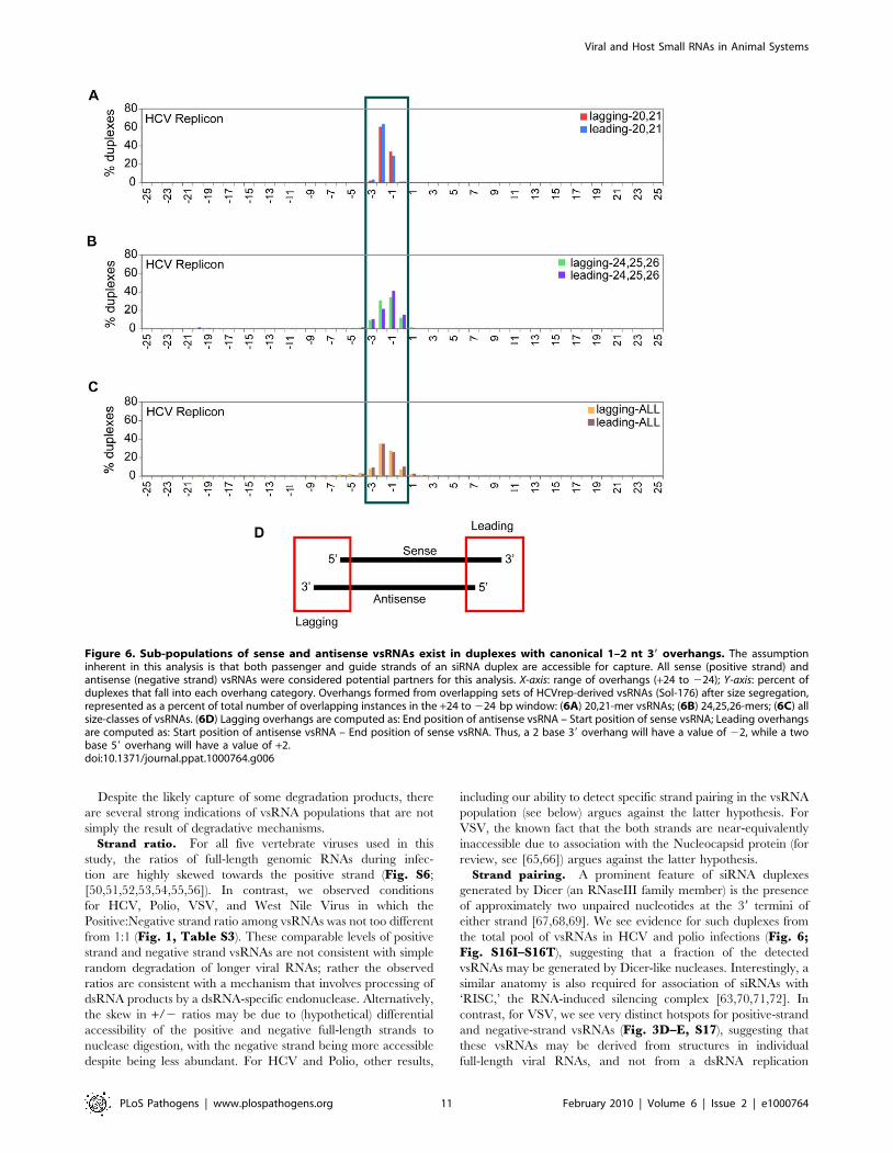
dsRNA duplex with a 0–3 base 39 overhang (Fig. 6A–C). These
are similar to canonical overhangs in Dicer-generated siRNAs. If
we first separated HCVrep-derived vsRNAs into different size
ranges and then calculated the distribution of overhangs, duplexes
formed by overlapping sets of 20–21 nt sense and antisense
vsRNAs had a strong bias for one or two nt 39 overhangs (Fig. 6A).
On the other hand, duplexes formed by vsRNAs that are 24–26 nt
long have a wider overhang range of zero to three nucleotides
(Fig. 6B; False Discovery Rate is less than 0.01%).
vsRNAs associate with Argonaute proteinsTo explore the possibility that vsRNAs may associate with core
components of the RISC machinery (the Argonaute, or ‘‘Ago’’
proteins) despite our inability to detect a role for vsRNAs in
silencing pathways, we used transient transfection to express
FLAG/HA-tagged Ago-1, Ago-2, Ago-3 or Ago-4 [12] in
HCVrep cell lines. We note a limitation of the Argonaute
immunoprecipitation (IP) assays in that a large fraction of small
RNAs from the cell may be capable of associating with Argonautes
in a specific or non-specific manner; nonetheless, the expectation
of such experiments is that immunoprecipitation will lead to
enrichment for small RNAs that specifically associate with the
tagged Argonaute. We compared RNA populations from each of
the four Argonaute IPs to RNAs from the Mock-IP (i.e. IP with
FLAG Ab, using lysates from mock-transfected cells), to give us an
indication of the specificity of the IPs, and conversely, of the
degree of non-specificity due to ‘‘stickiness’’ of the a-FLAG-M2
Antibody (Fig. 7A). Specifically, we compared the enrichment for
vsRNAs in the Ago IPs (relative to Mock IPs), first to the
enrichment for RNAs previously known to be Ago-associated
(miRNAs, some miRNA*s) [12,60,61,62], and second to the de-
enrichment for RNAs which have less (or no) specific association
with Argonaute (ribosomal RNAs). In the Ago IPs, we observed a
several-fold enrichment for vsRNAs (similar to that observed for
miRNAs), accompanied by a marked de-enrichment for rRNA
fragments (Fig. 7A; for raw data, see Fig. S15B–C). This
indicates that at least a subpopulation of vsRNAs associates
specifically with all four Argonautes.
A striking feature of Ago association in general is the rapid
reduction of the initial dsRNA duplex to a single-stranded guide
RNA [63]. We compared the duplex properties of vsRNA
populations in total cell lysates to those of vsRNAs that are
specifically associated with the Argonautes (Fig. 7B). The IP
datasets for Ago-2 and Ago-4 showed a notable feature: a striking
de-enrichment for duplexes with 0–3 nt 39 overhangs, compared
to their respective total RNA samples (P-values of 0 and 2.1E-49
respectively). Ago-1 IP and Ago-3 IP showed a de-enrichment for
such duplexes, but total RNA samples for Ago-1 and Ago-3 did
not have sufficient sequence coverage to allow for a comparison.
These data suggest that HCVrep cell lysates have populations of
duplexed (and some single-stranded) vsRNAs, with only a single
strand of each duplex reproducibly incorporated into an Ago
complex.
Discussion
We were interested in understanding the role played by the
RNAi machinery in shaping the course of viral pathogenesis in
vertebrate and invertebrate host systems. Our work builds on prior
Viral and Host Small RNAs in Animal Systems
PLoS Pathogens | www.plospathogens.org 9 February 2010 | Volume 6 | Issue 2 | e1000764

observations that effective replication by certain RNA viruses in
plants, C. elegans and in D. melanogaster requires suppression of the
antiviral RNAi response [15,16,64]. In each of these invertebrate
systems, there is strong evidence for an antiviral mechanism that is
directed by small RNAs derived from the virus genome
(‘vsRNAs’). Similar questions of great interest in mammals remain
unresolved. Using a sequencing approach to investigate the
involvement of small RNA-based responses in viral infection, we
detected vsRNAs from several mammalian host-viral systems.
vsRNAs have specific characteristics that distinguishthem from RNAs generated by non-specific degradationof viral full-length RNA
We consider two possible sources for the vsRNA populations
that were observed during infection: (i) the vsRNAs could be
participants in a specific pathway (or pathways) in which small
RNAs are generated from the viral genome for host or viral
functions; and (ii) the vsRNAs could be products of non-specific
degradation of longer (e.g. full-length or subgenomic) viral RNAs
mediated by ssRNA nucleases, chemicals, pH, mechanical shear
etc.
We note that the small RNA populations characterized
by sequencing may be a mixture of (i) biologically relevant
small RNAs, and (ii) degradation products with limited
significance. In particular, any population of larger RNAs,
on extraction and experimental manipulation, can yield a
sub-population of RNAs in every size range, including the
miRNA and siRNA size range of 19–30 nt. Since viral
genomic RNAs and mRNAs are abundant in infected cells,
we would certainly expect degraded derivatives to contribute
to sequenced pools.
Figure 5. vsRNAs are detectable in different models of Hepatitis C Virus infection. (5A) Sequence count: all RNAs, miRNAs, vsRNAs (Y-axis: logscale). vsRNAs from: (5B) Huh7 cells with HCV replicon (Sample: Sol-4); (5C) Huh7.5 cells infected with HCV virions, harvested 3 d.p.i (Sample: Sol-92).doi:10.1371/journal.ppat.1000764.g005
Viral and Host Small RNAs in Animal Systems
PLoS Pathogens | www.plospathogens.org 10 February 2010 | Volume 6 | Issue 2 | e1000764
Despite the likely capture of some degradation products, there
are several strong indications of vsRNA populations that are not
simply the result of degradative mechanisms.
Strand ratio. For all five vertebrate viruses used in this
study, the ratios of full-length genomic RNAs during infec-
tion are highly skewed towards the positive strand (Fig. S6;
[50,51,52,53,54,55,56]). In contrast, we observed conditions
for HCV, Polio, VSV, and West Nile Virus in which the
Positive:Negative strand ratio among vsRNAs was not too different
from 1:1 (Fig. 1, Table S3). These comparable levels of positive
strand and negative strand vsRNAs are not consistent with simple
random degradation of longer viral RNAs; rather the observed
ratios are consistent with a mechanism that involves processing of
dsRNA products by a dsRNA-specific endonuclease. Alternatively,
the skew in +/2 ratios may be due to (hypothetical) differential
accessibility of the positive and negative full-length strands to
nuclease digestion, with the negative strand being more accessible
despite being less abundant. For HCV and Polio, other results,
including our ability to detect specific strand pairing in the vsRNA
population (see below) argues against the latter hypothesis. For
VSV, the known fact that the both strands are near-equivalently
inaccessible due to association with the Nucleocapsid protein (for
review, see [65,66]) argues against the latter hypothesis.
Strand pairing. A prominent feature of siRNA duplexes
generated by Dicer (an RNaseIII family member) is the presence
of approximately two unpaired nucleotides at the 39 termini of
either strand [67,68,69]. We see evidence for such duplexes from
the total pool of vsRNAs in HCV and polio infections (Fig. 6;Fig. S16I–S16T), suggesting that a fraction of the detected
vsRNAs may be generated by Dicer-like nucleases. Interestingly, a
similar anatomy is also required for association of siRNAs with
‘RISC,’ the RNA-induced silencing complex [63,70,71,72]. In
contrast, for VSV, we see very distinct hotspots for positive-strand
and negative-strand vsRNAs (Fig. 3D–E, S17), suggesting that
these vsRNAs may be derived from structures in individual
full-length viral RNAs, and not from a dsRNA replication
Figure 6. Sub-populations of sense and antisense vsRNAs exist in duplexes with canonical 1–2 nt 39 overhangs. The assumptioninherent in this analysis is that both passenger and guide strands of an siRNA duplex are accessible for capture. All sense (positive strand) andantisense (negative strand) vsRNAs were considered potential partners for this analysis. X-axis: range of overhangs (+24 to 224); Y-axis: percent ofduplexes that fall into each overhang category. Overhangs formed from overlapping sets of HCVrep-derived vsRNAs (Sol-176) after size segregation,represented as a percent of total number of overlapping instances in the +24 to 224 bp window: (6A) 20,21-mer vsRNAs; (6B) 24,25,26-mers; (6C) allsize-classes of vsRNAs. (6D) Lagging overhangs are computed as: End position of antisense vsRNA – Start position of sense vsRNA; Leading overhangsare computed as: Start position of antisense vsRNA – End position of sense vsRNA. Thus, a 2 base 39 overhang will have a value of 22, while a twobase 59 overhang will have a value of +2.doi:10.1371/journal.ppat.1000764.g006
Viral and Host Small RNAs in Animal Systems
PLoS Pathogens | www.plospathogens.org 11 February 2010 | Volume 6 | Issue 2 | e1000764

Figure 7. Only one strand of the vsRNA duplex is incorporated into Argonaute complexes. (7A) Percent enrichment for various RNAs inAgo-IPs, compared to Mock-IPs, computed as: [(xRNA/totSeq)IP/(xRNA/totSeq)MockIP]; xRNA = vsRNA, miRNA, miRNA*, or rRNA; totSeq = total numberof sequences. The number of vsRNAs varied from 86 to 2472, and the number of total sequences varied from 126,022 to 2,147,467 in these samples.Fractionation of any specific RNA with Argonaute-bound complexes is evidenced in this analysis by retention of representation (compared tomiRNAs) and enrichment (beyond that observed for rRNA-derived segments) in the immunoprecipitated pool. (7B) Comparison between leading andlagging overhangs formed by HCVrep-derived vsRNAs that either associate with an Argonaute (IP), or are present in cell lysates (totalRNA). Alldetected sense (positive strand) and antisense (negative strand) vsRNAs were considered potential partners for this analysis.doi:10.1371/journal.ppat.1000764.g007
Viral and Host Small RNAs in Animal Systems
PLoS Pathogens | www.plospathogens.org 12 February 2010 | Volume 6 | Issue 2 | e1000764
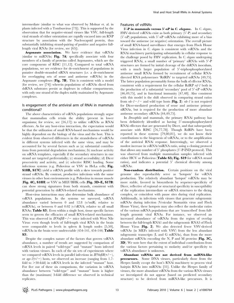
intermediate (similar to what was observed by Molnar et. al. in
plants infected with a Tombusvirus [73]). This is supported by the
observation that for negative-strand viruses like VSV, full-length
viral strands of either orientation are rapidly encased into an RNP
structure by association with the Nucleocapsid protein, thus
substantially inhibiting strand-pairing of positive and negative full-
length viral RNAs (for review, see [65]).
Argonaute association. We have evidence that vsRNAs
(similar to miRNAs; Fig. 7A) specifically associate with four
members of a family of proteins called Argonautes, which are the
core components of RISC [11,12]. Compared to total vsRNA
populations, we see evidence for de-enrichment of populations in
putative double-stranded siRNA structures (i.e. a de-enrichment
for overlapping sets of sense and antisense vsRNAs) in the
Argonaute complexes (Fig. 7B). This is consistent with a model
(for review, see [74]) wherein populations of vsRNAs diced from
dsRNA substrates persist as duplexes in cellular compartments,
with only one strand of the duplex stably maintained by Argonaute
complexes.
Is engagement of the antiviral arm of RNAi in mammalsconditional?
The above characteristics of vsRNA populations strongly argue
that mammalian cells retain the ability (present in lower
organisms; for review, see [16,17]) to utilize vsRNAs as RNAi
effectors. As for any host-virus interaction, the expectation would
be that the utilization of small RNA-based mechanisms would be
highly dependent on the biology of the virus and the host. This is
evident from observed differences in the strandedness of vsRNAs
in different systems infected with the same virus, and may be
accounted for by several factors such as (a) substantial contribu-
tions from potential degradation mechanisms; (b) nuclease activity
on ssRNA templates, especially if the secondary structures in one
strand are targeted preferentially; (c) strand accessibility; (d) Dicer
processivity and activity, and (e) selective RISC loading. Some
infectious systems (e.g. Poliovirus or VSV in HeLa cells; Fig.S18B, S17C) yield a vsRNA profile with a skew towards positive
strand vsRNAs. By contrast, productive infections with the same
viruses in other host environments (e.g. Poliovirus in mouse muscle
(Fig. 4C, S18H) or VSV in MEF/BHK cells (Fig. 3D–E, S17B)
can show strong signatures from both strands, consistent with
potential generation by dsRNA-related mechanisms.
Host-virus interactions may also determine bulk abundance of
vsRNA populations. In the systems we surveyed, vsRNA
abundance varied between 0 and 12.8 (v/miR; relative to
miRNAs), or between 0 and 0.02 (v/sRNA; relative to all small
RNAs; Table S3). Even within a single host, tissue specific factors
seem to govern the efficacies of small RNA-related mechanisms.
This was observed in IFNabR2/2 mice infected with West Nile
Virus: even though levels of full-length viral RNA in the brain
were comparable to levels in spleen & lymph nodes [5,50],
vsRNAs in the brain were undetectable (454-141, 454-144; TableS3).
Despite the complex nature of the factors that govern vsRNA
abundance, a number of trends are suggested by comparison of
vsRNA levels in paired ‘‘wild-type’’ and ‘‘mutant’’ hosts infected
with various viruses. In each of the six sets of experiments where
we compared vsRNA levels in parallel infections in IFNabR(+/2),
or ago-2(+/2) hosts, we observed an increase (ranging from 1.7-
fold to .30-fold) in vsRNA levels in the infected ‘‘mutant’’ host.
For five out of these six comparisons, the variation in vsRNA
abundance between ‘‘wild-type’’ and ‘‘mutant’’ hosts is higher
than the (maximum) 3-fold difference we observed in technical
replicates.
Features of vsRNAs59-P in mammals versus 59-xP in C. elegans. In C. elegans,
FHV-derived vsRNAs exist as both primary (59-P) and secondary
(59-xP) populations, with 59-xP vsRNAs exhibiting more of a bias
toward the antisense (or negative) orientation. The overall picture
of small RNA-based surveillance that emerges from Flock House
Virus infection in C. elegans is consistent with vsRNAs and the
RNAi machinery participating substantially in cellular response to
the challenge posed by FHV replication. In C. elegans undergoing
triggered RNAi, a small number of ‘primary’ siRNAs with 59-P
structures are formed by initial cleavage of the dsRNA inoculum,
with a much larger population of 59-triphosphosphorylated
antisense small RNAs formed by recruitment of cellular RNA-
directed RNA polymerases ‘RdRPs’ to targeted mRNAs [49,75].
The latter population presumably forms the bulk of effector RNAs,
consistent with a requirement for the cellular RdRP rrf-1 both in
the production of a substantial ‘secondary’ pool of 59-xP vsRNAs
[46,49,75], and in functional immunity [47,48]. Also consistent
with this model is the shift observed in comparing strand ratios
from rde-12/2 and wild type hosts (Fig. 2): rde-1 is not required
for Dicer-mediated production of sense and antisense primary
siRNAs, but is required for the production of more abundant
antisense secondary siRNAs [44,49,75].
In Drosophila and mammals, the primary RNAi pathway has
been definitively identified as having 59-monophosphorylated
RNAi effectors that are generated by Dicer, and that subsequently
associate with RISC [76,77,78]. Though RdRPs have been
reported in these systems [79,80,81], we do not know their
contributions to the biogenesis of vsRNAs, or to the amplification
of the antiviral RNAi response. Interestingly, we observed a
modest increase in vsRNA/miRNA ratio, using a cloning protocol
that allows any number of 59 phosphates (59-P-IND protocol). This
was observed from multiple mammalian samples infected with
either HCV or Poliovirus (Table S3; Fig. S19 for vsRNA strand
ratios), and indicates a potential 59 chemical diversity among
vsRNAs.
Non-random distribution. Certain positions on the viral
genome also reproducibly serve as ‘hotspots’ for vsRNA
production. The relatively abundant ‘hotspot’ vsRNAs may be
more stable, favored in biosynthesis by sequence-specificity of
Dicer, reflective of regional or structural specificity in susceptibility
of the replication intermediate or ssRNA structures to the dicing
complex, or coincident with pause sites during viral replication.
Additionally, in infections with viruses that generate subgenomic
mRNAs during infection (Vesicular Stomatitis virus and Flock
House Virus), these hotspots may also reflect the molecular ratios
of the various mRNA populations that are ‘transcribed’ from full-
length genomic viral RNAs. For instance, we observed an
increased abundance of vsRNAs from the region of overlap
between the full-length RNA1 and the subgenomic RNA3 of Flock
House Virus (Fig. 2). We also detected fewer VSV-derived
vsRNAs (in MEFs infected with VSV) from the less abundant
subgenomic transcripts (L and G mRNAs), than from the more
abundant mRNAs encoding the N, P and M proteins (Fig. 3C,3D). We note here that the extent of individual contributions from
the various factors pertaining to molarity and/or specificity to
vsRNA abundance is unknown.
Abundant vsRNAs are not derived from miRNA-like
precursors. Some DNA viruses, particularly those from the
Herpes family co-opt the host’s RNAi machinery to process viral
hairpin RNAs into miRNAs [31]. Unlike the Herpes family of
viruses, the more abundant vsRNAs from the various RNA viruses
we investigated do not appear (based on predicted secondary
structure) to be derived from miRNA-like precursors. It is
Viral and Host Small RNAs in Animal Systems
PLoS Pathogens | www.plospathogens.org 13 February 2010 | Volume 6 | Issue 2 | e1000764
conceivable, however, that some of the rare vsRNAs may be
derived from miRNA-like precursors, or that miRNA precursors
from RNA viruses have non-canonical structures.
Origins and functionalities of vsRNAsAre vsRNAs derived from the virus or the host? While
majority of vsRNAs (99.97%) shared perfect homology only with
the genome of the infecting virus, we detected a small percent of
sRNAs (0.033%) that mapped to both the viral and the host
genomes, confounding the source of their origin, and making
their classification as ‘vsRNAs’ difficult (Table S3, S4). This
observation raises the intriguing possibility of some vsRNAs with
perfect homology to the host genome (or host sRNAs with perfect
homology to the viral genome) participating in potential RNAi-
based cross-regulation between host and virus.
Are vsRNAs abundant enough for biological relevance?
The miRNA population is an aggregate of hundreds of different
biological effector RNAs. A diverse range of concentrations has
been reported for functional miRNAs. If we compare bulk
vsRNA populations to individual miRNAs, vsRNA populations
are more abundant than some functional miRNAs, and less
abundant than others. By extrapolation, low abundance of
vsRNAs does not imply lack of functionality. However, we do
stress that we do not yet know what fraction of the vsRNA pool is
functional.
Can vsRNAs access full-length viral RNA? The action of
Dicer alone (in producing vsRNAs) is not sufficient to halt virus
replication, as has been shown in Drosophila [82,83,84]. For RNAi
to be effective in antiviral defense, it is essential that vsRNAs get
incorporated into RISC, and that the vsRNA-programmed RISCs
find and cleave their targets. From the HCV replicon system, we
have evidence for vsRNA-primed Ago complexes (Fig. 7).
However, we do not know the extent to which vsRNAs
complexed with Ago mediate silencing of full-length replicating/
translating/quiescent viral RNAs, as viruses may use counter-
defenses to protect themselves from being seen by RISC effectors
[29].
Other roles for vsRNAs? vsRNAs have been postulated to
mediate roles other than viral gene silencing. One report suggests
that vsRNAs from Hepatitis Delta Virus ‘HDV’ may be involved
in RNA synthesis [33]. From our studies, vsRNAs from VSV
overlay tightly with Leader RNAs that are thought to play a role in
regulating viral transcription/replication [85,86,87]. We have
identified multiple populations of vsRNAs in several systems, both
of the 59-P, and the 59-xP type (Table S3), and it is plausible that
while some of them may direct the associated Ago complexes to
silence host transcripts in a vsRNA-mediated manner, others
may participate in pathways distinct from silencing, such as
stimulating/regulating the balance of viral transcription and
replication.
Viral modulation of the small RNA machineryViruses can hijack the RNAi machinery at various levels: at
the level of Dicer, Argonaute, RISC-mediated silencing, or a
combination of the above (for review, see [19]). Downregulation of
Dicer has an attenuating effect on Hepatitis C Virus replication
[88]. Several studies have shown that HCV proteins inhibit Dicer
and Argonaute [34,89,90]. In these experiments, inhibition is not
complete (,60–70%; [89]), concordant with our ability to detect
vsRNA populations with structures consistent with synthesis by
Dicer. We also see evidence for loading of vsRNAs onto Ago
complexes, indicating that some downstream steps are not entirely
impaired.
Is there cross talk between other antiviral pathways andRNAi?
Key players in the piRNA pathway, Piwi and Aubergine, are
required for protection against Drosophila X Virus infections [84].
The PIWI-piRNA pathway produces 26–31 nt RNAs in a Dicer-
independent manner, and is mostly active in the germline [91],
and in adjacent somatic tissues [92,93]. We note that animal
systems with PIWI proteins (vertebrates), we observe a wide size
range for vsRNAs (Fig. S2). These diverse size classes, together
with our observation that vsRNAs are still present in systems that
lack Dicer (Poliovirus infections in dcr2/2 MEFs), suggest that the
Dicer pathway (which is thought to primarily produce RNAs
shorter than 27 nt) may not be the only source for these vsRNAs,
and that there may some contribution from baseline levels of PIWI
proteins (or other novel proteins) in the various systems. We also
identified a population of Polio-derived vsRNAs in dcr-12/2 and
in eri-1+/+ MEFs that can form duplexes with 9 or 10 bp
overhangs (Fig. S16I, S16L), which are hallmarks of piRNAs that
are formed by a ping-pong mechanism [94,95]. This observation
brings up the question of how the piRNA pathway interfaces with
the RNAi pathway during viral infections, and whether in the
absence of the RNAi (and perhaps the IFN) machineries, we could
uncover a role for the piRNA pathway in antiviral immunity.
Long dsRNAs, such as those found in the viral replication
intermediate, primarily induce the non-specific interferon response
in mammalian cells. In the absence of the robust IFN pathway,
long dsRNA becomes a trigger for the sequence-directed RNAi
pathway [96,97,98,99]. Accordingly, we detected a more abun-
dant signature for dsRNA-derived vsRNAs in IFNabR2/2 mice
(Poliovirus and West Nile Virus; Table S3). Conversely, when we
stimulated the Interferon pathway by providing an exogenous
supply of IFN-a to a culture of HCVrep-harboring cells, we found
that concurrent with reduction in full-length genomic RNA (as
reported in: [55,100]), HCV-derived vsRNAs also dropped several
fold (Fig. S20). Thus vsRNA abundance seems to associate with
the strength of the IFN response: the more robust the IFN
response, the fewer the number of vsRNAs. The observed boost in
vsRNA abundance in IFN knockout conditions could conceivably
reflect a number of distinct effects including augmented viral
replication in the absence of IFN, and/or specific interactions
between IFN stimulation and the RNAi machineries.
miRNA modulationsWe observed consistent effects of viral infection on miRNA
profiles in distinct experimental systems (Fig. S7). For example:
i. miR-17-5p levels significantly dropped in Polio, VSV,
HCVrep, Dengue and WNV infected systems (with some
exceptions). Other members of the miR-17-92 cluster were
also diminished across cell types.
ii. Abundance of miR-125b decreased in all infected models,
except in brain and leg muscle of Poliovirus-infected
IFNabR2/2 mice.
iii. miR-21 levels increased in infected immune cells in culture,
and in spleens and lymph nodes of infected mice.
Downregulation of the miR-17-92 cluster (i) in virus-infected
cells may be a pro-apoptotic indicator, since an increase in
expression of the miR-17-92 cluster is associated with an inhibition
of apoptosis [101,102,103]. A decrease in miR-125b (ii) is observed
post-LPS-stimulation of macrophages, and causes de-repression of
TNF-a in a sequence-specific manner [104]. We speculate that
miR-125b may be one of the regulators of the TNF-a response
during viral infection, and that regulation of miR-125b may require
Viral and Host Small RNAs in Animal Systems
PLoS Pathogens | www.plospathogens.org 14 February 2010 | Volume 6 | Issue 2 | e1000764
an intact IFN response. Increased levels of the anti-apoptotic miR-
21 (iii) were found in memory and effector T cells, compared to
naıve T cells [105]. It is tempting to speculate that this
upregulation of miR-21 may be indicative of proliferating immune
cells post-recognition of viral antigens.
Therapeutic potential of vsRNAsThe identification of multiple vsRNAs, some of which are
derived from ‘hotspot’ locations in diverse viral genomes may be
useful for designing cocktails of siRNAs for therapeutic purposes,
and for mapping areas of the viral genome that are more
susceptible to RNAi machineries. Much work still remains to be
done in designing siRNA duplexes such that the most accessible
strand of the virus may be successfully targeted by siRNAs. A
major concern is whether RNAi would be effective in combating
viruses with fast replication kinetics. Poliovirus [106] and Semliki
Forest Virus [107] replication in cultured cells have been
effectively attenuated by an exogenous supply of siRNA triggers
against the virus. Whether this holds true for clearing infections in
whole organisms remains to be tested.
Materials and Methods
Ethics statementMice used for experimental infections with West Nile Virus
were genotyped and bred in the animal facilities of the
Washington University School of Medicine, and experiments
were performed with approval from, and according to the
guidelines of, the Washington University Animal Studies Com-
mittee (which is IACUC approved). For infections with Poliovirus,
mice that express the human Poliovirus Receptor gene were
maintained in BSL-2 animal facilities at Stanford University. The
methods for mouse use and care were approved by the Stanford
University Administrative Panel on Laboratory Animal Care
(APLAC), and are in accordance with the USDA Animal Welfare
Act and the Public Health Service Policy on Human Care and Use
of Laboratory Animals.
Host systems and infection conditionsInvertebrates. C. elegans: Worms of various genotypes
(N2, rde-1, rde-4, rrf-1; [43,46,108]) with extra-chromosomal copies
of hs::FHVRNA1DB2 [48] were heat-shocked for 3 hours at 33uCas L4s/young adults and were harvested and frozen in liquid
Nitrogen 24 hours post-heat-shock. RNA1 encodes a functional
FHV RNA replicase (FHV Protein A), and a second protein (FHV
Protein B2) that contributes to viral infectivity by inhibiting the
RNAi pathway (by binding dsRNA in bulk [109,110]). The
replicon RNA1DB2 lacks the ability to encode B2 protein, and can
effectively replicate only in RNAi-deficient genetic backgrounds
[48].
Mammals. Dengue Virus: Dengue virus-2 (DENV-2)
16681 (Accession# M19197.1) was used for all infections. Huh7
(human hepatoma) cells were infected at an MOI = 1, and were
harvested at 0, 2.5, 12, and 24 h.p.i. For infection of U937 cells (a
human monocytic cell line), virus (MOI = 5) was complexed with
the anti-DENV antibody 3H5, before addition to cells. After
2 hours, the medium was replaced, and cells were harvested 2, 15,
and 35 h.p.i. Primary monocyte-derived dendritic cells (MDDCs)
were infected at an MOI of 2 and harvested 4 and 24 h.p.i [111].
Hepatitis C Virus: HCV subgenomic Replicon-harboring cells
(RP7 cells) were established by electroporation of viral RNA from
a modified I377/NS3-39UTR replicon of genotype 1b ([55]; for
sequence, see accession# AJ242652.1) into Huh7 cell lines, and
maintained under G418 selection. For determining the effect of
IFN-a on the production of vsRNAs, RP7 cells were treated with
low (5U/mL) or high (100 U/mL) IFN-a concentrations for
72 hours pre-harvest. Virions for infections of Huh7.5 cells (a
derivative of Huh7) were obtained by transfection of infectious
pFL-J6/JFH1 RNA (Accession# AB047639.1) into Huh7.5 cells
[58]. These virions were subsequently used to infect Huh7.5 cells,
and infected cells were harvested at 1, 3, 5, 6, 9, 11 and 15 d.p.i.
Infected cells were split at the 3, 5, 6, 9 and 11 day time-points,
with one half of the cells propagated for subsequent time-points,
and the other half used for harvesting RNA. Poliovirus: The
Mahoney strain (Accession# NC_002058.3) was used for all
infections. For infection of HeLa cells, an MOI = 5 (harvest points:
2 and 5.5 h.p.i) was used. PVR+/+; IFNabR2/2 and PVR+/+;
IFNabR+/+ mouse embryonic fibroblasts ‘MEFs’ were infected at
an MOI = 1. K562 cells in which persistent infection was
established and confirmed were thawed and passaged a couple
of times before harvest. Infection in PVR2/2 MEFs (ago-2+/+,
ago-22/2, eri-1+/+, eri-12/2, dcr-1+/+**, dcr-12/2**) was
established by co-transfection of cells in 10-cm dishes with 100ug
of DNA plasmid encoding for the full-length Poliovirus 1 genome
under the control of a T7 promoter (pGEM-PV1), and 10 ug of a
plasmid encoding for T7 promoter protein. ago(+/2) MEFs were
harvested 5 hours post-transfection, while the other cell lines were
harvested 6 hours post-transfection. For infections in mice, 106
PFU of virus was injected into one hind leg of 6-week old male
mice (intramuscular inoculation; PVR+/+; IFNabR2/2 and
PVR+/+; IFNabR+/+). At 4 d.p.i, the brain and the inoculated
leg muscle were dissected from both paralyzed and non-paralyzed
individuals. **Wild type and Dicer fl/fl mouse embryonic fibroblasts were
generated and immortalized through infection with a retrovirus that expresses
SV40 large T as previously described [112]. Cells were then re-infected with a
retrovirus that expresses Cre recombinase and confers puromycin resistance.
Cells were treated with puromycin (2 ug/ml) for 5 days, after which clones
were selected by limiting dilution. Vesicular Stomatitis Virus: A
replication-competent, GFP-expressing recombinant virus (VSV-
GFP) derived from the Indiana strain (Accession#: NC_001560.1)
was used for all infections [47]. For infection in MEFs (ago-2+/+and ago-22/2), BHK-21, and HeLa cells, an MOI of 5 PFU/cell
was used, and all cells were harvested 4 h.p.i. West Nile Virus:The NY99-eqhs strain (Accession# AF260967.1) was used for
all infections. Primary cultures of Macrophages and Dendritic
cells were established from WT C57BL/6 mice and congenic
PKR2/2; RNaseL2/2, and IFNabR2/2 mice. MEFs and
cortical neurons were established from WT C57BL/6 mice
[113,114]. These were infected with virus at an MOI = 0.01,
and harvested 4 and 16 h.p.i. For infections in mice (WT B6,
PKR2/2; RNaseL2/2, and IFNabR2/2), 100 PFU of virus was
injected subcutaneously into the footpad, and mice were sacrificed
1 and 3 d.p.i. Spleen, brain and lymph nodes were dissected from
these mice [113,114].
Sample processing and library preparationWorms and mouse tissues were flash frozen in liquid nitrogen,
powdered and lysed. Cell lines were trypsinized and washed in
PBS pre-lysis. The mirVana kit (Ambion) was used for isolation of
RNAs shorter than 200 nt from all samples. Different protocols
were used to prepare libraries of RNAs with mono-phosphory-
lated, or with modified 59 termini. Briefly, in the 59-P-Dep
protocol, RNA was linkered at the 39 terminus, size-selected,
linkered at the 59 terminus, reverse-transcribed, amplified using
ten-nucleotide barcoded PCR primers [115], and sequenced on
the Roche/454 GS-20 or the GS-FLX platforms. In one version
of the 59-P-IND protocol (used for all samples sequenced on the
GS-20/FLX), the RNA was linkered on the 39 end, size-selected
Viral and Host Small RNAs in Animal Systems
PLoS Pathogens | www.plospathogens.org 15 February 2010 | Volume 6 | Issue 2 | e1000764

and reverse-transcribed before addition of the second linker [49].
In the 59-P-IND protocol for preparing Solexa libraries, RNA was
linkered on the 39 end, dephosphorylated and re-phosphorylated
(to replace any multi-phosphate moieties with a monophosphate),
linkered on the 59 end, reverse-transcribed and amplified
(method courtesy of Guoping Gu). All 59-P-Dep and 59-P-
IND libraries for Solexa were prepared by introducing four-
nucleotide barcodes as part of the 59 linker, rather than during
PCR amplification (Lui WO, Parameswaran P; unpub-lished). Shorter barcodes allowed for the use of non-barcoded
primers for amplification, and most importantly, for greater
allocation of sequence space to the sequence of interest. After
preparation of barcoded libraries, the libraries were pooled
together in molar ratios that were proportional to the sequencing
depth required from each sample. The presence of a large number
of libraries required multiple sequencing runs on the Roche/454
and the Illumina sequencers.
Data analysisSequences obtained from the Roche/454 platform were
handled differently from those obtained on the Solexa platform
due to different amplicon structures. Sequences from the Roche/
454 platform were binned based on their barcodes using Barsort
[115], trimmed using perl scripts (PP) and aligned using a local
copy of the multiple alignment program Blast (word size = 11).
Sequences from Illumina’s platform were segregated into
individual datasets based on perfect barcode match, trimmed
to ensure removal of the flanking adapter sequences before
analysis, and aligned using Blat (tile size = 11; step size = 5, run
on Mac OS X). Alignments were performed to databases of
species-specific miRNAs, and of the various viral genomes. The
alignments were subsequently parsed to yield unique hits with the
highest homology for each matching read. We filtered for
matches of .16 nt for Roche/454 sequencing, and of .19 nt for
Solexa sequencing.
For comparing incidence of miRNAs across samples, the
frequency of miRNAs and standard error were computed as per
the following formulae [116]:
a. Relative frequency of miRNAx (p,) = 100 * (Count of
miRNAx +2)/(Total miRNA count +4)
b. Standard Error of p, for a 95% confidence interval
(Std.Error) = Sqrt[((p, * (1002p,))/(Total miRNA count +4))]
c. 95% Confidence Interval = 1.96 * Std.Error
The distribution, length and strandedness of vsRNAs (i.e. RNAs
that mapped to the genome of the infecting virus) were plotted as a
function of the length of the viral genome. Starts and ends of
positive-strand and negative-strand vsRNAs were then compared
to generate a matrix of the percent incidence of different types of
overhangs. For generating the nucleotide bias, the vsRNAs were
aligned at the 59 termini (or at the 39 termini), and the nucleotide
frequencies were counted at each position ten nucleotides
upstream and ten nucleotides from the 59 end of the vsRNA
population (or ten nucleotides downstream and ten nucleotides
from the 39 end of the vsRNA population). These numbers were
then fed into Pictogram (Chris Burge, MIT; http://genes.mit.
edu/pictogram.html), and were normalized to the total numbers of
A, C, G and T in the population of cloned vsRNAs, thus
circumventing any bias that arose from a skewed ratio of
nucleotides inherent to either the cloning process, or to the
genome of the virus.
Calculation of P-values: For establishing statistical signifi-
cance, two-tailed P-values were calculated using either the Z-test
for two proportions, or Fisher’s Exact test. Fisher’s Exact test
was used in test cases with small sample or population sizes,
while the Z-test was used if sample or population sizes were
large.
Calculation of False Discovery Rate (FDR): Populations of
20,21-mers and 24,25,26-mer HCV vsRNAs were pooled, and
positive strand and negative strand vsRNAs were randomly
selected from the pool. 10,000 datasets that mirrored positive
strand and negative strand vsRNA abundances from the original
20,21-mer population, and another 10,000 datasets that
mirrored vsRNA abundances (of both polarities) from the
original 24,25,26-mer population were generated. The percent
of randomly generated datasets wherein 1–2 nt 39overhang
duplexes were as abundant as in the original 20,21-mer
population, or as depleted as in the 24,25,26-mer population
was calculated (for example, FDR of 0.01% indicates that we
were unable to detect percent incidence of duplexes similar to
what was observed, in at least 10,000 randomly-generated
datasets).
Argonaute immunoprecipitations (adapted from [117])Cultures of HCVrep cells were grown to ,80% confluency in
10-cm. plates, and transfected with 20ug each of FLAG/HA-
tagged Ago-1, Ago-2, Ago-3 or Ago-4 (codon-optimized), and
harvested 24 hours post-transfection. Cells were washed twice with
4mL PBS, and 0.75mL of ISOB/NP40 (10mM Tris pH 7.9,
0.15M NaCl, 1.5mM MgCl2, 0.8% NP40, proteinase inhibitor at
1 tablet per 13mL solution) was added to each plate. Cell lysates
were vortexed, incubated on ice for 20 minutes, spun at 4uC for
10 min at 13,000 rpm, and the supernatants were transferred to
new tubes. 10 uL ‘Fake’ (uncoated) Sepharose 4B beads were
washed twice with 1.5ml NET-1 buffer (16TBS-0.2% Tween),
and incubated with supernatants at 4uC for 2 hrs. The
supernatants post-‘fake-bead’ IP were subsequently incubated
with 10uL of washed Anti-Flag M2 beads [Sigma] at 4uC for
4 hrs. Finally, beads were washed 36with 500uL NET-1 buffer,
and RNA was extracted with lysis buffer (mirVana kit, Ambion).
Transfections were performed in duplicate: 1 plate was used as
input for IPs, while the other was used for cloning of total small
RNA populations. For Mock-IPs, lysates from mock-transfected
cells were used as described above.
Northern analysisThe loading control was a 5.7 kb region of the pGEM plasmid
carrying the Poliovirus 1 genome (pGEM-PV1), obtained by
digestion of the plasmid with HindIII and AgeI. Equivalent
amounts of RNA (as measured using a NanoDrop) from each
sample were run on a 1% agarose-formaldehyde gel, and
transferred under basic conditions to a Hybond+ membrane.
The loading control was visualized by staining the blot with
methylene blue. 60-mer DNA probes to specifically detect either
the positive strand (AF-PP-350: GTCACCGCTTGTAGAATT-
GTCATTGCCCTGTTGATGTTCCTTTCTGTTTGAACCT-
GGCTG & AF-PP-351: TCATCTATGGTTTGCCGATACGT-
GGTGTTGCTAATCCATGGCACT ACCATAGTACATGAG),
or the negative strand (AF-PP-84: TTCACGGGTACGTTC AC-
TCCTGACAACAACCAGACATCACCTGCCCGCAGGTTC-
TGCCCG, AF-PP-86: ATTC GGACACCAAAACAAAGCG-
GTGTACACTGCAGGTTACAAAATTTGCAACTACCACTT)
were end-labeled with 32P, and were successively used on the same
blot (the blot was stripped in between the two hybridizations). The
blots were hybridized and washed at 50uC, and exposed using a
phosphorimager screen.
Viral and Host Small RNAs in Animal Systems
PLoS Pathogens | www.plospathogens.org 16 February 2010 | Volume 6 | Issue 2 | e1000764

Supporting Information
Text S1 Supporting text containing supplementary results,
methods and references
Found at: doi:10.1371/journal.ppat.1000764.s001 (0.18 MB PDF)
Figure S1 Host-encoded miRNAs with partial homology to
HCV (summarized from published data). Human miRNAs that
demonstrate a sequence-directed effect on HCV RNA levels are
indicated [19,20,21]. These miRNAs are encoded in the host
genome, and are hence distinct from HCVrep-derived vsRNAs
that are viral-encoded.
Found at: doi:10.1371/journal.ppat.1000764.s002 (0.19 MB PDF)
Figure S2 Frequency-Length profiles for vsRNAs. X-axis:
vsRNA lengths; Y-axis: number of vsRNA instances.
Found at: doi:10.1371/journal.ppat.1000764.s003 (2.09 MB PDF)
Figure S3 Rare Dengue-derived vsRNAs are detectable in
certain host backgrounds. (S3A) Sequence count: all RNAs,
miRNAs, vsRNAs (Y-axis: log scale). (S3B) vsRNAs with a 59
monophosphate moiety from Huh7 cells infected with DENV-2,
25 h.p.i. (Sample: 454-75).
Found at: doi:10.1371/journal.ppat.1000764.s004 (0.23 MB PDF)
Figure S4 RNase-protection confirms that VSV-specific
vsRNAs are more abundant in ago22/2 MEFs. RNA was
isolated from mock-infected and VSV-infected ago2+/+ or
ago22/2 MEFs at 4 h.p.i. vsRNAs derived from positive strand
viral RNAs were detected by RNase protection using a
radiolabeled probe specific to the 59 end of the VSV-N gene.
Found at: doi:10.1371/journal.ppat.1000764.s005 (0.69 MB PDF)
Figure S5 (Profiles from GS-20/FLX-sequenced libraries)
Poliovirus vsRNAs are more abundant in MEFs deficient in
Argonaute-2. (S5A) Sequence count: all RNAs, miRNAs, vsRNAs
(Y-axis: log scale). vsRNAs with a 59-monophosphate moiety from
(S5B) ago-2+/+ MEFs (Sample: 454-193) and (S5C) ago-22/2
MEFs (Sample: 454-194) transfected with a plasmid encoding for
self-replicating full-length Poliovirus RNA.
Found at: doi:10.1371/journal.ppat.1000764.s006 (0.29 MB PDF)
Figure S6 Abundance of the two polarities of Poliovirus full-
length RNAs varies across systems, with full-length positive strand
generally present in excess over full-length negative strand.
Visualization of full-length Poliovirus in various samples by
Northern analysis: (S6A) Positive strand; (S6B) Negative strand.
Normalization control: fragment of plasmid with homology to
Poliovirus (see Materials and Methods).
Found at: doi:10.1371/journal.ppat.1000764.s007 (0.59 MB PDF)
Figure S7 Only a handful of miRNAs are modulated during
viral infection. (7A) Log2-transformed ratios of specific miRNA
abundances between infected and uninfected samples (or between
early and late time-points) were represented as a heat map. Only
those miRNAs that exhibited a . = 2-fold, and a statistically
significant upregulation or downregulation (i.e. no overlap of 95%
confidence intervals) were considered. Fold changes were log2-
transformed. No row-wise or column-wise normalizations were
performed. The yellow squares represent miRNAs that are
upregulated in infected cells/late time-points compared to
uninfected/early time-points. The blue squares represent miRNAs
that are downregulated in infected cells/late time-points compared
to uninfected/early time-points. Higher intensities correlate with a
greater fold change. Note: The baseline frequency for each miRNA
was set to 0.0001 (rather than 0), to enable computation of ratios.
(7B) Clustering of miRNA profiles across various infected and
uninfected mouse tissues/cell lines reveals that majority of samples
cluster according to tissue type/cell line origin. miRNA frequen-
cies were first normalized against the total number of miRNAs
for each sample. Cube-root values of the frequencies were
subsequently used in uncentered clustering of the data set,
both row-wise and column-wise. These plots were generated
using GenePattern [http://www.broad.mit.edu/cancer/software/
genepattern/].
Found at: doi:10.1371/journal.ppat.1000764.s008 (7.91 MB TIF)
Figure S8 Abundance and distribution of HCVrep-derived 59-P
vsRNAs varies between early and late passage cells, but 59-xP
vsRNAs are enriched (relative to 59-P) in both early and late
passages. (S8A) Sequence count: all RNAs, miRNAs, vsRNAs (Y-
axis: log scale). vsRNAs from a late passage (later than passage 15)
of cells: (S8B) captured using the 59-P-dependent protocol (Sol-
107); (S8C) captured using the 59-P-INDependent protocol (Sol-
109). vsRNAs from an early passage (passage 3) of cells: (S8D)
captured using the 59-P-dependent protocol (Sol-176); (S8E)
captured using the 59-P-INDependent protocol (Sol-179).
Found at: doi:10.1371/journal.ppat.1000764.s009 (0.34 MB PDF)
Figure S9 Increase in the abundance of HCVvir-derived
vsRNAs (relative to miRNAs) at later time-points in infection
(Solexa-sequenced libraries). Positive strand vsRNAs are shown as
blue bars, and negative strand vsRNAs as red bars. (S9A)
Sequence count: all RNAs, miRNAs, vsRNAs (Y-axis: log scale).
vsRNAs with 59 monophosphates from Huh7.5 cells infected with
HCV virions: (S9B) 1 d.p.i (Sample: Sol-91); (S9C) 3 d.p.i (Sample
Sol-92); (S9D) 6 d.p.i (Sample Sol-93); (S9E) 9 d.p.i (Sample Sol-
94); (S9F) 11 d.p.i (Sample Sol-95); (S9G) 15 d.p.i (Sample Sol-96).
Found at: doi:10.1371/journal.ppat.1000764.s010 (0.37 MB PDF)
Figure S10 Temporal changes in the abundance of HCVvir-
derived vsRNAs (GS-20/FLX-sequenced libraries). (S10A) Se-
quence count: all RNAs, miRNAs, vsRNAs (Y-axis: log scale).
vsRNAs with 59 monophosphates from Huh7.5 cells infected with
HCV virions: (S10B) 3 d.p.i (Sample: 454-84); (S10C) 5 d.p.i
(Sample: 454-85); (S10D) 11 d.p.i (Sample 454-86).
Found at: doi:10.1371/journal.ppat.1000764.s011 (0.27 MB PDF)
Figure S11 vsRNA coincidence plots show high degree of
similarity between independent amplicon libraries from the same
starting sample, and reproducible detection of hotspots. (S11A)
Coincidences are defined by vsRNAs of the same orientation, with
the same Start and End positions. Percent coincidence between
‘Sample-1’ and ‘Sample-2’ was calculated as follows: (# of
coincidences between Sample-1 and Sample-2)/[(# of vsRNAs
in Sample-1) + (# of vsRNAs in Sample-2)] * 100. These values
are plotted as a heat map, with dark-to-light blue representing low-
to-high values. (S11B-D) Pairwise comparison of the relative
abundance of individual vsRNAs between independent amplicon
libraries of early-passage HCV replicon cells. Each point
represents a unique vsRNA, and the X- and Y-axes represent
relative abundance (with respect to all vsRNAs) in the indicated
amplicon libraries. (S11E) Sequence count: all RNAs, miRNAs,
vsRNAs (Y-axis: log scale). vsRNAs with 59 monophosphates from
HCVrep cells: (S11F) Sample: Sol-176; (S11G) Sample Sol-201;
(S11H) Sample Sol-203; (S11I) Sample Sol-205. Positive strand
vsRNAs are shown as blue bars, and negative strand vsRNAs as
red bars.
Found at: doi:10.1371/journal.ppat.1000764.s012 (0.70 MB PDF)
Figure S12 A map of vsRNA start and end positions,
superimposed on the secondary structure of the HCV IRES (see
[19] for source of IRES structure). Only positions that had an
incidence of . = 2 Starts/Ends were mapped.
Found at: doi:10.1371/journal.ppat.1000764.s013 (0.32 MB PDF)
Viral and Host Small RNAs in Animal Systems
PLoS Pathogens | www.plospathogens.org 17 February 2010 | Volume 6 | Issue 2 | e1000764

Figure S13 A map of vsRNA start and end positions,
superimposed on the secondary structure of the EMCV IRES
(which is part of the HCV Replicon genome; see [20] for source of
IRES structure). Only positions that had an incidence of . = 2
starts/ends were mapped.
Found at: doi:10.1371/journal.ppat.1000764.s014 (0.42 MB PDF)
Figure S14 vsRNAs from cells with the HCV Replicon (Sol-4)
exhibit a bias for Cytidines at their 59 termini, and a bias for
Guanines at their 39 termini. vsRNAs were first aligned at their 59
ends (or at their 39 ends), and the nucleotide frequencies for 10
upstream (or 10 downstream) positions, plus for the first 10 (or last
10) vsRNA base positions were determined separately for positive
strand and negative strand vsRNAs. This analysis was performed
on all vsRNA instances, or post-collapse of the vsRNA dataset
(‘unique instances’), such that multiple instances of vsRNAs were
represented only once. Arrows indicate Start/End positions of
vsRNAs. Nucleotide frequencies have been normalized to the
overall nucleotide composition within each dataset. (S14A–S14D)
Pictogram of 10 bases upstream+10 bases (from 59 termini) of
vsRNAs derived from (S14A) Positive strand: unique instances;
(S14B) Negative strand: unique instances; (S14C) Positive strand:
all instances; (S14D) Negative strand: all instances. (S14E–S14H)
Pictogram of 10 bases downstream+10 bases (from 39 termini) of
vsRNAs derived from (S14E) Positive strand: unique instances;
(S14F) Negative strand: unique instances; (S14G) Positive strand:
all instances; (S14H) Negative strand: all instances.
Found at: doi:10.1371/journal.ppat.1000764.s015 (0.67 MB PDF)
Figure S15 Mock-IP enriches for rRNAs. (S15A) [(xRNA/
totSeq)MockIP/(xRNA/totSeq)totalRNA] was computed for the
various IPs; xRNA: vsRNA, miRNA, miRNA*, or rRNA;
totSeq = total number of sequences from each experiment. The
number of vsRNAs varied from 86 to 1,857, and the number of
total sequences varied from 126,022 to 891,858 in these samples.
(S15B–C) Raw data: [(xRNA/totSeq)] for the various IPs
(including the mock IP), and for the totalRNA populations;
xRNA: vsRNA, miRNA, miRNA*, or rRNA; totSeq: total
number of sequences from each experiment.
Found at: doi:10.1371/journal.ppat.1000764.s016 (0.25 MB PDF)
Figure S16 Start-to-Start, Start-to-End, and duplex overhang
plots for positive strand and negative strand vsRNAs show
differences between Poliovirus-infected hosts with wild-type or
mutant copies of dcr-1 and eri-1. Y-axis: percent of vsRNA pairs
with specified Start-to-Start or Start-to-End distances on a scale of
+120 to 2120. (S16A, S16B, S16E, S16F) Start-to-End distances
for positive strand and negative strand vsRNAs cloned from dcr-
1+/+, dcr-12/2, eri-1+/+, eri-12/2 MEFs; (S16C, S16D, S16G,
S16H) Start-to-Start distances for positive strand and nega-
tive strand vsRNAs cloned from dcr-1+/+, dcr-12/2, eri-1+/+,
eri-12/2 MEFs. Predicted overhangs formed by overlapping
sets of sense and antisense 20 and 21 nt vsRNAs sequenced
from: (S16I) eri-1+/+ MEFs +Poliovirus; (S16J) eri-12/2 MEFs
+Poliovirus; (S16K) dcr-1+/+ MEFs +Poliovirus; (S16L) dcr-12/2
MEFs +Poliovirus. (S16M-P) Overhangs formed by 24,25,26-mers
from systems listed above. (S16Q-T) Overhangs formed by all size-
classes of vsRNAs from systems listed above. All captured vsRNAs
of either polarity were considered to be potential partners for this
analysis.
Found at: doi:10.1371/journal.ppat.1000764.s017 (0.50 MB PDF)
Figure S17 Abundance, distribution and orientation biases of
VSV-derived vsRNAs are a function of cell-type. (S17A) Sequence
count: all RNAs, miRNAs, vsRNAs (Y-axis: log scale). vsRNAs
with 59 monophosphates from Vesicular Stomatitis virus infections
in: (S17B) BHK-21 cells, harvested 4 h.p.i (Sample: 454-179);
(S17C) HeLa cells, harvested 4 h.p.i (Sample: 454-181).
Found at: doi:10.1371/journal.ppat.1000764.s018 (0.28 MB PDF)
Figure S18 vsRNAs are present in both lytic and persistent
models of Poliovirus infection, and can be captured using both the
59-P-dependent, and the 59-P-INDependent cloning protocols.
(S18A) Sequence count: all RNAs, miRNAs, vsRNAs (Y-axis: log
scale). vsRNAs with a 59 monophosphate from: (S18B) HeLa cells,
that are prone to lysis upon infection with the Mahoney strain of
poliovirus (MOI = 5, 5.5 h.p.i, Sample: Sol-1); (S18C) K562 cells,
in which Poliovirus establishes a persistent infection (Sample: 454-
49); (S18D) brain of 6wk-old paralyzed Poliovirus-infected
IFNabR2/2; PVR+/+ male mouse, 4 d.p.i. (Sol-81). vsRNAs
captured using the 59-Phosphate-INDependent cloning protocol,
from: (S18E) K562 cells infected with Poliovirus (Sample: 454-50);
(S18F) ago-2+/+ MEFs +Poliovirus (Sample: Sol-79)*; (S18G)
ago-22/2 MEFs +Poliovirus (Sample: Sol-80)*; leg muscle
(S18H) and brain (S18I) of a 6wk-old male mouse (genotype:
IFNabR2/2; PVR+/+) infected with Poliovirus, 4 d.p.i. *These
MEFs were transfected with a plasmid encoding for self-replicating
Poliovirus RNA, and harvested 5 hours post-transfection.
Found at: doi:10.1371/journal.ppat.1000764.s019 (0.35 MB PDF)
Figure S19 The 59-P-INDependent cloning protocol subtly
enriches for antisense vsRNAs. Comparison of antisense vsRNAs
(as a % of all vsRNAs; S19A) and of sense vsRNAs (as a % of all
vsRNAs; S19B), between the 59-P-dependent and the 59-P-
INDependent cloning protocols.
Found at: doi:10.1371/journal.ppat.1000764.s020 (0.23 MB PDF)
Figure S20 IFN responsiveness reduces the incidence of
HCVrep-derived vsRNAs. (S20A) Sequence count: all RNAs,
miRNAs, vsRNAs (Y-axis: log scale). vsRNA profiles from: (S20B)
untreated HCVrep cells; (S20C) HCVrep cells treated with 5 U/
mL IFN, and harvested 72 hours post-treatment; (S20D) HCVrep
cells treated with 100 U/mL IFN, and harvested 72 hours post-
treatment.
Found at: doi:10.1371/journal.ppat.1000764.s021 (0.27 MB PDF)
Table S1 Reference genomes of all viruses used.
Found at: doi:10.1371/journal.ppat.1000764.s022 (0.04 MB PDF)
Table S2 Most abundant vsRNAs. The five most abundant
vsRNAs captured in infections with FHV, VSV, Polio, HCVrep
and HCVvir are listed. Start position, length, orientation and
sequence of these vsRNAs are indicated, as are the number of
datasets in which they were identified, and their respective total
and normalized counts. vsRNAs were first defined by start
position, length and orientation. All defined vsRNAs were then
ranked based on (a) number of samples in which they were
identified, and (b) their total count across all samples, normalized
to the total vsRNA count (or to the total miRNA count) for those
samples. For West Nile and Dengue Viruses, because of poor
coverage, only four and two vsRNAs (respectively) are shown.
Found at: doi:10.1371/journal.ppat.1000764.s023 (0.07 MB PDF)
Table S3 Comprehensive list of virus-host systems that were
surveyed, with sample descriptions, bulk frequencies and ratios of
various classes of small RNAs, and frequencies of ‘rogue’ viral and
host genomic matches to vsRNAs. Samples are ordered first by
sequencing platform, second by type of virus used in the infection,
third by genotype, fourth by cell/tissue type, and fifth by time-
point.
Found at: doi:10.1371/journal.ppat.1000764.s024 (0.41 MB PDF)
Table S4 List of vsRNAs that map perfectly to both host and viral
genomes. Due to the complete nature of sRNA homology to both
Viral and Host Small RNAs in Animal Systems
PLoS Pathogens | www.plospathogens.org 18 February 2010 | Volume 6 | Issue 2 | e1000764

viral and host genomes, the origin of the listed vsRNAs cannot be
determined. These numbers are also indicated in Table S3.
Found at: doi:10.1371/journal.ppat.1000764.s025 (0.06 MB PDF)
Acknowledgments
We thank Chaya Krishna, Guoping Gu, Ayelet Lamm, Li Tao, Weng-
Onn Lui, Julia Pak, Shahnewaz Siddique, Thaisan Tonthat, Michael
Stadler, Scott Boyd, Jay Maniar, Jonathan Gent, Daniel Blanchard, Steve
Johnson, Rosa Alcazar, Cecilia Mello, Kyle Sha, Frederick Tan, Lia
Gracey, Ellie Marshall, Sofia Gkountela, Menashe Elazar, Robin Trujillo,
and Chang-Zheng Chen for technical assistance, reagents, and for helpful
suggestions. We also thank Arend Sidow, Ziming Weng, Cheryl Smith,
Anton Valouev and Phil Lacroute for their contributions in making the
high throughput sequencing possible.
Author Contributions
Conceived and designed the experiments: PP AZF. Performed the
experiments: PP. Analyzed the data: PP AZF. Contributed reagents/
materials/analysis tools: PP ES CW TB MAS RL KMA VH SE WJ TD
SP CP FBdS RJ FB BG DG MK SK PS MR SWD EH MC MSD KK
JSG AZF. Wrote the paper: PP AZF.
References
1. Severa M, Fitzgerald KA (2007) TLR-mediated activation of type I IFN during
antiviral immune responses: fighting the battle to win the war. Curr Top
Microbiol Immunol 316: 167–192.
2. Takeuchi O, Akira S (2008) MDA5/RIG-I and virus recognition. Curr Opin
Immunol 20: 17–22.
3. Sadler AJ, Williams BR (2008) Interferon-inducible antiviral effectors. Nat Rev
Immunol 8: 559–568.
4. O’Connell RM, Taganov KD, Boldin MP, Cheng G, Baltimore D (2007)
MicroRNA-155 is induced during the macrophage inflammatory response.
Proc Natl Acad Sci U S A 104: 1604–1609.
5. Ida-Hosonuma M, Iwasaki T, Yoshikawa T, Nagata N, Sato Y, et al. (2005)
The alpha/beta interferon response controls tissue tropism and pathogenicity
of poliovirus. J Virol 79: 4460–4469.
6. Carthew RW (2001) Gene silencing by double-stranded RNA. Curr Opin Cell
Biol 13: 244–248.
7. Bernstein E, Caudy AA, Hammond SM, Hannon GJ (2001) Role for a
bidentate ribonuclease in the initiation step of RNA interference. Nature 409:
363–366.
8. Hammond SM (2005) Dicing and slicing: the core machinery of the RNA
interference pathway. FEBS Lett 579: 5822–5829.
9. Joshua-Tor L (2006) The Argonautes. Cold Spring Harb Symp Quant Biol 71:
67–72.
10. Collins RE, Cheng X (2005) Structural domains in RNAi. FEBS Lett 579:
5841–5849.
11. Liu J, Carmell MA, Rivas FV, Marsden CG, Thomson JM, et al. (2004)
Argonaute2 is the catalytic engine of mammalian RNAi. Science 305:
1437–1441.
12. Meister G, Landthaler M, Patkaniowska A, Dorsett Y, Teng G, et al. (2004)
Human Argonaute2 mediates RNA cleavage targeted by miRNAs and siRNAs.
Mol Cell 15: 185–197.
13. Rivas FV, Tolia NH, Song JJ, Aragon JP, Liu J, et al. (2005) Purified
Argonaute2 and an siRNA form recombinant human RISC. Nat Struct Mol
Biol 12: 340–349.
14. Song JJ, Smith SK, Hannon GJ, Joshua-Tor L (2004) Crystal structure of
Argonaute and its implications for RISC slicer activity. Science 305:
1434–1437.
15. Vance V, Vaucheret H (2001) RNA silencing in plants–defense and counter-
defense. Science 292: 2277–2280.
16. Ding SW, Voinnet O (2007) Antiviral immunity directed by small RNAs. Cell
130: 413–426.
17. Aliyari R, Ding SW (2009) RNA-based viral immunity initiated by the Dicer
family of host immune receptors. Immunol Rev 227: 176–188.
18. Burgyan J (2008) Role of silencing suppressor proteins. Methods Mol Biol 451:
69–79.
19. de Vries W, Berkhout B (2008) RNAi suppressors encoded by pathogenic
human viruses. Int J Biochem Cell Biol 40: 2007–2012.
20. Svoboda P (2007) Off-targeting and other non-specific effects of RNAi
experiments in mammalian cells. Curr Opin Mol Ther 9: 248–257.
21. Schlee M, Hornung V, Hartmann G (2006) siRNA and isRNA: two edges of
one sword. Mol Ther 14: 463–470.
22. O’Malley RP, Mariano TM, Siekierka J, Mathews MB (1986) A mechanism
for the control of protein synthesis by adenovirus VA RNAI. Cell 44: 391–
400.
23. Chang HW, Watson JC, Jacobs BL (1992) The E3L gene of vaccinia virus
encodes an inhibitor of the interferon-induced, double-stranded RNA-
dependent protein kinase. Proc Natl Acad Sci U S A 89: 4825–4829.
24. Langland JO, Pettiford S, Jiang B, Jacobs BL (1994) Products of the porcine
group C rotavirus NSP3 gene bind specifically to double-stranded RNA and
inhibit activation of the interferon-induced protein kinase PKR. J Virol 68:
3821–3829.
25. Mibayashi M, Martinez-Sobrido L, Loo YM, Cardenas WB, Gale M Jr, et al.
(2007) Inhibition of retinoic acid-inducible gene I-mediated induction of beta
interferon by the NS1 protein of influenza A virus. J Virol 81: 514–524.
26. Hale BG, Randall RE, Ortin J, Jackson D (2008) The multifunctional NS1
protein of influenza A viruses. J Gen Virol 89: 2359–2376.
27. Loo YM, Owen DM, Li K, Erickson AK, Johnson CL, et al. (2006) Viral and
therapeutic control of IFN-beta promoter stimulator 1 during hepatitis C virus
infection. Proc Natl Acad Sci U S A 103: 6001–6006.
28. Li WX, Li H, Lu R, Li F, Dus M, et al. (2004) Interferon antagonist proteins of
influenza and vaccinia viruses are suppressors of RNA silencing. Proc Natl
Acad Sci U S A 101: 1350–1355.
29. Itaya A, Zhong X, Bundschuh R, Qi Y, Wang Y, et al. (2007) A structured
viroid RNA serves as a substrate for dicer-like cleavage to produce biologically
active small RNAs but is resistant to RNA-induced silencing complex-mediated
degradation. J Virol 81: 2980–2994.
30. Gottwein E, Cullen BR (2008) Viral and cellular microRNAs as determinants
of viral pathogenesis and immunity. Cell Host Microbe 3: 375–387.
31. Umbach JL, Cullen BR (2009) The role of RNAi and microRNAs in animal
virus replication and antiviral immunity. Genes Dev 23: 1151–1164.
32. Matskevich AA, Moelling K (2007) Dicer is involved in protection against
influenza A virus infection. J Gen Virol 88: 2627–2635.
33. Haussecker D, Cao D, Huang Y, Parameswaran P, Fire AZ, et al. (2008)
Capped small RNAs and MOV10 in human hepatitis delta virus replication.
Nat Struct Mol Biol 15: 714–721.
34. Wang Y, Kato N, Jazag A, Dharel N, Otsuka M, et al. (2006) Hepatitis C virus
core protein is a potent inhibitor of RNA silencing-based antiviral response.
Gastroenterology 130: 883–892.
35. Kent WJ (2002) BLAT–the BLAST-like alignment tool. Genome Res 12:
656–664.
36. Altschul SF, Gish W, Miller W, Myers EW, Lipman DJ (1990) Basic local
alignment search tool. J Mol Biol 215: 403–410.
37. Pedersen IM, Cheng G, Wieland S, Volinia S, Croce CM, et al. (2007)
Interferon modulation of cellular microRNAs as an antiviral mechanism.
Nature 449: 919–922.
38. Jopling CL, Yi M, Lancaster AM, Lemon SM, Sarnow P (2005) Modulation of
hepatitis C virus RNA abundance by a liver-specific MicroRNA. Science 309:
1577–1581.
39. Murakami Y, Aly HH, Tajima A, Inoue I, Shimotohno K (2009) Regulation of
the hepatitis C virus genome replication by miR-199a. J Hepatol 50: 453–460.
40. Griffiths-Jones S, Saini HK, van Dongen S, Enright AJ (2008) miRBase: tools
for microRNA genomics. Nucleic Acids Res 36: D154–158.
41. Griffiths-Jones S, Grocock RJ, van Dongen S, Bateman A, Enright AJ (2006)
miRBase: microRNA sequences, targets and gene nomenclature. Nucleic Acids
Res 34: D140–144.
42. Griffiths-Jones S (2004) The microRNA Registry. Nucleic Acids Res 32:
D109–111.
43. Tabara H, Sarkissian M, Kelly WG, Fleenor J, Grishok A, et al. (1999) The
rde-1 gene, RNA interference, and transposon silencing in C. elegans. Cell 99:
123–132.
44. Parrish S, Fire A (2001) Distinct roles for RDE-1 and RDE-4 during RNA
interference in Caenorhabditis elegans. Rna 7: 1397–1402.
45. Tabara H, Yigit E, Siomi H, Mello CC (2002) The dsRNA binding protein
RDE-4 interacts with RDE-1, DCR-1, and a DExH-box helicase to direct
RNAi in C. elegans. Cell 109: 861–871.
46. Sijen T, Fleenor J, Simmer F, Thijssen KL, Parrish S, et al. (2001) On the role
of RNA amplification in dsRNA-triggered gene silencing. Cell 107: 465–476.
47. Wilkins C, Dishongh R, Moore SC, Whitt MA, Chow M, et al. (2005) RNA
interference is an antiviral defence mechanism in Caenorhabditis elegans.
Nature 436: 1044–1047.
48. Lu R, Maduro M, Li F, Li HW, Broitman-Maduro G, et al. (2005) Animal
virus replication and RNAi-mediated antiviral silencing in Caenorhabditis
elegans. Nature 436: 1040–1043.
49. Pak J, Fire A (2007) Distinct populations of primary and secondary effectors
during RNAi in C. elegans. Science 315: 241–244.
50. Samuel MA, Diamond MS (2005) Alpha/beta interferon protects against lethal
West Nile virus infection by restricting cellular tropism and enhancing neuronal
survival. J Virol 79: 13350–13361.
51. Cleaves GR, Ryan TE, Schlesinger RW (1981) Identification and character-
ization of type 2 dengue virus replicative intermediate and replicative form
RNAs. Virology 111: 73–83.
Viral and Host Small RNAs in Animal Systems
PLoS Pathogens | www.plospathogens.org 19 February 2010 | Volume 6 | Issue 2 | e1000764

52. Novak JE, Kirkegaard K (1991) Improved method for detecting poliovirus
negative strands used to demonstrate specificity of positive-strand encapsidation
and the ratio of positive to negative strands in infected cells. J Virol 65:
3384–3387.
53. Lohmann V, Korner F, Koch J, Herian U, Theilmann L, et al. (1999)
Replication of subgenomic hepatitis C virus RNAs in a hepatoma cell line.
Science 285: 110–113.
54. Lanford RE, Chavez D, Chisari FV, Sureau C (1995) Lack of detection of
negative-strand hepatitis C virus RNA in peripheral blood mononuclear cells
and other extrahepatic tissues by the highly strand-specific rTth reverse
transcriptase PCR. J Virol 69: 8079–8083.
55. Blight KJ, Kolykhalov AA, Rice CM (2000) Efficient initiation of HCV RNA
replication in cell culture. Science 290: 1972–1974.
56. Conzelmann KK (1998) Nonsegmented negative-strand RNA viruses: genetics
and manipulation of viral genomes. Annu Rev Genet 32: 123–162.
57. Pfeffer S, Sewer A, Lagos-Quintana M, Sheridan R, Sander C, et al. (2005)
Identification of microRNAs of the herpesvirus family. Nat Methods 2:
269–276.
58. Lindenbach BD, Evans MJ, Syder AJ, Wolk B, Tellinghuisen TL, et al. (2005)
Complete replication of hepatitis C virus in cell culture. Science 309: 623–626.
59. Chang J, Nicolas E, Marks D, Sander C, Lerro A, et al. (2004) miR-122, a
mammalian liver-specific microRNA, is processed from hcr mRNA and may
downregulate the high affinity cationic amino acid transporter CAT-1. RNA
Biology 1: 106–113.
60. Nelson PT, De Planell-Saguer M, Lamprinaki S, Kiriakidou M, Zhang P, et al.
(2007) A novel monoclonal antibody against human Argonaute proteins reveals
unexpected characteristics of miRNAs in human blood cells. RNA 13:
1787–1792.
61. Mourelatos Z, Dostie J, Paushkin S, Sharma A, Charroux B, et al. (2002)
miRNPs: a novel class of ribonucleoproteins containing numerous microRNAs.
Genes Dev 16: 720–728.
62. Okamura K, Liu N, Lai EC (2009) Distinct Mechanisms for MicroRNA Strand
Selection by Drosophila Argonautes. Molecular Cell 36: 431–444.
63. Nykanen A, Haley B, Zamore PD (2001) ATP requirements and small
interfering RNA structure in the RNA interference pathway. Cell 107: 309–
321.
64. Aliyari R, Wu Q, Li HW, Wang XH, Li F, et al. (2008) Mechanism of
induction and suppression of antiviral immunity directed by virus-derived small
RNAs in Drosophila. Cell Host Microbe 4: 387–397.
65. Banerjee AK, Barik S (1992) Gene expression of vesicular stomatitis virus
genome RNA. Virology 188: 417–428.
66. Barik S (2004) Control of nonsegmented negative-strand RNA virus replication
by siRNA. Virus Res 102: 27–35.
67. Robertson HD, Webster RE, Zinder ND (1968) Purification and properties of
ribonuclease III from Escherichia coli. J Biol Chem 243: 82–91.
68. Gan J, Tropea JE, Austin BP, Court DL, Waugh DS, et al. (2006) Structural
insight into the mechanism of double-stranded RNA processing by ribonucle-
ase III. Cell 124: 355–366.
69. Zhang H, Kolb FA, Jaskiewicz L, Westhof E, Filipowicz W (2004) Single
processing center models for human Dicer and bacterial RNase III. Cell 118:
57–68.
70. Elbashir SM, Martinez J, Patkaniowska A, Lendeckel W, Tuschl T (2001)
Functional anatomy of siRNAs for mediating efficient RNAi in Drosophila
melanogaster embryo lysate. Embo J 20: 6877–6888.
71. Elbashir SM, Lendeckel W, Tuschl T (2001) RNA interference is mediated by
21- and 22-nucleotide RNAs. Genes Dev 15: 188–200.
72. Elbashir SM, Harborth J, Lendeckel W, Yalcin A, Weber K, et al. (2001)
Duplexes of 21-nucleotide RNAs mediate RNA interference in cultured
mammalian cells. Nature 411: 494–498.
73. Molnar A, Csorba T, Lakatos L, Varallyay E, Lacomme C, et al. (2005) Plant
virus-derived small interfering RNAs originate predominantly from highly
structured single-stranded viral RNAs. J Virol 79: 7812–7818.
74. Carthew RW, Sontheimer EJ (2009) Origins and Mechanisms of miRNAs and
siRNAs. Cell 136: 642–655.
75. Sijen T, Steiner FA, Thijssen KL, Plasterk RH (2007) Secondary siRNAs result
from unprimed RNA synthesis and form a distinct class. Science 315: 244–247.
76. Myers JW, Jones JT, Meyer T, Ferrell JE Jr (2003) Recombinant Dicer
efficiently converts large dsRNAs into siRNAs suitable for gene silencing. Nat
Biotechnol 21: 324–328.
77. Provost P, Dishart D, Doucet J, Frendewey D, Samuelsson B, et al. (2002)
Ribonuclease activity and RNA binding of recombinant human Dicer. Embo J
21: 5864–5874.
78. Ma JB, Yuan YR, Meister G, Pei Y, Tuschl T, et al. (2005) Structural basis for
59-end-specific recognition of guide RNA by the A. fulgidus Piwi protein.
Nature 434: 666–670.
79. Maida Y, Yasukawa M, Furuuchi M, Lassmann T, Possemato R, et al. (2009)
An RNA-dependent RNA polymerase formed by TERT and the RMRP RNA.
Nature 461: 230–235.
80. Lipardi C, Paterson BM (2009) Identification of an RNA-dependent RNA
polymerase in Drosophila involved in RNAi and transposon suppression. Proc
Natl Acad Sci U S A 106: 15645–15650.
81. Lehmann E, Brueckner F, Cramer P (2007) Molecular basis of RNA-dependent
RNA polymerase II activity. Nature 450: 445–449.
82. van Rij RP, Saleh MC, Berry B, Foo C, Houk A, et al. (2006) The RNA
silencing endonuclease Argonaute 2 mediates specific antiviral immunity in
Drosophila melanogaster. Genes Dev 20: 2985–2995.
83. Wang XH, Aliyari R, Li WX, Li HW, Kim K, et al. (2006) RNA interference
directs innate immunity against viruses in adult Drosophila. Science 312:
452–454.
84. Zambon RA, Vakharia VN, Wu LP (2006) RNAi is an antiviral immune
response against a dsRNA virus in Drosophila melanogaster. Cell Microbiol 8:
880–889.
85. Wilusz J, Kurilla MG, Keene JD (1983) A host protein (La) binds to a unique
species of minus-sense leader RNA during replication of vesicular stomatitis
virus. Proc Natl Acad Sci U S A 80: 5827–5831.
86. Kurilla MG, Keene JD (1983) The leader RNA of vesicular stomatitis virus is
bound by a cellular protein reactive with anti-La lupus antibodies. Cell 34:
837–845.
87. Leppert M, Rittenhouse L, Perrault J, Summers DF, Kolakofsky D (1979) Plus
and minus strand leader RNAs in negative strand virus-infected cells. Cell 18:
735–747.
88. Randall G, Panis M, Cooper JD, Tellinghuisen TL, Sukhodolets KE, et al.
(2007) Cellular cofactors affecting hepatitis C virus infection and replication.
Proc Natl Acad Sci U S A 104: 12884–12889.
89. Chen W, Zhang Z, Chen J, Zhang J, Zhang J, et al. (2008) HCV core protein
interacts with Dicer to antagonize RNA silencing. Virus Res 133: 250–258.
90. Ji J, Glaser A, Wernli M, Berke JM, Moradpour D, et al. (2008) Suppression of
short interfering RNA-mediated gene silencing by the structural proteins of
hepatitis C virus. J Gen Virol 89: 2761–2766.
91. Klattenhoff C, Theurkauf W (2008) Biogenesis and germline functions of
piRNAs. Development 135: 3–9.
92. Malone CD, Brennecke J, Dus M, Stark A, McCombie WR, et al. (2009)
Specialized piRNA pathways act in germline and somatic tissues of the
Drosophila ovary. Cell 137: 522–535.
93. Li C, Vagin VV, Lee S, Xu J, Ma S, et al. (2009) Collapse of germline piRNAs
in the absence of Argonaute3 reveals somatic piRNAs in flies. Cell 137:
509–521.
94. Gunawardane LS, Saito K, Nishida KM, Miyoshi K, Kawamura Y, et al.
(2007) A slicer-mediated mechanism for repeat-associated siRNA 59 end
formation in Drosophila. Science 315: 1587–1590.
95. Brennecke J, Aravin AA, Stark A, Dus M, Kellis M, et al. (2007) Discrete small
RNA-generating loci as master regulators of transposon activity in Drosophila.
Cell 128: 1089–1103.
96. Stein P, Zeng F, Pan H, Schultz RM (2005) Absence of non-specific effects of
RNA interference triggered by long double-stranded RNA in mouse oocytes.
Dev Biol 286: 464–471.
97. Wianny F, Zernicka-Goetz M (2000) Specific interference with gene function
by double-stranded RNA in early mouse development. Nat Cell Biol 2: 70–75.
98. Svoboda P, Stein P, Hayashi H, Schultz RM (2000) Selective reduction of
dormant maternal mRNAs in mouse oocytes by RNA interference.
Development 127: 4147–4156.
99. Ui-Tei K, Zenno S, Miyata Y, Saigo K (2000) Sensitive assay of RNA
interference in Drosophila and Chinese hamster cultured cells using firefly
luciferase gene as target. FEBS Lett 479: 79–82.
100. Crance JM, Scaramozzino N, Jouan A, Garin D (2003) Interferon, ribavirin, 6-
azauridine and glycyrrhizin: antiviral compounds active against pathogenic
flaviviruses. Antiviral Res 58: 73–79.
101. O’Donnell KA, Wentzel EA, Zeller KI, Dang CV, Mendell JT (2005) c-Myc-
regulated microRNAs modulate E2F1 expression. Nature 435: 839–843.
102. Lu Y, Thomson JM, Wong HY, Hammond SM, Hogan BL (2007) Transgenic
over-expression of the microRNA miR-17-92 cluster promotes proliferation
and inhibits differentiation of lung epithelial progenitor cells. Dev Biol 310:
442–453.
103. Xiao C, Srinivasan L, Calado DP, Patterson HC, Zhang B, et al. (2008)
Lymphoproliferative disease and autoimmunity in mice with increased miR-17-
92 expression in lymphocytes. Nat Immunol 9: 405–414.
104. Tili E, Michaille JJ, Cimino A, Costinean S, Dumitru CD, et al. (2007)
Modulation of miR-155 and miR-125b levels following lipopolysaccharide/
TNF-alpha stimulation and their possible roles in regulating the response to
endotoxin shock. J Immunol 179: 5082–5089.
105. Wu H, Neilson JR, Kumar P, Manocha M, Shankar P, et al. (2007) miRNA
profiling of naive, effector and memory CD8 T cells. PLoS ONE 2: e1020.
doi:10.1371/journal.pone.0001020.
106. Gitlin L, Stone JK, Andino R (2005) Poliovirus escape from RNA interference:
short interfering RNA-target recognition and implications for therapeutic
approaches. J Virol 79: 1027–1035.
107. Seyhan AA, Alizadeh BN, Lundstrom K, Johnston BH (2007) RNA
interference-mediated inhibition of Semliki Forest virus replication in
mammalian cells. Oligonucleotides 17: 473–484.
108. Brenner S (1974) The genetics of Caenorhabditis elegans. Genetics 77: 71–94.
109. Lingel A, Simon B, Izaurralde E, Sattler M (2005) The structure of the flock
house virus B2 protein, a viral suppressor of RNA interference, shows a novel
mode of double-stranded RNA recognition. EMBO Rep 6: 1149–1155.
110. Chao JA, Lee JH, Chapados BR, Debler EW, Schneemann A, et al. (2005)
Dual modes of RNA-silencing suppression by Flock House virus protein B2.
Nat Struct Mol Biol 12: 952–957.
Viral and Host Small RNAs in Animal Systems
PLoS Pathogens | www.plospathogens.org 20 February 2010 | Volume 6 | Issue 2 | e1000764
111. Kwan WH, Helt AM, Maranon C, Barbaroux JB, Hosmalin A, et al. (2005)
Dendritic cell precursors are permissive to dengue virus and humanimmunodeficiency virus infection. J Virol 79: 7291–7299.
112. Ansel KM, Pastor WA, Rath N, Lapan AD, Glasmacher E, et al. (2008) Mouse
Eri1 interacts with the ribosome and catalyzes 5.8S rRNA processing. NatStruct Mol Biol 15: 523–530.
113. Samuel MA, Whitby K, Keller BC, Marri A, Barchet W, et al. (2006) PKR andRNase L contribute to protection against lethal West Nile Virus infection by
controlling early viral spread in the periphery and replication in neurons. J Virol
80: 7009–7019.
114. Klein RS, Lin E, Zhang B, Luster AD, Tollett J, et al. (2005) Neuronal
CXCL10 directs CD8+ T-cell recruitment and control of West Nile virusencephalitis. J Virol 79: 11457–11466.
115. Parameswaran P, Jalili R, Tao L, Shokralla S, Gharizadeh B, et al. (2007) A
pyrosequencing-tailored nucleotide barcode design unveils opportunities forlarge-scale sample multiplexing. Nucleic Acids Res 35: e130.
116. Samuels MLWJA (2002) Statistics for the Life Sciences, Chapter 6.6. 206–208.117. Xu N, Segerman B, Zhou X, Akusjarvi G (2007) Adenovirus virus-associated
RNAII-derived small RNAs are efficiently incorporated into the rna-induced
silencing complex and associate with polyribosomes. J Virol 81: 10540–10549.
Viral and Host Small RNAs in Animal Systems
PLoS Pathogens | www.plospathogens.org 21 February 2010 | Volume 6 | Issue 2 | e1000764