single-crystal x-ray crystallography ● the most common experimental means of obtaining a detailed...
TRANSCRIPT

Single-crystal X-ray Crystallography
● The most common experimental means of obtaining a detailed picture of a large molecule like a protein. ● Allows the resolution of individual atoms.
● NOT an imaging technique!
● Interpretation of the scattering (diffraction) of X-rays from many
identical molecules in an ordered array like a crystal.

Creation of a molecule’s image from a crystal has similarities to creating an image with a lens
Visible
Lens
Computer
LIGHT
X-Rays
Detector
Fourier
Synthesis
Object Image
Imageplane

Why do we use x-rays?
• The features we’re trying to see are on the
order of the distance between atoms: 10-10
meters.
• To “see” the atoms, we need to use light
with a wavelength that is close to this
distance.
• X-Rays (x-ray light) have a suitable
wavelength.

What is a crystal?
• A crystal is a periodic arrangement of objects (molecules) repeating in two or three dimensions.
• The repeating unit is a parallelepiped (in 3-D) or a parallelogram (in 2-D).
• A crystal of a typical protein will be half a mm on a side and contain 1015 molecules.

Why do we use crystals when we’d like to see one molecule?
• We can’t focus enough x-rays into a small enough
volume to “see” a molecule.• Even if that would work, we don’t have a lens to
focus x-rays.• A single molecule is a weak scatterer of X-rays.
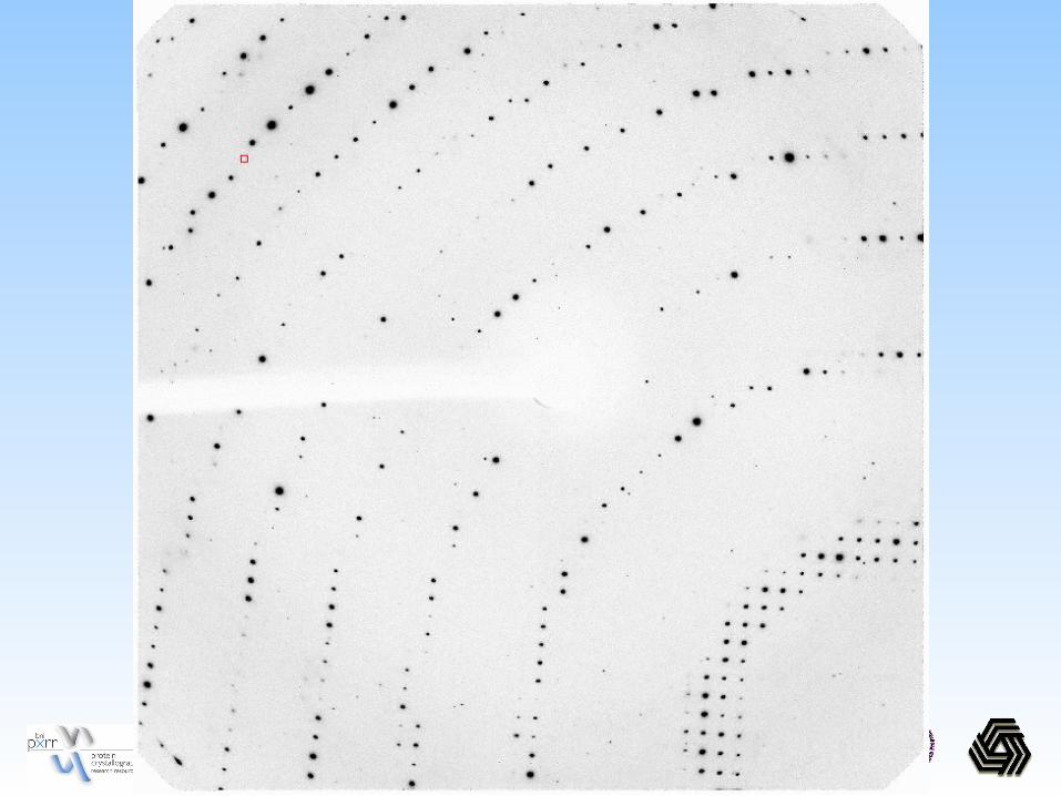
Diffraction from a periodic arrangement of
molecules (crystal) results in an amplified signal.

●Growing crystals
● Slow, controlled precipitation from aqueous solutions that do not denature the protein.
● Precipitants: ionic compounds(salts), organic solvents,
polymers like polyethylene glycols.

Electron-density maps
● When X-rays are directed at the crystal, the actual diffractors of the X-rays are the clouds of electrons in the molecules of the crystals. Therefore, diffraction reveals the distribution of electrons or electron density of the molecules.●Because protein molecules are in an ordered array in the crystal, electron
density can be described mathematically by a periodic function or as a
Fourier series.●Using the mathematical Fourier transforms the diffraction pattern can be
converted to electron density maps.●Computer programs are then used to come up with 3-d spatial coordinates.

From crystals to X-ray diffractometer, From crystals to X-ray diffractometer, computer, electron density maps, and computer, electron density maps, and molecular modelsmolecular models