simultaneous reduction of mad2 and bubr1 … · simultaneous reduction of mad2 and bubr1 expression...
TRANSCRIPT
Cell Biology International ISSN 1065-6995doi: 10.1002/cbin.10277
RESEARCH ARTICLE
Simultaneous reduction of MAD2 and BUBR1 expression inducesmitotic spindle alterations associated with p53 dependent cell cyclearrest and deathLaura Lentini1*y, Desirèe Piscitello1y, Lorena Veneziano1 and Aldo Di Leonardo1,2
1 Department of Biological Chemical and Pharmaceutical Sciences and Technologies (STEBICEF), University of Palermo Viale delle Scienze,Palermo 90128, Italy2 Center of Experimental OncoBiology (COBS), Via San Lorenzo, Palermo 90146, Italy
Abstract
Most human tumors are characterized by aneuploidy that is believed to be the consequence of chromosomal instability (CIN).Themechanism(s) leading to aneuploidy and the pathways that allow its tolerance are not completely understood. The SpindleAssembly Checkpoint (SAC) is a cellular surveillance mechanismworking duringmitosis, and alterations of genes that encodecomponents of the SAC weakening the mitotic checkpoint, induce aneuploidy by chromosome mis-segregation. We inducedaneuploidy in near-diploid tumor cells by simultaneous depletion of the SAC proteins MAD2 and BUBR1 by RNAinterference in the attempt to gain further insight on the cellular responses to aneuploidy. Individual reduction of MAD2 andBUBR1 protein levels caused defective mitosis and aneuploidy, while co-depletion of MAD2 and BUBR1 caused cell cyclearrest and cell death in addition to aneuploidy. The simultaneous reduction of the two SAC proteins induced high percentageof hyperdiploid cells and p53 stabilization suggesting that hyperdiploidy could activate a p53 controlled pathway. The resultsindicate that p53 is required to induce cell cycle arrest and cell death when the mitotic checkpoint is strongly perturbed,thereby preventing aneuploid cell propagation.
Keywords: cell cycle; cell death; chromosomes
Introduction
Segregation of genetic material into 2 daughter cells duringmitosis is a highly regulated event and errors during thisphase of the cell cycle can generate cells with aberrantchromosome numbers (aneuploidy). Precise control ofprogression through mitosis is essential to maintaingenomic stability and prevent aneuploidy (Ulrich, in press).Mutations/alterations in genes encoding mitotic regulatorsand defects in genes controlling centrosome numbers andtumor suppressors have been suggested as possible causesof aneuploidy (Lentini et al., 2002; Hernando et al., 2004;Fukasawa, 2005; Iovino et al., 2006; Lentini et al., 2006).Cells have evolved a surveillance mechanism, the SpindleAssembly Checkpoint (SAC), which prevents anaphaseonset until all chromosomes are properly attached tothe mitotic spindle ensuring that mitosis is faithfullyaccomplished preserving genome stability. The molecular
components of the SAC include Mad1, Mad2, Bubr1,Bub1, Bub3, and Mps1 proteins. Alteration of MAD2and BUBR1 expression may because of aneuploidy (Guoet al., 2012). In particular, human colorectal cancer cells(HCT116) and murine primary embryonic fibroblasts(MEFs), in which one MAD2 allele was deleted byhomologous recombination, are highly aneuploid (Michelet al., 2001; Lentini et al., 2012). Haplo-insufficiency ofMAD2 also increases the frequency of aneuploid tumors in ap53 background (Amato et al., 2009). Intriguingly, aneu-ploidy and tumorigenesis are also driven by hyperactivationof MAD2 (Sotillo et al., 2007; Schvartzman et al., 2011).The Bubr1 protein kinase, encoded by the BUB1B gene, isa crucial component for several processes controllingchromosome segregation during mitosis. Genetic mutationsof the Bubr1 kinase occur in the cancer susceptibledisorder, mosaic variegated aneuploidy (MVA) (Matsuuraet al., 2006), which is a rare disorder characterized by
�Corresponding author: e-mail: [email protected] authors contributed equally.
933Cell Biol Int 38 (2014) 933–941 © 2014 International Federation for Cell Biology
constitutional mosaic aneuploidies associated in most caseswith premature chromatid separation (PCS), highlightingthe key role of human Bubr1 in chromosome segregation(Matsuura et al., 2006). Bubr1 also functions in a positiveregulatory loop with the tumor suppressor p53, thusenhancing apoptosis of polyploid cells (Shin et al., 2003).It facilitates phosphorylation and stabilization of p53 aftermitotic spindle damage, as well as being a direct p53transcriptional target (Aylon and Oren, 2011). Inhibition ofBubr1 in the presence of mitotic poisons acceleratespolyploidy in a p53-null background, reinforcing the ideathat reduction of chromosome segregation fidelity andacquisition of aneuploid tolerance work synergistically topromote cancer (Shin et al., 2003). However, the genomicimbalance caused by aneuploidy reduces fitness of euploidcells (Torres et al., 2008). Similarly, stable near-diploidtumor cells that become aneuploid after treatment withmitotic poisons are removed from the cell population withinfew generations (Thompson and Compton, 2008). Thesefindings suggest that cancer cells must acquire the ability toovercome these barriers and that mutations triggeringaneuploidy tolerance should accompany chromosomemissegregation defects. The mechanisms leading to aneu-ploid tolerance in tumor cells are not yet completelyunderstood. We found that partial depletion of MAD2triggered aneuploidy in primary human fibroblasts followedby p53 stabilization that induced premature cellularsenescence to avoid the deleterious effects of aneuploidy(Lentini et al., 2012). p53 could be responsible for theproliferative disadvantage of aneuploidy cells, as suggestedalso by p53�/� cells in which aneuploidy remains high. Onlya fraction of MAD2 depleted HCT116 cells have wholechromosome aneuploidy associated with p53 activation(Li et al., 2010). Furthermore, the presence of aneuploid cellsin vivo and primary MEFs showing aneuploidy has noproliferative disadvantage, despite having an intact p53pathway, which suggest that p53 is activated in only someaneuploid cells (Zasadil et al., 2013).
Partial or complete inhibition of themitotic kinaseAurora Bresults in chromosomemisaggregationor failure of cytokinesis.To limit proliferation of aneuploid cells, a p53-mediated cellcycle arrest is induced, where p53 might be regulated in twoseparate ways, i.e. by being stabilized by inhibition of Aurora Bitself, or by damage to the mitotic spindle apparatus. Thestabilization is insufficient to activate p53 and a secondpathway, the MAP kinase signaling pathway MAP3K4-p38, isrequired for transcriptional activation of p53. MAP3K4 mightbe activated bymitotic spindle disruption and trigger the signaltop38b. Presumably, p38b targets a transcriptional co-factorofp53 leading to full activity of p53 and induction of p21.The cellgrowth arrest induced by these pathways might preventchromosomal instability and therefore suppress tumorigenesis(Ulrich, in press).
We triggered aneuploidy by weakening the SAC afterpost-transcriptional silencing of MAD2 and BUBR1 genesand investigated whether a p53 controlled pathway isactivated in near-diploid tumor cells (HCT116) with a MIN(Microsatellite Instability) phenotype to avoid propagationof highly aneuploidy cells. Individual reduction ofMad2 andBubr1 protein levels partially affected cell proliferation andcaused mitotic spindle defects and aneuploidy in near-diploid HCT116 cells. Conversely, simultaneous Mad2 andBubr1 depletion resulted in greater effects, leading to cellcycle arrest and high mortality.
Materials and methods
Cells and cell culture
Near-diploidHCT116 andHCT116 p53KO cells (a generousgift of Prof. B. Vogelstein, Ludwig Center for CancerGenetics and Therapeutics, Johns Hopkins, Baltimore, MD,USA) with a MIN phenotype were cultured in D-MEMsupplemented with 10% FBS (GIBCO, Invitrogen),100 units/mL penicillin and 0.1mg/mL streptomycin in ahumidified atmosphere of 5% CO2 in air at 37�C.
RNA interference
For the RNAi experiments, 2� 105 HCT116 cells wereplated in 6-well dishes, incubated at 37�C and transfected24 h after plating with specific siRNA duplexes. Briefly, thesiRNAs and the transfection reagent (Lipofectamine 2000,Invitrogen) were diluted separately in Opti-MEM (Invi-trogen) mixed gently, and then incubated for 5min at RT.After incubation the siRNAs and Lipofectamine 2000(Invitrogen) were mixed gently, allowed to sit 30min atRT to allow complex formation and added to the plates for72 h. After 6 h at 37�C, the transfection medium wasreplaced with fresh medium. To silence genes of interest,post-transcriptionally, cells were transfected with siRNAstargeting MAD2 (siMAD2: 50-AUACGGACUCAC-CUUUtt-30) (Michel et al., 2001), siRNAs targeting p53 (sip53: 50-GCA UGA ACC GGA GGC CCC AUtt-30) (Frameet al., 2006) and BUBR1 (siBUBR1: 50-GUCUCACA-GAUUGCUGCCUtt-30) (Choi and Lee, 2008) at 60 nM.The control siRNA (siGFP: 50-GGCUACGUCCAG-GAGCGCACCtt-30) targets the Green-Fluorescent-Proteinand was used at 60 nM. All siRNAs (21-nucleotide duplexes)were synthesized by Eurofins-MWG (Germany).
Real time RT-PCR
Primers to be used in Real time RT-PCR experiments weredesigned with Primer Express software (Applied Biosys-tems) choosing amplicons of 70–100 bp. The selectedsequences were tested on public databases (BLAST) to
MAD2/BUBR1 depletion induces cell death L. Lentini et al.
934 Cell Biol Int 38 (2014) 933–941 © 2014 International Federation for Cell Biology
confirm the identity of the genes. Total RNA was extractedfrom cells by using the RNAeasyMini kit (Qiagen). RNAwasreverse-transcribed in a final volume of 50mL using theHighCapacity c-DNA Archive kit (Applied Biosystems) for10min at 25�C and 2 h at 37�C. For each sample 2mL ofcDNA, corresponding to 100 ng of reverse transcribed RNA,were analyzed by Real time RT-PCR (95�C for 15 s, 60�C for60 s repeated for 40 cycles), in quadruplicate, using the ABIPRISM 7300 instrument (Applied Biosystems). Real-timeRT-PCR was done in 20mL comprising 1� Master MixSYBR Green (Applied Biosystems) and 0.3mM of forwardand reverse primers for: MAD2 (Fwd:50-GCCGAGTT-TTTCTCATTTGG-30; Rev:50-CCGATTCTTCCCACTTT-TCA-30), p53 (Fwd:50-TTCGACATAGTGTGGTGGTGC-30, Rev:50-AGTCAGAGCCAACCTGAGGC-30), p21waf1
(Fwd: 50-CTGGAGACTCTCAGGGTCGA-30 Rev:50-CGG-ATTAGGGCTTCCTCTTG-30), BUBR1 (Fwd:50-TACAC-TGGAAATGACCCTCTGGAT-30, Rev: 50-TATATTATC-GTTTTTCTCCTTGTAGTGCTT-30), GAPDH (Fwd:50-CTCATGACCACAGTCCATGCC-30; Rev:50-GCCAATC-CACAGTCTTCTGGGT-30). Data were analyzed by averag-ing quadruplicates Ct (cycle threshold). Levels of RNAexpression were determined by using the SDS softwareversion (Applied Biosystems) according to the 2-DDctmethod. Levels of RNA expression of selected genes werenormalized to the internal control GAPDH.
Western blotting
Protein concentrationwasmeasuredusing theBio-RadProteinAssay (Bio-Rad Laboratories). Proteins (50mg) were separatedby 10% SDS-PAGE containing 0.1% SDS and transferred toHybond-C nitrocellulose membranes (Amersham Life Sci-ence) by electroblotting (Lentini et al., 2014). The membraneswere sequentially incubated with p53-DO1 (mouse, 1:1000),p21(mouse, 1:1000), Bubr1 (goat, 1:500),Mad2 (goat, 1:200) asprimary antibodies (Santa Cruz,CA), and HRP-conjugatedmouse (1:5000), goat (1:5000), and rabbit IgG (1:5000) (SantaCruz,CA) as secondary antibodies. The target protein wasdetected with enhanced chemiluminescence Western blottingdetection reagents (PIERCE). Membranes were stained withPonceau-Red to confirm equivalent loading of total protein inall lanes.b-tubulin antibody (mouse; SIGMA-ALDRICH, Italy1:10.000) was used to confirm proteins loading.
Cell cycle analysis
Asynchronously growing cells were transfected with 60 nMMAD2 siRNA for 72 h before being pulse labeled with10mM bromodeoxyuridine (BrdU) for the last 3 h post-transfection. Cells were fixed and stained with anti-BrdU-FITC antibody, to detect S-phase cells, and propidium iodide(PI) to assess the DNA content and analyzed by flowcytometry. Labeling with BrdU allowed monitoring of cells
actively engaged in DNA synthesis. DNA content wasdetermined by treating the cells with PBS containing 4mg/mLof PI and 40mg/mL RNase. BrdU-labeled cells were analyzedas previously described (Barra et al., 2012; Lentini et al., 2012)with by FACSCanto (Becton Dickinson). Experiments wererepeated at least twice and >10,000 events were analyzed byusing the FACSDiva software.
Determination of ploidy
Cells were transfected with the specific siRNA (siGFP,siMAD2, siMAD2/siBUBR1) for 72 h treated with 0.2mg/mLcolcemid (Demecolcine, SIGMA-ALDRICH, Italy) and 1h,harvested by trypsinization, swollen in 75mM KCl at 37�C,fixed with 3:1 methanol/acetic acid (v/v), and dropped ontoclean, ice-cold glass microscope slides. The slides were air-dried and stained with a 3% GIEMSA solution in phosphate-buffered saline for 10min. Chromosome numbers werecounted using a Zeiss Axioskopmicroscope at a magnificationof 100 � objective.
Trypan blue exclusion test of cell viability
Cells were transfected with the specific siRNAs (siGFP,siMAD2, siBUBR1, siMAD2/siBUBR1) for 72 h, harvested bytrypsinization and collected in a tubewith 4mLof phosphate-buffered saline (PBS). Cell suspension (100mL) were mixedwith 100mL of Trypan Blue (Sigma-Aldrich, Italy) and 10mLwere placed in a Burker chamber for counting.
Immunofluorescence microscopy
To visualize b-tubulin, cells were grown on round glasscoverslips and fixed with ethanol/acetic acid 95:5 for 10min,permeabilized with 0.01% TritonX (Sigma-Aldrich, Italy) inPBS for 15min and blocked with 0.1% BSA for 30min atroom temperature. Coverslips were incubated with a mousemonoclonal antibody against b-tubulin mouse (Sigma-Aldrich, Italy, diluted 1:200) overnight at 4�C, followed by agoat anti-mouse IgG-FITC secondary antibody (SIGMA-ALDRICH, Italy, diluted 1:200) for 1 h at 37�C. Nuclei werestained with 1mg/mL 40,6-diamidino-2-phenylindole(DAPI) and examined on a Zeiss Axioskop microscopeequipped for fluorescence; 200 nuclei for sample wereanalyzed and images were captured with a CCD digitalcamera (AxioCam, Zeiss) and printed by Adobe PhotoShop.
Results
Co-depletion of MAD2 and BUBR1 by RNAi reduces cellproliferation
We partially depleted MAD2 and BUBR1 (crucial compo-nents of the SAC) by RNA interference in HCT116 cells toinvestigate if a pathway p53 controlled is activated also in
L. Lentini et al. MAD2/BUBR1 depletion induces cell death
935Cell Biol Int 38 (2014) 933–941 © 2014 International Federation for Cell Biology
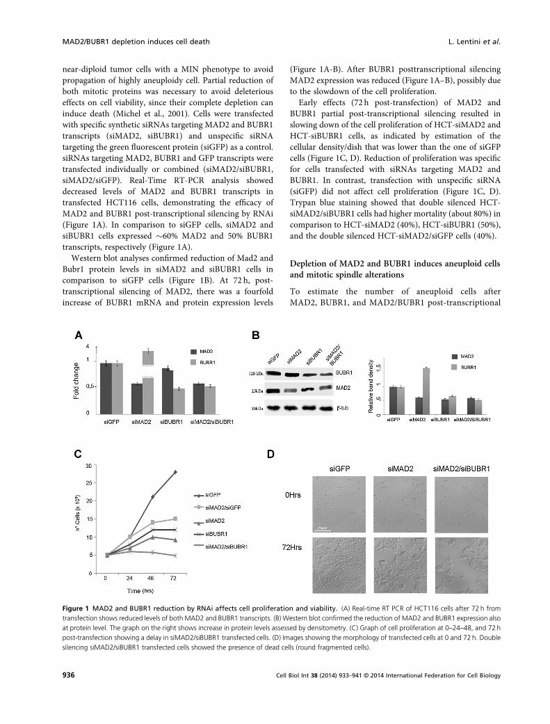
near-diploid tumor cells with a MIN phenotype to avoidpropagation of highly aneuploidy cell. Partial reduction ofboth mitotic proteins was necessary to avoid deleteriouseffects on cell viability, since their complete depletion caninduce death (Michel et al., 2001). Cells were transfectedwith specific synthetic siRNAs targeting MAD2 and BUBR1transcripts (siMAD2, siBUBR1) and unspecific siRNAtargeting the green fluorescent protein (siGFP) as a control.siRNAs targeting MAD2, BUBR1 and GFP transcripts weretransfected individually or combined (siMAD2/siBUBR1,siMAD2/siGFP). Real-Time RT-PCR analysis showeddecreased levels of MAD2 and BUBR1 transcripts intransfected HCT116 cells, demonstrating the efficacy ofMAD2 and BUBR1 post-transcriptional silencing by RNAi(Figure 1A). In comparison to siGFP cells, siMAD2 andsiBUBR1 cells expressed �60% MAD2 and 50% BUBR1transcripts, respectively (Figure 1A).
Western blot analyses confirmed reduction of Mad2 andBubr1 protein levels in siMAD2 and siBUBR1 cells incomparison to siGFP cells (Figure 1B). At 72 h, post-transcriptional silencing of MAD2, there was a fourfoldincrease of BUBR1 mRNA and protein expression levels
(Figure 1A-B). After BUBR1 posttranscriptional silencingMAD2 expression was reduced (Figure 1A–B), possibly dueto the slowdown of the cell proliferation.
Early effects (72 h post-transfection) of MAD2 andBUBR1 partial post-transcriptional silencing resulted inslowing down of the cell proliferation of HCT-siMAD2 andHCT-siBUBR1 cells, as indicated by estimation of thecellular density/dish that was lower than the one of siGFPcells (Figure 1C, D). Reduction of proliferation was specificfor cells transfected with siRNAs targeting MAD2 andBUBR1. In contrast, transfection with unspecific siRNA(siGFP) did not affect cell proliferation (Figure 1C, D).Trypan blue staining showed that double silenced HCT-siMAD2/siBUBR1 cells had higher mortality (about 80%) incomparison to HCT-siMAD2 (40%), HCT-siBUBR1 (50%),and the double silenced HCT-siMAD2/siGFP cells (40%).
Depletion of MAD2 and BUBR1 induces aneuploid cellsand mitotic spindle alterations
To estimate the number of aneuploid cells afterMAD2, BUBR1, and MAD2/BUBR1 post-transcriptional
Figure 1 MAD2 and BUBR1 reduction by RNAi affects cell proliferation and viability. (A) Real-time RT PCR of HCT116 cells after 72 h fromtransfection shows reduced levels of both MAD2 and BUBR1 transcripts. (B) Western blot confirmed the reduction of MAD2 and BUBR1 expression alsoat protein level. The graph on the right shows increase in protein levels assessed by densitometry. (C) Graph of cell proliferation at 0–24–48, and 72hpost-transfection showing a delay in siMAD2/siBUBR1 transfected cells. (D) Images showing the morphology of transfected cells at 0 and 72h. Doublesilencing siMAD2/siBUBR1 transfected cells showed the presence of dead cells (round fragmented cells).
MAD2/BUBR1 depletion induces cell death L. Lentini et al.
936 Cell Biol Int 38 (2014) 933–941 © 2014 International Federation for Cell Biology
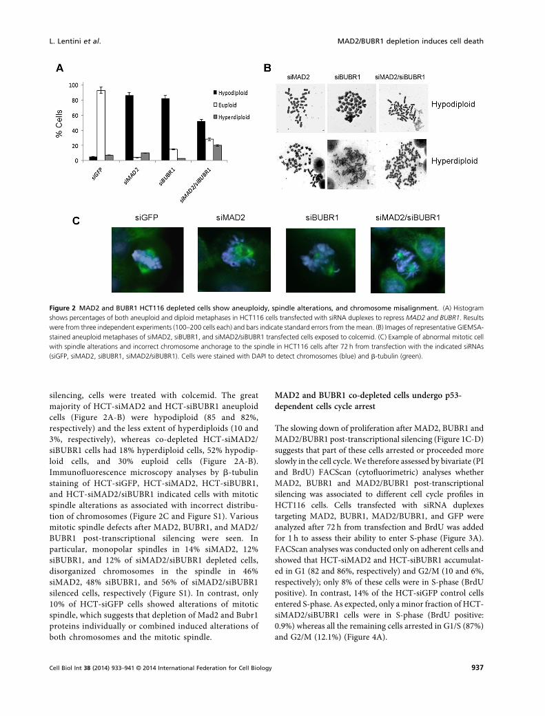
silencing, cells were treated with colcemid. The greatmajority of HCT-siMAD2 and HCT-siBUBR1 aneuploidcells (Figure 2A-B) were hypodiploid (85 and 82%,respectively) and the less extent of hyperdiploids (10 and3%, respectively), whereas co-depleted HCT-siMAD2/siBUBR1 cells had 18% hyperdiploid cells, 52% hypodip-loid cells, and 30% euploid cells (Figure 2A-B).Immunofluorescence microscopy analyses by b-tubulinstaining of HCT-siGFP, HCT-siMAD2, HCT-siBUBR1,and HCT-siMAD2/siBUBR1 indicated cells with mitoticspindle alterations as associated with incorrect distribu-tion of chromosomes (Figure 2C and Figure S1). Variousmitotic spindle defects after MAD2, BUBR1, and MAD2/BUBR1 post-transcriptional silencing were seen. Inparticular, monopolar spindles in 14% siMAD2, 12%siBUBR1, and 12% of siMAD2/siBUBR1 depleted cells,disorganized chromosomes in the spindle in 46%siMAD2, 48% siBUBR1, and 56% of siMAD2/siBUBR1silenced cells, respectively (Figure S1). In contrast, only10% of HCT-siGFP cells showed alterations of mitoticspindle, which suggests that depletion of Mad2 and Bubr1proteins individually or combined induced alterations ofboth chromosomes and the mitotic spindle.
MAD2 and BUBR1 co-depleted cells undergo p53-dependent cells cycle arrest
The slowing down of proliferation after MAD2, BUBR1 andMAD2/BUBR1 post-transcriptional silencing (Figure 1C-D)suggests that part of these cells arrested or proceeded moreslowly in the cell cycle.We therefore assessed by bivariate (PIand BrdU) FACScan (cytofluorimetric) analyses whetherMAD2, BUBR1 and MAD2/BUBR1 post-transcriptionalsilencing was associated to different cell cycle profiles inHCT116 cells. Cells transfected with siRNA duplexestargeting MAD2, BUBR1, MAD2/BUBR1, and GFP wereanalyzed after 72 h from transfection and BrdU was addedfor 1 h to assess their ability to enter S-phase (Figure 3A).FACScan analyses was conducted only on adherent cells andshowed that HCT-siMAD2 and HCT-siBUBR1 accumulat-ed in G1 (82 and 86%, respectively) and G2/M (10 and 6%,respectively); only 8% of these cells were in S-phase (BrdUpositive). In contrast, 14% of the HCT-siGFP control cellsentered S-phase. As expected, only a minor fraction of HCT-siMAD2/siBUBR1 cells were in S-phase (BrdU positive:0.9%) whereas all the remaining cells arrested in G1/S (87%)and G2/M (12.1%) (Figure 4A).
Figure 2 MAD2 and BUBR1 HCT116 depleted cells show aneuploidy, spindle alterations, and chromosome misalignment. (A) Histogramshows percentages of both aneuploid and diploid metaphases in HCT116 cells transfected with siRNA duplexes to repress MAD2 and BUBR1. Resultswere from three independent experiments (100–200 cells each) and bars indicate standard errors from the mean. (B) Images of representative GIEMSA-stained aneuploid metaphases of siMAD2, siBUBR1, and siMAD2/siBUBR1 transfected cells exposed to colcemid. (C) Example of abnormal mitotic cellwith spindle alterations and incorrect chromosome anchorage to the spindle in HCT116 cells after 72 h from transfection with the indicated siRNAs(siGFP, siMAD2, siBUBR1, siMAD2/siBUBR1). Cells were stained with DAPI to detect chromosomes (blue) and b-tubulin (green).
L. Lentini et al. MAD2/BUBR1 depletion induces cell death
937Cell Biol Int 38 (2014) 933–941 © 2014 International Federation for Cell Biology
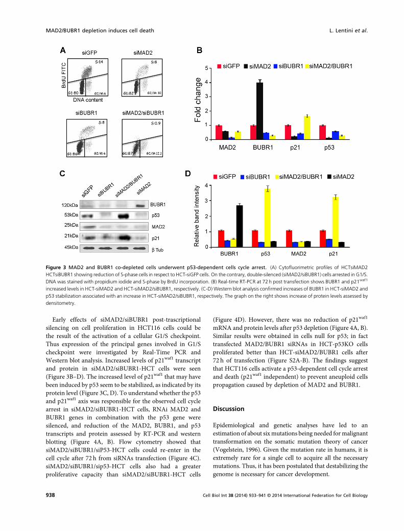
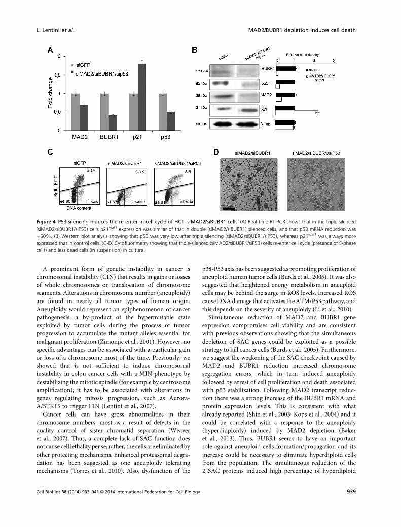
Early effects of siMAD2/siBUBR1 post-trascriptionalsilencing on cell proliferation in HCT116 cells could bethe result of the activation of a cellular G1/S checkpoint.Thus expression of the principal genes involved in G1/Scheckpoint were investigated by Real-Time PCR andWestern blot analysis. Increased levels of p21waf1 transcriptand protein in siMAD2/siBUBR1-HCT cells were seen(Figure 3B–D). The increased level of p21waf1 that may havebeen induced by p53 seem to be stabilized, as indicated by itsprotein level (Figure 3C, D). To understand whether the p53and p21waf1 axis was responsible for the observed cell cyclearrest in siMAD2/siBUBR1-HCT cells, RNAi MAD2 andBUBR1 genes in combination with the p53 gene weresilenced, and reduction of the MAD2, BUBR1, and p53transcripts and protein assessed by RT-PCR and westernblotting (Figure 4A, B). Flow cytometry showed thatsiMAD2/siBUBR1/siP53-HCT cells could re-enter in thecell cycle after 72 h from siRNAs transfection (Figure 4C).siMAD2/siBUBR1/sip53-HCT cells also had a greaterproliferative capacity than siMAD2/siBUBR1-HCT cells
(Figure 4D). However, there was no reduction of p21waf1
mRNA and protein levels after p53 depletion (Figure 4A, B).Similar results were obtained in cells null for p53; in facttransfected MAD2/BUBR1 siRNAs in HCT-p53KO cellsproliferated better than HCT-siMAD2/BUBR1 cells after72 h of transfection (Figure S2A-B). The findings suggestthat HCT116 cells activate a p53-dependent cell cycle arrestand death (p21waf1 independent) to prevent aneuploid cellspropagation caused by depletion of MAD2 and BUBR1.
Discussion
Epidemiological and genetic analyses have led to anestimation of about sixmutations being needed formalignanttransformation on the somatic mutation theory of cancer(Vogelstein, 1996). Given the mutation rate in humans, it isextremely rare for a single cell to acquire all the necessarymutations. Thus, it has been postulated that destabilizing thegenome is necessary for cancer development.
Figure 3 MAD2 and BUBR1 co-depleted cells underwent p53-dependent cells cycle arrest. (A) Cytofluorimetric profiles of HCTsiMAD2HCTsiBUBR1 showing reduction of S-phase cells in respect to HCT-siGFP cells. On the contrary, double-silenced (siMAD2/siBUBR1) cells arrested in G1/S.DNA was stained with propidium iodide and S-phase by BrdU incorporation. (B) Real-time RT-PCR at 72 h post transfection shows BUBR1 and p21waf1
increased levels in HCT-siMAD2 and HCT-siMAD2/siBUBR1, respectively. (C-D) Western blot analysis confirmed increases of BUBR1 in HCT-siMAD2 andp53 stabilization associated with an increase in HCT-siMAD2/siBUBR1, respectively. The graph on the right shows increase of protein levels assessed bydensitometry.
MAD2/BUBR1 depletion induces cell death L. Lentini et al.
938 Cell Biol Int 38 (2014) 933–941 © 2014 International Federation for Cell Biology
A prominent form of genetic instability in cancer ischromosomal instability (CIN) that results in gains or lossesof whole chromosomes or translocation of chromosomesegments. Alterations in chromosome number (aneuploidy)are found in nearly all tumor types of human origin.Aneuploidy would represent an epiphenomenon of cancerpathogenesis, a by-product of the hypermutable stateexploited by tumor cells during the process of tumorprogression to accumulate the mutant alleles essential formalignant proliferation (Zimonjic et al., 2001). However, nospecific advantages can be associated with a particular gainor loss of a chromosome most of the time. Previously, weshowed that is not sufficient to induce chromosomalinstability in colon cancer cells with a MIN phenotype bydestabilizing themitotic spindle (for example by centrosomeamplification); it has to be associated with alterations ingenes regulating mitosis progression, such as Aurora-A/STK15 to trigger CIN (Lentini et al., 2007).
Cancer cells can have gross abnormalities in theirchromosome numbers, most as a result of defects in thequality control of sister chromatid separation (Weaveret al., 2007). Thus, a complete lack of SAC function doesnot cause cell lethality per se; rather, the cells are eliminated byother protecting mechanisms. Enhanced proteasomal degra-dation has been suggested as one aneuploidy toleratingmechanisms (Torres et al., 2010). Also, dysfunction of the
p38-P53 axis has been suggested as promoting proliferationofaneuploid human tumor cells (Burds et al., 2005). It was alsosuggested that heightened energy metabolism in aneuploidcells may be behind the surge in ROS levels. Increased ROScauseDNAdamage that activates theATM/P53 pathway, andthis depends on the severity of aneuploidy (Li et al., 2010).
Simultaneous reduction of MAD2 and BUBR1 geneexpression compromises cell viability and are consistentwith previous observations showing that the simultaneousdepletion of SAC genes could be exploited as a possiblestrategy to kill cancer cells (Burds et al., 2005). Furthermore,we suggest the weakening of the SAC checkpoint caused byMAD2 and BUBR1 reduction increased chromosomesegregation errors, which in turn induced aneuploidyfollowed by arrest of cell proliferation and death associatedwith p53 stabilization. Following MAD2 transcript reduc-tion there was a strong increase of the BUBR1 mRNA andprotein expression levels. This is consistent with whatalready reported (Shin et al., 2003; Kops et al., 2004) and itcould be correlated with a response to the aneuploidy(hyperdidploidy) induced by MAD2 depletion (Bakeret al., 2013). Thus, BUBR1 seems to have an importantrole against aneuploid cells formation/propagation and itsincrease could be necessary to eliminate hyperdiploid cellsfrom the population. The simultaneous reduction of the2 SAC proteins induced high percentage of hyperdiploid
Figure 4 P53 silencing induces the re-enter in cell cycle of HCT- siMAD2/siBUBR1 cells (A) Real-time RT PCR shows that in the triple silenced(siMAD2/siBUBR1/siP53) cells p21waf1 expression was similar of that in double (siMAD2/siBUBR1) silenced cells, and that p53 mRNA reduction was�50%. (B) Western blot analysis showing that p53 was very low after triple silencing (siMAD2/siBUBR1/siP53), whereas p21waf1 was always moreexpressed that in control cells. (C-D) Cytofluorimetry showing that triple-silenced (siMAD2/siBUBR1/siP53) cells re-enter cell cycle (presence of S-phasecells) and less dead cells (in suspension) in culture.
L. Lentini et al. MAD2/BUBR1 depletion induces cell death
939Cell Biol Int 38 (2014) 933–941 © 2014 International Federation for Cell Biology
cells and p53 stabilization, suggesting that hyperdiploidycould activate a p53 controlled pathway accordingly topublished reports (Thompson and Compton DA:, 2010;Stuabach, in press). On the contrary, aneuploid cellsgenerated by single silencing of MAD2 or BUBR1 did notshow high levels of hyperdiploidy and did not activate thep53-controlled checkpoint. This conclusion is supported byp53 post-transcriptional silencing by siRNA in MAD2 andBUBR1co-depleted cells allowing cells to re-enter the cell cycle. Thiswas also seen in cells null for p53 (HCT-p53KO), whenMAD2 and BUBR1 were simultaneously silenced. After p53depletion by RNAi, we unexpectedly did not find a decreaseof p21waf1 protein levels, which suggests that p21waf1 is onlypartially involved in the arrest of the cell cycle observed inHCT116-MAD2/BUBR1 cells and that its increase inaneuploidy cells is not only under the p53 control.
In conclusion, our results indicate thatMAD2 and BUBR1reduction causes mitotic spindle dis-organization andincorrect distribution of chromosomes along the spindlein the absence of mitotic poisons, and, importantly, that ap53-controlled pathway is required to induce cell cycle arrestor cell death to avoid aneuploid cells proliferation.
Acknowledgments
This work was partly supported by a grant (ex 60%) fromUniversity of Palermo to ADL.
Conflict of interest
The authors declare that they have no competing interests.
References
Amato A, Lentini L, Schillaci T, Iovino F, Di Leonardo A (2009)RNAi mediated acute depletion of retinoblastoma protein(pRb) promotes aneuploidy in human primary cells viamicronuclei formation. BMC Cell Biol 10: 79.
Aylon Y, OrenM (2011) P53: guardian of ploidy. Mol Oncol 5(4):315–23.
Baker DJ, Dawlaty MM, Wijshake T, Jeganathan KB, MalureanuL, van Ree JH, Crespo-Diaz R, Reyes S, Seaburg L, Shapiro V(2013) Increased expression of BubR1 protects againstaneuploidy and cancer and extends healthy lifespan. Nat CellBiol 15(1): 96–102.
Barra V, Schillaci T, Lentini L, Costa G, Di Leonardo A (2012)Bypass of cell cycle arrest induced by transient DNMT1 post-transcriptional silencing triggers aneuploidy in human cells.Cell Div 7(1): 2.
Burds AA, Lutum AS, Sorger PK (2005) Generating chromosomeinstability through the simultaneous deletion ofMad2 and p53.Proc Natl Acad Sci USA 102(32): 11296–301.
Choi E, Lee H (2008) Chromosome damage in mitosis inducesBubR1 activation and prometaphase arrest. FEBS Lett 582(12):1700–6.
Frame FM, Rogoff HA, Pickering MT, Cress WD, Kowalik TF(2006) E2F1 induces MRN foci formation and a cell cyclecheckpoint response in human fibroblasts. Oncogene 25(23):3258–66.
Fukasawa K (2005) Centrosome amplification, chromosomeinstability and cancer development. Cancer Lett 230(1): 6–19.
Guo Y, Kim C, Ahmad S, Zhang J, Mao Y (2012) CENP-E–dependent BubR1 autophosphorylation enhances chromo-some alignment and the mitotic checkpoint. J Cell Biol 198(2):205–17.
Hernando E, Nahle Z, Juan G, Diaz-Rodriguez E, Alaminos M,Hemann M, Michel L, Mittal V, Gerald W, Benezra R (2004)Rb inactivation promotes genomic instability by uncouplingcell cycle progression from mitotic control. Nature 430(7001):797–802.
Iovino F, Lentini L, Amato A, Di Leonardo A (2006) RB acute lossinduces centrosome amplification and aneuploidy in murineprimary fibroblasts. Mol Cancer 5(1): 38.
Kops GJ, Foltz DR, Cleveland DW (2004) Lethality to humancancer cells through massive chromosome loss by inhibition ofthemitotic checkpoint. Proc Natl Acad Sci USA 101(23): 8699–704.
Lentini L, Pipitone L, Di Leonardo A (2002) Functionalinactivation of pRB results in aneuploid mammalian cellsafter release from a mitotic block. Neoplasia 4(5): 380–7.
Lentini L, Iovino F, Amato A, Di Leonardo A (2006) Centrosomeamplification induced by hydroxyurea leads to aneuploidyin pRB deficient human and mouse fibroblasts. Cancer Lett238(1): 153–60.
Lentini L, Amato A, Schillaci T, Di Leonardo A (2007)Simultaneous Aurora-A/STK15 overexpression and centro-some amplification induce chromosomal instability in tumourcells with a MIN phenotype. BMC Cancer 7–212.
Lentini L, Barra V, Schillaci T, Di Leonardo A (2012) MAD2depletion triggers premature cellular senescence in humanprimary fibroblasts by activating a p53 pathway preventinganeuploid cells propagation. J Cell Physiol 227(9): 3324–32.
Lentini L, Melfi R, Di Leonardo A, Spinello A, Barone G, Pace A,Palumbo Piccionello A, Pibiri I (2014) Toward a rationale forthe PTC124 (Ataluren) promoted readthrough of prematurestop codons: a computational approach and GFP-reporter cell-based assay. Mol Pharm.
LiM, Fang X, Baker DJ, Guo L, Gao X,Wei Z, Han S, van DeursenJM, Zhang P (2010) The ATM-p53 pathway suppressesaneuploidy-induced tumorigenesis. Proc Natl Acad Sci USA107(32): 14188–93.
Matsuura S, Matsumoto Y, Morishima K, Izumi H, MatsumotoH, Ito E, Tsutsui K, Kobayashi J, Tauchi H, Kajiwara Y (2006)Monoallelic BUB1B mutations and defective mitotic-spindlecheckpoint in seven families with premature chromatidseparation (PCS) syndrome. Am J Med Genet A 140(4):358–67.
MAD2/BUBR1 depletion induces cell death L. Lentini et al.
940 Cell Biol Int 38 (2014) 933–941 © 2014 International Federation for Cell Biology
Michel LS, Liberal V, Chatterjee A, Kirchwegger R, Pasche B,Gerald W, Dobles M, Sorger PK, Murty VV, Benezra R (2001)MAD2 haplo-insufficiency causes premature anaphase andchromosome instability inmammalian cells. Nature 409(6818):355–9.
Schvartzman JM, Duijf PH, Sotillo R, Coker C, Benezra (2011)Mad2 is a critical mediator of the chromosome instabilityobserved upon Rb and p53 pathway inhibition. Cancer Cell19(6): 701–14.
Shin HJ, Baek KH, Jeon AH, Park MT, Lee SJ, Kang CM, Lee HS,Yoo SH, Chung DH, Sung YC (2003) Dual roles of humanBubR1, a mitotic checkpoint kinase, in the monitoring ofchromosomal instability. Cancer Cell 4(6): 483–97.
Sotillo R, Hernando E, Diaz-Rodriguez E, Teruya-Feldstein J,Cordon-Cardo C, Lowe SW, Benezra R (2007) Mad2 over-expression promotes aneuploidy and tumorigenesis in mice.Cancer Cell 11(1): 9–23.
Stuabach AE (2004) The regulation of BubR1 expression byp53: a role for p53 in the mitotic spindle checkpoint andchromosome instability. Thesis submitted to the Divisionof Research and advanced studies of the University ofCincinnati.
Thompson SL, Compton DA (2010) Proliferation of aneuploidhuman cells is limited by a p53-dependent mechanism. J CellBiol 188(3): 369–81.
Thompson SL, Compton DA (2008) Examining the link betweenchromosomal instability and aneuploidy in human cells. J CellBiol 180(4): 665–72.
Torres EM,Williams BR, AmonA (2008) Aneuploidy: cells losingtheir balance. Genetics 179(2): 737–46.
Torres EM, Dephe N, Panneerselvam A, Tucker CM, WhittakerCA, Gygi SP, Dunham MJ, Amon A (2010) Identification ofaneuploidy-tolerating mutations. Cell 143(1): 71–83.
Ulrich T (2012) Function of Lin9 in vivo and MAP3K4-p38signaling regulates p 53mediated cell cycle arrest after defectivemitosis. Dissertation of science doctoral degree the BavarianJulius Maximilian University of Wurzburg.
Weaver BA, Silk AD, Montagna C, Verdier-Pinard P, ClevelandDW (2007) Aneuploidy acts both oncogenically and as a tumorsuppressor. Cancer Cell 11(1): 25–36.
Zasadil LM, Britigan EM, Weaver BA (2013) 2n or not 2n:Aneuploidy, polyploidy and chromosomal instability inprimary and tumor cells. Semin Cell Dev Biol 24(4): 370–9.
Zimonjic D, Brooks MW, Popescu N, Weinberg RA, Hahn WC(2001) Derivation of human tumor cells in vitro withoutwidespread genomic instability. Cancer Res 61(24): 8838–44.
Received 7 January 2014; accepted 20 March 2014.Final version published online 10 April 2014.
Supporting Information
Additional supporting information may be found in theonline version of this article at the publisher’s web-site
Figure S1. Histogram showing the distribution of differentmitotic alterations observed in wt and posttranscriptionalsilenced HCT116 cells.
Figure S2. MAD2 and BUBR1 reduction by RNAi didnot affect cell proliferation and viability inHCTp53KO cells.
L. Lentini et al. MAD2/BUBR1 depletion induces cell death
941Cell Biol Int 38 (2014) 933–941 © 2014 International Federation for Cell Biology