simulation study of microwave heating with nanoparticle...
TRANSCRIPT
INT. J. BIOAUTOMATION, 2019, 23(4), 421-434 doi: 10.7546/ijba.2019.23.3.000583
421
Simulation Study of Microwave Heating
with Nanoparticle Diffusion for Tumor Ablation
Kazi Mahdi Mahmud, Md. Maruf Hossain Shuvo*
Department of Electronics and Communication Engineering
Khulna University of Engineering & Technology
Khulna-9203, Bangladesh
E-mails: [email protected], [email protected]
*Corresponding author
Received: March 10, 2018 Accepted: April 19, 2019
Published: December 31, 2019
Abstract: Microwave heating is one of the prominent treatment procedure that elevates body
temperature using microwave energy to damage tumor cells. However, healthy tissues can
also absorb microwave energy causing undesired damage. This research study focuses on
optimizing the surrounding healthy tissue damage by diffusing magnetic nanoparticles (NPs)
in the tumor region. A rectangular liver tissue is modeled using Finite Element Methods (FEM)
and then a half-elliptical shaped tumor is incorporated in the model. A coaxial antenna
covered with a polytetrafluoroethylene catheter is inserted at the edge of the liver tissue.
Then the magnetic NPs are diffused within the tumor region. The performance of microwave
heating with and without nanoparticle diffusion is compared using the performance
parameters: power dissipation density, temperature distribution, and resultant tissue necrosis.
Simulation results show that the heating procedure coupled with ferromagnetic nanoparticle
diffusion has approximately 14% less damage to the healthy tissue. The study also shows that
433/915 MHz frequency value provides 3% less damage to the healthy tissue than 2.45 GHz.
Analyzing the performance of different nanoparticle we found that the ferromagnetic
nanoparticle provides 15% and 5% less damage to the healthy tissue than gold and manganese
iron oxide NPs respectively. The obtained result was also verified for kidney, breast, and lung
tumor ablations to confirm the findings.
Keywords: Microwave hyperthermia, Magnetic nanoparticle, Tumor necrosis, Liver tumor
ablation, Finite element analysis.
Introduction The common treatments for cancer are surgical tumor removal, radiation therapy, and
chemotherapy. In spite of being effective, these treatments are far from ideal. Surgical tumor
removal is extremely invasive, while chemo and radiation therapies have the potentiality to
cause serious side effects. Thus, there is an increasing demand for treatment procedure that is
effective but minimizes invasiveness and undesirable side effects. The treatments that can meet
these requirements involve mechanisms for specifically targeting cancer cells while keeping
healthy tissues unaffected. One technique for specifically targeting tumors that have gained
popularity due to its economic feasibility and minimal side effects is the thermal therapy.
The thermal therapy that applies extreme heat to the tumor with the intention of killing all the
malignant tissue is known as hyperthermia [14].
The limitation of hyperthermia therapy is that patients experience local pain and discomfort as
well as while heating the tumor tissue for destruction, some surrounding healthy tissues are also
damaged [15]. Thus, the temperature increase needs to be controlled to a specific region so that
the damage to the normal tissue is minimized [5]. Several methods have been developed to
localize electromagnetic radiation and its subsequent heating to the tumor. One of the prominent

INT. J. BIOAUTOMATION, 2019, 23(4), 421-434 doi: 10.7546/ijba.2019.23.3.000583
422
methods is inserting a microwave-emitting probe directly into the tumor [2]. Another proven
method of localization is injecting magnetic nanoparticles into the tumor, which then excited
with microwave radiation [13]. In this study, we will examine the effect of these two
localization strategies.
Mild hyperthermia treatments (40-43 °C) induce heat shock and cause changes in the cell cycle
that lead to faster denaturation of the pathogenic cells. Temperatures of 43 °C and above allow
for necrosis, or direct cell death adding the benefit of fully killing some tumor tissues [10].
Temperatures above 50 °C cause coagulation and temperatures of 60-90 °C results in
thermoablation. Temperatures above 100 °C cause vaporization and anything around 200 °C
results in tissue charring [8].
During the ablation procedure, an antenna is inserted into the target tissue that radiates
electromagnetic energy at the microwave frequencies. Most currently available devices operate
within the frequency bands approved for industrial, scientific, and medical (ISM) use, centered
at 433 MHz, 915 MHz and 2.45 GHz [7]. Conventional microwave ablation antennas are
coaxial antenna having axially symmetric radiation patterns [16]. Electromagnetic energy
radiated from the antenna is deposited in the lossy tissue leading to heating via dielectric
hysteresis. While thermal damage following ablation is a complex function of the time-
temperature history during heating, temperatures in excess of 60 °C lead to near instantaneous
cell death. A fundamental principle of successful ablation is the creation of an ablation zone
that sufficiently covers the entire tumor and margin of healthy tissue providing a margin of
safety for adjacent structures.
There are many constituents of NPs, such as gold, silver, carbon nanotubes, fullerenes,
manganese oxide, lipids, and micelles [3]. Magnetic iron oxide nanoparticles (IONPs) are
highly used for diagnostic and therapeutic agents because of low cost, tunable properties and
biocompatibility [1]. Since the sizes of nanoparticles are smaller than or comparable to those of
cells, it is easier for nanoparticles to get close to the targets. When exciting with an external
magnetic field, the NPs generate localized heat that can be exploited for therapeutic
hyperthermia treatment of tumors [11].
Our model consists of a liver tissue mimicking phantom containing a tumor and a coaxial
antenna at the edge. At first, hyperthermia is induced by microwave heating initiated by the
coaxial antenna probe at which the microwave input power is fed. After that, ferromagnetic
nanoparticles are diffused within the tumor region using transport of diluted species physics
domain and microwave heating is coupled to induce hyperthermia. The performance evaluation
parameters: power dissipation density, temperature distribution, and resultant tissue necrosis
are observed for both microwave heating with and without nanoparticle diffusion. Furthermore,
the same simulation is carried out by varying the microwave frequency and using different
nanoparticles.
Through comparative analysis, our study confirms that the most effective frequency is 433/915
MHz and the magnetic nanoparticle shows superior performance in minimizing damage to
healthy tissue compared to other NPs. We performed the same analysis on kidney, breast and
lung tissue mimicking phantoms and observed the similar performance in the nanoparticle-
mediated hyperthermia. In this study, we performed the simulation in COMSOL Multiphysics
(COMSOL Inc., ver. 5.2a) environment.

INT. J. BIOAUTOMATION, 2019, 23(4), 421-434 doi: 10.7546/ijba.2019.23.3.000583
423
Materials and methods
Model definition In this study, the problem domain consists of a liver tissue, a tumor, and a coaxial antenna.
Firstly, we built the geometry for liver with a rectangle of 80 mm height and 30 mm wide as
shown in Fig. 1. A half-elliptical shaped tumor of a) semiaxis 0.012 m, and b) semiaxis 0.015 m
was incorporated at the left edge of the liver.
a) b)
Fig. 1 Geometry of liver tissue with the tumor for (a) microwave heating without NPs;
(b) microwave heating coupled with NP дiffusion.
Horizontal and vertical axis represents the domain dimension in meterс.
The rectangle in Fig. 1a presents liver tissue and the black line at the left edge represent the
coaxial antenna while the half-elliptical shaped portion is the tumor. The polyvinyl chloride
pipe is inserted into the tumor for nanoparticle transportation as shown in Fig. 1b. The coaxial
cable is excited with a microwave input power of 10 W at the feed point. Due to this excitation,
an electromagnetic wave propagated in the coaxial cable. This electromagnetic heating
distributed temperature in the biological tissue through bio-heat transfer. The tissues and tumors
are defined in using the parameters given in Table 1 [2, 6, 12]. The physical properties of the
materials involved in the model are shown in Table 2 [3, 17].
Table 1. Different tissue and tumor properties in human
Tissue /
Tumor
Electrical
conductivity,
(S/m)
Relative
permittivity
Thermal
conductivity,
(W/mK)
Density,
(kg/m3)
Specific heat
capacity,
(J/kgK)
Liver 1.69 43.03 0.56 1040 3540
Liver tumor 2.00 45.00 0.60 1160 3540
Kidney 2.43 52.74 0.539 1050 3980
Kidney tumor 3.00 54.00 0.60 1120 3980
Breast 0.59 4.49 0.37 980 2960
Breast tumor 1.10 5.00 0.43 1060 2960
Lung 2.50 20.50 0.302 260 2560
Lung tumor 3.00 23.0 0.310 350 2560
In this study we have used a single thin coaxial antenna for microwave heating shown in Fig. 2.
It is composed of an inner conductor, a dielectric and an outer conductor [9]. The main part of
the antenna is a thin coaxial cable. The antenna has a ring-shaped slot of 1 mm cut on the outer

INT. J. BIOAUTOMATION, 2019, 23(4), 421-434 doi: 10.7546/ijba.2019.23.3.000583
424
conductor and is 5.5 mm away from the short-circuited tip. The antenna enclosed in a sleeve
catheter made of polytetrafluoroethylene is inserted from the left edge directly into the tumor.
It can be operated at different ISM band frequencies like 433 MHz, 915 MHz, and 2.4 GHz.
The microwave power input at the feeding point is 10 W. In the antenna the electromagnetic
wave propagates in Transverse Electromagnetic fields (TEM) while in the liver tissue, the
electromagnetic wave is characterized by the Transverse Magnetic fields (TM) [6].
Table 2. Parameter values used in the development of the model
Description Value Description Value
Density of blood 1E3 kg/m3 Input microwave power 10 W
Specific heat of blood 3639 J/(kgK) Specific heat of liver 3500 J/(kgK)
Blood perfusion rate 3.6E-3 1/s Density of liver 1050 kg/m3
Blood temperature 37 °C Nanoparticle volume 6.5450E-23 m3
Relative permittivity
of liver 43.03 Initial temperature everywhere 310.15 K
Electric conductivity
of liver 1.69 S/m
Real permittivity of nanoparticles
at 25% volume fraction 5.5
Thermal conductivity
of liver 0.56 W/(mK)
Imaginary permittivity of
nanoparticles at 25% volume
fraction
0.7
Relative permittivity
of dielectric 2.03
Real permeability of nanoparticles
at 25% volume fraction 1.5
Relative permittivity
of catheter 2.6
Imaginary permeability of
nanoparticles at 25% volume
fraction
0.7
Microwave frequency 2.45 GHz Volume fraction 6.55E-5
Magnetic nanoparticles are transported to the tumor region through the polyvinyl chloride pipe
which is inserted right into the tumor and the nanoparticles disperse within the tumor over time
through mass transfer. We defined six cut points to visualize the effects of microwave heating
in different regions which is shown in Fig. 3. The first three points are inside the tumor region
and the rest are within the healthy tissue region.
Fig. 2 Single coaxial antenna
for microwave heating Fig. 3 Cut points

INT. J. BIOAUTOMATION, 2019, 23(4), 421-434 doi: 10.7546/ijba.2019.23.3.000583
425
Domain and boundary equations The electromagnetic wave in coaxial cable is denoted by TEM. The governing equations [2]
are:
j t kz
r
CE e e
r
, (1)
j t kzCH e e
z
, (2)
21
Re 2 ln ,2
outer
inner
router
av zr
inner
rCP E H rdr e
Z r
(3)
where C represents amplitude and ω denotes the angular frequency. Here, z is the propagation
direction and r, φ, z are cylindrical coordinates centered on the coaxial cable axis. Z is the wave
impedance in the dielectric, while rinner and router is the dielectric’s inner and outer radius
respectively.
The electric and magnetic field flowing in the coaxial cable are defined in Eqs. (1) and (2)
respectively. Eq. (3) is the time-averaged power flow Pav in cable. The relation between
propagation constant, k, and the wavelength in the medium, λ, and is shown using Eq. (4):
2
.k
(4)
In the tissue, the electric field also has a finite axial component whereas the magnetic field is
purely in the azimuthal direction. Thus, the model antenna uses an axisymmetric transverse
magnetic (TM) formula as in Eq. (5). The wave equation becomes scalar
2
0 0
0
1,r
r
H k Hj
(5)
where, σ is the electric conductivity, µ represents dielectric permeability and ε is dielectric
permittivity. The electric field E is obtained from the Maxwell equations (Ampère’s law) and
the magnetic field H is solved from Eq. (5). The subscript 0 of a quantity represents the value
of that quantity in the free space, and the subscript r represents the relative value (ratio) of that
quantity to the free space value.
Let n denotes the unit normal vector for a surface. Then 0n E shows the boundary
conditions for the metallic surfaces. A first-order low-reflecting boundary condition is used at
outer boundaries of tissue which is shown in Eq. (7):
02 .n E H H (7)
Eq. (8) defines the input field at the coaxial cable port. The power level is set to 10 W.

INT. J. BIOAUTOMATION, 2019, 23(4), 421-434 doi: 10.7546/ijba.2019.23.3.000583
426
0
ln /.
av
outer inner
P Z
r r rH
r
(8)
The antenna radiates into the tissue and a damping wave propagates. The bio-heat equation at
(9) describes the stationary heat transfer problem as:
,
bb b b met extb T T
TC k T C Q Q
t
(9)
where k represents the liver’s thermal conductivity (W/(m.K)); ρb – blood density (kg/m3);
Cb – blood’s specific heat capacity (J/(kg.K)); ωb – blood perfusion rate (1/s); Qmet – heat source
from metabolism, and Qext – external heat source, both measured in W/m3. This model neglects
the heat source from metabolism.
Eq. (10) presents the external heat source which is the resistive heat generated by the
electromagnetic field:
1
Re .2
extQ j EE (10)
Mass transfer of the nanoparticles throughout the tissue after needle injection can be modeled
as a transient process, with the governing equations simplified as in Eq. (11):
,ii i i
cD c R
t
(11)
where ci is the nanoparticle concentration (mole/m3), Di – diffusivity (m2/s) of the nanoparticles
through the tissue; Ri – reaction rate expression (mole/m3s) for the species.
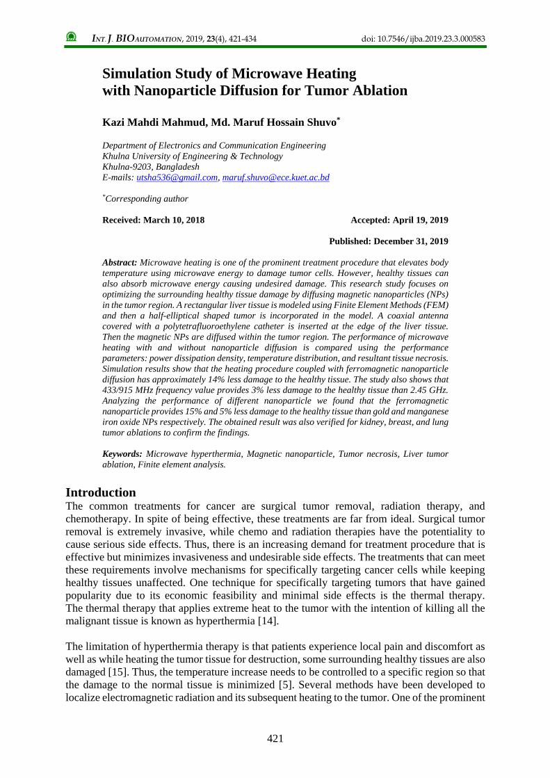
Results analysis and discussions Fig. 4 depicts the transportation mechanism of magnetic nanoparticles. They are injected from
the top of the injection pipe and released at the bottom of the pipe.
NPs spreads throughout the tumor region where their concentration is 1.661E-6 mole/m3.
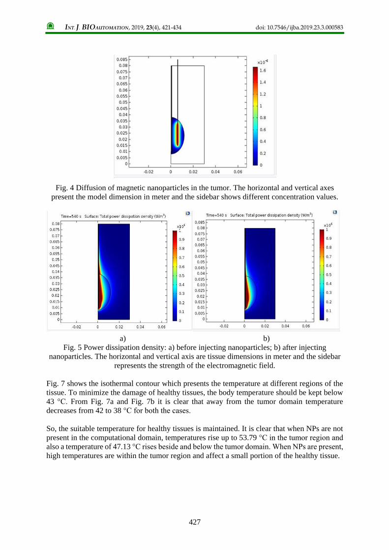
Fig. 5a and Fig. 5b shows the distribution of the source power before and after injecting NPs
respectively. Before injecting NPs some portion of the strong source power is seen to be
dissipated outside the tumor region resulting in a strong electromagnetic field and high-
temperature distribution. It is evident that the strong electromagnetic field is concentrated inside
the tumor region after diffusing NPs. Hence, a higher temperature distribution is observed
inside the tumor region than the healthy tissue region.
Fig. 6 shows the resulting steady-state temperature distribution before and after injecting NPs
in the liver tissue for an input microwave power of 10 W. We observed that near the antenna,
the heat source is strong, which leads to high temperatures as depicted in Fig. 6a, while far from
the antenna, the heat source is weaker and the normal body temperature of 37 °C is maintained.
Fig. 6b shows that the nanoparticles are effectively blocking the passage of energy into the
tissue area. Therefore, around the nanoparticle site, almost 3-4 °C low temperature is monitored.
This phenomenon indicates that the NPs are absorbing the microwave radiation.
INT. J. BIOAUTOMATION, 2019, 23(4), 421-434 doi: 10.7546/ijba.2019.23.3.000583
427
Fig. 4 Diffusion of magnetic nanoparticles in the tumor. The horizontal and vertical axes
present the model dimension in meter and the sidebar shows different concentration values.
a) b)
Fig. 5 Power dissipation density: a) before injecting nanoparticles; b) after injecting
nanoparticles. The horizontal and vertical axis are tissue dimensions in meter and the sidebar
represents the strength of the electromagnetic field.
Fig. 7 shows the isothermal contour which presents the temperature at different regions of the
tissue. To minimize the damage of healthy tissues, the body temperature should be kept below
43 °C. From Fig. 7a and Fig. 7b it is clear that away from the tumor domain temperature
decreases from 42 to 38 °C for both the cases.
So, the suitable temperature for healthy tissues is maintained. It is clear that when NPs are not
present in the computational domain, temperatures rise up to 53.79 °C in the tumor region and
also a temperature of 47.13 °C rises beside and below the tumor domain. When NPs are present,
high temperatures are within the tumor region and affect a small portion of the healthy tissue.

INT. J. BIOAUTOMATION, 2019, 23(4), 421-434 doi: 10.7546/ijba.2019.23.3.000583
428
a) b)
Fig. 6 Temperature distribution: a) before injecting nanoparticles; b) after injecting
nanoparticles. The horizontal and vertical axes are tissue dimensions in meter
and the sidebar represents temperature in °C.
a) b)
Fig. 7 Isothermal contour: a) before injecting nanoparticles; b) after injecting nanoparticles.
The horizontal and vertical axes are tissue dimensions in meter and the sidebar represents
20 different temperature values indicated by different colors.
Fig. 8 shows the fraction of necrotic tissue resulting from the microwave heating. When NPs
are not present a noticeable portion of healthy tissue is damaged around the tumor region.
On the other hand in the presence of NPs, a small portion of healthy tissue is damaged.
Fig. 9 represents how much of the tissue is damaged over time in different regions defined in
the cut points. The blue curve corresponds to the innermost point’s necrosis and the yellow one
corresponds to the outermost point’s necrosis.
Considering Fig. 9a and Fig. 9b, we observed that the tumor cell is almost destroyed for both
the cases. In case of nanoparticle diffusion, it took a little bit long time. Observing the curves,
we found that microwave heating alone hampers 69% of the healthy tissue. In contrast, when
heating is combined with magnetic NPs only 55% of the healthy tissue is hampered. Away from
the tumor less the difference is very acute. The same scenario observed in the vertical direction
also.

INT. J. BIOAUTOMATION, 2019, 23(4), 421-434 doi: 10.7546/ijba.2019.23.3.000583
429
a) b)
Fig. 8 Fraction of necrotic tissue: a) before injecting nanoparticles; b) after injecting
nanoparticles. The horizontal and vertical axes are tissue dimensions in meter and the sidebar
represents how much portion of the tissue is damaged (1 dictates 100% damage).
a) b)
Fig. 9 Point graph for damaged tissue: a) before injecting nanoparticles; b) after injecting
nanoparticles. The horizontal axis represents treatment time in the minute and the vertical axis
represents the portion of tissue necrosis due to heating (1 represents 100% necrosis).
Fig. 10 represents how much of the tissue is damaged at the 433/915 MHz frequency over time
in the cut points. We observed that at 433/915 MHz the tumor damage decreased from 95% to
93% compared to the 2.45 GHz. On the other hand, the healthy tissue damage decreased from
55% to 52%. So, in this case, the healthy tissue damage around the tumor site decreased by 3%.
Therefore, 433/915 MHz frequency would be more effective in minimizing healthy tissue
damage around the tumor site for magnetic nanoparticle hyperthermia.
We observed the effects of different NPs for liver tumor ablation using microwave radiation.
Fig. 11a and Fig. 11b shows the performance analysis of different NPs for liver tumor ablation
in terms of percent damage versus time. In case of gold NP no significant improvement
achieved in minimizing healthy tissue damage compared to the ferromagnetic NPs. Only 2%
less damage is monitored around the tumor. Manganese iron oxide (MnFe2O4) nanoparticle
shows almost similar performance to that of Fe2O3. We observed that MnFe2O4 provides 96%
tumor damage which is 3% more than the Fe2O3 NP. In case of healthy tissue, MnFe2O4 causes

INT. J. BIOAUTOMATION, 2019, 23(4), 421-434 doi: 10.7546/ijba.2019.23.3.000583
430
5% more damage than the Fe2O3. Therefore, ferromagnetic nanoparticle (Fe2O3) outperforms
gold and manganese iron oxide NPs. Therefore, ferromagnetic (Fe2O3) NPs outperforms in
hyperthermia treatment.
a) b)
Fig. 10 Point graph for damaged liver tissue with 433/915 MHz frequency:
a) before injecting NPs; b) after injecting NPs.
a) b)
Fig. 11 Performance analysis of different NPs for liver:
a) tumor damage; b) healthy tissue damage.
We have also analyzed the treatment procedure on human kidney, breast, and lung tissue while
considering tumors on relevant tissue-mimicking phantoms. We have used the 433/915 MHz
frequency and magnetic nanoparticle because of their better performance proved from the
previous analysis.

INT. J. BIOAUTOMATION, 2019, 23(4), 421-434 doi: 10.7546/ijba.2019.23.3.000583
431
Fig. 12a shows the percent of tumor damage over time on different human tissues caused by
the microwave heating in absence of ferromagnetic NPs at 433/915 MHz. We see that the liver,
kidney, breast, and lung tumors are fully damaged in 9, 6, 5 and 2 minutes of heating
respectively. Fig. 12b represents the percent of tumor damage over time in presence of
ferromagnetic NPs. The lung, breast, and kidney tumors are fully destroyed over 2, 5 and 6
minutes of heating respectively. Liver tumor damage is 95% within 9 minutes of heating.
a) b)
Fig. 12 Comparative analysis of magnetic nanoparticle hyperthermia on the different human
tissue: a) tumor damage without NP diffusion; b) tumor damage with NPs.
Fig. 13a illustrates the percent of healthy tissue damage on different human tissue caused by
microwave heating.
a) b)
Fig. 13 Comparative analysis of magnetic nanoparticle hyperthermia on different human
tissue considering: a) healthy tissue damage without NP diffusion;
b) healthy tissue damage with NPs.

INT. J. BIOAUTOMATION, 2019, 23(4), 421-434 doi: 10.7546/ijba.2019.23.3.000583
432
The damage on healthy tissues surrounding the tumor of liver, kidney, breast, and lung is 69%,
33%, 31% and 30% for 9, 6, 5 and 2 minutes of heating respectively. Fig. 13b presents the
percent of healthy tissue damage caused by the magnetic nanoparticle hyperthermia. The least
damage of 15% occurs in lung tissue in 2 minutes of heating time and this is 15% less than that
obtained with the heating procedure in absence of magnetic NPs. The highest damage of 52%
is caused in the liver tissue in 9 minutes of heating which provides 17% less damage. Breast and
kidney tissue damage percent is 29% and 31% over 5 and 6 minutes of heating respectively
each providing 2% less damage to surrounding healthy tissue.
Conclusions This research focuses on the improvement in Microwave coagulation therapy by incorporating
ferromagnetic nanoparticles in the treatment domain. Firstly, we performed the simulation of
microwave heating with and without diffusing the ferromagnetic nanoparticles in the targeted
tumor region. Our study illustrates that the nanoparticles absorb the microwave radiation.
Hence, the temperature is concentrated within the tumor thereby serving the purpose of
destructing tumor cell with less hampering the healthy tissues. The same simulation is
performed by varying microwave frequency and using different nanoparticles. The simulated
result confirms that the 415/915 MHz frequency and ferromagnetic nanoparticle perform better
in reducing healthy tissue damage. Moreover, the simulation was done for ablating lung, kidney
and breast tumors using ferromagnetic nanoparticle and 415/933 MHz frequency value. In case
of liver tumor ablation, the simulation findings are most promising providing 17% less damage
of healthy tissue and maintaining healthy tissue temperature below 43 °C in most of the region.
So, the findings of this research reveal that magnetic nanoparticle hyperthermia initiated by
microwave heating can be effective in practical treatment cases by ensuring rapid ablation of
the tumor while minimizing healthy tissue damage. The potential future directions this work
can lead to is the experiment considering the effect of blood flow velocity and vessel location.
References 1. Abenojar E. C., S. Wickramasinghe, J. Bas-Concepcion, A. C. S. Samia (2016). Structural
Effects on the Magnetic Hyperthermia Properties of Iron Oxide Nanoparticles, Progress in
Natural Science: Materials International, 26, 440-448.
2. Ali S. M., A. K. Jha, S. Mahapatra, M. Panigrahi (2014). Cancer Detection Using
Coagulation Therapy with Coaxial Antenna, Comsol International Conference, Bangalore,
India, https://www.comsol.com/paper/download/210861/ali_paper.pdf (Last access
December 04, 2019 )
3. Blagoeva R., A. Nedev, V. Michailova (2017). An Approach to Modeling Drug Release
from Polymersome Nanoparticles Based on PNIPAM-g-PEO Graft Copolymer,
International Journal Bioautomation, 21(2), 179-188.
4. Deshan Y., J. M. Bertram, M. C. Converse, A. P. O’Rourke, J. G. Webster, S. C. Hagness,
J. A. Will, D. M. Mahvi (2006). A Floating Sleeve Antenna Yields Localized Hepatic
Microwave Ablation, IEEE Transactions on Biomedical Engineering, 53(3), 533-537.
5. Dutz S., R. Hergt (2014). Magnetic Particle Hyperthermia – A Promising Tumor Therapy,
Nanotechnology, 25(45), 1-28.
6. Elkayal H. A., N. E. Ismail, M. Lotfy (2015). Microwaves for Breast Cancer Treatments,
Alexandria Engineering Journal, 54(4), 1105-1113.
7. Huo X., U. M. Jow, M. Ghovanloo (2010). Radiation Characterization of an Intra-oral
Wireless Device at Multiple ISM Bands: 433 MHZ, 915 MHZ, and 2.42 GHz, Annual
International Conference of the IEEE Engineering in Medicine and Biology, 1425-1428.
8. Javidi M., M. Heydari, A. Karimi, M. Haghpanahi, M. Navidbakhsh, A. Razmkon (2014).
Evaluation of the Effects of Injection Velocity and Different Gel Concentrations on

INT. J. BIOAUTOMATION, 2019, 23(4), 421-434 doi: 10.7546/ijba.2019.23.3.000583
433
Nanoparticles in Hyperthermia Therapy, Journal of Biomedical Physics and Engineering,
4(4), 151-162.
9. Keangin P., T. Wessapan, P. Rattanadecho (2011). Analysis of Heat Transfer in Deformed
Liver Cancer Modeling Treated Using a Microwave Coaxial Antenna, Applied Thermal
Engineering, 31, 3243-3254.
10. Kobayashi T. (2011). Cancer Hyperthermia Using Magnetic Nanoparticles, Biotechnology
Journal, 6(11), 1342-1347.
11. Krishnan K. M. (2010). Biomedical Nanomagnetics: A Spin through Possibilities in
Imaging, Diagnostics, and Therapy, IEEE Transactions on Magnetics, 46(7), 2523-2558.
12. Pavel M., G. Gradinariu, A. Stancu (2008). Study of the Optimum Dose of Ferromagnetic
Nanoparticles Suitable for Cancer Therapy Using MFH, IEEE Transactions on Magnetics,
44(11), 3205-3208.
13. Pearce J. A., J. R. Cook, S. Y. Emelianov (2010). Ferrimagnetic Nanoparticles Enhance
Microwave Heating for Tumor Hyperthermia Therapy, Annual International Conference of
the IEEE Engineering in Medicine and Biology, 2751-2754.
14. Salloum M., R. Ma, L. Zhu (2008). An in vivo Experimental Study of Temperature
Elevations in Animal Tissue during Magnetic Nanoparticle Hyperthermia, International
Journal of Hyperthermia, 24(7), 589-601.
15. Santos E. D., J. A. Green, N. Bhandari, A. Hong, P. Guitera, G. B. Fogarty (2015).
Tangential Volumetric Modulated Radiotherapy – A New Technique for Large Scalp
Lesions with a Case Study in Lentigo Maligna, International Journal Bioautomation, 19(2),
223-236.
16. Ward R. C., T. T. Healey, D. E. Dupuy (2013). Microwave Ablation Devices for
Interventional Oncology, Expert Review of Medical Devices, 10(2), 225-238.
17. Yang D., M. Converse, D. Mahvi (2007). Expanding the Bio-heat Equation to Include
Tissue Internal Water Evaporation during Heating, IEEE Transactions on Biomedical
Engineering, 54(8), 1382-1388.
Kazi Mahdi Mahmud, B.Sc.
E-mail: [email protected]
Mr. Kazi Mahdi Mahmud received his B.Sc. in Electronics and
Communication Engineering (ECE) degree from Khulna University of
Engineering & Technology (KUET), Khulna, Bangladesh, in 2018.
His research interests are finite element modeling, microwave
hyperthermia, and nanotechnology. Currently he is preparing for higher
studies and research.

INT. J. BIOAUTOMATION, 2019, 23(4), 421-434 doi: 10.7546/ijba.2019.23.3.000583
434
Assist. Prof. Md. Maruf Hossain Shuvo
E-mail: [email protected]
Md. Maruf Hossain Shuvo received his B.Sc. in Electronics and
Communication Engineering (ECE) degree from Khulna University of
Engineering & Technology (KUET), Bangladesh, in 2014. He is the
University Gold Medalist and Prime Minister Gold Medalist.
He received many awards including best paper awards at the
international conferences in recognition of his research.
After graduation, he has been working as a faculty member at ECE,
KUET, since 2015, and currently works as an Assistant Professor.
His research interests are medical imaging and analysis, finite element
modeling, and computational biomedical engineering. He is a member
of different professional societies and a reviewer of different
conferences and journals.
© 2019 by the authors. Licensee Institute of Biophysics and Biomedical Engineering,
Bulgarian Academy of Sciences. This article is an open access article distributed under
the terms and conditions of the Creative Commons Attribution (CC BY) license
(http://creativecommons.org/licenses/by/4.0/).