simulation of normal and pathological gaits using a fusion
TRANSCRIPT
J N E R JOURNAL OF NEUROENGINEERINGAND REHABILITATION
Martínez et al. Journal of NeuroEngineering and Rehabilitation 2013, 10:73http://www.jneuroengrehab.com/content/10/1/73
METHODOLOGY Open Access
Simulation of normal and pathological gaitsusing a fusion knowledge strategyFabio Martínez, Christian Cifuentes and Eduardo Romero*
Abstract
Gait distortion is the first clinical manifestation of many pathological disorders. Traditionally, the gait laboratory hasbeen the only available tool for supporting both diagnosis and prognosis, but under the limitation that any clinicalinterpretation depends completely on the physician expertise. This work presents a novel human gait model whichfusions two important gait information sources: an estimated Center of Gravity (CoG) trajectory and learned heelpaths, by that means allowing to reproduce kinematic normal and pathological patterns. The CoG trajectory isapproximated with a physical compass pendulum representation that has been extended by introducing energyaccumulator elements between the pendulum ends, thereby emulating the role of the leg joints and obtaining acomplete global gait description. Likewise, learned heel paths captured from actual data are learned to improve theperformance of the physical model, while the most relevant joint trajectories are estimated using a classical inversekinematic rule. The model is compared with standard gait patterns, obtaining a correlation coefficient of 0.96.Additionally,themodel simulates neuromuscular diseases like Parkinson (phase 2, 3 and 4) and clinical signs like theCrouch gait, case in which the averaged correlation coefficient is 0.92.
BackgroundQuantification of complex movements such as humanlocomotion is a fundamental step towards an objectivecharacterization of particular patterns associated to acertain degree of a disease [1-3]. The gait is the resultof complex interactions between several sub-systems:neuromuscular, musculo-tendinous and osteo-articular,which work together to generate the body dynamics thatunderlies the bipedal displacement [4,5]. In despite ofthe intensive research in biomechanics [6], robotics [7,8],medicine [9] and computer animation [10,11], the biologi-cal complexity has hindered a proper understanding of thelocomotor system. This problem has been partially over-come in the clinical routine by a gait estimation inferredfrom the gait laboratory [9,12,13]. Usually, a physicianor rehabilitation expert determines whether there existpathological gait patterns using exclusively her/his exper-tise [4,14,15]. Overall, diagnosis is supported using sta-tistical tests carried out on the acquired gait laboratorydata [16-20], with an inherent high degree of variability. Inconsequence, development of gait models that provide a
*Correspondence: [email protected]&Lab - School of Medicine, Universidad Nacional de Colombia, Bogotá DC,Colombia
quantitative gait description has become important in theprocess of supporting physician decisions [4,9,14,21].The main contribution of the present work is a human
gait model that accurately describes a set of kinematicgait patterns, normal or pathological. Themodel fuses twoimportant gait information sources: an estimated Centerof Gravity (CoG) trajectory and heel paths learned fromactual gaits. The global motion is governed by the CoGtrajectory of a compass physical pendulum representa-tion, coupled to a spring that emulates the muscle func-tion. This trajectory is regulated by learned heel paths,while the remaining joint patterns are estimated usinga classical inverse kinematic method. The models bene-fit is demonstrated by accurately simulating two differentsorts of neuromuscular gaits: Parkinson and Crouch pat-terns. Finally, a human-like leg structure is animated withthe obtained trajectories, allowing the clinician to inter-act with the model and facilitating the interpretation of anobservational analysis.Many models have been previously proposed for sim-
ulating the human gait, with different complexity levels,depending on the application area. A first group includesbipedal descriptions that exclusively use structural infor-mation so that they are able only to determine globalrelationships between muscles and joint angles. These
© 2013 Martínez et al.; licensee BioMed Central Ltd. This is an Open Access article distributed under the terms of the CreativeCommons Attribution License (http://creativecommons.org/licenses/by/2.0), which permits unrestricted use, distribution, andreproduction in any medium, provided the original work is properly cited.
Martínez et al. Journal of NeuroEngineering and Rehabilitation 2013, 10:73 Page 2 of 12http://www.jneuroengrehab.com/content/10/1/73
models exploit the conceptual simplicity of mechanicalsystems such as the inverted pendulum or mass-springs[22-26]. Basically, these approximations provide a loco-motion description from an energy standpoint, simulatingthe change from the kinematic to potential energy dur-ing the gait cycle. These models are devised to coarselyclassify normal and pathological patterns [18,27]. How-ever, a main drawback of these approximations is thatabout a 20% of the gait cycle, corresponding to the dou-ble stance phase, is completely eliminated. These physicalmodels are useful in areas like robotics since they elimi-nate the dependence on a robust control mechanism. Nev-ertheless, they are very limited for medical applicationsbecause of their strong simplifications, missing relevantgait aspects such as the non-linearities introduced by theheel strike.A second group of human gait models are capable of
simulating muscles and tendons during the gait. Thesemodels have obtained better gait representations, intro-ducing muscular information that is required from aclinical standpoint in terms of interpretability, i.e., spe-cific activity of certain muscle groups in musculoskele-tal disorders like hemiplegic movements. These modelshave introduced new elements to simulate the controland energy storage of the locomotion process. Specifi-cally, some gait approximations include the Hill modelas the base of the muscle representation [4,5,15], butwith no relation between the muscle and the locomo-tor structure and hence without any clinical meaning[28]. In these approaches, each model accelerates a spe-cific body segment, obtaining a simplistic simulationof pathological movements. Likewise, these models arenot accurate enough to describe the complex interac-tion among different groups of muscles. In addition,they require a certain number of parameters that needto be tuned, with the consequent dependence on an
expert knowledge. Scott Delp [10,29] introduced a com-putational strategy that combines the Hill muscle modeland structural information, accomplishing realistic nor-mal and pathological simulations, but again, with a highdegree of subjectivity at tuning the model parameters.Currently, several approaches have used some control-based strategies, requiring relatively few data to simulatesimple human structures and predicting new motions[30]. These approaches include a large number of degrees-of-freedomwhile joint force profiles remain subjected to alarge number of constraints [9,11,19,27,31]. These meth-ods approximate human control systems and simulatesome neurological pathologies [15], but these strategiesrequire specific information about each particular motionto be simulated and therefore they demand a high degreeof interaction and prior knowledge [27]. Moreover, thesemethods necessitate a large group of experimental data togenerate natural motions so that their clinical usefulnessstill remains very limited.
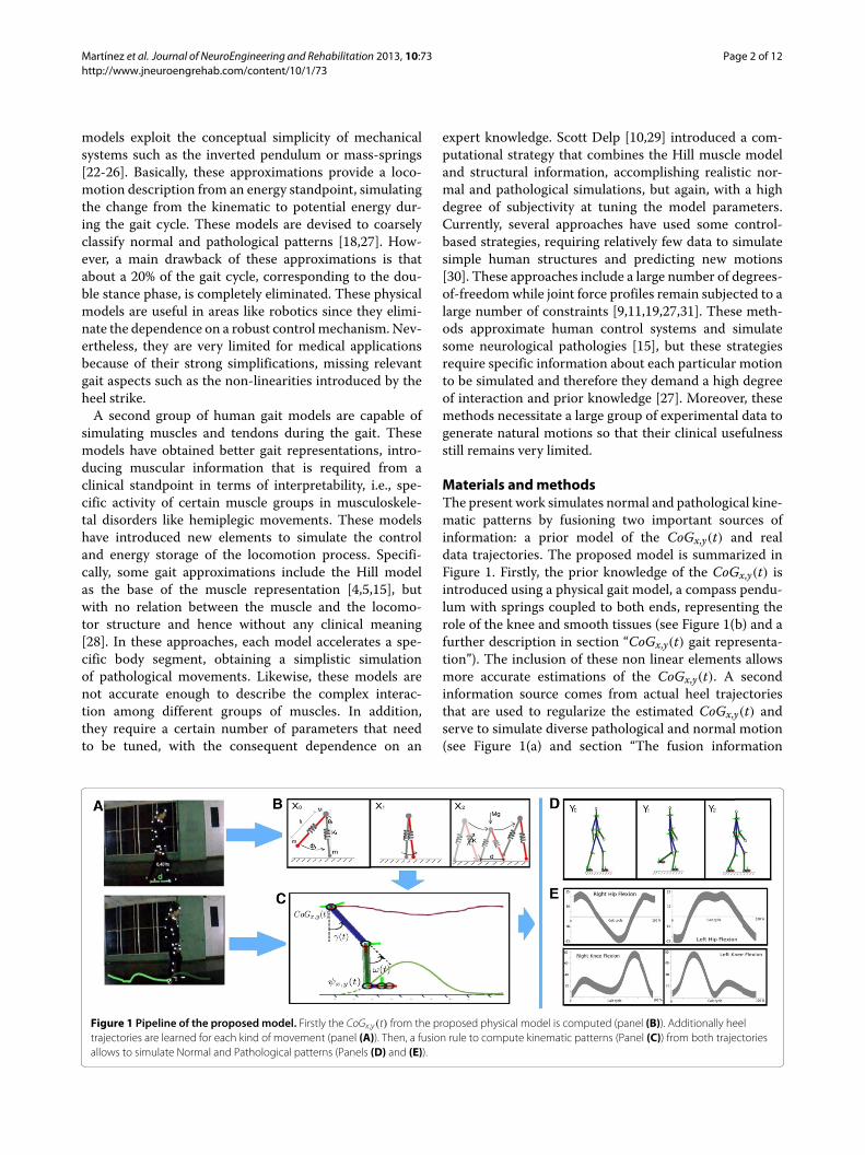
Materials andmethodsThe present work simulates normal and pathological kine-matic patterns by fusioning two important sources ofinformation: a prior model of the CoGx,y(t) and realdata trajectories. The proposed model is summarized inFigure 1. Firstly, the prior knowledge of the CoGx,y(t) isintroduced using a physical gait model, a compass pendu-lum with springs coupled to both ends, representing therole of the knee and smooth tissues (see Figure 1(b) and afurther description in section “CoGx,y(t) gait representa-tion”). The inclusion of these non linear elements allowsmore accurate estimations of the CoGx,y(t). A secondinformation source comes from actual heel trajectoriesthat are used to regularize the estimated CoGx,y(t) andserve to simulate diverse pathological and normal motion(see Figure 1(a) and section “The fusion information
Figure 1 Pipeline of the proposedmodel. Firstly the CoGx,y(t) from the proposed physical model is computed (panel (B)). Additionally heeltrajectories are learned for each kind of movement (panel (A)). Then, a fusion rule to compute kinematic patterns (Panel (C)) from both trajectoriesallows to simulate Normal and Pathological patterns (Panels (D) and (E)).
Martínez et al. Journal of NeuroEngineering and Rehabilitation 2013, 10:73 Page 3 of 12http://www.jneuroengrehab.com/content/10/1/73
strategy”). Additionally, this fusion facilitates an accu-rate estimation of the remaining joint trajectories, usinga classical inverse kinematic framework. Finally the setof obtained trajectories animates a human-like leg struc-ture that provides the clinician with a interpretable tool(see Figure 1(c-d)).
Ethical approvalThe study was approved by the Ethics Committees of theInstitute. Written informed consent was obtained by theparents or, when applicable, by the patients.
CoGx,y(t) gait representationIn human movement analysis, the gait is divided in cycles,coarsely classified as double and single stance phases[14,32,33]. The double stance period accounts for around20% of gait cycle and stands for the body movement withboth limbs touching the ground, while the single stancerepresents around 80% of gait cycle and corresponds tothe interval in which a single limb supports the wholebody weight. In this work theCoGx,y(t) for a complete gaitcycle is approached using two complementary strategies: acompass pendulum for the single stance and a spring masssystem for the double stance, as follows.
The single support phaseThe single support phase conserves a regular periodic-ity which is properly captured using a compass pendulumrepresentation. This strategy represents the upper part ofthe body by a massM which moves forwards with respectto each fixed point (with mass m), describing a harmonicoscillating trajectory, similar to the inverted pendulum[22,34]. Likewise, the free foot swings with respect to thismass, establishing a simple pendulum pattern. Providedthat these processes are coupled together, the humangait is modeled by a compass pendulum as two couplednon-linear differential equations:
β(1 − cosφ)(3θ − φ) − β sinφ(φ2 − 2θφ)
+ (g sin θ
l )(β(sin(θ − φ) − 1)) = 0
θ (β(1 − cosφ)) − βφ + βθ2 sinφ + (βgl ) sin(θ − φ) = 0
(1)
where β = m/M , θ is the angle of the stance leg at the par-ticular time t with respect to the slope and φ is the anglebetween the stance leg, and l0 = lr = ll. This model alsoallows to simulate the swing foot when it hits the groundat the heelstrike, a time in the cycle that corresponds toφ(t) − 2θ(t) = 0 [34], when the double stance starts.
Double stance phaseClassical gait models often ignore the double supportstance since they have been devised to simplify the gait
rather than to accurately follow gait patterns. These sim-plifications have ended up by considering the leg struc-tures as rigid segments, a hypothesis that easily leads toconclude for instance that the percentage of gait recov-ery is inefficient in energy terms, a reason why this phasehas been eliminated in most of these strategies [4,9,21,35].Additionally, important elastic contributions which pro-duce relevant changes in the CoGx,y(t), during the doublestance, are often neglected. These strong simplificationsreduce an appropriate gait understanding and may leadto wrong interpretations when these models are used assupporting tools of clinical decisions.A more accurate CoGx,y(t) description of the double
stance phase was herein achieved by coupling a planarspring-mass system [36] to the compass pendulum, pre-viously introduced. This change of the leg length l duringthe gait stance phase, allows to estimate the reaction forceduring the whole gait cycle, as illustrated in Figure 1 (B).Notice that each leg reaction forces points out towardsopposite sides, separated by a distance d (the distancebetween the heelstrike and the other toe-off phase). Thecoupling is obtained as:
Mx = llx − lr(d − x)My = lly + lry − gM
(2)
where g is the gravity, ll and lr are the left and right legs,respectively and their length changes as:
ll = k(l0√
x2 + y2− 1)
lr = k(l0√
(d − x)2 + y2− 1)
(3)
These equations simulate the periodic vertical groundforces, with a period defined by T = 2π
√mk . This inde-
pendent formulation of each reaction force allows an inde-pendent analysis of each link, whereby gait abnormalitiesthat asymmetrically affect each leg, such as the diplegia,can be simulated. Finally, the CoGx,y(t) is simulated by theintegration of the two gait phases described as follows:
CoGx,y(t)=
⎧⎪⎨⎪⎩l0 [sin θ(t), cos θ(t)] if φ(t)−2θ(t)<0;[ll x
36 −lr(d− x3
6 )
M , lly36 +lr y
36 −gM
M
]elsewhere.
(4)
The fusion information strategyAlthough the CoGx,y(t) is a fundamental clinical descrip-tor [18], a useful identification of a particular disorder alsorequires a proper gait analysis of other anatomical jointtrajectories. Accordingly, a more complete gait descrip-tion was herein achieved by the fusion of two important
Martínez et al. Journal of NeuroEngineering and Rehabilitation 2013, 10:73 Page 4 of 12http://www.jneuroengrehab.com/content/10/1/73
sources of information: the physical gait strategy previ-ously described and the learned heel trajectories.The learned heel trajectories were modeled as a set
of normal distributions with mean μi and variance σ 2i
from three different groups of patients captured in a gaitlaboratory as:
ψx,y(t) =I∑
i=1wiN(t|μi, σ 2
i )
where I represents the total number of learned gait move-ments (normal, Crouch and Parkinsonian gaits). Each gaitdistribution was computed from 30 gait cycles belong-ing to 10 patients (7 men and 3 women). From thismulti-gaussian distribution model, we can select a heeltrajectory i to regularize the CoGx,y(t) associated to aparticular gait movement. Likewise, the normal motiondistribution allows a large variety of gait patterns of thesame pathology. New relationships are inferred from thesetwo trajectories by assuming the knee joint position asrx,y = l0x,y
2 [37]. Afterward, a classical inverse kinematicmethod is adapted to obtain two main kinematic patterns:the flexion-extension patterns of the hip ω(t) and kneeγ (t). For doing so, at each time t of the gait cycle, a CCDmethod performs an iterative rigid transformation overeach couple of joints. The two patterns are defined as:
γ (t) = acos(CoGx,y(t)2 − r2x,y − r2x,y
2
)(5)
ω(t) = atan2(ψx,y) + atan2(rx,y sin γ , rx,y + rx,y cos γ )
(6)
where r is the distance between the CoGx,y(t) and ψx,y(t).Unlike other approaches, this model estimates kinematicpatterns with medical meaning, but the model can alsoobtain energy and ground force patterns for normal andpathological cases, obtained from the CoGx,y(t).
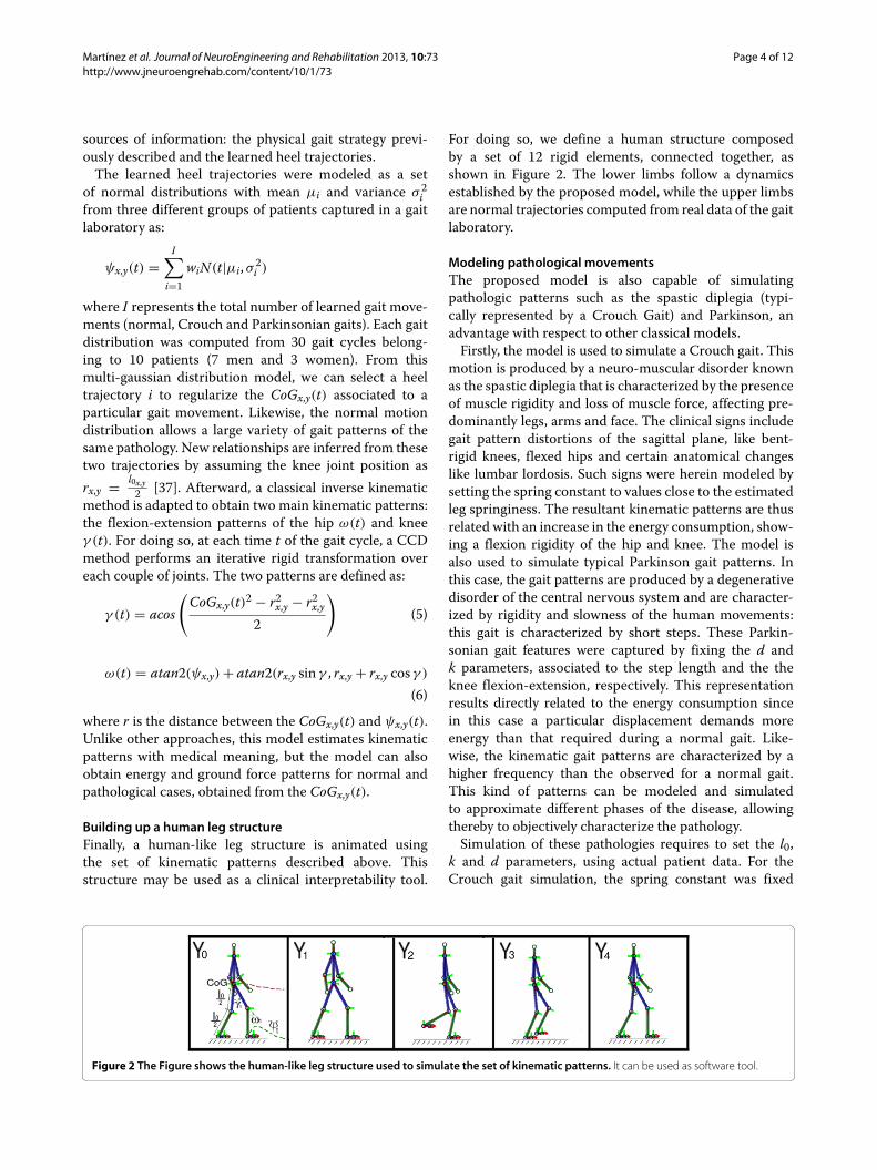
Building up a human leg structureFinally, a human-like leg structure is animated usingthe set of kinematic patterns described above. Thisstructure may be used as a clinical interpretability tool.
For doing so, we define a human structure composedby a set of 12 rigid elements, connected together, asshown in Figure 2. The lower limbs follow a dynamicsestablished by the proposed model, while the upper limbsare normal trajectories computed from real data of the gaitlaboratory.
Modeling pathological movementsThe proposed model is also capable of simulatingpathologic patterns such as the spastic diplegia (typi-cally represented by a Crouch Gait) and Parkinson, anadvantage with respect to other classical models.Firstly, the model is used to simulate a Crouch gait. This
motion is produced by a neuro-muscular disorder knownas the spastic diplegia that is characterized by the presenceof muscle rigidity and loss of muscle force, affecting pre-dominantly legs, arms and face. The clinical signs includegait pattern distortions of the sagittal plane, like bent-rigid knees, flexed hips and certain anatomical changeslike lumbar lordosis. Such signs were herein modeled bysetting the spring constant to values close to the estimatedleg springiness. The resultant kinematic patterns are thusrelated with an increase in the energy consumption, show-ing a flexion rigidity of the hip and knee. The model isalso used to simulate typical Parkinson gait patterns. Inthis case, the gait patterns are produced by a degenerativedisorder of the central nervous system and are character-ized by rigidity and slowness of the human movements:this gait is characterized by short steps. These Parkin-sonian gait features were captured by fixing the d andk parameters, associated to the step length and the theknee flexion-extension, respectively. This representationresults directly related to the energy consumption sincein this case a particular displacement demands moreenergy than that required during a normal gait. Like-wise, the kinematic gait patterns are characterized by ahigher frequency than the observed for a normal gait.This kind of patterns can be modeled and simulatedto approximate different phases of the disease, allowingthereby to objectively characterize the pathology.Simulation of these pathologies requires to set the l0,
k and d parameters, using actual patient data. For theCrouch gait simulation, the spring constant was fixed
Figure 2 The Figure shows the human-like leg structure used to simulate the set of kinematic patterns. It can be used as software tool.
Martínez et al. Journal of NeuroEngineering and Rehabilitation 2013, 10:73 Page 5 of 12http://www.jneuroengrehab.com/content/10/1/73
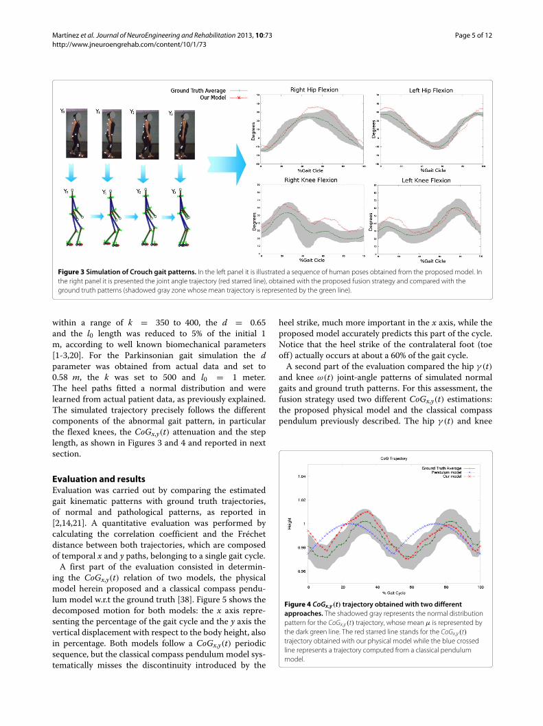
Figure 3 Simulation of Crouch gait patterns. In the left panel it is illustrated a sequence of human poses obtained from the proposed model. Inthe right panel it is presented the joint angle trajectory (red starred line), obtained with the proposed fusion strategy and compared with theground truth patterns (shadowed gray zone whose mean trajectory is represented by the green line).
within a range of k = 350 to 400, the d = 0.65and the l0 length was reduced to 5% of the initial 1m, according to well known biomechanical parameters[1-3,20]. For the Parkinsonian gait simulation the dparameter was obtained from actual data and set to0.58 m, the k was set to 500 and l0 = 1 meter.The heel paths fitted a normal distribution and werelearned from actual patient data, as previously explained.The simulated trajectory precisely follows the differentcomponents of the abnormal gait pattern, in particularthe flexed knees, the CoGx,y(t) attenuation and the steplength, as shown in Figures 3 and 4 and reported in nextsection.
Evaluation and resultsEvaluation was carried out by comparing the estimatedgait kinematic patterns with ground truth trajectories,of normal and pathological patterns, as reported in[2,14,21]. A quantitative evaluation was performed bycalculating the correlation coefficient and the Fréchetdistance between both trajectories, which are composedof temporal x and y paths, belonging to a single gait cycle.A first part of the evaluation consisted in determin-
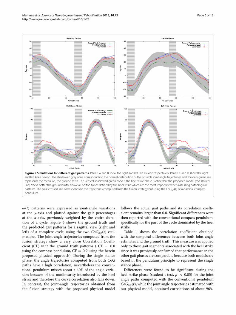
ing the CoGx,y(t) relation of two models, the physicalmodel herein proposed and a classical compass pendu-lum model w.r.t the ground truth [38]. Figure 5 shows thedecomposed motion for both models: the x axis repre-senting the percentage of the gait cycle and the y axis thevertical displacement with respect to the body height, alsoin percentage. Both models follow a CoGx,y(t) periodicsequence, but the classical compass pendulum model sys-tematically misses the discontinuity introduced by the
heel strike, much more important in the x axis, while theproposed model accurately predicts this part of the cycle.Notice that the heel strike of the contralateral foot (toeoff ) actually occurs at about a 60% of the gait cycle.A second part of the evaluation compared the hip γ (t)
and knee ω(t) joint-angle patterns of simulated normalgaits and ground truth patterns. For this assessment, thefusion strategy used two different CoGx,y(t) estimations:the proposed physical model and the classical compasspendulum previously described. The hip γ (t) and knee
Figure 4 CoGx,y(t) trajectory obtained with two differentapproaches. The shadowed gray represents the normal distributionpattern for the CoGx,y(t) trajectory, whose mean μ is represented bythe dark green line. The red starred line stands for the CoGx,y(t)trajectory obtained with our physical model while the blue crossedline represents a trajectory computed from a classical pendulummodel.
Martínez et al. Journal of NeuroEngineering and Rehabilitation 2013, 10:73 Page 6 of 12http://www.jneuroengrehab.com/content/10/1/73
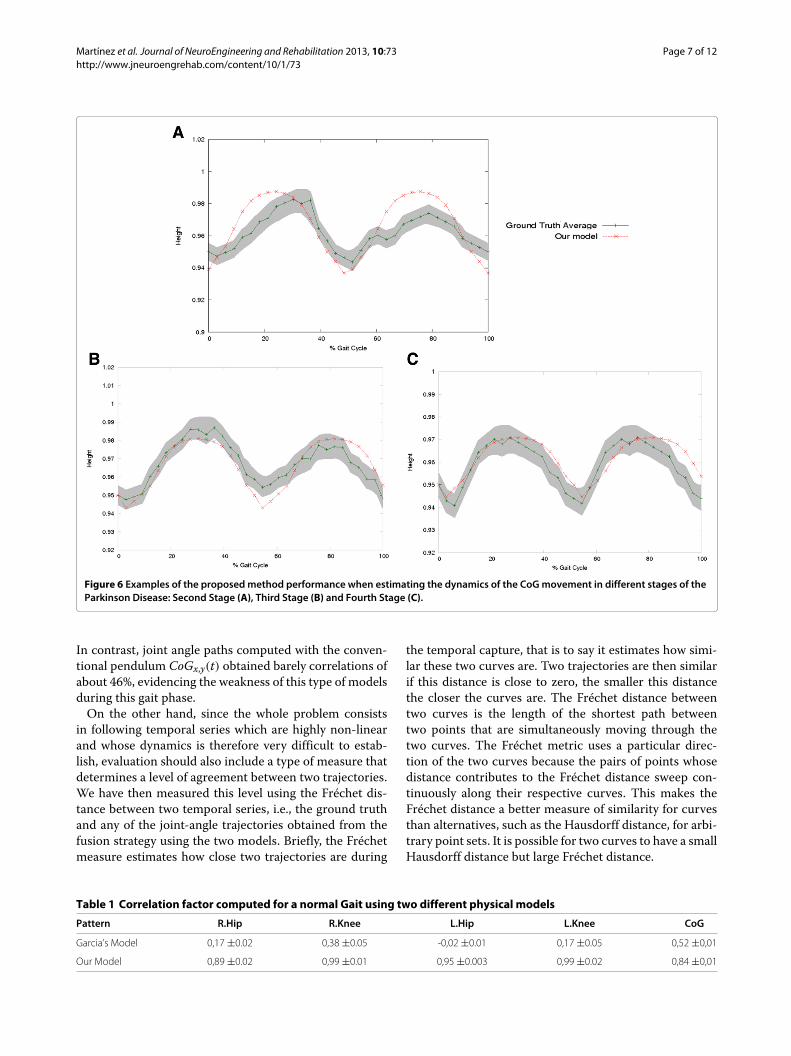
Figure 5 Simulations for different gait patterns. Panels A and B show the right and left Hip Flexion respectively. Panels C and D show the rightand left knee flexion. The shadowed gray zone corresponds to the normal distribution of the possible joint angle trajectories and the dark green linerepresents the mean, i.e., the ground truth. The vertical shadowed green zone is the heel strike phase. Notice that the proposed model (red starredline) tracks better the ground truth, above all on the zones defined by the heel strike which are the most important when assessing pathologicalpatterns. The blue crossed line corresponds to the trajectories computed from the fusion strategy but using the CoGx,y(t) of a classical compasspendulum.
ω(t) patterns were expressed as joint-angle variationsat the y-axis and plotted against the gait percentagesat the x-axis, previously weighted by the entire dura-tion of a cycle. Figure 6 shows the ground truth andthe predicted gait patterns for a sagittal view (right andleft) of a complete cycle, using the two CoGx,y(t) esti-mations. The joint-angle trajectories computed from thefusion strategy show a very close Correlation Coeffi-cient (CF) w.r.t the ground truth patterns ( CF = 0.8using the compass pendulum, CF = 0.9 using the hereinproposed physical approach). During the single stancephase, the angle trajectories computed from both CoGpaths have a high correlation, nevertheless the conven-tional pendulum misses about a 40% of the angle varia-tion because of the nonlinearity introduced by the heelstrike and therefore the curve correlation also falls down.In contrast, the joint-angle trajectories obtained fromthe fusion strategy with the proposed physical model
follows the actual gait paths and its correlation coeffi-cient remains larger than 0.8. Significant differences werethen reported with the conventional compass pendulum,specifically for the part of the cycle dominated by the heelstrike.Table 1 shows the correlation coefficient obtained
with the temporal differences between both joint angleestimates and the ground truth. This measure was appliedonly to those gait segments associated with the heel strikesince it was previously confirmed that performance in theother gait phases are comparable because both models arebased in the pendulum principle to represent the singlestance phase.Differences were found to be significant during the
heel strike phase (student t-test, p < 0.05) for the jointangle paths computed with the conventional pendulumCoGx,y(t), while the joint angle trajectories estimated withour physical model, obtained correlations of about 96%.
Martínez et al. Journal of NeuroEngineering and Rehabilitation 2013, 10:73 Page 7 of 12http://www.jneuroengrehab.com/content/10/1/73
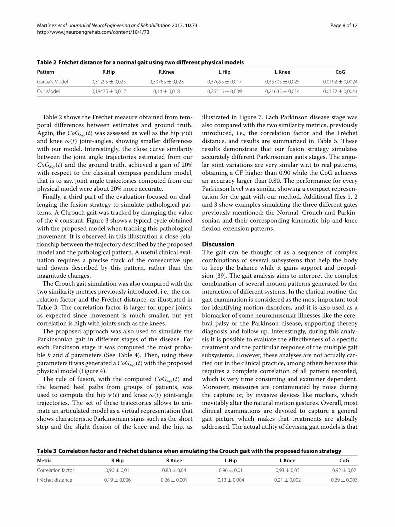
Figure 6 Examples of the proposedmethod performance when estimating the dynamics of the CoGmovement in different stages of theParkinson Disease: Second Stage (A), Third Stage (B) and Fourth Stage (C).
In contrast, joint angle paths computed with the conven-tional pendulum CoGx,y(t) obtained barely correlations ofabout 46%, evidencing the weakness of this type of modelsduring this gait phase.On the other hand, since the whole problem consists
in following temporal series which are highly non-linearand whose dynamics is therefore very difficult to estab-lish, evaluation should also include a type of measure thatdetermines a level of agreement between two trajectories.We have then measured this level using the Fréchet dis-tance between two temporal series, i.e., the ground truthand any of the joint-angle trajectories obtained from thefusion strategy using the two models. Briefly, the Fréchetmeasure estimates how close two trajectories are during
the temporal capture, that is to say it estimates how simi-lar these two curves are. Two trajectories are then similarif this distance is close to zero, the smaller this distancethe closer the curves are. The Fréchet distance betweentwo curves is the length of the shortest path betweentwo points that are simultaneously moving through thetwo curves. The Fréchet metric uses a particular direc-tion of the two curves because the pairs of points whosedistance contributes to the Fréchet distance sweep con-tinuously along their respective curves. This makes theFréchet distance a better measure of similarity for curvesthan alternatives, such as the Hausdorff distance, for arbi-trary point sets. It is possible for two curves to have a smallHausdorff distance but large Fréchet distance.
Table 1 Correlation factor computed for a normal Gait using two different physical models
Pattern R.Hip R.Knee L.Hip L.Knee CoG
Garcia’s Model 0,17 ±0.02 0,38 ±0.05 -0,02 ±0.01 0,17 ±0.05 0,52 ±0,01
Our Model 0,89 ±0.02 0,99 ±0.01 0,95 ±0.003 0,99 ±0.02 0,84 ±0,01
Martínez et al. Journal of NeuroEngineering and Rehabilitation 2013, 10:73 Page 8 of 12http://www.jneuroengrehab.com/content/10/1/73
Table 2 Fréchet distance for a normal gait using two different physical models
Pattern R.Hip R.Knee L.Hip L.Knee CoG
Garcia’s Model 0,31295 ± 0,025 0,30765 ± 0,023 0,37695 ± 0,017 0,35305 ± 0,025 0,0192 ± 0,0024
Our Model 0,18475 ± 0,012 0,14 ± 0,018 0,26515 ± 0,009 0,21635 ± 0,014 0,0132 ± 0,0041
Table 2 shows the Fréchet measure obtained from tem-poral differences between estimates and ground truth.Again, the CoGx,y(t) was assessed as well as the hip γ (t)and knee ω(t) joint-angles, showing smaller differenceswith our model. Interestingly, the close curve similaritybetween the joint angle trajectories estimated from ourCoGx,y(t) and the ground truth, achieved a gain of 20%with respect to the classical compass pendulum model,that is to say, joint angle trajectories computed from ourphysical model were about 20% more accurate.Finally, a third part of the evaluation focused on chal-
lenging the fusion strategy to simulate pathological pat-terns. A Chrouch gait was tracked by changing the valueof the k constant. Figure 3 shows a typical cycle obtainedwith the proposed model when tracking this pathologicalmovement. It is observed in this illustration a close rela-tionship between the trajectory described by the proposedmodel and the pathological pattern. A useful clinical eval-uation requires a precise track of the consecutive upsand downs described by this pattern, rather than themagnitude changes.The Crouch gait simulation was also compared with the
two similarity metrics previously introduced, i.e., the cor-relation factor and the Fréchet distance, as illustrated inTable 3. The correlation factor is larger for upper joints,as expected since movement is much smaller, but yetcorrelation is high with joints such as the knees.The proposed approach was also used to simulate the
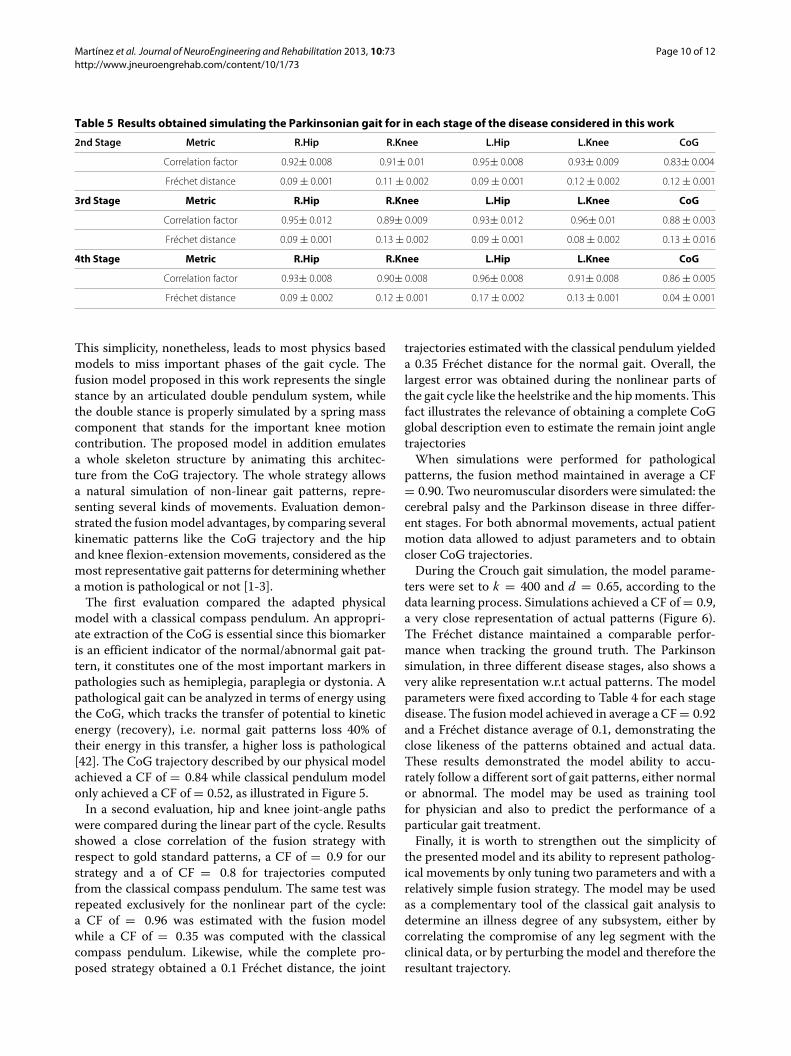
Parkinsonian gait in different stages of the disease. Foreach Parkinson stage it was computed the most proba-ble k and d parameters (See Table 4). Then, using theseparameters it was generated aCoGx,y(t)with the proposedphysical model (Figure 4).The rule of fusion, with the computed CoGx,y(t) and
the learned heel paths from groups of patients, wasused to compute the hip γ (t) and knee ω(t) joint-angletrajectories. The set of these trajectories allows to ani-mate an articulated model as a virtual representation thatshows characteristic Parkinsonian signs such as the shortstep and the slight flexion of the knee and the hip, as
illustrated in Figure 7. Each Parkinson disease stage wasalso compared with the two similarity metrics, previouslyintroduced, i.e., the correlation factor and the Fréchetdistance, and results are summarized in Table 5. Theseresults demonstrate that our fusion strategy simulatesaccurately different Parkinsonian gaits stages. The angu-lar joint variations are very similar w.r.t to real patterns,obtaining a CF higher than 0.90 while the CoG achievesan accuracy larger than 0.80. The performance for everyParkinson level was similar, showing a compact represen-tation for the gait with our method. Additional files 1, 2and 3 show examples simulating the three different gatespreviously mentioned: the Normal, Crouch and Parkin-sonian and their corresponding kinematic hip and kneeflexion-extension patterns.
DiscussionThe gait can be thought of as a sequence of complexcombinations of several subsystems that help the bodyto keep the balance while it gains support and propul-sion [39]. The gait analysis aims to interpret the complexcombination of several motion patterns generated by theinteraction of different systems. In the clinical routine, thegait examination is considered as the most important toolfor identifying motion disorders, and it is also used as abiomarker of some neuromuscular illnesses like the cere-bral palsy or the Parkinson disease, supporting therebydiagnosis and follow up. Interestingly, during this analy-sis it is possible to evaluate the effectiveness of a specifictreatment and the particular response of the multiple gaitsubsystems. However, these analyses are not actually car-ried out in the clinical practice, among others because thisrequires a complete correlation of all pattern recorded,which is very time consuming and examiner dependent.Moreover, measures are contaminated by noise duringthe capture or, by invasive devices like markers, whichinevitably alter the natural motion gestures. Overall, mostclinical examinations are devoted to capture a generalgait picture which makes that treatments are globallyaddressed. The actual utility of devising gait models is that
Table 3 Correlation factor and Fréchet distance when simulating the Crouch gait with the proposed fusion strategy
Metric R.Hip R.Knee L.Hip L.Knee CoG
Correlation factor 0,96 ± 0,01 0,88 ± 0,04 0,96 ± 0,01 0,93 ± 0,03 0.92 ± 0,02
Fréchet distance 0,19 ± 0,006 0,26 ± 0,001 0,13 ± 0,004 0,21 ± 0,002 0,29 ± 0,003
Martínez et al. Journal of NeuroEngineering and Rehabilitation 2013, 10:73 Page 9 of 12http://www.jneuroengrehab.com/content/10/1/73
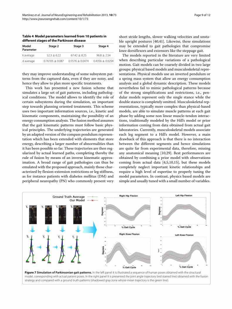
Table 4 Model parameters learned from 10 patients indifferent stages of the Parkinson disease
ModelParameter
Stage 2 Stage 3 Stage 4
k average 52.3 ± 6.22 67.42 ± 8.25 96.8 ± 2.54
d average 0.76105 ± 0.087 0.5576 ± 0.0474 0.4356 ± 0.0294
they may improve understanding of some subsystem pat-terns from the captured data, even if they are noisy, andhence they allow to plan more specific treatments.This work has presented a new fusion scheme that
simulates a large set of gait patterns, including patholog-ical conditions. The model allows to identify the role ofcertain subsystems during the simulation, an importantstep towards planning oriented treatments. This schemeuses two important information sources, i.e., kinetic andkinematic components, maintaining the possibility of anenergy consumption analysis. The fusionmethod assumesthat the gait kinematic patterns must follow basic phys-ical principles. The underlying trajectories are generatedby an adapted version of the compass pendulum represen-tation which has been extended with elements that storeenergy, describing a larger number of abnormalities thanit has been possible so far. These trajectories are then reg-ularized by actual learned paths, completing thereby therule of fusion by means of an inverse kinematic approx-imation. A broad range of gait pathologies can thus beemulated with the proposed approach, mainly those char-acterized by flexion-extension restrictions or leg stiffness,as for instance patients with diabetes mellitus (DM) andperipheral neuropathy (PN) who commonly present very
short stride lengths, slower walking velocities and unsta-ble upright postures [40,41]. Likewise, these simulationsmay be extended to gait pathologies that compromiseknee dorsiflexors and extensors like the steppage gait.The models reported in the literature are very limited
when describing particular variations of a pathologicalmotion. Gait models can be coarsely divided in two largegroups: physical based models and musculoskeletal repre-sentations. Physical models use an inverted pendulum ora spring mass system that allow an energy consumptionanalysis and a global dynamic description. These modelsnevertheless fail to mimic pathological patterns becauseof the strong simplifications and restrictions, i.e., pen-dular models represent only the single stance while thedouble stance is completely omitted. Musculoskeletal rep-resentations, typically more complex than physical-basedmodels, are able to simulate muscle patterns at each gaitphase by adding some non linear muscle-tendon interac-tions, traditionally modeled by the Hill’s model or priorinformation coming from data obtained from actual gaitlaboratories. Currently, musculoskeletal models associateeach leg segment to a Hill’s model. However, a maindrawback of this approach is that there is no interactionbetween the different segments and hence simulationsare quite far from experimental data, therefore, missingany anatomical meaning [10,29]. Best performances areobtained by combining a prior model with observationscoming from actual data [4,5,10,15], but these modelscompletely neglect important kinetic relationships andrequire a high level of expertise to properly tuning themodel parameters. In contrast, physics based models aresimple and usually tuned with a small number of variables.
Figure 7 Simulation of Parkinsonian gait patterns. In the left panel it is illustrated a sequence of human poses obtained with the structuralmodel, corresponding with actual patient poses. In the right panel it is presented the joint angle trajectory (red starred line) obtained with the fusionstrategy and compared with a ground truth patterns (shadowed gray zone whose mean trajectory is the green line).
Martínez et al. Journal of NeuroEngineering and Rehabilitation 2013, 10:73 Page 10 of 12http://www.jneuroengrehab.com/content/10/1/73
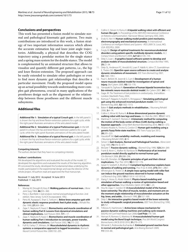
Table 5 Results obtained simulating the Parkinsonian gait for in each stage of the disease considered in this work
2nd Stage Metric R.Hip R.Knee L.Hip L.Knee CoG
Correlation factor 0.92± 0.008 0.91± 0.01 0.95± 0.008 0.93± 0.009 0.83± 0.004
Fréchet distance 0.09 ± 0.001 0.11 ± 0.002 0.09 ± 0.001 0.12 ± 0.002 0.12 ± 0.001
3rd Stage Metric R.Hip R.Knee L.Hip L.Knee CoG
Correlation factor 0.95± 0.012 0.89± 0.009 0.93± 0.012 0.96± 0.01 0.88 ± 0.003
Fréchet distance 0.09 ± 0.001 0.13 ± 0.002 0.09 ± 0.001 0.08 ± 0.002 0.13 ± 0.016
4th Stage Metric R.Hip R.Knee L.Hip L.Knee CoG
Correlation factor 0.93± 0.008 0.90± 0.008 0.96± 0.008 0.91± 0.008 0.86 ± 0.005
Fréchet distance 0.09 ± 0.002 0.12 ± 0.001 0.17 ± 0.002 0.13 ± 0.001 0.04 ± 0.001
This simplicity, nonetheless, leads to most physics basedmodels to miss important phases of the gait cycle. Thefusion model proposed in this work represents the singlestance by an articulated double pendulum system, whilethe double stance is properly simulated by a spring masscomponent that stands for the important knee motioncontribution. The proposed model in addition emulatesa whole skeleton structure by animating this architec-ture from the CoG trajectory. The whole strategy allowsa natural simulation of non-linear gait patterns, repre-senting several kinds of movements. Evaluation demon-strated the fusionmodel advantages, by comparing severalkinematic patterns like the CoG trajectory and the hipand knee flexion-extension movements, considered as themost representative gait patterns for determining whethera motion is pathological or not [1-3].The first evaluation compared the adapted physical
model with a classical compass pendulum. An appropri-ate extraction of the CoG is essential since this biomarkeris an efficient indicator of the normal/abnormal gait pat-tern, it constitutes one of the most important markers inpathologies such as hemiplegia, paraplegia or dystonia. Apathological gait can be analyzed in terms of energy usingthe CoG, which tracks the transfer of potential to kineticenergy (recovery), i.e. normal gait patterns loss 40% oftheir energy in this transfer, a higher loss is pathological[42]. The CoG trajectory described by our physical modelachieved a CF of = 0.84 while classical pendulum modelonly achieved a CF of = 0.52, as illustrated in Figure 5.In a second evaluation, hip and knee joint-angle paths
were compared during the linear part of the cycle. Resultsshowed a close correlation of the fusion strategy withrespect to gold standard patterns, a CF of = 0.9 for ourstrategy and a of CF = 0.8 for trajectories computedfrom the classical compass pendulum. The same test wasrepeated exclusively for the nonlinear part of the cycle:a CF of = 0.96 was estimated with the fusion modelwhile a CF of = 0.35 was computed with the classicalcompass pendulum. Likewise, while the complete pro-posed strategy obtained a 0.1 Fréchet distance, the joint
trajectories estimated with the classical pendulum yieldeda 0.35 Fréchet distance for the normal gait. Overall, thelargest error was obtained during the nonlinear parts ofthe gait cycle like the heelstrike and the hipmoments. Thisfact illustrates the relevance of obtaining a complete CoGglobal description even to estimate the remain joint angletrajectoriesWhen simulations were performed for pathological
patterns, the fusion method maintained in average a CF= 0.90. Two neuromuscular disorders were simulated: thecerebral palsy and the Parkinson disease in three differ-ent stages. For both abnormal movements, actual patientmotion data allowed to adjust parameters and to obtaincloser CoG trajectories.During the Crouch gait simulation, the model parame-
ters were set to k = 400 and d = 0.65, according to thedata learning process. Simulations achieved a CF of = 0.9,a very close representation of actual patterns (Figure 6).The Fréchet distance maintained a comparable perfor-mance when tracking the ground truth. The Parkinsonsimulation, in three different disease stages, also shows avery alike representation w.r.t actual patterns. The modelparameters were fixed according to Table 4 for each stagedisease. The fusionmodel achieved in average a CF= 0.92and a Fréchet distance average of 0.1, demonstrating theclose likeness of the patterns obtained and actual data.These results demonstrated the model ability to accu-rately follow a different sort of gait patterns, either normalor abnormal. The model may be used as training toolfor physician and also to predict the performance of aparticular gait treatment.Finally, it is worth to strengthen out the simplicity of
the presented model and its ability to represent patholog-ical movements by only tuning two parameters and with arelatively simple fusion strategy. The model may be usedas a complementary tool of the classical gait analysis todetermine an illness degree of any subsystem, either bycorrelating the compromise of any leg segment with theclinical data, or by perturbing the model and therefore theresultant trajectory.
Martínez et al. Journal of NeuroEngineering and Rehabilitation 2013, 10:73 Page 11 of 12http://www.jneuroengrehab.com/content/10/1/73
Conclusions and perspectivesThis work has presented a fusion model to simulate nor-mal and pathological kinematic gait patterns. Two maincontributions are introduced in this work, a fusion strat-egy of two important information sources which allowsthe accurate estimation hip and knee joint angle trajec-tories. Additionally, a physical that describes the COGtrajectory using a pendular motion for the single stanceand a springmass system for the double stance. Themodelis complemented by an animated structure that allows tovisualize and quantify different gait patterns, i.e., the hipand knee flexion-extension. The proposed approach canbe easily extended to simulate other pathologies or evento find more dynamic gait relationships that describe aparticular movement. Finally the proposed model opensup an actual possibility towards understandingmore com-plex gait phenomena, crucial in many applications of theprostheses design such as the alignment or the relation-ship between those prostheses and the different musclesubsystems.
Additional files
Additional file 1: Simulation of a typical Crouch gait. In the left panel itis shown the hip and knee flexion-extension patterns for a gait cycle, whilethe right panel illustrates animations of the articulated model.
Additional file 2: Simulation of a typical Parkinsonian gait. In the leftpanel it is shown the hip and knee flexion-extension patterns for a gaitcycle, while the right panel illustrates animations of the articulated model.
Additional file 3: Simulation of a typical Normal gait. In the left panel itis shown the hip and knee flexion-extension patterns for a gait cycle, whilethe right panel illustrates animations of the articulated model.
Competing interestsThe authors declare that they have no competing interests.
Authors’ contributionsFM developed the algorithms and evaluated the results of the model. CCdeveloped the algorithms and evaluated the results of the learning algorithm.ER conceived the study, developed the fundamental ideas underlying thismodel, participated in the experimental design and was the director of thewhole project. All authors read and approved the final manuscript.
Received: 11 July 2012 Accepted: 14 June 2013Published: 11 July 2013
References1. Murray M, Drought B, Kory R:Walking patterns of normal men. J Bone
Joint Surg 1964, 46:335–360.2. Perry J, Burnfield J: Gait analysis: normal and pathological function. 2nd
edition. New Jersey: SLACK Incorporated; 1992.3. Perry M, Ayyappa E, Shan S, Torburn L: Below knee amputee gait with
dynamic elastic response prosthetic feet A pilot study. J Rehabil ResDev 1990, 27:369–384.
4. Zajac F, Neptune R, Kautz S: Biomechanics andmuscle coordination ofhuman walking Part II: Lessons from dynamical simulations andclinical implications. Gait Posture 2003, 17:1–17.
5. Zajac F, Neptune R, Kautz S: Biomechanics andmuscle coordination ofhuman walking Part I: Introduction to concepts, power transfer,dynamics and simulations. Gait Posture 2002, 16:215–232.
6. Full RJ, Farley CT, Winters JM:Musculoskeletal dynamics in rhythmicsystems: a comparative approach to legged locomotion. BiomechNeural Control Posture Mov 2000:192–205.
7. Collins S, Steven H, Ruina A: A bipedal walking robot with efficient andhuman-like gait. In Proceedings of the 2005 IEEE International Conferenceon Robotics and Automation. Hannover Messe, Germany: IEEE; 2005.
8. Endo K, Herr H: Human walking model predicts joint mechanics,electromyography andmechanical economy. In InternationalConference on Intelligent Robots and Systems - IROS 2009. St. Louis, MO,USA: IEEE/RSJ; 2009.
9. Fregly BJ: Design of optimal treatments for neuromusculoskeletaldisorders using patient-specific multibody dynamic models. Int JComput Vision and Biomech 2009, Jul 1;2(2):145–155.
10. Delp S, Loan J: A graphics based software system to develop andanalyze models of musculoskeletal structure. Comput Biol Med 1995,25:22–34.
11. Delp SL, Anderson FC, Arnold AS, Loan P, Habib A, John CT, GuendelmanE, Thelen DG: OpenSim open source software to create and analyzedynamic simulations of movement. IEEE Trans Biomed Eng 2007,54:1940–1951.
12. Paul C, Bellotti M, Jezernik S, Curt A: Development of a humanneuro-musculo-skeletal model for investigation of spinal cordinjury. Biol Cybern 2005, 93:153–170.
13. Yamazaki N, Ogihara N: Generation of human bipedal locomotion by abio-mimetic neuro-musculo-skeletal model. Bio Cybern 2001, 84:1–11.
14. Gage JR: The Treatment of Gait problems in cerebral palsy. 2nd edition.London: MacKeith Press; 2004.
15. Komura T, Nagano A, Kudoh S, Shinagawa Y: Simulating pathologicalgait using the enhanced inverted pendulummodel. IEEE TransBiomech Eng 2005, 52:1502–1513.
16. Baker R: Gait analysis methods in rehabilitation. J Neuroeng Rehabil2006, 3:4–14.
17. Collins S, Wisse M, Ruina A: A three dimensional passive-dynamicwalking robot with two legs and knees. Int J Rob Res 2001, 17:607–615.
18. Martinez F, Gomez F, Romero E: A kinematic method for computingthe motion of the body center-of-Mass (CoM) during walking: abayesian approach. CompMeth Biomech Biomed Eng 2010, 14:561–572.
19. Alvarez-Alvarez A, Trivino G, Cordon O: Human gait modeling using agenetic fuzzy finite state machine. IEEE Trans Fuzzy Syst 2012,20:205–223.
20. Hausdorff JM: Gait variability: methods, modeling andmeaning.J Neuroeng Rehabil 2005, 2:19.
21. Simon S: Gait Analysis, Normal and Pathological Function. J Bone JointSurg 1993, 75:476–b–477.
22. McGeer T: Passive dynamic walking. J Biomech Eng 1990, 123:264–269.23. Frank B, Kevin C, Walker M, Rainbow M: Performance of an inverted
pendulummodel directly applied to normal human gait.Clin Biomech 2006, 21:288–296.
24. Kuo AD, Donelan JM: Dynamic principles of gait and their clinicalimplications. Phys Ther 2010, 90(2):157–176.
25. Geyer H, Seyfarth A, Blickhan R: Compliant leg behaviour explains basicdynamics of walking and running. Proc R Soc B 2006, 273:2861–2867.
26. Whittington B, Thelen D: A simple mass-spring model with roller feetcan induce the ground reactions observed in human walking.J of Biomech Eng 2009, 131:011013-1-011013-8.
27. Xiang Y, Arora JS, Abdel-Malek K: Physics-based modeling andsimulation of human walking: a review of optimization-based andother approaches. Struct Multidisc Optim 2010, 42:1–23.
28. Hoy M, Zajac F, Gordon M: Amusculoskeletal model of the humanlower extremity: The efect of muscle, tendon, andmoment arm onthe moment-angle relationship of musculotendon actuators at thehip, knee, and ankle. J Biomech 1990, 23:157–169.
29. Delp S: An interactive graphics based model of the lower extremityto study orthopaedic surgical procedures. IEEE Trans Biomed Eng 1990,37:757.
30. Trifonov K, Hashimoto S: Active knee-release mechanism forpassive-dynamic walking machines and walking cycle research.IEEE/RSJ Int Conf Intell Robots Syst 2008, Conf Pub:179–184.
31. Hernani R, Romero G, Martinez M: Amusculoskeletal human gaitmodel using the Bond Graph technique. Proc 6thWorld CongressBiomechanics (WCB 2010) 2010, 31:270–273.
32. Winiarski S, Rutkowska-Kucharska A: Estimated ground reaction forcein normal and pathological gait. Acta Bioeng Biomech 2009,11(1):53–60.
Martínez et al. Journal of NeuroEngineering and Rehabilitation 2013, 10:73 Page 12 of 12http://www.jneuroengrehab.com/content/10/1/73
33. B P, S W, S J: Three-dimensional human gait pattern - reference datafor normal men. Acta Bioeng Biomech 2012, 14(3):9–16.
34. Kuo AD: Energetics of actively powered locomotion using thesimplest walking model. J Biomech Eng 2002, 124:113–120.
35. Garcia M, Chatterjee A, Ruina A, Coleman M: The simplest walkingmodel: stability, complexity, and scaling. J Biomech Eng 1998,120:281–288.
36. Blickhan R: The Spring-mass model for running and hopping.J Biomechanics 1989, 22:1217–1227.
37. Dempster W, Gaughran G: Properties of body segments based on sizeand weight. Am J Anat 1967, 120:33–54.
38. Kuo A: A simple model of bipedal walking predicts the preferredspeed step length relationship. J Biomech Eng 2001, 123:264–269.
39. Whittlesey S, van Emmerik R, Hamill J: The swing phase of humanwalking not a passive movement.Motor Control 2000, 4:273–292.
40. Dingwell JB, Ulbrecht JS, Boch J, Becker MB, O’Gorman JT, Cavanagh PR:Neuropathic gait shows only trends towards increased variability ofsagittal plane kinematics during treadmill locomotion. Gait Posture1999, 10:21–29.
41. Brach JS, Talkowsi JB, Strotmeyer elsa S, Newman AB: Diabetes mellitusand gait dysfunction: possible explanatory factors. J Phys Ther 2008,88(11):1365–1374.
42. Detrembleur C, van den Hecke A, Dierick F:Motion of the body centreof gravity as a summary indicator of the mechanics of humanpathological gait. Gait Posture 2000, 12(3):243–250.
doi:10.1186/1743-0003-10-73Cite this article as: Martínez et al.: Simulation of normal and pathologicalgaits using a fusion knowledge strategy. Journal of NeuroEngineering andRehabilitation 2013 10:73.
Submit your next manuscript to BioMed Centraland take full advantage of:
• Convenient online submission
• Thorough peer review
• No space constraints or color figure charges
• Immediate publication on acceptance
• Inclusion in PubMed, CAS, Scopus and Google Scholar
• Research which is freely available for redistribution
Submit your manuscript at www.biomedcentral.com/submit