silver nanoparticles-loaded calcium alginate beads embedded in gelatin scaffolds and their release...
TRANSCRIPT

SILVER NANOPARTICLES-LOADED CALCIUM ALGINATE BEADS EMBEDDED IN GELATIN SCAFFOLDS AND THEIR RELEASE CHARACTERISTICS
Presented by:
Porntipa Pankongadisak
Materials Science, School of Science
Mae Fah Luang University
Mae Fah Luang University

INTRODUCTION
2

3
SILVER NANOPARTICLES (AgNPs)
• Antibacterial agent• Ultra fine particles of silver (10 – 100 nm)• High surface area to volume ratio
More potent than Ag+ ions:• Interactions with the bacterial cell membrane• Release of Ag ions• Penetration into the cell interior

4
SILVER NANOPARTICLES (AgNPs)
• Silver salts (e.g. nitrate, sulfate)• Reduction of Ag+ ions
Chemical methods• Sodium borohydride• Formamide• Dimethylformamide• Triethanolamine
Microwave Ultrasonic Gamma ray or UV irradiation

ELECTROHYDRODYNAMIC SPRAYING (EHDA)
5
EHDA is a process of liquid atomization using electrical forces.
• Easy to set up• Control the size of the particle
(micrometer – nanometer)• Automatic drop wise
Negative electrode
Ground electrode

ALGINATE
6
• A natural polysaccharide• A heteropolysaccharide compound
o α-L-guluronic (G)o β-D-mannuronic (M)
• Biodegradability• Biocompatibility• Gel ability
*Sodium alginate cross-linked with calcium to produce calcium alginate
The egg-box model*
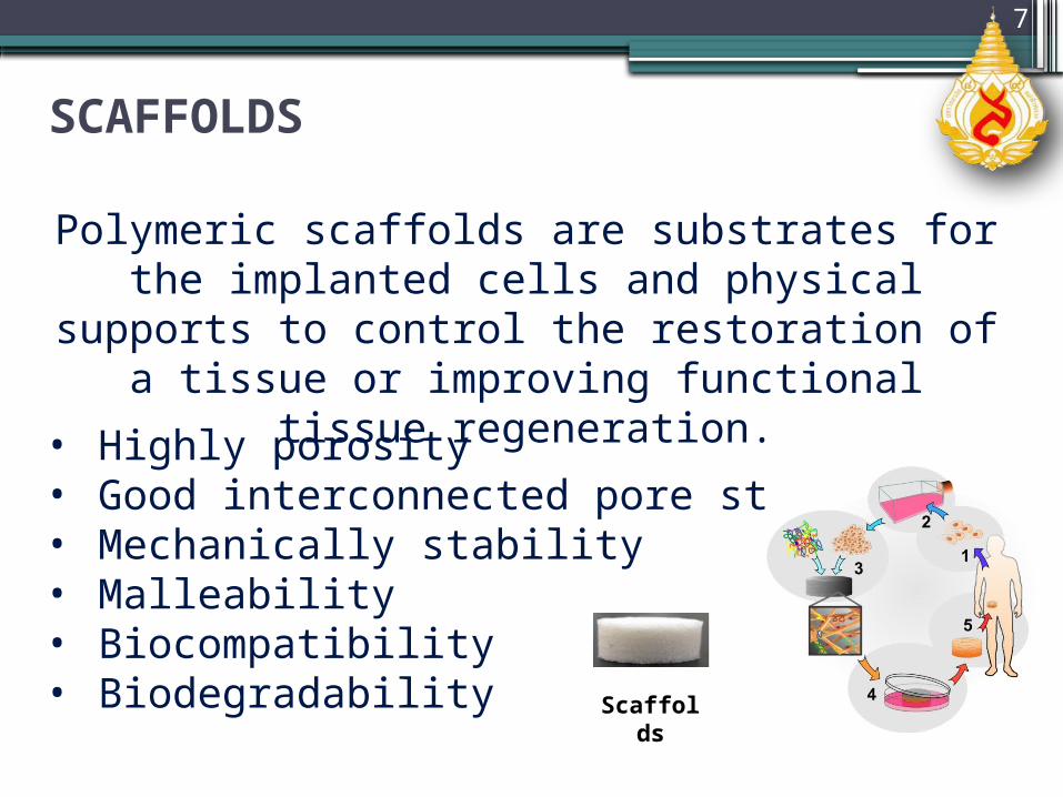
SCAFFOLDS
7
Polymeric scaffolds are substrates for the implanted cells and physical supports to control the restoration of a tissue
or improving functional tissue regeneration.
• Highly porosity• Good interconnected pore structure• Mechanically stability• Malleability• Biocompatibility• Biodegradability Scaffol
ds

GELATIN
8
• A natural polymer • A derivative of collagen• Amino acid• Biological origin • Biodegradability• Biocompatibility
Figure 1 : Chemical structure of gelatin

PROBLEMS
SOLVE THE PROBLEMS“Embedding them in a polymer matrix
may reduce their cytotoxic effect”
9
9
• AgNPs are strong bactericidal agents but they are also cytotoxic.
• AgNPs in smaller sizes had better antibacterial properties but higher cytotoxicity.

OBJECTIVES
1) To prepare the AgNPs-loaded calcium alginate beads embedded in
gelatin scaffolds
2) To characterize the AgNPs-loaded calcium alginate beads embedded
in gelatin scaffolds (morphology, thermal properties, mechanical
properties, water swelling and weight loss behavior and, release
characteristic of Ag+ ions)
10

EXPERIMENT

Part 1: Preparation of pure calcium alginate beads and AgNPs-loaded calcium alginate beads
4%w/w AgNO3 solution was initially prepared and mixed with 1.5% w/v of Na alginate.
AgNO3/alginate solution was irradiated by UV light for 1 h.
Alginate beads incorporated with AgNPs were fabricated by EHDA.
The wet and dry beads were observed the morphology and size by Optical Microscope (OM) and Scanning Electron Microscope (SEM), respectively.
12
Negative electrode
Ground electrode
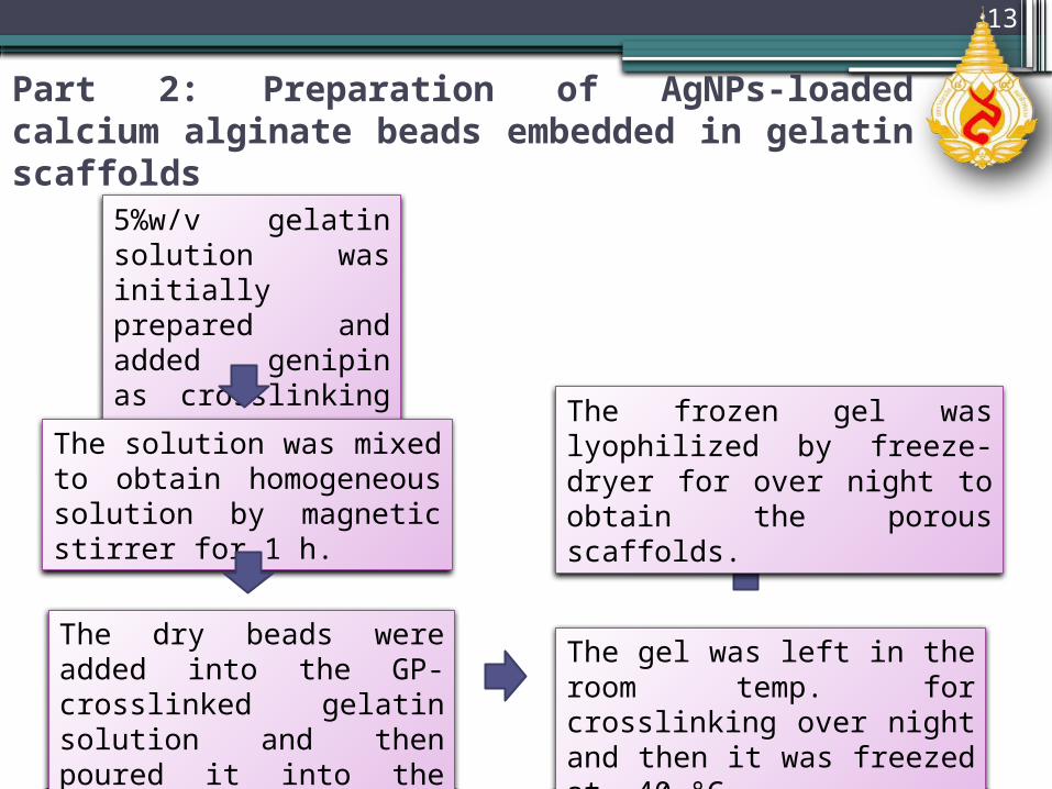
Part 2: Preparation of AgNPs-loaded calcium alginate beads embedded in gelatin scaffolds
5%w/v gelatin solution was initially prepared and added genipin as crosslinking agent.
The solution was mixed to obtain homogeneous solution by magnetic stirrer for 1 h.
The dry beads were added into the GP-crosslinked gelatin solution and then poured it into the mold to form gel.
The gel was left in the room temp. for crosslinking over night and then it was freezed at -40 °C.
The frozen gel was lyophilized by freeze-dryer for over night to obtain the porous scaffolds.
13
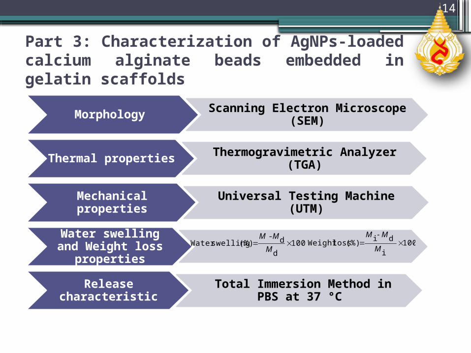
Part 3: Characterization of AgNPs-loaded calcium alginate beads embedded in gelatin scaffolds
Morphology Scanning Electron Microscope (SEM)
Thermal properties Thermogravimetric Analyzer (TGA)
Mechanical properties Universal Testing Machine (UTM)
Water swelling and Weight loss properties
Release characteristic Total Immersion Method in PBS at 37 °C
14
100,d
d- (%) swellingWater
M
MM100
i
di(%) lossWeight
M
M-M

RESULTS & DISCUSSION

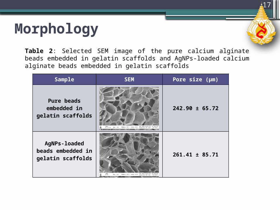
Morphology Table 1: Optical micrographs (OM), scanning electron micrographs (SEM) and diameters of the pure calcium-alginate beads and AgNPs-loaded calcium-alginate beads
16
SampleObserved by
OM Diameter (µm) SEM Diameter(µm)
Control 523.68 ± 17.36 149.46 ± 12.26
AgNPs 475.97 ± 16.09 143.65 ± 13.35
Adding Ag into the beads affected the size of beads to decrease.
Morphology
Table 2: Selected SEM image of the pure calcium alginate beads embedded in gelatin scaffolds and AgNPs-loaded calcium alginate beads embedded in gelatin scaffolds
17
Sample SEM Pore size (µm)
Pure beads embedded in gelatin scaffolds 242.90 ± 65.72
AgNPs-loaded beads embedded in gelatin
scaffolds 261.41 ± 85.71

Thermal properties
18
Temperature (oC)0 100 200 300 400 500 600
Wei
ght l
oss
(%)
20
40
60
80
100
120Pure beads embedded in scaffoldsAgNPs-loaded beads embedded in scaffolds
Figure 2: Thermogravimetric analysis (TGA) of the pure calcium alginate beads embedded in gelatin scaffolds and AgNPs-loaded calcium alginate beads embedded in gelatin scaffolds
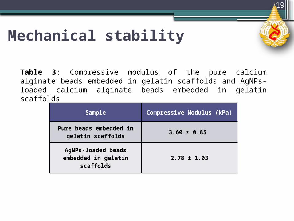
Mechanical stability
19
Sample Compressive Modulus (kPa)
Pure beads embedded in gelatin scaffolds 3.60 ± 0.85
AgNPs-loaded beads embedded in gelatin scaffolds 2.78 ± 1.03
Table 3: Compressive modulus of the pure calcium alginate beads embedded in gelatin scaffolds and AgNPs-loaded calcium alginate beads embedded in gelatin scaffolds
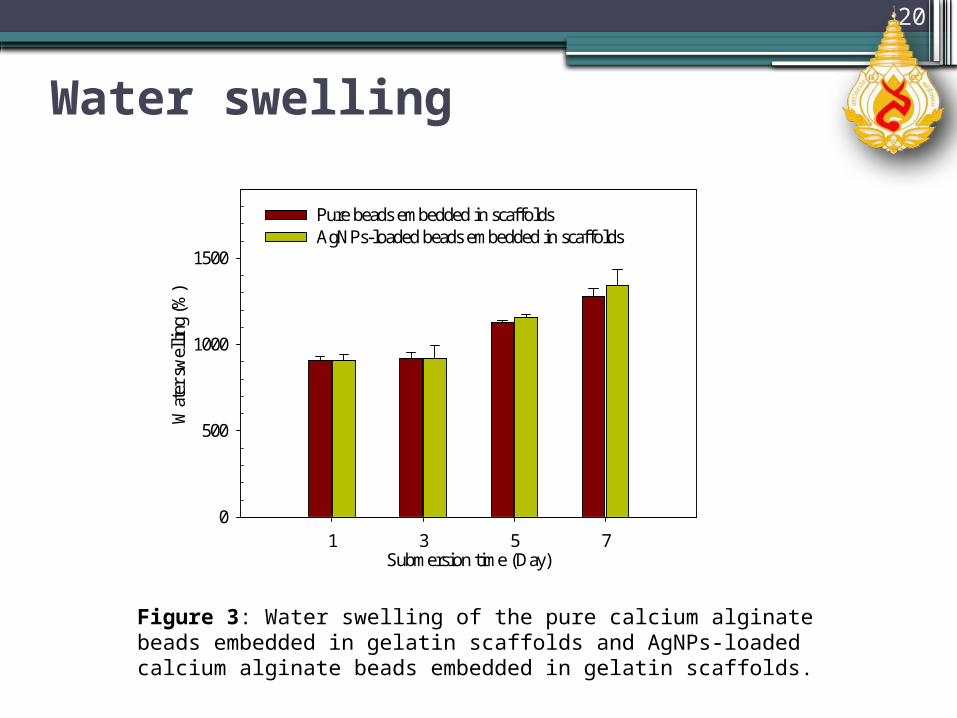
Water swelling
20
Submersion time (Day)1 3 5 7
Wat
er s
wel
ling
(%)
0
500
1000
1500
Pure beads embedded in scaffoldsAgNPs-loaded beads embedded in scaffolds
Figure 3: Water swelling of the pure calcium alginate beads embedded in gelatin scaffolds and AgNPs-loaded calcium alginate beads embedded in gelatin scaffolds.

Weight loss
21
Figure 4: Weight loss of the pure calcium alginate beads embedded in gelatin scaffolds and silver nanoparticles-loaded calcium alginate beads embedded in gelatin scaffolds.
Submersion time (Day)1 3 5 7
Wei
ght l
oss
(%)
0
10
20
30
40Pure beads embedded in scaffoldsAgNPs-loaded beads embedded in scaffolds

Release characteristic
22
Submersion time (Day)
0 1 2 3 4 5 6 7 8
Cum
ula
tive
rele
ase
of
silv
er i
ons
(%, bas
ed o
n a
ctual
am
ount
of
silv
er
0
20
40
60
80
100
Figure 5: Cumulative release profiles of Ag+ ions from AgNPs-loaded calcium alginate beads embedded in gelatin scaffolds in phosphate buffer solution at 37 °C.

CONCLUSIONS
23
The AgNPs-loaded calcium alginate beads embedded in gelatin scaffolds were successfully fabricated.
The incorporation of AgNPs-loaded into the calcium alginate beads decreased the size of beads.
The morphology of the AgNPs-loaded calcium alginate beads embedded in gelatin scaffolds showed the interconnected pore structure .
The incorporation of AgNPs into the calcium alginate beads embedded in gelatin scaffolds decreased the strength of materials.

CONCLUSIONS
24
The water swelling and weight loss behavior of the scaffolds increased with an increase in the submersion time.
The release of Ag ions from the scaffolds gradually increased with increase in submersion time and then reached a plateau value at day 3, and continually increased to reach 80% release at day 7.

REFERENCES1) Lee YM, Kim SS, Park MH, Song KW, Sung YK, Kang K (2000) J
Mater Sci 11:817-823 2) del Valle LJ, Roa M, Díaz A, Casas MT, Puiggalí J, Rodríguez-Galán A
(2012) J Polym Res 19:97923) Sharma VK, Yngard RA, Lin Y (2009) Adv Colloid Interface Sci
145:83-964) Yoksan R, Chirachachai S (2009) Mater Chem Phys 115:296–3025) Ogończyk D, Siek M, Garstecki P (2011) Biomicrofluidics 5:013405 6) Knight CG (1982) J Membr Sci 12:131–1327) Ding L, Lee T, Wang C (2005) J Control Release 102:395–413 8) Xie J, Lim LK, Phua Y, Hua J, Wang C (2006) J Colloid Interf Sci
302:103–112 9) Ciach T (2006) Int J Pharm 324:51–5510)Cloupeau M (1994) J Aerosol Sci 25:1143-1157
25

ACKNOWLEDGEMENTS
“The authors would like to acknowledge the financial support from the Research, Development and Engineering (RD&E) fund through The National Nanotechnology Center (NANOTEC), The National Science and Technology Development Agency (NSTDA), Thailand (P-11-00986) to Mae Fah Luang University (MFU) and Thailand Graduate Institute of Science and Technology (TGIST) (TG-55-99-55-048M)”
26

Thank You For Your Attention