silencingofthe drosophila orthologof sox5 inheart
TRANSCRIPT

Silencing of the Drosophila ortholog of SOX5 in heartleads to cardiac dysfunction as detected by opticalcoherence tomography
Airong Li1,{, Osman O. Ahsen4,{, Jonathan J. Liu4, Chuang Du5, Mary L. McKee2, Yan Yang1,
Wilma Wasco1, Christopher H. Newton-Cheh3, Christopher J. O’Donnell6, James G. Fujimoto4,
Chao Zhou4,7,∗ and Rudolph E. Tanzi1,∗
1Genetics and Aging Research Unit, Department of Neurology, MassGeneral Institute for Neurodegenerative Diseases,2Program in Membrane Biology and 3Department of Medicine, Massachusetts General Hospital and Harvard Medical
School, Charlestown, MA, USA, 4Department of Electrical Engineering and Computer Science, Research Laboratory of
Electronics, Massachusetts Institute of Technology, Cambridge, MA, USA, 5Center for Neuroscience Research, Tufts
University School of Medicine, Boston, MA, USA, 6Framingham Heart Study, The National Heart, Lung, and Blood
Institute (NHLBI), Framingham, MA, USA and 7Department of Electrical and Computer and Engineering, Lehigh
University, Bethlehem, PA, USA
Received January 24, 2013; Revised and Accepted May 16, 2013
The SRY-related HMG-box 5 (SOX5) gene encodes a member of the SOX family of transcription factors. Recently,genome-wide association studies have implicated SOX5 as a candidate gene for susceptibility to four cardiac-related endophenotypes: higher resting heart rate (HR), the electrocardiographic PR interval, atrial fibrillationand left ventricular mass. We have determined that human SOX5 has a highly conserved Drosophila ortholog,Sox102F, and have employed transgenic Drosophila models to quantitatively measure cardiac function inadult flies. For this purpose, we have developed a high-speed and ultrahigh-resolution optical coherence tom-ography imaging system, which enables rapid cross-sectional imaging of the heart tube over various cardiaccycles for the measurement of cardiac structural and dynamical parameters such as HR, dimensions andareas of heart chambers, cardiac wall thickness and wall velocities. We have found that the silencing ofSox102F resulted in asignificant decrease in HR, heart chamber sizeand cardiac wall velocities,and asignificantincrease in cardiac wall thickness that was accompanied by disrupted myofibril structure in adult flies. In add-ition, the silencing of Sox102F in the wing led to increased L2, L3 and wing marginal veins and increased anddisorganized expression of wingless, the central component of the Wnt signaling pathway. Collectively, the si-lencing of Sox102F resulted in severe cardiac dysfunction and structural defects with disrupted Wnt signalingtransduction in flies. This implicates an important functional role for SOX5 in heart and suggests that the altera-tions in SOX5 levels may contribute to the pathogenesis of multiple cardiac diseases or traits.
INTRODUCTION
The SRY-related HMG-box 5 (SOX5) gene is localized onchromosome 12p12 (OMIN 604975) and encodes a member ofthe SOX family of transcription factors that bind to DNA (1).
SOX5 is expressed in multiple human tissues, including heart,liver, lung, kidney, spleen, fetal brain and testis (2). Studieshave shown that SOX5 can modulate cell fate, control cell pro-liferation and regulate cartilage formation and neuron
†These authors contributed equally.
∗To whom correspondence should be addressed at: Genetics and Aging Research Unit, Department of Neurology, Massachusetts General Hospital, 114,16th Street, Charlestown, MA 02129, USA. Tel: +1 6177266845; Fax: +1 6177241949; Email: [email protected] (R.E.T.); Department ofElectrical and Computer and Engineering, Lehigh University, 19 Memorial Drive West, Bethlehem, PA 18015-3084, USA. Tel: +1 6107585092;Fax: +1 6107586279; Email: [email protected] (C.Z.)
# The Author 2013. Published by Oxford University Press. All rights reserved.For Permissions, please email: [email protected]
Human Molecular Genetics, 2013 1–9doi:10.1093/hmg/ddt230
HMG Advance Access published June 5, 2013 at L
ehigh University on June 7, 2013
http://hmg.oxfordjournals.org/
Dow
nloaded from

development (3–5). Recently, genome-wide association studies(GWASs) have implicated SOX5 as a candidate gene for suscep-tibility to four cardiac-related endophenotypes: higher restingheart rate (RHR) (6), the electrocardiographic PR interval (7),atrial fibrillation (AF) (8) and left ventricular mass (LVM) (9).These findings suggest an important functional role for SOX5in the heart. Mice with Sox5 gene single null mutation wereearly lethal and had mild skeletal abnormalities (3). Sox5-deficient mice died at birth with respiratory distress and abnor-mal lung development, indicating that Sox5 is critical forproper in utero lung morphogenesis (10). To date, early lethalityin loss of function Sox5 mouse models has prohibited analysis ofadult cardiac function; a role for SOX5 in cardiac function hasnot been reported previously.
Drosophila melanogaster or fruit fly has been successfullyused to characterize multiple genes associated with cardiac dis-eases (11–14). The basic mechanisms of heart development andcontrol of cardiac function are highly conserved between humanand Drosophila. Thus, discoveries made in the fly can be appliedto higher species including human. In addition, Drosophila havea short life span and an oxygen transport system independentfrom its heart, which makes the organism more viable togenetic alterations that effect cardiac function (15). Anatomical-ly, the fly heart lies close to the dorsal surface of the abdomen(14). Therefore, the morphological and rhythm changes can bereadily analyzed in the relatively simple organization of the flyheart with the emerging biomedical imaging technologyoptical coherence tomography (OCT) (16).
Non-invasive OCT enables real-time, in vivo, micron-scaleand three-dimensional (3D) imaging of biological tissueswithout the need to sacrifice and process specimens. OCT hasbeen used for a wide range of clinical applications in human, in-cluding ophthalmology (16–19), endoscopy (20–26) and car-diovascular imaging (27–31). Drosophila’s heart beats at arate of several hundred beats per minute (BPM), making highimaging speed critical in order to capture the dynamics of itsheart during various cardiac cycles. The utility of OCT for study-ing Drosophila cardiac functions has been reported by severalstudies (32–37). With a custom-built OCT imaging platform,we previously demonstrated acquiring cross-sectional OCTimages at �120 frames/s, fast enough to capture the dynamicsof the beating Drosophila heart, in order to assess the effect ofAlzheimer’s disease and dilated cardiomyopathy-associatedpresenilin gene. We found that either the overexpression or thesilencing of the Drosophila ortholog of presenilin in the heartleads to cardiac dysfunction (36). To date, OCT has been usedby our group and others to evaluate cardiac function usingM-mode imaging at a single location of the Drosophila hearttube over time and concentrated on inferring structural informa-tion, such as heart dimensions during systole and diastole phase,as well as functional information, such as HR and arrhythmiaprevalence, and to detect significant differences in thesecardiac parameters in response to genetic alterations related tocardiac diseases (32–34,36). Previous studies, however,lacked the resolution and speed to resolve fine details such ascardiac wall dimensions, as well as the ability to measure walldynamics using information from the entire cross-sectionalheart chamber (32–34,36). With the advancements in theimaging speed as well as Doppler and phase sensitive detection,the use of OCT for Drosophila studies was further extended to
study the dynamics of cardiac wall movement during variouscardiac cycles (35,37).
In this study, we have developed a high-speed and ultrahigh-resolution OCT imaging system to non-invasively quantifyDrosophila cardiac function more precisely and more compre-hensively than previously possible. The new OCT systemenables the 3D volumetric imaging of the Drosophila andrapidly captures the cross-sectional images of the heart tubeover various cardiac cycles. By searching the Drosophilagenome database, we determined that human SOX5 is highlyconserved to a Drosophila ortholog Sox102F. SOX5 andSox102F share 71% identity and 82% similarity in amino acidsequences. To circumvent the lethality of mouse models nullfor Sox5, we have employed transgenic Drosophila modelsand high-performance OCT imaging to quantitatively measurecardiac function in adult flies to assess the functional role ofSox102F in the heart. Our studies have demonstrated that the si-lencing of Sox102F in the heart results in cardiac dysfunction.
RESULTS
Drosophila cardiac function and heart structural analysis
We employed UAS-Sox102F-RNAi transgenic flies and anestablished 24B-GAL4 driver (38) to silence Sox102F specifical-ly in the fly heart (38,39). The 24B-GAL4 line allows targeted ex-pression in mesoderm and all cardiac and muscular cells with auniform and high level of expression in the most anterior heartcells (40). Flies in which Sox102F was silenced by 24B-GAL4(UAS-Sox102F-RNAi; 24B-GAL4) appeared normal at eclo-sion. The silencing of Sox102F led to a decrease in Sox102Fexpression to 55% compared with control flies that expressed aheterozygous 24B-GAL4 alone (24B-GAL4/+), as assessed byreal-time reverse transcriptase (RT)–polymerase chain reaction(PCR) of cDNA from the hearts of the UAS-Sox102F-RNAi;24B-GAL4 and 24B-GAL4/+ flies.
We quantitatively measured cardiac function in 30-day-oldUAS-Sox102F-RNAi; 24B-GAL4 flies (n ¼ 35) and age-matched control flies (n ¼ 36) using the newly developed OCTsystem. Since the imaging resolution and speed were dramatic-ally improved compared with our previous system (36), this ana-lysis allows us to have a more precise measurement of thechamber dimensions, as well as enables us to use DopplerOCT to measure cardiac wall velocity versus time. As can beseen in Figure 1, where an example of volumetric and cross-sectional OCT images of an adult Drosophila are shown, OCTcan readily identify at least the first three heart segments(ostia) of the fly (Fig. 1B).
Analyses of OCT imaging data for assessing cardiac functionwere categorized as structural parameters, including the end sys-tolic and diastolic vertical and transverse dimensions (ESDv,ESDt, EDDv and EDDt), end systolic and diastolic areas (ESAand EDA) and cardiac wall thickness and dynamical parameters,including HR, systolic and diastolic wall velocities and theprevalence of arrhythmia (Table 1). Compared with the age-matched controls, structurally, the silencing of Sox102F inflies (UAS-Sox102F-RNAi; 24B-GAL4) resulted in a signifi-cant decrease in each of the structural parameters, includingESDv, ESDt, EDDv, EDDt, ESA and EDA, and a significantincrease in cardiac wall thickness from 10.80+ 0.28 mm in
2 Human Molecular Genetics, 2013
at Lehigh U
niversity on June 7, 2013http://hm
g.oxfordjournals.org/D
ownloaded from

control to 12.23+ 0.30 mm in UAS-Sox102F-RNAi; 24B-GAL4 flies (Table 1, Fig. 2). Dynamically, the silencing ofSox102F caused a significant decrease in HR from 239+ 8 to211+ 10 BPM in UAS-Sox102F-RNAi flies and both systolicand diastolic wall velocities (Table 1, Figs 2 and 3) and an in-crease in the cardiac arrhythmia prevalence, although arrhyth-mia prevalence did not reach statistical significance (Table 1).
We analyzed 30-day-old flies’ whole heart by F-actin immu-nostaining and heart ultrastructure by transmission electron mi-croscopy (TEM). Fluorescent labeling of the whole adult heartwith F-actin demonstrated that the silencing of Sox102F resultedin an enlarged and irregular cardiac tube and loss of myofibrilstructure (Fig. 4B) compared with that of control (Fig. 4A).TEM analysis of the adult hearts of control flies showednormal myofibril structure (Fig. 4C and D). In contrast, heartsfrom flies in which Sox102F was silenced revealed irregularand broken myofilament arrays with larger gaps between the in-dividual filaments, discontinuous Z discs, irregular and smallersarcoplasmic reticulum (SR) and degenerative mitochondria(Fig. 4E and F) consistent with whole heart and cardiac function-al alterations. Collectively, these data demonstrate that the silen-cing of Sox102F led to cardiac hypertrophy, a decrease in the sizeof the heart chamber and a decrease in HR and wall velocities,accompanied by disrupted myofibril structure in adult flies.
Wing phenotype and wingless expression
Wing development is regulated by multiple signaling pathways,including Notch, Wnt, epidermal growth factor receptor, hedge-hog and decapentaplegic. The wing pattern has provided an im-portant tool in isolating and characterizing genes affecting thesesignaling pathways (41). Wnts are a family of secreted signaling
proteins including wingless (wg), the central component of Wntsignaling pathway (42). To identify the functional pathway ofSOX5, we silenced Sox102F specifically in the wing.SD-GAL4 drives the silencing of Sox102F in the wing disc inthe pattern of the scalloped (sd) gene, which regulates the expres-sion of a number of targeted genes including wg (42). Over 200flies were analyzed for the wing phenotype. HeterozygousSD-GAL4 driver alone (SD-GAL4/+) showed normal wing(Fig. 5A–C). The silencing of Sox102F in the wing (UAS-Sox102F-RNAi; SD-GAL4) resulted in a significant increasein the L2, L3 and wing marginal veins; in particular, extraveins were formed in the distal part of L3 (Fig. 5D–F). Wenext examined the expression of the wg protein by immunostain-ing wing imaginal discs dissected from the third instar larvaewith an anti-wg antibody. In control flies with heterozygousSD-GAL4 driver alone (SD-GAL4/+), wg was expressed as abroad strip in the notum, a thinner strip in the prospective wingmargin-dorsal/ventral (D/V) boundary and a strip encirclingthe prospective wing blade (Fig. 5G). Consistent with the pheno-type observed in adult flies, we observed remarkably increasedwg expression levels and disorganized wg expression patternin the third instar wing discs in which Sox102F was silenced,leading to extra strips in the prospective wing blade region(Fig. 5H). These data strongly support Sox102F as a regulatorof wg expression.
DISCUSSION
In this study, we have provided clear evidence indicating that thesilencing of Sox102F leads to severe cardiac dysfunction andstructural defects in adult flies, including cardiac hypertrophy,reduced heart chamber size and decrease HR and wall velocities,accompanied by disrupted myofibril structure, and that the ex-pression of Sox102F can regulate wg expression. Variants inSOX5 have been significantly associated with multiple cardiacdiseases or traits, including the higher RHR (6), electrocardio-graphic PR interval (7), AF (8) and LVM (9). This is the firstreport of a systematic assessment of the functional role of theDrosophila ortholog of SOX5 in a whole animal in vivo. Thesefindings implicate that SOX5 may play an important role in regu-lating cardiac function.
We have developed a high-speed and ultrahigh-resolutionOCT imaging system that enables successful characterizationof structural and dynamical parameters of cardiac function inadult flies. Notably, the cardiac wall thickness is �10 mm, andthe differences between the two groups included in this studywere ,2 mm. This highlights the necessity of using a high-resolution imaging system in order to be able to distinguishthese subtle differences. Moreover, as the velocity of the heartwall is relatively low, as opposed to the velocity of blood flowtraditionally measured with Doppler OCT methods, imagingand analysis protocols need to be carefully designed in order toovercome the deleterious effects of phase noise and bulksample motion. A high-speed imaging system is imperative inthis respect, as extracting Doppler information from rapidlyscanned cross-sectional images is more reliable and robust com-pared with using an M-mode image from a single transverse pos-ition. This is due to the additional structural information gainedfrom cross-sections. Higher speed imaging systems will provide
Figure 1. OCT imaging of adult Drosophila. (A) 3D reconstruction from a volu-metric data set of an adult Drosophila. (B) Cross-sectional OCT view along thefly body showing detailed structures in the sagittal plane. Arrows point to the firstthree ostia, identified as empty, elongated tubes. (C) Cross-sectional OCT viewacross the fly’s abdomen showing a detailed structure of the main heart chamber(conical heart chamber). As indicated by the arrow, the heart is observed as anempty space in the images. Images are false color coded and the higher intensitysignal is represented by a color closer to red. Scale bars: 250 mm.
Human Molecular Genetics, 2013 3
at Lehigh U
niversity on June 7, 2013http://hm
g.oxfordjournals.org/D
ownloaded from

4D data sets (3D + time) in high frame rates, which will allowcharacterizing the velocity and other functional parametersalong the entire heart tube of the fly. Important parameterssuch as cardiac output (Q) can then be extracted from the 4D(3D + time) data sets for the comprehensive assessment ofcardiac function.
The Wnt signal transduction has been implicated as an import-ant event that regulates cardiac development and function (43).The Wnt signaling pathway controlsb-catenin, which enters thenucleus, binds to DNA and triggers expression of genes (44).SOX5 acts synergistically with a stable form of b-catenin andincreases the expression of axin2, a negative regulator of theWnt pathway in neural tube cells (5). Consistent with previousfindings, we have demonstrated that the silencing of Sox102Fpromoted the formation of wing marginal veins and a remark-ably increased and disorganized level of wg expression in flies.These data support the contention that SOX5 negatively regu-lates Wnt signaling via reducing wg expression in addition to in-creasing axin2 expression. The finding that the silencing ofSox102F resulted in cardiac hypertrophy accompanied with re-markably increased wg expression is consistent with theknown role of Wnt signaling in heart formation. Down-regulation of the Wnt pathway leads to a thinner cardiac tubeby gene expression profiling in Drosophila heart formation(45). The inhibition of Sox102F on the Wnt signaling may con-tribute to the pathogenesis that led to cardiac dysfunction in fliesin which Sox102F was silenced.
A single-nucleotide polymorphism (SNP) rs17287293 whichis located 3′ to SOX5 was significantly associated with the higherRHR in a meta-analysis of 15 GWASs (6). Epidemiologicstudies have demonstrated that the higher RHR is significantlyassociated with increased cardiovascular disease and mortalityrisk independent of traditional risk factors in diverse subgroups,including healthy people in the general population (46,47),hypertensives (48) and those with established ischemic heartdisease (47,49,50). A higher RHR also has been associatedwith poorer prognosis within subgroups of patients with cardio-vascular disease (51–53). Compared with those whose RHR wasconsistent at ,70 BPM, individuals whose rates increased from,70 to .85 BPM had a 90% higher risk of death from heartdisease after 12 years of follow-up of 29 325 healthy men andwomen who had no known heart disease at the start of thestudy (50). In a 5-year follow-up, 5139 healthy middle-aged
men who showed that an increase of .3 BPM in the RHR wasassociated with a 19% increase in all-cause mortality (54).
In other GWASs, the SNP rs11047543 located 3′ to SOX5 wassignificantly associated with both the electrocardiographic PRinterval (7) and AF (8). The disturbances of the PR interval isconsidered an intermediate phenotype for AF and an increaserisk of AF, which is independently associated with an increasedrisk of stroke, heart failure, dementia and death. The minor alleleof SNP rs11047543 was significantly associated with a decreasein AF risk (7). Furthermore, the SNP rs10743465 3′ to SOX5 wasthe most significantly associated with LVM in a follow-up studyfor LVM on chromosome 12p11 (9). An increase in LVM is oneof the most important cardiac risk factors for stroke, myocardialinfarction and chronic heart failure, independent of age, sex andrace-ethnicity (55,56). In a landmark study, each 50 g/m increasein LVM was associated with an adjusted relative risk of cardio-vascular disease of 1.49 and 1.57 in men and women, respective-ly (57). Moreover, diastolic dysfunction is linked to increasedmortality in a range of conditions, including post-acute myocar-dial infarction, hypertension and chronic renal failure (58–62).Closely linked to diastolic function, alterations in cardiac wallvelocities are associated with several cardiovascular disorders,mostly pronounced with hypertrophic cardiomyopathy (63–68).
An action potential is the first step in the chain of eventsleading to contraction in cardiac cells. The action potential sti-mulates Ca++ entry into cardiomyocytes through voltage-gatedL-type Ca++ channels (LTCCs). Antisense oligonucleotidesagainst SOX5 have been shown to significantly decrease themaximum charge movement in mouse myoblast cells (69).Charge movement is regulated by LTCCs (69), and the ampli-tude of LTCCs was increased in hypertrophied and failing myo-cytes, contributing to cardiac hypertrophy (70). Moreover, thenitric oxide (NO) level is significantly correlated with the occur-rence of LVM (71). SOX5 regulated shear stress-modulatedgene expression in an NO-dependent manner in endothelialcells (72), which may contribute to LVM pathogenesis. Collect-ively, these findings suggest that Sox5 may regulate LTCCs andthat genetic variants in SOX5 may alter atrial action potentialand atrioventricular conduction influencing risk for the higherRHR, disturbed PR interval, AF and LVM (73).
In addition to associations with cardiac diseases or traits, theSNP rs11046966 3′ to SOX5 has been associated with chronic ob-structive pulmonary disease (10); the intronic SNP rs1522232 in
Table 1. Comparison of the cardiac structural and dynamical parameters in control flies and UAS-Sox102F-RNAi; 24B-GAL4 flies derived from the OCT images
Parameter (mean+SE) 24B-GAL4/+ (n ¼ 36) UAS-Sox102F-RNAi; 24B-GAL4 (n ¼ 35) P-value
Structural parameters ESDt (mm) 32+4 19+3 0.001∗
EDDt (mm) 64+4 54+3 0.02∗
ESDv (mm) 37+4 19+3 ,0.0001∗
EDDv (mm) 71+4 53+4 0.0006∗
ESA (mm2) 1233+160 562+149 0.002∗
EDA (mm2) 3473+300 2241+265 0.002∗
Cardiac wall thickness (mm) 10.80+0.28 12.23+0.30 0.0006∗
Dynamical parameters HR (BPM) 239+8 211+10 0.02∗
Systolic wall velocity (mm/s) 877+64 724+33 0.02∗
Diastolic wall velocity (mm/s) 762+41 575+29 0.0002∗
Arrhythmia prevalence 15/35 (42%) 21/36 (60%) 0.1
∗Statistically significant difference, P , 0.05.
4 Human Molecular Genetics, 2013
at Lehigh U
niversity on June 7, 2013http://hm
g.oxfordjournals.org/D
ownloaded from
SOX5 was associated with the rapid progression of AIDS (74);the other intronic SNP rs1464500 in SOX5 was associatedwith metabolic side effects of antipsychotic drugs, such as high-density lipoprotein levels in patients taking perphenazine (75)and triglyceride levels in subjects taking statins (76). Moreover,two missense mutations in SOX5, Gln362Pro and Glu367Gln,five amino acids apart, were identified in 2 of the 190 amyotroph-ic lateral sclerosis patients and not detected in 190 normal con-trols (77). Further studies are warranted to elucidate thefunctional role(s) of SOX5 in multiple human diseases or traits.
In summary, we have demonstrated that the silencing ofSox102F in the Drosophila heart results in cardiac dysfunction.Together, these data provide the first in vivo evidence for an im-portant functional role for SOX5 in the heart and suggest that thealterations in SOX5 levels may contribute to the pathogenesis ofmultiple cardiac diseases or traits.
MATERIALS AND METHODS
Transgenic flies and fly culture
Fly culture and crosses were carried out according to the standardprocedures. The following fly strains were used: UAS-Sox102F-RNAi from the TRiP Project at Harvard Medical
School (http://www.flyrnai.org/TRiP-HOME.html), 24B-GAL4(38) and SD-GAL4 (42).
Animal preparation
Flies were anesthetized in a closed container using FlyNapw
(Carolina Biological Supply Company) for 2 min. FlyNapw isan established anesthetizing agent used in Drosophila studies,and its main constituent, triethylamine, is shown to haveminimal effect on influencing fly cardiac functions (13). Afteranesthesia, flies were taped on a microscope cover slide bytheir wings with the dorsal side facing up. OCT imaging was con-ducted within 30 min, before the effects of anesthesia wore off inorder to minimize motion artifacts.
High-speed and ultrahigh-resolution OCT imaging systemand OCT imaging for the Drosophila cardiac functionassessment
A custom-built spectral domain OCT system was used for thestudy. The OCT system was optimized for high-speed andultrahigh-resolution imaging, where a broadband superlumines-cent diode (Superlum, Dublin, Ireland) centered at �850 nmwas employed to provide �3 mm axial resolutions in tissue.A long working distance near infrared objective (Thorlabs,Newton, NJ, USA) was used to achieve 8 mm resolution in thetransverse dimensions. A 25 kHz line rate line-scan camera(e2v, Chelmsford, UK) was used to generate cross-sectionalimages at 110 frames/s with 226 axial scans per frame, whichis sufficient to capture the chamber morphology at various
Figure 2. Obtaining functional information about cardiac function from struc-tural OCT images. (A) A pseudo M-mode OCT image from the center of themain heart chamber (top) and the automatically generated plots for the heartdimensions (below) of a fly from the control group. Rapidly repeated cross-sectional OCT images over the clearest and largest heart chamber were per-formed for each fly where the single A-scan over the center of the heartchamber in each cross-sectional image is extracted to create a pseudo M-modeOCT image. The regularity of the heart beats can be visualized from theM-mode image as well as the resulting dimension plot. This particular fly hasbeen characterized with a HR of 258 BPM, EDDt and EDDv of 80 and108 mm, respectively, ESDt and ESDv of 49 and 71 mm, respectively, ESAand EDA of 2605 and 5644 mm2, respectively. (B) A pseudo M-mode OCTimage (top) and the automatically generated plots for the heart dimensions(bottom) of a fly from the UAS-Sox102F-RNAi; 24B-GAL4 group. TheM-mode image shows irregular beating patterns. This particular fly has beencharacterized with a HR of 138 BPM, EDDt and EDDv of 54 and 32 mm, respect-ively, ESDt and ESDv of 1 and 1.4 mm, respectively, ESA and EDA of 6 and1036 mm2, respectively. Red lines in the dimension plots correspond to axialdimensions, whereas the blue lines correspond to transverse dimensions. Scalebars: 50 mm in the vertical dimension; 1 s in the horizontal dimension.
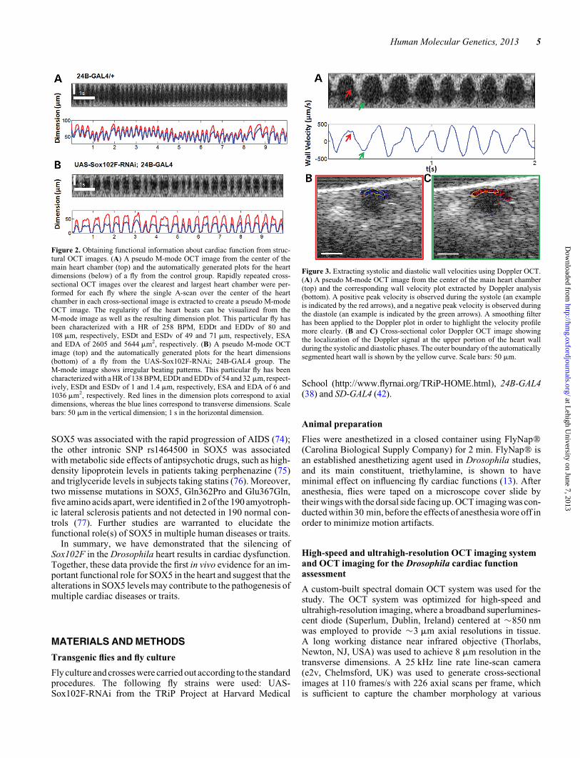
Figure 3. Extracting systolic and diastolic wall velocities using Doppler OCT.(A) A pseudo M-mode OCT image from the center of the main heart chamber(top) and the corresponding wall velocity plot extracted by Doppler analysis(bottom). A positive peak velocity is observed during the systole (an exampleis indicated by the red arrows), and a negative peak velocity is observed duringthe diastole (an example is indicated by the green arrows). A smoothing filterhas been applied to the Doppler plot in order to highlight the velocity profilemore clearly. (B and C) Cross-sectional color Doppler OCT image showingthe localization of the Doppler signal at the upper portion of the heart wallduring the systolic and diastolic phases. The outer boundary of the automaticallysegmented heart wall is shown by the yellow curve. Scale bars: 50 mm.
Human Molecular Genetics, 2013 5
at Lehigh U
niversity on June 7, 2013http://hm
g.oxfordjournals.org/D
ownloaded from

cardiac cycles of a beating fly heart. The imaging speed and thetransverse imaging resolution were improved by a factor of �2,and the axial resolution is improved by more than a factor of 3compared with the OCT system used in our previous studieson Drosophila (36). Dynamical and structural parameters in-cluding HR, ESDv, ESDt, EDDv, EDDt, ESA, EDA, wallthickness and systolic and diastolic wall velocities of the heartwall were extracted from cross-sectional images obtained fromthe heart chamber �50 mm away from the chest. This cross-sectional imaging plane was selected for analysis as it consistent-ly presents the clearest and largest heart chamber in all the flies.Each cross-sectional imaging session was acquired continuouslyfor 10 s and repeated three times in order to capture multiplecardiac cycles for accurate analysis. Post-processing of theimage data was performed with the custom-written MATLAB(Mathworks, Natick, MA, USA) code. The prevalence of ar-rhythmia in each fly group (%) was quantitatively analyzed viaidentifying the irregular heartbeat rhythm in the dimension plots.
Since the Drosophila hemolymph is essentially transparentdue to lack of red blood cells, obtaining flow information fromthe hemolymph is not possible without using extrinsic contrastagents. However, it is possible to extract Doppler OCT informa-tion from the heart wall as demonstrated in previous studies(35,37). Doppler analysis of the cardiac wall relied on an estab-lished method in the OCT literature, which requires over-sampling in the spatial dimension and extracting motioninformation by finding the phase difference between two succes-sive A-scans and correcting the calculated phase for bulk motioneffects (78,79). We were able to extract systolic and diastolic
cardiac wall velocity profiles from the flies using DopplerOCT. The upper portion of the heart wall was automatically seg-mented in order to calculate velocity from the region of the wallthat has movement in the vertical direction, as the Doppler ana-lysis method employed in this study can only measure velocity inparallel to the scanning beam. From the resulting M-modeDoppler velocity plots, systolic and diastolic wall velocitieswere calculated by finding the mean of positive and negativepeak velocity values, respectively.
Drosophila heart structural analysis by F-actinimmunostaining microscopy and TEM
Hearts from 30-day-old control or silencing of Sox102F flieswere dissected for whole heart and cardiac ultrastructure ana-lyses. Fluorescent labeling of the whole adult heart tube withF-actin and imaging were done as described previously (80).In brief, 15 hearts from control or silencing of Sox102F flieswere dissected in oxygenated hemolymph and fixed with 4% for-maldehyde. The sarcomeric F-actin was stained with Alexa-Fluorw 594 phalloidin (red, Life Technologies, Grand Island,NY, USA) and imaged with a Zeiss confocal microscope. ForTEM, 15 hearts from control or silencing of Sox102F flieswere fixed, embedded, sectioned, examined and imaged asdescribed previously (81). In brief, tissues were fixed in 2.0%glutaraldehyde in 0.1 M sodium cacodylate buffer, pH 7.4 (Elec-tron Microscopy Sciences, Hatfield, PA, USA) overnight at 48C.They were rinsed in buffer, post-fixed in 1.0% osmium tetroxidein cacodylate buffer for 1 h at room temperature, rinsed in buffer
Figure 4. Whole heart and cardiac ultrastructure analysis. (A and B) Micrographs of the whole adult heart by F-actin immuno-fluorescent staining. (A) Control fly(24B-GAL4/+) showed a normal cardiac tube. (B) Silencing of Sox102F in the heart (UAS-Sox102F-RNAi; 24B-GAL4) resulted in an enlarged and irregular cardiactube and loss of myofibril structure. (C–F) Ultrastructure of adult heart longitudinal sections between A1 and A3 segments by TEM. (C and D) Control fly showednormal myofibril structure. (E and F) Silencing of Sox102F in the heart led to irregular myofilament arrays, discontinuous Z discs, irregular and smaller SR and de-generative mitochondria. Myofilaments, Z discs, SR and mitochondrion were labeled and pointed with arrows. Magnification: (A and B) 10×, scale bars: 100 mm;(C and E) 20 000×; (D and F) 30 000×, scale bars: 500 nm.
6 Human Molecular Genetics, 2013
at Lehigh U
niversity on June 7, 2013http://hm
g.oxfordjournals.org/D
ownloaded from

and dehydrated through a graded series of ethanol to 100%. Theywere then infiltrated with Epon resin (Ted Pella, Redding, CA,USA) in a 1:1 solution of Epon:ethanol. The following daythey were placed in fresh Epon for several hours and then embed-ded in Epon overnight at 608C. Thin sections were cut on a LeicaEM UC7 ultramicrotome, collected on formvar-coated grids,stained with uranyl acetate and lead citrate and examined in aJEOL JEM 1011 transmission electron microscope at 80 kV.Images were collected using an AMT digital imaging system(Advanced Microscopy Techniques, Danvers, MA, USA).
Analysis of the wing phenotype and immunostainingfor wg expression
Wings from adult flies were dissected in isopropanol andmounted in Canada Balsam mounting medium. A total of 30
third instar wing discs were dissected, fixed, blocked andprobed with primary and secondary antibodies accordingly.The following antibodies were used: mouse anti-wg antibody(1:1000, Developmental Studies Hybridoma Bank, Iowa City,IA, USA) and the secondary anti-mouse Alexa 546 (1:200,Life Technologies).
cDNA synthesis and real-time RT–PCR
The cDNAs used for real-time RT–PCR were synthesized fromtotal RNA isolated from the heart muscle of 30-day-old adultDrosophila. The cDNA was synthesized using SUPERSCRIPTPreamplification System for first-strand cDNA synthesis (LifeTechnologies). Real-time RT–PCR quantification withSox102F or dActin-specific sense and antisense primers wasdone on an iCycler (BIO-RAD, Hercules, CA, USA) usingSYBR Green PCR Core Reagents (Qiagen, Germantown, MD,USA) according to the manufacturer’s instructions. Thesequences of primers are: Sox102F 5′-TGGTGGCTCAGGAAGTCTCT, 3′-AACTGCTGAGGGGGTGTATG; RT–PCR product size 171 bp; Actin5C 5′-TACCCCATTGAGCACGGTAT, 3′-GGTCATCTTCTCACGGTTGG; RT–PCRproduct size 156 bp. The house-keeping gene dActin was usedas an internal control and was co-amplified under the samePCR conditions. All standards and unknown samples were runin triplicates per reaction. The fluorescence intensity was calcu-lated using iCycler software version 3.1. The expressions ofSox102F were given as a relative number of copies (%) ofmRNA molecules, as calibrated by co-amplification of dActin.
Statistical analysis of cardiac function
A custom MATLAB (Mathworks) script was used to automatic-ally segment the fly heart chamber from cross-sectional OCTimages and extract functional and structural parametersdescribed previously. Average values for these parameterswere calculated from the measurement of multiple cardiaccycles and repeated imaging sessions. The mean+SE of eachparameter was reported for each fly group.
The Student’s t-test was performed to statistically compare thedifference between the UAS-Sox102F-RNAi; 24B-GAL4 groupand the aged matched 24B-GAL4/+ control group and P , 0.05was defined as statistically significant. Irregular heartbeatrhythm was identified if it occurred during imaging and theprevalence of cardiac arrhythmia was statistically comparedbetween the two groups using a two-proportion z-test.
Conflict of Interest statement. None declared.
FUNDING
This work was supported by the Cure Alzheimer’s Fund toR.E.T., the National Institute of Health (R01AG014713 andR01MH60009 to R.E.T.; R01CA75289 and R01HL095717 toJ.G.F.; R00EB010071 to C.Z.; R03AR063271 to A.L.), the AirForce Office of Scientific Research (FA9550-07-1-0014 toJ.G.F.) and a Massachusetts General Hospital ECOR Award toA.L. The Microscopy Core Facility at the MGH Programin Membrane Biology receives support from the Boston
Figure 5. Silencing of Sox102F in the wing resulted in wing vein phenotypes andalteredwgexpression inwingdisc. (A–C) Control flywithheterozygousSD-GAL4driver alone (SD-GAL4/+) showed normal wing structure. (D–F) Silencing ofSox102F in the wing (UAS-Sox102F-RNAi; SD-GAL4) led to a significant in-crease in the L2, L3 and wing marginal veins; in particular, extra veins wereformed in the distal part of L3 vein (arrows). (G) In control flies, wg was expressedas a broad strip in the notum, a thinner strip in the prospective wing margin-D/Vboundary and a strip encircling the prospective wing blade. (H) In flies in whichSox102F was silenced, there were increased wg expression level and disorganizedwg expression pattern, forming extra strips in the prospective wing blade region.
Human Molecular Genetics, 2013 7
at Lehigh U
niversity on June 7, 2013http://hm
g.oxfordjournals.org/D
ownloaded from

Area Diabetes and Endocrinology Research Center (DK 57521)and the Center for the Study of Inflammatory Bowel Disease(DK 43351).
REFERENCES
1. Remenyi, A., Lins, K., Nissen, L.J., Reinbold, R., Scholer, H.R. andWilmanns, M. (2003) Crystal structure of a POU/HMG/DNA ternarycomplex suggests differential assembly of Oct4 and Sox2 on two enhancers.Genes Dev., 17, 2048–2059.
2. Wunderle, V.M., Critcher, R., Ashworth, A. and Goodfellow, P.N. (1996)Cloning and characterization of SOX5, a new member of the human SOXgene family. Genomics, 36, 354–358.
3. Smits, P., Li, P., Mandel, J., Zhang, Z., Deng, J.M., Behringer, R.R., deCrombrugghe, B. and Lefebvre, V. (2001) The transcription factors L-Sox5and Sox6 are essential for cartilage formation. Dev. Cell, 1, 277–290.
4. Shim, S., Kwan, K.Y., Li, M., Lefebvre, V. and Sestan, N. (2012)Cis-regulatory control of corticospinal system development and evolution.Nature, 486, 74–79.
5. Martinez-Morales, P.L., Quiroga, A.C., Barbas, J.A. and Morales, A.V.(2010) SOX5 controls cell cycle progression in neural progenitors byinterfering with the WNT-beta-catenin pathway. EMBO Rep., 11, 466–472.
6. Eijgelsheim, M., Newton-Cheh, C., Sotoodehnia, N., de Bakker,P.I., Muller,M., Morrison, A.C., Smith, A.V., Isaacs, A., Sanna, S., Dorr, M. et al. (2010)Genome-wide association analysis identifies multiple loci related to restingheart rate. Hum. Mol. Genet., 19, 3885–3894.
7. Pfeufer, A.,vanNoord,C., Marciante,K.D., Arking,D.E., Larson, M.G., Smith,A.V., Tarasov, K.V., Muller, M., Sotoodehnia, N., Sinner, M.F. et al. (2010)Genome-wide association study of PR interval. Nat. Genet., 42, 153–159.
8. Olesen, M.S., Holst, A.G., Jabbari, J., Nielsen, J.B., Christophersen, I.E.,Sajadieh, A., Haunso, S. and Svendsen, J.H. (2012) Genetic loci onchromosomes 4q25, 7p31, and 12p12 are associated with onset of lone atrialfibrillation before the age of 40 years. Can. J. Cardiol., 28, 191–195.
9. Della-Morte, D., Beecham, A., Rundek, T., Wang, L., McClendon, M.S.,Slifer, S., Blanton, S.H., Di Tullio, M.R. and Sacco, R.L. (2011) A follow-upstudy for left ventricular mass on chromosome 12p11 identifies potentialcandidate genes. BMC Med. Genet., 12, 100.
10. Hersh, C.P., Silverman, E.K., Gascon, J., Bhattacharya, S., Klanderman,B.J., Litonjua, A.A., Lefebvre, V., Sparrow, D., Reilly, J.J., Anderson, W.H.et al. (2011) SOX5 is a candidate gene for chronic obstructive pulmonarydisease susceptibility and is necessary for lung development. Am. J. Respir.Crit. Care Med., 183, 1482–1489.
11. Ocorr, K., Reeves, N.L., Wessells, R.J., Fink, M., Chen, H.S., Akasaka, T.,Yasuda, S., Metzger, J.M., Giles, W., Posakony, J.W. et al. (2007) KCNQpotassium channel mutations cause cardiac arrhythmias in Drosophila thatmimic the effects of aging. Proc. Natl Acad. Sci. USA, 104, 3943–3948.
12. Vigoreaux, J.O. (2001) Genetics of the Drosophila flight muscle myofibril: awindow into the biology of complex systems. Bioessays, 23, 1047–1063.
13. Paternostro, G., Vignola, C., Bartsch, D.U., Omens, J.H., McCulloch, A.D.and Reed, J.C. (2001) Age-associated cardiac dysfunction in Drosophilamelanogaster. Circ. Res., 88, 1053–1058.
14. Curtis, N.J., Ringo, J.M. and Dowse, H.B. (1999) Morphology of the pupalheart, adult heart, and associated tissues in the fruit fly, Drosophilamelanogaster. J. Morphol., 240, 225–235.
15. Nishimura, M., Ocorr, K., Bodmer, R. and Cartry, J. (2011) Drosophila as amodel to study cardiac aging. Exp. Gerontol., 46, 326–330.
16. Huang, D., Swanson, E.A., Lin, C.P., Schuman, J.S., Stinson, W.G., Chang,W., Hee, M.R., Flotte, T., Gregory, K., Puliafito, C.A. et al. (1991) Opticalcoherence tomography. Science, 254, 1178–1181.
17. Drexler, W., Morgner, U., Ghanta, R.K., Kartner, F.X., Schuman, J.S. andFujimoto, J.G. (2001) Ultrahigh-resolution ophthalmic optical coherencetomography. Nat. Med., 7, 502–507.
18. Sakata, L.M., DeLeon-Ortega, J., Sakata, V. and Girkin, C.A. (2009) Opticalcoherence tomography of the retina and optic nerve - a review. Clin. Exp.Ophthalmol., 37, 90–99.
19. Geitzenauer, W., Hitzenberger, C.K. and Schmidt-Erfurth, U.M. (2011)Retinal optical coherence tomography: past, present and future perspectives.Br. J. Ophthalmol., 95, 171–177.
20. Tearney, G.J., Brezinski, M.E., Bouma, B.E., Boppart, S.A., Pitvis, C.,Southern, J.F. and Fujimoto, J.G. (1997) In vivo endoscopic optical biopsywith optical coherence tomography. Science, 276, 2037–2039.
21. Bouma, B.E., Tearney, G.J., Compton, C.C. and Nishioka, N.S. (2000)High-resolution imaging of the human esophagus and stomach in vivo usingoptical coherence tomography. Gastrointest. Endosc., 51, 467–474.
22. Sivak, M.V., Kobayashi, K., Izatt, J.A., Rollins, A.M., Ung-runyawee, R.,Chak, A., Wong, R.C.K., Isenberg, G.A. and Willis, J. (2000)High-resolution endoscopic imaging of the GI tract using optical coherencetomography. Gastrointest. Endosc., 51, 474–479.
23. Suter, M.J., Jillella, P.A., Vakoc, B.J., Halpern, E.F., Mino-Kenudson, M.,Lauwers, G.Y., Bouma, B.E., Nishioka, N.S. and Tearney, G.J. (2010)Image-guided biopsy in the esophagus through comprehensive opticalfrequency domain imaging and laser marking: a study in living swine.Gastrointest. Endosc., 71, 346–353.
24. Li, X.D., Boppart, S.A., Van Dam, J., Mashimo, H., Mutinga, M., Drexler,W., Klein, M., Pitris, C., Krinsky, M.L., Brezinski, M.E. et al. (2000) Opticalcoherence tomography: advanced technology for the endoscopic imaging ofBarrett’s esophagus. Endoscopy, 32, 921–930.
25. Hatta, W., Uno, K., Koike, T., Yokosawa, S., Iijima, K., Imatani, A. andShimosegawa, T. (2010) Optical coherence tomography for the staging oftumor infiltration in superficial esophageal squamous cell carcinoma.Gastrointest. Endosc., 71, 899–906.
26. Zhou, C., Tsai, T.H., Lee, H.C., Kirtane, T., Figueiredo, M., Tao, Y.K.K.,Ahsen, O.O., Adler, D.C., Schmitt, J.M., Huang, Q. et al. (2012)Characterization of buried glands before and after radiofrequency ablationby using 3-dimensional optical coherence tomography (with videos).Gastrointest. Endosc., 76, 32–40.
27. Tearney, G.J., Brezinski, M.E., Southern, J.F., Bouma, B.E., Boppart, S.A.and Fujimoto, J.G. (1997) Optical biopsy in human gastrointestinal tissueusing optical coherence tomography. Am. J. Gastroenterol., 92, 1800–1804.
28. Barlis, P., van Soest, G., Serruys, P.W. and Regar, E. (2009) Intracoronaryoptical coherence tomography and the evaluation of stents. Exp. Rev. Med.
Devices, 6, 157–167.29. Bezerra, H.G., Costa, M.A., Guagliumi, G., Rollins, A.M. and Simon, D.I.
(2009) Intracoronary optical coherence tomography: a comprehensivereview clinical and research applications. JACC Cardiovasc. Interv., 2,1035–1046.
30. Jang, I.K., Bouma, B.E., Kang, D.H., Park, S.J., Park, S.W., Seung, K.B.,Choi, K.B., Shishkov, M., Schlendorf, K., Pomerantsev, E. et al. (2002)Visualization of coronary atherosclerotic plaques in patients usingoptical coherence tomography: comparison with intravascular ultrasound.J. Am. Coll. Cardiol., 39, 604–609.
31. Gutierrez-Chico, J.L., Alegria-Barrero, E., Teijeiro-Mestre, R., Chan, P.H.,Tsujioka, H., de Silva, R., Viceconte, N., Lindsay, A., Patterson, T., Foin, N.et al. (2012) Optical coherence tomography: from research to practice. Eur.
Heart J. Cardiovasc. Imaging, 13, 370–384.32. Wolf, M.J., Amrein, H., Izatt, J.A., Choma, M.A., Reedy, M.C. and
Rockman, H.A. (2006) Drosophila as a model for the identification of genescausing adult human heart disease. Proc. Natl Acad. Sci. USA, 103, 1394–1399.
33. Choma, M.A., Izatt, S.D., Wessells, R.J., Bodmer, R. and Izatt, J.A. (2006)Images in cardiovascular medicine: in vivo imaging of the adult Drosophila
melanogaster heart with real-time optical coherence tomography.Circulation, 114, e35–e36.
34. Allikian, M.J., Bhabha, G., Dospoy, P., Heydemann, A., Ryder, P., Earley,J.U., Wolf, M.J., Rockman, H.A. and McNally, E.M. (2007) Reduced lifespan with heart and muscle dysfunction in Drosophila sarcoglycan mutants.Hum. Mol. Genet., 16, 2933–2943.
35. Choma, M.A., Suter, M.J., Vakoc, B.J., Bouma, B.E. and Tearney, G.J.(2010) Heart wall velocimetry and exogenous contrast-based cardiac flowimaging in Drosophila melanogaster using Doppler optical coherencetomography. J. Biomed. Optics, 15, 056020.
36. Li, A., Zhou, C., Moore, J., Zhang, P., Tsai, T.H., Lee, H.C., Romano, D.M.,McKee, M.L., Schoenfeld, D.A., Serra, M.J. et al. (2011) Changes in theexpression of the Alzheimer’s disease-associated presenilin gene inDrosophila heart leads to cardiac dysfunction. Curr. Alzheimer Res., 8,313–322.
37. Choma, M.A., Suter, M.J., Vakoc, B.J., Bouma, B.E. and Tearney, G.J.(2011) Physiological homology between Drosophila melanogaster andvertebrate cardiovascular systems. Dis. Models Mech., 4, 411–420.
38. Brand, A.H. and Perrimon, N. (1993) Targeted gene expression as a means ofaltering cell fates and generating dominant phenotypes. Development, 118,401–415.
39. Fischer, J.A., Giniger, E., Maniatis, T. and Ptashne, M. (1988) Gal4 activatestranscription in Drosophila. Nature, 332, 853–856.
8 Human Molecular Genetics, 2013
at Lehigh U
niversity on June 7, 2013http://hm
g.oxfordjournals.org/D
ownloaded from

40. Zikova, M., Da Ponte, J.P., Dastugue, B. and Jagla, K. (2003) Patterning ofthe cardiac outflow region in Drosophila. Proc. Natl Acad. Sci. USA, 100,12189–12194.
41. Sturtevant, M.A. and Bier, E. (1995) Analysis of the genetic hierarchyguidingwing veindevelopment in Drosophila. Development, 121, 785–801.
42. Paumard-Rigal, S., Zider, A., Vaudin, P. and Silber, J. (1998) Specificinteractions between vestigial and scalloped are required to promote wingtissue proliferation in Drosophila melanogaster. Dev. Genes. Evol., 208,440–446.
43. Eisenberg, L.M. and Eisenberg, C.A. (2006) Wnt signal transduction and theformation of the myocardium. Dev. Biol., 293, 305–315.
44. Liebner, S., Cattelino, A., Gallini, R., Rudini, N., Iurlaro, M., Piccolo, S. andDejana, E. (2004) Beta-catenin is required for endothelial-mesenchymaltransformation during heart cushion development in the mouse.J. Cell. Biol.,166, 359–367.
45. Zeitouni, B., Senatore, S., Severac, D., Aknin, C., Semeriva, M. andPerrin,L. (2007)Signallingpathways involved inadultheart formationrevealedby gene expression profiling in Drosophila. PLoS Genet., 3, 1907–1921.
46. Cooney, M.T., Vartiainen, E., Laatikainen, T., Juolevi, A., Dudina, A. andGraham, I.M. (2009) Elevated resting heart rate is an independent risk factorfor cardiovascular disease in healthy men and women. Am. Heart J., 159,612–619 e613.
47. Kannel, W.B., Kannel, C., Paffenbarger, R.S. Jr and Cupples, L.A. (1987)Heart rate and cardiovascular mortality: the Framingham study. Am. HeartJ., 113, 1489–1494.
48. Benetos, A., Rudnichi, A., Thomas, F., Safar, M. and Guize, L. (1999)Influence of heart rate on mortality in a French population: role of age,gender, and blood pressure. Hypertension, 33, 44–52.
49. Woodward, M., Webster, R., Murakami, Y., Barzi, F., Lam, T.H., Fang, X.,Suh, I., Batty, G.D., Huxley, R. and Rodgers, A. (2012) The associationbetween resting heart rate, cardiovascular disease and mortality: evidencefrom 112,680 men and women in 12 cohorts. Eur. J. Prev. Cardiol.doi:10.1177/2047487312452501.
50. Nauman, J., Janszky, I., Vatten, L.J. and Wisloff, U. (2011) Temporalchanges in resting heart rate and deaths from ischemic heart disease. JAMA,306, 2579–2587.
51. Fox, K., Ford, I., Steg, P.G., Tendera, M., Robertson, M. and Ferrari, R.(2008) Heart rate as a prognostic risk factor in patients with coronary arterydisease and left-ventricular systolic dysfunction (BEAUTIFUL): a subgroupanalysis of a randomised controlled trial. Lancet, 372, 817–821.
52. Fox, K. and Steg, P.G. (2008) Elevated heart rate proven to increase coronaryevents. Cardiovasc. J. Afr., 19, 276–278.
53. Diaz, A., Bourassa, M.G., Guertin, M.C. and Tardif, J.C. (2005) Long-termprognostic value of resting heart rate in patients with suspected or provencoronary artery disease. Eur. Heart J., 26, 967–974.
54. Jouven, X., Empana, J.P., Escolano, S., Buyck, J.F., Tafflet, M., Desnos, M.and Ducimetiere, P. (2009) Relation of heart rate at rest and long-term (.20years) death rate in initially healthy middle-aged men. Am. J. Cardiol., 103,279–283.
55. Gosse, P. and Dallocchio, M. (1993) Left ventricular hypertrophy:epidemiological prognosis and associated critical factors. Eur. Heart J., 14(Suppl. D), 16–21.
56. Haider, A.W., Larson, M.G., Benjamin, E.J. and Levy, D. (1998) Increasedleft ventricular mass and hypertrophy are associated with increased risk forsudden death. J. Am. Coll. Cardiol., 32, 1454–1459.
57. Levy, D., Garrison, R.J., Savage, D.D., Kannel, W.B. and Castelli, W.P.(1990) Prognostic implications of echocardiographically determined leftventricular mass in the Framingham Heart Study. N. Engl. J. Med., 322,1561–1566.
58. Moller, J.E., Egstrup, K., Kober, L., Poulsen, S.H., Nyvad, O. andTorp-Pedersen, C. (2003) Prognostic importance of systolic and diastolicfunction after acute myocardial infarction. Am. Heart J., 145, 147–153.
59. Hillis, G.S., Moller, J.E., Pellikka, P.A., Gersh, B.J., Wright, R.S., Ommen,S.R., Reeder, G.S. and Oh, J.K. (2004) Noninvasive estimation of leftventricular filling pressure by E/e’ is a powerful predictor of survival afteracute myocardial infarction. J. Am. Coll. Cardiol., 43, 360–367.
60. Wang, M.,Yip, G.W.,Wang, A.Y., Zhang,Y., Ho, P.Y., Tse, M.K., Yu, C.M.and Sanderson, J.E. (2005) Tissue Doppler imaging provides incrementalprognostic value in patients with systemic hypertension and left ventricularhypertrophy. J. Hypertens., 23, 183–191.
61. Sharma, R., Pellerin, D., Gaze, D.C., Mehta, R.L., Gregson, H., Streather,C.P.,Collinson,P.O. and Brecker, S.J. (2006) Mitral peak DopplerE-wave topeak mitral annulus velocity ratio is an accurate estimate of left ventricular
filling pressure and predicts mortality in end-stage renal disease. J. Am. Soc.Echocardiogr., 19, 266–273.
62. Nagueh, S.F., Appleton, C.P., Gillebert, T.C., Marino, P.N., Oh, J.K.,Smiseth, O.A., Waggoner, A.D., Flachskampf, F.A., Pellikka, P.A. andEvangelista, A. (2009) Recommendations for the evaluation of leftventricular diastolic function by echocardiography. J. Am. Soc.Echocardiogr., 22, 107–133.
63. Matsumura, Y., Elliott, P.M., Virdee, M.S., Sorajja, P., Doi, Y. andMcKenna, W.J. (2002) Left ventricular diastolic function assessed usingDoppler tissue imaging in patients with hypertrophic cardiomyopathy:relation to symptoms and exercise capacity. Heart, 87, 247–251.
64. Eckberg, D.L., Gault, J.H., Bouchard, R.L., Karliner, J.S. and Ross, J. (1973)Mechanics of left ventricular contraction in chronic severe mitralregurgitation. Circulation, 47, 1252–1259.
65. Kovick, R.B., Fogelman, A.M., Abbasi, A.S., Peter, J.B. and Pearce, M.L.(1975) Echocardiographic evaluation of posterior left-ventricular wallmotion in muscular-dystrophy. Circulation, 52, 447–454.
66. Chikamori, T., Dickie, S., Poloniecki, J.D., Myers, M.J., Lavender, J.P. andMckenna, W.J. (1990) Prognostic-significance of radionuclide-assesseddiastolic function in hypertrophic cardiomyopathy. Am. J. Cardiol., 65,478–482.
67. Pak, P.H., Maughan, W.L., Baughman, K.L. and Kass, D.A. (1996) Markeddiscordance between dynamic and passive diastolic pressure-volumerelations in idiopathic hypertrophic cardiomyopathy. Circulation, 94,52–60.
68. Galetta, F., Franzoni, F. and Santoro, G. (2004) Left ventricular diastolicfunction assessed using tissue Doppler imaging in elderly athletes.Int. J. Cardiol., 94, 339–340.
69. Zheng, Z., Wang, Z.M. and Delbono, O. (2002) Charge movement andtranscription regulation of L-type calcium channel alpha(1S) in skeletalmuscle cells. J. Physiol., 540, 397–409.
70. Benitah, J.P., Alvarez, J.L. and Gomez, A.M. (2010) L-type Ca(2+) currentin ventricular cardiomyocytes. J. Mol. Cell. Cardiol., 48, 26–36.
71. Hua, L., Li, C., Xia, D., Qu, P., Li, Z., Zhang, W. and Feng, X. (2000)Relationship between hypertensive left ventricular hypertrophyand levels ofendothelin and nitric oxide. Hypertens. Res., 23, 377–380.
72. Braam, B., de Roos, R., Bluyssen, H., Kemmeren, P., Holstege, F., Joles, J.A.and Koomans, H. (2005) Nitric oxide-dependent and nitricoxide-independent transcriptional responses to high shear stress inendothelial cells. Hypertension, 45, 672–680.
73. Olsson, S.B., Cotoi, S. and Varnauskas, E. (1971) Monophasic actionpotential and sinus rhythm stability after conversion of atrial fibrillation.Acta Med. Scand., 190, 381–387.
74. Le Clerc, S., Limou, S., Coulonges, C., Carpentier, W., Dina, C., Taing, L.,Delaneau, O., Labib, T., Sladek, R., Deveau, C. et al. (2009) Genomewideassociation study of a rapid progression cohort identifies new susceptibilityalleles for AIDS (ANRS Genomewide Association Study 03). J. Infect. Dis.,200, 1194–1201.
75. Adkins, D.E., Aberg, K., McClay, J.L., Bukszar, J., Zhao, Z., Jia, P., Stroup,T.S., Perkins, D., McEvoy, J.P., Lieberman, J.A. et al. (2011) Genomewidepharmacogenomic study of metabolic side effects to antipsychotic drugs.Mol. Psychiatry, 16, 321–332.
76. Barber, M.J., Mangravite, L.M., Hyde, C.L., Chasman, D.I., Smith, J.D.,McCarty, C.A., Li, X., Wilke, R.A., Rieder, M.J., Williams, P.T. et al. (2010)Genome-wide association of lipid-lowering response to statins in combinedstudy populations. PLoS One, 5, e9763.
77. Daoud, H., Valdmanis, P.N., Gros-Louis, F., Belzil, V., Spiegelman, D.,Henrion, E., Diallo, O., Desjarlais, A., Gauthier, J., Camu, W. et al. (2011)Resequencing of 29 candidate genes in patients with familial and sporadicamyotrophic lateral sclerosis. Arch. Neurol., 68, 587–593.
78. White, B., Pierce, M., Nassif, N., Cense, B., Park, B., Tearney, G., Bouma,B., Chen, T. and de Boer, J. (2003) In vivo dynamic human retinal blood flowimaging using ultra-high-speed spectral domain optical coherencetomography. Optics Express, 11, 3490–3497.
79. Leitgeb, R., Schmetterer, L., Drexler, W., Fercher, A., Zawadzki, R. andBajraszewski, T. (2003) Real-time assessment of retinal blood flow withultrafast acquisition by color Doppler Fourier domain optical coherencetomography. Optics Express, 11, 3116–3121.
80. Alayari, N.N., Vogler, G., Taghli-Lamallem, O., Ocorr, K., Bodmer, R. andCammarato, A. (2009) Fluorescent labeling of Drosophila heart structures.J. Vis. Exp., 32, e1423.
81. Sullivan William, M.A. and Scott Hawley, R. (2000) Drosophila Protocols.Cold Spring Harbor Labroratory Press, Woodbury, NY, 245 pp.
Human Molecular Genetics, 2013 9
at Lehigh U
niversity on June 7, 2013http://hm
g.oxfordjournals.org/D
ownloaded from