signal transduction - sin título 2 ·...
TRANSCRIPT

McCormick and Cristina SánchezFernández-Ruiz, Manuel Guzmán, Peter J. Irving, Carme Lluís, Enric I. Canela, JavierGómez-Cañas, M. Ruth Pazos, Andrew J. Pérez-Gómez, Sandra Blasco-Benito, MaríaMedrano, María M. Caffarel, Eduardo Estefanía Moreno, Clara Andradas, Mireia SignalingHeteromers Modulates Cancer Cell
-GPR55 Receptor2Targeting CBSignal Transduction:
doi: 10.1074/jbc.M114.561761 originally published online June 18, 20142014, 289:21960-21972.J. Biol. Chem.
10.1074/jbc.M114.561761Access the most updated version of this article at doi:
.JBC Affinity SitesFind articles, minireviews, Reflections and Classics on similar topics on the
Alerts:
When a correction for this article is posted•
When this article is cited•
to choose from all of JBC's e-mail alertsClick here
http://www.jbc.org/content/289/32/21960.full.html#ref-list-1
This article cites 38 references, 11 of which can be accessed free at
at UN
IVE
RSID
AD
CO
MPL
UT
EN
SE D
E M
AD
RID
on September 9, 2014
http://ww
w.jbc.org/
Dow
nloaded from
at UN
IVE
RSID
AD
CO
MPL
UT
EN
SE D
E M
AD
RID
on September 9, 2014
http://ww
w.jbc.org/
Dow
nloaded from

Targeting CB2-GPR55 Receptor Heteromers ModulatesCancer Cell Signaling*
Received for publication, March 4, 2014, and in revised form, June 17, 2014 Published, JBC Papers in Press, June 18, 2014, DOI 10.1074/jbc.M114.561761
Estefanía Moreno,a,b,c,1 Clara Andradas,d,e,1 Mireia Medrano,a,b,c,1 María M. Caffarel,d,2 Eduardo Pérez-Gómez,d,e,3
Sandra Blasco-Benito,d,e María Gómez-Cañas,b,f,g M. Ruth Pazos,b,f Andrew J. Irving,h Carme Lluís,a,b,c
Enric I. Canela,a,b,c Javier Fernández-Ruiz,b,f,g Manuel Guzmán,b,d,g Peter J. McCormick,a,b,c,i,4 and Cristina Sánchezd,e,5
From the aDepartment of Biochemistry and Molecular Biology, University of Barcelona, 08028 Barcelona, Spain, the bCentro deInvestigación Biomédica en Red de Enfermedades Neurodegenerativas (CIBERNED) , 28031 Madrid, Spain, the cInstitute ofBiomedicine of the University of Barcelona, 08028 Barcelona, Spain, the dDepartment of Biochemistry and Molecular Biology I,School of Biology and fDepartment of Biochemistry and Molecular Biology III/Instituto Universitario de Investigación enNeuroquímica, School of Medicine, Complutense University, 28040 Madrid, Spain, the eInstituto de Investigación Hospital 12 deOctubre, 28041 Madrid, Spain, the gInstituto Ramón y Cajal de Investigación Sanitaria (IRYCIS), Madrid 28034, Spain, the hDivisionof Neuroscience, Ninewells Hospital, University of Dundee, Dundee DD1 9SY, United Kingdom, and the iSchool of Pharmacy,University of East Anglia, Norwich Research Park, Norwich NR4 7TJ, United Kingdom
Background: Cannabinoid receptor CB2 (CB2R) and GPR55 are overexpressed in cancer cells and control cell fate.Results: In cancer cells, CB2R and GPR55 form heteromers that impact the signaling of each protomer.Conclusion: CB2R-GPR55 heteromers drive biphasic signaling responses as opposed to the individual receptors viacross-antagonism.Significance: These heteromers may explain some of the biphasic effects of cannabinoids and, therefore, constitute potentialnew targets in oncology.
The G protein-coupled receptors CB2 (CB2R) and GPR55 areoverexpressed in cancer cells and human tumors. Because amodulation of GPR55 activity by cannabinoids has been sug-gested, we analyzed whether this receptor participates in canna-binoid effects on cancer cells. Here we show that CB2R andGPR55 form heteromers in cancer cells, that these structurespossess unique signaling properties, and that modulation ofthese heteromers can modify the antitumoral activity of canna-binoids in vivo. These findings unveil the existence of previouslyunknown signaling platforms that help explain the complexbehavior of cannabinoids and may constitute new targets fortherapeutic intervention in oncology.
G protein-coupled receptors participate in the control ofmany different physiological processes, and their deregulationcontributes to numerous human diseases (1, 2). Two decades
ago, cannabinoid receptor type 1 (CB1R)6 and type 2 (CB2R)were identified and cloned (3). They are part of the endocan-nabinoid system, which consists at least of these two receptors,their endogenous ligands (the endocannabinoids), and theenzymes that produce and metabolize these signaling lipids (3).This system modulates a wide variety of physiological func-tions, including cell fate (3, 4). Therefore, it has been describedthat cannabinoids, in most cases via CB1R and/or CB2R, directcells toward proliferation, differentiation, or death, dependingon the cell type and its specific context (5). In tumor cells inparticular, these compounds usually produce proliferation-in-hibiting and death-inducing effects both in vitro and in vivo (6),making them promising therapeutic options for the manage-ment of cancer. More recently, another G protein-coupledreceptor, G protein-coupled receptor 55 (GPR55), has beenrelated to cannabinoids (7). In this case, the pharmacology ofthe receptor is controversial, and, although some authors havereported cannabinoid actions via GPR55, to date, this receptordoes not formally belong to the cannabinoid receptor family (8).Several publications support that lysophosphatidylinositol(LPI), another signaling lipid, is a putative GPR55 endogenousligand (9, 10). Like its close relatives CB1R and CB2R, GPR55has been implicated in the control of cancer cell fate (11). Spe-cifically, this receptor promotes cancer cell proliferation bothin cell cultures and in animal models of cancer (12–14). How-ever, the mechanistic details behind these effects remain
* This work was supported by Spanish Ministry of Economy and Competitive-ness Grant PI11/00295 (to C. S.), by a Ramón y Cajal fellowship (to P. J. M.),by Madrid Regional Government Grant S2010/BMD-2308 (to M. G.), and byfunds from TV3 Marató Project 308/C/2013 (to P. J. M.).
1 These authors contributed equally to this work.2 Present address: Department of Pathology, University of Cambridge, Cam-
bridge, CB2 1QP, United Kingdom.3 Recipient of a postdoctoral research contract from the Fundación Científica
Asociación Española Contra el Cáncer.4 To whom correspondence may be addressed: School of Pharmacy, CP
1.44, University of East Anglia, Norwich Research Park, Norwich NR47TJ, United Kingdom. Tel.: 44-1603-597197; E-mail: [email protected].
5 To whom correspondence may be addressed: Dept. of Biochemistry andMolecular Biology I, School of Biology, Complutense University, C/JoséAntonio Novais, 2, 28040 Madrid, Spain. Tel.: 34-913944668; Fax: 34-913944672; E-mail: [email protected].
6 The abbreviations used are: CB1R, type 1 cannabinoid receptor; CB2R, type 2cannabinoid receptor; LPI, lysophosphatidylinositol; BRET, biolumines-cence resonance energy transfer; Rluc, Renilla luciferase; PLA, proximityligation assay; DMR, dynamic mass redistribution; THC, �9-tetrahydrocan-nabinol; PTX, pertussis toxin; CTX, cholera toxin; HBA, 4-[4-(3-hydroxyphe-nyl)-3-(4-methylphenyl)-6-oxo-1H,4H,5H,6H-pyrrolo[3,4-c]pyrazol-5-yl]benzoic acid; FK, forskolin; ANOVA, analysis of variance.
THE JOURNAL OF BIOLOGICAL CHEMISTRY VOL. 289, NO. 32, pp. 21960 –21972, August 8, 2014© 2014 by The American Society for Biochemistry and Molecular Biology, Inc. Published in the U.S.A.
21960 JOURNAL OF BIOLOGICAL CHEMISTRY VOLUME 289 • NUMBER 32 • AUGUST 8, 2014
at UN
IVE
RSID
AD
CO
MPL
UT
EN
SE D
E M
AD
RID
on September 9, 2014
http://ww
w.jbc.org/
Dow
nloaded from
unclear, in part because of the lack of clarity regarding the phar-macology of the receptor.
The classical pharmacological paradigm associating oneligand with one receptor and one receptor with one signalingpathway is being replaced with the view that G protein-coupledreceptor-receptor interactions are an important mechanismthat can modulate the pharmacological properties of eachprotomer (15). Here we aimed to determine whether CB2R andGPR55, two receptors that are overexpressed in most humantumors and control cancer cell fate (6, 12, 13), can form hetero-mers in cancer cells and, if so, whether these complexes mightplay a role in cannabinoid signaling in tumors.
EXPERIMENTAL PROCEDURES
Cells, Cell Cultures, and Transfections—HEK293 AD cellsstably expressing CB2R (HEK-CB2) or HA-GPR55 (HEK-GPR55) or coexpressing both receptors (HEK-CB2-GPR55)were developed as described previously (16, 17). All HEK293-derived cells were grown in DMEM (Invitrogen) supplementedwith 2 mM L-glutamine, 100 �g/ml sodium pyruvate, 100units/ml penicillin/streptomycin, minimal essential mediumnon-essential amino acid solution (1/100), and 10% (v/v) heat-inactivated FBS (Invitrogen) in the presence of the correspond-ing selection antibiotic (0.2 mg/ml of zeocin for HEK-CB2cells, 0.3 mg/ml of G418 for HEK-GPR55 cells, or 0.2 mg/ml ofzeocin and 0.3 mg/ml of G418 for HEK-CB2-GPR55 cells).BT474 human breast adenocarcinoma cells endogenously ex-pressing CB2R and GPR557 or stably transfected with a 3�HA-GPR55 construct (BT474-GPR55) and selected by FACS weremaintained in RPMI medium supplemented with 10% FBS,penicillin/streptomycin, and 0.4 mg/ml G418. Human glioblas-toma T98G cells endogenously expressing CB2R and GPR55 (atsimilar levels as BT474 cells)7 or stably transfected with selec-tive CB2R or GPR55 shRNAs (Genecopoeia, Rockville, MD)and selected by FACS were grown in DMEM supplementedwith 2 mM L-glutamine, 100 �g/ml sodium pyruvate, 100units/ml penicillin/streptomycin, minimal essential mediumnon-essential amino acid solution (1/100), and 10% (v/v) heat-inactivated FBS in the presence of the corresponding selectionantibiotic (5 �g/ml puromycin for T98G-shGPR55 and T98G-shCB2). For transient transfections, HEK293 and BT474 cellswere transfected with the corresponding fusion protein cDNAby the PEI (Sigma) method (18).
Bioluminescence Resonance Energy Transfer (BRET)—ForBRET, GPR55-Rluc, CB2R-YFP, and Ghrelin 1a receptor-YFPfusion proteins were obtained as follows. The human cDNAsfor CB2R, GPR55, or the Ghrelin 1a receptor were cloned intopcDNA3.1 and amplified without their stop codons using senseand antisense primers harboring unique EcoRI and BamHI sitesfor CB2R or the ghrelin receptor or harboring HindIII andBamHI for GPR55. The amplified fragments were subcloned tobe in-frame with Renilla luciferase (Rluc) into the EcoRI andBamHI restriction sites of the pcDNA3.1-RLuc vector (pRLuc-
N1, PerkinElmer Life Sciences) or the pEYFP-N1 vector(enhanced yellow variant of GFP, Clontech, Heidelberg, Ger-many) to generate the plasmids that express proteins fused toRLuc or YFP on the C-terminal end (GPR55-RLuc, CB2R-YFP,and Ghrelin 1a receptor-YFP). The expression of the constructswas tested as described previously (19). HEK293 or BT474 cellswere transiently cotransfected with a constant amount ofcDNA encoding for proteins fused to Rluc as a BRET donor andwith increasing amounts of the cDNA corresponding to pro-teins fused to YFP as a BRET acceptor. The fusion proteinexpression and BRET values were quantified as described pre-viously (20) using a Mithras LB 940 that allows the integrationof the signals detected in the short wavelength filter at 485 nm(440 –500 nm) and the long wavelength filter at 530 nm (510 –590 nm) (20). The net BRET is defined as [(long wavelengthemission)/(short wavelength emission)] � Cf, where Cf corre-sponds to [(long wavelength emission)/(short wavelength emis-sion)] for the donor construct expressed alone in the sameexperiment. Data were fitted to a non-linear regression equa-tion, assuming a single phase saturation curve with GraphPadPrism software (GraphPad, San Diego, CA). BRET is expressedas milliBRET units (net BRET � 1000). In saturation curves, therelative amount of BRET is given as a function of 100 � the ratiobetween the fluorescence of the acceptor (YFP) and the lucifer-ase activity of the donor (Rluc).
In Situ Proximity Ligation Assays (PLA)—Cells were grownon glass coverslips and fixed in 4% paraformaldehyde, washedwith PBS containing 20 mM glycine, permeabilized with thesame buffer containing 0.05% Triton X-100, and washed suc-cessively with PBS. CB2R-GPR55 heteromers were detectedusing the Duolink II in situ PLA detection kit (Olink, Biosci-ence, Uppsala, Sweden). After 1 h of incubation at 37°C with theblocking solution in a preheated humidity chamber, cells wereincubated overnight in the antibody dilution medium with amixture of equal amounts of mouse anti-HA antibody (1:100,Sigma) or rabbit anti-GPR55 antibody (1:100, Abcam, Cam-bridge, UK) coupled directly to a DNA minus chain to detectHA-GPR55 or endogenous GPR55 and rabbit anti-CB2R anti-body (1:100, Cayman Chemical, Ann Arbor, MI) coupleddirectly to a DNA plus chain. Cells were washed with washbuffer A at room temperature and incubated in a preheatedhumidity chamber for 30 min at 37°C with the ligation solution(Duolink II ligation stock, 1:5, and Duolink II ligase, 1:40) toinduce annealing and ligation of the two DNA probes. Ampli-fication was done with the Duolink II detection reagents red kit,which contains fluorescence nucleotides. After exhaustivewashing at room temperature with wash buffer B, cells weremounted using mounting medium with DAPI. The sampleswere observed under a Leica SP2 confocal microscope (LeicaMicrosystems, Mannheim, Germany). Red fluorescent imageswere processed with ImageJ software. PLA requires that bothreceptors be close enough to allow the two different antibody-DNA probes to be able to ligate (�17 nm) (21, 22). If the recep-tors are within sufficient proximity, a punctate fluorescent sig-nal can be detected by confocal microscopy.
Dynamic Mass Redistribution (DMR) Assays—The agonist-induced cell global signaling signature was determined by label-free technology measuring the DMR using an EnSpire� multi-
7 E. Moreno, C. Andradas, M. Medrano, M. M. Caffarel, E. Pérez-Gómez, S.Blasco-Benito, M. Gómez-Cañas, M. R. Pazos, A. J. Irving, C. Lluís, E. I. Canela,J. Fernández-Ruiz, M. Guzmán, P. J. McCormick, and C. Sánchez, unpub-lished data.
CB2R-GPR55 Heteromers in Cancer Cells
AUGUST 8, 2014 • VOLUME 289 • NUMBER 32 JOURNAL OF BIOLOGICAL CHEMISTRY 21961
at UN
IVE
RSID
AD
CO
MPL
UT
EN
SE D
E M
AD
RID
on September 9, 2014
http://ww
w.jbc.org/
Dow
nloaded from

mode plate reader (PerkinElmer Life Sciences) (23). Refractivewaveguide grating optical biosensors, integrated in 384-wellmicroplates, allowed measurements of changes in local opticaldensity in a detection zone up to 150 nm above the surface ofthe sensor. Cellular mass movements induced upon receptoractivation were detected by illuminating the underside of thebiosensor with polychromatic light and measured as changes inwavelength of the reflected monochromatic light that is a func-tion of the index of refraction. The magnitude of this wave-length shift (in picometers) is directly proportional to theamount of cell movement. Briefly, 24 h before the assay, cells(10,000 cells/well) were seeded in 384-well sensor microplatesand cultured to obtain 70 – 80% confluent monolayers. Beforethe assay, cells were washed twice with assay buffer (Hanks’balanced salt solution with 20 mM HEPES (pH 7.15)) and incu-bated for 2 h in assay buffer with 0.1% dimethyl sulfoxide in thereader at 24 °C. Thereafter, the sensor plate was scanned, and abaseline optical signature was recorded before adding the testcompounds dissolved in assay buffer containing 0.1% dimethylsulfoxide. DMR responses were monitored for at least 2000 s.Kinetic results were analyzed using EnSpire workstation soft-ware version 4.10.
cAMP Production—Homogeneous time-resolved (TR) fluo-rescence energy transfer (FRET) assays were performed usingthe Lance Ultra cAMP kit (PerkinElmer Life Sciences) on thebasis of the competitive displacement of a europium chelate-labeled cAMP tracer bound to a specific antibody conjugated toacceptor beads. We first established the optimal cell density foran appropriate fluorescent signal. This was done by measuringthe TR-FRET signal, determined as a function of forskolin con-centration using different cell densities. The forskolin dose-response curves were related to the cAMP standard curve toestablish which cell density provides a response that coversmost of the dynamic range of the cAMP standard curve. Cells(1000 cells/well) were pretreated with the antagonists or thecorresponding vehicle (dimethyl sulfoxide) in white ProxiPlate384-well microplates (PerkinElmer Life Sciences) at 25 °C for20 min and stimulated with agonists for 15 min before adding0.5 �M forskolin or vehicle and incubating for an additional15-min period. Fluorescence at 665 nm was analyzed on aPHERAstar Flagship microplate reader equipped with a homo-geneous time-resolved fluorescence energy transfer opticalmodule (BMG Lab Technologies, Offenburg, Germany).
ERK-1/2 Phosphorylation—Cells (35,000 cells/well) seededin 96-well poly-D-lysine-coated plates (Sigma-Aldrich) werepretreated at 25 °C for 20 min with the antagonists and stimu-lated for an additional 7 min with the indicated agonists. Phos-phorylation was determined in white ProxiPlate 384-wellmicroplates (PerkinElmer Life Sciences) by �-screen bead-based technology using the amplified luminescent proximityhomogeneous assay kit (PerkinElmer Life Sciences) and theEnspire multimode plate reader (PerkinElmer Life Sciences).Phosphorylation is expressed in arbitrary units, ALPHA counts,as measured by light emission at 520 – 620 nm by the acceptorbeads. To evaluate phospho-ERK-1/2 expression in tumors, aWestern blot analysis was performed. Tumor lysates were sub-jected to SDS-PAGE, and proteins were transferred onto poly-vinylidene fluoride membranes. Blots were incubated with anti-
phospho-ERK (Thr-202/Tyr-204), anti-ERK (Cell SignalingTechnology, Danvers, MA), and anti-�-tubulin (Sigma-Al-drich) antibodies. Luminograms were obtained with the Amer-sham Biosciences enhanced chemiluminescence detection kit(GE Healthcare), and the densitometric analysis was performedwith Quantity One software (Bio-Rad).
[35S]GTP�S Binding Assays—HEK-GPR55 cells were rinsedtwice in phosphate-buffered saline, detached from dishes byincubation with a buffer containing 5.6 mM glucose, 5 mM KCl,5 mM HEPES, 137 mM NaCl, and 1 mM EGTA (pH 7.4), andcollected by centrifugation (500 � g) at 4 °C. The pellets werethen resuspended in ice-cold lysis buffer (0.2 mM MgSO4, 0.38mM KH2PO4, 0.61 mM Na2HPO4, and 0.5% PMSF (pH 7.4)) andhomogenized by vortexing. HEK-GPR55 membranes were iso-lated by centrifugation (20,000 � g for 20 min), and pellets wereresuspended in 50 mM Tris-HCl buffer (pH 7.4). Protein con-centration was determined by detergent compatible proteinassay kit (Bio-Rad). Membranes were stored at �80 °C untilused for analysis of LPI-induced stimulation of [35S]GTP�Sbinding. For this analysis, we followed a procedure publishedpreviously (24) in which cell membranes (20 �g of protein/ml)were incubated for 120 min at 30 °C in assay buffer (100 mM
NaCl, 50 mM Tris-HCl, 10 mM MgCl2, 1 mM EGTA, 1 mM DTT,50 �M GDP, and 1 mg/ml BSA (pH 7.4)) containing 0.1 nM
[35S]GTP�S and increasing concentrations of LPI (10�10-10�5
M) in the presence or absence of 10�6 M �9-tetrahydrocannab-inol (THC, The Health Concept, Richelbach, Germany). Non-specific binding was determined in the presence of 10 �M unla-beled GTP�S. Reactions were terminated by rapid filtrationperformed by a Harvester Filtermate (PerkinElmer) with Filter-mate A GF/C filters. Filters were rinsed nine times with wash-ing buffer (50 mM Tris-HCl and 1 mg/ml BSA (pH 7.4)) and leftto dry, and melt-on scintillation pads (Meltilex A, Perkin ElmerLife Sciences) were melted onto them. The bound radioactivitywas quantified by a liquid scintillation spectrophotometer(Wallac MicroBeta Trilux, PerkinElmer Life Sciences). Resultswere normalized as percent change over basal level (set at100%) and corresponded to three separate experiments, eachperformed in triplicate. Data were analyzed by nonlinearregression analysis of sigmoidal dose-response curves usingGraphPad Prism 5.01.
Tumor Generation and Animal Treatments—Tumors wereinduced in 6-week-old athymic male mice (n � 6/experimentalgroup; Harlan Interfauna Iberica, Barcelona, Spain) by subcu-taneous injection of 10 � 106 T98G human glioblastoma cells inPBS supplemented with 0.1% glucose. Half of the animals weretreated with double-stranded RNA duplexes for human GPR55(ON-TARGETplus SMARTpools) from Dharmacon-ThermoScientific (Lafayette, CO). The sequences were 5�-GAAUUCC-GCAUGAACAUCAUU-3�, 5�-GAGAAACAGCUUUAUCG-UAUU-3�, 5�-AAGAACAGGUGGCCCGAUUUU-3�, and 5�-GCUACUACUUUGUCAUCAAUU-3�. The other half wastreated with a non-targeted control siRNA from AppliedBiosystems-Ambion (Austin, TX). The sequence was 5�-UUC-UCCGAACGUGUCACGUtt-3�. siRNA was mixed with Atelo-Gene (Koken, Tokyo, Japan) and injected locally when tumorsreached approximately 200 mm3 (day 1) and on day 7. At thesame time, each group was treated peritumorally with THC (1.5
CB2R-GPR55 Heteromers in Cancer Cells
21962 JOURNAL OF BIOLOGICAL CHEMISTRY VOLUME 289 • NUMBER 32 • AUGUST 8, 2014
at UN
IVE
RSID
AD
CO
MPL
UT
EN
SE D
E M
AD
RID
on September 9, 2014
http://ww
w.jbc.org/
Dow
nloaded from

FIGURE 1. Expression and functional characterization of CB2R-GPR55 heteromers in transfected HEK293 cells. A, BRET saturation experiments wereperformed in cells transfected with a fixed amount of GPR55-Rluc cDNA (0.5 �g) and increasing amounts (1–5 �g) of CB2R-YFP or Ghrelin 1a receptor-YFPcDNAs. Values are the mean � S.E. of three to six different experiments grouped as a function of the amount of BRET acceptor. mBU, milliBRET unit. B–J, DMRin HEK-CB2 (B and C), HEK-GPR55 (D and E), or HEK-CB2-GPR55 (F–I) cells not treated (B, D, F, and G) or treated overnight with 10 ng/ml PTX or with 100 ng/mlCTX prior to the addition of the antagonists HBA (B and G) or AM630 (D and F) and stimulation with LPI or HU-308. The resulting picometer (pm) shifts ofreflected light wavelength versus time were monitored. Each curve is the mean of a representative optical trace experiment carried out in triplicates.
CB2R-GPR55 Heteromers in Cancer Cells
AUGUST 8, 2014 • VOLUME 289 • NUMBER 32 JOURNAL OF BIOLOGICAL CHEMISTRY 21963
at UN
IVE
RSID
AD
CO
MPL
UT
EN
SE D
E M
AD
RID
on September 9, 2014
http://ww
w.jbc.org/
Dow
nloaded from

or 15 mg/kg/day) (The Health Concept) or the correspondingvehicle (PBS supplemented with 5% BSA) for 15 days. Tumorswere measured routinely with external calipers, and the volumewas calculated as (4�/3) � (width/2)2 � (length/2). At the endof the treatment, animals were sacrificed, and tumors werecollected.
RESULTSExpression and Functional Characterization of CB2R-GPR55
Heteromers in Transfected HEK293 Cells—To analyze the pos-sible molecular interaction between CB2R and GPR55, BRETexperiments were performed. HEK293 cells expressing a fixedamount of GPR55-Rluc as the BRET donor and increasingamounts of CB2R-YFP as the BRET acceptor generated a hyper-
bolic and saturable BRET signal (Fig. 1A) with a BRETmax of257 � 18 milliBRET units and a BRET50 of 7.3 � 1.2 that wasnot evident in cells expressing equivalent amounts of GPR55-Rluc and Ghrelin 1a receptor-YFP as a negative control (Fig.1A). These results support that CB2R and GPR55 form hetero-mers in cotransfected cells.
We then analyzed whether the formation of these complexesalters the signaling properties of the individual protomers. Totest which G proteins are coupled to the receptors whenexpressed alone, we used a label-free approach that measuresDMR in the bottom 150 nm of a cell monolayer through detec-tion of changes in light diffraction (23). In HEK293 cellsexpressing CB2R only (HEK-CB2), the CB2R-selective agonist
FIGURE 2. cAMP signaling in HEK293 cells expressing single receptors or CB2R-GPR55 heteromers. cAMP production in HEK-CB2 (A–C), HEK-GPR55 (D andE), or HEK-CB2-GPR55 cells (F and G) treated (A and F) or not treated (B–E and G) overnight with 10 ng/ml PTX or with 100 ng/ml CTX. Cells were preincubatedwith vehicle or with the antagonists AM630 or HBA and stimulated with increasing concentrations of HU-308 or LPI in the absence or presence of 0.5 �M FK.Values are mean � S.E. of n � 4 –7 and are expressed as a percentage of the FK-treated cells in each condition. One-way ANOVA followed by Bonferroni posthoc test showed a significant effect over vehicle-treated cells (*, p � 0.05; **, p � 0.01; ***, p � 0.001) or over the FK effect (##, p � 0.01; ###, p � 0.001).
CB2R-GPR55 Heteromers in Cancer Cells
21964 JOURNAL OF BIOLOGICAL CHEMISTRY VOLUME 289 • NUMBER 32 • AUGUST 8, 2014
at UN
IVE
RSID
AD
CO
MPL
UT
EN
SE D
E M
AD
RID
on September 9, 2014
http://ww
w.jbc.org/
Dow
nloaded from

HU-308 produced a robust DMR signal (Fig. 1B) that was sen-sitive to pertussis toxin (PTX) but not to cholera toxin (CTX)(Fig. 1C). This is in line with many previous reports showingcoupling of CB2R to Gi heterotrimeric proteins (3). In HEK293cells expressing GPR55 only (HEK-GPR55), we observed thatthe GPR55 agonist LPI produced a strong response (Fig. 1D)that was insensitive to CTX or PTX treatment (Fig. 1E), sugges-tive of coupling to G proteins other than Gi and Gs, as reportedpreviously (8). Importantly, neither LPI nor HU-308 showedany activity in non-transfected cells (Fig. 1J), and both CB2Rand GPR55 agonists and antagonists showed selectivity fortheir respective receptors with no agonist activation or antago-nist blockade of the partner receptor in single receptor-ex-pressing cells (Fig. 1, B and D). Interestingly, in HEK293 cellsoverexpressing both receptors (HEK-CB2-GPR55), we ob-served a similar coupling to G proteins but a different pharma-cological behavior. In these cells, LPI induced a robust DMRsignal (Fig. 1F) that was insensitive to CTX or PTX treatment
(Fig. 1, H and I), again suggesting coupling to G proteins differ-ent from Gi and Gs, and HU-308 induced a signal (Fig. 1G) thatwas blocked by PTX but not by CTX (Fig. 1, H and I), indicatinga Gi coupling. Surprisingly, the signal induced by LPI was com-pletely blocked by the CB2R antagonist AM630 (Fig. 1F), andthe signaling induced by HU-308 was blocked by the GPR55antagonist 4-[4-(3-hydroxyphenyl)-3-(4-methylphenyl)-6-oxo-1H,4H,5H,6H-pyrrolo[3,4-c]pyrazol-5-yl] benzoic acid (HBA)(Fig. 1G). This cross-antagonism phenomenon suggests that,through the heteromer, one receptor can be targeted by usingthe partner receptor antagonist.
Because DMR experiments are indicative of global receptorsignaling, we next investigated heteromer function in specificsignaling pathways. In HEK-CB2 cells, HU-308 (Fig. 2A), butnot LPI (Fig. 2B), prevented the increase in cAMP levels elicitedby forskolin (FK), an effect that was blocked by PTX but not byCTX (Fig. 2A) and by AM630 but not by HBA (Fig. 2C). InHEK-GPR55 cells, LPI produced no effect on FK-induced
FIGURE 3. ERK-1/2 phosphorylation in HEK293 cells expressing single receptors or CB2R-GPR55 heteromers. A and B, ERK-1/2 phosphorylation wasdetermined in HEK-CB2 (black columns) or HEK-GPR55 (white columns) cells stimulated with 0.1 �M HU-308 or 1 �M LPI for different times (A) or for 7 min withincreasing concentrations of HU-308 or LPI (B). C–E, ERK-1/2 phosphorylation was determined in HEK-CB2-GPR55 cells stimulated with 0.1 �M HU-308 or 1 �M
LPI for different times (C), with increasing concentrations of these compounds for 7 min (D), or in cells pretreated with vehicle (white columns), with HBA (graycolumns), or AM630 (black columns) prior to stimulation with HU-308, LPI, or both (E). Phosphorylation was expressed in arbitrary units (ALPHA counts, lightemission at 520 – 620 nm). Values are mean � S.E. of n � 6 –9 and are expressed as a percentage over vehicle-treated cells. One-way ANOVA followed byBonferroni post hoc test showed a significant effect over vehicle-treated cells (*, p � 0.05; **, p � 0.01; ***, p � 0.001) or of the antagonist plus agonist over theagonist treatment (E; ##, p � 0.01; ###, p � 0.001).
CB2R-GPR55 Heteromers in Cancer Cells
AUGUST 8, 2014 • VOLUME 289 • NUMBER 32 JOURNAL OF BIOLOGICAL CHEMISTRY 21965
at UN
IVE
RSID
AD
CO
MPL
UT
EN
SE D
E M
AD
RID
on September 9, 2014
http://ww
w.jbc.org/
Dow
nloaded from

cAMP levels (Fig. 2D), supporting coupling of this receptor to Gproteins different from Gi or Gs. HU-308 did not induce anyeffect in these cells either (Fig. 2E). As observed in the label-freeassays, HEK-CB2-GPR55 cells showed a different pharmaco-logical behavior. HU-308 alone was still able to block the FK-induced cAMP increase through a PTX-sensitive mechanism(Fig. 1F). As expected in these cells, LPI was not able to increaseor decrease (Fig. 1G) FK-stimulated cAMP levels. However,simultaneous activation of CB2R and GPR55 prevented HU-308 action (Fig. 1G), which is indicative of a negative cross-talkbetween both receptors. Moreover, in HEK-CB2-GPR55 cells,HU-308 effects on cAMP levels were blocked not only byAM630 but also by HBA (Fig. 1G). Similar negative cross-talkand cross-antagonism were detected in ERK-1/2 signaling.When expressed alone, activation of each receptor by its selec-tive ligand resulted in a time- and dose-dependent increase inERK-1/2 phosphorylation (Fig. 3, A and B). In cells expressingboth receptors simultaneously, the activation of any of theprotomers individually produced a similar response (Fig. 3, Cand D). However, coactivation of both receptors resulted inreduced ERK-1/2 phosphorylation (Fig. 3E). In addition, LPI-induced ERK-1/2 phosphorylation was prevented by the CB2Rantagonist, and HU-308 action was blocked by the GPR55
antagonist (i.e. cross-antagonist) (Fig. 3E). Together, theseresults support that CB2R and GPR55 form heteromers incotransfected cells and that, via these complexes, agonists andantagonists of one receptor are able to impair the signaling ofthe partner receptor.
Expression and Functional Characterization of CB2R-GPR55Heteromers in Human Breast Cancer Cells—Next we sought todetermine whether CB2R-GPR55 heteromers are present in amore physiological setting, i.e. human cancer cells. First, BRETsaturation curves performed in human breast adenocarcinomaBT474 cells transfected to express GPR55-Rluc and increasingamounts of CB2R-YFP indicated that these receptors also inter-act in cancer cells (Fig. 4A). This interaction was confirmedfurther by PLAs in BT474 cells endogenously expressing CB2Rand stably expressing HA-GPR55 (BT474-GPR55). Hetero-mers were readily detectable in these cells (Fig. 4B, upper panel)but not in cells not expressing CB2R or upon removal of one ofthe primary antibodies (Fig. 4B, bottom panels). Of interest, thePLA-positive BT474 cells showed the same signaling profile asthe aforementioned HEK-CB2-GPR55 cells. In label-free exper-iments, HU-308 induced a DMR signal that was sensitive toPTX and not to CTX, LPI induced a signal that was insensitiveto toxins, and both LPI- and HU308-induced signals were
FIGURE 4. Expression and functional characterization of CB2R-GPR55 heteromers in BT474 human breast cancer cells. A, BRET saturation experimentswere performed in BT474 cells transfected with 1 �g of GPR55-Rluc cDNA and increasing amounts of CB2R-YFP cDNA (1–3 �g). Values are given as the mean �S.E. of three to seven different experiments grouped as a function of the amount of BRET acceptor. mBU, milliBRET units. B, representative result of an in situ PLAperformed in BT474-HA-GPR55 cells (upper panel). In the confocal microscopy image (superimposed sections) heteromers appear as red spots. Cell nuclei werestained with DAPI (blue). As negative controls (bottom panels), PLA were performed in HEK-GPR55 cells in the presence of anti-HA and anti-CB2R antibodies orin BT474-GPR55 cells in the absence of the anti-HA (CB2R) or the anti-CB2R antibodies (GPR55). Scale bars � 20 �m. C–E, DMR analysis in BT474-HA-GPR55 cellsnot treated (C) or treated overnight with PTX (D, 10 ng/ml) or CTX (E, 100 ng/ml) prior to preincubation with the CB2R or the GPR55 antagonists (AM630 or HBA,respectively) and challenged with LPI or HU-308. The resulting picometer shifts of reflected light wavelength (picometer, pm) versus time were monitored. Eachcurve is the mean of a representative optical trace experiment carried out in triplicates.
CB2R-GPR55 Heteromers in Cancer Cells
21966 JOURNAL OF BIOLOGICAL CHEMISTRY VOLUME 289 • NUMBER 32 • AUGUST 8, 2014
at UN
IVE
RSID
AD
CO
MPL
UT
EN
SE D
E M
AD
RID
on September 9, 2014
http://ww
w.jbc.org/
Dow
nloaded from
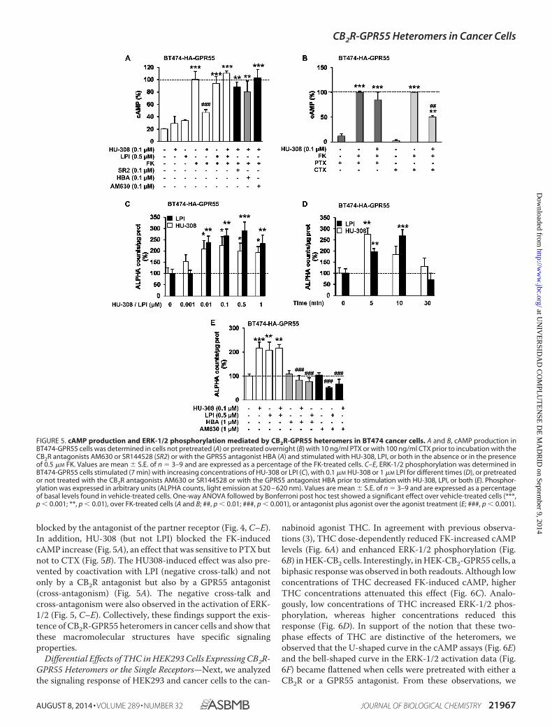
blocked by the antagonist of the partner receptor (Fig. 4, C–E).In addition, HU-308 (but not LPI) blocked the FK-inducedcAMP increase (Fig. 5A), an effect that was sensitive to PTX butnot to CTX (Fig. 5B). The HU308-induced effect was also pre-vented by coactivation with LPI (negative cross-talk) and notonly by a CB2R antagonist but also by a GPR55 antagonist(cross-antagonism) (Fig. 5A). The negative cross-talk andcross-antagonism were also observed in the activation of ERK-1/2 (Fig. 5, C–E). Collectively, these findings support the exis-tence of CB2R-GPR55 heteromers in cancer cells and show thatthese macromolecular structures have specific signalingproperties.
Differential Effects of THC in HEK293 Cells Expressing CB2R-GPR55 Heteromers or the Single Receptors—Next, we analyzedthe signaling response of HEK293 and cancer cells to the can-
nabinoid agonist THC. In agreement with previous observa-tions (3), THC dose-dependently reduced FK-increased cAMPlevels (Fig. 6A) and enhanced ERK-1/2 phosphorylation (Fig.6B) in HEK-CB2 cells. Interestingly, in HEK-CB2-GPR55 cells, abiphasic response was observed in both readouts. Although lowconcentrations of THC decreased FK-induced cAMP, higherTHC concentrations attenuated this effect (Fig. 6C). Analo-gously, low concentrations of THC increased ERK-1/2 phos-phorylation, whereas higher concentrations reduced thisresponse (Fig. 6D). In support of the notion that these two-phase effects of THC are distinctive of the heteromers, weobserved that the U-shaped curve in the cAMP assays (Fig. 6E)and the bell-shaped curve in the ERK-1/2 activation data (Fig.6F) became flattened when cells were pretreated with either aCB2R or a GPR55 antagonist. From these observations, we
FIGURE 5. cAMP production and ERK-1/2 phosphorylation mediated by CB2R-GPR55 heteromers in BT474 cancer cells. A and B, cAMP production inBT474-GPR55 cells was determined in cells not pretreated (A) or pretreated overnight (B) with 10 ng/ml PTX or with 100 ng/ml CTX prior to incubation with theCB2R antagonists AM630 or SR144528 (SR2) or with the GPR55 antagonist HBA (A) and stimulated with HU-308, LPI, or both in the absence or in the presenceof 0.5 �M FK. Values are mean � S.E. of n � 3–9 and are expressed as a percentage of the FK-treated cells. C–E, ERK-1/2 phosphorylation was determined inBT474-GPR55 cells stimulated (7 min) with increasing concentrations of HU-308 or LPI (C), with 0.1 �M HU-308 or 1 �M LPI for different times (D), or pretreatedor not treated with the CB2R antagonists AM630 or SR144528 or with the GPR55 antagonist HBA prior to stimulation with HU-308, LPI, or both (E). Phosphor-ylation was expressed in arbitrary units (ALPHA counts, light emission at 520 – 620 nm). Values are mean � S.E. of n � 3–9 and are expressed as a percentageof basal levels found in vehicle-treated cells. One-way ANOVA followed by Bonferroni post hoc test showed a significant effect over vehicle-treated cells (***,p � 0.001; **, p � 0.01), over FK-treated cells (A and B; ##, p � 0.01; ###, p � 0.001), or antagonist plus agonist over the agonist treatment (E; ###, p � 0.001).
CB2R-GPR55 Heteromers in Cancer Cells
AUGUST 8, 2014 • VOLUME 289 • NUMBER 32 JOURNAL OF BIOLOGICAL CHEMISTRY 21967
at UN
IVE
RSID
AD
CO
MPL
UT
EN
SE D
E M
AD
RID
on September 9, 2014
http://ww
w.jbc.org/
Dow
nloaded from
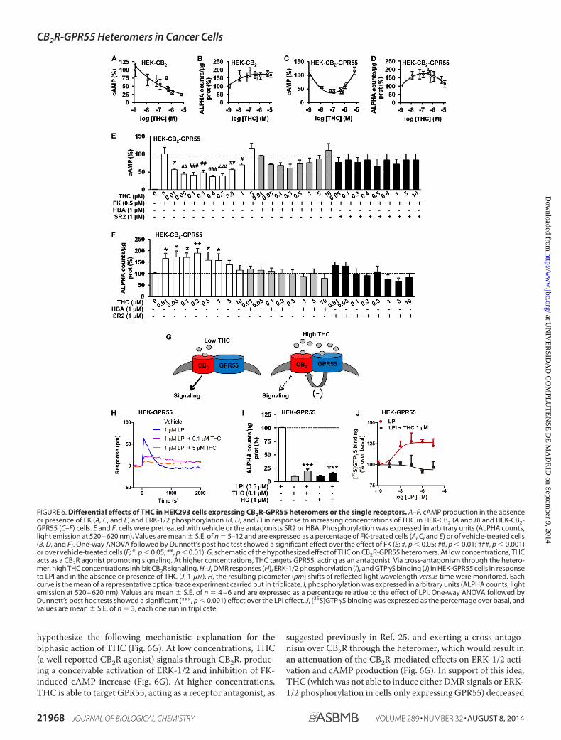
hypothesize the following mechanistic explanation for thebiphasic action of THC (Fig. 6G). At low concentrations, THC(a well reported CB2R agonist) signals through CB2R, produc-ing a conceivable activation of ERK-1/2 and inhibition of FK-induced cAMP increase (Fig. 6G). At higher concentrations,THC is able to target GPR55, acting as a receptor antagonist, as
suggested previously in Ref. 25, and exerting a cross-antago-nism over CB2R through the heteromer, which would result inan attenuation of the CB2R-mediated effects on ERK-1/2 acti-vation and cAMP production (Fig. 6G). In support of this idea,THC (which was not able to induce either DMR signals or ERK-1/2 phosphorylation in cells only expressing GPR55) decreased
FIGURE 6. Differential effects of THC in HEK293 cells expressing CB2R-GPR55 heteromers or the single receptors. A–F, cAMP production in the absenceor presence of FK (A, C, and E) and ERK-1/2 phosphorylation (B, D, and F) in response to increasing concentrations of THC in HEK-CB2 (A and B) and HEK-CB2-GPR55 (C–F) cells. E and F, cells were pretreated with vehicle or the antagonists SR2 or HBA. Phosphorylation was expressed in arbitrary units (ALPHA counts,light emission at 520 – 620 nm). Values are mean � S.E. of n � 5–12 and are expressed as a percentage of FK-treated cells (A, C, and E) or of vehicle-treated cells(B, D, and F). One-way ANOVA followed by Dunnett’s post hoc test showed a significant effect over the effect of FK (E; #, p � 0.05; ##, p � 0.01; ###, p � 0.001)or over vehicle-treated cells (F; *, p � 0.05; **, p � 0.01). G, schematic of the hypothesized effect of THC on CB2R-GPR55 heteromers. At low concentrations, THCacts as a CB2R agonist promoting signaling. At higher concentrations, THC targets GPR55, acting as an antagonist. Via cross-antagonism through the hetero-mer, high THC concentrations inhibit CB2R signaling. H–J, DMR responses (H), ERK-1/2 phosphorylation (I), and GTP�S binding (J) in HEK-GPR55 cells in responseto LPI and in the absence or presence of THC (J, 1 �M). H, the resulting picometer (pm) shifts of reflected light wavelength versus time were monitored. Eachcurve is the mean of a representative optical trace experiment carried out in triplicate. I, phosphorylation was expressed in arbitrary units (ALPHA counts, lightemission at 520 – 620 nm). Values are mean � S.E. of n � 4 – 6 and are expressed as a percentage relative to the effect of LPI. One-way ANOVA followed byDunnett’s post hoc tests showed a significant (***, p � 0.001) effect over the LPI effect. J, [35S]GTP�S binding was expressed as the percentage over basal, andvalues are mean � S.E. of n � 3, each one run in triplicate.
CB2R-GPR55 Heteromers in Cancer Cells
21968 JOURNAL OF BIOLOGICAL CHEMISTRY VOLUME 289 • NUMBER 32 • AUGUST 8, 2014
at UN
IVE
RSID
AD
CO
MPL
UT
EN
SE D
E M
AD
RID
on September 9, 2014
http://ww
w.jbc.org/
Dow
nloaded from

LPI-induced DMR responses (Fig. 6H) and ERK-1/2 activation(Fig. 6I) in HEK-GPR55 cells. The capability of THC to preventLPI-induced activation of GPR55 was further confirmed byGTP�S binding assays. LPI produced a marked increase in[35S]GTP�S binding in HEK-GPR55 membranes (Emax �129 � 2%; EC50 � 7.1 � 3.4 nM), an effect that was completelyblocked by coincubation with THC (Fig. 6J). Together, theseresults indicate that, at high concentrations, THC actuallybehaves as a GPR55 antagonist.
Involvement of CB2R-GPR55 Heteromers in the Response ofCancer Cells to THC—We then went back to cancer cells tochallenge this hypothesis. First, PLA experiments showed thatneither low nor high THC concentrations disrupt the CB2R-
GPR55 heteromers (Fig. 7A). Second, as in HEK-CB2-GPR55cells, a two-phase effect of THC was observed in BT474-GPR55cells on the modulation of both cAMP levels and ERK-1/2 phos-phorylation, in which the response found at low concentrationswas attenuated at higher concentrations (Fig. 7B). The U-shaped curve in the cAMP assays and the bell-shaped curve inthe ERK-1/2 activation became less pronounced or even flat-tened when BT474-GPR55 cells were pretreated with theGPR55 antagonist HBA (Fig. 7C), demonstrating that theantagonistic effect of THC on GPR55 modulates CB2R signal-ing through CB2R-GPR55 heteromers.
Our hypothesis was further corroborated in T98G cells, ahuman glioblastoma cell line that endogenously expresses bothCB2R (26) and GPR55 (12). By PLA, we detected red spots cor-responding to CB2R-GPR55 heteromers (Fig. 8A, top leftpanel). Treatment of cells with either low or high concentra-tions of THC did not alter this staining (Fig. 8A, top panels),suggesting that the heteromers are not disrupted by the canna-binoid. The CB2R-GPR55 complexes were not detected in thenegative controls, in which one of the primary antibodies wasomitted, or in T98G cells, in which CB2R (T98G-shCB2) orGPR55 (T98G-shGPR55) expression was silenced (Fig. 8A, bot-tom panels). As in transfected cells, a two-phase effect of THCwas observed in T98G cells on the modulation of both cAMPlevels and ERK-1/2 phosphorylation, in which the responsefound at low concentrations was attenuated at higher concen-trations (Fig. 8B). Finally, we analyzed the strength of ourhypothesis in an in vivo setting. Subcutaneous tumors weregenerated by injection of T98G cells into athymic male mice.Tumors increased their growth slightly in response to a lowTHC dose (although no statistical differences were observed),whereas a higher THC dose produced the opposite effect, i.e. asignificant reduction in tumor growth (Fig. 8C). According toour hypothesis, the low-dose effect would be produced mainlyvia activation of CB2R and the high-dose effect via cross-antag-onism of CB2R upon targeting of GPR55. The direct antago-nism of GPR55, a receptor that has been shown previously todrive tumorigenesis (12–14), by THC may contribute to thisstrong antitumoral response. Supporting the idea that GPR55behaves as a tumor growth brake when targeted by high dosesof THC, we observed that GPR55-silenced tumors increasedtheir growth when exposed to THC (Fig. 8C). The differentialeffects of THC on tumor growth occurred in concert with dif-ferential changes in the levels of activated ERK-1/2, i.e. a reduc-tion when CB2R and GPR55 were coexpressed and an enhance-ment when GPR55 was silenced (Fig. 8, D and E). These resultssupport our hypothesis and suggest that the well establishedcannabinoid target CB2R, as well as GPR55, coparticipate, inpart via direct receptor-receptor interaction, in the control oftumor growth in response to THC.
DISCUSSION
The findings reported in this study lead to three importantconclusions regarding the role of cannabinoids and their cog-nate receptors. First, we demonstrate the existence and func-tion of CB2R-GPR55 heteromers in cancer cells. Second, weshow that the expression of these receptor heteromers has amajor impact on cannabinoid signaling in these cells. Finally,
FIGURE 7. Involvement of CB2R-GPR55 heteromers in the response oftransfected cancer cells to THC. A, representative results of in situ PLAs per-formed in BT474-HA-GPR55 cells treated (30 min) with high and low THCconcentrations. In the confocal microscopy images (superimposed sections),heteromers appear as red spots. Cell nuclei were stained with DAPI (blue).Scale bars � 20 �m. B, the effect of THC on FK-induced cAMP production (leftpanel) and ERK-1/2 phosphorylation (right panel) in BT474-HA-GPR55 cells.Schematics depict the hypothesized THC mechanism of action. C, cAMP pro-duction (top panel) and ERK-1/2 phosphorylation (bottom panel) in BT474-GPR55 cells pretreated with vehicle or the GPR55 antagonist HBA prior tostimulation with THC. Top panel, cells were incubated in the absence or pres-ence of 0.5 �M forskolin. Phosphorylation was expressed in arbitrary units(ALPHA counts, light emission at 520 – 620 nm). Values are mean � S.E. of n �5–12 and are expressed as a percentage of FK-treated cells (cAMP determina-tion) or of vehicle-treated cells (ERK-1/2 phosphorylation). One-way ANOVAfollowed by Dunnett’s post hoc test showed a significant effect over the effectof FK (#, p � 0.05; ##, p � 0.01) or over vehicle-treated cells (F; *, p � 0.05;**, p � 0.01; ***, p � 0.001).
CB2R-GPR55 Heteromers in Cancer Cells
AUGUST 8, 2014 • VOLUME 289 • NUMBER 32 JOURNAL OF BIOLOGICAL CHEMISTRY 21969
at UN
IVE
RSID
AD
CO
MPL
UT
EN
SE D
E M
AD
RID
on September 9, 2014
http://ww
w.jbc.org/
Dow
nloaded from

our results suggest that direct targeting of CB2R-GPR55 viaappropriate doses of THC may be an effective approach toreducing tumor growth.
Receptor heteromers involving the sister cannabinoid recep-tor CB1 have been the focus of intense research. Therefore,CB1Rs have been shown previously to interact with other G
FIGURE 8. Involvement of CB2R-GPR55 heteromers in the response to THC of cancer cells endogenously expressing CB2R and GPR55. A, top panels,representative results of in situ PLAs performed in T98G cells treated (30 min) with vehicle and low and high THC concentrations. In the confocal microscopyimages (superimposed sections), heteromers appear as red spots. Cell nuclei were stained with DAPI (blue). Bottom panels, as negative controls, PLAs wereperformed in T98G cells in the absence of anti-CB2R antibody (GPR55) or anti-GPR55 antibody (CB2R) or in the presence of anti-GPR55 and anti-CB2R antibodiesin T98G cells in which CB2R (T98G-shCB2) or GPR55 (T89G-shGPR55) was silenced. Scale bars � 20 �m. B, FK-induced cAMP production (left panel) and ERK-1/2phosphorylation (right panel) in T98G cells in response to THC. Schematics depict the hypothesized THC mechanism of action. C, the volume of subcutaneoustumors generated by injection of T98G cells in immunodeficient mice was determined. Tumors were treated with a control siRNA (left panel) or a GPR55-selective siRNA (right panel), and animals received the indicated doses of THC or the corresponding vehicle (Veh). Tumor growth curves were compared byANOVA with a post hoc analysis by Student-Newman-Keuls test. D and E, Western blot analysis (D) and densitometric analysis (E) of phospho-ERK-1/2 (pERK) incontrol siRNA (siC) and GPR55-siRNA tumors treated with 15 mg/Kg THC. *, p � 0.05 versus vehicle-treated animals (C) or cells (E).
CB2R-GPR55 Heteromers in Cancer Cells
21970 JOURNAL OF BIOLOGICAL CHEMISTRY VOLUME 289 • NUMBER 32 • AUGUST 8, 2014
at UN
IVE
RSID
AD
CO
MPL
UT
EN
SE D
E M
AD
RID
on September 9, 2014
http://ww
w.jbc.org/
Dow
nloaded from

protein-coupled receptors, including dopamine D2 receptors(which promotes a switch in the preferential coupling from Gito Gs) (27), D2 receptors and adenosine A2A receptors simulta-neously (producing a negative modulation of D2 receptor func-tion by A2A and CB1R agonists) (28), opioid receptors (whichproduces a negative cross-talk between protomers) (29), orexinOX1 receptors (eliciting a positive cross-talk in response toorexin and cross-antagonism) (30), and angiotensin AT1 recep-tors (resulting in the potentiation of AT1 receptor signaling)(31). More recently, coimmunoprecipitation assays in HEK293cells have suggested that CB1R can form heteromers withGPR55 (32). In contrast to CB1R, very little is known about thepossible existence and functional relevance of heteromersinvolving CB2R. A recent study has shown that CB2R hetero-merizes with CB1R in neuronal cells in culture and in vivo (19).In these systems, coactivation of both receptors results in anegative cross-talk and a bidirectional cross-antagonism (19).However, CB2R signaling can be conceivably more relevant innon-differentiated cells, in which the receptor is highly abun-dant, than in terminally differentiated cells such as neurons, inwhich the receptor is scarce (3, 33). Specifically, CB2R, as well asGPR55, is notably overexpressed in a wide variety of cancer celllines and human malignant tumors (6, 11), in which they playpivotal roles in controlling cancer cell fate (6, 11–14). It istempting to speculate that CB2R-GPR55 heteromers may alsoexist and play pivotal signaling roles in other cells or tissues inwhich they are overexpressed, such as hematopoietic cells (16)or bones (34).
More and more studies have attempted to address the phys-iological role of GPR55. This receptor has been implicated incancer, where it is generally linked with growth and prolifera-tion (12–14). However, the molecular and cellular mechanismsbehind these effects are still unanswered. In addition, it hasbeen unclear whether GPR55’s effects on proliferation involveCB2R or are independent. Considering the receptor heteromersdiscussed above and knowing that CB2R and GPR55 have beenlinked functionally in hematopoietic cells (16), we pursued thehypothesis that CB2R-GPR55 heteromers might play a role inthe effects of GPR55 in cancer cells. Indeed, we found that thesecomplexes were able to form in HEK293 cells and in bothBT474 and T98G cancer cells and that they display a cross-talkand cross-antagonism at the level of the cAMP and p-ERK-1/2pathways. We also found different cell signaling effects at lowand high concentrations of THC and that this bimodal effectrequired the presence of the heteromer. Our findings that THCappears to be an antagonist of GPR55, at least at the level of cellsignaling both of the single receptor and within the CB2R-GPR55 heteromer, were particularly surprising. Previousreports have indeed suggested this (25), and the data weobtained in three different cell lines as well as in a mouse modelof cancer in vivo support these conclusions. This is in line withthe general idea that, despite the potential relationship betweencannabinoid receptors and GPR55, their pharmacology is verydifferent (8, 35).
Finally, our discovery that CB2R-GPR55 complexes haveunique pharmacological and signaling properties and are criti-cally involved in the response of cancer cells to THC both invitro and in vivo opens new doors to the development of com-
pounds targeting these heteromers as novel sites of interven-tion for future cancer studies. Our results also shed light on thepossible molecular mechanisms underlying the well known butstill poorly understood biphasic effects of cannabinoids, whichhave been reported for several decades regarding their actionon food intake, motor behavior, and anxiety, among others(36 –38).
Acknowledgments—We thank Dr. Nariman Balenga and Dr. JuliaKargl for generation of the HEK cell lines.
REFERENCES1. Dorsam, R. T., and Gutkind, J. S. (2007) G-protein-coupled receptors and
cancer. Nat. Rev. Cancer 7, 79 –942. Rosenbaum, D. M., Rasmussen, S. G., and Kobilka, B. K. (2009) The struc-
ture and function of G-protein-coupled receptors. Nature 459, 356 –3633. Pertwee, R. G., Howlett, A. C., Abood, M. E., Alexander, S. P., Di Marzo, V.,
Elphick, M. R., Greasley, P. J., Hansen, H. S., Kunos, G., Mackie, K.,Mechoulam, R., and Ross, R. A. (2010) International Union of Basic andClinical Pharmacology: LXXIX: cannabinoid receptors and their ligands:beyond CB and CB. Pharmacol. Rev. 62, 588 – 631
4. Pacher, P., Bátkai, S., and Kunos, G. (2006) The endocannabinoid systemas an emerging target of pharmacotherapy. Pharmacol. Rev. 58, 389 – 462
5. Guzmán, M., Sánchez, C., and Galve-Roperh, I. (2002) Cannabinoids andcell fate. Pharmacol. Ther. 95, 175–184
6. Velasco, G., Sánchez, C., and Guzmán, M. (2012) Towards the use ofcannabinoids as antitumour agents. Nat Rev Cancer 12, 436 – 444
7. Brown, A. J. (2007) Novel cannabinoid receptors. Br. J. Pharmacol. 152,567–575
8. Ross, R. A. (2009) The enigmatic pharmacology of GPR55. Trends Phar-macol. Sci. 30, 156 –163
9. Oka, S., Nakajima, K., Yamashita, A., Kishimoto, S., and Sugiura, T. (2007)Identification of GPR55 as a lysophosphatidylinositol receptor. Biochem.Biophys. Res. Commun. 362, 928 –934
10. Pertwee, R. G. (2010) Receptors and channels targeted by synthetic can-nabinoid receptor agonists and antagonists. Curr. Med. Chem. 17,1360 –1381
11. Henstridge, C. M., Balenga, N. A., Kargl, J., Andradas, C., Brown, A. J.,Irving, A., Sanchez, C., and Waldhoer, M. (2011) Minireview: recent de-velopments in the physiology and pathology of the lysophosphatidylinosi-tol-sensitive receptor GPR55. Mol. Endocrinol. 25, 1835–1848
12. Andradas, C., Caffarel, M. M., Pérez-Gómez, E., Salazar, M., Lorente, M.,Velasco, G., Guzmán, M., and Sánchez, C. (2011) The orphan G protein-coupled receptor GPR55 promotes cancer cell proliferation via ERK. On-cogene 30, 245–252
13. Pérez-Gómez, E., Andradas, C., Flores, J. M., Quintanilla, M., Paramio,J. M., Guzmán, M., and Sánchez, C. (2013) The orphan receptor GPR55drives skin carcinogenesis and is upregulated in human squamous cellcarcinomas. Oncogene 32, 2534 –2542
14. Piñeiro, R., Maffucci, T., and Falasca, M. (2011) The putative cannabinoidreceptor GPR55 defines a novel autocrine loop in cancer cell proliferation.Oncogene 30, 142–152
15. Rozenfeld, R., and Devi, L. A. (2010) Receptor heteromerization and drugdiscovery. Trends Pharmacol. Sci. 31, 124 –130
16. Balenga, N. A., Aflaki, E., Kargl, J., Platzer, W., Schröder, R., Blättermann,S., Kostenis, E., Brown, A. J., Heinemann, A., and Waldhoer, M. (2011)GPR55 regulates cannabinoid 2 receptor-mediated responses in humanneutrophils. Cell Res. 21, 1452–1469
17. Henstridge, C. M., Balenga, N. A., Ford, L. A., Ross, R. A., Waldhoer, M.,and Irving, A. J. (2009) The GPR55 ligand L-�-lysophosphatidylinositolpromotes RhoA-dependent Ca2 signaling and NFAT activation. FASEBJ. 23, 183–193
18. González, S., Rangel-Barajas, C., Peper, M., Lorenzo, R., Moreno, E., Ciru-ela, F., Borycz, J., Ortiz, J., Lluís, C., Franco, R., McCormick, P. J., Volkow,N. D., Rubinstein, M., Floran, B., and Ferré, S. (2012) Dopamine D4 recep-
CB2R-GPR55 Heteromers in Cancer Cells
AUGUST 8, 2014 • VOLUME 289 • NUMBER 32 JOURNAL OF BIOLOGICAL CHEMISTRY 21971
at UN
IVE
RSID
AD
CO
MPL
UT
EN
SE D
E M
AD
RID
on September 9, 2014
http://ww
w.jbc.org/
Dow
nloaded from

tor, but not the ADHD-associated D4.7 variant, forms functional hetero-mers with the dopamine D2S receptor in the brain. Mol. Psychiatry 17,650 – 662
19. Callén, L., Moreno, E., Barroso-Chinea, P., Moreno-Delgado, D., Cortés,A., Mallol, J., Casadó, V., Lanciego, J. L., Franco, R., Lluis, C., Canela, E. I.,and McCormick, P. J. (2012) Cannabinoid receptors CB1 and CB2 formfunctional heteromers in brain. J. Biol. Chem. 287, 20851–20865
20. Carriba, P., Navarro, G., Ciruela, F., Ferré, S., Casadó, V., Agnati, L., Cor-tés, A., Mallol, J., Fuxe, K., Canela, E. I., Lluís, C., and Franco, R. (2008)Detection of heteromerization of more than two proteins by sequentialBRET-FRET. Nat. Methods 5, 727–733
21. Söderberg, O., Leuchowius, K. J., Gullberg, M., Jarvius, M., Weibrecht, I.,Larsson, L. G., and Landegren, U. (2008) Characterizing proteins and theirinteractions in cells and tissues using the in situ proximity ligation assay.Methods 45, 227–232
22. Trifilieff, P., Rives, M. L., Urizar, E., Piskorowski, R. A., Vishwasrao, H. D.,Castrillon, J., Schmauss, C., Slättman, M., Gullberg, M., and Javitch, J. A.(2011) Detection of antigen interactions ex vivo by proximity ligation as-say: endogenous dopamine D2-adenosine A2A receptor complexes in thestriatum. BioTechniques 51, 111–118
23. Schröder, R., Janssen, N., Schmidt, J., Kebig, A., Merten, N., Hennen, S.,Müller, A., Blättermann, S., Mohr-Andrä, M., Zahn, S., Wenzel, J., Smith,N. J., Gomeza, J., Drewke, C., Milligan, G., Mohr, K., and Kostenis, E.(2010) Deconvolution of complex G protein-coupled receptor signaling inlive cells using dynamic mass redistribution measurements. Nat. Biotech-nol. 28, 943–949
24. Mato, S., Vidal, R., Castro, E., Díaz, A., Pazos, A., and Valdizán, E. M.(2010) Long-term fluoxetine treatment modulates cannabinoid type 1 re-ceptor-mediated inhibition of adenylyl cyclase in the rat prefrontal cortexthrough 5-hydroxytryptamine 1A receptor-dependent mechanisms. Mol.Pharmacol. 77, 424 – 434
25. Anavi-Goffer, S., Baillie, G., Irving, A. J., Gertsch, J., Greig, I. R., Pertwee,R. G., and Ross, R. A. (2012) Modulation of L-�-lysophosphatidylinositol/GPR55 mitogen-activated protein kinase (MAPK) signaling by cannabi-noids. J. Biol. Chem. 287, 91–104
26. Lorente, M., Torres, S., Salazar, M., Carracedo, A., Hernández-Tiedra, S.,Rodríguez-Fornés, F., García-Taboada, E., Meléndez, B., Mollejo, M.,Campos-Martín, Y., Lakatosh, S. A., Barcia, J., Guzmán, M., and Velasco,G. (2011) Stimulation of the midkine/ALK axis renders glioma cells resis-tant to cannabinoid antitumoral action. Cell Death Differ. 18, 959 –973
27. Marcellino, S., Attar, H., Lièvremont, D., Lett, M. C., Barbier, F., andLagarde, F. (2008) Heat-treated Saccharomyces cerevisiae for antimonyspeciation and antimony(III) preconcentration in water samples. Anal.Chim. Acta 629, 73– 83
28. Navarro, G., Ferré, S., Cordomi, A., Moreno, E., Mallol, J., Casadó, V.,Cortés, A., Hoffmann, H., Ortiz, J., Canela, E. I., Lluís, C., Pardo, L., Franco,R., and Woods, A. S. (2010) Interactions between intracellular domains askey determinants of the quaternary structure and function of receptorheteromers. J. Biol. Chem. 285, 27346 –27359
29. Rios, C., Gomes, I., and Devi, L. A. (2006) � Opioid and CB1 cannabinoidreceptor interactions: reciprocal inhibition of receptor signaling and neu-ritogenesis. Br. J. Pharmacol. 148, 387–395
30. Ellis, J., Pediani, J. D., Canals, M., Milasta, S., and Milligan, G. (2006)Orexin-1 receptor-cannabinoid CB1 receptor heterodimerization resultsin both ligand-dependent and -independent coordinated alterations ofreceptor localization and function. J. Biol. Chem. 281, 38812–38824
31. Rozenfeld, R., Gupta, A., Gagnidze, K., Lim, M. P., Gomes, I., Lee-Ramos,D., Nieto, N., and Devi, L. A. (2011) AT1R-CB(1)R heteromerization re-veals a new mechanism for the pathogenic properties of angiotensin II.EMBO J. 30, 2350 –2363
32. Kargl, J., Balenga, N., Parzmair, G. P., Brown, A. J., Heinemann, A., andWaldhoer, M. (2012) The cannabinoid receptor CB1 modulates the sig-naling properties of the lysophosphatidylinositol receptor GPR55. J. Biol.Chem. 287, 44234 – 44248
33. Pertwee, R. G. (2009) Emerging strategies for exploiting cannabinoid re-ceptor agonists as medicines. Br. J. Pharmacol. 156, 397– 411
34. Whyte, L. S., Ryberg, E., Sims, N. A., Ridge, S. A., Mackie, K., Greasley, P. J.,Ross, R. A., and Rogers, M. J. (2009) The putative cannabinoid receptorGPR55 affects osteoclast function in vitro and bone mass in vivo. Proc.Natl. Acad. Sci. U.S.A. 106, 16511–16516
35. Rempel, V., Volz, N., Gläser, F., Nieger, M., Bräse, S., and Müller, C. E.(2013) Antagonists for the orphan G-protein-coupled receptor GPR55based on a coumarin scaffold. J. Med. Chem. 56, 4798 – 4810
36. Moreira, F. A., and Wotjak, C. T. (2010) Cannabinoids and anxiety. Curr.Top. Behav. Neurosci. 2, 429 – 450
37. Sañudo-Peña, M. C., Romero, J., Seale, G. E., Fernandez-Ruiz, J. J., andWalker, J. M. (2000) Activational role of cannabinoids on movement. Eur.J. Pharmacol. 391, 269 –274
38. Sulcova, E., Mechoulam, R., and Fride, E. (1998) Biphasic effects of anan-damide. Pharmacol. Biochem. Behav. 59, 347–352
CB2R-GPR55 Heteromers in Cancer Cells
21972 JOURNAL OF BIOLOGICAL CHEMISTRY VOLUME 289 • NUMBER 32 • AUGUST 8, 2014
at UN
IVE
RSID
AD
CO
MPL
UT
EN
SE D
E M
AD
RID
on September 9, 2014
http://ww
w.jbc.org/
Dow
nloaded from