sign management psmba
TRANSCRIPT

7/29/2019 SIGN Management PSMBA
http://slidepdf.com/reader/full/sign-management-psmba 1/64
Scottish Intercollegiate Guidelines Network
SIGN
Management of acute upper andlower gastrointestinal bleeding
A national clinical guideline
September 2008
105

7/29/2019 SIGN Management PSMBA
http://slidepdf.com/reader/full/sign-management-psmba 2/64
This document is produced from elemental chlorine-free material and is sourced from sustainable forests
KEY TO EVIDENCE STATEMENTS AND GRADES OF RECOMMENDATIONS
LEVELS OF EVIDENCE
1++ High quality meta-analyses, systematic reviews of RCTs, or RCTs with a very low risk of bias
1+ Well conducted meta-analyses, systematic reviews, or RCTs with a low risk of bias
1 - Meta-analyses, systematic reviews, or RCTs with a high risk of bias
2++ High quality systematic reviews of case control or cohort studies
High quality case control or cohort studies with a very low risk of confounding or bias and ahigh probability that the relationship is causal
2+ Well conducted case control or cohort studies with a low risk of confounding or bias and amoderate probability that the relationship is causal
2 - Case control or cohort studies with a high risk of confounding or bias and a significant risk thatthe relationship is not causal
3 Non-analytic studies, eg case reports, case series
4 Expert opinion
GRADES OF RECOMMENDATION
Note: The grade of recommendation relates to the strength of the evidence on which therecommendation is based. It does not reect the clinical importance of the recommendation.
A At least one meta-analysis, systematic review, or RCT rated as 1++,and directly applicable to the target population; or
A body of evidence consisting principally of studies rated as 1+,directly applicable to the target population, and demonstrating overall consistency of results
B A body of evidence including studies rated as 2++,directly applicable to the target population, and demonstrating overall consistency of results; or
Extrapolated evidence from studies rated as 1++ or 1+
C A body of evidence including studies rated as 2+,directly applicable to the target population and demonstrating overall consistency of results; or
Extrapolated evidence from studies rated as 2++
D Evidence level 3 or 4; or
Extrapolated evidence from studies rated as 2+
GOOD PRACTICE POINTS
Recommended best practice based on the clinical experience of the guideline developmentgroup.
NHS Quality Improvement Scotland (NHS QIS) is committed to equality and diversity. Thisguideline has been assessed for its likely impact on the six equality groups defined by age, disability,gender, race, religion/belief, and sexual orientation.
For the full equality and diversity impact assessment report please see the “published guidelines”section of the SIGN website at www.sign.ac.uk/guidelines/published/numlist.html. The full reportin paper form and/or alternative format is available on request from the NHS QIS Equality andDiversity Officer.
Every care is taken to ensure that this publication is correct in every detail at the time of publication.However, in the event of errors or omissions corrections will be published in the web version of this
document, which is the definitive version at all times. This version can be found on our web sitewww.sign.ac.uk

7/29/2019 SIGN Management PSMBA
http://slidepdf.com/reader/full/sign-management-psmba 3/64
Scottish Intercollegiate Guidelines Network
Mm
A national clinical guideline
September 2008
7/29/2019 SIGN Management PSMBA
http://slidepdf.com/reader/full/sign-management-psmba 4/64
ManageMent of acute upper and lower gastrointestinal bleeding
isbn 978 1 905813 37 7
ph sm 2008
SIGN consents to the photocoping of this guideline for thepurpose of implementation in NHSScotland
sh i g ne H, 8 -10 H c
eh eH7 5ea
...

7/29/2019 SIGN Management PSMBA
http://slidepdf.com/reader/full/sign-management-psmba 5/64
contents
c
1 i ..................................................................................................................... 1
1.1 The need for a guideline.................................................................................................... 1
1.2 Remit of the guideline ....................................................................................................... 1
1.3 Definitions ........................................................................................................................ 2
1.4 Statement of intent ............................................................................................................ 3
2 am ....................................................................................................... 4
2.1 Assessing gastrointestinal bleeding in the communit ........................................................ 4
2.2 Assessing gastrointestinal bleeding in hospital ................................................................... 4
3 o v ................................................................................................... 10
3.1 Dedicated GI bleeding unit ............................................................................................... 10
4 r mm .............................................................................. 12
4.1 Airwa, breathing and circulation ...................................................................................... 12
4.2 Fluid resuscitation ............................................................................................................. 12
4.3 Earl pharmacological management .................................................................................. 13
4.4 Earl endoscopic intervention ........................................................................................... 14
5 Mm -v ........................................... 16
5.1 Risk stratification ............................................................................................................... 16
5.2 Endoscop......................................................................................................................... 16
5.3 Pharmacological therap ................................................................................................... 19
6 Mm v ........................................ 26
6.1 Endoscopic therap for acute variceal haemorrhage .......................................................... 27
6.2 Vasoactive drug therap for acute variceal haemorrhage ................................................... 28
6.3 Antibiotic therap .............................................................................................................. 30
6.4 Balloon tamponade ........................................................................................................... 31
6.5 Management of bleeding varices not controlled b endoscop .......................................... 31
7 pv v ..................................................................................... 32
7.1 Vasoactive drug therap .................................................................................................... 32
7.2 Endoscopic therap ........................................................................................................... 32
7.3 Portosstemic shunts ......................................................................................................... 33
8 Mm .............................................................. 34
8.1 Localising bleeding ........................................................................................................... 35
8.2 Interventions ..................................................................................................................... 35

7/29/2019 SIGN Management PSMBA
http://slidepdf.com/reader/full/sign-management-psmba 6/64
antibiotic propHylaxis in surgeryManageMent of acute upper and lower gastrointestinal bleeding
9 pv m ................................................................................................... 37
9.1 Areas of concern to patients .............................................................................................. 37
9.2 Sources of further information ........................................................................................... 38
10 imm h .............................................................................................. 39
10.1 Resource implications of ke recommendations ................................................................ 39
10.2 Auditing current practice ................................................................................................... 40
10.3 Advice to NHSScotland from the scottish medicines consortium ....................................... 40
11 th v ............................................................................................................ 41
11.1 Sstematic literature review ............................................................................................... 41
11.2 Recommendations for research.......................................................................................... 41
11.3 Review and updating ......................................................................................................... 42
12 dvm h .......................................................................................... 43
12.1 Introduction ...................................................................................................................... 43
12.2 The guideline development group ..................................................................................... 43
12.3 Acknowledgements ........................................................................................................... 44
12.4 Consultation and peer review ............................................................................................ 44
av .............................................................................................................................. 46
a 1....................................................................................................................................... 47
a 2....................................................................................................................................... 51
r .................................................................................................................................. 52

7/29/2019 SIGN Management PSMBA
http://slidepdf.com/reader/full/sign-management-psmba 7/641
introduction
1 i
1.1 tHe need for a guideline
Acute gastrointestinal (GI) bleeding (or haemorrhage) is a common maor medical emergenc,accounting for approximatel 7,000 admissions to hospitals in Scotland each ear. In a 2007UK-wide audit, overall mortalit of patients admitted with acute GI bleeding was 7%. In contrastthe mortalit in patients who bled during admissions to hospital for other reasons was 26%.1 Inan audit undertaken in the West of Scotland the incidence of acute GI bleeding was higher thanthat reported elsewhere at 170/100,000 people with a mortalit of 8.2%. 2 These differencesma relate to different case ascertainment in the two audits.
Over the last ten ears there has been a number of improvements in diagnosis and management.The increased involvement of acute care specialists during resuscitation and follow up, improveddiagnostic and therapeutic endoscop, advances in diagnostic and therapeutic radiolog, theuse of powerful ulcer healing drugs, more selective and less invasive surgical approaches maall improve outcome for patients. These changes have altered the diagnostic and treatment
pathwas for patients presenting with non-variceal and variceal upper GI bleeding andthose with acute colonic bleeding. There is a need to examine the evidence to clarif whichdiagnostic and management steps have proven benefit. The maor obectives of all involved inthe management of bleeding patients are to reduce mortalit and the need for maor surger. Asecondar obective is to prevent unnecessar hospital admission for patients presenting withbleeding that is not life threatening.
1.2 reMit of tHe guideline
1.2.1 OVERALL OBjECTIVES
This guideline provides recommendations based on current evidence for best practice in
the management of acute upper and lower GI bleeding. It includes the assessment andmanagement of variceal, non-variceal, and colonic bleeding in adults. The guideline dealswith the management of bleeding that is of sufficient severit to lead to emergenc admissionto hospital. Bleeding of lesser severit is subect to elective investigation and is not consideredhere. The management of patients under the age of 14 is not covered b this guideline.
1.2.2 TARGET USERS OF THE GUIDELINE
This guideline will be of interest to a range of medical professionals including acute phsicians,gastroenterologists, gastrointestinal surgeons, endoscopists, pharmacists, anaesthetists andnurses. It will also be of interest to patients who have suffered from acute GI bleeding and totheir carers.
7/29/2019 SIGN Management PSMBA
http://slidepdf.com/reader/full/sign-management-psmba 8/642
ManageMent of acute upper and lower gastrointestinal bleeding
1.3 definitions
u
Upper gastrointestinal bleeding (or haemorrhage) is that originating proximal to the ligamentof Treitz; in practice from the oesophagus, stomach and duodenum. Lower gastrointestinal
bleeding is that originating from the small bowel and colon. This guideline focuses upon upper GI and colonic bleeding since acute small bowel bleeding is uncommon.
Hmm (and coffee-ground vomitus)
Haematemesis is vomiting of blood from the upper gastrointestinal tract or occasionall after swallowing blood from a source in the nasopharnx. Bright red haematemesis usuall impliesactive haemorrhage from the oesophagus, stomach or duodenum. This can lead to circulatorcollapse and constitutes a maor medical emergenc. Patients presenting with haematemesishave a higher mortalit than those presenting with melaena alone.2
Coffee-ground vomitus refers to the vomiting of black material which is assumed to be blood.Its presence implies that bleeding has ceased or has been relativel modest.
MMelaena is the passage of black tarr stools usuall due to acute upper gastrointestinal bleedingbut occasionall from bleeding within the small bowel or right side of the colon.
Hmhz
Hematochezia is the passage of fresh or altered blood per rectum usuall due to colonic bleeding.Occasionall profuse upper gastrointestinal or small bowel bleeding can be responsible.
sh
Shock is circulator insufficienc resulting in inadeuate oxgen deliver leading to globalhpoperfusion and tissue hpoxia. In the context of GI bleeding shock is most likel to behpovolaemic (due to inadeuate circulating volume from acute blood loss). The shocked,
hpovolaemic patient generall exhibits one or more of the following signs or smptoms:a rapid pulse (tachcardia)
anxiet or confusion
a high respirator rate (tachpnoea)
cool clamm skin
low urine output (oliguria)
low blood pressure (hpotension).
It is important to remember that a patient with normal blood pressure ma still be shocked andreuire resuscitation.
V
Varices are abnormal distended veins usuall in the oesophagus (oesophageal varices) and lessfreuentl in the stomach (gastric varices) or other sites (ectopic varices) usuall occurring as aconseuence of liver disease. Bleeding is characteristicall severe and ma be life threatening.The size of the varices and their propensit to bleed is directl related to the portal pressure,which, in the maorit of cases, is directl related to the severit of underling liver disease.Large varices with red spots are at highest risk of rupture.
e
Endoscop is the visualisation of the inside of the gastrointestinal tract using telescopes.Examination of the upper gastrointestinal tract (oesophagus, stomach and duodenum) is knownas gastroscop or upper gastrointestinal endoscop. Examination of the colon (large bowel) iscalled colonoscop.
tTriage is a sstem of initial assessment and management whereb a group of patients is classifiedaccording to the seriousness of their inuries or illnesses so that treatment priorities can beallocated between them.

7/29/2019 SIGN Management PSMBA
http://slidepdf.com/reader/full/sign-management-psmba 9/643
introduction
1.4 stateMent of intent
This guideline is not intended to be construed or to serve as a standard of care. Standardsof care are determined on the basis of all clinical data available for an individual case andare subect to change as scientific knowledge and technolog advance and patterns of care
evolve. Adherence to guideline recommendations will not ensure a successful outcome inever case, nor should the be construed as including all proper methods of care or excludingother acceptable methods of care aimed at the same results. The ultimate udgement must bemade b the appropriate healthcare professional(s) responsible for clinical decisions regardinga particular clinical procedure or treatment plan. This udgement should onl be arrived atfollowing discussion of the options with the patient, covering the diagnostic and treatmentchoices available. It is advised, however, that significant departures from the national guidelineor an local guidelines derived from it should be full documented in the patient’s case notesat the time the relevant decision is taken.
1.4.1 ADDITIONAL ADVICE TO NHSSCOTLAND FROM NHS qUALITy IMPROVEMENTSCOTLAND AND THE SCOTTISH MEDICINES CONSORTIUM
NHS qIS processes multiple technolog appraisals (MTAs) for NHSScotland that have beenproduced b the National Institute for Health and Clinical Excellence (NICE) in England andWales.
The Scottish Medicines Consortium (SMC) provides advice to NHS Boards and their Area Drugand Therapeutics Committees about the status of all newl licensed medicines and an maor new indications for established products.
SMC advice and NHS qIS validated NICE MTAs relevant to this guideline are summarised inthe section on implementation.

7/29/2019 SIGN Management PSMBA
http://slidepdf.com/reader/full/sign-management-psmba 10/644
ManageMent of acute upper and lower gastrointestinal bleeding
3
3
2-
3
3
3
2
2 am
2.1 assessing gastrointestinal bleeding in tHe coMMunity
The assessment of GI bleeding from an cause in the communit involves the identificationof patients who reuire urgent admission, patients who reuire to be referred for outpatientassessment and patients who can be managed at home without involvement of hospital services.No studies were identified that were undertaken in primar care settings to address optimalreferral practice. The decision to refer must be based upon clinical experience, common senseand extrapolation of guidance derived from risk assessment studies undertaken in secondarcare settings.
2.2 assessing gastrointestinal bleeding in Hospital
The purpose of this section is to assist individual units to develop guidelines and protocolsbased on available evidence which are suitable for their local circumstances. Patients referredto hospital are initiall assessed in a variet of settings including emergenc departments,acute assessment units, gastroenterolog departments, dedicated GI bleeding units or surgicalwards.
Acute GI bleeding is a medical emergenc. Initial triage and assessment are generic withemphasis on identifing the sick patient with life threatening haemodnamic compromise andinitiating appropriate resuscitation (see section 4.2). Certain clinical features associated with GIbleeding have been studied in attempts to identif patients at increased risk of morbidit anddeath. Although acute upper and lower GI bleeding are distinct entities, the site of bleedingis not alwas immediatel apparent; for example, 15% of patients with severe haematocheziahave a source of bleeding in the upper GI tract.3 Despite this, the literature on upper and lower GI bleeding is largel separate and this section on assessment is similarl subdivided.
2.2.1 RISK FACTORS ASSOCIATED WITH POOR OUTCOMEa
There is a lack of good ualit studies on the initial assessment of patients with acute upper GIbleeding (UGIB). Limited evidence is available from cohort and case series which identif riskfactors associated with poor outcome (variousl defined) but usuall without formal scoring.Studies confirm an extremel high fatalit in inpatients of 42%.4,5
The following factors are associated with a poor outcome, defined in terms of severit of bleed,uncontrolled bleeding, rebleeding, need for intervention and mortalit. These factors should betaken into account when determining the need for admission or suitabilit for discharge.
a - mortalit due to UGIB increases with age across all age groups. Odds ratio (OR) for mortalit is from 1.8 to 3 for age >60 ears (compared to patients aged 45-59 ears), and
from4.5to12forage>75years(comparedtopatients≤75years).2,4,6
cm - the absence of significant comorbidit is associated with mortalit aslow as 4%.2,4,6,7 Even one comorbidit almost doubles mortalit (OR 1.8) and thepresence of cardiac failure (OR 1.8) or malignanc (OR 3.8) significantl worsensprognosis.
lv - cirrhosis is associated with a doubling of mortalit and much higher risk of interventions such as endoscopic haemostasis or transfusion.8 The overall mortalit of patientspresenting with varices is 14%.1
i have approximatel a threefold increased risk of death compared to patientsnewl admitted with GI bleeding. This is due to the presence of comorbidities in establishedinpatients rather than increased severit of bleeding.4,5
i h (hpotension and tachcardia) is associated with increased mortalit (OR 3.8)
and need for intervention.2,4,7

7/29/2019 SIGN Management PSMBA
http://slidepdf.com/reader/full/sign-management-psmba 11/645
assessMent and triage
3
3
3
3
3
3
3
4
3
3
3
3
3
3
3
c after admission is associated with high risk of intervention (OR 1.8)7 and up to a 50-fold increased mortalit.6
Hmm - the presence of initial haematemesis doubles mortalit.2,7
Hmhz - the presence of haematochezia doubles rebleeding, mortalit and surgerrates.9
ev is associated with a need for intervention.10
Non-steroidal anti-inflammator drugs (NSAIDs)2,11 and anticoagulants2,12 do not adversel affectthe clinical outcomes of patients presenting with UGIB.
There is conflicting evidence on the value of nasogastric aspiration. A blood aspirate maindicate a high-risk lesion (sensitivit 48%, specificit 76%) but no evidence has been identifiedthat it alters outcome.13,14
a
There is limited evidence available on the initial assessment of patients with acute lower gastrointestinal bleeding (LGIB). One general review of management15 and one guidelinewere identified.16 Other evidence comes from case series and epidemiolog, and from expert
opinion. Two uncontrolled case series analse earl predictors of severit, one prospective 17 and one retrospective.18 The available evidence identifies the following factors associated withuncontrolled bleeding and/or death.
a - acute lower GI bleeding occurs most often in the elderl. The precise relationshipbetween age and mortalit is statisticall less well defined than for UGIB.15,18,19
a hmm (OR 3 to 4.3) and gross rectal bleeding on initialexamination (OR 2.3 to 3) are important predictors of subseuent severe bleeding. 17,18
cm - the presence of two comorbid conditions doubles the chance of a severebleed (OR 1.9).18
s – patients taking aspirin or NSAIDs are at increased risk of severe lower GIbleeding (OR 1.8 to 2.7).18,20
i
who are hospitalised for another condition and who subseuentl bleed after admission have a mortalit rate of 23% compared with 3.6% in those admitted to hospitalbecause of rectal bleeding (p<0.001).19
The patient’s histor is important for accurate assessment of risk and can give important cluesto the diagnosis and need for admission. For example, a histor of previous LGIB from a knowndiagnosis of diverticular disease (the commonest cause of LGIB accounting for 23-48% of cases)predicts a further episode with a 10% chance of recurrence at one ear and 25% at four ears.Diverticular bleeds resolve spontaneousl in 75% of cases.19
2.2.2 PRE-ENDOSCOPIC RISK ASSESSMENT
a
Simple and widel validated scoring sstems to identif patients at high risk of rebleeding, deathand active intervention are needed for optimum management.
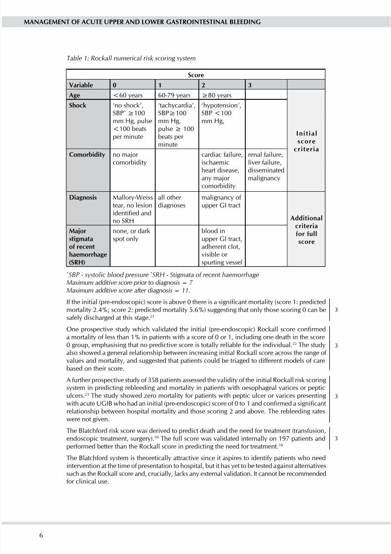
The Rockall scoring sstem was principall designed to predict death based on a combinationof clinical and endoscopic findings. Given that man of the risk factors for rebleeding areidentical to those for mortalit and that rebleeding itself is independentl predictive of death,the Rockall score ma also be used to estimate rebleeding risk.21 The initial (pre-endoscopic)Rockall score is derived from age (0 to 2 points), shock (0 to 2 points) and comorbidit (0 to3 points). The minimum score of 0 is assigned to patients with age <60 ears who have noevidence of shock and or comorbidit. A score of 0 identifies 15% of patients with acute UGIBat presentation who have an extremel low risk of death (0.2%) and rebleeding (0.2%), andwho ma be suitable for earl discharge or non-admission (see Table 1).21
7/29/2019 SIGN Management PSMBA
http://slidepdf.com/reader/full/sign-management-psmba 12/646
ManageMent of acute upper and lower gastrointestinal bleeding
3
3
3
3
Table 1: Rockall numerical risk scoring system
s
V 0 1 2 3
a <60 ears 60-79 ears ≥80years
i
sh ‘no shock’,SBP*≥100mm Hg, pulse<100 beatsper minute
‘tachcardia’,SBP≥100mm Hg,pulse≥100beats per minute
‘hpotension’,SBP <100mm Hg,
cm no maor comorbidit
cardiac failure,ischaemicheart disease,an maor comorbidit
renal failure,liver failure,disseminatedmalignanc
d Mallor-Weisstear, no lesionidentified andno SRH
all other diagnoses
malignanc of upper GI tract
a
Mjm hmh(srH)
none, or darkspot onl
blood inupper GI tract,adherent clot,visible or spurting vessel
*SBP - systolic blood pressure *SRH - Stigmata of recent haemorrhageMaximum additive score prior to diagnosis = 7
Maximum additive score after diagnosis = 11.
If the initial (pre-endoscopic) score is above 0 there is a significant mortalit (score 1: predictedmortalit 2.4%; score 2: predicted mortalit 5.6%) suggesting that onl those scoring 0 can besafel discharged at this stage.21
One prospective stud which validated the initial (pre-endoscopic) Rockall score confirmeda mortalit of less than 1% in patients with a score of 0 or 1, including one death in the score0 group, emphasising that no predictive score is totall reliable for the individual.22 The studalso showed a general relationship between increasing initial Rockall score across the range of values and mortalit, and suggested that patients could be triaged to different models of carebased on their score.
A further prospective stud of 358 patients assessed the validit of the initial Rockall risk scoring
sstem in predicting rebleeding and mortalit in patients with oesophageal varices or pepticulcers.23 The stud showed zero mortalit for patients with peptic ulcer or varices presentingwith acute UGIB who had an initial (pre-endoscopic) score of 0 to 1 and confirmed a significantrelationship between hospital mortalit and those scoring 2 and above. The rebleeding rateswere not given.
The Blatchford risk score was derived to predict death and the need for treatment (transfusion,endoscopic treatment, surger).10 The full score was validated internall on 197 patients andperformed better than the Rockall score in predicting the need for treatment.10
The Blatchford sstem is theoreticall attractive since it aspires to identif patients who needintervention at the time of presentation to hospital, but it has et to be tested against alternativessuch as the Rockall score and, cruciall, lacks an external validation. It cannot be recommendedfor clinical use.
7/29/2019 SIGN Management PSMBA
http://slidepdf.com/reader/full/sign-management-psmba 13/647
assessMent and triage
3
3
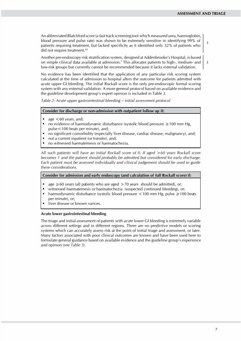
An abbreviated Blatchford score (a fast track screening tool which measured urea, haemoglobin,blood pressure and pulse rate) was shown to be extremel sensitive in identifing 99% of patients reuiring treatment, but lacked specificit as it identified onl 32% of patients whodid not reuire treatment.10
Another pre-endoscop risk stratification sstem, designed at Addenbrooke’s Hospital, is basedon simple clinical data available at admission.7 This allocates patients to high-, medium- andlow-risk groups but currentl cannot be recommended because it lacks external validation.
No evidence has been identified that the application of an particular risk scoring sstemcalculated at the time of admission to hospital alters the outcome for patients admitted withacute upper GI bleeding. The initial Rockall score is the onl pre-endoscopic formal scoringsstem with an external validation. A more general protocol based on available evidence andthe guideline development group’s expert opinion is included in Table 2.
Table 2: Acute upper gastrointestinal bleeding – initial assessment protocol
c h -m h :
age <60 ears, and;noevidenceofhaemodynamicdisturbance(systolicbloodpressure≥100mmHg,
pulse<100 beats per minute), and;no significant comorbidit (especiall liver disease, cardiac disease, malignanc), and;
not a current inpatient (or transfer), and;
no witnessed haematemesis or haematochezia.
All such patients will have an initial Rockall score of 0. If aged >60 years Rockall scorebecomes 1 and the patient should probably be admitted but considered for early discharge.Each patient must be assessed individually and clinical judgement should be used to guidethese considerations.
c m ( r ) :
age≥60years(allpatientswhoareaged>70yearsshouldbeadmitted),or;
witnessed haematemesis or haematochezia (suspected continued bleeding), or;
haemodynamicdisturbance(systolicbloodpressure<100mmHg,pulse≥100beats
per minute), or;liver disease or known varices.
a
The triage and initial assessment of patients with acute lower GI bleeding is extremel variableacross different settings and in different regions. There are no predictive models or scoringsstems which can accuratel assess risk at the point of initial triage and assessment, or later.Man factors associated with poor clinical outcomes are known and have been used here to
formulate general guidance based on available evidence and the guideline group’s experienceand opinion (see Table 3).

7/29/2019 SIGN Management PSMBA
http://slidepdf.com/reader/full/sign-management-psmba 14/648
ManageMent of acute upper and lower gastrointestinal bleeding
3
3
3
3
3
3
3
Table 3: Acute lower gastrointestinal bleeding – initial assessment protocol
c h -m h :
age <60 ears, and;
no evidence of haemodnamic compromise, and;
no evidence of gross rectal bleeding, and;
an obvious anorectal source of bleeding on rectal examination/sigmoidoscop.
c m :
age≥60years,or;
haemodnamic disturbance, or;
evidence of gross rectal bleeding, or;
taking aspirin or an NSAID, or;
significant comorbidit.
2.2.3 POST-ENDOSCOPIC RISK ASSESSMENT
a The full Rockall score comprises the initial score plus additional points for endoscopic diagnosis(0 to 2 points), and endoscopic stigmata of recent haemorrhage (SRH) (0 to 2 points) giving amaximum score of 11 points (see Table 1).
AroundathirdoftheoriginalcohortofpatientswithUGIBstudiedbyRockallscored≤2onthe full Rockall score. These patients had low mortalit (0.1%) and rebleeding (4.3%) in theacute phase. Earl endoscop identifies a substantial number of patients at low risk of rebleedingor death who should be considered for earl discharge and appropriate outpatient follow up,with conseuent resource savings.24
The full Rockall score has been validated in a number of studies. One stud analsed 951 Dutchpatients with acute UGIB.25 The overall mortalit was 14%, indicating a group with higher
baseline risk than Rockall’s original cohort. The Rockall score performed well in predictingmortalit but less well in predicting rebleeding. The mortalit in patients with full Rockall score<2 was zero, and mortalit in patients with full Rockall score of <3 was 0.8%. The rebleedingrate in patients with full Rockall score <3 was 6.7%. This stud suggests that patients with afull Rockall score <3 should be considered for earl discharge.
One Italian stud prospectivel validated the full Rockall score in patients with non-varicealUGIB. The stud found zero mortalit in patients with a full Rockall score <3, but, like theDutch stud, showed that prediction of rebleeding was poor.26
A further prospective stud confirmed that the full Rockall score predicted mortalit andrebleeding in patients with ulcer and varices with low scores but was unsatisfactor in predictingmortalit in patients with peptic ulcers with high scores. A full score <3 was associated withzero mortalit in patients with ulcers or varices.23
The usefulness of the full Rockall score for the triage of patients at higher risk of death has beenconsidered. One stud showed a progressive increase in mortalit from 2% with full Rockallscore 2 to 39% in patients with full Rockall score >8. There was a similar gradual increase inrebleeding from 5% to 47%. There was no obvious cut-off at which a different model of carecould be suggested.24
Another stud showed a mortalit risk of 11% and rebleeding risk of 16% in those with a fullRockall score of 5.25 This rose to a mortalit risk of 46% and rebleeding risk of 27% in patientswhoscored≥8.PredictionofrebleedingbyRockallscorewasstatisticallyunsatisfactory.
The reported rates for both mortalit and rebleeding have been shown to var markedl fromthe original Rockall rates at higher scores suggesting that the Rockall score ma be unreliablein the statistical prediction of mortalit at higher levels and is unlikel to be of value in triagingpatients to standard or intensive care.21

7/29/2019 SIGN Management PSMBA
http://slidepdf.com/reader/full/sign-management-psmba 15/649
assessMent and triage
3
3
3
3
2.2.4 SUMMARy
The initial Rockall scoring sstem is an appropriate tool for assessment prior to endoscop andis predictive of death and rebleeding in patients with ulcers or varices.21-23 Patients presentingwith an initial (pre-endoscopic) score of 0 (age <60 ears, no shock, no comorbidit) have anextremel low risk of death or rebleeding and should be considered for non-admission or earldischarge with appropriate outpatient follow up.21,22
d a h h hv (pre-endoscopic) r . p h r 0h -m h h .
A full (post-endoscopic) Rockall score is predictive of mortalit in unselected patients with acuteUGIB.23-26 This includes both patients with bleeding ulcers and varices.23 It is less satisfactorin predicting rebleeding.24,25
Approximatel 30% of all patients undergoing earl endoscop will have a Rockall score<3. These patients have an extremel low predicted mortalit (<1%) and rebleeding rate
(approximatel 5%) and should be considered for earl discharge and outpatient followup.24,25
d i h (pre-endoscopic) r >0 mm m .
d p h (post-endoscopic) r <3 hv h h h .
There is a general relationship between increasing Rockall score and both mortalit andrebleeding at Rockall score above 2,24 however this varies across studies.23,25 No studies haveaddressed the validit of triaging patients to different models of care, such as high dependencunit (HDU) according to Rockall score, and at present the Rockall score is not recommended
as a tool for this purpose.
d th r h h h v . i h hh .

7/29/2019 SIGN Management PSMBA
http://slidepdf.com/reader/full/sign-management-psmba 16/6410
ManageMent of acute upper and lower gastrointestinal bleeding
2-
3
2+
3 o v
No evidence for the management of patients with GI bleeding within primar care was identified.
Current practice is based upon immediate referral to an acute admitting unit.In the maorit of UK hospitals patients with UGIB are admitted to general medical wards andpatients with LGIB are admitted to surgical units. Over the last 10 to 15 ears several models of care have been introduced in an attempt to improve the outcomes of these patients. The mostprominent is the dedicated GI bleeding service.
3.1 dedicated gi bleeding unit
Several cohort studies were identified which described the management of upper GI bleeding.The maorit of these studies were conducted prior to the routine use of endoscopic interventionsto control bleeding and are therefore less relevant to current practice. However, there wasan improved mortalit associated with these bleeding units in which patients with acute
gastrointestinal bleeding are managed b dedicated teams. Improved outcome ma have beendue to protocolised care, prompt resuscitation and close medical and surgical liaison.
Four cohort studies27-30 and one single cohort stud31 that examined the role of bleeding unitswere identified from the “post-endoscopic intervention” era. Four of these studies were reecteddue to a high risk of bias.27-30
One stud was of adeuate methodological ualit.31 This stud described the effectiveness of a dedicated upper gastrointestinal bleeding unit in the UK. The outcomes from 900 patientsadmitted to the unit were described. Once stratified b Rockall scoring into low, moderate andhigh risk of death, outcomes were compared with those from the National Audit of UGIB4 bcalculating standardised mortalit ratios (SMRs) (see Table 4).
This stud expresses the relationship between outcomes in the two groups as a standardised
mortalit ratio. This compares actual numbers of deaths to expected numbers, adusting for ageand sex. In this case, the actual numbers of deaths in the stud sample was compared to theexpected number of deaths derived from the larger population of the UK audit. A population withan SMR of 1 has the same mortalit as the reference population, an SMR less than 1 indicateslower mortalit and an SMR more than 1 indicates greater mortalit.
Table 4: A comparison of mortality data from a dedicated GI bleeding unit and a National Audit
p sMr 95% v
All 0.63 0.48 to 0.78
Low-risk (full Rockall score 0-3) 0.35 0.00 to 1.04*
Medium-risk (full Rockall score 4-6) 0.56 0.34 to 0.78High-risk(fullRockallscore≥7) 0.70 0.49 to 0.91
* Not significant
This stud suffers from uncertain case ascertainment in the reference group, nevertheless thelarge number of patients and inclusion of a high proportion of patients with varices (a high riskgroup) make the conclusions of interest.
d p h hmh h m, m .

7/29/2019 SIGN Management PSMBA
http://slidepdf.com/reader/full/sign-management-psmba 17/6411
organisation of serVices
This evidence supports a dedicated GI bleeding unit with the following features:
a dedicated ward area,
nursing staff experienced in the care of UGIB, with the abilit to monitor vital signs at least
hourl,
all patients with suspected UGIB admitted to unit,unit guidelines for the management of UGIB,
consultant gastroenterolog 24 hour on-call service,
abilit to perform immediate interventional endoscop if needed,
abilit to manage central venous access,
shared care between gastroenterolog and the referring consultant.

7/29/2019 SIGN Management PSMBA
http://slidepdf.com/reader/full/sign-management-psmba 18/6412
ManageMent of acute upper and lower gastrointestinal bleeding
d
4
4 r mm
4.1 airway, breatHing and circulation
Patients with acute GI bleeding should have continual assessment and appropriate managementof airwa, breathing and circulation. These patients are at particular risk of airwa compromise.Staff involved in the care of these patients should be competent in the recognition of airwacompromise and its management with basic airwa manoeuvres. The should also be able tocall upon staff trained in advanced airwa manoeuvres when appropriate.
4.2 fluid resuscitation
Shock is associated with a greater risk of death in patients with acute GI haemorrhage (seesection 2.2.1). A ke part of their initial management is the recognition of shock and earlaggressive resuscitation.
4.2.1 INITIAL RESUSCITATION
The guideline on the management of massive blood loss from the British Committee for Standards in Haematolog recommends rapid volume expansion to maintain tissue oxgenationand perfusion.32 Transfusion of red cells is likel to be reuired after 30-40% of the circulationvolume is lost (see Table 5).
Table 5: Classification of hypovolaemic shock by blood loss in adults
c i c ii c iii c iV
b ,vm (m)
<750 750-1500 1500-2000 >2000
b (%
)
0-15 15-30 30-40 >40
s
No change Normal Reduced Ver reduced
d
No change Raised Reduced Verreduced/ unrecordable
p( m)
Slighttachcardia
100-120 120 (thread) >120(ver thread)
r Normal Normal Raised(>20/min)
Raised(>20/min)
M Alert, thirst Anxious or
aggressive
Anxious,
aggressive or drows
Drows,
confused or unconscious
Adapted from Baskett, PJF. ABC of major trauma. Management of Hypovolaemic Shock. BMJ1990; 300: 1453-1457.
sh h v m vm m.
r h 30% h
vm.

7/29/2019 SIGN Management PSMBA
http://slidepdf.com/reader/full/sign-management-psmba 19/6413
resuscitation and initial ManageMent
1+1+
1++
3
1++
1++
4.2.2 COLLOID AND CRySTALLOID FLUIDS
No studies of sufficient ualit comparing crstalloid and colloid fluid restoration were identifiedin patients with GI bleeding. Evidence from a broader population of criticall ill patients wasconsidered. One meta-analsis and one large RCT of sufficient ualit were identified.
A Cochrane review demonstrated no statistical difference between crstalloids and a widerange of colloids (hdroxethlstarch, modified gelatins, dextrans and colloid in hpertoniccrstalloid).33 This review includes the Saline versus Albumin Fluid Evaluation (SAFE) studwhich showed no difference in outcomes between the use of 4.5% human albumin solutionand normal saline in the resucitation of criticall ill ICU patients. 34
b eh m hv vm m .
4.2.3 USE OF MAjOR HAEMORRHAGE PROTOCOLS
The use of protocols ma form an integral part of the management of patients within a UGIB unit(see section 3.1). Maor haemorrhage protocols have become more common in practice in the
last 10 ears. No evidence was identified describing the use of maor haemorrhage protocolsin the management of patients with acute gastrointestinal haemorrhage.
Units which manage acutel bleeding patients should have a maor haemorrhage protocol;
in place.
4.3 early pHarMacological ManageMent
4.3.1 UNSELECTED PATIENTS WITH GASTROINTESTINAL BLEEDING BEFORE ENDOSCOPy
Maintaining gastric pH above 6 optimises platelet aggregation and clot formation.35 Patients athigh risk for rebleeding receive endoscopic therap to achieve haemostasis and are subseuentltreated with high-dose acid suppression to promote the formation of blood clots over thearterial defect that is responsible for bleeding (see section 5.3.2). Although there is evidenceof improved clinical outcome associated with post-endoscopic pharmacological managementof patients at high risk of rebleeding,36 there is a lack of evidence to support pre-endoscopictreatment with proton pump inhibitors (PPI).
In one meta-analsis, PPI treatment before diagnosis b endoscop in unselected outpatients withupper gastrointestinal bleeding showed no benefit in terms of mortalit, rebleeding or need for surger.37 Pooled mortalit rates were low for both the PPI group (6.1%) and the control group(5.5%). Comorbidities were not recorded. The low mortalit rate ma be partl explained bthe exclusion of inpatients, a group with high mortalit rate, from the main stud in the meta-analsis. Overall 37.3% of patients on PPI and 39.6% of patients in the control group reuiredendoscopic haemostatic treatment.
Pooled rebleeding rates were 13.9% for PPI treatment and 16.6% for control treatment, indicatingthat there was no statisticall significant effect of PPI treatment on pooled rebleeding rates (OR0.81, 95% confidence interval (CI) 0.61 to 1.09). Pooled rates for surger were 9.9% for PPItreatment and 10.2% for control treatment. PPI treatment did not significantl affect surgicalintervention rates (OR 0.96, 95% CI 0.68 to 1.35).
One RCT suggested that high-dose omeprazole infusion (80 mg bolus followed b 8 mg/hour)prior to endoscop accelerated the signs of resolution of bleeding and reduced the need for endoscopic therap.38 This stud ma not be generalisable to Scotland as it was carried out inan Asian population. The treatment effect is higher in Asian patients who are more sensitiveto PPI treatment (see section 5.3.2). The stud also excluded patients on long-term aspirintherap. The optimum dose and route of PPI is unclear and reuires to be evaluated in a non-Asian population.

7/29/2019 SIGN Management PSMBA
http://slidepdf.com/reader/full/sign-management-psmba 20/6414
ManageMent of acute upper and lower gastrointestinal bleeding
1++
2+
4
2+
4
Pre-endoscopic therap did not affect clinical outcome and should not be considered analternative to earl endoscop (see section 4.4.1). Endoscopic therap is indicated for onlhigh-risk lesions (active arterial bleeding, non-bleeding visible vessels and adherent clots). Thosewith a clean ulcer base or pigmented spots do not reuire intervention (see section 5.2). In thistrial, although more ulcers with clean bases were observed in the omeprazole group than in
the placebo group (p=0.001), there was no difference in the numbers of non-bleeding visiblevessels, clots and pigmented spots.
Pre-endoscopic therap with high-dose PPI ma reduce the numbers of patients who reuireendoscopic therap, but there is no evidence that it alters important clinical outcomes and thereis insufficient evidence to support this practice.
a p m h h h .
The earl pharmacological management of patients with suspected variceal bleeding is discussedin section 6.2.1.
4.4 early endoscopic interVention
Endoscop is an effective intervention for acute GI bleeding (see sections 5.2 and 6.1). Theoptimal timing of endoscop has not been clearl established and there is no consistent definitionof an “earl” or “delaed” procedure. The literature describes earl endoscop as ranging fromone to 24 hours after initial presentation.39,40
4.4.1 TIMING OF ENDOSCOPy
a
Current clinical practice involves endoscop being undertaken in working hours within 24hours of presentation. Earl endoscop allows risk to be estimated for bleeding patients. Low-risk patients who can be discharged from hospital at an earl stage, ma be identified thus
reducing costs of admission.40 No evidence was identified that urgent earl endoscop affectsmortalit, although a sstematic review suggested that earl endoscop is associated with areduced transfusion need and a reduction in length of sta in high-risk patients with non-varicealbleeding.41 Timing in these studies varied from four hours to 12 hours.
A small subgroup of patients is unstable because of active bleeding (active haematemesis and/or melaena, tachcardia and/or hpotension). Earl endoscop and endoscopic therap (<24 hoursfrom admission) is associated with reduced transfusion reuirements, a reduction in rebleedingand a lower need for surger compared to patients receiving later endoscop. 41-43
Endoscop should be undertaken in a dedicated endoscop area with the help of appropriateltrained endoscop assistants. Optimum resuscitation is essential before endoscop in order toreduce the potential cardiorespirator complications of the procedure.43

7/29/2019 SIGN Management PSMBA
http://slidepdf.com/reader/full/sign-management-psmba 21/6415
resuscitation and initial ManageMent
1+
3
a
One RCT comparing urgent colonoscop with elective colonoscop found little difference inoutcome between the two groups although a definite source of bleeding was found more oftenin urgent colonoscopies.44
A large cohort stud showed that length of hospital sta was shorter in patients who underwentcolonoscop within 24 hours of admission than those undergoing colonoscop after 24 hours.45 Afurther cohort stud suggested that colonoscop be deferred until patients are haemodnamicallstable, have adeuate bowel preparation to optimise diagnostic accurac and upper GI bleedinghas been excluded b upper endoscop. A higher diagnostic ield was found in patients withless severe bleeding.46
Most patients who present with haematochezia are investigated when stable. Urgent colonoscopis onl considered in activel bleeding and shocked patients. It should onl be done onceresuscitation has been optimised.
c e m h h 24 h , h .

7/29/2019 SIGN Management PSMBA
http://slidepdf.com/reader/full/sign-management-psmba 22/6416
ManageMent of acute upper and lower gastrointestinal bleeding
3
34
5 Mm -v
The reported rates of non-variceal gastrointestinal bleeding due to specific causes varconsiderabl, reflecting differing methodologies and definitions, and variations in caseascertainment. The most common cause of significant non-variceal bleeding is universallreported to be peptic ulcer disease, which accounts for up to half of all cases found at emergencendoscop (see Table 6).1,4
Table 6: Major causes of upper gastrointestinal bleeding
c rv q(% of those in whom any abnormality was identified at endoscopy)
Peptic ulcer 44
Oesophagitis 28
Gastritis/erosions 26
Erosive duodenitis 15
Varices 13
Portal hpertensive gastropath 7
Malignanc 5
Mallor Weiss tear 5
Vascular malformation 3
NB. In approximately 20% of patients presenting with apparent acute upper gastrointestinal
bleeding endoscopy does not reveal a cause.
5.1 risk stratification
Endoscopic stigmata are integral to the Rockall scoring sstem (see section 2.2.3). Ulcers withclean base, black or red spots have negligible rebleeding risk.47,48 The risk of rebleeding frompatients who have adherent blood clot is approximatel 35% whilst that for non-bleeding visiblevessels is 40-50%.42,43,49 Patients who are shocked and have active bleeding at endoscop havean 80% risk of continuing to bleed or rebleed unless endoscopic intervention is undertaken.
5.2 endoscopy
Whilst the rate of rebleeding, reuirements for blood transfusion and need for surgicalintervention are significantl reduced b endoscopic therapies (see sections 5.2.1 to 5.2.4),the impact upon reduced mortalit is generall not significant (number needed to treat, NNT35-500 ).42 This ma be because the maor determinant of survival is the number and severitof medical comorbidities rather than achievement of haemostasis.2,21 Onl high risk lesions(active arterial bleeding, non-bleeding visible vessels or an adherent blood clot) should betreated endoscopicall since onl these are at risk of further bleeding.43 Black or red spots or a clean ulcer base with oozing do not merit endoscopic intervention since these lesions havean excellent prognosis without intervention.43
d e h h v v , -v v , h h , h h .

7/29/2019 SIGN Management PSMBA
http://slidepdf.com/reader/full/sign-management-psmba 23/6417
ManageMent of non-Variceal upper gastrointestinal bleeding
1+
4
1++
1++
4
1++
1++
1+
1++
1++
5.2.1 INjECTION
Endoscopic inection of fluid around and into the bleeding point reduces the rate of rebleedingin patients with non-bleeding visible vessels from approximatel 50% to 15-20%.42 Rebleedingfollowing inection into ulcers with adherent blood clot is also significantl reduced fromapproximatel 35 to 10%.49,50 The commonest inection fluid is 1:10,000 adrenaline(epinephrine).
One RCT compared the effect of different volumes of inected adrenaline on haemostasisand complication rates in patients with activel bleeding ulcers.51 There were no significantdifferences in the rate of initial haemostasis between three groups with 20, 30 and 40 mlendoscopic inections of a 1:10,000 solution of adrenaline. The rate of peptic ulcer perforationwas significantl higher in the group receiving 40 ml adrenaline (p<0.05). The rate of recurrentbleeding was significantl higher in the 20 ml adrenaline group (20.3%) than in the 30 ml(5.3%) and 40 ml (2.8%) adrenaline groups (p<0.01). There were no significant differencesin the rates of mortalit, surgical intervention, the amount of transfusion reuirements, or thedas of hospitalisation between the three groups. The proportion of patients who developedepigastric pain associated with endoscopic inection, was significantl higher in the 40 ml
adrenaline group (67%) than in the 20 ml (3%) and 30 ml (7%) adrenaline groups (p<0.001).This stud concludes that the optimal inection volume of adrenaline for endoscopic treatmentof an activel bleeding ulcer is 30 ml.
Another RCT showed that inection of a large volume (>13 ml) of adrenaline can reduce therate of recurrent bleeding in patients with high-risk peptic ulcers and is superior to inection of lesser volumes of adrenaline (5-10 ml) when used to achieve sustained haemostasis. 52
Inection of sclerosants (poldochanol, sodium tetradecl sulphate (STD) or ethanolamine)and absolute alcohol is also effective but is associated with a significantl increased risk of complications including mucosal perforation and necrosis compared with adrenaline. 42
5.2.2 THERMAL
Coagulation using the heater probe or multipolar coagulation has similar clinical efficac toinection.53
Complications, including mucosal perforation are rare.54-56 Therap should be administereduntil the treated area is black and cavitated.
5.2.3 MECHANICAL
A meta-analsis compared the efficac of endoscopic clipping versus inection or thermocoagulation in the control of non-variceal gastrointestinal bleeding. Patients (n=1,156)were randomised in 15 RCTs.57 Definitive haemostasis was higher with clipping (86.5%) thaninection (75.4%; relative risk, RR 1.14, 95% CI 1.00 to 1.30). Use of clips significantl reducedrebleeding (9.5%) compared with inection (19.6%; RR 0.49, 95% CI 0.30 to 0.79) and the needfor surger (2.3% v 7.4%; RR 0.37, 95% CI 0.15 to 0.90). Clipping and thermocoagulation had
comparable efficac (81.5% and 81.3%; RR 1.00). No differences in mortalit were reportedbetween an interventions.
5.2.4 COMBINATION THERAPIES
Two meta-analses have demonstrated that combinations of endoscopic therap are superior to the use of a single modalit therap, and combination treatment does not increase the riskof complications.
One meta-analsis of 16 RCTs reported that adding a second endoscopic intervention (thermal,mechanical or inection) following an endoscopic adrenaline inection reduced the further bleeding rate from 18.4% to 10.6% (OR 0.53, 95% CI, 0.40 to 0.69) and emergenc surgerfrom 11.3% to 7.6% (OR 0.64, 95% CI, 0.46 to 0.90). Mortalit fell from 5.1% to 2.6% (OR
0.51, 95% CI 0.31 to 0.84).58

7/29/2019 SIGN Management PSMBA
http://slidepdf.com/reader/full/sign-management-psmba 24/6418
ManageMent of acute upper and lower gastrointestinal bleeding
1++
1++
1++
1++
1++
1+
3
Another meta-analsis showed that definitive haemostasis was higher with inection combinedwith clipping (88.5%) compared with inections alone (78.1%, RR 1.13, 95% CI 1.03 to 1.23),leading to a reduction in rebleeding (8.3% v 18.0%; RR 0.47, 95% CI 0.28 to 0.76) and reducedreuirement for surger (1.3% v 6.3%; RR 0.23, 95% CI 0.08 to 0.70). There was no differencein mortalit between single and combination therapies.57
a cm h m j 13 m 1:10,000 h h hm mh m mm m.
5.2.5 REPEAT ENDOSCOPy
The value of second look endoscop following endoscopic treatment for peptic ulcer bleedingwas examined in a meta-analsis of four RCTs involving a total of 785 patients. Patients whounderwent second look endoscop with further treatment when maor SRH were found, had areduced rate of rebleeding (12% v 18.2%; OR 0.64, 95% CI 0.44 to 0.95, p<0.001) comparedto those who underwent a single procedure (NNT=16). This was not associated with reducedmortalit or surgical operation rate.59
A second meta-analsis of 10 studies, including 1,202 patients, also showed reduction of rebleeding in patients undergoing second look endoscop (11.4% v 15.7%; OR 0.69; 95% CI0.49 to 0.96).57
These findings show that repeat endoscop has significant advantages in terms of reducingrebleeding but does not confer survival benefit. Repeat endoscop is safe and complicationsare rare.
b e -h h h 24 h h m -m (because of difficult access, poor visualisation,technical difficulties) hm h.
5.2.6 REBLEEDING FOLLOWING ENDOSCOPIC THERAPy
Patients who rebleed after endoscopic therap have increased mortalit and reuire urgentintervention.6,7,60
Optimum management is based upon clinical udgement, local expertise and is best undertakenfollowing discussion between phsicians and surgeons.
One trial randomised 100 patients who rebled following endoscopic therap for ulcer bleedingto operative surger or repeat endoscopic treatment. Thirt da mortalit and transfusionreuirements were low and similar in the two groups although more complications occurred inpatients randomised to surger.61 This trial was undertaken in a tertiar referral centre b expertendoscopists and its conclusions ma not be generalisable to less specialist units.
The use of digital subtraction angiograph to assist in the localisation of bleeding point andsimultaneous superselective coil transcatheter embolisation using coils and polvinl alcohol,and gelatine sponge, has been reported in small cohort studies. These indicate high rates of technical success (98%), no rebleeding within 30 das (68-76%), and low (4-5%) complicationrates (hepatic/splenic infarction, duodenal ischaemia).62-64 One retrospective stud reportedsimilar success rates with embolisation using N-butl-canoacrlate.65
A single retrospective comparison between embolisation and surger showed no difference inrebleeding or mortalit despite the more advanced age and greater prevalence of heart diseasein the embolisation group.66

7/29/2019 SIGN Management PSMBA
http://slidepdf.com/reader/full/sign-management-psmba 25/6419
ManageMent of non-Variceal upper gastrointestinal bleeding
3
1+
1++
1+
3
Embolisation has been used for a wider variet of causes of non-variceal upper GI haemorrhage,such as oesophageal haemorrhage,67 GI surger,68 pancreatitis,69 and haemobilia.70
A retrospective review of 163 patients with acute upper gastrointestinal haemorrhage andtranscatheter embolisation reviewed factors associated with clinical success and concludedsuch treatment had a positive impact on survival independent of clinical condition64 while afurther review indicated earl rebleeding was associated with abnormal coagulation and useof coils alone.71
d n-v hmh h m, v m .
5.3 pHarMacological tHerapy
The recommendations made in this section are based on evidence available to support therapeuticmanagement decisions in patients who present with non-variceal upper gastrointestinal bleeding.The recommendations cover the prevention of recurrent ulcer bleeding and do not address
primar prophlaxis of gastrointestinal bleeding.
Approximatel one third of patients who present with a bleeding ulcer will develop recurrentbleeding within two ears and 40-50% within 10 ears if left untreated after ulcer healing. 72
5.3.1 HELICOBACTER PyLORI
pv
The role of Helicobacter plori (H plori) eradication in reducing the recurrence rate of uncomplicated peptic ulcer disease is well established.73 In bleeding peptic ulcers, H Plorieradication therap also has a role in the prevention of recurrent bleeding.
One sstematic review which contained two meta-analses compared H plori eradicationtherap to antisecretor non-eradication therap and concluded that eradication of H plori ismore effective than antisecretor non-eradicating therap (with or without long term maintenanceantisecretor therap) in preventing recurrent bleeding from peptic ulcer.72 The NNT witheradication to prevent one episode of rebleeding was 6 when compared with no long termmaintenance and 20 when compared with long term antisecretor therap. Studies includedfollow up of at least six months. Studies excluded patients taking NSAIDs in order to removecomplications attributable to these drugs.
There is evidence to support discontinuing acid suppressing therap after one week eradicationtherap in uncomplicated peptic ulcer disease, however, the duration of ulcer healing treatmentin patients with bleeding peptic ulcer varied within the trials included in the meta-analses.One RCT confirmed that following successful eradication and three weeks of omeprazole20 mg dail in patients with bleeding ulcers, there was no difference in terms of ulcer recurrence
or H plori re-infection during a mean follow up of 56 months between groups randomisedto 16 weeks maintenance with antacid, colloidal bismuth subcitrate 300 mg four times dail,famotidine 20 mg twice dail or placebo.74 This stud confirmed there is no reuirement for maintenance therap beond a four week treatment course and, in the absence of evidence tosupport a shorter treatment course, three weeks of a usual healing dose of PPI should be givenfollowing the one week H plori eradication regimen.
There is no evidence to suggest that H plori eradication influences the rate of rebleeding in theacute phase of peptic ulcer bleeding. One prospective cohort stud showed that earl H plorieradication had no effect on the rate of rebleeding within three weeks of the index bleed.75 Thisstud suggests there is no need to treat patients before oral intake is established.

7/29/2019 SIGN Management PSMBA
http://slidepdf.com/reader/full/sign-management-psmba 26/64

7/29/2019 SIGN Management PSMBA
http://slidepdf.com/reader/full/sign-management-psmba 27/6421
ManageMent of non-Variceal upper gastrointestinal bleeding
1++
1-
1++
1-
5.3.2 ACID SUPPRESSION AND AGENTS TO ARREST BLEEDING
a
Patients at high risk of rebleeding (active arterial bleeding, non-bleeding visible vessels,adherent clots) receive endoscopic therap to achieve haemostasis. The aim of additional acid
suppression therap in this group of patients is to maintain intragastric pH above 6 to stabiliseclots and prevent rebleeding.35 The aim of acid suppression therap in patients in whom thereis no indication for endoscopic therap, is to commence usual therapeutic doses of oral PPI toinitiate the ulcer healing process. This section focuses on the effectiveness of acid suppressingagents in terms of mortalit, rebleeding or need for surger in those patients with high-riskpeptic ulcer bleeding.
A meta-analsis of 24 RCTs involving 4,373 patients confirmed that PPIs significantl reducethe rate of rebleeding (NNT=13), the need for surger (NNT=34) and reuirement for further endoscopic treatment (NNT=10).36 However, PPIs did not significantl affect overall mortalit.An updated meta-analsis and further subgroup analsis of the same patients reported thatreduction in mortalit was significant when analsis was confined to seven trials in high-riskpatients (active bleeding or non-bleeding visible vessel) who received endoscopic treatment.36
The reduction in mortalit remained significant when analsis was confined to four trials that usedhigh-dose PPI treatment (omeprazole 80 mg bolus inection followed b 8 mg/hour intravenousinfusion for 72 hours) following endoscopic treatment. There was no effect on mortalit in theother three trials that used lower-dose intravenous or oral PPI treatment. The trials included inthe meta-analsis used either H
2receptor antagonists or placebo as control treatment. Mortalit
benefit was greatest in Asian patients (NNT=34) and in patients with active bleeding or a non-bleeding visible vessel (NNT=50). The optimum dose and route of PPI is unclear and shouldbe evaluated in a non-Asian population (see section 4.3.1).
PPIs are not licensed for the reduction in rate of rebleeding in patients with bleeding pepticulcers.
a Hh- v m h h (eg omeprazole or pantoprazole
80 mg bolus followed by 8 mg/hour infusion for 72 hours) h h mj (active bleeding or non-bleeding visible vessel) hm h.
tm
The role of fibrinoltic inhibitors in gastrointestinal bleeding is unclear. Two meta-analsesincluding trials undertaken prior to the current practice of endoscopic treatment wereidentified.81,82 Studies were small and heterogeneous, varied in methodolog and the dosesof tranexamic acid used. Pooled analsis suggested that tranexamic acid did not significantlreduce the rate of rebleeding or need for surger but significantl reduced mortalit (5% v 8%;RR 0.61, 95% CI 0.42 to 0.89). No evidence was identified that evaluated tranexamic acid asan adunct to endoscop. Tranexamic acid ma be of benefit but large randomised trials are
reuired to investigate its role in the management of upper gastrointestinal bleeding.There is insufficient evidence to make a recommendation for the use of tranexamic acid in thetreatment of non-variceal gastrointestinal bleeding.
sm
The role of somatostatin in non-variceal gastrointestinal bleeding is unclear. Small individualtrials show inconsistent results, var in methodolog and are heterogeneous. One meta-analsis,undertaken prior to current practice of endoscopic treatment compared somatostatin 250 mcg/ hour or octreotide with H
2receptor antagonists or placebo controls.83 Somatostatin reduced the
risk of continued or rebleeding (NNT=5) and the risk of need for surger (NNT=8).
There is insufficient evidence to make a recommendation for the use of somatostatin or itssnthetic analogues in the treatment of non-variceal gastrointestinal bleeding.

7/29/2019 SIGN Management PSMBA
http://slidepdf.com/reader/full/sign-management-psmba 28/6422
ManageMent of acute upper and lower gastrointestinal bleeding
2++
2++
1+
1+
5.3.3 CONTINUATION OF THERAPy FOR OTHER MEDICAL CONDITIONS
Prior to the bleeding episode, patients ma have been taking medication which, if continuedma increase the risk of rebleeding. This section describes evidence available to support riskminimisation strategies when medicines associated with upper gastrointestinal complicationsare used.
Medicines known to increase the risk of upper gastrointestinal complications should,;
where possible, be given in monotherap and at the lowest effective dose to minimisethe risk of upper gastrointestinal complications.
n- -mm (nsaid)
There is a fourfold increase in acute upper gastrointestinal bleeding and perforation in peoplewho take NSAIDs (aspirin and non-aspirin NSAIDs) compared to people not taking thesemedications. Clinical factors reported to increase the risk of developing NSAID associatedupper gastrointestinal complications include a histor of ulcer or GI bleeding, increasing age,concomitant anticoagulation or corticosteroid therap and high-dose NSAID use.84 Patients withadvanced age or a histor of complicated ulcer disease have higher baseline risk for further
gastrointestinal complications whether or not the take NSAIDs.
Users of NSAIDs with a histor of ulcer complications have a greater absolute increased risk of upper gastrointestinal bleeding than those without a histor of ulcers. An incidence rate of 25-30 per 1,000 patient ears was shown in NSAID users with a previous histor of complicatedulcer. The risk associated with the NSAID persists for approximatel two months after thetreatment is stopped.84
A number of studies have examined the role of gastroprotective agents in minimising the riskof recurrent bleeding in patients who reuire continuing NSAID treatment.
One RCT examined the use of 400 mcg/da misoprostol in combination with 500 mg/danaproxen or 1,000 mg/da of the cclo-oxgenase 2 (COX-2) selective inhibitor nabumetonealone for 24 weeks.85 The proportion of patients suffering maor gastrointestinal events at 24
weeks was similar in both groups (31.1% in the naproxen/misoprostol group compared with28.9% in the nabumetone group, p=0.93). This stud suggested that neither misoprostol(400 mcg/da) nor nabumetone adeuatel reduces the risk of recurrent ulcer complications.Both drugs have a similar risk of complications. No studies were found where higher doses of misoprostol (associated with a high incidence of diarrhoea) were used in prevention of recurrentulcer complications.
Gastroprotection and eradication of H plori infection were assessed in another RCT whichcompared omeprazole 20 mg dail with H plori eradication for the prevention of recurrentUGIB in both users of low-dose aspirin (80 mg) and in patients with arthritis taking naproxen500 mg twice dail.86 After six months, the probabilit of recurrent bleeding among aspirinusers was 1.9% after eradication therap and 0.9% on omeprazole (absolute difference 1%;95% CI –1.9 to 3.9%). Among naproxen users, the probabilit of recurrent bleeding was 18.8%
after eradication therap and 4.4% on omeprazole (absolute difference 14.4%; 95% CI 4.4 to24.4%, p=0.005).
Omeprazole (20 mg dail) is superior to eradication of H plori in preventing recurrent bleedingin patients who are taking non-aspirin NSAIDs. Eradication of H plori alone is as effective asmaintenance treatment with omeprazole in preventing recurrent upper gastrointestinal bleedingin patients taking low-dose aspirin.
cox-2 ih
The safet of a COX-2 inhibitor in comparison to a combination of a non-selective NSAID anda PPI has been evaluated in three randomised controlled trials that assessed the freuenc of recurrent bleeding and ulcer complications in patients with previous peptic ulcer bleeding.Patients were similar with no other risk factors.

7/29/2019 SIGN Management PSMBA
http://slidepdf.com/reader/full/sign-management-psmba 29/6423
ManageMent of non-Variceal upper gastrointestinal bleeding
1++
;
1++
1+
Similar rates of rebleeding ulcers were found at six months: 6.4% in those taking diclofenac75 mg twice dail in combination with omeprazole 20 mg dail, and 4.9% in those takingcelecoxib 200 mg twice dail.87 In a similar stud, the probabilit of recurrent ulcers was 24%in the celecoxib group versus 32% in the diclofenac plus omeprazole group.88 Another studcompared celecoxib 200 mg dail to naproxen 750 mg dail in combination with lansoprazole
30 mg dail after healing of complicated NSAID ulcers and eradication of H plori.89 This studdid not demonstrate that COX-2 inhibitors alone are safer than a combination of non-selectiveNSAID in combination with a PPI. After 24 weeks 4/120 (3.7%) in the celecoxib group comparedwith 7/122 (6.3%) in the naproxen and lansoprazole group developed ulcer complications(absolute difference –2.6%; 95% CI –9.1% to 3.7%).
One RCT compared a combination of celecoxib 200 mg twice dail and esomeprazole 20 mgtwice dail with celecoxib alone for prevention of recurrent ulcer bleeding in patients withprevious NSAID induced ulcer bleeding who continued NSAID treatment.90 No patients in thecombination group and 12 patients (8.9%) in the celecoxib group had recurrent ulcer bleedingin the 13 month follow up period.
The optimum dose of PPI for prevention of NSAID induced ulcer complications is unclear. A
stud involving patients at increased risk of developing GI complications (age over 60 and/or previous peptic ulcer disease) but not a previous histor of recent GI haemorrhage, comparednon-selective NSAIDs and COX-2 inhibitors in combination with esomeprazole 20 mg, 40 mgor placebo.91 This stud demonstrated that esomeprazole 20 mg is as effective as 40 mg dailfor ulcer prevention. Subgroup analsis from this stud of patients who did not have ulcer complications, suggested that a COX-2 inhibitor in combination with a PPI was no more effectivethan a non-selective NSAID plus PPI in ulcer prevention. The combination of COX-2 inhibitor and PPI has not been compared to non-selective NSAID and PPI in patients with a histor of ulcer bleeding.
Although the rate of rebleeding varies among different studies, patients at the highest risk of NSAID induced ulcer complications (those with a histor of ulcer bleeding) have an increasedrisk of recurrent bleeding when taking a combination of NSAID and a PPI or COX-2 inhibitor
alone.It is not possible to recommend a COX-2 inhibitor in combination with a PPI in all high riskpatients who are not at cardiovascular risk. Further studies are reuired to compare the ratesof recurrent bleeding in patients receiving a combination of COX-2 inhibitor and PPI with acombination of non-selective NSAID and PPI.
Patients who have a histor of ulcer bleeding and reuire NSAID treatment for arthritic conditionsare usuall elderl and have coexisting medical conditions, freuentl including cardiovascular disease. The cardiovascular risk associated with both COX-2 inhibitors and non-selective NSAIDsshould be taken into account when assessing individual need for an NSAID and in selectingchoice, dose, route of administration and duration of therap.
a p h h h v H q
m m h h h nsaid, cox-2 h .
In patients in whom cardiovascular risk is a concern, naproxen with a proton pump
inhibitor is recommended when alternative analgesic therapies fail.
COX-2 inhibitors are not recommended in patients with cardiovascular risk.

7/29/2019 SIGN Management PSMBA
http://slidepdf.com/reader/full/sign-management-psmba 30/6424
ManageMent of acute upper and lower gastrointestinal bleeding
4
a
1+
1++
1++
2+
2+
a
At a dail dose of 75 mg, aspirin is associated with a twofold increase in risk of upper GIcomplications compared to people not taking aspirin (RR 2.0, 95% CI 1.6 to 2.6). The risk isnot reduced with enteric coated formulations.92
One RCT provided evidence that H plori eradication therap alone is as effective as maintenancetreatment with omeprazole in preventing rebleeding in low-dose aspirin users.86 The probabilitof recurrent bleeding was 1.9% after eradication therap and 0.9% on omeprazole (absolutedifference 1%; 95% CI –1.9 to 3.9%). A further RCT assessed whether the combination of lansoprazole 30 mg dail with H plori eradication adds an benefit to H plori eradication alonein prevention of rebleeding in aspirin users.93 After 12 months, addition of lansoprazole 30 mgdail reduced the freuenc of rebleeding (adusted hazard ratio 9.6, 95% CI 1.2 to 76.1).
The safet of clopidogrel in comparison to a combination of aspirin with esomeprazole has beenevaluated in two RCTs involving patients with previous aspirin-induced peptic ulcer bleeding.94,95 H plori eradication and ulcer healing were confirmed before randomisation. In one trial thecumulative incidence of recurrent bleeding during the 12 month period was 8.6% (95% CI 4.1to 13.1) in the clopidogrel group and 0.7% (95% CI 0 to 2.0) in those taking aspirin 80 mg plus
esomeprazole 20 mg twice dail (difference 7.9%; 95% CI 3.4 to 12.4, p=0.001).95
The second trial emploed a dose of 100 mg aspirin and esomeprazole 20 mg once dailcompared with clopidogrel 75 mg dail.94 No patients in the aspirin plus esomeprazole groupand nine patients in the clopidogrel group developed recurrent ulcer complications. A greater absolute difference in cumulative incidence was observed, 13.6% (95% CI 6.3 to 20.9,p=0.0019). Esomeprazole 20 mg once dail is an effective dose in the prevention of recurrentulcer bleeding. In patients with a histor of aspirin-induced ulcer bleeding, the combinationof aspirin plus esomeprazole is superior to clopidogrel in the prevention of recurrent ulcer bleeding.
All data comparing the recurrence of gastrointestinal bleeding associated with NSAIDs (aspirinand non-aspirin NSAIDs) with or without PPI are derived from studies where ulcer healing and
eradication of H plori was confirmed before randomisation.a nsaid h h h
.
o h H m,
nsaid h h .
sv h
A review of cohort and case control studies provides weak evidence that selective serotoninreuptake inhibitor (SSRI) use ma be associated with an increased risk of upper gastrointestinalbleeding especiall in those patients at high risk and those taking concomitant NSAIDs or aspirin.96 The relative risk is less with other antidepressants.
d sv h h h hhv , nsaid . a -ssri m h h .
a
The risk of recurrent bleeding in those patients taking oral anticoagulants and with a histor of GI bleeding is unknown and data must be extrapolated from studies of patients with no historof gastrointestinal bleeding. Concurrent use of oral anticoagulants in NSAID users has beenshown in a cohort stud to increase the risk of hospitalisation for bleeding ulcer approximatelthreefold compared with NSAID users not taking oral anticoagulants.97 This increase was similar to that found in users of anticoagulants compared with non-users of anticoagulants. These datasuggest that anticoagulant use is associated with a threefold increase in risk of bleeding ulcer.The relative risk of bleeding ulcer in patients taking a combination of anticoagulants and NSAIDscompared with non-users of either drug was 12.7 (95% CI 6.3 to 25.7). 97

7/29/2019 SIGN Management PSMBA
http://slidepdf.com/reader/full/sign-management-psmba 31/6425
ManageMent of non-Variceal upper gastrointestinal bleeding
2+
c
The risk of recurrent bleeding in those patients taking oral corticosteroids and with a histor of GI bleeding is unknown. Concurrent use of oral corticosteroids in NSAID users has been shownin a case control stud to increase the relative risk of peptic ulcer or ulcer complications from3.6 (95% CI 2.9 to 4.3) in those receiving NSAID monotherap to 8.5 (95% CI 3.9 to 13.9).92 Extrapolation of these data suggests that the risk of gastrointestinal bleeding associated withNSAIDs might be doubled in patients receiving corticosteroids.
d o h h m , h nsaid.

7/29/2019 SIGN Management PSMBA
http://slidepdf.com/reader/full/sign-management-psmba 32/6426
ManageMent of acute upper and lower gastrointestinal bleeding
3
6 Mm v
Variceal haemorrhage occurs from dilated veins (varices) at the unction between the portaland sstemic venous sstems. These tend to be in the distal oesophagus and/or the proximalstomach, but isolated varices ma be found in the distal stomach, large and small intestine. Themaorit of patients with variceal bleeding have chronic liver disease. Patients with varicealhaemorrhage will often present with overt upper GI bleeding with haematemesis and/or melaena,but ma also present with a decompensation of chronic liver disease including encephalopathor with anaemia.
Around 11% of patients undergoing endoscop for upper GI bleeding have variceal bleeding,1 of which the large maorit have bleeding oesophageal varices (see Table 7 ). Variceal haemorrhagehas a poor prognosis (see section 2.2.1) and prompt recognition and treatment are reuired.
Table 7: Relative frequency of variceal gastrointestinal bleeding
V rv q (%)
oesophageal varices 90
gastric varices 8
ectopic varices 2
The outcome for patients with variceal haemorrhage is closel related to the severit of theunderling liver disease.98 The severit of liver disease is stratified b Childs-Pugh grade (seeTable 8). In patients with alcoholic liver disease who were treated with inection sclerotherapfor bleeding oesophageal varices mortalit was reported at 32% for Childs A, 46% for ChildsB and 79% for Childs C patients three ears after endoscopic therap. Survival rates declined
in all patients as length of follow up increased.
98
There is evidence that outcomes from varicealhaemorrhage are improving over time as new treatment strategies (eg variceal band ligationand vasoactive drugs) are introduced. 99,100
Table 8: Childs-Pugh grading of chronic liver disease
s
c/
1 2 3
ehh None Mild (grade 1-2) Severe (grade 3-4)
a None Mild/Slight Moderate/Large
b (mm/) <34 34-51 >51
am (/) ≥35 28-35 <28
phm m ()
<4 4-6 >6
m (inr)
<1.3 1.3 – 1.5 >1.5
Chronic liver disease is classified into Child-Pugh class A to C, emploing the total score fromthe above table.
t p ch-ph
5-6 A
7-9 B
10-15 C

7/29/2019 SIGN Management PSMBA
http://slidepdf.com/reader/full/sign-management-psmba 33/6427
ManageMent of acute Variceal upper gastrointestinal bleeding
4
1++
1+
1+
1++
1+
4
Patients presenting with variceal haemorrhage should be assessed and resuscitated as for another patient with evidence of UGIB. Variceal haemorrhage ma be suspected when there is ahistor of previous variceal bleeding, known liver disease or when clinical assessment identifies‘stigmata’ of chronic liver disease or portal hpertension. These include the presence of aundice,ascites, splenomegal (enlargement of the spleen), encephalopath, caput medusae (dilated
periumbilical veins) and spider naevi. The initial approaches to treating patients presentingwith variceal haemorrhage are endoscopic treatment, pharmacological therap, and balloontamponade.
6.1 endoscopic tHerapy for acute Variceal HaeMorrHage
Variceal haemorrhage is confirmed at the time of upper gastrointestinal endoscop. In patientswith suspected variceal haemorrhage endoscop should be performed once appropriateresuscitation has been undertaken.101,102
6.1.1 OESOPHAGEAL VARICES
A meta-analsis of seven RCTs showed that variceal band ligation therap was superior to
sclerotherap in terms of rebleeding (OR 0.52, 95% CI 0.37 to 0.74), all-cause mortalit (OR0.67 CI 0.46 to 0.98), and death due to bleeding (OR 0.49, CI 0.24 to 0.996) in patients withbleeding oesophageal varices.103
In a subseuent randomised trial better control of variceal bleeding was achieved with ligationthan sclerotherap (97% v 76%, p=0.12). Complications were greater in the sclerotherap group(29% v 5%, p=0.007), particularl in regard to sepsis and oesophageal ulceration. 104
Variceal band ligation has been shown to be superior to sclerotherap in patients who are alsoprescribed somatostatin.105 The advantage was seen in immediate haemostasis (OR 2.4, 95%CI 1.1 to 4.9), and in significantl greater six week survival without continued acute bleeding,rebleeding or death (p=0.01).
A meta-analsis of vasoactive drug treatment versus sclerotherap indicated similar rates of
haemostasis, rebleeding and mortalit for both interventions, with greater adverse events inthe sclerotherap group (RR 0.14, 95% CI 0.07 to 0.22).106
One trial compared somatostatin with variceal band ligation in the management of activevariceal bleeding. The ligation group had a significantl lower failure rate (4.8% v 31.7%,p=0.0001).107
a p h m h v hmh h v .
Banding ma be technicall difficult in cases of continued bleeding, and sclerotherap mathen be necessar.102,108,109
6.1.2 GASTRIC VARICESGastric varices can be classified according to their position and their association with oesophagealvarices. Gastric varices which are in continuit with oesophageal varices extending less than5 cm along the lesser curve of the stomach are classified gastro-oesophageal (GOV) Tpe 1. Thosewhich are in continuit with oesophageal varices but which extend further towards the fundusare classified GOV Tpe 2. Isolated gastric varices (IGV) are classified according to whether the are found in the fundus (IGV Tpe 1) or elsewhere in the stomach (IGV Tpe 2).
Two RCTs have compared the efficac and complications of canoacrlate inection and bandingligation for the management of bleeding gastric varices.

7/29/2019 SIGN Management PSMBA
http://slidepdf.com/reader/full/sign-management-psmba 34/6428
ManageMent of acute upper and lower gastrointestinal bleeding
1+
1+
1+
3
3
1++
1++
In the first of these studies endoscopic obturation using canoacrlate was more effective andsafer than band ligation.110 Initial haemostatic rate (defined as no bleeding for 72 hours after treatment) was 87% in the inection group and 45% in the ligation group (p=0.03). Rebleedingrates were significantl higher in the ligation group (54%) than the inection group (31%,p=0.0005). Treatment-induced ulcer bleeding occurred in two patients (7%) in the inection
group and eight patients (28%) in the ligation group (p=0.03). The amount of blood transfusionsreuired was also higher in the ligation group than the inection group (4.2 ± 1.3 v 2.6 ± 0.9units, respectivel, p<0.01). Nine patients in the inection group and 14 patients in the ligationgroup died (p=0.05).
In the second stud there was no difference in control of bleeding, but the rebleeding rate wassignificantl less in those treated with canoacrlate (OR 2.45).111
The maorit of patients in both studies had GOV Tpe 1 rather than fundal varices (GOVTpe 2 or IGV Tpe 1). The benefits of canoacrlate inection therap were not limited to anspecific tpe of gastric varix in the first stud. In the second stud the reduction in rebleedingwas most clearl seen in patients with IGV Tpe 1.
A further RCT, involving onl IGV Tpe 1 compared canoacrlate with alcohol inection and
suggested an advantage in the use of canoacrlate for controlling acute bleeding. 112
A retrospective stud compared canoacrlate inection with transugular intrahepaticportosstemic stent shunt (TIPSS) for acute gastric variceal haemorrhage.113 This stud suggestedthat canoacrlate was more cost effective than TIPSS. There were no significant differences inmortalit or rebleeding between the two treatments. Most patients had GOV Tpe 1 varices.
Although not subect to RCTs, thrombin inection of gastric varices has been described for themanagement of acute bleeding. The largest of these studies reported a 94% initial haemostasisrate with a low (8%) six week mortalit.114
b p h m v hmh h hv h, h j.
6.2 VasoactiVe drug tHerapy for acute Variceal HaeMorrHage
Two sstematic reviews considered the use of either terlipressin115 or somatostatin and itsanalogues116 for the management of acute variceal haemorrhage.
In the first sstematic review seven RCTs compared terlipressin with placebo.115 There was astatisticall significant mortalit benefit in favour of terlipressin with a relative risk of 0.66 (95%CI 0.49 to 0.88). The NNT for terlipressin to prevent one death was 8.3.
The sstematic review of somatostatin and its analogues identified 21 RCTs comparing thesedrugs with placebo.116 There was no reduction in mortalit (relative risk 0.97, 95% CI 0.75 to1.25, for the trials with a low risk of bias, and 0.80, 95% CI 0.63 to 1.01, for the other trials),although there was an improvement in initial haemostasis with drug therap (relative risk 0.68;
95% CI 0.54 to 0.87).
Neither meta-analsis presented results according to whether the drugs were used before or after endoscop. In clinical practice the decision to use drug treatment is based either on suspicionof variceal haemorrhage or endoscopic confirmation of variceal haemorrhage. In addition,neither review separated those trials which used vasoactive drug treatment in combination with,or instead of endoscopic therap. Therefore, trials relating to these differing clinical situationswere reviewed separatel.
6.2.1 VASOACTIVE DRUG THERAPy PRIOR TO ENDOSCOPy
In the studies reviewed, vasoactive drug treatment was initiated prior to an endoscopic diagnosisof variceal haemorrhage. Most patients went on to receive endoscopic treatment with either
variceal band ligation or sclerotherap.

7/29/2019 SIGN Management PSMBA
http://slidepdf.com/reader/full/sign-management-psmba 35/64

7/29/2019 SIGN Management PSMBA
http://slidepdf.com/reader/full/sign-management-psmba 36/64

7/29/2019 SIGN Management PSMBA
http://slidepdf.com/reader/full/sign-management-psmba 37/64

7/29/2019 SIGN Management PSMBA
http://slidepdf.com/reader/full/sign-management-psmba 38/6432
ManageMent of acute upper and lower gastrointestinal bleeding
4
1+
1+
1+
1+
1++
1+
7 pv v
Once acute bleeding is successfull controlled, the recurrence of variceal rebleeding can be
as high as 50% within the first da of the acute episode and 80% within one ear.149,150
Dueto the high risk of mortalit, consideration must be given to secondar prophlaxis of varicealhaemorrhage.
7.1 VasoactiVe drug tHerapy
7.1.1 OESOPHAGEAL VARICES
A meta-analsis of 895 patients in 12 trials comparing propranolol with placebo in the secondarprevention of variceal haemorrhage found propranolol monotherap more effective thanplacebo in reducing risk of death (pooled risk difference -5%, 95% CI -9% to 1%, p=0.002)and rebleeding (pooled risk difference -25%, 95% CI -39% to -10%, p<0.001).151
The combination of beta blocker and nitrate is superior to beta blocker therap alone and of eual efficac to variceal band ligation.152-154
There is no evidence that octreotide has an role in the secondar prevention of varicealbleeding.
7.1.2 GASTRIC VARICES
No placebo controlled studies have been identified that examine the efficac of non-selectivebeta blocker drugs in preventing rebleeding from gastric varices.
7.2 endoscopic tHerapy
7.2.1 OESOPHAGEAL VARICESSecondar prevention of variceal bleeding can be achieved with endoscopic sclerotherap or band ligation.
In a meta-analsis of secondar prevention studies (1,111 patients) sclerotherap was shown tobe superior to placebo in reducing the risk of rebleeding, (OR 0.63, 95% CI 0.49 to 0.79) anddeath (OR 0.77, 95% CI 0.61 to 0.98). Nine trials involving a total of 787 patients comparedsclerotherap to beta blocker drugs and found rebleeding to be significantl reduced in the groupreceivcing sclerotherap (OR 0.71, 95% CI 0.51 to 0.99), despite considerable heterogeneitin the data analsed (p=0.07).155
This meta-analsis also showed band ligation to be more effective than sclerotherap inpreventing rebleeding (OR 0.49, 95% CI 0.31 to 0.78).155
In another meta-analsis of seven RCTs involving 547 patients which compared varicealsclerotherap to band ligation, band ligation was associated with a lower rebleeding rate (OR0.52, 95% CI 0.37 to 0.74) and mortalit (OR 0.67, 95% CI 0.46 to 0.98) and had significantlfewer complications (OR 0.10, 95% CI 0.03 to 0.29).103
A meta-analsis of eight RCTs compared combination sclerotherap and band ligation toband ligation alone in the prevention of rebleeding.156 No difference was found in rebleeding(RR=1.05, 95% CI 0.67 to 1.64, p=0.83) or death rates (RR=0.99, 95% CI 0.68 to 1.44) buta high stricture rate was noted in those treated with combination therap.

7/29/2019 SIGN Management PSMBA
http://slidepdf.com/reader/full/sign-management-psmba 39/6433
preVention of Variceal rebleeding
1+
1+
1++
1+
1+
1-
1++
In two RCTs the combination of nadolol (a non-selective beta blocker) and variceal band ligationwas shown to be superior to band ligation alone for reduction in rebleeding. The varicealbleeding recurrence rates were 12% and 14% in the ligation plus beta blocker groups and 29%and 38% in the ligation onl groups respectivel (p=0.001 and p=0.006).157,158
a V m h mm v h v hmh.
a i v m -v mm v h vhmh.
7.2.2 GASTRIC VARICES
One RCT suggested that endoscopic inection therap with histoacrl glue is not more effectivethan non-selective beta blockers in secondar prevention of variceal haemorrhage.159
7.3 portosysteMic sHunts
7.3.1 OESOPHAGEAL VARICES
A meta-analsis of 22 trials compared portosstemic shunts (TIPSS and surgical shunts) versusendoscopic therap.160 Shunt therap reduced the rate of rebleeding (OR 0.24, 95% CI 0.18to 0.30) but this was at the cost of an increased incidence of chronic hepatic encephalopath(OR 2.07, 95% CI 1.20 to 3.62) with no differences in mortalit. Higher shunt dsfunction andreintervention was observed in TIPSS patients (59%, range 18% to 72%) compared to thosereceiving a distal splenorenal surgical shunt operation (7.8%, range 3.8% to 13.9%).
Surgical shunts and TIPSS have similar rates of rebleeding and encephalopath but TIPSS isassociated with a higher rate of shunt dsfunction.161
One RCT found that poltetrafluoroethlene covered stents had lower shunt dsfunction anda reduced reintervention rate compared to uncovered stents.162
One RCT showed that a combination of propranolol and nitrates was less effective than TIPSSin preventing variceal rebleeding. Hepatic encephalopath was less prevalent and treatmentcosts were lower in patients receiving the drug combination whilst survival and changes in theChild-Pugh scores were similar in both groups.163
As surgical shunts are not readil available and reuire specialised surgical skills, and becauseman patients with chronic liver disease are unfit for maor surger, TIPSS should be consideredto prevent rebleeding when combination pharmacological and band ligation therap are notavailable, cannot be tolerated or fail.
a tj h m h h v
h v h , / hm h.
7.3.2 GASTRIC VARICES
One RCT has demonstrated that TIPSS is more effective than canoacrlate inection in preventingrebleeding from gastric varices, with similar survival and freuenc of complications.164 After a median follow up of 33 months, rebleeding from gastric varices was recorded in 11% of patients who received TIPSS and in 38% of patients who received canoacrlate inection (OR3.6, 95% CI 1.2 to 11.1, p=0.014). Blood transfusion reuirements were lower in the TIPSSgroup than in the canoacrlate group (p<0.01). There was no significant difference betweengroups in survival or freuenc of complications.
b tj h m h h v v .

7/29/2019 SIGN Management PSMBA
http://slidepdf.com/reader/full/sign-management-psmba 40/6434
ManageMent of acute upper and lower gastrointestinal bleeding
34
3
4
8 Mm
Lower gastrointestinal bleeding of modest severit is a common problem in primar care.This guideline addresses the management of bleeding that is of sufficient severit to warrantemergenc admission to hospital. Bleeding of lesser severit, subect to elective investigation, isnot considered. This section considers the management of the small group of patients admittedwith severe colonic haemorrhage.
Around 25% of patients presenting with GI haemorrhage in hospital have bleeding that originatesin the lower GI tract. A large maorit of these (80-85%) will stop bleeding spontaneouslwithout an specific treatment.165 These patients should receive resuscitation and transfusion,if reuired, to restore circulator volume. Colonic imaging is an appropriate investigation toexclude neoplasia and determine an underling cause.
Although lower GI haemorrhage is defined as bleeding that originates from a source distal tothe ligament of Trietz, approximatel 15% of patients with acute severe haematochezia willhave an upper GI source of bleeding identified on upper endoscop. 3 Small bowel sourcesaccount for 0.7-9.0% of cases of severe hamatochezia.15
The incidence of underling causes of lower GI bleeding varies between age groups. The mostcommon causes are listed in Table 9.19
Table 9: Major causes of colonic bleeding
Mj
diverticular disease
vascular malformations (angiodsplasia)
ischaemic colitishaemorrrhoids
inflammator bowel disease (eg ulcerative proctitis, Crohn’s disease)
neoplasia (carcinoma or polps)
radiation enteropath
A case series of 88 patients with radiation-induced rectal bleeding following radiotherap for gnaecological malignancies showed that most patients presented with bleeding within oneear (69% of patients). Within two ears of radiotherap 96% of patients had presented withrectal bleeding and the remaining 4% presented later than two ears. Sigmoidoscop showedactive proctitis and occasionall bleeding was severe.166
A histor of pain and weight loss in combination with bleeding suggests cancer. Most rectalcancers are palpable. Rectal examination in patients presenting with haematochezia is essentialto detect ongoing bleeding and enable diagnosis of local anorectal conditions (accounting for 14% of acute LGIB).167 The maorit of these are haemorrhoids which can cause severe bleeding.Rectal examination and proctoscop will allow confident diagnosis of trivial anorectal conditionsin a health oung person permitting safe discharge and outpatient follow up.
All patients with rectal bleeding should have a full histor taken, abdominal examination;
and should undergo digital rectal examination and proctoscop.

7/29/2019 SIGN Management PSMBA
http://slidepdf.com/reader/full/sign-management-psmba 41/6435
ManageMent of lower gastrointestinal bleeding
1+
3
3
3
3
8.1 localising bleeding
Localisation of the site and determination of the cause of bleeding in acute colonic haemorrhageallows treatment to be appropriatel focused. Localisation techniues utilise endoscopic,radiological and nuclear scintigraphic modalities.
The uantit of evidence on which this practice is based is limited. Few studies have compareddiagnostic modalities.
c
One RCT compared urgent colonoscop (within eight hours) with standard colonoscop(within 48 hours) and found improved diagnosis with urgent colonoscop but not improvedoutcomes.44
cm mh h/h
Cohort studies have showed that computed tomograph angiograph (CTA) ma have a roleprior to angiograph.168 One stud compared multislice CTA with flexible sigmoidoscop,and found favourable results for CTA but concluded that further work was needed to define
sensitivit and specificit.
169
n h
Several single-cohort studies examined the role of technetium-labelled red blood cell scintigraphin the preoperative localisation of acute LGIB. In contrast to CT angiograph, whilst the siteof bleeding ma be identified b scintigraph, this modalit cannot determine the underlingcause.170-173
A single-cohort stud showed that technetium-labelled red blood cell scintigraph in acutelower GI haemorrhage is more useful with patients with active significant haemorrhage(>2 units transfused in previous 24 hours).174
d th mv hmh h m h m mh ,
m mh h h.
d n h h h hmh.
8.2 interVentions
In patients with poor localisation and ongoing bleeding, earl catheter mesenteric angiographand embolisation using superselective techniues is often attempted. The uantit of evidenceon which this practice is based is limited. There are few studies that allow direct comparisonbetween modalities.
8.2.1 COLONOSCOPIC HAEMOSTATIC TECHNIqUES
A number of case series and cohort studies were identified that describe the effectiveness of colonoscopic haemostatic techniues (adrenaline inections, bipolar coagulation or endoscopichemoclipping).175-178
In patients who were identified to be bleeding secondar to diverticulosis, colonoscopichaemostatic techniues were associated with:
high technical success in 90-100% of cases
clinical success rates of 70-100% of cases
no significant complications.

7/29/2019 SIGN Management PSMBA
http://slidepdf.com/reader/full/sign-management-psmba 42/6436
ManageMent of acute upper and lower gastrointestinal bleeding
3
3
2+
2
-
2+
In patients who had bleeding following colonoscopic polpectom or colonoscopic biopscolonoscopic haemostatic techniues were associated with:
high technical success in 99-100% of cases
clinical success rates of 95-100% of cases
no significant complications.
177,178
d i h mv hmh, hm v m hmh m v v -m , h v.
8.2.2 EMBOLISATION
Several single-cohort studies were identified that analsed embolisation and superselectiveembolisation in the treatment of lower GI haemorrhage.63,179-184
Embolisation was associated with:
high technical success in 89-100% of cases
clinical success rates of 80-91% of cases (complete 68%, partial in 16%). Delaed rebleeding
(in another bowel segment) in 27%
some complications - in one stud, 11% of patients reuired colectom for colonic
ischaemia.181
d i h mv hmh, hmh, h m mm v m hmh.
8.2.3 SURGERy
Eight cohort studies and two case control studies were identified that investigated the surgical
management of lower GI haemorrhage.185-194
Surger was associated with:
rebleeding rates of 0 - 18% of cases
mortalit rates of 0 - 33% of cases.
In the cohort studies the rebleeding and mortalit rates for blind segmental resectionwere considerabl higher than those for either directed segmental resection or subtotalcolectom.193,194
Two case control studies comparing subtotal colectom with segmental colectom have producedconflicting conclusions regarding the supremac of one techniue over the other.192, 193 In thesestudies, where preoperative localisation was not possible, a subtotal colectom was a safeprocedure with acceptable functional results.
d l m m mm hmm hmh h hq.

7/29/2019 SIGN Management PSMBA
http://slidepdf.com/reader/full/sign-management-psmba 43/6437
proVision of inforMation
9 pv mThis section reflects the issues likel to be of most concern to patients and their carers. Thesepoints are provided for use b health professionals when discussing GI bleeding with patientsand carers and in guiding the production of locall produced information materials.
9.1 areas of concern to patients
The following section suggests uestions which ma arise and information which ma be desiredb patients at different stages of their illness.
9.1.1 AT TIME OF PRESENTATION
what is happening?
wh has this happened and how serious is this?
will I have to be admitted to hospital and if so for how long?
9.1.2 AT TIME OF INITIAL ASSESSMENTwhat is an endoscop/colonoscop?
will I be sedated?
could there be complications and if so, what are the?
9.1.3 AT TIME OF TREATMENT
what risks are there in an of the procedures?
what alternatives are there?
what if I do not agree to the procedure, what will happen?
do I have to sign a consent form?
what is the prognosis in both short term and long term?
will I have to have medication and what are the possible side effects of taking thismedication?
assuming other medical conditions exist, will I be able to continue with m normal
medication and if not, what alternatives do I have?
9.1.4 AFTER TREATMENT
will I have a re-occurrence of the bleed?
will I have to attend an outpatients clinic? How do I get an appointment?

7/29/2019 SIGN Management PSMBA
http://slidepdf.com/reader/full/sign-management-psmba 44/64

7/29/2019 SIGN Management PSMBA
http://slidepdf.com/reader/full/sign-management-psmba 45/6439
iMpleMenting tHe guideline
10 imm h
This section provides advice on the resource implications associated with implementing the
ke clinical recommendations, and advice on audit as a tool to aid implementation.Implementation of national clinical guidelines is the responsibilit of each NHS Board and is anessential part of clinical governance. Mechanisms should be in place to review care providedagainst the guideline recommendations. The reasons for an differences should be assessedand addressed where appropriate. Local arrangements should then be made to implement thenational guideline in individual hospitals, units and practices.
10.1 resource iMplications of key recoMMendations
This section is based on discussions with the guideline development group regarding currentresource use in Scotland and the likel impact of implementing the recommendations made inthe guideline. Where current practice will not change as a result of the recommendations it is
unlikel there will be resource implications.
10.1.1 UNSELECTED PATIENTS WITH GASTROINTESTINAL BLEEDING BEFORE ENDOSCOPy
a p m h h h .
10.1.2 ACID SUPPRESSION AND AGENTS TO ARREST BLEEDING
a Hh v m h h h hmj hm h.
Implementation of these recommendations should lead to a significant decrease in the use
of PPI therap for these patients across NHSScotland, leading to reductions in the cost to thedrugs budget.
10.1.3 DEDICATED GI BLEEDING UNIT
d p h hmh h m, m .
There are approximatel 7,000 admissions per annum in Scotland for acute GI bleeding.
Current practice varies from dedicated GI bleeding units to ad hoc management in surgical or GI units. As a result, the exact resource implications across NHSScotland are unclear.
The guideline development group estimates that, in some areas, the features of a dedicated GI
bleeding unit identified in the guideline can be achieved with reorganisation of existing services.This is likel to have training and awareness raising elements. For areas where the features arenot currentl available, such as availabilit of 24 hour interventional endoscop, then therema be significant resource implications associated with additional staffing.
For areas where there is a low caseload and a local GI bleeding unit is unfeasible, implementationof this recommendation will reuire collaboration with other sites. This ma encourage thedevelopment of clinical networks for the management of GI bleeding.
7/29/2019 SIGN Management PSMBA
http://slidepdf.com/reader/full/sign-management-psmba 46/6440
ManageMent of acute upper and lower gastrointestinal bleeding
10.2 auditing current practice
A first step in implementing a clinical practice guideline is to gain an understanding of currentclinical practice. Audit tools designed around guideline recommendations can assist in thisprocess. Audit tools should be comprehensive but not time consuming to use. Successful
implementation and audit of guideline recommendations reuires good communication betweenstaff and multidisciplinar team working.
The guideline development group has identified the following as ke points to audit to assistwith the implementation of this guideline:
Hospital audit of outcomes before/after adoption of suggested protocols for initial
assessment.
The proportion of patients with non-variceal gastrointestinal bleeding who are tested for H
plori which is subseuentl eradicated.
In what proportion of patients is antisecretor therap continued unnecessaril following
eradication of H plori in non-NSAID users?
What proportion of patients with and without maor peptic ulcer bleeding are prescribed
high-dose intravenous PPI?
10.3 adVice to nHsscotland froM tHe scottisH Medicines consortiuM
There is no relevant SMC advice.

7/29/2019 SIGN Management PSMBA
http://slidepdf.com/reader/full/sign-management-psmba 47/6441
tHe eVidence base
11 th v
11.1 systeMatic literature reView
The evidence base for this guideline was snthesised in accordance with SIGN methodolog.A sstematic review of the literature was carried out using a search strateg devised b a SIGNInformation Officer. Databases searched include Medline, Embase, Cinahl, PscINFO and TheCochrane Librar. For most searches the ear range covered was 2000-2007, but some wentback to 1990. Internet searches were carried out on various websites including the New ZealandGuidelines Programme, NELH Guidelines Finder, and the US National Guideline Clearinghouse.The Medline version of the main search strategies can be found on the SIGN website, in thesection covering supplementar guideline material. The main searches were supplemented bmaterial identified b individual members of the development group.
11.1.1 LITERATURE SEARCH FOR PATIENT ISSUES
At the start of the guideline development process, a SIGN Information Officer conducted a
literature search for ualitative and uantitative studies that addressed patient issues of relevanceto gastrointestinal bleeding. The search was run in Medline, Embase, CINAHL and PscINFO,and the results were summarised and presented to the guideline development group.
A number of themes were identified from the literature, the main ones being ‘Patient Anxiet’,‘Doctor-Patient Relationships’ and ‘Patient Education and Information’.
A cop of the Medline version of the patient search strateg is available on the SIGN website.
11.2 recoMMendations for researcH
The guideline development group was not able to identif sufficient evidence to answer all of the ke uestions asked in this guideline. The following areas for further research have beenidentified:
Existing risk assessment scores should be validated in populations other than those with
which the were developed.
Research is reuired to clarif the management of GI bleeding in the communit. This
should include the natural histor of the bleeding episodes, modes of presentation, indicationsfor admission, outpatient referral and appropriate follow up methods.
Research is reuired to determine whether an initial Rockall Score of 0 or 1 could be used
for GP triage to identif the group not reuiring admission.
Research is reuired to determine the risks of rebleeding and cardiovascular mortalit in
patients presenting with ulcer bleeding who are taking aspirin.
Research is reuired to determine the role of tranexamic acid in the treatment of non-variceal
gastrointestinal bleeding following endoscopic haemostasis.
Research is reuired to determine the optimum dose of PPI to use in European patients toprevent rebleeding in high-risk patients.
Research is reuired to determine the optimum dose of PPI to be used in combination with a
non-selective NSAID or COX-2 inhibitor in the prevention of recurrent peptic ulcer bleedingin high-risk patients who need to continue anti-inflammator treatment.
Is there a need to test for H plori in patients with bleeding peptic ulcer or can empirical
eradication therap be prescribed? Is there a need to confirm eradication success? What isthe rate of recurrent bleeding in those who fail eradication?
What is the diagnostic ield of gastric cancer resulting from following up endoscop to
confirm ulcer healing?

7/29/2019 SIGN Management PSMBA
http://slidepdf.com/reader/full/sign-management-psmba 48/64

7/29/2019 SIGN Management PSMBA
http://slidepdf.com/reader/full/sign-management-psmba 49/6443
deVelopMent of tHe guideline
12 dvm h
12.1 introduction
SIGN is a collaborative network of clinicians, other healthcare professionals and patientorganisations and is part of NHS qualit Improvement Scotland. SIGN guidelines are developedb multidisciplinar groups of practising clinicians using a standard methodolog based on asstematic review of the evidence. The views and interests of NHS qualit Improvement Scotlandas the funding bod have not influenced an aspect of guideline development, including the finalrecommendations. Further details about SIGN and the guideline development methodolog arecontained in “SIGN 50: A Guideline Developer’s Handbook”, available at ...
12.2 tHe guideline deVelopMent group
Dr Kelvin Palmer Consultant Gastroenterologist, Western General Hospital,(Chair) Edinburgh
Dr Robin Balfour General Practitioner, Edinburgh
Dr Chris Cairns Consultant in Anaesthesia and Intensive Care,Stirling Royal Infirmary
Ms Lilian D’Arc Lay Representative, Edinburgh
Dr Ewan Forrest Consultant Gastroenterologist, Glasgow Royal Infirmary
Mr Malcolm Green Lay Representative, Edinburgh
Dr Graeme Houston Consultant Radiologist, Ninewells Hospital, Dundee
Ms Moira Kinnear Principal Pharmacist, Western General Hospital, Edinburgh
Mr Colin MacKa General Upper GI Surgeon, Victoria Infirmary, Glasgow
Ms Sheila Mair Gastroenterology Nurse Practitioner, Hairmyres Hospital,
East KilbrideMr james Mander Consultant Colorectal Surgeon, Western General Hospital,
Edinburgh
Dr john Morris Consultant Gastroenterologist, Glasgow Royal Infirmary
Dr Mora Nairn Programme Manager, SIGN
Dr Rodd Neilson Consultant Haematologist, Falkirk and District RoyalInfirmary
Dr William Ruddell Consultant Physician, Forth Valley Acute Hospital Trust
Ms joanna Kell Information Officer, SIGN
The membership of the guideline development group was confirmed following consultation
with the member organisations of SIGN. All members of the guideline development groupmade declarations of interest and further details of these are available on reuest from theSIGN Executive.
Guideline development and literature review expertise, support and facilitation were providedb the SIGN Executive.

7/29/2019 SIGN Management PSMBA
http://slidepdf.com/reader/full/sign-management-psmba 50/6444
ManageMent of acute upper and lower gastrointestinal bleeding
12.2.1 PATIENT INVOLVEMENT
In addition to the identification of relevant patient issues from a broad literature search, SIGNinvolves patients and carers throughout the guideline development process in several was.SIGN recruits a minimum of two patient representatives to guideline development groupsb inviting nominations from the relevant “umbrella”, national and/or local patient focusedorganisations in Scotland. Where organisations are unable to nominate, patient representativesare sought via other means, eg from consultation with health board public involvement staff.
Further patient and public participation in guideline development was achieved b involvingpatients, carers and voluntar organisation representatives at the National Open Meeting (seesection 12.4.1). Patient representatives were invited to take part in the peer review stage of the guideline and specific guidance for la reviewers was circulated. Members of the SIGNpatient network were also invited to comment on the draft guideline section on provision of information.
12.3 acknowledgeMents
SIGN is grateful to the following former members of the guideline development group whohave contributed to the development of this guideline.
Mr David Chong Consultant Surgeon, Glasgow Royal Infirmary
Dr Brian McLelland Strategy Director, Scottish National Blood TransfusionService, Edinburgh
Professor Ashle Mowat Consultant Gastroenterologist, Aberdeen Royal Infirmary
Mr Rowan Parks Senior Lecturer in Surgery, The University of Edinburgh
Ms janice Ross Nurse Endoscopist, Stirling Royal Infirmary
12.4 consultation and peer reView
12.4.1 NATIONAL OPEN MEETING
A national open meeting is the main consultative phase of SIGN guideline development, atwhich the guideline development group presents its draft recommendations for the first time.The national open meeting for this guideline was held on 4 Ma 2007 and was attended brepresentatives of all the ke specialties relevant to the guideline. The draft guideline was alsoavailable on the SIGN website for a limited period at this stage to allow those unable to attendthe meeting to contribute to the development of the guideline.
12.4.2 SPECIALIST REVIEW
This guideline was also reviewed in draft form b the following independent expert referees,who were asked to comment primaril on the comprehensiveness and accurac of interpretationof the evidence base supporting the recommendations in the guideline. The guideline groupaddresses ever comment made b an external reviewer, and must ustif an disagreementwith the reviewers’ comments.
SIGN is ver grateful to all of these experts for their contribution to the guideline.
Dr Alan Begg General Practitioner, Townhead Surgery, Montrose
Dr Rob Boulton-jones Consultant Physician and Gastroenterologist,Victoria Infirmary, Glasgow
Dr Rodne Burnham Registrar, Royal College of Physicians, London
Dr Nicholas Church Consultant Gastroenterologist, Queen Margaret Hospital,
DunfermlineMs Ruth Forrest Lead Clinical Pharmacist, ITU and Theatres,
Western Infirmary, Glasgow

7/29/2019 SIGN Management PSMBA
http://slidepdf.com/reader/full/sign-management-psmba 51/64

7/29/2019 SIGN Management PSMBA
http://slidepdf.com/reader/full/sign-management-psmba 52/6446
ManageMent of acute upper and lower gastrointestinal bleeding
av
a&e accident and emergenc
aau acute assessment unit
ci confidence interval
ct computed tomograph
cox-2 cclo-oxegenase 2
cta computed tomograph angiograph
dsa digital subtraction angiograph
gi gastrointestinal
giH gastrointestinal haemorrhage
goV gastro-oespohageal varixgtn glcerl trinitrate
Hdu high dependenc unit
H Helicobacter Plori
igV isolated gastric varix
inr international normalised ratio
lgib lower gastrointestinal bleeding
Mta multiple technolog assessment
nnt number needed to treat
nHs Qis NHS qualit Improvement Scotland
nice National Institute for Health and Clinical Excellence
nsaid non-steroidal anti-inflammator drug
or odds ratio
ppi proton pump inhibitor
rct randomised controlled trial
rr relative risk
safe Saline versus Albumin Fluid Evaluation trial
sbp sstolic blood pressuresign Scottish Intercollegiate Guidelines Network
sMc Scottish Medicines Consortium
sMr standardised mortalit ratio
srH stigmata of recent haemorrhage
ssri selective serotonin reuptake inhibitor
std sodium tetradecl sulphate
tipss transugular intrahepatic portosstemic stent shunt
ugib upper gastrointestinal bleeding

7/29/2019 SIGN Management PSMBA
http://slidepdf.com/reader/full/sign-management-psmba 53/6447
annexes
a 1Ke uestions used to develop the guideline
tHe key Questions used to deVelop tHe guideline
ASSESSMENT
k q s
In patients presenting in the pre-hospital setting with acute1.GI bleeding, are there an subgroups that do not needimmediate referral to hospital, and can the be managed inthe communit setting?
2.1
In patients presenting in hospital with GI bleeding, what2.signs, smptoms and features can be used to determinethose at high risk and reuiring immediate intervention, and
those at low risk who can be safel discharged? a) hematemesisb) shockc) aged) medical comorbiditiese) patients on aspirin, warfarin, SSRI, NSAIDs, steroidsf) basic tests (HB, urea, renal function, creatinine)
2.2.1
In patients with GI bleeding, (with or without liver disease)3.is there an accurate scoring sstem for determining whichpatients are high risk and reuire immediate intervention?
2.2.2 and 2.2.3
In patients with GI bleeding who reuire immediate4.
intervention, what is the most appropriate model of care interms of length of hospital sta, mortalit, rebleeding, needfor surger and blood transfusion?
a) dedicated GI bleeding serviceb) resuscitation and triage b acute team (ITU v general ward)
3.1
What follow up is necessar in patients with a GI bleed5.who are sent home from A&E to ensure optimum outcomein terms of mortalit, rebleeding, need for surger or transfusion?
a) outpatient endoscopb) omission of causative drugs ie NSAIDs, aspirin, SSRI,
warfarin
c) treatment with proton-pump inhibitorsd) referral/review b GP or GI outpatient department
4.3.1, 5.3.1 and 5.3.3

7/29/2019 SIGN Management PSMBA
http://slidepdf.com/reader/full/sign-management-psmba 54/64

7/29/2019 SIGN Management PSMBA
http://slidepdf.com/reader/full/sign-management-psmba 55/6449
annexes
14. What is the evidence that the following drugs improvemortalit and risk of re-bleeding in patients with non-variceal bleeding?
a) proton-pump inhibitor b) H
2
receptor antagonistsc) somatostatin analogues (octreotide)d) tranexamic acid
5.3
15. Is there evidence that H plori testing and treatment affectsearl outcomes (rebleeding, surger, mortalit) or lateoutcomes (recurrent bleeding, recurrent smptoms). If so,when and how should it be done?
5.3.1
16. What is the evidence that one or more of the followingdrugs alters the risk of rebleeding in patients with a previousbleed?
a) aspirinb) SSRIs
c) NSAIDsd) steroidse) clopidogrelf) warfaring) PPI/ H
2receptor antagonists
5.3.3
17. In this group of patients, does a second-look endoscopaffect outcomes (further bleeding), in:
a) the acute situationb) interval endoscop (after discharge, 2-3 months)
5.2.5
VARICEAL UPPER GI BLEEDING(Including patients suspected of having variceal bleed, but not et confirmed b endoscop)
k q s
18. In this group of patients, what is the evidence that anintervention (tube or drug) alters pre-endoscopic continuedbleeding, blood transfusion reuirement, finding of activebleeding or immediate survival at the time of eventualendoscop?
a) drugs: vasopressin, glpressin, somatostatin analogues,octreotide
b) tubes: balloon tamponade, sengstaken tube/Minnesota
6.2.1 and 6.4
19. In this group of patients, what is the optimum time toperform an endoscop to reduce mortalit? (less or more
that 8 hours)
4.4.1

7/29/2019 SIGN Management PSMBA
http://slidepdf.com/reader/full/sign-management-psmba 56/6450
ManageMent of acute upper and lower gastrointestinal bleeding
VARICEAL UPPER GI BLEEDING(Including patients with m v hmh)
k q s
20. In patients with confirmed variceal bleed at time of
endoscop, which of the following therapies should be usedfor improved survival and transfusion reuirements, andhaemostasis?
a) sclerotherapb) variceal bandingc) drugs – glpressin, octreotide, vasopressin, nitrates,
somatostatin analogues
6.1 and 6.2.2
21. In patients where variceal bleed remains uncontrolledafter or during endoscopic treatment, what is the evidencethat the following therapies improve survival and risk of rebleeding?
a) TIPSS
b) balloon tamponadec) repeat endoscopd) drugs – glpressin, octreotide, vasopressin, nitrates,
somatostatin analogues
6.5
22. In patients where the variceal bleed is successfullcontrolled after endoscopic treatment, what is the evidencethat the following treatments (or combination of) reduce therisk or rebleeding and mortalit? (and hepatorenal failure)
a) glpressinb) antibioticsc) nitratesd) beta-blockers
e) somatostatin analogues (octreotide)f) bandingg) TIPSSh) repeat endoscop
7
COLONIC BLEEDING
k q s
23. What is the most accurate diagnostic tool in patientspresenting with lower massive/maor GI bleeding?
a) colonoscopb) angiographc) contrast enhanced CT
d) operative endoscope) radio nucleotide scanf) capsule endoscop
8.1
24. Which of the following interventions influence theoutcomes (rebleeding, mortalit, perforation, transfusionreuirements, repeat surger ) of colonic bleeding?
a) clippingb) laser c) embolisationd) surger
8.2
25. In patients presenting with maor/massive bleeding, what
is the value of localising the precise anatomical site of bleeding in terms of rebleeding, repeat surger, mortalitand bowel function?
8.1
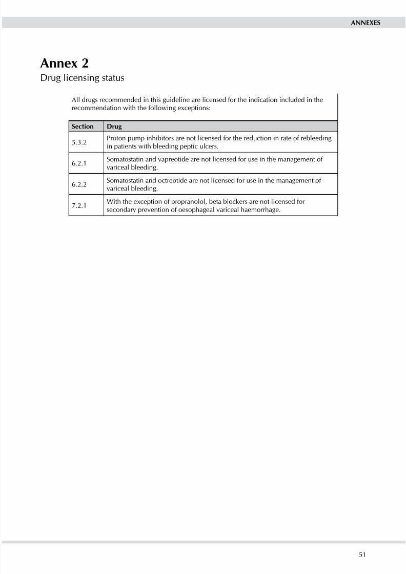
7/29/2019 SIGN Management PSMBA
http://slidepdf.com/reader/full/sign-management-psmba 57/64

7/29/2019 SIGN Management PSMBA
http://slidepdf.com/reader/full/sign-management-psmba 58/64

7/29/2019 SIGN Management PSMBA
http://slidepdf.com/reader/full/sign-management-psmba 59/64

7/29/2019 SIGN Management PSMBA
http://slidepdf.com/reader/full/sign-management-psmba 60/64

7/29/2019 SIGN Management PSMBA
http://slidepdf.com/reader/full/sign-management-psmba 61/6455
references
129. Feu F, Ruiz del Arbol L, Banares R, Planas R, Bosch j. Double-blind randomized controlled trial comparing terlipressin andsomatostatin for acute variceal hemorrhage. Variceal BleedingStud Group. Gastroenterolog 1996;111(5):1291-9.
130. Walker S, Kreichgauer HP, Bode jC. Terlipressin vs. somatostatinin bleeding esophageal varices: a controlled, double-blind stud.Hepatolog 1992;15(6):1023-30.
131. Banares R, Albillos A, Rincon D, Alonso S, Gonzalez M, Ruiz-del-Arbol L, et al. Endoscopic treatment versus endoscopic pluspharmacologic treatment for acute variceal bleeding: a meta-analsis. Hepatolog 2002;35(3):609-15.
132. Sung jj, Chung SC, yung My, Lai CW, Lau jy, Lee yT, et al.Prospective randomised stud of effect of octreotide on rebleedingfrom oesophageal varices after endoscopic ligation. Lancet1995;346(8991-8992):1666-9.
133. Freitas DS, Sofia C, Pontes jM, Gregório C, Cabral jP, Andrade P, etal. Octreotide in acute bleeding esophageal varices: a prospectiverandomized stud. Hepatogastroenterolog 2000;47(35):1310-4.
134. Farooi jI, Farooi Rj, Ha N, Siddi Ur R, Mahmood S. Treatmentand outcome of variceal bleeding - A comparison of two methods.
j Coll Phsicians Surg Pak 2000;10(4):131-3.135. Shah HA, Mumtaz K, jafri W, Abid S, Hamid S, Ahmad A, et al.
Sclerotherap plus octreotide versus sclerotherap alone in themanagement of gastro-oesophageal variceal hemorrhage. j AubMed Coll Abbottabad 2005;17(1):10-4.
136. Morales GF, Pereira Lima jC, Hornos AP, Marues DL, Costa CSD,Pereira Lima L, et al. Octreotide for esophageal variceal bleedingtreated with endoscopic sclerotherap: A randomized, placebo-controlled trial. Hepatogastroenterolog 2007;54(73):195-200.
137. Palazon jM, Such j, Sanchez-Paa j, Compan L, de MadariaE, Sempere L, et al. A comparison of two different dosages of somatostatin combined with sclerotherap for the treatment of acute esophageal variceal bleeding: a prospective randomizedtrial. Rev Esp Enferm Dig 2006;98(4):249-54.
138. Soderlund C, Magnusson I, Torngren S, Lundell L. Terlipressin(triglcl-lsine vasopressin) controls acute bleeding oesophagealvarices. A double-blind, randomized, placebo-controlled trial.Scand j Gastroenterol 1990;25(6):622-30.
139. Soares-Weiser K, Brezis M, Tur-Kaspa R, Leibovici L. Antibioticprophlaxis for cirrhotic patients with gastrointestinal bleeding(Cochrane Review). In: The Cochrane Librar, Issue 4, 2005.London: john Wile & Sons Ltd.
140. Fernandez j, del Arbol LR, Gomez C, Durandez R, Serradilla R,Guarner C, et al. Norfloxacin vs Ceftriaxone in the Prophlaxis of Infections in Patients With Advanced Cirrhosis and Hemorrhage.Gastroenterolog 2006;131(4):1049-56.
141. McKee RF, Garden Oj, Anderson jR, Carter DC. A comparison of SMS 201-995 and oesophageal tamponade in the control of acutevariceal haemorrhage. HPB Surg 1992;6(1):7-17.
142. Teres j, Planas R, Panes j, Salmeron jM, Mas A, Bosch j, et al.Vasopressin/nitroglcerin infusion vs. esophageal tamponade inthe treatment of acute variceal bleeding: a randomized controlledtrial. Hepatolog 1990;11(6):964-8.
143. Fort E, Sautereau D, Silvain C, Ingrand P, Pillegand B, BeauchantM. A randomized trial of terlipressin plus nitroglcerin vs.balloon tamponade in the control of acute variceal hemorrhage.Hepatolog 1990;11(4):678-81.
144. Garcia-Compean D, Blanc P, Bories jM, Michel j, Desprez D,Pageaux GP, et al. Treatment of active gastroesophageal varicealbleeding with terlipressin or hemostatic balloon in patients
with cirrhosis. A randomized controlled trial. Arch Med Res1997;28(2):241-5.145. Lo GH, Lai KH, Ng WW, Tam TN, Lee SD, Tsai yT, et al.
Inection sclerotherap preceded b esophageal tamponadeversus immediate sclerotherap in arresting active varicealbleeding: a prospective randomized trial. Gastrointest Endosc1992;38(4):421-4.
146. Panes j, Teres j, Bosch j, Rodes j. Efficac of balloon tamponadein treatment of bleeding gastric and esophageal varices. Resultsin 151 consecutive episodes. Dig Dis Sci 1988;33(4):454-9.
147. jalan R, john T, Redhead D, Garden O, Simpson K, FinlasonN, et al. A comparative stud of emergenc transugular intrahepatic portosstemic stent-shunt and esophageal transectionin the management of uncontrolled variceal hemorrhage. Am jGastroenterol 1995;90(11):1932-7.
148. Rosemurg A, Goode S, Zwiebel B, Black T, Brad P. A prospectivetrial of transugular intrahepatic portasstemic stent shunts versussmall-diameter prosthetic H-graft portacaval shunts in the treatment
of bleeding varices. Ann Surg 1996;224(3):378-84.149. D’Amico G, Morabito A, Pagliaro L, Marubini E. Survival and
prognostic indicators in compensated and decompensate cirrhosis.Dig Dis Sci 1986;31 (5):468-75.
150. Graham Dy, Smith jL. The course of patients after varicealhemorrhage. Gastroenterolog 1981;80(4):800–980.
151. Cheng jW, Zhu L, Gu Mj, Song ZM. Meta analsis of propranololeffects on gastrointestinal hemorrhage in c irrhotic patients. World
j Gastroenterol 2003;9(8):1836-9.152. Lo GH, Chen WC, Chen MH, Hsu PI, Lin CK, Tsai WL, et al.
Banding ligation versus nadolol and isosorbide mononitrate for
the prevention of esophageal variceal rebleeding. Gastroenterolog2002;123(3):728-34.
153. Gourna j, Masliah C, Martin T, Perrin D, Galmiche jP. Isosorbidemononitrate and propranolol compared with propranololalone for the prevention of variceal rebleeding. Hepatolog2000;31(6):1239-45.
154. Romero G, Kravetz D, Argonz j, Vulcano C, Suarez A, Fassio E, et al.Comparative stud between nadolol and 5-isosorbide mononitratevs. endoscopic band ligation plus sclerotherap in the preventionof variceal rebleeding in cirrhotic patients: A randomizedcontrolled trial. Alimentar Pharmacolog & Therapeutics. Vol.2006;24(4):601-11.
155. D’Amico G, Pagliaro L, Bosch j. The treatment of portalhpertension: a meta-analtic review. Hepatolog 1995;22(1):332-54.
156. Karsan HA, Morton SC, Shekelle PG, Spiegel BMR, Suttorp Mj,Edelstein MA, et al. Combination endoscopic band ligation andsclerotherap compared with endoscopic band ligation alone for
the secondar prophlaxis of esophageal variceal hemorrhage: Ameta-analsis. Dig Dis Sci 2005;50(2):399-406.
157. Lo GH, Lai KH, Cheng jS, Chen MH, Huang HC, Hsu PI, et al.Endoscopic variceal ligation plus nadolol and sucralfate comparedwith ligation alone for the prevention of variceal rebleeding: aprospective, randomized trial. Hepatolog 2000;32(3):461-5.
158. de la Pena j, Brullet E, Sanchez-Hernandez E, Rivero M, VergaraM, Martin-Lorente jL, et al. Variceal ligation plus nadolol comparedwith ligation for prophlaxis of variceal rebleeding: a multicenter trial. Hepatolog 2005;41(3):572-8.
159. Evrard S, Dumonceau jM, Delhae M, Golstein P, Deviere j, LeMoine O. Endoscopic histoacrl obliteration vs. propranolol in theprevention of esophagogastric variceal rebleeding: a randomizedtrial. Endoscop 2003;35(9):729-35.
160. Khan S, Tudur SC, Williamson P, Sutton R. Portosstemic shuntsversus endoscopic therap for variceal rebleeding in patients withcirrhosis (Cochrane Review). In: The Cochrane Librar, Issue 4,2006. London: john Wile & Sons Ltd.
161. Henderson jM, Boer TD, Kutner MH, Gallowa jR, RikkersLF, jeffers Lj, et al. Distal splenorenal shunt versus transugular intrahepatic portal sstematic shunt for variceal bleeding: arandomized trial. Gastroenterolog 2006;130(6):1643-51.
162. Tripathi D, Ferguson j, Barkell H, Macbeth K, Ireland H, RedheadDN, et al. Improved clinical outcome with transugular intrahepaticportosstemic stent-shunt utilizing poltetrafluoroethlene-coveredstents. Eur j Gastroenterol Hepatol 2006;18(3):225-32.
163. Escorsell A, Bañares R, García-Pagán jC, Gilabert R, Moitinho E,Piueras B, et al. TIPS versus drug therap in preventing varicealrebleeding in advanced cirrhosis: a randomized controlled trial.Hepatolog 2002;35(2):385-92.
164. Lo GH, Liang HL, Chen WC, Chen MH, Lai KH, Hsu PI, etal. A prospective, randomized controlled trial of transugular intrahepatic portosstemic shunt versus canoacrlate inectionin the prevention of gastric variceal rebleeding. Endoscop2007;39(8):679-85.
165. Farrell jj, Friedman jS. Gastrointesinal bleeding in the elderl.Gastroenterol Clin North Am 2001;30(2):377-407.166. Gilinsk NH, Burns DG, Barbezat GO, Levin W, Mers HS, Marks
IN. The natural histor of radiation-induced proctosigmoiditis: ananalsis of 88 patients. q j Med 1983;52(205):40-53.
167. Scottish Executive. Scottish Referral Guidelines for SuspectedCancer. Edinburgh: Scottish Executive; 2007. (ref. NHSHDL(2007)09). Available from http://www.sehd.scot.nhs.uk/mels/ HDL2007_09.pdf: [Accessed. 19 August 2008.
168. Sabharwal R, Vladica P, Chou R, Law WP. Helical CT in thediagnosis of acute lower gastrointestinal haemorrhage. Eur j Radiol2006;58(2):273-9.
169. Talor SA, Halligan S, Vance M, Windsor A, Atkin W, Bartram CI.Use of multidetector-row computed tomographic colonographbefore flexible sigmoidoscop in the investigation of rectalbleeding. Br j Surg 2003;90(9):1163-4.
170. Suzman MS, Talmor M, jennis R, Binkert B, Barie PS.Accurate localization and surgical management of active lower
gastrointestinal hemorrhage with technetium-labeled erthroctescintigraph. Ann Surg 1996;224(1):29-36.

7/29/2019 SIGN Management PSMBA
http://slidepdf.com/reader/full/sign-management-psmba 62/6456
ManageMent of acute upper and lower gastrointestinal bleeding
171. Hunter jM, Pezim ME. Limited value of technetium 99m-labeled redcell scintigraph in localization of lower gastrointestinal bleeding.Am j Surg 1990;159(5):504-6.
172. Garofalo TE, Abdu RA. Accurac and efficac of nuclear scintigraph for the detection of gastrointestinal bleeding. ArchSurg 1997;132(2):196-9.
173. Voeller GR, Bunch G, Britt LG. Use of technetium-labeled red
blood cell scintigraph in the detection and management of gastrointestinal hemorrhage. Surger 1991;110(4):799-804.
174. Olds GD, Cooper GS, Chak A, Sivak MV, jr., Chitale AA, WongRC. The ield of bleeding scans in acute lower gastrointestinalhemorrhage. j Clin Gastroenterol 2005;39(4):273-7.
175. jensen DM, Machicado GA, jutabha R, Kovacs TO. Urgentcolonoscop for the diagnosis and treatment of severe diverticular hemorrhage. N Engl j Med 2000;342(2):78-82.
176. Ohama T, Sakurai y, Ito M, Daito K, Sezai S, Sato y. Analsisof urgent colonoscop for lower gastrointestinal tract bleeding.Digestion 2000;61(3):189-92.
177. Rex DK, Lewis BS, Wae jD. Colonoscop and endoscopic therapfor delaed post-polpectom hemorrhage. Gastrointest Endosc1992;38(2):127-9.
178. Parra-Blanco A, Kaminaga N, Koima T, Endo y, Uragami N,Okawa N, et al. Hemoclipping for postpolpectom and postbiopscolonic bleeding. Gastrointest Endosc 2000;51(1):37-41.
179. Bandi R, Shett PC, Sharma RP, Burke TH, Burke MW, Kastan D.
Superselective arterial embolization for the treatment of lower gastrointestinal hemorrhage. j Vasc Interv Radiol 2001;12(12):1399-405.
180. Defrene L, Vanlangenhove P, Decruenaere j, Van MaeleG, De Vos M, Troisi R, et al. Outcome of acute nonvaricealgastrointestinal haemorrhage after nontherapeutic arteriographcompared with embolization. Eur Radiol 2003;13(12):2604-14.
181. d’Othee Bj, Surapaneni P, Rabkin D, Nasser I, Clouse M. Microcoilembolization for acute lower gastrointestinal bleeding. CardiovascIntervent Radiol 2006;29(1):49-58.
182. Keeling WB, Armstrong PA, Stone PA, Zweibel BR, KudrkBT, johnson BL, et al. Risk factors for recurrent hemorrhageafter successful mesenteric arterial embolization. Am Surg2006;72(9):802-6.
183. Lungdahl M, Eriksson LG, Nman R, Gustavsson S. Arterialembolisation in management of massive bleeding from gastric andduodenal ulcers. Eur j Surg 2002;168(7):384-90.
184. Silver A, Bendick P, Wasvar H. Safet and efficac of superselectiveangioembolization in control of lower gastrointestinal hemorrhage.Am j Surg 2005;189(3):361-3.
185. Britt LG, Warren L, Moore OF, 3rd. Selective management of lower gastrointestinal bleeding. Am Surg 1983;49(3):121-5.
186. Casarella Wj, Gallowa Sj, Taxin RN, Follett DA, Pollock Ej,Seaman WB. “Lower” gastrointestinal tract hemorrhage: newconcepts based on arteriograph. Am j Roentgenol Radium Ther Nucl Med 1974;121(2):357-68.
187. Colacchio TA, Forde KA, Patsos Tj, Nunez D. Impact of moderndiagnostic methods on the management of active rectal bleeding.Ten ear experience. Am j Surg 1982;143(5):607-10.
188. Drapanas T, Pennington DG, Kappelman M, Lindse ES. Emergencsubtotal colectom: preferred approach to management of massivelbleeding diverticular disease. Ann Surg 1973;177(5):519-26.
189. Leitman IM, Paull DE, Shires GT, 3rd. Evaluation and managementof massive lower gastrointestinal hemorrhage. Ann Surg1989;209(2):175-80.
190. Welch CE, Athanasoulis CA, Galdabini jj. Hemorrhage fromthe large bowel with special reference to angiodsplasia anddiverticular disease. World j Surg 1978;2(1):73-83.
191. Wright HK, Pelliccia O, Higgins EF, jr., Sreenivas V, Gupta A.Controlled, semielective, segmental resection for massive colonichemorrhage. Am j Surg 1980;139(4):535-8.
192. Farner R, Lichliter W, Kuhn j, Fisher T. Total colectom versuslimited colonic resection for acute lower gastrointestinal bleeding.Am j Surg 1999;178(6):587-91.
193. Baker R, Senagore A. Abdominal colectom offers safe managementfor massive lower GI bleed. Am Surg 1994;60(8):578-82.
194. Renzulli P, Maurer CA, Netzer P, Dinkel HP, Buchler MW. Subtotalcolectom with primar ileorectostom is effective for unlocalized,diverticular hemorrhage. Langenbecks Arch Surg 2002;387(2):67-71.

7/29/2019 SIGN Management PSMBA
http://slidepdf.com/reader/full/sign-management-psmba 63/64

7/29/2019 SIGN Management PSMBA
http://slidepdf.com/reader/full/sign-management-psmba 64/64