sigma xi presentation
TRANSCRIPT

THE EFFECTS OF
GRANULOCYTE
COLONY
STIMULATING
FACTOR ON
STROKE INDUCED
RATSMatthew Busel
Pine Crest School
Work conducted at Florida Atlantic University

INTRODUCTION: GCSF
Granulocyte Colony Stimulating Factor (GCSF) is a growth factor
and cytokine that stimulates the proliferation and mobilization of
hematopoietic stem cells
Its actions are mediated by binding to specific GCSF receptors
(GCSFRs)
Although its main clinical use has been to counteract neutropenia,
recent studies have reported that GCSFRs are located on the
brain leading to neuroprotective effects of the drug
By decreasing the rate of neuronal death many central nervous
system (CNS) disorders may be counteracted against

INTRODUCTION: CEREBRAL ISCHEMIA
Focal cerebral ischemia (stroke) is a CNS disorder in which the
cerebral blood vessels are blocked, resulting in an inadequate
blood flow to the brain and subsequent oxygen (hypoxic) –
glucose deprived environment
In this state the brain cannot meet metabolic demands and
neuronal death will eventually be initiated via apoptosis
The resulting ischemic infarct (area of dead cell tissue) includes a
central core (an area of severe ischemia) and a surrounding
penumbra, which is vulnerable if the cell death is not arrested
White = core
Red = penumbra
INTRODUCTION: APOPTOTIC
PATHWAYS
The endoplasmic reticulum (ER) is responsible for the proper folding and sorting of proteins
During an ischemic insult the ER becomes stressed and will try to restore normal function by deactivating protein translation signaling pathways while activating other pathways that increase the production of chaperone molecules that facilitates protein folding
If it is unsuccessful in rectifying its stressed state and restoring normal function, the ER will initiate an apoptotic cascade
Caspase 12 is an ER-membrane associated protein and one of the initiators of the caspase-mediated apoptotic pathway
Glucose regulated protein 78 (GRP78) is a chaperon molecule associated with the ER
INTRODUCTION: EXPERIMENT
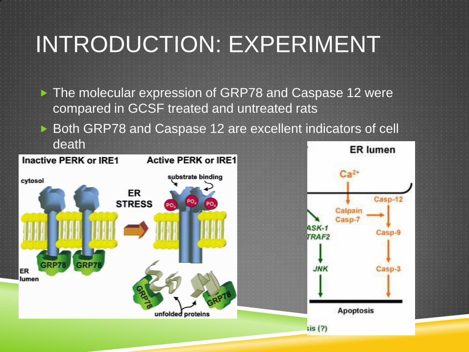
The molecular expression of GRP78 and Caspase 12 were
compared in GCSF treated and untreated rats
Both GRP78 and Caspase 12 are excellent indicators of cell
death

HYPOTHESIS
If the molecular expression of GRP78 and Caspase 12 are
measured in GCSF treated and untreated rats after focal cerebral
ischemia, treated rats will have lower expression of the selected
indicators and therefore lower cell death in both the core and
penumbra of the brain
METHODS: WESTERN BLOT
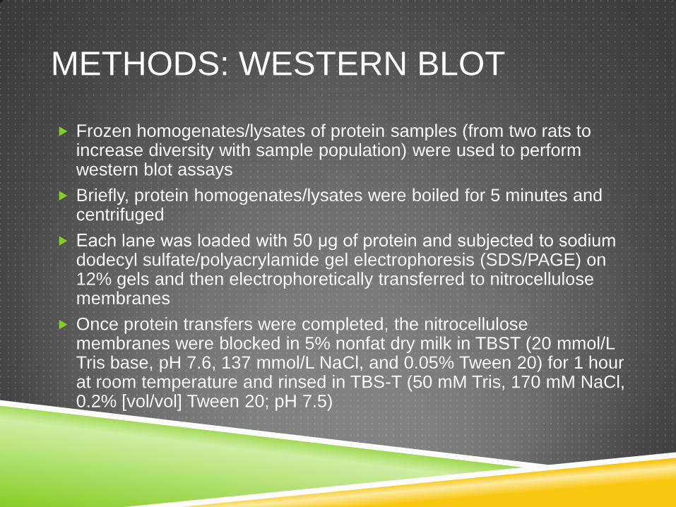
Frozen homogenates/lysates of protein samples (from two rats to increase diversity with sample population) were used to perform western blot assays
Briefly, protein homogenates/lysates were boiled for 5 minutes and centrifuged
Each lane was loaded with 50 μg of protein and subjected to sodium dodecyl sulfate/polyacrylamide gel electrophoresis (SDS/PAGE) on 12% gels and then electrophoretically transferred to nitrocellulose membranes
Once protein transfers were completed, the nitrocellulose membranes were blocked in 5% nonfat dry milk in TBST (20 mmol/L Tris base, pH 7.6, 137 mmol/L NaCl, and 0.05% Tween 20) for 1 hour at room temperature and rinsed in TBS-T (50 mM Tris, 170 mM NaCl, 0.2% [vol/vol] Tween 20; pH 7.5)

METHODS: WESTERN BLOT
They were then incubated with primary antibodies (anti-caspase
12, anti-Grp78 and anti-GAPDH, all at 1:1000 dilution) and
incubated in TBST and the corresponding primary antibody
(1:1000) overnight at 4°C
The membranes were subsequently washed in TBST and
incubated for 1.5 hours at room temperature with the same
corresponding secondary antibody in a dilution of 1:3000 with
TBST
Immunoreactive bands were visualized in the linear range with
enhanced chemoluminescence
Films were scanned and the optical density was determined with
ImageJ
METHODS: ANALYSIS
All statistical data shown was expressed as the mean optical
density for each treatment
A one-way ANOVA test was used to compare means between
groups and determine statistical significance
Differences of P<0.05 were considered statistically significant

RESULTS: CASPASE 12
This figure shows the mean normalized optical density values
determined by ImageJ for Caspase 12
GAPDH was used as the internal control to normalize values
Click to next slide for
more explanation
Legend(50 mg protein lysate in each well)
1. Naïve- non-operated controls
2. Sham- operated controls not given focal ischemia
3. MCAO- rats were given focal ischemia by middle cerebral artery occlusion
4. MCAO+GCSF- rats were given focal ischemia by middle cerebral artery occlusion and administered GCSF
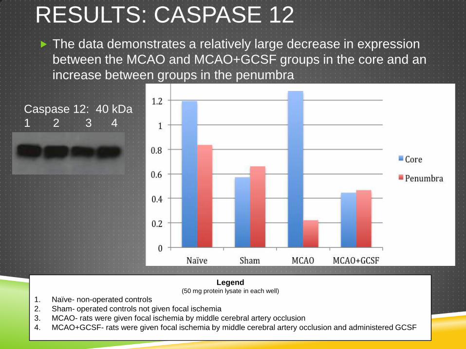
RESULTS: CASPASE 12 The data demonstrates a relatively large decrease in expression
between the MCAO and MCAO+GCSF groups in the core and an
increase between groups in the penumbra
Caspase 12: 40 kDa
1 2 3 4
Legend(50 mg protein lysate in each well)
1. Naïve- non-operated controls
2. Sham- operated controls not given focal ischemia
3. MCAO- rats were given focal ischemia by middle cerebral artery occlusion
4. MCAO+GCSF- rats were given focal ischemia by middle cerebral artery occlusion and administered GCSF

RESULTS: CASPASE 12 ANOVA Tests were run for the results of both the core and
penumbra
Since both had p values > .05, results were determined to be
statistically insignificantCore
Penumbra
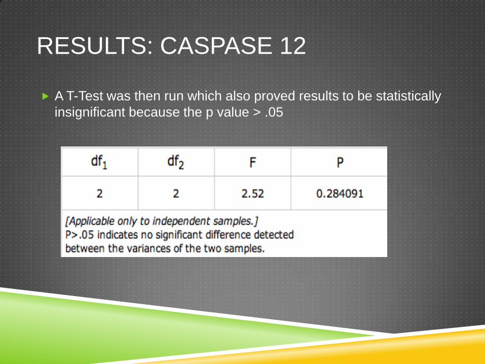
RESULTS: CASPASE 12
A T-Test was then run which also proved results to be statistically
insignificant because the p value > .05

RESULTS: GRP78
This figure displays the mean normalized optical density values
determined by ImageJ for GRP78
GAPDH was used as the internal control to normalize values
Legend(50 mg protein lysate in each well)
1. Naïve- non-operated controls
2. Sham- operated controls not given focal ischemia
3. MCAO- rats were given focal ischemia by middle cerebral artery occlusion
4. MCAO+GCSF- rats were given focal ischemia by middle cerebral artery occlusion and administered GCSF
Click to next slide for
more explanation
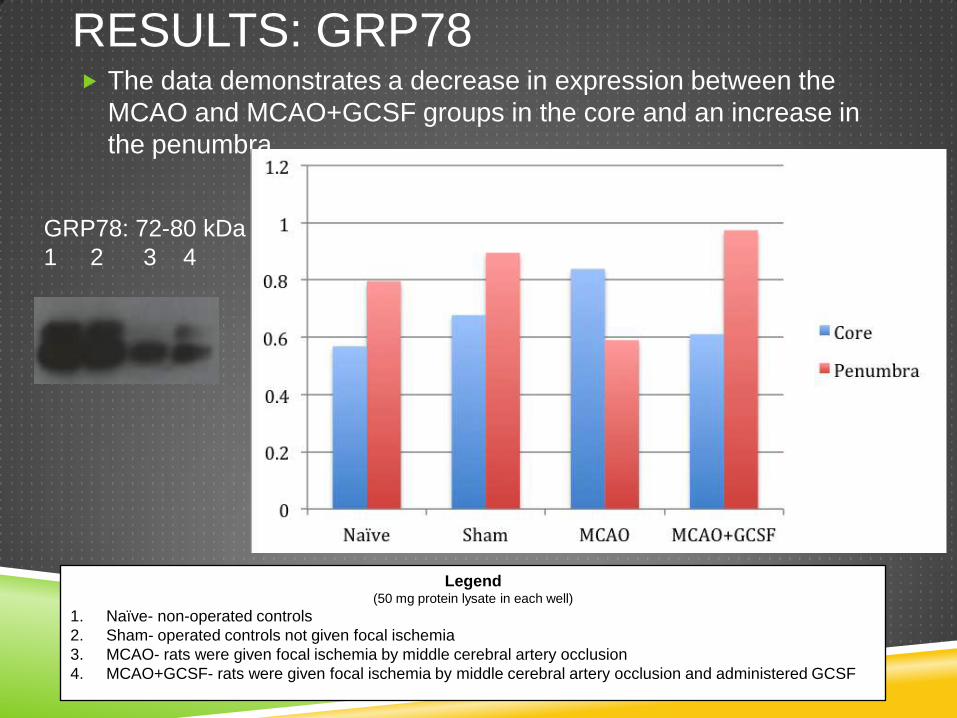
RESULTS: GRP78 The data demonstrates a decrease in expression between the
MCAO and MCAO+GCSF groups in the core and an increase in
the penumbra
GRP78: 72-80 kDa
1 2 3 4
Legend(50 mg protein lysate in each well)
1. Naïve- non-operated controls
2. Sham- operated controls not given focal ischemia
3. MCAO- rats were given focal ischemia by middle cerebral artery occlusion
4. MCAO+GCSF- rats were given focal ischemia by middle cerebral artery occlusion and administered GCSF
RESULTS: SUMMARY
With both Caspase 12 and GRP78, there was a decrease in
expression in the treated rats in the core, but an increase in
expression in the treated rats in the penumbra

DISCUSSION: CASPASE 12
RESULTS
In response to excess calcium in the ER lumen, an apoptotic
cascade is initiated involving Caspase 12 with the final death
marker prior to apoptosis being Caspase 3
Consistent with previous studies, the core GCSF-treated rats
showed reduced Caspase 12 expression
The penumbra GCSF-treated rats showed increased Caspase 12
expression in the penumbra in complete disagreement with
previous studies
DISCUSSION: GRP78 RESULTS
In the absence of ER stress, the chaperone protein GRP78
protects cells from apoptosis by binding to IRE1, PERK, ATF6,
and Caspase 14
GRP78 normally inhibits the activation of these intracellular
cytoplasmic kinases but when the ER is stressed, GRP78
dissociates from the cytoplasmic molecules to sequester the extra
calcium
Similar to the results with Caspase 12, the core-GCSF treated
groups decreased GRP78 expression, but in the penumbra the
opposite effect occurred.

DISCUSSION: STATISTICS
There was no statistical significance between any of the treatment groups analyzed
A possible reason for this result is the relatively small sample size of data used in this experiment compared to similar studies
Also there was a relatively high level of variation within each group, which may have contributed to some of the observed discrepancies in the data analysis
Another statistical discrepancy may have come from GAPDH, which was used to normalize OD values
As demonstrated in Figure 1, the expression of GAPDH in the MCAO group was significantly smaller than any other group making the normalized value of the MCAO group relatively smaller
FUTURE RESEARCH
In future research, additional Western Blots with Caspase 12 and
GRP78 would be run in order to compile more data and create
more reliable results
As this research only investigated pro-death markers (Caspase
12) and ER stress indicators (GRP78), it would be interesting to
assay BCl2, a pro-survival marker in future research
SOURCES
Http://www.weizmann.ac.il/immunology/Lapidot/documents/5.pdf (2002): n. pag. 17 June 2002. Web.
"Annals of Oncology." G-CSF Prevents the Suppression of Bone Marrow Hematopoiesis Induced by IL-12 and Augments Its Antitumor Activity in a Melanoma Model in Mice. N.p., n.d.Web. 26 Sept. 2012. <http://annonc.oxfordjournals.org/content/9/1/63.short>.
"Phenotypic Changes in Neutrophil Granulocytes after G-CSF Administration in Patients with Acute Lymphoblastic Leukemia under Chemotherapy." Phenotypic Changes in Neutrophil Granulocytes after G-CSF Administration in Patients with Acute Lymphoblastic Leukemia under Chemotherapy. N.p., n.d. Web. 26 Sept. 2012. <http://www.haematologica.org/content/83/6/573.abstract>.
"A Role for G-CSF ( Granulocyte-Colony Stimulating Factor ) Review ND ABBREVIATIONS SC." Free Reference Manager and PDF Organizer. N.p., n.d. Web. 26 Sept. 2012. <http://www.mendeley.com/research/a-role-for-gcsf-granulocytecolony-stimulating-factor-review-nd-abbreviations-sc/>.
Khan, Shaheen N., and Fridoon J. Ahmad. "IGF-1 and G-CSF Complement Each Other in BMSC Migration towards Infarcted Myocardium in a Novel in Vitro Model." Cell Biology International 33.6 (2009): n. pag. June 2009. Web. <IGF-1 and G-CSF complement each other in BMSC migration towards infarcted myocardium in a novel in vitro model>.
Chopp, M., S. Frinak, D. R. Walton, and M. B. Smith. "Intracellular Acidosis during and after Cerebral Ischemia: In Vivo Nuclear Magnetic Resonance Study of Hyperglycemia in Cats." N.p., n.d. Web. <http://stroke.ahajournals.org/content/18/5/919.full.pdf>.
SOURCES
“Neuroprotective Effect of Recombinant Human Granulocyte Colony-stimulating Factor in Transient Focal Ischemia of Mice." Nature.com. Nature Publishing Group, n.d. Web. 26 Sept. 2012.
"Anti-apoptotic Effect of Granulocyte-colony Stimulating Factor after Focal Cerebral Ischemia in the Rat." Neuroscience143.4 (2006): 965-74. 28 Dec. 2006. Web. <http://www.sciencedirect.com/science/article/pii/S0306452206012310>.
Pan, Chunliu, Amit Gupta, Howard Prentice, and Jang-yen Wu. "Protection of Taurine and Granulocyte Colony-Stimulating Factor against Excitotoxicity Induced by Glutamate in Primary Cortical Neurons." N.p., 2010. Web. <http://www.biomedcentral.com/content/pdf/1423-0127-17-S1-S18.pdf>.
"Neuroprotective Effect of Granulocyte ColonyâStimulating Factor After Focal Cerebral Ischemia." Neuroprotective Effect of Granulocyte ColonyâStimulating Factor After Focal Cerebral Ischemia. N.p., n.d. Web. 26 Sept. 2012. <http://stroke.ahajournals.org/content/34/3/745.long>.
"Anti-apoptotic Effect of Granulocyte-colony Stimulating Factor after Focal Cerebral Ischemia in the Rat." Neuroscience143.4 (2006): 965-74. 28 Dec. 2006. Web. <http://www.sciencedirect.com/science/article/pii/S0306452206012310>.
"Caspase-12 Mediates Endoplasmic-reticulum-specific Apoptosis and Cytotoxicity by Amyloid-beta." National Center for Biotechnology Information. U.S. National Library of Medicine, n.d. Web. 26 Sept. 2012. <http://www.ncbi.nlm.nih.gov/pubmed/10638761>.
"Research." School of Biological and Biomedical Sciences Interorganellar Signalling Laboratory. N.p., n.d. Web. <http://www.dur.ac.uk/martin.schroeder/Research%20Summary.htm>.
"The Critical Roles of Endoplasmic Reticulum Chaperones and Unfolded Protein Response in Tumorigenesis and Anticancer Therapies." Nature.com. Nature Publishing Group, n.d. Web. 26 Sept. 2012. <http://www.nature.com/onc/journal/vaop/ncurrent/fig_tab/onc2012130f1.html>.