show and tell: clinical vignettes (primary pci, pci vs
TRANSCRIPT
Presenter Disclosures
Dr. Yin Ge
Show and tell: clinical vignettes (primary PCI, PCI vs CABG, TAVI)
Relationships with financial sponsors:
• Grants/Research Support: N/A
• Speakers Bureau/Honoraria: N/A
• Consulting Fees: N/A
• Patents: N/A
• Other: N/A
Case 1

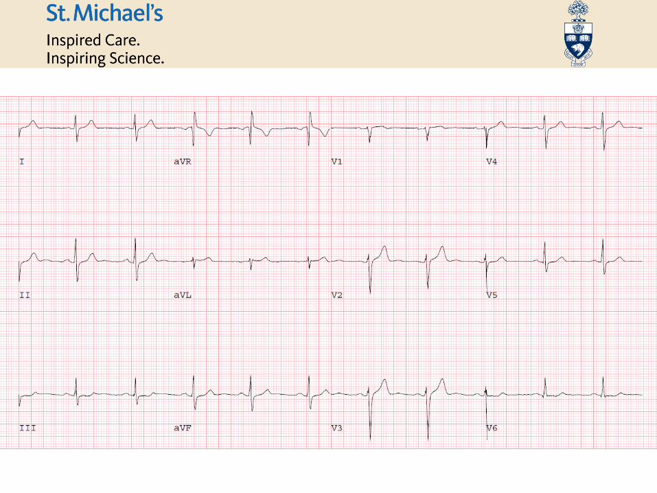
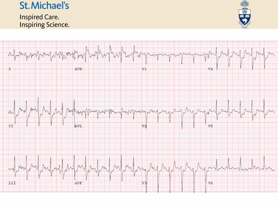
HPI
62 M with no cardiac risk factors
5 day history of intermittent chest pain
Pressure-like; lasts a few minutes
Occurs at rest, not precipitated by exercise
Sometimes accompanied by headaches and dizziness
Past Medical History
Back surgery - 2014
Physical Exam
VS: BP 147/83 mmHg, HR 88 and regular.CV: S1, S2 with physiologic split, no murmurs.Chest: No crackles.
Extremities: No edema. Good equal bilateral pulses
Home Medications
None
Labs
Electrolytes: WNLCBC: WNLHs-TnI: 17>27>21

SSS: 13 (19%)
SDS: 11 (16%)
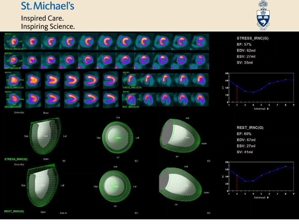
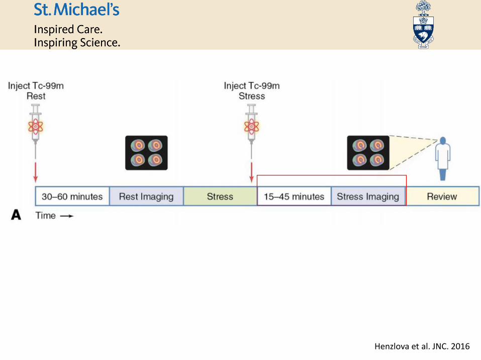
Henzlova et al. JNC. 2016
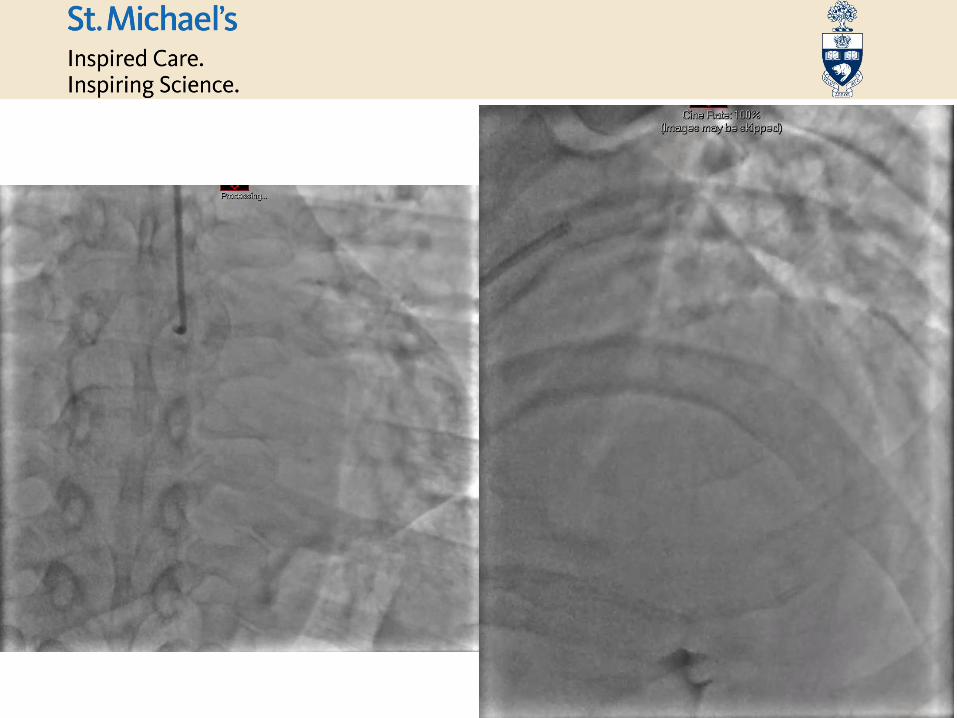
• Cath
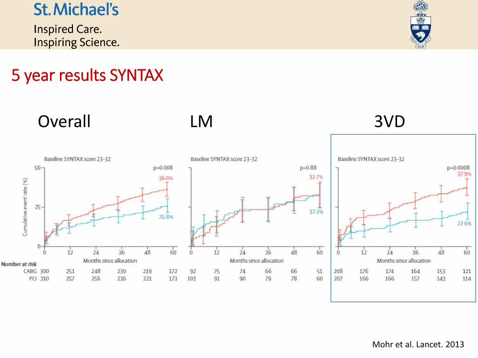
Overall LM 3VD
5 year results SYNTAX
Mohr et al. Lancet. 2013
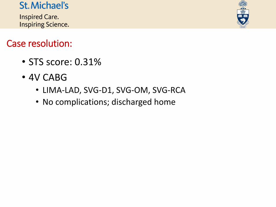
• STS score: 0.31%
• 4V CABG• LIMA-LAD, SVG-D1, SVG-OM, SVG-RCA
• No complications; discharged home
Case resolution:

Case 2
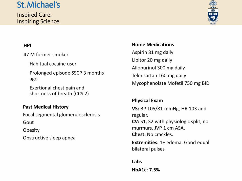
HPI
47 M former smoker
Habitual cocaine user
Prolonged episode SSCP 3 months ago
Exertional chest pain and shortness of breath (CCS 2)
Past Medical History
Focal segmental glomerulosclerosis
Gout
Obesity
Obstructive sleep apnea
Physical Exam
VS: BP 105/81 mmHg, HR 103 and regular.CV: S1, S2 with physiologic split, no murmurs. JVP 1 cm ASA.Chest: No crackles.
Extremities: 1+ edema. Good equal bilateral pulses
Home Medications
Aspirin 81 mg daily
Lipitor 20 mg daily
Allopurinol 300 mg daily
Telmisartan 160 mg daily
Mycophenolate Mofetil 750 mg BID
Labs
HbA1c: 7.5%


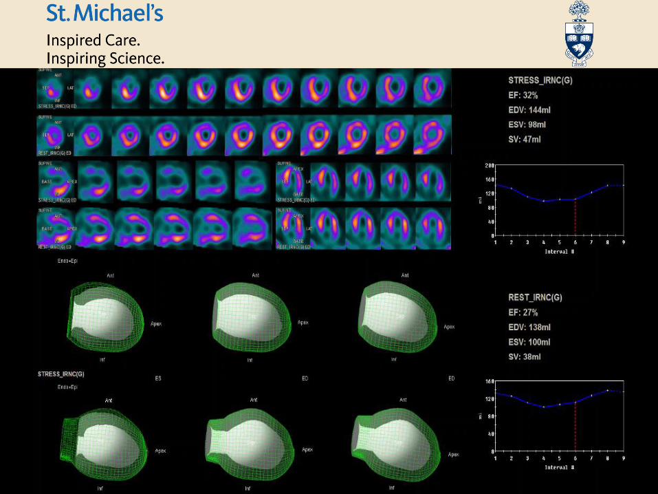
SSS: 19 (28%)
SDS: 17 (25%)

• Cath

FREEDOM
Esper et al. JACC. 2018

• STS score: 0.94%
• Renal failure: 3.6%
• 4V CABG• LIMA to LAD, left radial to PDA and SVG to OM.
• Vasoplegia and renal failure; didn’t require dialysis
Case resolution:
Case 3
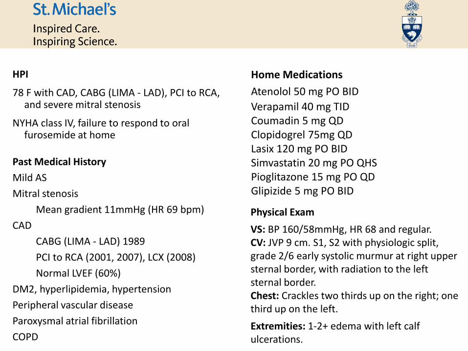
HPI
78 F with CAD, CABG (LIMA - LAD), PCI to RCA, and severe mitral stenosis
NYHA class IV, failure to respond to oral furosemide at home
Past Medical History
Mild AS
Mitral stenosis
Mean gradient 11mmHg (HR 69 bpm)
CAD
CABG (LIMA - LAD) 1989
PCI to RCA (2001, 2007), LCX (2008)
Normal LVEF (60%)
DM2, hyperlipidemia, hypertension
Peripheral vascular disease
Paroxysmal atrial fibrillation
COPD
Physical Exam
VS: BP 160/58mmHg, HR 68 and regular.CV: JVP 9 cm. S1, S2 with physiologic split, grade 2/6 early systolic murmur at right upper sternal border, with radiation to the left sternal border.Chest: Crackles two thirds up on the right; one third up on the left.
Extremities: 1-2+ edema with left calf ulcerations.
Home Medications
Atenolol 50 mg PO BID
Verapamil 40 mg TIDCoumadin 5 mg QDClopidogrel 75mg QDLasix 120 mg PO BIDSimvastatin 20 mg PO QHS Pioglitazone 15 mg PO QD Glipizide 5 mg PO BID

Echocardiogram
STS calculated at 9% and TMVR was offered
HR 55

Transcatheter mitral valve in MAC
26mm Sapien 3 valve
TEE and fluoroscopy guided
Uncomplicated procedure; MV gradient 4 mmHg at HR 72 bpm

Post procedure:
Symptomatically improved. Diuresing.
Increased intensity of murmur in upper sternal border.
TTE: Peak gradient across LVOT 58 mmHg at rest (unable to perform Valsalva).
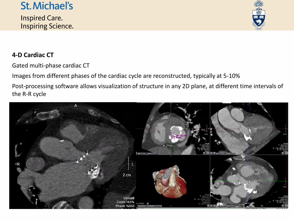
4-D Cardiac CT
Gated multi-phase cardiac CT
Images from different phases of the cardiac cycle are reconstructed, typically at 5-10%
Post-processing software allows visualization of structure in any 2D plane, at different time intervals of the R-R cycle

Multimodality evaluation of the mitral valve
Blanke et al. JACC imaging. 2015

Multimodality evaluation of the mitral valve
Blanke et al. JACC imaging. 2015

LVOT obstruction post TMVR
Blanke et al. JACC imaging. 2015

Risk factors for LVOT obstruction post TMVR
Blanke et al. JACC imaging. 2017
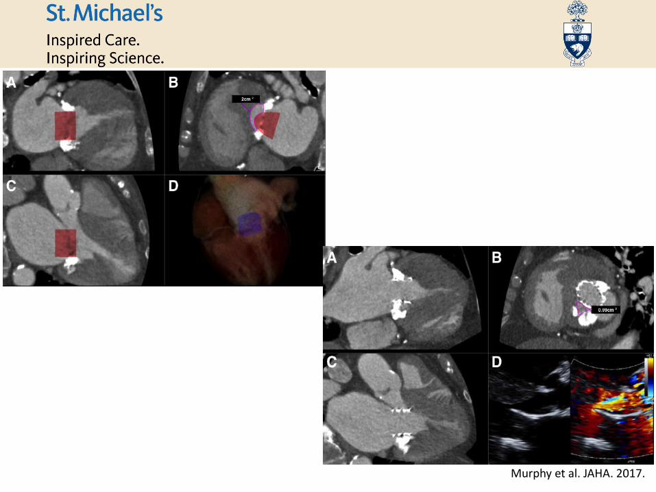
Murphy et al. JAHA. 2017.