shoulder labral tears mri
TRANSCRIPT

SHOULDER – LABRAL TEARS, VARAINTS.

The glenohumeral joint has the following supporting structures:
Superiorly coracoacromial arch and coracoacromial ligament long head of the biceps tendon tendon of the supraspinatus muscle
Anteriorly anterior labrum glenohumeral ligaments - SGHL, MGHL, IGHL (anterior band) subscapularis tendon
Posteriorly posterior labrum posterior band of the IGHL infraspinatus and teres minor tendon

Glenoid Labrum
The glenoid labrum is a fibrocartilaginous structure that attaches to the glenoid rim and is about 4 mm wide.
The labrum may show considerable variation in shape and in mechanism of attachment to the glenoid. It is usually rounded or triangular on cross-sectional images.
Biceps Tendon
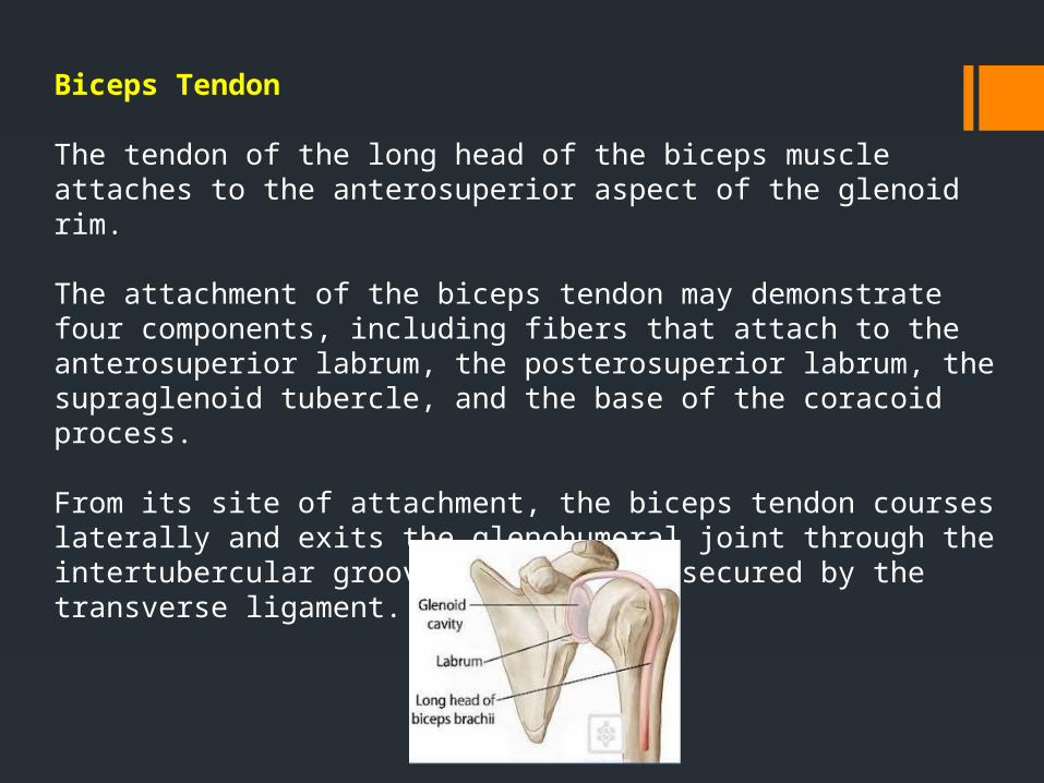
The tendon of the long head of the biceps muscle attaches to the anterosuperior aspect of the glenoid rim.
The attachment of the biceps tendon may demonstrate four components, including fibers that attach to the anterosuperior labrum, the posterosuperior labrum, the supraglenoid tubercle, and the base of the coracoid process.
From its site of attachment, the biceps tendon courses laterally and exits the glenohumeral joint through the intertubercular groove, where it is secured by the transverse ligament.

coronal section obtained at the level of the labral-bicipital complex illustrates the biceps tendon (B), superior labrum (L), and glenoid cartilage (C), all of which are intimately related in this region.
biceps tendon attachment at the level of the superior labrum and glenoid illustrates attachments to the superior glenoid rim (1), the posterior (2) and anterior (3) labrum, and the base of the coracoid process (4).

Glenohumeral Ligaments
Superior Glenohumeral Ligament.—The glenohumeral ligaments play a role as shoulder stabilizers and consist of thickened bands of the joint capsule.
The superior glenohumeral ligament is the most consistently identified capsular ligament. It can arise from the anterosuperior labrum, the attachment of the tendon of the long head of the biceps muscle, or the middle glenohumeral ligament.

Middle Glenohumeral Ligament.—
The middle glenohumeral ligament varies most in size and site of attachment to the glenoid. It may attach to the superior portion of the anterior glenoid labrum but more frequently attaches medially on the glenoid neck.
The middle glenohumeral ligament may be absent or may appear thick and cordlike (as, for example, in Buford complex).
CT arthrogram (2-mm section thickness) shows the middle glenohumeral ligament (arrowhead) attached to the anterior labrum (arrow).

Transverse fat-saturated MR arthrogram (560/14) demonstrates the middle glenohumeral ligament attaching medially on the glenoid neck (arrow).
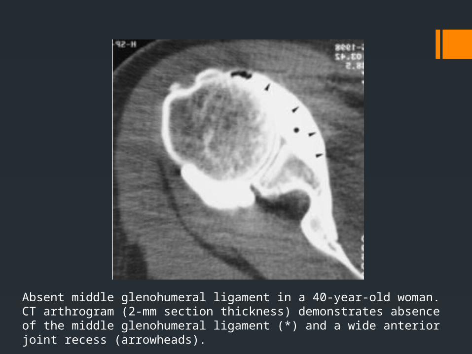
Absent middle glenohumeral ligament in a 40-year-old woman. CT arthrogram (2-mm section thickness) demonstrates absence of the middle glenohumeral ligament (*) and a wide anterior joint recess (arrowheads).

Inferior Glenohumeral Ligament.—
The inferior glenohumeral ligament is an important stabilizer of the anterior shoulder joint and consists of the axillary pouch and anterior and posterior bands. The anterior band inserts along the inferior two-thirds of the anterior glenoid labrum. When redundant, it may overlap the anterior edge of the glenoid cartilage. The anterior band is usually quite prominent, although in approximately 25% of cases it is very thin.The posterior band is usually thinner than the anterior band.

Sagittal fat-saturated T1-weighted MR arthrogram (750/15) demonstrates the biceps tendon (t), subscapularis tendon (S), and anterior and posterior bands of the inferior glenohumeral ligament (arrows).
CORONAL

Labral variants
There are many labral variants. These normal variants are all located in the 11-3 o'clock position.

It is important to recognize these variants, because they can mimick a SLAP tear.These normal variants will usually not mimick a Bankart-lesion, since it is located at the 3-6 o'clock position, where these normal variants do not occur.However labral tears may originate at the 3-6 o'clock position and subsequently extend superiorly.

Sublabral recess
There are 3 types of attachment of the superior labrum at the 12 o'clock position where the biceps tendon inserts.
In type I there is no recess between the glenoid cartilage and the labrum.In type II there is a small recess.In type III there is a large sublabral recess.
This sublabral recess can be difficult to distinguish from a SLAP-tear or a sublabral foramen.

These images illustrate the differences between an sublabral recess and a SLAP-tear.

Type I labral ---On a coronal MR arthrogram , the labrum (black arrow) is tightly attached to the glenoid cartilage and biceps tendon (white arrow)
Type II labral attachment. Coronal fat-saturated T1-weighted MR arthrogram shows a small recess between the labrum and the glenoid cartilage (arrow).
Type III labral attachment ----coronal CT arthrogram shows a large recess between the labrum and the glenoid (arrow).

Sublabral Foramen
A sublabral foramen or sublabral hole is an unattached anterosuperior labrum at the 1-3 o'clock position. It is seen in 11% of individuals.
On a MR-arthtrogram a sublabral foramen should not be confused with a sublabral recess or SLAP-tear, which are also located in this region.
A sublabral recess however is located at the site of the attachment of the biceps tendon at 12 o'clock and does not extend to the 1-3 o?lock position.
A SLAP tear may extend to the 1-3 o'clock position, but the attachment of the biceps tendon to the superior labrum should always be involved.
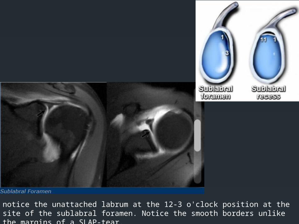
notice the unattached labrum at the 12-3 o'clock position at the site of the sublabral foramen. Notice the smooth borders unlike the margins of a SLAP-tear.

Buford complex
A Buford complex is a congenital labral variant.The anterosuperior labrum is absent in the 1-3 o'clock position and the middle glenohumeral ligament is usually thickened. It is present in approximately 1.5% of individuals.

On these axial images a Buford complex can be identified. The anterior labrum is absent in the 1-3 o'clock position and there is a thickened middle GHL.
The thickened middle GHL should not be confused with a displaced labrum.It should always be possible to trace the middle GHL upwards to the glenoid rim and downwards to the humerus.

Variation in the shapes of labrum

Labral pathology
• BANKART LESION• BONY BANKART• REVERSE BANKART• PERTHES• REVERSE PERTHES• APLPSA• POLPSA• ALIPSA• POLIPSA• GLAD• GARD• GAGL• HAGL• BHAGL• DOUBLE LESION• TRIPLE LESION
Following are various labral pathologies :

Labral pathologyA Clockwise approach to the labrum is the easiest way to diagnose labral tears and to differentiate them from normal labral variants.
There are two types of labral tears: SLAP tears and Bankart lesions.
SLAP is an acronym that stands for 'Superior Labral tear from Anterior to Posterior'.SLAP tears start at the 12 o'clock position where the biceps anchor is located, which tears the labrum off the glenoid.SLAP tears typically extend from the 10 to the 2 o'clock position, but can extend more posteriorly or anteriorly and even extend into the biceps tendon.
Bankart lesions are typically located in the 3-6 o'clock position because that's where the humeral head dislocates.

DislocationAnterior dislocationThe shoulder is a very mobile and therefore unstable joint.
The humeral head is almost always displaced anteriorly, inferiorly and medially below the coracoid process (95% of cases). Motion to superior is limited by the acromion, coracoid process and rotator cuff (figure). Motion in a posterior direction is limited by the posterior rim of the glenoid which is in an anteverted position.

The dislocation of the humeral head to antero-inferior causes damage to the antero-inferior rim of the glenoid in the 3 - 6 o'clock position (marked in red).
Especially in younger patients this results in a Bankart fracture or a Bankart lesion which is a tear of the anteroinferior labrum. This results in instability and recurrent dislocations.

The images show a subtle Bankart fracture (arrows).

Hill-Sachs On MR a Hill-Sachs defect is seen at or above the level of the coracoid process.
Hill-Sachs is a posterolateral depression of the humeral head.
It is above or at the level of the coracoid in the first 18 mm of the proximal humeral head. It is seen in 75-100% of patients with anterior instability. It is chondral or osteochondral.

Bankart and variantsBankart-lesions and variants like Perthes and ALPSA are injuries to the anteroinferior labrum. These injuries are always located in the 3-6 o'clock position because they are caused by an anterior-inferior dislocation.

Bankart lesion
Bankart lesions are labral tears without an osseus fragment.MR arthrography or arthroscopy are optimal to diagnose Bankart or Bankart-like lesions.There is a detachment of the anteroinferior labrum (3-6 o'clock) with complete tearing of the anterior scapular periosteum.The arrow points to the disrupted periosteum.

On MR-athrography the labrum is missing on the anterior glenoid and the labral fragment is displaced anteriorly (arrow).

Osseus BankartBankart lesions with an osseus fragment are common findings in patients with an anterior dislocation and are frequently seen on the x-rays or CT-scan.

On MR-arthrography it may be difficult to depict the osseus fragment. On CT it is easy to appreciate the osseus fragment of the anterior glenoid (arrow).

Reverse BankartCT-images in another patient show a reversed osseus Bankart in a patient with posterior dislocation.
Axial MR-arthrogram of a reverse Bankart.

Perthes lesion
A Perthes lesion is a labroligamentous avulsion like a Bankart, but with a medially stripped intact periosteum.
On images of the shoulder with the arm in a neutral position, the torn labrum may be held in its normal anatomic position by the intact scapular periosteum, which thereby prevents contrast media from entering the tear.
This means that MR-arthrography with the arm in the neutral position may fail to detect the labral tear.

In the ABER-position it is obvious that there is a Perthes lesion (black arrow).Due to the ABER-position the anterior band of the inferior GHL creates tension on the anteroinferior labrum and contrast fills the tear.
In the ABER position however there is tension on the antero-inferior labrum by the stretched anterior band of the inferior glenohumeral ligament and you have more chance to detect the tear. The arrow points to the intact periosteum.


ALPSA
An ALPSA-lesion is an Anterior Labral Periosteal Sleeve Avulsion.
The anterior labrum is absent on the glenoid rim.
The arrow points to the medially displaced labroligamentous complex.

Images of a patient with an ALPSA-lesion. Notice the medially displaced labrum.
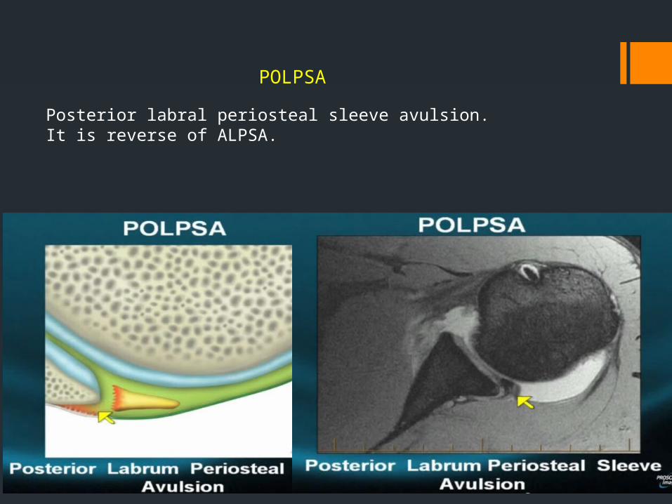
POLPSA
Posterior labral periosteal sleeve avulsion.It is reverse of ALPSA.

GLADA GLAD-lesion is a GlenoLabral Articular Disruption.
It represents a patial tear of the anteroinferior labrum with adjacent cartilage damage.
The arrow points to the cartilage defect.

The images show a partial tear of the anteroinferior labrum with adjacent cartilage damage at the 4-6 o 'clock position (arrows).
GLAD-lesion

Defect is at the base of the labrum, predominantly in the glenoid articular hyaline cartilage.

HAGL is a Humeral Avulsion of the inferior Glenohumeral Ligament.
There is discontinuity of the IGHL attachment on the humerus with leakage of contrast.
HAGL
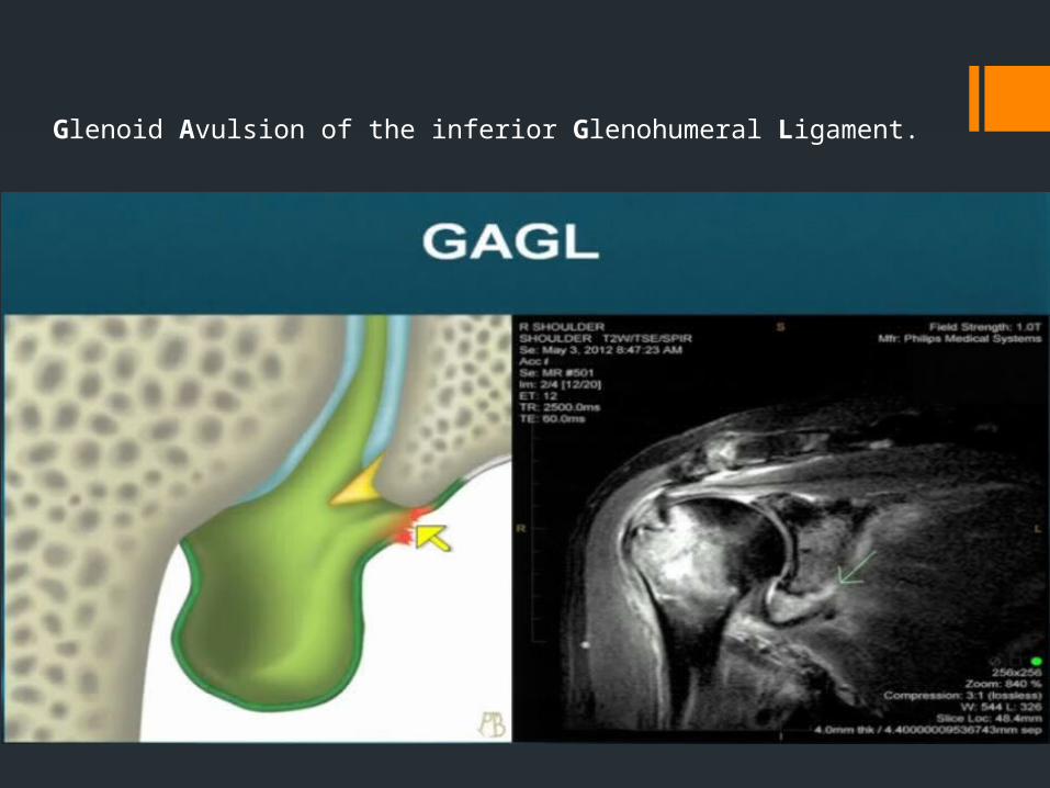
Glenoid Avulsion of the inferior Glenohumeral Ligament.


THANK YOU