shoulder dystocia: managing an obstetric emergency
TRANSCRIPT

84 American Family Physician www.aafp.org/afp Volume 102, Number 2 ◆ July 15, 2020
Shoulder dystocia is an obstetric emergency in which gentle downward traction of the fetal head does not lead to delivery and additional maneuvers are required to deliver the fetal shoulders.1 Shoulder dystocia is usually attributed to impaction of the anterior shoulder against the maternal symphysis after delivery of the fetal head; less commonly, it is caused by impaction of the posterior shoulder against the sacral promontory.2
Shoulder dystocia complicates 0.3% to 3% of all vag-inal deliveries.3,4 The exact incidence can be difficult to determine because the diagnosis is subjective and there are no agreed upon diagnostic criteria for shoulder dysto-cia. Objective criteria of a head-to-body delivery interval of 60 seconds or the need for additional delivery maneu-vers are proposed based on the incidence of significantly more birth injuries and lower Apgar scores during these deliveries.5
Risk Factors and PreventionRisk factors for shoulder dystocia include fetal macro-somia (odds ratio = 16.1), prior shoulder dystocia (odds ratio = 8.25), and preexisting or gestational diabetes melli-tus (odds ratio = 1.8).6-8 Other risk factors include maternal obesity, excessive maternal weight gain during pregnancy, oxytocin (Pitocin) use, prolonged first or second stage labor, and operative vaginal delivery (forceps or vacuum); however, these are poorly predictive of shoulder dystocia.9 There are no accurate models to predict or prevent shoulder dystocia.10,11
Fetal macrosomia is difficult to accurately predict. At term, fetal sonography has at least a 10% margin of error for diagnosis of macrosomia.11 Although the incidence of shoulder dystocia increases with increasing fetal weight and maternal diabetes, one study of pregnancies compli-cated by shoulder dystocia found that half of the neonates weighed less than 4,000 g (8 lb, 13 oz) and that only 20% of the patients had diabetes.12 Results from studies evaluating labor induction for suspected macrosomia are inconsistent, and induction is not recommended to prevent shoulder dys-tocia.13 Given the increased risk of shoulder dystocia with
Shoulder Dystocia: Managing an Obstetric Emergency
D. Ashley Hill, MD, AdventHealth Graduate Medical Education Program, Orlando, Florida
Jorge Lense, MD, AdventHealth Orlando Hospital, Orlando, Florida
Fay Roepcke, MD, MPH, AdventHealth Orlando Family Medicine Residency Program, Orlando, Florida
CME This clinical content conforms to AAFP criteria for CME. See CME Quiz on page 81.
Author disclosure: No relevant financial affiliations.
Shoulder dystocia is an obstetric emergency in which normal traction on the fetal head does not lead to delivery of the shoulders. This can cause neonatal brachial plexus injuries, hypoxia, and maternal trauma, including damage to the bladder, anal sphincter, and rectum, and postpartum hemorrhage. Although fetal macrosomia, prior shoulder dystocia, and preex-isting or gestational diabetes mellitus increases the risk of shoulder dystocia, most cases occur without warning. Labor and delivery teams should always be prepared to recognize and treat this emergency. Training and simulation exer-cises improve physician and team performance when shoulder dystocia occurs. Unequivocally announcing that dystocia is happening, summoning extra assis-tance, keeping track of the time from delivery of the head to full delivery of the neonate, and communicating with the patient and health care team are helpful. Calm and thoughtful use of release maneuvers such as knee to chest (McRoberts maneuver), suprapubic pressure, posterior arm or shoulder delivery, and inter-nal rotational maneuvers will almost always result in successful delivery. When these are unsuccessful, additional maneuvers, including intentional clavicular fracture or cephalic replacement, may lead to delivery. Each institution should consider the length of time it will take to prepare the operating room for general inhalational anesthesia and abdominal rescue and practice this during simulation exercises. (Am Fam Physician. 2020; 102(2): 84-90. Copyright © 2020 American Academy of Family Physicians.)
Illu
stra
tio
n b
y C
hri
sty
Kra
me
s
Downloaded from the American Family Physician website at www.aafp.org/afp. Copyright © 2020 American Academy of Family Physicians. For the private, non-commercial use of one individual user of the website. All other rights reserved. Contact [email protected] for copyright questions and/or permission requests.
Downloaded for Betty Burns ([email protected]) at National Certification Corporation from ClinicalKey.com by Elsevier on August 11, 2020.For personal use only. No other uses without permission. Copyright ©2020. Elsevier Inc. All rights reserved.

July 15, 2020 ◆ Volume 102, Number 2 www.aafp.org/afp American Family Physician 85
SHOULDER DYSTOCIA
increasing fetal weights, the American College of Obstetricians and Gynecol-ogists (ACOG) recommends consideration of cesarean delivery for a patient who does not have diabetes and is carrying a fetus with an estimated fetal weight of 5,000 g (11 lb). ACOG also recommends consideration of cesarean delivery for a patient who has diabetes and is carrying a fetus with an estimated fetal weight of 4,500 g (9 lb, 15 oz).10
ACOG and the Advanced Life Support in Obstetrics program recommend that labor and delivery teams conduct regular team train-ing drills that include iden-tification and management of shoulder dystocia.10,14
ComplicationsShoulder dystocia can cause several maternal and neona-tal complications (Table 1).10 The most common maternal complications are postpartum hemorrhage (11%) and obstetric anal sphincter injuries (3.8%).15 The most common
neonatal injuries are brachial plexus injuries and clavicular or humeral fractures.16 Transient brachial plexus injuries may occur in up to 20% of deliveries complicated by shoul-der dystocia.3 Most resolve without permanent disability, although approximately 10% may result in permanent neu-rologic injury.17 The head-to-body delivery interval does not predict fetal asphyxia or death.10 However, due to the potential for serious maternal or neonatal harm, a system-atic approach to expeditious delivery is necessary.10,15
Initial ResponsePhysicians should announce delivery of the fetal head so that an assistant can start a timer. If the fetus fails to deliver using normal traction or if retraction of the fetal head against the perineum (turtle sign) occurs, the physician should announce that there is a shoulder dystocia, and the delivery team should call for additional team members to assist. A longitudinal study of a shoulder dystocia simulation program found a significant reduction in neonatal brachial plexus injuries at discharge (7.6% to 1.3%) when the delivery team performed specified actions during shoulder dystocia deliveries.18 These included an unequivocal announcement of the shoulder dystocia, calling for additional assistance from qualified personnel, and having an assistant announce the time from delivery of the fetal head every 30 seconds.
TABLE 1
Complications of Shoulder Dystocia
Maternal
Lacerations of the bladder, urethra, vagina, anal sphincter, or rectum
Lateral femoral cutaneous neuropathy
Postpartum hemorrhage
Symphyseal separation
Uterine rupture
Neonatal
Fetal death
Fetal hypoxic ischemic encephalopathy
Fracture of the clavicle or humerus
Neurologic: brachial plexus injury, diaphragmatic paraly-sis, facial nerve injuries, Horner syndrome
Information from reference 10.
SORT: KEY RECOMMENDATIONS FOR PRACTICE
Clinical recommendationEvidence
rating Comments
Conduct team training simulation drills that include identification to improve performance during actual shoulder dystocia emergencies.18
B Longitudinal study of a mandatory shoulder dystocia training program
Announce unequivocally that there is a shoulder dystocia when it occurs.18
B Longitudinal study of a mandatory shoulder dystocia training program
Elevate both knees to the chest (McRob-erts maneuver) as the first therapeutic maneuver during shoulder dystocia.10
B Retrospective analysis of shoulder dystocia cases
Consider posterior arm delivery if McRoberts maneuver and suprapubic pressure are unsuccessful.10,14,21
C Clinical guidelines based on con-sensus, computer modeling, and a retrospective analysis of shoulder dystocia cases
Document precisely the head-to-body delivery interval and maneuvers per-formed after every shoulder dystocia.10
C Consensus-based clinical guidelines
A = consistent, good-quality patient-oriented evidence; B = inconsistent or limited-quality patient-oriented evidence; C = consensus, disease-oriented evidence, usual practice, expert opinion, or case series. For information about the SORT evidence rating system, go to https:// www.aafp.org/afpsort.
Downloaded from the American Family Physician website at www.aafp.org/afp. Copyright © 2020 American Academy of Family Physicians. For the private, non-commercial use of one individual user of the website. All other rights reserved. Contact [email protected] for copyright questions and/or permission requests.
Downloaded for Betty Burns ([email protected]) at National Certification Corporation from ClinicalKey.com by Elsevier on August 11, 2020.For personal use only. No other uses without permission. Copyright ©2020. Elsevier Inc. All rights reserved.

86 American Family Physician www.aafp.org/afp Volume 102, Number 2 ◆ July 15, 2020
Physicians should also obtain assistance from a physi-cian qualified to perform cesarean delivery and someone to resuscitate the neonate. Additional helpful actions that have not been studied include communicating with the patient so that she knows when to push, lowering the bed, using a stool for the assistant applying suprapubic pressure, and having someone record events for precise documentation.
Delivery ManeuversIf the fetus does not deliver using gentle traction, release maneuvers can be used in a thoughtful and sequential manner to deliver the impacted shoulder (Figure 1). Aggres-sive lateral or downward traction on the fetal head and neck should be avoided because it can injure the brachial
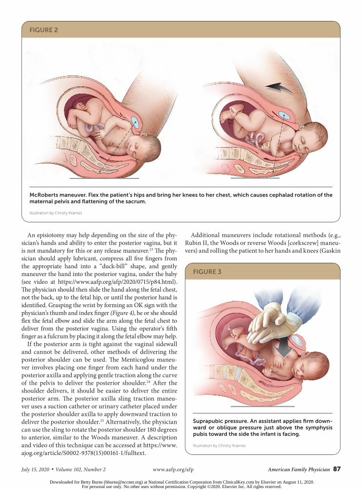
plexus.4 There are no randomized trials comparing the various maneuvers used to release an impacted shoul-der 10 (Table 210,18,19). ACOG, the Royal College of Obstetri-cians and Gynaecologists, and the Advanced Life Support in Obstetrics program recommend using the McRoberts maneuver first, followed by suprapubic pressure if neces-sary.10,14,19 The McRoberts maneuver, performed by flexing the hips and bringing both knees toward the chest, rotates the symphysis pubis cephalad and further opens the pel-vic outlet (Figure 2). This is a simple and proven method to manage shoulder dystocia, with a success rate of up to 42% as the sole maneuver.10,15
If delivery does not occur, firm, steady suprapubic pres-sure should be performed concurrently with the McRoberts maneuver. An assistant should apply firm downward or oblique pressure just above the symphysis pubis toward the side the infant is facing. This decreases the distance between the infant’s shoulders (bisacromial distance), potentially assisting anterior shoulder dislodgement (Figure 3). Fundal pressure increases the risk of uterine rupture.20
If the McRoberts maneuver and suprapubic pressure are unsuccessful, delivery of the posterior arm should be con-sidered10,14,21 (Figure 4). A retrospective review revealed that the combination of the McRoberts maneuver, suprapubic pressure, and posterior arm delivery resulted in successful delivery within four minutes in 95% of cases.21 Computer modeling suggests that delivery of the posterior arm results in the least amount of brachial plexus stretch compared with other maneuvers.22 Delivery of the posterior arm requires patience and communication to keep the patient calm. Training with a birth simulator will likely improve operator confidence and performance of this procedure.
TABLE 2
Delivery Maneuvers for Shoulder Dystocia
Initial maneuvers*
McRoberts
Suprapubic pressure
Delivery of the posterior arm
Menticoglou posterior shoulder delivery
Posterior axillary sling traction
Secondary maneuvers*
Rubin II rotational
Woods corkscrew rotational
Reverse Woods corkscrew rotational
Gaskin all-fours
Catastrophic maneuvers
Abdominal rescue under general anesthesia
Cephalic replacement (Zavanelli)
Intentional clavicular fracture
*—Listed in the suggested order that they should be performed.
Information from references 10, 18, and 19.
FIGURE 1
Management algorithm for shoulder dystocia.
CALL FOR HELP
Additional nurses, surgical obstetrics specialist, operat-ing room team, anesthesia provider, and neonatal staff
McRoberts maneuver
Elevate both knees to chest
Suprapubic pressure
Assistant pushes above pubic bone toward the side the fetus is facing
Consider episiotomy for exposure
Deliver posterior arm or shoulder
Place hand between perineum and fetus and slide across fetal
chest, grasp posterior arm, and sweep arm across chest
or
Place fingers from both hands, or a suction catheter, under
the posterior axilla and deliver the posterior shoulder and arm
Rotational maneuvers
Rubin II: Place two to four fingers along back of anterior
shoulder and rotate 30 degrees
Woods: Continue Rubin II and add fingers to front of posterior shoulder to rotate 180 degrees
Reverse Woods: Place fingers on the front side of the
anterior shoulder and back side of the posterior shoul-der and rotate 180 degrees
Gaskin all-fours maneuver
Consider preparing operating room
or
Repeat above maneuvers
Consider catastrophic maneuvers (abdominal rescue, cephalic replacement, intentional clavicular fracture)
Downloaded for Betty Burns ([email protected]) at National Certification Corporation from ClinicalKey.com by Elsevier on August 11, 2020.For personal use only. No other uses without permission. Copyright ©2020. Elsevier Inc. All rights reserved.
July 15, 2020 ◆ Volume 102, Number 2 www.aafp.org/afp American Family Physician 87
SHOULDER DYSTOCIA
An episiotomy may help depending on the size of the phy-sician’s hands and ability to enter the posterior vagina, but it is not mandatory for this or any release maneuver.23 The phy-sician should apply lubricant, compress all five fingers from the appropriate hand into a “duck-bill” shape, and gently maneuver the hand into the posterior vagina, under the baby (see video at https://www.aafp.org/afp/2020/0715/p84.html). The physician should then slide the hand along the fetal chest, not the back, up to the fetal hip, or until the posterior hand is identified. Grasping the wrist by forming an OK sign with the physician’s thumb and index finger (Figure 4), he or she should flex the fetal elbow and slide the arm along the fetal chest to deliver from the posterior vagina. Using the operator’s fifth finger as a fulcrum by placing it along the fetal elbow may help.
If the posterior arm is tight against the vaginal sidewall and cannot be delivered, other methods of delivering the posterior shoulder can be used. The Menticoglou maneu-ver involves placing one finger from each hand under the posterior axilla and applying gentle traction along the curve of the pelvis to deliver the posterior shoulder.24 After the shoulder delivers, it should be easier to deliver the entire posterior arm. The posterior axilla sling traction maneu-ver uses a suction catheter or urinary catheter placed under the posterior shoulder axilla to apply downward traction to deliver the posterior shoulder.25 Alternatively, the physician can use the sling to rotate the posterior shoulder 180 degrees to anterior, similar to the Woods maneuver. A description and video of this technique can be accessed at https:// www.ajog.org/article/S0002-9378(15)00161-1/fulltext.
Additional maneuvers include rotational methods (e.g., Rubin II, the Woods or reverse Woods [corkscrew] maneu-vers) and rolling the patient to her hands and knees (Gaskin
FIGURE 2
McRoberts maneuver. Flex the patient’s hips and bring her knees to her chest, which causes cephalad rotation of the maternal pelvis and flattening of the sacrum.
Illustration by Christy Krames
FIGURE 3
Suprapubic pressure. An assistant applies firm down-ward or oblique pressure just above the symphysis pubis toward the side the infant is facing.
Illustration by Christy Krames
Downloaded for Betty Burns ([email protected]) at National Certification Corporation from ClinicalKey.com by Elsevier on August 11, 2020.For personal use only. No other uses without permission. Copyright ©2020. Elsevier Inc. All rights reserved.

88 American Family Physician www.aafp.org/afp Volume 102, Number 2 ◆ July 15, 2020
all-fours maneuver). To perform the Rubin II maneuver, the physician places two fingers into the vagina to push the scapula of the anterior fetal shoulder toward the fetal face to attempt to rotate the fetus 30 degrees (Figure 5; see video at https://www.aafp.org/afp/2020/0715/p84.html).
The Woods maneuver combines the hand placement for the Rubin II maneuver with two fingers on the anterior aspect of the posterior fetal shoulder with the intent of rotating the fetus 180 degrees (Figure 5; see video at https://www.aafp.org/afp/2020/0715/p84.html). For the reverse Woods maneuver, fingers or hands are placed on the front side of the anterior shoulder and back side of the posterior shoulder to rotate the fetus 180 degrees. An episiotomy may be helpful for the Woods maneuvers to be able to gain access with two hands. The Gaskin all-fours maneuver requires the patient to roll onto her hands and knees. This had an 83% success rate as the sole maneuver used in one series.26 This maneuver may be more difficult if the patient is fatigued or has neuraxial anesthesia.
Maneuvers for Catastrophic Shoulder DystociaIf these maneuvers do not result in delivery, options include performing the maneuvers again (Figure 1) or enlisting assistance from another experienced physician who might try the previously attempted maneuvers again or who can
1. Grasp wrist with thumb and
index finger and flex elbow
2. Sweep hand across chest and deliver
Elbow
FIGURE 4
Posterior arm release. The physician’s hand enters the pelvis posteriorly and travels along the fetal chest to grasp the fetal posterior wrist using an OK sign. The operator’s hand should slide along the fetal chest, not the back, which may involve using the physician’s nondominant hand depending on the direction the fetus is facing. Hooking the little finger around the fetal elbow may facilitate the maneuver. The arm is then swept across the fetal chest.
Illustration by Christy Krames
30° 180°
FIGURE 5
Rubin II maneuver (left): Place two fingers on the back (posterior) side of the anterior shoulder and rotate 30 degrees toward the fetal face. Woods maneuver (right): Continue the finger placement for the Rubin II maneuver and add two fingers to the front (anterior) of the posterior shoulder and rotate the fetus 180 degrees.
Illustration by Christy Krames
Downloaded for Betty Burns ([email protected]) at National Certification Corporation from ClinicalKey.com by Elsevier on August 11, 2020.For personal use only. No other uses without permission. Copyright ©2020. Elsevier Inc. All rights reserved.

July 15, 2020 ◆ Volume 102, Number 2 www.aafp.org/afp American Family Physician 89
SHOULDER DYSTOCIA
collaborate to attempt less proven maneuvers, such as abdominal rescue, cephalic replacement (Zavanelli maneu-ver), and intentional clavicular fracture (Table 3).10,27,28 Each institution should consider the length of time it will take to prepare the operating room for general inhalational anesthesia and abdominal rescue and practice this during simulation exercises.
DocumentationPrecise documentation is extremely important after a shoulder dystocia to inform the clinical team of the deliv-ery events, including the head-to-body delivery interval and maneuvers used. ACOG has provided a checklist for documenting the occurrence of shoulder dystocia (https://journals.lww.com/greenjournal/Citation/2012/08000/Patient_Safety_Checklist_No__6___Documenting.43.aspx).29
This article updates a previous article by Baxley and Gobbo.30
Data Sources: A PubMed search was completed in Clinical Que-ries using the key terms shoulder dystocia, shoulder, brachial plexus, and abnormal labor. The search included meta-analyses, randomized controlled trials, clinical trials, and reviews. We also searched Ovid, Clinical Key, Cochrane Library, Web of Sci-ence, the Agency for Healthcare Research and Quality evidence reports, and Essential Evidence Plus. Search dates: September 5, 2019, and April 13, 2020.
The Authors
D. ASHLEY HILL, MD, is the chair of the Department of Obstetrics and Gynecology at the AdventHealth Gradu-ate Medical Education Program, Orlando, Fla.; a professor of obstetrics and gynecology at the University of Central Florida College of Medicine, Orlando; and the medical direc-tor of the Multiprofessional Obstetrics Simulation Training (M.O.S.T.®) program at AdventHealth Orlando.
JORGE LENSE, MD, is the medical director of the Advent-Health Obstetric Hospitalists at the AdventHealth Altamonte, Celebration, Orlando (Fla.) and Winter Park Hospitals and AdventHealth Gynecology Hospitalists at AdventHealth Orlando Hospital.
FAY ROEPCKE, MD, MPH, is a fellow at the Women’s Health Junior Faculty Fellowship at AdventHealth Orlando Fam-ily Medicine Residency and is an Advanced Life Support in Obstetrics (ALSO) course instructor.
Address correspondence to D. Ashley Hill, MD, 235 East Prince- ton St., Ste. 200, Orlando, FL 32804 (email: D.Hill.MD@ Advent Health.com). Reprints are not available from the authors.
References 1. Resnik R. Management of shoulder girdle dystocia. Clin Obstet Gyne-
col. 1980; 23(2): 559-564.
2. Hankins GD, Clark SL. Brachial plexus palsy involving the posterior shoul-der at spontaneous vaginal delivery. Am J Perinatol. 1995; 12(1): 44-45.
3. Gherman RB, Chauhan S, Ouzounian JG, et al. Shoulder dystocia: the unpreventable obstetric emergency with empiric management guide-lines. Am J Obstet Gynecol. 2006; 195(3): 657-672.
4. American College of Obstetricians and Gynecologists; Task Force on Neonatal Brachial Plexus Palsy. Neonatal Brachial Plexus Palsy. Ameri-can College of Obstetricians and Gynecologists; 2014.
5. Beall MH, Spong C, McKay J, et al. Objective definition of shoulder dystocia: a prospective evaluation. Am J Obstet Gynecol. 1998; 179(4): 934-937.
6. Tsur A, Sergienko R, Wiznitzer A, et al. Critical analysis of risk factors for shoulder dystocia. Arch Gynecol Obstet. 2012; 285(5): 1225-1229.
7. Bingham J, Chauhan SP, Hayes E, et al. Recurrent shoulder dystocia: a review. Obstet Gynecol Surv. 2010; 65(3): 183-188.
8. Zhang C, Wu Y, Li S, et al. Maternal prepregnancy obesity and the risk of shoulder dystocia: a meta-analysis. BJOG. 2018; 125(4): 407-413.
9. Ouzounian JG, Gherman RB. Shoulder dystocia: are historic risk factors reliable predictors? Am J Obstet Gynecol. 2005; 192(6): 1933-1935.
10. Committee on Practice Bulletins—Obstetrics. Practice bulletin no. 178: shoulder dystocia. Obstet Gynecol. 2017; 129(5): e123-e133.
11. Gupta M, Hockley C, Quigley MA, et al. Antenatal and intrapartum pre-diction of shoulder dystocia. Eur J Obstet Gynecol Reprod Biol. 2010; 151(2): 134-139.
12. Ouzounian JG, Korst LM, Miller DA, et al. Brachial plexus palsy and shoulder dystocia: obstetric risk factors remain elusive. Am J Perinatol. 2013; 30(4): 303-307.
13. American College of Obstetricians and Gynecologists; Committee on Practice Bulletins—Obstetrics. Practice bulletin number 173: fetal mac-rosomia. Obstet Gynecol. 2016; 128(5): e195-e209.
14. Shields SG, Ratcliffe S. Chapter F: labor dystocia. In: Leeman L, Quinlan JD, Dresang LT, et al. Advanced Life Support in Obstetrics Provider Man-ual. 8th edition. American Academy of Family Physicians; 2017: 1-14.
TABLE 3
Delivery Maneuvers for Catastrophic Shoulder Dystocia
Abdominal rescue
Perform a hysterotomy incision and then manipulate the fetus to dislodge the shoulders, allowing vaginal delivery.27
Cephalic replacement (Zavanelli maneuver)
Rotate the fetal head to a direct occiput anterior position, flexing the neck so the chin presses against the perineum, then push the head gently into the vagina.28 Apply con-tinuous pressure to hold the head in place while another physician performs a cesarean delivery to extract the baby abdominally. Relaxing the uterus with oral or intravenous nitroglycerin or inhalational anesthetics will likely make this procedure more successful, although this is unproven.
Intentional clavicular fracture
Pull the clavicles outward to fracture one or both, collaps-ing the shoulders inward and allowing delivery. However, this can be difficult to perform because of the strength of the clavicles, and it may damage underlying vasculature. Blunt manipulation is recommended; avoid the use of scissors or other sharp instruments.10
Information from references 10, 27, and 28.
Downloaded for Betty Burns ([email protected]) at National Certification Corporation from ClinicalKey.com by Elsevier on August 11, 2020.For personal use only. No other uses without permission. Copyright ©2020. Elsevier Inc. All rights reserved.

90 American Family Physician www.aafp.org/afp Volume 102, Number 2 ◆ July 15, 2020
SHOULDER DYSTOCIA
15. Gherman RB, Goodwin TM, Souter I, et al. The McRoberts’ maneu-ver for the alleviation of shoulder dystocia: how successful is it? Am J Obstet Gynecol. 1997; 176(3): 656-661.
16. Gherman RB, Ouzounian JG, Goodwin TM. Obstetric maneuvers for shoulder dystocia and associated fetal morbidity. Am J Obstet Gynecol. 1998; 178(6): 1126-1130.
17. Gherman RB, Ouzounian JG, Miller DA, et al. Spontaneous vaginal delivery: a risk factor for Erb’s palsy? Am J Obstet Gynecol. 1998; 178(3): 423-427.
18. Grobman WA, Miller D, Burke C, et al. Outcomes associated with intro-duction of a shoulder dystocia protocol. Am J Obstet Gynecol. 2011; 205(6): 513-517.
19. Royal College of Obstetricians and Gynaecologists. Shoulder dystocia (green-top guideline No. 42). March 28, 2012. Updated Feb-ruary 2017. Accessed March 11, 2020. https:// www.rcog.org.uk/en/guidelines-research-services/guidelines/gtg42/
20. Sturzenegger K, Schäffer L, Zimmermann R, et al. Risk factors of uterine rupture with a special interest to uterine fundal pressure. J Perinat Med. 2017; 45(3): 309-313.
21. Leung TY, Stuart O, Suen SS, et al. Comparison of perinatal outcomes of shoulder dystocia alleviated by different type and sequence of manoeu-vres: a retrospective review. BJOG. 2011; 118(8): 985-990.
22. Grimm MJ, Costello RE, Gonik B. Effect of clinician-applied maneuvers on brachial plexus stretch during a shoulder dystocia event: investiga-
tion using a computer simulation model. Am J Obstet Gynecol. 2010; 203(4): 339.e1-339.e5.
23. Sagi-Dain L, Sagi S. The role of episiotomy in prevention and manage-ment of shoulder dystocia: a systematic review. Obstet Gynecol Surv. 2015; 70(5): 354-362.
24. Menticoglou SM. A modified technique to deliver the posterior arm in severe shoulder dystocia. Obstet Gynecol. 2006; 108(3 pt 2): 755-757.
25. Cluver CA, Hofmeyr GJ. Posterior axilla sling traction for shoulder dys-tocia: case review and a new method of shoulder rotation with the sling. Am J Obstet Gynecol. 2015; 212(6): 784.e1-784.e7.
26. Bruner JP, Drummond SB, Meenan AL, et al. All-fours maneuver for reducing shoulder dystocia during labor. J Reprod Med. 1998; 43(5): 439-443.
27. O’Shaughnessy MJ. Hysterotomy facilitation of the vaginal delivery of the posterior arm in a case of severe shoulder dystocia. Obstet Gynecol. 1998; 92(4 pt 2): 693-695.
28. Sandberg EC. The Zavanelli maneuver: 12 years of recorded experi-ence. Obstet Gynecol. 1999; 93(2): 312-317.
29. American College of Obstetricians and Gynecologists. Patient safety checklist no. 6: documenting shoulder dystocia. Obstet Gynecol. 2012; 120(2 pt 1): 430-431.
30. Baxley EG, Gobbo RW. Shoulder dystocia. Am Fam Physician. 2004; 69(7): 1707-1714. Accessed March 11, 2020. https:// www.aafp.org/afp/2004/0401/p1707.html
Downloaded for Betty Burns ([email protected]) at National Certification Corporation from ClinicalKey.com by Elsevier on August 11, 2020.For personal use only. No other uses without permission. Copyright ©2020. Elsevier Inc. All rights reserved.