short course i - sfn
TRANSCRIPT

SHORT COURSE IUsing iPS Cells and Reprogramming to Model Neural Development and Disease Organized by Kevin Eggan, PhD

Short Course IUsing iPS Cells and Reprogramming to
Model Neural Development and Disease Organized by Kevin Eggan, PhD

Please cite articles using the model:[AUTHOR’S LAST NAME, AUTHOR’S FIRST & MIDDLE INITIALS] (2015)
[CHAPTER TITLE] In: Using iPS Cells and Reprogramming to Model Neural Development and Disease. (Eggan K, ed) pp. [xx-xx]. Chicago, IL: Society for Neuroscience.
All articles and their graphics are under the copyright of their respective authors.
Cover graphics and design © 2015 Society for Neuroscience.

Table of Contents
Probing Disorders of the Nervous System Using Reprogramming Approaches Evangelos Kiskinis, PhD . . . . . . . . . . . . . . . . . . . . . . . . . . . . . . . . . . . . . . . . . . . . . . . . . . . . 7
Stem Cells As a Tool for Studying the Developmental Regulation of Gene Expression
Hynek Wichterle, PhD, Michael Closser, BS, Esteban O . Mazzoni, PhD, Shaun Mahony, PhD, Yuchun Guo, PhD, Rachel Kopunova, and David K . Gifford, PhD . . . . . . . . . . . . . . . . . . . . . . . 17
Generating a Functional Human Cortex In Vitro From Induced Pluripotent Stem Cells Sergiu P . Pasca, MD . . . . . . . . . . . . . . . . . . . . . . . . . . . . . . . . . . . . . . . . . . . . . . . . . . . . . 27
Generating 3D Cerebral Organoids From Human Pluripotent Stem Cells to Model Cortical Development and Disease
Paola Arlotta, PhD . . . . . . . . . . . . . . . . . . . . . . . . . . . . . . . . . . . . . . . . . . . . . . . . . . . . . . 41
Drug-Based Modulation of Endogenous Stem Cells Promotes Functional Remyelination In Vivo Fadi J . Najm, MBA, Mayur Madhavan, PhD, Anita Zaremba, BA, Elizabeth Shick, BS,
Robert T . Karl, BS, Daniel C . Factor, BA, Tyler E . Miller, BS, Zachary S . Nevin, BS, Christopher Kantor, Alex Sargent, Kevin L . Quick, Daniela M . Schlatzer, Hong Tang, Ruben Papoian, PhD, Kyle R . Brimacombe, MS, Min Shen, Matthew B . Boxer, Ajit Jadhav, Andrew P . Robinson, Joseph R . Podojil, PhD, Stephen D . Miller, Robert H . Miller, and Paul J . Tesar, PhD . . . . . . . . . . . . . . . . . . . . . . . . . . . . . . . . . . . . . . . . . . . . . . . . . . . . . . 51
Developing Stem Cell Models to Study Neuropsychiatric Diseases Lindy Barrett, PhD, and Kevin Eggan, PhD . . . . . . . . . . . . . . . . . . . . . . . . . . . . . . . . . . . . . . 63
Modeling Predisposition to Schizophrenia, a Genetically Heterogeneous Neuropsychiatric Disorder, Using Induced Pluripotent Stem Cells
Seok-Man Ho, BSc, Erin Flaherty, BSc, and Kristen J . Brennand, PhD . . . . . . . . . . . . . . . . . . . . 71


© 2015 Kiskinis
Assistant Professor of Neurology and Physiology Feinberg School of Medicine
Northwestern University Chicago, Illinois
Probing Disorders of the Nervous System Using Reprogramming Approaches
Evangelos Kiskinis, PhD

9
NOTES
© 2015 Kiskinis
Probing Disorders of the Nervous System Using Reprogramming Approaches
IntroductionDiseases of the nervous system represent an enormous burden for society in terms of human suffering and financial cost. While significant advancements have been achieved over the last few decades particularly in terms of genetic linkage, clinical classification, and patient care, effective treatments are lacking. The inaccessibility of the relevant tissues and cell types in the CNS and the complex, multifactorial nature of most neurological disorders have hampered research progress. Animal models have been crucial in the investigation of disease mechanisms, but fundamental developmental, biochemical, and physiological differences exist between animals and humans. The importance of utilizing human cells for these purposes is evident by the large number of drugs that show efficacy and safety in rodent models of diseases but subsequently fail in human clinical trials, failures that are attributed partly to these species differences (Rubin, 2008). Furthermore, the overwhelming majority of neurological disease is of a sporadic nature, rendering animal modeling ineffective, while it remains unclear whether the relatively rare monogenic forms of disease truly represent the vast majority of sporadic cases.
The simultaneous development of methods for reprogramming adult cells into induced pluripotent stem cells (iPSCs) (Takahashi et al., 2007; Yu et al., 2007; Park et al., 2008) and the directed differentiation of pluripotent stem cells into distinct neuronal subtypes (Williams et al., 2012) suggested an attractive route to a novel model system for the study of neurological disorders. Patient-specific iPSCs can be generated using epigenetic reprogramming of various adult cell types, such as skin fibroblasts and blood mononuclear cells, and just like embryonic stem cells (ESCs), self-renew indefinitely and retain the potential to give rise to all cell types in the human body (Takahashi et al., 2007). More recently, sophisticated lineage-conversion approaches have allowed for the direct generation of neurons and neural cell types from adult cells by means of overexpressing key transcription factors (Tsunemoto et al., 2014). These methods have overcome some of the limitations of directed differentiation and have enabled the generation of cell types that, in many cases, were previously unattainable.
The overwhelming advantages of using iPSCs and lineage conversion to develop models of diseases of the nervous system are that they allow one to study disease mechanisms in the context of human neurons and in the context of each patient’s unique genetic constellation. In many cases, established
differentiation protocols allow for the generation of the particular neuronal subtype that is most vulnerable to the particular disease, such as spinal motor neurons (Davis-Dusenbery et al., 2014) and dopaminergic neurons (Kriks et al., 2011). These neurons can be produced in abundance from variable genetic backgrounds and could provide useful platforms for drug discovery.
The concept of using iPSCs and lineage conversion to study neurological disease appears straightforward: both approaches allow for the generation of patient-specific neurons, which are relevant to the disease of interest. In addition, when these neurons are compared with neurons generated from healthy controls, any differences identified could be related to the disease. In practice, however, this approach has proven to be more challenging than initially believed. What is the right cell type to make and study? How should quality control of neurons be performed? What are the right controls to use when assessing a disease-related phenotype? How do phenotypes identified in vitro relate to the clinical presentation of patients? These are just some of the questions that the community has struggled with since the initial description of iPSCs and the onset of the development of in vitro patient-specific disease models. Perhaps the seemingly biggest advantage of this approach—the ability to study disease in the genetic background of the patient—has created the biggest challenge, as genetic background contributes to high variability in the properties of the patient-derived cells. This variability is a reality that neurologists have been facing for years, as often, two patients diagnosed with the same condition might present with very different clinical profiles. The technology of cellular reprogramming has brought this reality of clinical heterogeneity seen in patients from the bedside to the lab bench.
Since the initial description of reprogramming technologies, neuroscientists, neurologists, and stem cell researchers have generated and characterized hundreds of patient-specific stem cell lines as well as neuronal cells derived from them. The first “wave” of disease-modeling studies focused on generating patient-specific human neurons and confirming previously described pathologies (Dimos et al., 2008; Ebert et al., 2009; Marchetto et al., 2010; Brennand et al., 2011; Seibler et al., 2011; Bilican et al., 2012; Israel et al., 2012). More recent studies have revealed novel insights into disease mechanisms and employed gene editing approaches to clearly demonstrate the association of identified phenotypes with known genetic variants that contribute to disease (An
10
NOTES
© 2015 Kiskinis
et al., 2012; Corti et al., 2012; Fong et al., 2013; Reinhardt et al., 2013; Kiskinis et al., 2014; Wainger et al., 2014; Wen et al., 2014). At the same time, our ability to generate neuronal subtypes via directed differentiation and the exogenous expression of transcription factors has made tremendous progress.
Specificity of Phenotypes: The Importance of ControlsSignificant technical advancements achieved during the past few years have allowed for the generation of patient-specific iPSCs that are free from genomic integration of the reprogramming factors (Malik and Rao, 2013). The essential quality of any newly derived iPSC can be easily assessed by (1) immunocytochemistry for pluripotency markers (e.g., NANOG/SSEA3); (2) a quantitative pluripotency assay, such as TaqMan® hPSC Scorecard™ Assay (ThermoFisher Scientific, Waltham, MA) or PluriTest™ (Scripps Research Institute, available at pluritest.org); and (3) analysis of genomic integrity (e.g., karyotyping, array comparative genomic hybridization).
Disease-modeling studies based on iPSC technology have relied on the use of diseased cells derived from patients as a model for disease and cells derived from healthy individuals as controls. However, genetic and potentially epigenetic heterogeneity of iPSC lines contributes to functional variability of differentiated somatic cells, confounding the evaluation of disease-modeling experiments (Sandoe and Eggan, 2013). Such variability can be introduced at multiple levels, including the generation of stem cell lines, continuous in vitro culture, variation in cell culture reagents, differential efficiencies of neural generation, and genetic background. Different approaches can be taken to overcoming this variation. One approach is through the use of targeted gene editing that results in the generation of a control stem cell line that is isogenic to the patient one, except for the disease-causing mutation. Such an approach effectively minimizes line-to-line differences and is a crucial tool for iPSC-based disease modeling.
CRISPR/Cas9, a recent technology that has emerged, allows for the efficient generation of such isogenic stem cell lines (CRISPR stands for clustered regularly interspaced short palindromic repeats, and Cas9 is a class of RNA-guided endonucleases) (Hsu et al., 2014). The system contains two essential components: an enzyme that can cleave DNA so that a double-strand break or a single nick is generated, and a guide RNA that targets the enzyme to a specific genomic location. By simultaneously introducing either a
single-stranded oligodeoxynucleotide containing the desired edit, or a targeting plasmid with larger desired sequence alterations, the genomic sequence can be precisely edited via the cells’ own endogenous repair mechanism, homologous recombination. Given the incredible versatility of the CRISPR/Cas9 system and the continuous evolvement of the technical aspects of this approach, it should be expected that every iPSC study that focuses on genetic forms of disease should include an isogenic control cell line. The rescue of a phenotype by genetic correction can lead to the conclusion that the genetic lesion is necessary for the onset of the phenotype. The same technique can be used to introduce a disease-associated mutation in a healthy iPSC line in order to assess whether the mutation in itself is sufficient for the onset of particular phenotypes.
An alternative approach to the concern of variation would be to utilize multiple stem cell clones from each individual patient and compare the desired measurement against multiple healthy individuals. The use of multiple patient clones would ensure that the phenotype is not an artifact of a defective clonal cell line, while the use of multiple healthy controls should encapsulate sufficient technical and genetic variation so that the measured cellular properties (e.g., neuronal firing, dendritic density) will represent a true average. This approach will be important in studies of sporadic disease.
An important point to consider when assessing the specificity of an identified phenotype is whether it is apparent only in the cell type known to be most vulnerable to the disease being modeled. In amyotrophic lateral sclerosis (ALS) patients, for example, it is the upper and lower motor neurons that are initially targeted by disease mechanisms and gradually lost, while sensory neurons remain relatively unaffected. It would therefore be predicted that a phenotype that is truly relevant to the disease would not be evident in a sensory neuron generated from the same individual. Although this approach could be valuable, it should be taken with caution for two reasons: (1) because a sensory neuron might simply be resistant to a phenotype, and therefore it is the effect of the phenotype on the sensory cell that should be considered, not simply the presence of the phenotype in itself; and (2) because it might be the in vivo microenvironment of a sensory neuron that confers resistance and not a cell-autonomous trait. Nevertheless, studies have demonstrated neuronal-type specificity of certain phenotypes. These include the sensitivity of mutant Parkinson’s disease (PD) tyrosine hydroxylase (TH)–positive neurons but

11
NOTESnot TH-negative neurons to H2O2-induced toxicity (Nguyen et al., 2011) and morphometric deficiencies of mutant ALS, Islet (ISL)–positive motor neurons but not ISL-negative neurons grown in the same culture dishes (Kiskinis et al., 2014).
A major advantage of using reprogramming approaches to study neurological disease is the ability to assess the biological variation associated with a specific neuronal defect. Consider that a phenotype (e.g., defective lysosomal function) has been identified in neurons derived from a patient cell line and that this phenotype is mutation dependent (i.e., it is corrected in an isogenic control line). The first level of biological variation can be addressed by examining neurons derived from a different individual that harbors the exact same mutation in the same gene. If the phenotype is not present, then additional genetic or epigenetic factors might be necessary for the onset of the defect. The next level of biological variability can be addressed by examining neurons from a patient with a different mutation in the same gene. Lastly, the broader relevance of the identified phenotype for the disease can be assessed by examining the lysosomal function of neurons from patients with mutations in different disease-causing genes as well as in a large number of sporadic cases.
A Shift in Focus: From Developing Neurons to Maturing and Aging ThemA critical area that deserves further investigation is the maturity and aging of cells derived in vitro. We like to think that there are three stages we need to consider when setting up in vitro models of disease: the development, the maturation, and the natural aging process of a neural cell type. Although significant advancements have been achieved in generating and maturing neural cell types (either by directed differentiation or lineage conversion), little has been done in terms of affecting the aging of cells. For late-onset diseases such as ALS, frontotemporal dementia, Huntington’s disease (HD), PD, and Alzheimer’s disease (AD), it is possible that changes elicited by aging are required to induce the disease process. Age is the strongest risk factor for neurodegenerative diseases, and although there are rare cases with early-onset presentation, the overwhelming majority of patients develop clinical symptoms in the later stages of their lives. The nature of age-related risk remains largely unknown, and whether it arises from cell-autonomous mechanisms or as a result of a systemic dysfunction remains to be determined. A number of studies support the notion that cellular
epigenetic changes in the CNS correlate with aging. For example, recent work has demonstrated that profound changes in DNA methylation levels occur in the brains of mice with age (Lister et al., 2013), while aging oligodendrocytes lose their ability to effectively remyelinate damaged nerves (Ruckh et al., 2012). Importantly, under conditions of heterochronic parabiosis in mice, the effects on oligodendrocytes were reversible, implicating some aspect of epigenetic regulation.
Current studies suggest that the transcriptional and electrophysiological properties of both iPSC-derived and lineage-converted neurons are more similar to fetal neurons than adult ones (Son et al., 2011; Takazawa et al., 2012). It is likely that extrinsic factors present during normal development or aging are required to activate the maturation process. We and others have shown, for example, that adding primary astrocytes to lineage-conversion cultures significantly improves the maturation of induced neurons (Son et al., 2011; Chanda et al., 2013; Wainger et al., 2015). Additional progress in generating more mature and aged cells will require a better understanding of the gene expression and functional changes associated with maturation and aging. This has been difficult to obtain for specific neuronal subtypes because of the scarcity of available human tissue. Efforts such as those of the Allen Brain Institute have shed some light on these markers, but future studies will need to analyze specific neuronal subtypes in order to be sure that differences between aged neurons and young neurons are truly the result of aging and not of different neuronal subtypes.
In addition to glial-derived factors, Rubin and colleagues recently showed that circulatory factors contribute to the aging process in the CNS (Katsimpardi et al., 2014). They were able to identify a single factor: growth differentiation factor 11 (GDF11), whose expression normally declines with age. Interestingly, restoring GDF11 levels in old mice rejuvenated the proliferative and neurogenic properties of neural stem cells in the mouse (Katsimpardi et al., 2014). This finding suggests that other factors may control the aging of neurons and could be exploited to regulate this process in vitro.
Studer and colleagues took a more intrinsic approach to inducing aging in iPSC-derived neurons by expressing progerin, which is a mutant form of the Lamin A protein that causes accelerated aging phenotypes in humans (Miller et al., 2013). The expression of progerin induced higher levels of DNA damage and mitochondrial reactive oxygen species
© 2015 Kiskinis
Probing Disorders of the Nervous System Using Reprogramming Approaches

12
NOTES in dopaminergic neurons derived from PD patients, which enabled the detection of PD-associated disease phenotypes such as dendrite degeneration, mitochondrial enlargement, Lewy body–precursor inclusions, and suppression of TH expression (Miller et al., 2013). It remains unclear whether this approach induces the recapitulation of bona fide disease processes, but it does represent a new line of targeted aging procedures.
From Cell Autonomy to More Sophisticated SystemsNeurons do not exist in isolation in the human nervous system. Rather, they form elaborate and functional networks with other neurons and rely on a sophisticated microenvironment that is created by the interactions with other neural and nonneural cell types, which provide structural, metabolic, and functional support as well as effective communication (Abbott et al., 2006). Glial cells, astrocytes, oligodendrocytes, microglia, and endothelial cells exist in abundance in the nervous system and play vital functional roles. Glial cells buffer harmful ions, astrocytes provide nutrients and circulate neurotransmitters around synapses, oligodendrocytes form myelin sheaths around axons, microglia scavenge and degrade dead cells, and endothelial cells are important for maintaining the blood–brain barrier. Cell–cell interactions and the microenvironment as a whole might mediate important neuroprotective or neurotoxic activities in response to disease or injury. In fact, a number of studies during the past few years have clearly demonstrated that non–cell-autonomous processes involving astrocytes, oligodendrocytes, and microglia play a critical role in mediating disease progression and, potentially, onset in neurodegeneration in such diseases as ALS, HD, PD, prion disease, the spinal cerebellar ataxias, and AD in vivo (Ilieva et al., 2009). The strength of utilizing iPSCs to study neurological disease is found in their ability to generate a range of different cell types from the same genetic background. This versatility allows for the assessment of how, for example, a specific genetic lesion might differentially impact neuronal subtypes. It also allows for a rational step-by-step approach for assessing how cellular interactions might contribute to the evolution of a disease-associated phenotype or a cellular response to stress.
The coculture of spinal motor neurons with cortical astrocytes has been utilized in one of the first stem cell–based models of ALS to demonstrate how mutant or healthy astrocytes significantly
compromise or maintain, respectively, the health of a pure population of motor neurons (Di Giorgio et al., 2008; Marchetto et al., 2008). The coculture of cortical excitatory with cortical inhibitory neurons, and the establishment of functional circuitry, might be beneficial when studying epileptic syndromes. The clinical presentation of epileptic patients is the result of the functional control (or lack thereof) of a network of neurons, so recapitulating such a network could be an essential step toward the development of a cellular disease model. The importance of the local microenvironment to neuronal function (and potentially, dysfunction during disease) is also relevant in the context of the three-dimensionality that it creates. Neither the brain nor the spinal cord hosts isolated neurons surrounded by an entirely liquid trophic support (akin to culture media) in which nutrients, molecules, and proteins can freely diffuse and float around. Recently, Kim, Tanzi, and colleagues were able to successfully recapitulate amyloid-beta (Aβ) plaques and tau neurofibrillary tangles—the two pathological hallmarks of AD—in a single three-dimensional human neural-cell culture system (Choi et al., 2014). Although this system was not based on iPSCs, and their cell lines expressed slightly elevated protein levels of PSEN1 and APP, they designed a simple but innovative cell culture system with neurons grown embedded within a 0.3 mm layer of an extracellular matrix composed of BD Matrigel™ Basement Membrane Matrix (BD Biosciences, Erembodegem, Belgium). This viscous layer reduced the diffusion of secreted Aβ and led to the accumulation of aggregated plaques. This was the first time this had been achieved in a cell-based in vitro system and demonstrates the importance of using a three-dimensional environment for disease-modeling assays.
The recent description of cerebral organoids generated from human pluripotent stem cells and resembling the three-dimensional regional organization of a developing brain has created an exciting opportunity for iPSC-based disease-modeling approaches (Lancaster et al., 2013). These brain-like structures, formed by the combination of external growth factor patterning and intrinsic and environmental cues, exhibit distinct regional identities that functionally interact and, most importantly, recapitulate human cortical organization. The authors utilized this method to study microcephaly and demonstrate that patient-specific organoids show premature neuronal differentiation and are capable of developing only to a smaller size. Significantly, mouse models have failed to effectively recapitulate these disease
© 2015 Kiskinis

13
NOTESphenotypes for microcephaly, probably owing to the dramatic differences in the development and regional organization of their brain, as mice do not have an outer subventricular zone. This system may be suitable for the study of other neurodevelopmental and neuropsychiatric syndromes in which moderate but crucial defects in cortical organization and function are present. This approach also may be useful for recapitulating human neurodegenerative models that primarily affect brain function because it may allow for the establishment of neuronal circuitry as well as biochemical networks.
AcknowledgmentsThis paper was adapted from Ichida JK, Kiskinis E, 2015, Probing disorders of the nervous system using reprogramming approaches. EMBO J 34:1456–1477.
ReferencesAbbott NJ, Ronnback L, Hansson E (2006)
Astrocyte-endothelial interactions at the blood–brain barrier. Nat Rev Neurosci 7:41–53.
An MC, Zhang N, Scott G, Montoro D, Wittkop T, Mooney S, Melov S, Ellerby LM (2012) Genetic correction of Huntington’s disease phenotypes in induced pluripotent stem cells. Cell Stem Cell 11:253–263.
Bilican B, Serio A, Barmada SJ, Nishimura AL, Sullivan GJ, Carrasco M, Phatnani HP, Puddifoot CA, Story D, Fletcher J, Park IH, Friedman BA, Daley GQ, Wyllie DJ, Hardingham GE, Wilmut I, Finkbeiner S, Maniatis T, Shaw CE, Chandran S (2012) Mutant induced pluripotent stem cell lines recapitulate aspects of TDP-43 proteinopathies and reveal cell-specific vulnerability. Proc Natl Acad Sci USA 109:5803–5808.
Brennand KJ, Simone A, Jou J, Gelboin-Burkhart C, Tran N, Sangar S, Li Y, Mu Y, Chen G, Yu D, McCarthy S, Sebat J, Gage FH (2011) Modelling schizophrenia using human induced pluripotent stem cells. Nature 473:221–225.
Chanda S, Marro S, Wernig M, Sudhof TC (2013) Neurons generated by direct conversion of fibroblasts reproduce synaptic phenotype caused by autism-associated neuroligin-3 mutation. Proc Natl Acad Sci USA 110:16622–16627.
Choi SH, Kim YH, Hebisch M, Sliwinski C, Lee S, D’Avanzo C, Chen H, Hooli B, Asselin C, Muffat J, Klee JB, Zhang C, Wainger BJ, Peitz M, Kovacs DM, Woolf CJ, Wagner SL, Tanzi RE, Kim DY (2014) A three-dimensional human neural cell culture model of Alzheimer’s disease. Nature 515:274–278.
Corti S, Nizzardo M, Simone C, Falcone M, Nardini M, Ronchi D, Donadoni C, Salani S, Riboldi G, Magri F, Menozzi G, Bonaglia C, Rizzo F, Bresolin N, Comi GP (2012) Genetic correction of human induced pluripotent stem cells from patients with spinal muscular atrophy. Sci Transl Med 4:165ra162.
Davis-Dusenbery BN, Williams LA, Klim JR, Eggan K (2014) How to make spinal motor neurons. Development 141:491–501.
Di Giorgio FP, Boulting GL, Bobrowicz S, Eggan KC (2008) Human embryonic stem cell–derived motor neurons are sensitive to the toxic effect of glial cells carrying an ALS-causing mutation. Cell Stem Cell 3:637–648.
Dimos JT, Rodolfa KT, Niakan KK, Weisenthal LM, Mitsumoto H, Chung W, Croft GF, Saphier G, Leibel R, Goland R, Wichterle H, Henderson CE, Eggan K (2008) Induced pluripotent stem cells generated from patients with ALS can be differentiated into motor neurons. Science 321:1218–1221.
Ebert AD, Yu J, Rose FF, Jr., Mattis VB, Lorson CL, Thomson JA, Svendsen CN (2009) Induced pluripotent stem cells from a spinal muscular atrophy patient. Nature 457:277–280.
Fong H, Wang C, Knoferle J, Walker D, Balestra ME, Tong LM, Leung L, Ring KL, Seeley WW, Karydas A, Kshirsagar MA, Boxer AL, Kosik KS, Miller BL, Huang Y (2013) Genetic correction of tauopathy phenotypes in neurons derived from human induced pluripotent stem cells. Stem Cell Rep 1:226–234.
Hsu PD, Lander ES, Zhang F (2014) Development and applications of CRISPR-Cas9 for genome engineering. Cell 157:1262–1278.
Ilieva H, Polymenidou M, Cleveland DW (2009) Non-cell autonomous toxicity in neurodegenerative disorders: ALS and beyond. J Cell Biol 187:761–772.
© 2015 Kiskinis
Probing Disorders of the Nervous System Using Reprogramming Approaches

14
NOTES
© 2015 Kiskinis
Israel MA, Yuan SH, Bardy C, Reyna SM, Mu Y, Herrera C, Hefferan MP, Van Gorp S, Nazor KL, Boscolo FS, Carson CT, Laurent LC, Marsala M, Gage FH, Remes AM, Koo EH, Goldstein LS (2012) Probing sporadic and familial Alzheimer’s disease using induced pluripotent stem cells. Nature 482:216–220.
Katsimpardi L, Litterman NK, Schein PA, Miller CM, Loffredo FS, Wojtkiewicz GR, Chen JW, Lee RT, Wagers AJ, Rubin LL (2014) Vascular and neurogenic rejuvenation of the aging mouse brain by young systemic factors. Science 344:630–634.
Kiskinis E, Sandoe J, Williams LA, Boulting GL, Moccia R, Wainger BJ, Han S, Peng T, Thams S, Mikkilineni S, Mellin C, Merkle FT, Davis-Dusenbery BN, Ziller M, Oakley D, Ichida J, Di Costanzo S, Atwater N, Maeder ML, Goodwin MJ, et al. (2014) Pathways disrupted in human ALS motor neurons identified through genetic correction of mutant SOD1. Cell Stem Cell 14:791–795.
Kriks S, Shim JW, Piao J, Ganat YM, Wakeman DR, Xie Z, Carrillo-Reid L, Auyeung G, Antonacci C, Buch A, Yang L, Beal MF, Surmeier DJ, Kordower JH, Tabar V, Studer L (2011) Dopamine neurons derived from human ES cells efficiently engraft in animal models of Parkinson’s disease. Nature 480:547–551.
Lancaster MA, Renner M, Martin CA, Wenzel D, Bicknell LS, Hurles ME, Homfray T, Penninger JM, Jackson AP, Knoblich JA (2013) Cerebral organoids model human brain development and microcephaly. Nature 501:373–379.
Lister R, Mukamel EA, Nery JR, Urich M, Puddifoot CA, Johnson ND, Lucero J, Huang Y, Dwork AJ, Schultz MD, Yu M, Tonti-Filippini J, Heyn H, Hu S, Wu JC, Rao A, Esteller M, He C, Haghighi FG, Sejnowski TJ, et al. (2013) Global epigenomic reconfiguration during mammalian brain development. Science 341:1237905.
Malik N, Rao MS (2013) A review of the methods for human iPSC derivation. Methods Mol Biol 997:23–33.
Marchetto MC, Carromeu C, Acab A, Yu D, Yeo GW, Mu Y, Chen G, Gage FH, Muotri AR (2010) A model for neural development and treatment of Rett syndrome using human induced pluripotent stem cells. Cell 143:527–539.
Marchetto MC, Muotri AR, Mu Y, Smith AM, Cezar GG, Gage FH (2008) Non-cell-autonomous effect of human SOD1 G37R astrocytes on motor neurons derived from human embryonic stem cells. Cell Stem Cell 3:649–657.
Miller JD, Ganat YM, Kishinevsky S, Bowman RL, Liu B, Tu EY, Mandal PK, Vera E, Shim JW, Kriks S, Taldone T, Fusaki N, Tomishima MJ, Krainc D, Milner TA, Rossi DJ, Studer L (2013) Human iPSC-based modeling of late-onset disease via progerin-induced aging. Cell Stem Cell 13:691–705.
Nguyen HN, Byers B, Cord B, Shcheglovitov A, Byrne J, Gujar P, Kee K, Schule B, Dolmetsch RE, Langston W, Palmer TD, Pera RR (2011) LRRK2 mutant iPSC-derived DA neurons demonstrate increased susceptibility to oxidative stress. Cell Stem Cell 8:267–280.
Park IH, Arora N, Huo H, Maherali N, Ahfeldt T, Shimamura A, Lensch MW, Cowan C, Hochedlinger K, Daley GQ (2008) Disease-specific induced pluripotent stem cells. Cell 134:877–886.
Reinhardt P, Schmid B, Burbulla LF, Schondorf DC, Wagner L, Glatza M, Hoing S, Hargus G, Heck SA, Dhingra A, Wu G, Muller S, Brockmann K, Kluba T, Maisel M, Kruger R, Berg D, Tsytsyura Y, Thiel CS, Psathaki OE, et al. (2013) Genetic correction of a LRRK2 mutation in human iPSCs links parkinsonian neurodegeneration to ERK-dependent changes in gene expression. Cell Stem Cell 12:354–367.
Rubin LL (2008) Stem cells and drug discovery: the beginning of a new era? Cell 132:549–552.
Ruckh JM, Zhao JW, Shadrach JL, van Wijngaarden P, Rao TN, Wagers AJ, Franklin RJ (2012) Rejuvenation of regeneration in the aging central nervous system. Cell Stem Cell 10:96–103.
Sandoe J, Eggan K (2013) Opportunities and challenges of pluripotent stem cell neurodegenerative disease models. Nat Neurosci 16:780–789.
Seibler P, Graziotto J, Jeong H, Simunovic F, Klein C, Krainc D (2011) Mitochondrial Parkin recruitment is impaired in neurons derived from mutant PINK1 induced pluripotent stem cells. J Neurosci 31:5970–5976.
Son EY, Ichida JK, Wainger BJ, Toma JS, Rafuse VF, Woolf CJ, Eggan K (2011) Conversion of mouse and human fibroblasts into functional spinal motor neurons. Cell Stem Cell 9:205–218.

15
NOTES
Probing Disorders of the Nervous System Using Reprogramming Approaches
© 2015 Kiskinis
Takahashi K, Tanabe K, Ohnuki M, Narita M, Ichisaka T, Tomoda K, Yamanaka S (2007) Induction of pluripotent stem cells from adult human fibroblasts by defined factors. Cell 131:861–872.
Takazawa T, Croft GF, Amoroso MW, Studer L, Wichterle H, Macdermott AB (2012) Maturation of spinal motor neurons derived from human embryonic stem cells. PLoS One 7:e40154.
Tsunemoto RK, Eade KT, Blanchard JW, Baldwin KK (2015) Forward engineering neuronal diversity using direct reprogramming. EMBO J 34:1445–1455.
Wainger BJ, Kiskinis E, Mellin C, Wiskow O, Han SS, Sandoe J, Perez NP, Williams LA, Lee S, Boulting G, Berry JD, Brown RH, Jr., Cudkowicz ME, Bean BP, Eggan K, Woolf CJ (2014) Intrinsic membrane hyperexcitability of amyotrophic lateral sclerosis patient-derived motor neurons. Cell Rep 7:1–11.
Wainger BJ, Buttermore ED, Oliveira JT, Mellin C, Lee S, Saber WA, Wang AJ, Ichida JK, Chiu IM, Barrett L, Huebner EA, Bilgin C, Tsujimoto N, Brenneis C, Kapur K, Rubin LL, Eggan K, Woolf CJ (2015) Modeling pain in vitro using nociceptor neurons reprogrammed from fibroblasts. Nat Neurosci 18:17–24.
Wen Z, Nguyen HN, Guo Z, Lalli MA, Wang X, Su Y, Kim NS, Yoon KJ, Shin J, Zhang C, Makri G, Nauen D, Yu H, Guzman E, Chiang CH, Yoritomo N, Kaibuchi K, Zou J, Christian KM, Cheng L, et al. (2014) Synaptic dysregulation in a human iPS cell model of mental disorders. Nature 515:414–418.
Williams LA, Davis-Dusenbery BN, Eggan KC (2012) SnapShot: directed differentiation of pluripotent stem cells. Cell 149:1174–1174.e1.
Yu J, Vodyanik MA, Smuga-Otto K, Antosiewicz-Bourget J, Frane JL, Tian S, Nie J, Jonsdottir GA, Ruotti V, Stewart R, Slukvin, II, Thomson JA (2007) Induced pluripotent stem cell lines derived from human somatic cells. Science 318:1917–1920.

© 2013
1Departments of Pathology and Cell Biology, Neurology, and Neuroscience
Center for Motor Neuron Biology and Disease, Columbia Stem Cell Initiative
Columbia University Medical Center New York, New York
2Department of Biology New York University
New York, New York
3Department of Biochemistry and Molecular Biology Center for Eukaryotic Gene Regulation
The Pennsylvania State University University Park, Pennsylvania
4Computer Science and Artificial Intelligence Laboratory Massachusetts Institute of Technology
Cambridge, Massachusetts
Stem Cells As a Tool for Studying the Developmental Regulation
of Gene ExpressionHynek Wichterle, PhD,1 Michael Closser, BS,1
Esteban O. Mazzoni, PhD,2 Shaun Mahony, PhD,3 Yuchun Guo, PhD,4
Rachel Kopunova,1 and David K. Gifford, PhD4
© 2015 Wichterle


19
NOTESIntroductionDespite extensive functional analysis of transcription factors, the detailed mechanisms by which they regulate gene expression and specify cell identity in developing organisms remain poorly understood. Recent advances in chromatin mapping technologies have provided unprecedented insight into the organization of regulatory regions, chromatin structure, and the exact positions of transcription factor binding sites. The emerging picture of extremely plastic chromatin organization prevents simple extrapolation of a regulatory landscape from one cell lineage or even one developmental stage to another. We have developed a pluripotent stem cell–based differentiation system that facilitates systematic mapping and probing of transcriptional regulatory networks that control the specification of spinal motor neuron identity. The systematic analysis of mechanisms controlling cell type–specific regulation of gene expression is facilitated by combining inducible stem cell lines, in which gain-of-function studies can be performed, with unlimited access to relatively homogenous populations of cells differentiating along the motor neuron lineage. Identifying regulatory motifs, transcription factors, and cofactors engaged in the specification of motor neuron identity provides novel insights into ways to efficiently program and derive clinically relevant cell types.
Progress in Cell ProgrammingRecent progress in programming cell fate using transcription factors has given hope to those pursuing the goal of producing clinically relevant cell types for modeling disease and developing new therapeutic strategies. Muscle cells, pluripotent stem cells, pancreatic beta cells, hepatocytes, and several types of neurons have all been created by the forced expression of transcription factor combinations known as “programming modules” (Tapscott et al., 1988; Mann and Carroll, 2002; Takahashi and Yamanaka, 2006; Zhou et al., 2008; Son et al., 2011). However, the process of transcriptional programming remains largely enigmatic. Understanding the mechanism through which programming modules convert one expression profile to another would accomplish two main goals: illuminating the process of cell-fate specification during normal embryonic development, and aiding the rational design of programming modules for producing cell types that are difficult to generate using available methodologies.
Motor neurons are cholinergic cells located in the ventral and caudal CNS, whose developmental program is particularly well mapped (Jessell, 2000). Spinal somatic motor neurons innervating skeletal muscles are derived from the ventral spinal progenitor domain and are characterized by the coexpression of Isl1, Lhx3, and Hb9 (Mnx1) at the time of their birth (Jessell, 2000). The combined expression of Isl1, Lhx3, and Ngn2 transcription factors (NIL factors) is sufficient to bestow spinal motor neuron identity on dorsal spinal progenitors and on spinal progenitors derived from embryonic stem cells (ESCs) (Lee and Pfaff, 2003; Hester et al., 2011). This finding indicates that NIL factors act as a principal motor neuron identity–specifying programming module.
To study the process of motor neuron programming, we established inducible ESC lines that harbor the NIL programming module under the control of doxycycline (Dox)–regulated promoter (TetO) (Iacovino et al., 2011; Mazzoni et al., 2011). We demonstrated that NIL induction in differentiating ESCs results in rapid and highly efficient specification of spinal motor neuron identity. Taking advantage of these robust and efficient programming systems, we mapped genome-wide binding sites of programming factors in both inducible lines (Mazzoni et al., 2013). Computational analysis of occupied cis-regulatory elements demonstrated that Isl1 directly interacts and synergizes with Lhx3. The Isl1/Lhx3 heterodimers cooperate with additional cis-regulatory elements to establish active enhancers controlling the expression of motor neuron genes.
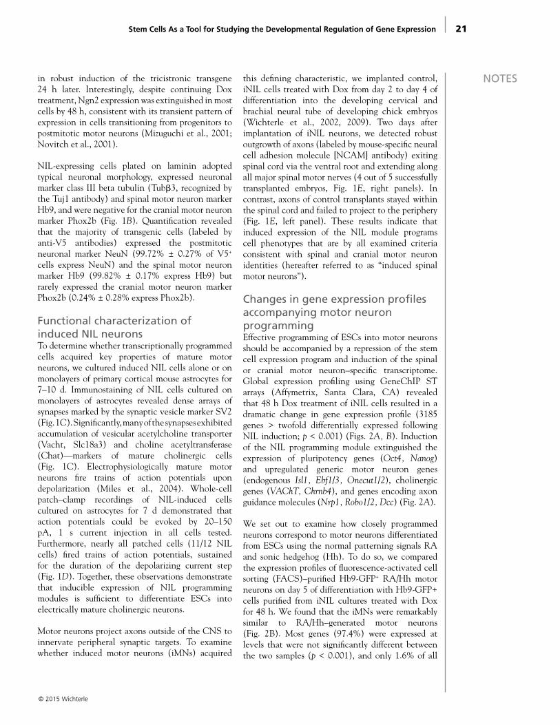
ResultsSpecification of cells expressing spinal motor markers upon inducible expression of Ngn2, Isl1, and Lhx3To study the programming of spinal and cranial motor neuron identity, we generated two Dox-inducible ESC lines (Mazzoni et al., 2011), one of which harbors a polycistronic expression construct in which the open reading frames of spinal motor neuron determinants Ngn2, Isl1, and Lhx3 (Lee and Pfaff, 2003; Hester et al., 2011; Lee et al., 2012) are separated by 2A peptides (the iNIL line) (Fig. 1A). NIL factors were previously shown to activate the specification of motor neuron identity in retinoic acid (RA)–treated differentiating ESCs (Hester et al., 2011; Lee et al., 2012). We established that NIL factors are sufficient to induce the expression of spinal motor neuron markers even in the absence of RA. Treating differentiating ESCs with Dox resulted
Stem Cells As a Tool for Studying the Developmental Regulation of Gene Expression
© 2015 Wichterle

20
NOTES
© 2015 Wichterle
Figure 1. Ngn2, Isl1, and Lhx3 (NIL) transcription factors program spinal motor neurons. A, Schematic representation of Dox-inducible NIL programming modules. TRE: tetracycline response element, F2A, T2A–2A peptide sequences from foot-and-mouth disease virus. B, In the absence of patterning signals, NIL-programmed spinal motor neuron exhibit neuronal morphology with multiple Tuj1 immunoreactive processes, express Hb9, but do not express the cranial marker Phox2b. Day 2 embryoid bodies treated with Dox for 48 h were dissociated, plated on laminin-coated substrate, and analyzed 24 h later. C, NIL-programmed cells contain cholinergic synaptic vesicles. Dissociated iNIL cells induced with Dox were cultured on astrocyte monolayers for 7 d and stained with the synaptic marker SV2 and the cholinergic markers Vacht and Chat. D, NIL-programmed neurons cultured for 7 d on astrocyte monolayers fire repetitive action potentials. Calibration: 20 mV, 250 ms. E, Control and Dox-induced day 4 embryoid bodies were implanted into the stage 16 developing chick cervical spinal cord in vivo. Embryos were fixed 2 d later, sectioned, and stained with a mouse-specific NCAM antibody. Dense bundles of axons emanating from NIL-induced transplants were observed within the ventral root and in axial (left arrow) and limb (right arrow) nerve branches (4 of 5 successfully transplanted embryos). Scale bars: B, 50 μm; C, 10 μm; E, 100 μm. Reprinted with permission from Mazzoni et al. (2013), their Figs. 1a, b, f, g, h.
21
NOTESin robust induction of the tricistronic transgene 24 h later. Interestingly, despite continuing Dox treatment, Ngn2 expression was extinguished in most cells by 48 h, consistent with its transient pattern of expression in cells transitioning from progenitors to postmitotic motor neurons (Mizuguchi et al., 2001; Novitch et al., 2001).
NIL-expressing cells plated on laminin adopted typical neuronal morphology, expressed neuronal marker class III beta tubulin (Tubβ3, recognized by the Tuj1 antibody) and spinal motor neuron marker Hb9, and were negative for the cranial motor neuron marker Phox2b (Fig. 1B). Quantification revealed that the majority of transgenic cells (labeled by anti-V5 antibodies) expressed the postmitotic neuronal marker NeuN (99.72% ± 0.27% of V5+
cells express NeuN) and the spinal motor neuron marker Hb9 (99.82% ± 0.17% express Hb9) but rarely expressed the cranial motor neuron marker Phox2b (0.24% ± 0.28% express Phox2b).
Functional characterization of induced NIL neuronsTo determine whether transcriptionally programmed cells acquired key properties of mature motor neurons, we cultured induced NIL cells alone or on monolayers of primary cortical mouse astrocytes for 7–10 d. Immunostaining of NIL cells cultured on monolayers of astrocytes revealed dense arrays of synapses marked by the synaptic vesicle marker SV2 (Fig. 1C). Significantly, many of the synapses exhibited accumulation of vesicular acetylcholine transporter (Vacht, Slc18a3) and choline acetyltransferase (Chat)—markers of mature cholinergic cells (Fig. 1C). Electrophysiologically mature motor neurons fire trains of action potentials upon depolarization (Miles et al., 2004). Whole-cell patch–clamp recordings of NIL-induced cells cultured on astrocytes for 7 d demonstrated that action potentials could be evoked by 20–150 pA, 1 s current injection in all cells tested. Furthermore, nearly all patched cells (11/12 NIL cells) fired trains of action potentials, sustained for the duration of the depolarizing current step (Fig. 1D). Together, these observations demonstrate that inducible expression of NIL programming modules is sufficient to differentiate ESCs into electrically mature cholinergic neurons.
Motor neurons project axons outside of the CNS to innervate peripheral synaptic targets. To examine whether induced motor neurons (iMNs) acquired
this defining characteristic, we implanted control, iNIL cells treated with Dox from day 2 to day 4 of differentiation into the developing cervical and brachial neural tube of developing chick embryos (Wichterle et al., 2002, 2009). Two days after implantation of iNIL neurons, we detected robust outgrowth of axons (labeled by mouse-specific neural cell adhesion molecule [NCAM] antibody) exiting spinal cord via the ventral root and extending along all major spinal motor nerves (4 out of 5 successfully transplanted embryos, Fig. 1E, right panels). In contrast, axons of control transplants stayed within the spinal cord and failed to project to the periphery (Fig. 1E, left panel). These results indicate that induced expression of the NIL module programs cell phenotypes that are by all examined criteria consistent with spinal and cranial motor neuron identities (hereafter referred to as “induced spinal motor neurons”).
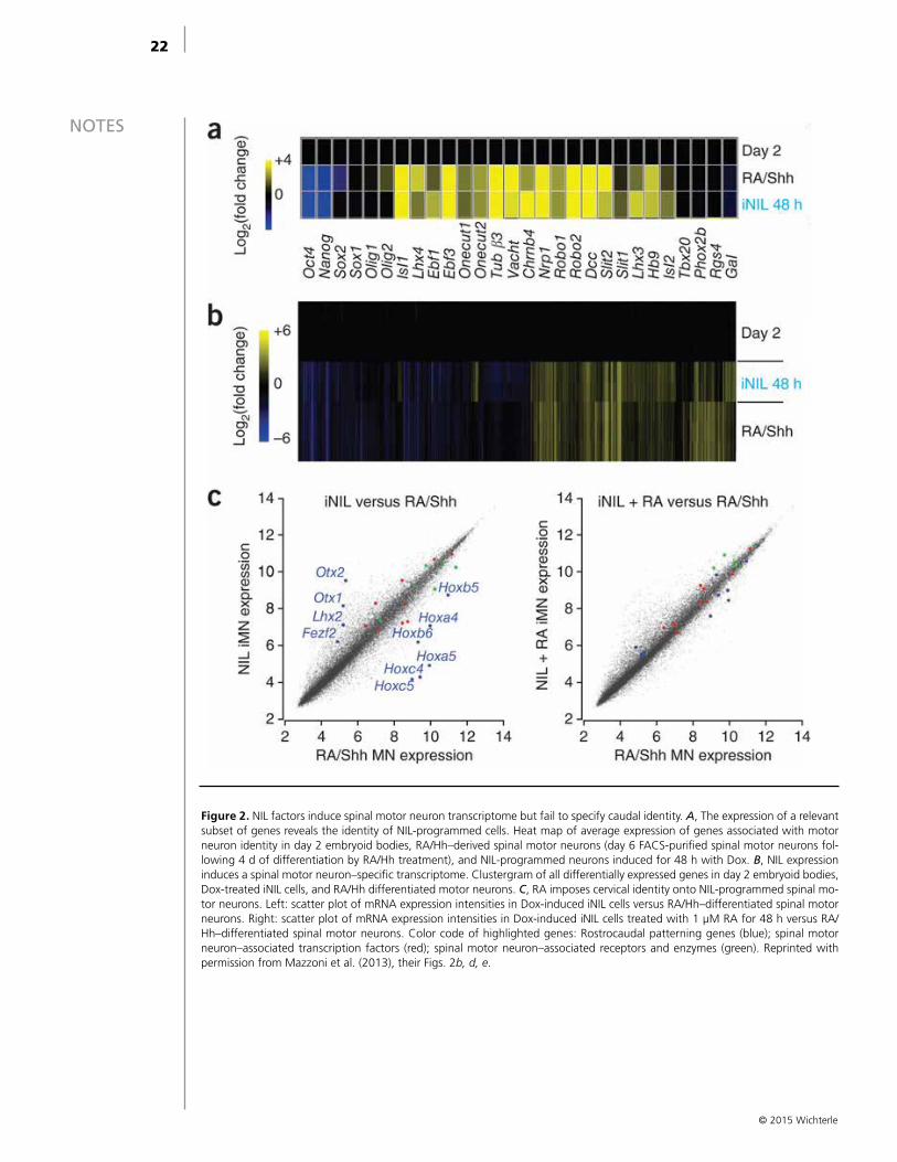
Changes in gene expression profiles accompanying motor neuron programmingEffective programming of ESCs into motor neurons should be accompanied by a repression of the stem cell expression program and induction of the spinal or cranial motor neuron–specific transcriptome. Global expression profiling using GeneChIP ST arrays (Affymetrix, Santa Clara, CA) revealed that 48 h Dox treatment of iNIL cells resulted in a dramatic change in gene expression profile (3185 genes > twofold differentially expressed following NIL induction; p < 0.001) (Figs. 2A, B). Induction of the NIL programming module extinguished the expression of pluripotency genes (Oct4, Nanog) and upregulated generic motor neuron genes (endogenous Isl1, Ebf1/3, Onecut1/2), cholinergic genes (VAChT, Chrnb4), and genes encoding axon guidance molecules (Nrp1, Robo1/2, Dcc) (Fig. 2A).
We set out to examine how closely programmed neurons correspond to motor neurons differentiated from ESCs using the normal patterning signals RA and sonic hedgehog (Hh). To do so, we compared the expression profiles of fluorescence-activated cell sorting (FACS)–purified Hb9-GFP+ RA/Hh motor neurons on day 5 of differentiation with Hb9-GFP+ cells purified from iNIL cultures treated with Dox for 48 h. We found that the iMNs were remarkably similar to RA/Hh–generated motor neurons (Fig. 2B). Most genes (97.4%) were expressed at levels that were not significantly different between the two samples (p < 0.001), and only 1.6% of all
Stem Cells As a Tool for Studying the Developmental Regulation of Gene Expression
© 2015 Wichterle
22
NOTES
© 2015 Wichterle
Figure 2. NIL factors induce spinal motor neuron transcriptome but fail to specify caudal identity. A, The expression of a relevant subset of genes reveals the identity of NIL-programmed cells. Heat map of average expression of genes associated with motor neuron identity in day 2 embryoid bodies, RA/Hh–derived spinal motor neurons (day 6 FACS-purified spinal motor neurons fol-lowing 4 d of differentiation by RA/Hh treatment), and NIL-programmed neurons induced for 48 h with Dox. B, NIL expression induces a spinal motor neuron–specific transcriptome. Clustergram of all differentially expressed genes in day 2 embryoid bodies, Dox-treated iNIL cells, and RA/Hh differentiated motor neurons. C, RA imposes cervical identity onto NIL-programmed spinal mo-tor neurons. Left: scatter plot of mRNA expression intensities in Dox-induced iNIL cells versus RA/Hh–differentiated spinal motor neurons. Right: scatter plot of mRNA expression intensities in Dox-induced iNIL cells treated with 1 μM RA for 48 h versus RA/Hh–differentiated spinal motor neurons. Color code of highlighted genes: Rostrocaudal patterning genes (blue); spinal motor neuron–associated transcription factors (red); spinal motor neuron–associated receptors and enzymes (green). Reprinted with permission from Mazzoni et al. (2013), their Figs. 2b, d, e.

23
NOTES
genes exhibited divergent expression (i.e., they were induced in one cell type but repressed in the other). While key motor neuron–specific genes were correctly regulated, a set of genes controlling rostrocaudal neural identity and motor neuron subtype identity was differentially expressed in RA/Hh and induced iNIL cells (Fig. 2C). Induced iNIL motor neurons expressed low levels of Hox transcription factors and high levels of rostral neural markers (Otx1, Otx2). To rectify this difference, we asked whether programmed iNIL motor neurons would be responsive to the caudalizing RA signal (Wichterle et al., 2002; Mahony et al., 2011). Treatment of iNIL cells with RA during Dox treatment resulted in correct specification of cervical spinal identity, marked by the expression of Hox genes from paralogous groups 4 and 5 and suppression of rostral markers Otx1/2 (Fig. 2C). Thus, although programmed cells acquire generic motor neuron identity following induction of NIL factors, the specification of rostrocaudal subtype identity depends on the treatment of the cells with caudalizing patterning signals.
Isl binds to a large number of genomic regionsEfficient and rapid transcriptional programming of ESCs into cells exhibiting fundamental motor
neuron properties provides an ideal system in which to study whether individual transcription factors act independently or engage in synergistic interactions. We performed chromatin immunoprecipitation–sequencing (ChIP-seq) analyses of Isl1 in iNIL cells 48 h after Dox induction. Inducible Isl1 factor was not epitope-tagged, and therefore, we optimized ChIP using a pool of monoclonal antibodies raised against Isl1. Because these antibodies cross-react with both Isl1 and the closely related Isl2 transcription factor, we refer to the data as Isl ChIP-seq. We observed extensive Isl recruitment to genomic loci in the iNIL-induced cells (Fig. 3A). We identified approximately 22,000 significant Isl binding events characterized by the presence of a canonical homeodomain binding motif (Fig. 3B) at the majority of binding sites.
Next, we examined whether identified Isl binding sites are distributed randomly across the genome or whether their position correlates with tissue-specific cis-regulatory elements. We took advantage of project data from ENCODE (Encyclopedia of DNA Elements) that identified putative regulatory regions in mouse ESCs, whole brain, heart, kidney, liver, and spleen, defined using combinations of DNaseI hypersensitivity and enrichment in H3K4me1 and H3K27ac histone modifications. Of all tissues examined, Isl binding sites correlated best with
© 2015 Wichterle
Stem Cells As a Tool for Studying the Developmental Regulation of Gene Expression
Figure 3. Isl1 and Lhx3 bind to many common sites harboring a complex homeodomain motif. A, Isl ChIP-seq signals over Lhx3, Chat, and Phox2b. Blue peaks represent significant (p < 0.01) read enrichment over control. Genomic loci coordinates are shown next to the x-axis. B, Primary DNA motifs overrepresented under enriched peaks obtained from Isl ChIP-seq experiments in iNIL cells treated for 48 h with Dox. C, Lhx3 colocalizes with Isl genomic binding sites in iNIL cells. Comparison of read enrichment from Isl with Lhx3 at all detected peaks. Blue represents peaks significantly differentially enriched for Isl or Lhx3 binding. Adapted with permission from Mazzoni et al. (2013), their Figs. 4a, 5b, c.
24
NOTES whole-brain putative regulatory regions. Interestingly, the overlap with regulatory regions in ESCs was as low as in unrelated tissues. These findings indicate that expressed NIL factors are not passively recruited to existing stem cell regulatory regions, but rather, actively engage neuronal regulatory regions.
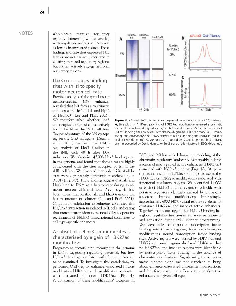
Lhx3 co-occupies binding sites with Isl to specify motor neuron cell fatePrevious analysis of the spinal motor neuron–specific Hb9 enhancer revealed that Isl1 forms a multimeric complex with Lhx3, Ldb1, and Ngn2 or Neurod4 (Lee and Pfaff, 2003). We therefore asked whether Lhx3 co-occupies other sites selectively bound by Isl in the iNIL cell line. Taking advantage of the V5 epitope tag on the Lhx3 transgene (Mazzoni et al., 2011), we performed ChIP-seq analysis of Lhx3 binding in the iNIL cells 48 h after Dox induction. We identified 47,908 Lhx3 binding sites in the genome and found that these sites are highly coincidental with the sites occupied by Isl in the iNIL cell line. We observed that only 1.7% of all Isl sites were significantly differentially enriched (p < 0.001) (Fig. 3C). These findings suggest that Isl1 and Lhx3 bind to DNA as a heterodimer during spinal motor neuron differentiation. Previously, it had been shown that purified Isl1 and Lhx3 transcription factors interact in solution (Lee and Pfaff, 2003). Coimmunoprecipitation experiments confirmed this Isl1/Lhx3 interaction in induced iNIL cells, indicating that motor neuron identity is encoded by cooperative recruitment of Isl/Lhx3 transcriptional complexes to cell type–specific enhancers.
A subset of Isl/Lhx3–cobound sites is characterized by a gain of H3K27ac modificationProgramming factors bind throughout the genome in iMNs, suggesting regulatory potential, but how Isl/Lhx3 binding correlates with function has yet to be examined. To investigate this correlation, we performed ChIP-seq for enhancer-associated histone modification H3K4me1 and a modification associated with activated enhancers H3K27ac (Fig. 4). A comparison of these modifications’ locations in
ESCs and iMNs revealed dramatic remodeling of the chromatin regulatory landscape. Remarkably, a large fraction of newly gained active enhancers (H3K27ac) coincided with Isl/Lhx3 binding (Figs. 4A, B), yet a significant fraction of Isl/Lhx3 binding sites lacked the H3K4me1 or H3K27ac modifications associated with functional regulatory regions. We identified 14,000 or 63% of Isl/Lhx3 binding events to coincide with putative regulatory elements marked by enhancer-associated histone modifications. Interestingly, approximately 6000 (40%) distal regulatory elements contained H3K27ac, the mark of active enhancers. Together, these data suggest that Isl/Lhx3 binding has a global regulatory function in enhancer recruitment and activation during iMN identity programming. We were able to annotate transcription factor binding into three categories, based on chromatin modifications around transcription factor binding sites. Active regions were marked by H3K4me1 and H3K27ac, primed regions displayed H3K4me1 but no H3K27ac, and inactive regions were identifiable by transcription factor binding in the absence of chromatin modifications. Significantly, transcription factor binding alone was not sufficient to bring about enhancer-associated chromatin modifications, and therefore, it was not sufficient to identify active enhancers in a given cell type.
© 2015 Wichterle
Figure 4. Isl1 and Lhx3 binding is accompanied by acetylation of H3K27 histone. A, Line plots of ChIP-seq profiling of H3K27ac modification revealed a dramatic shift in these activated regulatory regions between ESCs and iMNs. The majority of Isl/Lhx3 binding sites coincides with the newly gained H3K27ac mark. B, Cumula-tive quantitative analysis of H3K27ac level at Isl/Lhx3 binding sites in iMNs (red line) and in ESCs (blue line). C, Genomic sites bound by Isl and Lhx3 (red line) in iMNs are not occupied by Oct4, Nanog, or Sox2 transcription factors in ESCs (blue line).
25
NOTES
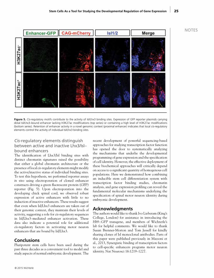
Cis-regulatory elements distinguish between active and inactive Lhx3/Isl–bound enhancersThe identification of Lhx3/Isl binding sites with distinct chromatin signatures raised the possibility that either a global chromatin architecture or the presence of local cis-regulatory elements might modify the active/inactive status of individual binding sites. To test this hypothesis, we performed reporter assays in vivo using electroporation of cloned enhancer constructs driving a green fluorescent protein (GFP) reporter (Fig. 5). Upon electroporation into the developing chick spinal cord, we observed robust expression of active enhancers with little to no induction of inactive enhancers. These results suggest that even when Isl/Lhx3 enhancers are taken out of their genomic context, they maintain their levels of activity, suggesting a role for cis-regulatory sequences in Isl/Lhx3–mediated enhancer activation. These data also indicate a potential role for additional cis-regulatory factors in activating motor neuron enhancers that are bound by Isl/Lhx3.
ConclusionsPluripotent stem cells have been used during the past three decades as a convenient tool to model and study aspects of normal embryonic development. The
recent development of powerful sequencing-based approaches for studying transcription factor function has opened the door to systematically analyzing the mechanisms that underlie the developmental programming of gene expression and the specification of cell identity. However, the effective deployment of these biochemical approaches will critically depend on access to a significant quantity of homogenous cell populations. Here we demonstrated how combining an inducible stem cell differentiation system with transcription factor binding studies, chromatin analysis, and gene expression profiling can reveal the fundamental molecular mechanisms underlying the specification of spinal motor neuron identity during embryonic development.
AcknowledgmentsThe authors would like to thank Ivo Lieberam (King’s College, London) for assistance in introducing the Hb9::GFP transgene, and members of Wichterle’s lab for helpful comments. We would like to thank Susan Brenner-Morton and Tom Jessell for kindly sharing clones of Isl monoclonal antibodies. Parts of this paper were published previously in Mazzoni et al., 2013, Synergistic binding of transcription factors to cell-specific enhancers programs motor neuron identity. Nat Neurosci 16:1219–1227.
© 2015 Wichterle
Stem Cells As a Tool for Studying the Developmental Regulation of Gene Expression
Figure 5. Cis-regulatory motifs contribute to the activity of Isl/Lhx3 binding sites. Expression of GFP reporter plasmids carrying distal Isl/Lhx3–bound enhancer lacking H3K27ac modifications (top series) or containing a high level of H3K27ac modifications (bottom series). Retention of enhancer activity in a novel genomic context (proximal enhancer) indicates that local cis-regulatory elements control the activity of individual Isl/Lhx3 binding sites.
26
NOTES ReferencesHester ME, Murtha MJ, Song S, Rao M, Miranda CJ,
Meyer K, Tian J, Boulting G, Schaffer DV, Zhu MX, Pfaff SL, Gage FH, Kaspar BK (2011) Rapid and efficient generation of functional motor neurons from human pluripotent stem cells using gene delivered transcription factor codes. Mol Ther 19:1905–1912.
Iacovino M, Bosnakovski D, Fey H, Rux D, Bajwa G, Mahen E, Mitanoska A, Xu Z, Kyba M (2011) Inducible cassette exchange: a rapid and efficient system enabling conditional gene expression in embryonic stem and primary cells. Stem Cells 29:1580–1588.
Jessell TM (2000) Neuronal specification in the spinal cord: inductive signals and transcriptional codes. Nat Rev Genet 1:20–29.
Lee SK, Pfaff SL (2003) Synchronization of neurogenesis and motor neuron specification by direct coupling of bHLH and homeodomain transcription factors. Neuron 38:731–745.
Lee S, Cuvillier JM, Lee B, Shen R, Lee JW, Lee SK (2012) Fusion protein Isl1-Lhx3 specifies motor neuron fate by inducing motor neuron genes and concomitantly suppressing the interneuron programs. Proc Natl Acad Sci USA 109:3383–3388.
Mahony S, Mazzoni EO, McCuine S, Young RA, Wichterle H, Gifford DK (2011) Ligand-dependent dynamics of retinoic acid receptor binding during early neurogenesis. Genome Biol 12:R2.
Mann RS, Carroll SB (2002) Molecular mechanisms of selector gene function and evolution. Curr Opin Genet Dev 12:592–600.
Mazzoni EO, Mahony S, Iacovino M, Morrison CA, Mountoufaris G, Closser M, Whyte WA, Young RA, Kyba M, Gifford DK, Wichterle H (2011) Embryonic stem cell–based mapping of developmental transcriptional programs. Nat Methods 8:1056–1058.
Mazzoni EO, Mahony S, Closser M, Morrison CA, Nedelec S, Williams DJ, An D, Gifford DK, Wichterle H (2013) Synergistic binding of transcription factors to cell-specific enhancers programs motor neuron identity. Nat Neurosci 16:1219–1227.
Miles GB, Yohn DC, Wichterle H, Jessell TM, Rafuse VF, Brownstone RM (2004) Functional properties of motoneurons derived from mouse embryonic stem cells. J Neurosci 24:7848–7858.
Mizuguchi R, Sugimori M, Takebayashi H, Kosako H, Nagao M, Yoshida S, Nabeshima Y, Shimamura K, Nakafuku M (2001) Combinatorial roles of olig2 and neurogenin2 in the coordinated induction of pan-neuronal and subtype-specific properties of motoneurons. Neuron 31:757–771.
Novitch BG, Chen AI, Jessell TM (2001) Coordinate regulation of motor neuron subtype identity and pan-neuronal properties by the bHLH repressor Olig2. Neuron 31:773–789.
Son EY, Ichida JK, Wainger BJ, Toma JS, Rafuse VF, Woolf CJ, Eggan K (2011) Conversion of mouse and human fibroblasts into functional spinal motor neurons. Cell Stem Cell 9:205–218.
Takahashi K, Yamanaka S (2006) Induction of pluripotent stem cells from mouse embryonic and adult fibroblast cultures by defined factors. Cell 126:663–676.
Tapscott SJ, Davis RL, Thayer MJ, Cheng PF, Weintraub H, Lassar AB (1988) MyoD1: a nuclear phosphoprotein requiring a Myc homology region to convert fibroblasts to myoblasts. Science 242:405–411.
Wichterle H, Lieberam I, Porter JA, Jessell TM (2002) Directed differentiation of embryonic stem cells into motor neurons. Cell 110:385–397.
Wichterle H, Peljto M, Nedelec S (2009) Xenotransplantation of embryonic stem cell-derived motor neurons into the developing chick spinal cord. Methods Mol Biol 482:171–183.
Zhou B, Zhong Q, Minoo P, Li C, Ann DK, Frenkel B, Morrisey EE, Crandall ED, Borok Z (2008) Foxp2 inhibits Nkx2.1-mediated transcription of SP-C via interactions with the Nkx2.1 homeodomain. Am J Respir Cell Mol Biol 38:750–758.
© 2015 Wichterle

© 2015 Pasca
Department of Psychiatry and Behavioral Sciences Neurosciences Institute and Institute for Stem Cell Biology
Stanford ChEM-H Institute Stanford University
Stanford, California
Generating a Functional Human Cortex In Vitro From
Induced Pluripotent Stem CellsSergiu P. Pasca, MD

29
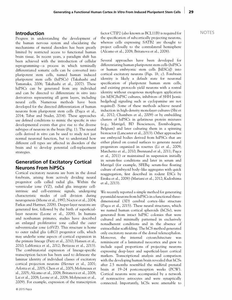
NOTESIntroductionProgress in understanding the development of the human nervous system and elucidating the mechanisms of mental disorders has been greatly limited by restricted access to functional human brain tissue. In recent years, a paradigm shift has been achieved with the introduction of cellular reprogramming—a process in which terminally differentiated somatic cells can be converted into pluripotent stem cells, named human induced pluripotent stem cells (hiPSCs) (Takahashi and Yamanaka, 2006; Takahashi et al., 2007). These hiPSCs can be generated from any individual and can be directed to differentiate in vitro into derivatives representing all germ layers, including neural cells. Numerous methods have been developed for the directed differentiation of human neurons from pluripotent stem cells (Pasca et al., 2014; Tabar and Studer, 2014). These approaches use defined conditions to mimic the specific in vivo developmental events that give rise to the diverse subtypes of neurons in the brain (Fig. 1). The neural cells derived in vitro can be used to study not just normal neuronal function, but to understand how different cell types are affected in disorders of the brain and to develop potential cell-replacement strategies.
Generation of Excitatory Cortical Neurons From hiPSCsCortical excitatory neurons are born in the dorsal forebrain, arising from actively dividing neural progenitor cells called radial glia. Within the ventricular zone (VZ), radial glia integrate cell-intrinsic and cell-extrinsic signals, undergoing characteristic modes of cell division during neurogenesis (Mione et al., 1997; Noctor et al., 2004; Farkas and Huttner, 2008). Deeper-layer neurons are generated first, followed by the birth of superficial-layer neurons (Leone et al., 2008). In humans and nonhuman primates, studies have described an enlarged proliferative zone called the outer subventricular zone (oSVZ). This structure is home to outer radial glia (oRG) progenitor cells, which may underlie some aspects of cortical expansion in the primate lineage (Fietz et al., 2010; Hansen et al., 2010; LaMonica et al., 2012; Betizeau et al., 2013). The combinatorial expression of lineage-specific transcription factors has been used to delineate the laminar identity of individual classes of excitatory cortical projection neurons (Hevner et al., 2001; Arlotta et al., 2005; Chen et al., 2005; Molyneaux et al., 2005; Alcamo et al., 2008; Britanova et al., 2008; Lai et al., 2008; Leone et al., 2008; Molyneaux et al., 2009). For example, expression of the transcription
factor CTIP2 (also known as BCL11B) is required for the specification of subcortically projecting neurons, whereas cells expressing SATB2 are thought to project callosally to the contralateral hemisphere (Alcamo et al., 2008; Britanova et al., 2008).
Several approaches have been developed for differentiating human pluripotent stem cells (hiPSCs or human embryonic stem cells [hESCs]) into cortical excitatory neurons (Figs. 1b, c). Forebrain identity is likely a default state for neuronal specification of pluripotent human stem cells, and existing protocols yield neurons with a rostral identity without exogenous morphogen application (in hESC/hiPSC cultures, inhibitors of SHH [sonic hedgehog] signaling such as cyclopamine are not required). Some of these methods achieve neural induction in high-density monolayer cultures (Shi et al., 2012; Chambers et al., 2009) or by embedding clusters of hiPSCs in gelatinous protein mixtures (e.g., Matrigel, BD Biosciences, Erembodegem, Belgium) and later culturing them in a spinning bioreactor (Lancaster et al., 2013). Other approaches use embryoid bodies derived from hiPSCs that are either plated on coated surfaces to generate neural progenitors organized in rosettes (Li et al., 2009; Marchetto et al., 2010; Brennand et al., 2011; Pasca et al., 2011) or maintained in suspension initially in serum-free conditions and later in serum and Matrigel (for example, SFEBq: serum-free floating culture of embryoid body–like aggregates with quick reaggregation, first described in rodent ESCs by Eiraku et al., 2008) (Mariani et al., 2012; Kadoshima et al., 2013).
We recently reported a simple method for generating pyramidal neurons from hiPSCs in a functional three-dimensional (3D) cerebral cortex–like structure (Pasca et al., 2015). These neural structures, which we named human cortical spheroids (hCSs), were generated from intact hiPSC colonies that were cultured and minimally patterned in exclusively nonadherent conditions and in the absence of extracellular scaffolding. The hCS method generated only excitatory neurons of the dorsal telencephalon. Moreover, the internal cytoarchitecture was reminiscent of a laminated neocortex and grew to include equal proportions of projecting neurons expressing deep-layer and superficial-layer cortical markers. Transcriptional analysis and comparison with the developing human brain revealed that hCSs after 2.5 months resembled the midfetal prenatal brain at 19–24 postconception weeks (PCW). Cortical neurons were accompanied by a network of nonreactive astrocytes and were synaptically connected. Importantly, hCSs were amenable to
© 2015 Pasca
Generating a Functional Human Cortex In Vitro From Induced Pluripotent Stem Cells

30
NOTES
© 2015 Pasca
Figure 1. Approaches for the generation of excitatory cortical projection neurons. a, The developmental events underlying the generation of cortical excitatory neurons in vivo are recapitulated in vitro using (b) adherent cultures or (c) suspended aggregate methods. Reprinted with permission from Pasca et al., 2014, their Fig. 2.
31
NOTESacute-slice physiology, making it possible to record and electrically stimulate neurons while preserving a relatively intact network. Lastly, this method is scalable and reproducible between hiPSC lines, across and within differentiations. hCSs have the potential to reveal cellular phenotypes associated with neuropsychiatric disorders, identify biomarkers for early diagnosis and clinical stratification, and provide a platform for drug and teratologic agent screenings in vitro.
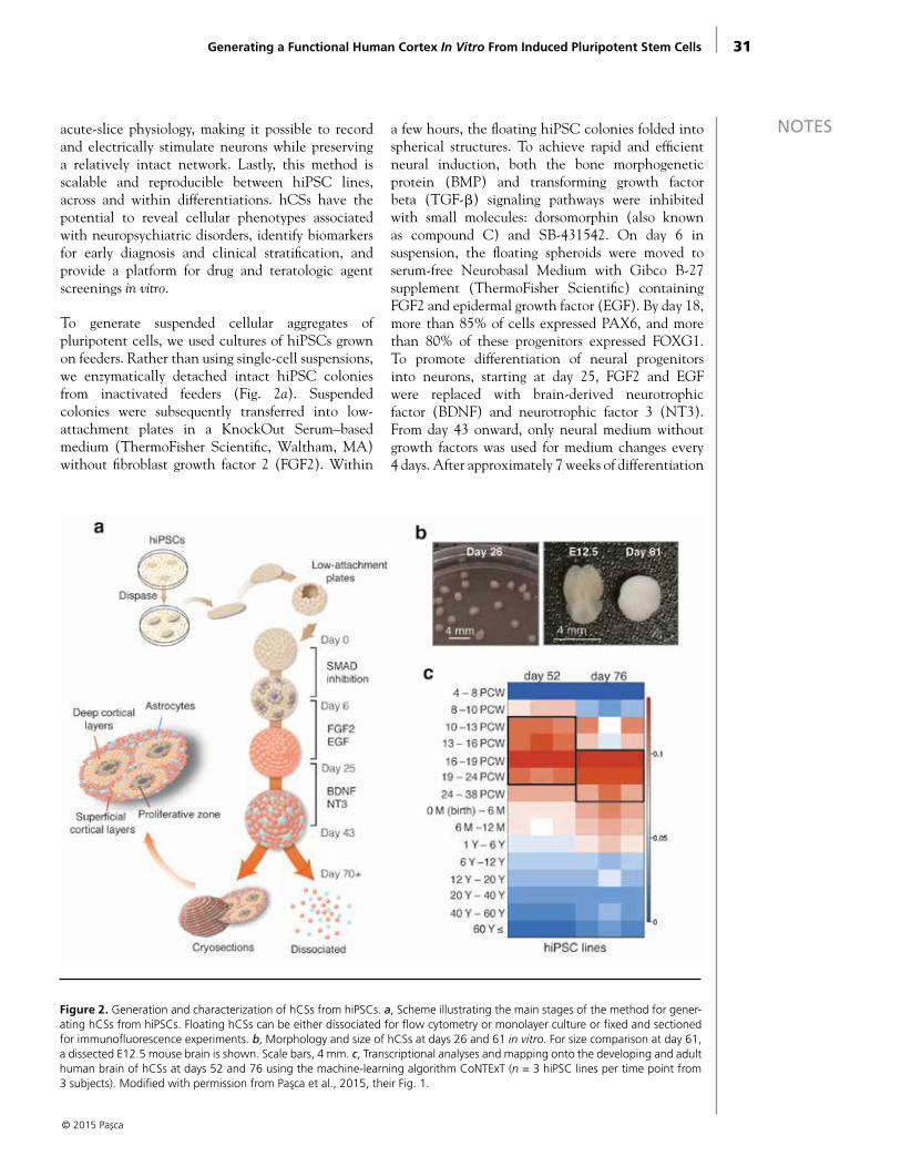
To generate suspended cellular aggregates of pluripotent cells, we used cultures of hiPSCs grown on feeders. Rather than using single-cell suspensions, we enzymatically detached intact hiPSC colonies from inactivated feeders (Fig. 2a). Suspended colonies were subsequently transferred into low-attachment plates in a KnockOut Serum–based medium (ThermoFisher Scientific, Waltham, MA) without fibroblast growth factor 2 (FGF2). Within
a few hours, the floating hiPSC colonies folded into spherical structures. To achieve rapid and efficient neural induction, both the bone morphogenetic protein (BMP) and transforming growth factor beta (TGF-β) signaling pathways were inhibited with small molecules: dorsomorphin (also known as compound C) and SB-431542. On day 6 in suspension, the floating spheroids were moved to serum-free Neurobasal Medium with Gibco B-27
supplement (ThermoFisher Scientific) containing FGF2 and epidermal growth factor (EGF). By day 18, more than 85% of cells expressed PAX6, and more than 80% of these progenitors expressed FOXG1. To promote differentiation of neural progenitors into neurons, starting at day 25, FGF2 and EGF were replaced with brain-derived neurotrophic factor (BDNF) and neurotrophic factor 3 (NT3). From day 43 onward, only neural medium without growth factors was used for medium changes every 4 days. After approximately 7 weeks of differentiation
© 2015 Pasca
Generating a Functional Human Cortex In Vitro From Induced Pluripotent Stem Cells
Figure 2. Generation and characterization of hCSs from hiPSCs. a, Scheme illustrating the main stages of the method for gener-ating hCSs from hiPSCs. Floating hCSs can be either dissociated for flow cytometry or monolayer culture or fixed and sectioned for immunofluorescence experiments. b, Morphology and size of hCSs at days 26 and 61 in vitro. For size comparison at day 61, a dissected E12.5 mouse brain is shown. Scale bars, 4 mm. c, Transcriptional analyses and mapping onto the developing and adult human brain of hCSs at days 52 and 76 using the machine-learning algorithm CoNTExT (n = 3 hiPSC lines per time point from 3 subjects). Modified with permission from Pasca et al., 2015, their Fig. 1.
32
NOTES in vitro, 78.8% ± 2.5% of the cells expressed the neuronal marker β3-tubulin (TUBB3), and 36.2% ± 3.6% of neurons expressed the mature neuronal marker NeuN (neuronal nuclear antigen), which is present in the human forebrain only after 20 weeks of gestation (Sarnat et al., 1998). The hCSs grew in size up to 4 mm in diameter by 2.5 months (4.2 ± 0.3 mm) (Fig. 2b).
We used transcriptional profiling to assess developmental maturity and observed a strong overlap between hCSs and cortical developmental stages up to late midfetal periods (19–24 PCW) (Fig. 2c). This was in contrast to monolayer methods as well as other 3D approaches for neural differentiation of hiPSCs (Mariani et al., 2012; Stein, 2014) that yield neurons mapping onto earlier fetal stages. When we looked for genes whose expression was changing in the same direction in hCSs and human fetal cortex between stages 1 and 2 (4–10 PCW) and stage 6 (19–24 PCW), but not in hiPSC-derived neural cultures differentiated in monolayer, we found that upregulated genes were enriched for synaptic transmission genes, whereas the downregulated genes were enriched for cell-cycle and cell-division genes.
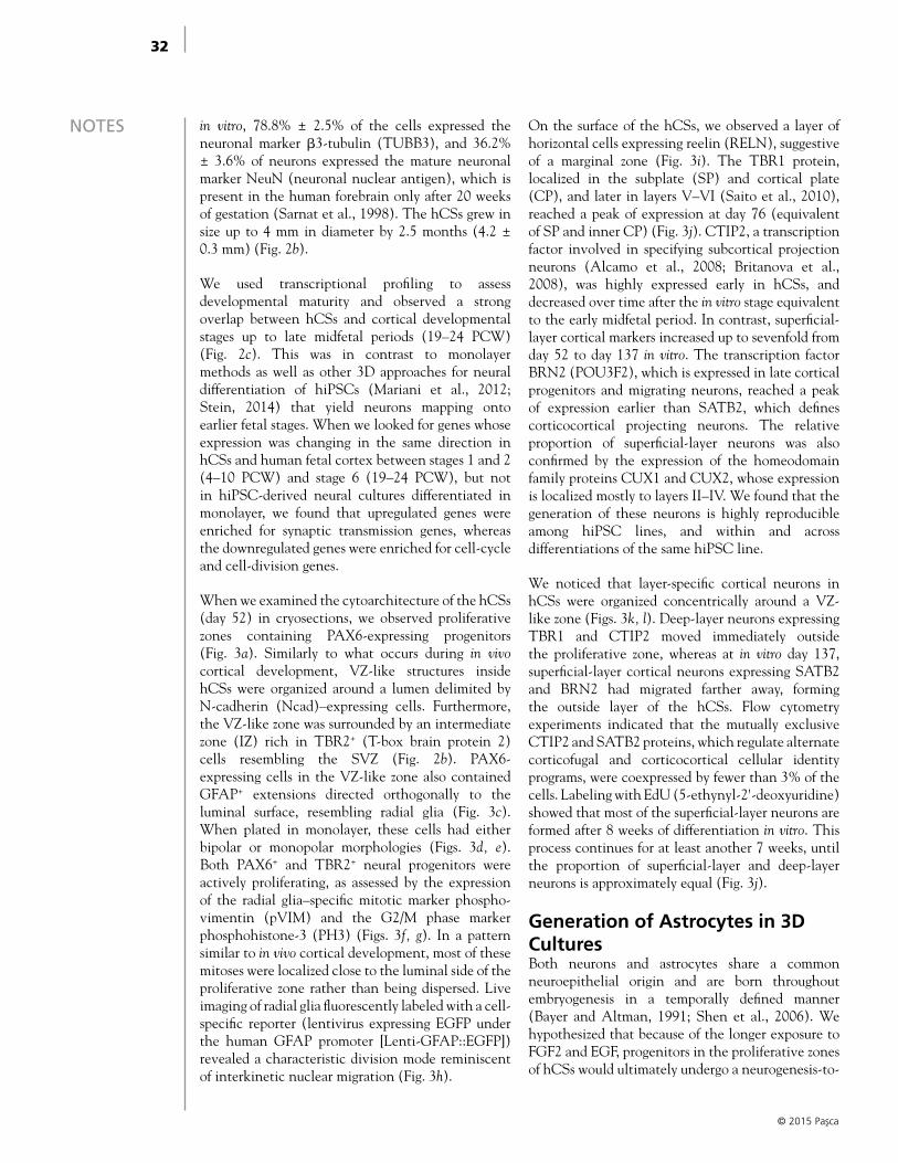
When we examined the cytoarchitecture of the hCSs (day 52) in cryosections, we observed proliferative zones containing PAX6-expressing progenitors (Fig. 3a). Similarly to what occurs during in vivo cortical development, VZ-like structures inside hCSs were organized around a lumen delimited by N-cadherin (Ncad)–expressing cells. Furthermore, the VZ-like zone was surrounded by an intermediate zone (IZ) rich in TBR2+ (T-box brain protein 2) cells resembling the SVZ (Fig. 2b). PAX6-expressing cells in the VZ-like zone also contained GFAP+ extensions directed orthogonally to the luminal surface, resembling radial glia (Fig. 3c). When plated in monolayer, these cells had either bipolar or monopolar morphologies (Figs. 3d, e). Both PAX6+ and TBR2+ neural progenitors were actively proliferating, as assessed by the expression of the radial glia–specific mitotic marker phospho-vimentin (pVIM) and the G2/M phase marker phosphohistone-3 (PH3) (Figs. 3f, g). In a pattern similar to in vivo cortical development, most of these mitoses were localized close to the luminal side of the proliferative zone rather than being dispersed. Live imaging of radial glia fluorescently labeled with a cell-specific reporter (lentivirus expressing EGFP under the human GFAP promoter [Lenti-GFAP::EGFP]) revealed a characteristic division mode reminiscent of interkinetic nuclear migration (Fig. 3h).
On the surface of the hCSs, we observed a layer of horizontal cells expressing reelin (RELN), suggestive of a marginal zone (Fig. 3i). The TBR1 protein, localized in the subplate (SP) and cortical plate (CP), and later in layers V–VI (Saito et al., 2010), reached a peak of expression at day 76 (equivalent of SP and inner CP) (Fig. 3j). CTIP2, a transcription factor involved in specifying subcortical projection neurons (Alcamo et al., 2008; Britanova et al., 2008), was highly expressed early in hCSs, and decreased over time after the in vitro stage equivalent to the early midfetal period. In contrast, superficial-layer cortical markers increased up to sevenfold from day 52 to day 137 in vitro. The transcription factor BRN2 (POU3F2), which is expressed in late cortical progenitors and migrating neurons, reached a peak of expression earlier than SATB2, which defines corticocortical projecting neurons. The relative proportion of superficial-layer neurons was also confirmed by the expression of the homeodomain family proteins CUX1 and CUX2, whose expression is localized mostly to layers II–IV. We found that the generation of these neurons is highly reproducible among hiPSC lines, and within and across differentiations of the same hiPSC line.
We noticed that layer-specific cortical neurons in hCSs were organized concentrically around a VZ-like zone (Figs. 3k, l). Deep-layer neurons expressing TBR1 and CTIP2 moved immediately outside the proliferative zone, whereas at in vitro day 137, superficial-layer cortical neurons expressing SATB2 and BRN2 had migrated farther away, forming the outside layer of the hCSs. Flow cytometry experiments indicated that the mutually exclusive CTIP2 and SATB2 proteins, which regulate alternate corticofugal and corticocortical cellular identity programs, were coexpressed by fewer than 3% of the cells. Labeling with EdU (5-ethynyl-2'-deoxyuridine) showed that most of the superficial-layer neurons are formed after 8 weeks of differentiation in vitro. This process continues for at least another 7 weeks, until the proportion of superficial-layer and deep-layer neurons is approximately equal (Fig. 3j).
Generation of Astrocytes in 3D CulturesBoth neurons and astrocytes share a common neuroepithelial origin and are born throughout embryogenesis in a temporally defined manner (Bayer and Altman, 1991; Shen et al., 2006). We hypothesized that because of the longer exposure to FGF2 and EGF, progenitors in the proliferative zones of hCSs would ultimately undergo a neurogenesis-to-
© 2015 Pasca

33
NOTES
gliogenesis switch. After 7 weeks of differentiation in vitro, we noticed astrocytes with thin GFAP+ processes intermingled with neurons cells in the hCS parenchyma (Fig. 4a). As expected, we observed few GFAP+ cells during the first 35 days of differentiation (Figs. 4b, c), but this proportion increased to approximately 8% by day 76 and almost 20% after 180 days (Fig. 4b). We also closely examined the morphology of GFAP+ cells after dissociation. When maintained in monolayer in defined serum-free culture conditions (Foo et al., 2011), astrocytes extended abundant thin projections (Fig. 4d). To investigate whether these cells could respond to
reactive cues in vitro, we added serum, which is a potent activator of reactive astrogliosis, to the culture medium. Within several days, the cells adopted a reactive phenotype with polygonal morphologies and upregulated expression of genes associated with in vivo astrogliosis, including GFAP, VIM, and LCN2 (lipocalin 2). Finally, using electron microscopy, we confirmed that the thin GFAP+ processes dispersed throughout the hCSs contained numerous glycogen granules, which are localized predominantly in astrocytes in the mammalian brain (Brown and Ransom, 2007).
© 2015 Pasca
Generating a Functional Human Cortex In Vitro From Induced Pluripotent Stem Cells
Figure 3. Corticogenesis in the hCS. a, Cryosection of an hCS at day 52 stained for PAX6 (progenitors) and NEUN (neurons), demonstrating the presence of a VZ-like region organized around a lumen. b, Intermediate progenitor cells (TBR2+) are present in a SVZ-like region beyond the VZ; Ncad stains the luminal side of the progenitors. c–f, Radial glial cells expressing combina-tions of GFAP, PAX6, or TBR2 and pVIM are present in proliferative zones, extend processes perpendicular to the lumen (L) and, when plated in monolayer, have either one or two processes. White arrowheads indicate the cell body and yellow arrowheads the processes. g, Mitoses (PH3+) are spatially restricted to the luminal side of the proliferative zones. h, Live imaging showing interkinetic nuclear migration (Lenti-GFAP::EGFP). i, RELN+ neurons are positioned horizontally on the surface of hCSs. j, Quanti-fication in cryosections of the proportion of cells expressing layer-specific cortical markers at three time points of differentiation (mean ± SEM; n = 3–13 hCSs [numbers listed within each bar] from 4 hiPSC lines derived from 4 individuals; 2-way ANOVA, F2,48 = 32.96, p < 0.0001 for time point [day 52 vs day 76 vs day 137]; Tukey’s multiple-comparison tests: *p < 0.05, **p < 0.01, ****p < 0.0001). k, l, Cryosections of hCSs at 137 d stained for the indicated markers, showing organization of layer-specific neurons. Scale bars: a, i, 100 μm; b, g, l, 50 μm; c–f, 10 μm; h, 20 μm; k, 200 μm. Modified with permission from Pasca et al., 2015, their Fig. 2.

34
NOTES
Synaptogenesis and Functional Assessment of 3D Cortical SpheroidsWe also tested the ability of the cells in hCSs to differentiate into electrically active mature neurons. We observed that neurons displayed abundant spontaneous calcium spikes and found that all neurons recorded by patch–clamping produced a transient inward current after depolarization beyond –30 mV, which was blocked by tetrodotoxin (TTX). This inward voltage-gated Na+ current was followed by activation of a more sustained K+ current. Importantly, depolarizing current injection revealed that all the recorded neurons reliably produced action potentials (Fig. 5a).
Considering the functional maturity of the cortical neurons and the presence of astrocytes, we investigated synaptogenesis within 180-day-old hCSs. To identify
individual synapses with a high degree of confidence, we used array tomography to visualize individual synapses (Micheva and Smith, 2007) within the dense neuropil of the hCSs and antibodies against multiple presynaptic and postsynaptic proteins. We found that the presynaptic protein synapsin-1 (SYN-1) and postsynaptic protein PSD-95 were expressed throughout the interior of the hCSs in both large and small puncta (Fig. 5b). Larger SYN-1 puncta were often found adjacent to PSD-95 puncta, indicating the presence of a synapse. To examine these larger puncta in further detail, we constructed synaptograms consisting of a series of high-resolution sections through a single synapse where we could probe for potential colocalization of at least three independent synaptic markers. In many cases, we observed colocalization of the presynaptic proteins SYN-1 and the glutamate transporter VGLUT-1 in close apposition to the postsynaptic protein PSD-95,
© 2015 Pasca
Figure 4. Astrogenesis in cortical hCSs. a, Volume rendering by array tomography of the interior of an hCS (74 × 88 × 2.45 μm) revealing the commingling of MAP2 (red) staining of neuronal dendrites and GFAP (cyan) staining of glial processes. DAPI stain-ing for nuclei is rendered in white. b–c, Developmental time course for the generation of GFAP+ cells. Quantification performed in dissociated hCSs (mean ± SEM; n = 3 for all time points except day 63, when n = 4; ANOVA F8,19 = 66.75, p < 0.0001). d, Astrocyte morphology after the indicated periods of in vitro culture in monolayer in defined, serum-free medium and after 1-week exposure to serum. Scale bars: a, 10 μm; c, 40 μm; d, 20 μm. Modified with permission from Pasca et al., 2015, their Fig. 3.
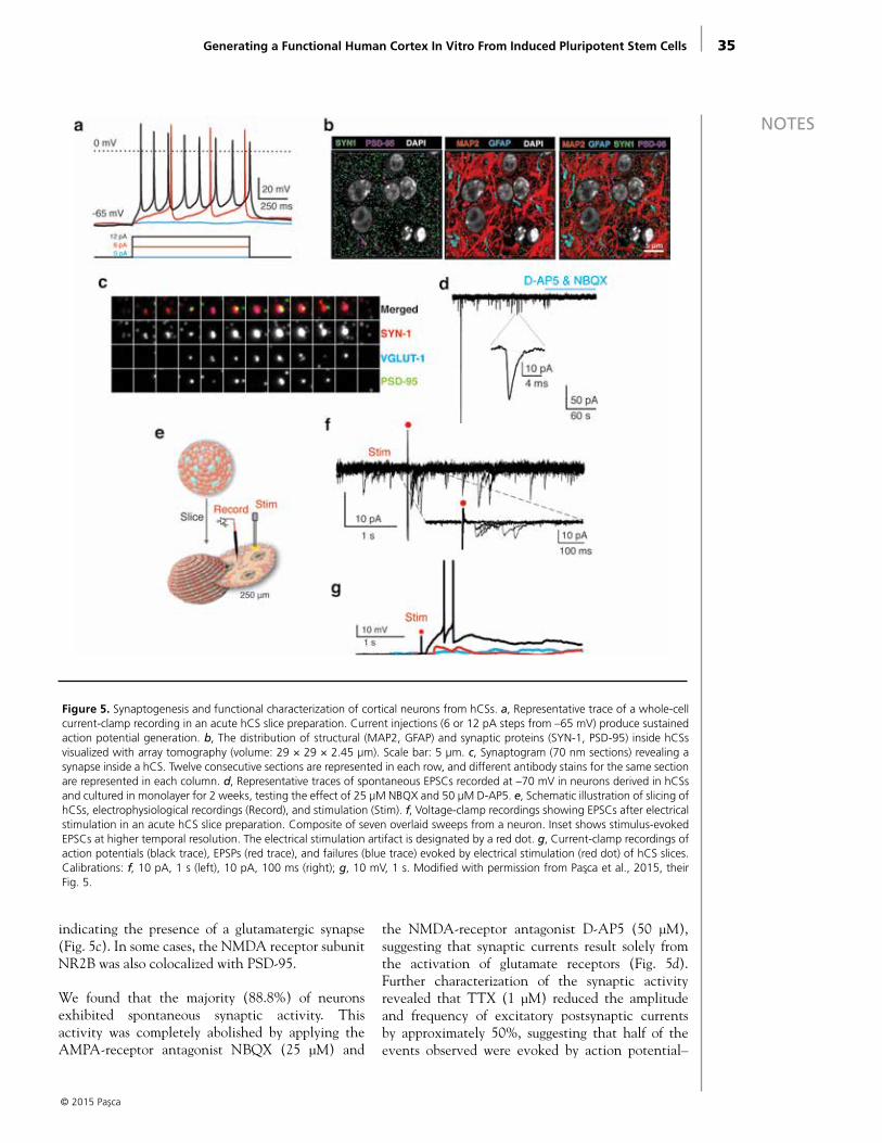
35
NOTES
Generating a Functional Human Cortex In Vitro From Induced Pluripotent Stem Cells
© 2015 Pasca
indicating the presence of a glutamatergic synapse (Fig. 5c). In some cases, the NMDA receptor subunit NR2B was also colocalized with PSD-95.
We found that the majority (88.8%) of neurons exhibited spontaneous synaptic activity. This activity was completely abolished by applying the AMPA-receptor antagonist NBQX (25 μM) and
the NMDA-receptor antagonist D-AP5 (50 μM), suggesting that synaptic currents result solely from the activation of glutamate receptors (Fig. 5d). Further characterization of the synaptic activity revealed that TTX (1 μM) reduced the amplitude and frequency of excitatory postsynaptic currents by approximately 50%, suggesting that half of the events observed were evoked by action potential–
Figure 5. Synaptogenesis and functional characterization of cortical neurons from hCSs. a, Representative trace of a whole-cell current-clamp recording in an acute hCS slice preparation. Current injections (6 or 12 pA steps from –65 mV) produce sustained action potential generation. b, The distribution of structural (MAP2, GFAP) and synaptic proteins (SYN-1, PSD-95) inside hCSs visualized with array tomography (volume: 29 × 29 × 2.45 μm). Scale bar: 5 μm. c, Synaptogram (70 nm sections) revealing a synapse inside a hCS. Twelve consecutive sections are represented in each row, and different antibody stains for the same section are represented in each column. d, Representative traces of spontaneous EPSCs recorded at –70 mV in neurons derived in hCSs and cultured in monolayer for 2 weeks, testing the effect of 25 μM NBQX and 50 μM D-AP5. e, Schematic illustration of slicing of hCSs, electrophysiological recordings (Record), and stimulation (Stim). f, Voltage-clamp recordings showing EPSCs after electrical stimulation in an acute hCS slice preparation. Composite of seven overlaid sweeps from a neuron. Inset shows stimulus-evoked EPSCs at higher temporal resolution. The electrical stimulation artifact is designated by a red dot. g, Current-clamp recordings of action potentials (black trace), EPSPs (red trace), and failures (blue trace) evoked by electrical stimulation (red dot) of hCS slices. Calibrations: f, 10 pA, 1 s (left), 10 pA, 100 ms (right); g, 10 mV, 1 s. Modified with permission from Pasca et al., 2015, their Fig. 5.

36
NOTES
© 2015 Pasca
dependent vesicle release and the remainder resulted from spontaneous vesicle release.
To characterize the neural network in an increasingly intact system, we sliced hCSs into 250 μm sections and performed acute whole-cell recordings (Fig. 5e). We found that 80% of neurons fire action potentials in response to depolarizing current steps from a holding potential of –65 mV. The large majority of neurons (86%) showed spontaneous synaptic activity that was reduced by kynurenic acid, a glutamate receptor blocker. Importantly, in response to extracellular electrical stimulation, we observed large-amplitude excitatory postsynaptic potentials (EPSPs; > 20 pA), demonstrating that cortical neurons in hCSs participate in network activity (Fig. 5f). To determine whether these synaptic responses are capable of driving spike firing in hCS neurons, we performed current-clamp recordings in which no holding current was applied while administering periodic electrical stimulation. Single spikes as well as burst events were observed after stimulation (Fig. 5g). Spontaneous spiking events were also observed in the absence of stimulation. These results demonstrate that an active network exists within hCSs that is capable of producing complex synaptic events associated with postsynaptic neuronal spike firing.
SummaryThe hCS system provides a platform on which, together with deep and superficial cortical neurons, human astrocytes are generated from an identical genetic background. Astrocytes powerfully control synapse formation and function and are critical for proper neural development (Pfrieger and Barres, 1997; Ullian et al., 2001). In contrast to existing protocols for generating astrocytes from hiPSCs or by direct conversion, in the hCS system, the astrocytes are dispersed throughout the hCS and develop spontaneously, without the need for ciliary neurotrophic factor (CNTF), leukemia inhibitory factor (LIF), or serum, which are known activators of astrocytes. It is likely that the generation and close spatial integration of astrocytes inside hCSs mediate synaptogenic mechanisms that ultimately produce robust synaptic responses (Eroglu et al., 2009; Allen et al., 2012). In fact, neurons in the hCS are more transcriptionally and electrophysiologically mature than neurons generated through conventional methods. Finally, and importantly, hCSs are amenable to acute-slice physiology techniques, which have been extensively used to study circuits in animal brain slices. Excitatory neurons within acutely prepared slices are capable of complex synaptic events that result in postsynaptic neuronal
spike firing, making hCSs an attractive model for investigating neural network activity in human cells. hCSs represent a versatile platform for patterning and specification of various neuronal and glial cell types as well as for designing large-scale drug screening in vitro. With the ability to derive hiPSCs from unique patient populations, this approach may prove a convenient and physiologically relevant platform for studying unique aspects of human corticogenesis as well as the pathophysiology of neuropsychiatric disorders, including synaptopathies and epilepsies.
Future DirectionsHuman cellular reprogramming technologies were introduced only recently, but their application to human disease modeling has been abundant. A PubMed search using the terms “induced pluripotent stem cells” and “disease” reveals more than 2300 articles. To date, in vitro models of brain diseases encompass both developmental and degenerative disorders and are based on the directed differentiation of neurons. Although cellular models for neuropsychiatric disorders were initially met with skepticism, a number of well-controlled studies to date indicate that we can surmount the variability associated with cellular reprogramming and differentiation in vitro and, more significantly, that these models can be utilized as a reliable platform for understanding disease pathogenesis. A major goal of this approach is the ability to run large-scale drug screening and perhaps even in vitro clinical trials for rare disorders, for which sufficient numbers of patients may not be available. It is becoming increasingly clear that drug responses within specific psychiatric conditions are quite variable. This observation has paved the way for another feasible application of iPSC technology: the development of iPSC-based assays that reliably predict drug responses in individuals. It remains to be seen whether large-scale, multidimensional cellular phenotyping in neurons from patients with idiopathic schizophrenia or autism spectrum disorders will yield novel operational parameters to improve our Kraepelian view of these disorders. The following is a series of issues to be addressed and future directions for the field of neural differentiation and in vitro disease modeling:
1. A comprehensive exploration of the functional properties and transcriptional signatures of forebrain neurons derived from multiple human and nonhuman primate species will provide novel evolutionary insights and guide the study of disorders of social cognition.
37
NOTES
Generating a Functional Human Cortex In Vitro From Induced Pluripotent Stem Cells
© 2015 Pasca
2. A major missing element in our understanding of in vitro cellular reprogramming and human neural differentiation is a detailed mapping of epigenetics, imprinting, and X-inactivation phenomena that occur during these processes. This information will be essential for us to reliably address, in in vitro cellular models, neuropsychiatric conditions caused by disruptions in genes governed by such biological events.
3. Further studies are required to establish the actual age of neurons generated in vitro and to concretely identify their corresponding in vivo human developmental stage. In addition, better approaches for aging human neurons in the dish should be developed.
4. Integrated models of neuropsychiatric disease that address cell type–specific defects should be built to address complexities related to the intimate multicellular milieu existing in the brain. Specifically, these models should consider the relative contributions made by astrocytes, neurons, endothelial cells, microglia, or oligodendrocytes in modulating a specific cellular phenotype. Particularly in this setting, the requirement to carefully assess neuronal identity becomes apparent.
5. The field needs to create better infrastructure for sharing hiPSC clones among laboratories and institutions, and for collecting clinical details from patients whose cells are reprogrammed for subsequent cellular phenotype–clinical correlation studies.
6. Studies of neuropsychiatric disorders should be highly integrative, using complementary rodent disease models, human cellular models, and postmortem tissue.
7. With appropriate resources, studies of neurons derived from large populations of patients with idiopathic forms of psychiatric disease (i.e., enrolling hundreds to thousands of subjects) should be conducted to identify whether, with more statistical power, we can isolate disease subtypes, predict drug responses, or make clinical prognoses on the basis of cellular endophenotypes.
8. Human cellular models of disease are likely to reveal numerous cellular phenotypes for a given disorder. Some of these abnormalities will be core pathophysiological processes, whereas others will be homeostatic compensatory events or in vitro artifacts. We must develop novel approaches to determine the nature of identified cellular abnormalities so that we can efficiently target therapies to them.
9. The ultimate proof of the therapeutic potential of patient-derived neurons will come from demonstrating that a drug identified as correcting cellular phenotypes in vitro can result in clinical improvement in patients with a specific disorder. This outcome could arise from high-throughput in vitro screening with FDA-approved drugs and drug repurposing, or by running clinical trials in the dish for rare disorders for which the number of drugs to test surpasses the number of available patients.
AcknowledgmentsThis chapter was adapted from Pasca AM, Sloan SA, Clarke LE, Tian Y, Makinson CD, Huber N, Kim CH, Park JY, O’Rourke NA, Nguyen KD, Smith SJ, Huguenard JR, Geschwind DH, Barres BA, Pasca SP, 2015, Functional cortical neurons and astrocytes from human pluripotent stem cells in 3D culture, Nat Methods 12:671–678, and from Pasca SP, Panagiotakos G, Dolmetsch RE, 2014, Generating human neurons in vitro and using them to understand neuropsychiatric disease, Ann Rev Neurosci 37:479–501.
ReferencesAlcamo EA, Chirivella L, Dautzenberg M, Dobreva G,
Farinas I, Grosschedl R, McConnell SK (2008) Satb2 regulates callosal projection neuron identity in the developing cerebral cortex. Neuron 57:364–377.
Allen NJ, Bennett ML, Foo LC, Wang GX, Chakraborty C, Smith SJ, Barres BA (2012) Astrocyte glypicans 4 and 6 promote formation of excitatory synapses via GluA1 AMPA receptors. Nature 486:410–414.
Arlotta P, Molyneaux BJ, Chen J, Inoue J, Kominami R, Macklis JD (2005) Neuronal subtype-specific genes that control corticospinal motor neuron development in vivo. Neuron 45:207–221.
Bayer SA, Altman, J. (1991) Neocortical development. New York, NY: Raven Press.

38
NOTES
© 2015 Pasca
Betizeau M, Cortay V, Patti D, Pfister S, Gautier E, Bellemin-Menard A, Afanassieff M, Huissoud C, Douglas RJ, Kennedy H, Dehay C (2013) Precursor diversity and complexity of lineage relationships in the outer subventricular zone of the primate. Neuron 80:442–457.
Brennand KJ, Simone A, Jou J, Gelboin-Burkhart C, Tran N, Sangar S, Li Y, Mu Y, Chen G, Yu D, McCarthy S, Sebat J, Gage FH (2011) Modelling schizophrenia using human induced pluripotent stem cells. Nature 473:221–225.
Britanova O, de Juan Romero C, Cheung A, Kwan KY, Schwark M, Gyorgy A, Vogel T, Akopov S, Mitkovski M, Agoston D, Sestan N, Molnar Z, Tarabykin V (2008) Satb2 is a postmitotic determinant for upper-layer neuron specification in the neocortex. Neuron 57:378–392.
Brown AM, Ransom BR (2007) Astrocyte glycogen and brain energy metabolism. Glia 55:1263–1271.
Chambers SM, Fasano CA, Papapetrou EP, Tomishima M, Sadelain M, Studer L (2009) Highly efficient neural conversion of human ES and iPS cells by dual inhibition of SMAD signaling. Nat Biotechnol 27:275–280.
Chen B, Schaevitz LR, McConnell SK (2005) Fezl regulates the differentiation and axon targeting of layer 5 subcortical projection neurons in cerebral cortex. Proc Natl Acad Sci USA 102:17184–17189.
Eiraku M, Watanabe K, Matsuo-Takasaki M, Kawada M, Yonemura S, Matsumura M, Wataya T, Nishiyama A, Muguruma K, Sasai Y (2008) Self-organized formation of polarized cortical tissues from ESCs and its active manipulation by extrinsic signals. Cell Stem Cell 3:519–532.
Eroglu C, Allen NJ, Susman MW, O’Rourke NA, Park CY, Ozkan E, Chakraborty C, Mulinyawe SB, Annis DS, Huberman AD, Green EM, Lawler J, Dolmetsch R, Garcia KC, Smith SJ, Luo ZD, Rosenthal A, Mosher DF, Barres BA (2009) Gabapentin receptor alpha2delta-1 is a neuronal thrombospondin receptor responsible for excitatory CNS synaptogenesis. Cell 139:380–392.
Farkas LM, Huttner WB (2008) The cell biology of neural stem and progenitor cells and its significance for their proliferation versus differentiation during mammalian brain development. Curr Opin Cell Biol 20:707–715.
Fietz SA, Kelava I, Vogt J, Wilsch-Brauninger M, Stenzel D, Fish JL, Corbeil D, Riehn A, Distler W, Nitsch R, Huttner WB (2010) OSVZ progenitors of human and ferret neocortex are epithelial-like and expand by integrin signaling. Nat Neurosci 13:690–699.
Foo LC, Allen NJ, Bushong EA, Ventura PB, Chung WS, Zhou L, Cahoy JD, Daneman R, Zong H, Ellisman MH, Barres BA (2011) Development of a method for the purification and culture of rodent astrocytes. Neuron 71:799–811.
Hansen DV, Lui JH, Parker PR, Kriegstein AR (2010) Neurogenic radial glia in the outer subventricular zone of human neocortex. Nature 464:554–561.
Hevner RF, Shi L, Justice N, Hsueh Y, Sheng M, Smiga S, Bulfone A, Goffinet AM, Campagnoni AT, Rubenstein JL (2001) Tbr1 regulates differentiation of the preplate and layer 6. Neuron 29:353–366.
Kadoshima T, Sakaguchi H, Nakano T, Soen M, Ando S, Eiraku M, Sasai Y (2013) Self-organization of axial polarity, inside-out layer pattern, and species-specific progenitor dynamics in human ES cell-derived neocortex. Proc Natl Acad Sci USA 110:20284–20289.
Lai T, Jabaudon D, Molyneaux BJ, Azim E, Arlotta P, Menezes JR, Macklis JD (2008) SOX5 controls the sequential generation of distinct corticofugal neuron subtypes. Neuron 57:232–247.
LaMonica BE, Lui JH, Wang X, Kriegstein AR (2012) OSVZ progenitors in the human cortex: an updated perspective on neurodevelopmental disease. Curr Opin Neurobiol 22:747–753.
Lancaster MA, Renner M, Martin CA, Wenzel D, Bicknell LS, Hurles ME, Homfray T, Penninger JM, Jackson AP, Knoblich JA (2013) Cerebral organoids model human brain development and microcephaly. Nature 501:373–379.
Leone DP, Srinivasan K, Chen B, Alcamo E, McConnell SK (2008) The determination of projection neuron identity in the developing cerebral cortex. Curr Opin Neurobiol 18:28–35.
Li XJ, Zhang X, Johnson MA, Wang ZB, Lavaute T, Zhang SC (2009) Coordination of sonic hedgehog and Wnt signaling determines ventral and dorsal telencephalic neuron types from human embryonic stem cells. Development 136:4055–4063.
Marchetto MC, Carromeu C, Acab A, Yu D, Yeo GW, Mu Y, Chen G, Gage FH, Muotri AR (2010) A model for neural development and treatment of Rett syndrome using human induced pluripotent stem cells. Cell 143:527–539.
39
NOTES
Generating a Functional Human Cortex In Vitro From Induced Pluripotent Stem Cells
© 2015 Pasca
Mariani J, Simonini MV, Palejev D, Tomasini L, Coppola G, Szekely AM, Horvath TL, Vaccarino FM (2012) Modeling human cortical development in vitro using induced pluripotent stem cells. Proc Natl Acad Sci USA 109:12770–12775.
Micheva KD, Smith SJ (2007) Array tomography: a new tool for imaging the molecular architecture and ultrastructure of neural circuits. Neuron 55:25–36.
Mione MC, Cavanagh JF, Harris B, Parnavelas JG (1997) Cell fate specification and symmetrical/asymmetrical divisions in the developing cerebral cortex. J Neurosci 17:2018–2029.
Molyneaux BJ, Arlotta P, Hirata T, Hibi M, Macklis JD (2005) Fezl is required for the birth and specification of corticospinal motor neurons. Neuron 47:817–831.
Molyneaux BJ, Arlotta P, Fame RM, MacDonald JL, MacQuarrie KL, Macklis JD (2009) Novel subtype-specific genes identify distinct subpopulations of callosal projection neurons. J Neurosci 29:12343–12354.
Noctor SC, Martinez-Cerdeno V, Ivic L, Kriegstein AR (2004) Cortical neurons arise in symmetric and asymmetric division zones and migrate through specific phases. Nat Neurosci 7:136–144.
Pasca AM, Sloan SA, Clarke LE, Tian Y, Makinson CD, Huber N, Kim CH, Park JY, O’Rourke NA, Nguyen KD, Smith SJ, Huguenard JR, Geschwind DH, Barres BA, Pasca SP (2015) Functional cortical neurons and astrocytes from human pluripotent stem cells in 3D culture. Nat Methods 12:671–678.
Pasca SP, Portmann T, Voineagu I, Yazawa M, Shcheglovitov A, Pasca AM, Cord B, Palmer TD, Chikahisa S, Nishino S, Bernstein JA, Hallmayer J, Geschwind DH, Dolmetsch RE (2011) Using iPSC-derived neurons to uncover cellular phenotypes associated with Timothy syndrome. Nat Med 17:1657–1662.
Pasca SP, Panagiotakos G, Dolmetsch RE (2014) Generating human neurons in vitro and using them to understand neuropsychiatric disease. Ann Rev Neurosci 37:479–501.
Pfrieger FW, Barres BA (1997) Synaptic efficacy enhanced by glial cells in vitro. Science 277:1684–1687.
Saito T, Hanai S, Takashima S, Nakagawa E, Okazaki S, Inoue T, Miyata R, Hoshino K, Akashi T, Sasaki M, Goto YI, Hayashi M, Itoh M (2010) Neocortical layer formation of human developing brains and lissencephalies: consideration of layer-specific marker expression. Cereb Cortex 21:588–596.
Sarnat HB, Nochlin D, Born DE (1998) Neuronal nuclear antigen (NeuN): a marker of neuronal maturation in early human fetal nervous system. Brain Dev 20:88–94.
Shen Q, Wang Y, Dimos JT, Fasano CA, Phoenix TN, Lemischka IR, Ivanova NB, Stifani S, Morrisey EE, Temple S (2006) The timing of cortical neurogenesis is encoded within lineages of individual progenitor cells. Nat Neurosci 9:743–751.
Shi Y, Kirwan P, Livesey FJ (2012) Directed differentiation of human pluripotent stem cells to cerebral cortex neurons and neural networks. Nat Protoc 7:1836–1846.
Stein JL, Torre-Ubieta L, Tian Y, Parikshak NP, Hernandez IA, Marchetto MC, Baker DK, Lu D, Hinman CR, Lowe JK, Wexler EM, Muotri AR, Gage FH, Kosik KS, Geschwind DH (2014) A quantitative framework to evaluate modeling of cortical development by neural stem cells. Neuron 83:69–86.
Tabar V, Studer L (2014) Pluripotent stem cells in regenerative medicine: challenges and recent progress. Nat Rev Genet 15:82–92.
Takahashi K, Yamanaka S (2006) Induction of pluripotent stem cells from mouse embryonic and adult fibroblast cultures by defined factors. Cell 126:663–676.
Takahashi K, Tanabe K, Ohnuki M, Narita M, Ichisaka T, Tomoda K, Yamanaka S (2007) Induction of pluripotent stem cells from adult human fibroblasts by defined factors. Cell 131:861–872.
Ullian EM, Sapperstein SK, Christopherson KS, Barres BA (2001) Control of synapse number by glia. Science 291:657–661.


© 2015 Arlotta
Department of Stem Cell and Regenerative Biology Harvard University
Cambridge, Massachusetts
Generating 3D Cerebral Organoids From Human Pluripotent Stem Cells to Model
Cortical Development and DiseasePaola Arlotta, PhD


43
NOTESIntroductionThe mammalian cerebral cortex stands uncontested as the most complex region of the CNS, being composed of billions of neurons and glia whose subtype-specific classification remains to this day incomplete. Using model organisms such as mice, great progress has been made toward understanding how the cortex develops and the logic that shapes its cellular diversity. However, the study of both development and disease of the human cortex is hampered by the limited availability of primary tissue as well as differences in brain development and function between humans and standard animal models. For example, entire regions of the human cerebral cortex are not present in mice, and evidence indicates that therapeutics that show promise in rodents often fail to reproduce the predicted beneficial effects when tested in humans (Institute of Medicine, 2013).
Protocols have recently become available to generate brain “organoids”: multicellular, three-dimensional (3D) structures resembling developing human brain tissue that can be derived via self-assembly of cells derived from induced pluripotent stem cells (iPSCs) (Lancaster et al., 2013; Lancaster and Knoblich, 2014). Although much work is still needed to characterize this system, emerging data suggest that cerebral organoids recapitulate aspects of human cortical development, including the generation of some of the latter’s cellular diversity and complex tissue architecture. Cerebral organoids therefore represent a potentially important model for studying human cortical development and disease in vitro.
In this course, we will cover the advances made in generating 3D brain organoids from pluripotent stem cells, compare and contrast events of organoid formation with embryonic development of the brain, and consider applications for modeling and understanding human brain disease. A particular focus will be placed on the use of brain organoids to model function and dysfunction of the cerebral cortex.
The Mammalian Cerebral Cortex: Elements of Organization and Neuronal DiversityThe neocortex processes information that regulates higher-order functions, including cognition, sensory perception, regulation of fine motor skills, and, in humans, language. These complex behaviors are centrally executed by two major groups of neurons: excitatory projection neurons (PNs) and inhibitory interneurons (INs), both present in a plethora of
subtypes (Greig et al., 2013; Kepecs and Fishell, 2014; Lodato et al., 2015). Excitatory PNs are born from neural progenitors located in the developing proliferative zones of the dorsal telencephalon; they are glutamatergic and send long-distance axons to targets within and outside the cortex (Greig et al., 2013; Lodato et al., 2015). The activity of PNs is finely modulated by cortical INs, which are generated from neural progenitors residing in the ventral telencephalon (Anderson et al., 1997) and display a great diversity of molecular signatures, electrophysiological properties, connectivity, and synaptic dynamics; INs are GABAergic and connect locally within the cortical microcircuitry (Kepecs and Fishell, 2014).
The mammalian cerebral cortex is organized radially into six layers (lamina) and horizontally into multiple functional areas (Greig et al., 2013) (Fig. 1). Distinct PN subtypes can be recognized and canonically classified based on the laminar position of their cell bodies, somatic and dendritic morphology, electrophysiological properties, and above all, axonal connectivity (Migliore and Shepherd, 2005; Lodato et al., 2015) (Fig. 2). Indeed, PNs derive their classic nomenclature from their axonal targets and can be broadly classified into intracortical PNs (commissural and associative PNs) and corticofugal PNs (corticothalamic and subcerebral PNs). Intracortical neurons, although present in all six cortical layers, reside in larger numbers in the upper cortical layers (L2/3) and extend axons across the midline to the opposite hemisphere. The majority of intracortical neurons project to contralateral targets via the corpus callosum and are thus dubbed “callosal PNs” (CPNs), whereas a smaller percentage projects via the anterior commissure, the most ancient commissure of the brain. Commissural neurons have been identified in all areas of the neocortex, where they are responsible for integrating bilateral information between homologous areas of the two cerebral hemispheres (Lodato et al., 2015). Neurons projecting contralaterally through the anterior commissure are located mainly in the most lateral cortical areas, which are part of the olfactory–limbic system (Aboitiz and Montiel, 2003). Associative PNs extend axons within the same cortical hemisphere. They can project to either short-distance targets or long-distance targets, in the frontal cortex for example.
We have previously defined the early molecular signatures of CPNs (Arlotta et al., 2005; Molyneaux et al., 2009, 2015; Lodato et al., 2015). While these neurons express a complex, temporally regulated combinations of genes, in this short course, we will
Generating 3D Cerebral Organoids From Human Pluripotent Stem Cells to Model Cortical Development and Disease
© 2015 Arlotta

44
NOTES
often identify them by the expression of Cux1, Cux2, Satb2, Inhba, Lpl, Tle3, and Hspb3.
Corticofugal PNs (CFuPNs), located in the deep layers of the cortex (L5 and L6), send axons to distal targets outside the cortex. Corticothalamic PNs are a heterogeneous group of neurons that target different nuclei of the thalamus, while subcerebral PNs (ScPNs) extend axons to multiple targets below the brain, most prominently connecting the cortex to the nuclei of the brainstem and the spinal cord. ScPN somas are found in L5b (across different cortical areas), and different subgroups of ScPNs extend axons to distinct anatomical and functional targets. ScPNs include the corticospinal motor neurons that connect to the spinal cord, the corticopontine PNs that connect to the brainstem motor nuclei, and the corticotectal PNs that project to the superior colliculus (Lodato et al., 2015). We have defined the early molecular signatures of CFuPNs (Arlotta et al., 2005; Lodato et al., 2015; Molyneaux et al., 2015). Like CPNs, these neurons express a complex, temporally regulated combination of genes; in this short course, we will often identify them by the expression of Ctip2, Fezf2, Tle4, ER81, Foxp2, and EphB1.
Cortical INs represent approximately 20–30% of the total number of cortical neurons and make local connections within the cortex (Anderson et al., 2002; Fishell and Kriegstein, 2005; Brandão and Romcy-Pereira, 2015). INs of the cortex are extremely diverse, and their classification is still incomplete. Cortical GABAergic IN subtypes differ in morphology, molecular identity, firing properties, and patterns of local connectivity (Markram et al., 2004).
At a superficial level, three comprehensive and nonoverlapping groups of INs can be found in the neocortex. They express one of three markers: parvalbumin (PV), somatostatin (SST), or the ionotropic serotonin receptor 5HT3a (5HT3aR) (Marín et al., 2012; Kepecs and Fishell, 2014). PV-positive and SST-positive INs are found primarily in the deep layers of the cortex, and 5HT3aR-positive INs preferentially populate the upper layers (Lee et al., 2010).
Within these three classes, many other subtypes can be identified based on the morphology of the soma, axons, and dendrites and their electrophysiological properties. For example, PV-positive INs include fast-
© 2015 Arlotta
Figure 1. Schematic representing the development of the cerebral cortex of the mouse. Mammalian cortical development occurs along a developmental timeline in which diverse subtypes of both neurons and glia are produced in a defined sequence. OPC, oligodendrocyte precursor cell. Adapted with permission from Harris et al., 2015, their Fig. 2.
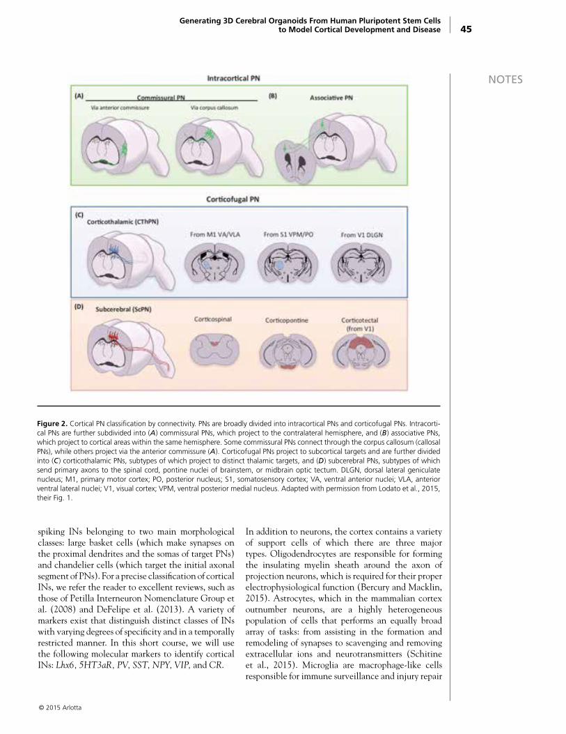
45
NOTES
spiking INs belonging to two main morphological classes: large basket cells (which make synapses on the proximal dendrites and the somas of target PNs) and chandelier cells (which target the initial axonal segment of PNs). For a precise classification of cortical INs, we refer the reader to excellent reviews, such as those of Petilla Interneuron Nomenclature Group et al. (2008) and DeFelipe et al. (2013). A variety of markers exist that distinguish distinct classes of INs with varying degrees of specificity and in a temporally restricted manner. In this short course, we will use the following molecular markers to identify cortical INs: Lhx6, 5HT3aR, PV, SST, NPY, VIP, and CR.
In addition to neurons, the cortex contains a variety of support cells of which there are three major types. Oligodendrocytes are responsible for forming the insulating myelin sheath around the axon of projection neurons, which is required for their proper electrophysiological function (Bercury and Macklin, 2015). Astrocytes, which in the mammalian cortex outnumber neurons, are a highly heterogeneous population of cells that performs an equally broad array of tasks: from assisting in the formation and remodeling of synapses to scavenging and removing extracellular ions and neurotransmitters (Schitine et al., 2015). Microglia are macrophage-like cells responsible for immune surveillance and injury repair
© 2015 Arlotta
Generating 3D Cerebral Organoids From Human Pluripotent Stem Cells to Model Cortical Development and Disease
Figure 2. Cortical PN classification by connectivity. PNs are broadly divided into intracortical PNs and corticofugal PNs. Intracorti-cal PNs are further subdivided into (A) commissural PNs, which project to the contralateral hemisphere, and (B) associative PNs, which project to cortical areas within the same hemisphere. Some commissural PNs connect through the corpus callosum (callosal PNs), while others project via the anterior commissure (A). Corticofugal PNs project to subcortical targets and are further divided into (C) corticothalamic PNs, subtypes of which project to distinct thalamic targets, and (D) subcerebral PNs, subtypes of which send primary axons to the spinal cord, pontine nuclei of brainstem, or midbrain optic tectum. DLGN, dorsal lateral geniculate nucleus; M1, primary motor cortex; PO, posterior nucleus; S1, somatosensory cortex; VA, ventral anterior nuclei; VLA, anterior ventral lateral nuclei; V1, visual cortex; VPM, ventral posterior medial nucleus. Adapted with permission from Lodato et al., 2015, their Fig. 1.
46
NOTES as well as synaptic pruning and remodeling (Harris et al., 2015; Michell-Robinson et al., 2015).
A Tale of Mice and Men: Progenitors in the Cerebral Cortex of Mice and HumansThe mouse cortex differs significantly from that of humans, so modeling the cellular variety of the human neocortex has been challenging, both in vivo and in vitro. Thus, the current understanding of human cortical development is limited largely to analyses of postmortem samples of human fetal cortex. For an in-depth treatment of the development and evolution of the human cerebral cortex, we refer the reader to excellent reviews, such as those of Taverna et al. (2014), Geschwind and Rakic (2013), and Florio and Huttner (2014). We will limit ourselves here to a short comparison of cortical progenitor classification and biology between mice and humans because of their relevance to human diseases (e.g., microcephaly) that have been modeled using human cerebral organoids.
The human cortex is vastly expanded in surface area relative to its size, producing a gyrencephalic rather than a smooth, lissencephalic cortex, as in mice (Borrell and Gotz, 2014). It also contains a much increased diversity of cellular types and distinct functional areas. The increase in size and cellular diversity of the human cortex is supported at least in part by an expanded subventricular zone (SVZ), which contains a vastly increased number and diversity of progenitors (De Juan Romero and Borrell, 2015). In mice, after neural tube closure, neuroepithelial (NE) cells with stem cell–like properties initially divide symmetrically to expand the progenitor pool. Later, they differentiate into more restricted progenitors known as radial glial cells (RGCs), which are bipolar cells with radial fibers contacting the apical ventricular zone and the pial surface. RGCs serve as a scaffold for neuronal migration, and they are also multipotent progenitor cells able to generate neurons, astrocytes, and oligodendrocytes (Malatesta et al., 2000; Anthony et al., 2004). At the onset of neurogenesis, the majority of RGCs exhibit asymmetric divisions in the ventricular zone to produce an RGC daughter cell and either a neuron or an intermediate precursor cell (IPC) (Pontious et al., 2008). IPCs then migrate basally to form the SVZ, where they further divide symmetrically to give rise to two to four neurons (Haubensak et al., 2004; Miyata et al., 2004; Noctor et al., 2004). The progenitor composition of the human developing cortex is more complex. One
key distinction of the SVZ of humans (and that of primates, more broadly) is that, in addition to increased numbers of IPCs, it contains an expanded new population of progenitor cells named outer radial glia (oRG), which lack apical contacts but retain a basal process to pia (Hansen et al., 2010). Interestingly, oRGs are also present in mice but at a very low frequency (Wang et al., 2011). A striking difference between oRGs in humans and mice is that murine oRGs directly produce neurons by symmetric division, whereas oRGs in humans divide asymmetrically to self-renew and generate a self-amplifying IPC, which then generates neurons (Hansen et al., 2010; Wang et al., 2011; LaMonica et al., 2013). Thus, oRG cells might contribute to the increased number and tangential dispersion of human neurons and to cortical folding (Sun and Hevner, 2014). Recent studies in primates have also shown that, in addition to IPCs, at least four types of oRG cells are present in the SVZ, contributing to increased progenitor diversity (Betizeau et al., 2013).
Modeling Human Cortical Development With Stem Cell–Derived Cerebral OrganoidsResearch using embryonic stem cells (ESCs) and iPSCs has demonstrated a surprising capacity of initially homogenous cultures to spontaneously self-assemble under permissive conditions into complex structures resembling endogenous tissue (Sasai, 2013). The Sasai laboratory first described an ESC aggregation protocol for producing relatively simple assemblies of cortical-like neurons (Eiraku et al., 2008); subsequently, using a modified culture protocol, they were able to generate tissue resembling cortical neuroepithelium (Nasu et al., 2012). Most strikingly, spontaneously self-assembling 3D optic-cup structures could be generated by these methods from both mouse (Eiraku et al., 2011) and human (Nakano et al., 2012) ESCs. These ESCs undergo a morphogenetic process resembling endogenous optic-cup development and subsequently form laminated retina tissue containing major classes of neural retinal cell types. Similar ESC-derived or iPSC-derived “organoids” have been produced for a variety of other tissues, including intestine and pituitary adenohypophysis (Sasai, 2013).
In vitro differentiation of stem cells to cortical neurons was first reported several years ago (Watanabe et al., 2005; Eiraku et al., 2008; Gaspard et al., 2008). Recently, methods have been adapted to create more complex, 3D structures in which iPSCs, triggered to differentiate into neuroectoderm
© 2015 Arlotta

47
NOTESby short-term application of appropriate growth factors, spontaneously differentiate further and self-assemble into a 3D organoid (Fig. 3). This organoid mimics some properties of the developing embryonic brain, including the presence of radial glia–like stem cells and neurons that appear to recapitulate the migration and intercalation that occur during embryonic cortical development (Lancaster et al., 2013; Lancaster and Knoblich, 2014). Much work remains to be done to understand the extent to which 3D cerebral organoids mimic the architecture, developmental trajectory, and cellular diversity of the developing human cortex; however, initial data indicate a significantly improved system for recapitulating these key steps of development in the dish.
This system has several unmet goals and limitations that will be discussed in the course. For example, it is clear that organoids currently reflect the cellular composition of the embryonic human brain, and
thus, a major unmet goal is to produce tissue that resembles the postnatal human brain. This is not a trivial task, as native human cortical development covers several years of prenatal and postnatal development. Although organoids are amenable to long-term culture (Lancaster et al., 2013, reported maintaining healthy cultures for more than 10 months), they lack blood vessels to support tissue oxygenation and appear to exhaust their capacity to grow and develop past early embryonic stages. In addition, it is unclear whether the organoids contain the full range of neuronal and glial populations found in the embryonic brain, although studies are ongoing to address these limitations. In addition, the question of whether circuit organization within organoids resembles that of endogenous tissue has not been investigated.
Nonetheless, the organoid system holds great promise for studying basic developmental biology of the human cortex because it allows ready access to
© 2015 Arlotta
Generating 3D Cerebral Organoids From Human Pluripotent Stem Cells to Model Cortical Development and Disease
Figure 3. Schematic representing the generation of cortical cell types from pluripotent stem cells in vitro. Traditional methods (top) use sequential application of growth factors to promote differentiation of predetermined cell types. The organoid method (bottom) exploits the self-organizing ability of induced neuroectodermal cells to form an organized, multilayer structure contain-ing multiple differentiated cell types. EGF, epidermal growth factor; FGF, fibroblast growth factor; IGF, insulin-like growth factor; NT3, neurotrophic factor 3; PDGF, platelet-derived growth factor; SHH, sonic hedgehog. Adapted with permission from Harris et al., 2015, their Fig. 3.

48
NOTES
© 2015 Arlotta
cells for comparative study with human fetal tissue. In particular, transcriptome and proteome analyses will benefit from an on-demand supply of fresh, healthy cells. In addition, both the starting iPSCs and the organoids themselves can be genetically manipulated to introduce, for example, cell type–specific fluorescent markers to trace cell populations, or disease-causing mutations to dissect molecular mechanisms of development and disease.
Lancaster et al. (2013) have already demonstrated, in a proof-of-concept experiment, the use of patient-derived iPSCs to model microencephaly, a condition characterized by an abnormally small brain. Although causative mutations of this condition have been identified, mouse models do not recapitulate the patient phenotype, possibly because of the difference in human versus mouse progenitor behavior. Using the organoid system, Lancaster et al. observed a reduction in early neural stem cell populations along with premature neural differentiation, suggesting that the CDK5RAP2 mutation found in patients disrupts the timing of the switch between proliferative and neurogenic divisions in neural progenitors. Similar experiments can be imagined for a variety of human diseases, including complex polygenic diseases and those for which the causative mutations have not been identified.
More recently, a modified protocol for organoid generation has been used by Mariani et al. (2015), who studied neurodevelopmental defects in organoids derived from patients with severe idiopathic autism spectrum disorder (ASD). These ASD-derived organoids exhibited enhanced proliferation of progenitors and overproduction of GABAergic inhibitory neurons, leading to phenotypes consistent with studies of patient-derived tissue. Mariani et al. further demonstrated that abnormally high expression of the transcription factor FOXG1 plays a part in these differences (Mariani et al., 2015).
These studies provide promising initial examples of the use of organoids not only to study normal human brain development but also to model human diseases that have a developmental origin. In this course, we will review different methods for generating human cerebral organoids from pluripotent stem cells. We will compare and contrast available protocols and consider the limitations and advantages of this system for studying cortical development and disease.
ReferencesAboitiz F, Montiel J (2003) One hundred million
years of interhemispheric communication: the history of the corpus callosum. Braz J Med Biol Res 36:409–420.
Anderson SA, Eisenstat DD, Shi L, Rubenstein JL (1997) Interneuron migration from basal forebrain to neocortex: dependence on Dlx genes. Science 278:474–476.
Anderson SA, Kaznowski CE, Horn C, Rubenstein JL, McConnell SK (2002) Distinct origins of neocortical projection neurons and interneurons in vivo . Cereb Cortex 12:702–709.
Anthony TE, Klein C, Fishell G, Heintz N (2004) Radial glia serve as neuronal progenitors in all regions of the central nervous system. Neuron 41:881–890.
Arlotta P, Molyneaux BJ, Chen J, Inoue J, Kominami R, Macklis JD (2005) Neuronal subtype-specific genes that control corticospinal motor neuron development in vivo. Neuron 45:207–221.
Bercury KK, Macklin WB (2015) Dynamics and mechanisms of CNS myelination. Dev Cell 32:447–458.
Betizeau M, Cortay V, Patti D, Pfister S, Gautier E, Bellemin-Menard A, Afanassieff M, Huissoud C, Douglas RJ, Kennedy H, Dehay C (2013) Precursor diversity and complexity of lineage relationships in the outer subventricular zone of the primate. Neuron 80:442–457.
Borrell V, Gotz M (2014) Role of radial glial cells in cerebral cortex folding. Curr Opin Neurobiol 27:39–46.
Brandão JA, Romcy-Pereira RN (2015) Interplay of environmental signals and progenitor diversity on fate specification of cortical GABAergic neurons. Front Cell Neurosci 9:149.
DeFelipe J, López-Cruz PL, Benavides-Piccione R, Bielza C, Larrañaga P, Anderson S, Burkhalter A, Cauli B, Fairén A, Feldmeyer D, Fishell G, Fitzpatrick D, Freund TF, González-Burgos G, Hestrin S, Hill S, Hof PR, Huang J, Jones EG, Kawaguchi Y, et al. (2013) New insights into the classification and nomenclature of cortical GABAergic interneurons. Nat Rev Neurosci 14:202–216.
De Juan Romero C, Borrell V (2015) Coevolution of radial glial cells and the cerebral cortex. Glia 63:1303–1319.

49
NOTESEiraku M, Watanabe K, Matsuo-Takasaki M, Kawada M, Yonemura S, Matsumura M, Wataya T, Nishiyama A, Muguruma K, Sasai Y (2008) Self-organized formation of polarized cortical tissues from ESCs and its active manipulation by extrinsic signals. Cell Stem Cell 3:519–532.
Eiraku M, Takata N, Ishibashi H, Kawada M, Sakakura E, Okuda S, Sekiguchi K, Adachi T, Sasai Y (2011) Self-organizing optic–cup morphogenesis in three-dimensional culture. Nature 472:51–56.
Fishell G, Kriegstein A (2005) Cortical development: new concepts. Neuron 46:361–362.
Florio M, Huttner WB (2014) Neural progenitors, neurogenesis and the evolution of the neocortex. Development 141:2182–2194.
Gaspard N, Bouschet T, Hourez R, Dimidschstein J, Naeije G, van den Ameele J, Espuny-Camacho I, Herpoel A, Passante L, Schiffmann SN, Gaillard A, Vanderhaeghen P (2008) An intrinsic mechanism of corticogenesis from embryonic stem cells. Nature 455:351–357.
Geschwind DH, Rakic P (2013) Cortical evolution: judge the brain by its cover. Neuron 80:633–647.
Greig LC, Woodworth MB, Galazo MJ, Padmanabhan H, Macklis JD (2013) Molecular logic of neocortical projection neuron specification, development and diversity. Nat Rev Neurosci 14:755–769.
Hansen DV, Lui JH, Parker PR, Kriegstein AR (2010) Neurogenic radial glia in the outer subventricular zone of human neocortex. Nature 464:554–561.
Harris J, Tomassy GS, Arlotta P (2015) Building blocks of the cerebral cortex: from development to the dish. Wiley Interdiscip Rev Dev Biol 4:529–544.
Haubensak W, Attardo A, Denk W, Huttner WB (2004) Neurons arise in the basal neuroepithelium of the early mammalian telencephalon: a major site of neurogenesis. Proc Natl Acad Sci USA 101:3196–3201.
Institute of Medicine (2013) Improving the utility and translation of animal models for nervous system disorders: Workshop Summary. Washington, DC: The National Academies Press.
Kepecs A, Fishell G (2014) Interneuron cell types are fit to function. Nature 505:318–326.
LaMonica BE, Lui JH, Hansen DV, Kriegstein AR (2013) Mitotic spindle orientation predicts outer radial glial cell generation in human neocortex. Nat Commun 4:1665.
Lancaster MA, Knoblich JA (2014) Generation of cerebral organoids from human pluripotent stem cells. Nat Protoc 9:2329–2340.
Lancaster MA, Renner M, Martin CA, Wenzel D, Bicknell LS, Hurles ME, Homfray T, Penninger JM, Jackson AP, Knoblich JA (2013) Cerebral organoids model human brain development and microcephaly. Nature 501:373–379.
Lee S, Hjerling-Leffler J, Zagha E, Fishell G, Rudy B (2010) The largest group of superficial neocortical GABAergic interneurons expresses ionotropic serotonin receptors. J Neurosci 30:16796–16808.
Lodato S, Shetty AS, Arlotta P (2015) Cerebral cortex assembly: generating and reprogramming projection neuron diversity. Trends Neurosci 38:117–125.
Lui JH, Hansen DV, Kriegstein AR (2011) Development and evolution of the human neocortex. Cell 146:18–36.
Malatesta P, Hartfuss E, Gotz M (2000) Isolation of radial glial cells by fluorescent-activated cell sorting reveals a neuronal lineage. Development 127:5253–5263.
Mariani J, Coppola G, Zhang P, Abyzov A, Provini L, Tomasini L, Amenduni M, Szekely A, Palejev D, Wilson M, Gerstein M, Grigorenko EL, Chawarska K, Pelphrey KA, Howe JR, Vaccarino FM (2015) FOXG1-dependent dysregulation of GABA/glutamate neuron differentiation in autism spectrum disorders. Cell 162:375–390.
Marín O, Noebels JL, Avoli M, Rogawski MA, Olsen RW, Delgado-Escueta AV, Gelman DM, Marín O, Rubenstein JLR (2012) The generation of cortical interneurons. Nat Rev Neurosci 13:107–120.
Markram H, Toledo-Rodriguez M, Wang Y, Gupta A, Silberberg G, Wu C (2004) Interneurons of the neocortical inhibitory system. Nat Rev Neurosci 5:793–807.
Michell-Robinson MA, Touil H, Healy LM, Owen DR, Durafourt BA, Bar-Or A, Antel JP, Moore CS (2015) Roles of microglia in brain development, tissue maintenance and repair. Brain 138:1138–1159.
Migliore M, Shepherd GM (2005) Opinion: an integrated approach to classifying neuronal phenotypes. Nat Rev Neurosci 6:810–818.
Miyata T, Kawaguchi A, Saito K, Kawano M, Muto T, Ogawa M (2004) Asymmetric production of surface-dividing and non-surface-dividing cortical progenitor cells. Development 131:3133–3145.
© 2015 Arlotta

© 2013
50
NOTES Molyneaux BJ, Arlotta P, Fame RM, MacDonald JL, MacQuarrie KL, Macklis JD (2009) Novel subtype-specific genes identify distinct subpopulations of callosal projection neurons. J Neurosci 29:12343–12354.
Molyneaux BJ, Goff LA, Brettler AC, Chen HH, Brown JR, Hrvatin S, Rinn JL, Arlotta P (2015) DeCoN: genome-wide analysis of in vivo transcriptional dynamics during pyramidal neuron fate selection in neocortex. Neuron 85:275–288.
Nakano T, Ando S, Takata N, Kawada M, Muguruma K, Sekiguchi K, Saito K, Yonemura S, Eiraku M, Sasai Y (2012) Self-formation of optic cups and storable stratified neural retina from human ESCs. Cell Stem Cell 10:771–785.
Nasu M, Takata N, Danjo T, Sakaguchi H, Kadoshima T, Futaki S, Sekiguchi K, Eiraku M, Sasai Y (2012) Robust formation and maintenance of continuous stratified cortical neuroepithelium by laminin-containing matrix in mouse ES cell culture. PLoS One 7:e53024.
Noctor SC, Martinez-Cerdeno V, Ivic L, Kriegstein AR (2004) Cortical neurons arise in symmetric and asymmetric division zones and migrate through specific phases. Nat Neurosci 7:136–144.
Petilla Interneuron Nomenclature Group, Ascoli GA, Alonso-Nanclares L, Anderson SA, Barrionuevo G, Benavides-Piccione R, Burkhalter A, Buzsáki G, Cauli B, Defelipe J, Fairén A, Feldmeyer D, Fishell G, Fregnac Y, Freund TF, Gardner D, Gardner EP, Goldberg JH, Helmstaedter M, Hestrin S, et al. (2008) Petilla terminology: nomenclature of features of GABAergic interneurons of the cerebral cortex. Nat Rev Neurosci 9:557–568.
Pontious A, Kowalczyk T, Englund C, Hevner RF (2008) Role of intermediate progenitor cells in cerebral cortex development. Dev Neurosci 30:24–32.
Sasai Y (2013) Cytosystems dynamics in self-organization of tissue architecture. Nature 493:318–326.
Schitine C, Nogaroli L, Costa MR, Hedin-Pereira C (2015) Astrocyte heterogeneity in the brain: from development to disease. Front Cell Neurosci 9:76.
Sun T, Hevner RF (2014) Growth and folding of the mammalian cerebral cortex: from molecules to malformations. Nat Rev Neurosci 15:217–232.
Taverna E, Gotz M, Huttner WB (2014) The cell biology of neurogenesis: toward an understanding of the development and evolution of the neocortex. Annu Rev Cell Dev Biol 30:465–502.
Wang X, Tsai JW, LaMonica B, Kriegstein AR (2011) A new subtype of progenitor cell in the mouse embryonic neocortex. Nat Neurosci 14:555–561.
Watanabe K, Kamiya D, Nishiyama A, Katayama T, Nozaki S, Kawasaki H, Watanabe Y, Mizuseki K, Sasai Y (2005) Directed differentiation of telencephalic precursors from embryonic stem cells. Nat Neurosci 8:288–296.

© 2015 Tesar
Departments of 1Genetics and Genome Sciences, 2Neurosciences, and 3Pathology
and 4Center for Proteomics and Bioinformatics Case Western Reserve University School of Medicine
Cleveland, Ohio
5Department of Stem Cell Biology and Regenerative Medicine Lerner Research Institute, Cleveland Clinic
Cleveland, Ohio
6PerkinElmer Waltham, Massachusetts
7Drug Discovery Center University of Cincinnati College of Medicine
Cincinnati, Ohio
8National Center for Advancing Translational Sciences National Institutes of Health
Rockville, Maryland
9Department of Microbiology–Immunology and Interdepartmental Immunobiology Center
Feinberg School of Medicine, Northwestern University Chicago, Illinois
Drug-Based Modulation of Endogenous Stem Cells Promotes Functional Remyelination In Vivo
Fadi J. Najm, MBA,1 Mayur Madhavan, PhD,1 Anita Zaremba, BA,2 Elizabeth Shick, BS,1 Robert T. Karl, BS,1
Daniel C. Factor, BA,1 Tyler E. Miller, BS,1,3,5 Zachary S. Nevin, BS,1 Christopher Kantor,2 Alex Sargent,2 Kevin L. Quick,6
Daniela M. Schlatzer,4 Hong Tang,7 Ruben Papoian, PhD,7 Kyle R. Brimacombe, MS,8 Min Shen,8 Matthew B. Boxer,8
Ajit Jadhav,8 Andrew P. Robinson,9 Joseph R. Podojil, PhD,9 Stephen D. Miller,9 Robert H. Miller,2 and Paul J. Tesar, PhD1,2

© 2015 Tesar

53
NOTESIntroductionMultiple sclerosis (MS) involves an aberrant autoimmune response and progressive failure of remyelination in the CNS. Prevention of neural degeneration and subsequent disability requires remyelination through the generation of new oligodendrocytes, but current treatments exclusively target the immune system. Oligodendrocyte progenitor cells (OPCs) are stem cells in the CNS and the principal source of myelinating oligodendrocytes (Goldman et al., 2012). These progenitor cells are abundant in demyelinated regions of patients with MS yet fail to differentiate, thereby representing a cellular target for pharmacological intervention (Chang et al., 2002). To discover therapeutic compounds for enhancing myelination from endogenous OPCs, we screened a library of bioactive small molecules on mouse pluripotent epiblast stem cell (EpiSC)–derived OPCs (Brons et al., 2007; Tesar et al., 2007; Najm et al., 2011). Here we discuss seven drugs that function at nanomolar doses to selectively enhance the generation of mature oligodendrocytes from progenitor cells in vitro.
Two drugs—miconazole and clobetasol—are effective in vitro for promoting precocious myelination in organotypic cerebellar slice cultures, and in vivo, in early postnatal mouse pups. Systemic delivery of each of the two drugs significantly increases the number of new oligodendrocytes and enhances remyelination in a lysolecithin-induced mouse model of focal demyelination. Administering each drug at the peak of disease in an experimental autoimmune encephalomyelitis mouse model of chronic progressive MS results in a striking reversal of disease severity. Immune response assays show that miconazole functions directly as a remyelinating drug with no effect on the immune system, whereas clobetasol is a potent immunosuppressant as well as a remyelinating agent. Mechanistic studies show that miconazole and clobetasol function in OPCs through mitogen-activated protein (MAP) kinase and glucocorticoid receptor signaling, respectively. Furthermore, both drugs enhance the generation of human oligodendrocytes from human OPCs in vitro. Collectively, our results provide a rationale for testing miconazole and clobetasol (or structurally modified derivatives) to enhance remyelination in patients.
ResultsLead generationRepair of damaged myelin may provide therapeutic benefit in MS and other demyelinating disorders (Dubois-Dalcq et al., 2005; Franklin et al., 2008;
Mi et al., 2009; Fancy et al., 2010; Bai et al., 2012; Deshmukh et al., 2013; Mei et al., 2014). Therefore, we set out to identify drugs that could be repurposed as remyelinating therapeutics. We selected the US National Institutes of Health (NIH) Clinical Collection I and II libraries comprising 727 drugs with a history of safe use in clinical trials, to test for maturation of OPCs into myelinating oligodendrocytes. Using mouse EpiSC–derived OPCs, we developed an in vitro phenotypic screen that accurately quantified differentiation into mature oligodendrocytes using high-content imaging of myelin protein expression (Fig. 1a).
Two batches (>100 million cells each) of pure OPCs were generated from independent mouse pluripotent EpiSC lines of opposite sex. EpiSC-derived OPCs shared virtually all defining molecular and cellular properties, including gene expression profiles, with in vivo isolated OPCs but provided the key advantage of being highly scalable (Najm et al., 2011). For in vitro screening, the seeding density, endpoint assays, and DMSO (vehicle) tolerance were optimized in pilot studies to ensure accurate and reproducible measurement of OPC differentiation in a 96-well format.
For the primary screen, OPCs were treated with vehicle alone (0.05% [v/v] DMSO) as a negative control, thyroid hormone (a known OPC differentiation inducer) as a positive control (Barres et al., 1994), or drug dissolved in DMSO at a concentration of 5 μM. After 72 h, cells were fixed and labeled with antibodies to MBP, and the length and intensity of MBP-labeled oligodendrocyte processes were measured (Fig. 1a). These features were reliable indicators of alteration in cellular phenotype, as indicated by consistency and high signal-to-background ratio of positive and vehicle controls across all screening plates. We then normalized the experimental data for the tested drugs against thyroid hormone (set value of 100) on a per-plate basis. On the basis of this analysis, we identified the 22 drugs that enhanced oligodendrocyte formation > 5 SD above DMSO treatment and outperformed thyroid hormone in the measured parameters (Fig. 1b). Notably, one of the top 22 drugs was benztropine, a muscarinic receptor antagonist recently shown to induce OPC differentiation and remyelination (Deshmukh et al., 2013; Mei et al., 2014).
To validate and prioritize the 22 drug hits, the assay was repeated using alternative OPCs, reagents, and parameters to eliminate screen-specific artifacts. Drugs were ranked by their dose-dependent ability
Drug-Based Modulation of Endogenous Stem Cells Promotes Functional Remyelination In Vivo
© 2015 Tesar
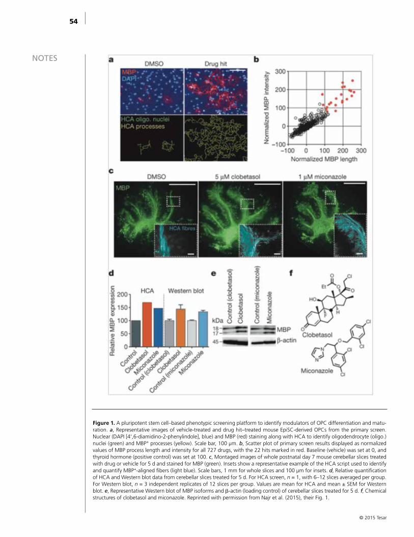
54
NOTES
© 2015 Tesar
Figure 1. A pluripotent stem cell–based phenotypic screening platform to identify modulators of OPC differentiation and matu-ration. a, Representative images of vehicle-treated and drug hit–treated mouse EpiSC-derived OPCs from the primary screen. Nuclear (DAPI [4‘,6-diamidino-2-phenylindole], blue) and MBP (red) staining along with HCA to identify oligodendrocyte (oligo.) nuclei (green) and MBP+ processes (yellow). Scale bar, 100 μm. b, Scatter plot of primary screen results displayed as normalized values of MBP process length and intensity for all 727 drugs, with the 22 hits marked in red. Baseline (vehicle) was set at 0, and thyroid hormone (positive control) was set at 100. c, Montaged images of whole postnatal day 7 mouse cerebellar slices treated with drug or vehicle for 5 d and stained for MBP (green). Insets show a representative example of the HCA script used to identify and quantify MBP+-aligned fibers (light blue). Scale bars, 1 mm for whole slices and 100 μm for insets. d, Relative quantification of HCA and Western blot data from cerebellar slices treated for 5 d. For HCA screen, n = 1, with 6–12 slices averaged per group. For Western blot, n = 3 independent replicates of 12 slices per group. Values are mean for HCA and mean ± SEM for Western blot. e, Representative Western blot of MBP isoforms and β-actin (loading control) of cerebellar slices treated for 5 d. f, Chemical structures of clobetasol and miconazole. Reprinted with permission from Najr et al. (2015), their Fig. 1.
55
NOTESto induce oligodendrocyte generation from OPCs without toxicity. To demonstrate reproducibility, an independent laboratory tested selected drug hits using distinct equipment, plate format (1536-well), personnel, and imaging/analysis scripts. Of the 16 hits tested at the external screening site, 14 were validated as potent inducers of oligodendrocyte differentiation.
Testing drug leads in vitroWe next tested whether the drug hits could promote the maturation of native OPCs in CNS tissue. Cerebellar slices were generated from mice at postnatal day 7—a time that precedes widespread myelination—and treated ex vivo with drug or DMSO (vehicle) for 5 d and labeled with anti-MBP antibodies (Fig. 1c) (Mi et al., 2009; Bai et al., 2012). We screened 11 of the top drugs and used a high-content analysis (HCA) algorithm developed in house to rank them on the basis of their ability to increase the extent of MBP+ aligned fibers in whole cerebellar slices. The so-called high-performing group consisted of four drugs that increased the number of MBP+ aligned fibers by approximately 150% or greater (Fig. 1d). We validated the accuracy of our high-content screen using semiquantitative western blotting of MBP protein isoforms in independent slice-culture experiments (Figs. 1d, e) (Woodruff et al., 1998, 1999).
Analysis of structure–activity relationships revealed that the top hits from the primary screen segregated into two specific classes containing either a 1,3-diazole with monosubstitution at the 1-position or a sterane base structure. We selected miconazole and clobetasol, the top overall performing hits in each of the imidazole and sterane classes, respectively, for further mechanistic and functional testing after confirming that both drugs readily cross the blood–brain barrier in mice (Fig. 1f). Miconazole is a topical antifungal agent functioning through cytochrome P450 inhibition, and clobetasol is a potent topical corticosteroid, but their functions in OPCs were unknown.
Testing miconazole and clobetasol in vivoTo test whether miconazole or clobetasol enhance remyelination in vivo, we used a toxin-induced model whereby focal demyelinated lesions are generated in dorsal white matter of the spinal cord of adult mice by localized injection of lysolecithin (lysophosphatidylcholine [LPC]). In lesioned
animals, demyelination is complete within 4 d, after which OPCs are recruited into the lesion. Widespread remyelination does not normally start until 14–21 d postlesion (d.p.l.), which provides a defined window from days 4–14 to test the efficacy of drugs to enhance the extent and rate of remyelination (Jeffery et al., 1995). Both miconazole (10 or 40 mg/kg body weight) and clobetasol (2 mg/kg) treatment induced a marked improvement within the lesions of treated mice compared with vehicle-treated controls. At 8 d.p.l., both drugs induced a significant increase in the number of newly generated CC1+ oligodendrocytes in the lesion core (Figs. 2a, b). This effect was coincident with extensive MBP staining in the lesions of miconazole-treated and clobetasol-treated, but not vehicle-treated, animals at both 8 and 12 d.p.l. (Fig. 2a). Electron micrographs and tissue sections stained with toluidine blue demonstrated that miconazole and clobetasol each induced a striking increase in the extent of remyelination (Figs. 2c, d). At 12 d.p.l., lesions of vehicle-treated mice consisted mostly of unmyelinated axons (6% myelinated), while those of miconazole-treated and clobetasol-treated mice contained more than 70% remyelinated axons throughout the extent of the lesion (Fig. 2d). Analysis of myelin thickness relative to axon diameter (g ratio) at 12 d.p.l. revealed that miconazole-induced and clobetasol-induced myelin was thinner than intact myelin, a defining characteristic of remyelination (Fig. 2d).
We also evaluated whether miconazole or clobetasol could promote precocious myelination during development, in the absence of injury or disease. We treated mice at postnatal day 2 (a time point that precedes widespread CNS myelination) daily for 4 d with drug or vehicle. In miconazole-treated and clobetasol-treated mice, we found a significant increase in the number of CC1+ oligodendrocytes in the lateral corpus callosum compared with vehicle-treated mice (not shown). Additionally, we found that a significantly larger portion of the corpus callosum was populated by MBP+ fiber tracts in miconazole-treated and clobetasol-treated mice. This finding suggests that miconazole and clobetasol enhance myelination in the absence of damage or disease. Collectively, the LPC demyelination and developmental mouse models demonstrated that miconazole and clobetasol each function to induce the differentiation of endogenous OPCs in the CNS and promote enhanced myelination.
To determine whether the drugs were working at a particular stage of the OPC differentiation process,
Drug-Based Modulation of Endogenous Stem Cells Promotes Functional Remyelination In Vivo
© 2015 Tesar

56
NOTES
© 2015 Tesar
Figure 2. Miconazole and clobetasol each enhance remyelination in the LPC lesion mouse model. a, Representative immuno-histochemical images of treated mice showing newly generated oligodendrocytes (CC1, red) and MBP (green) within the lesion (approximated by white dashed outline) at 8 and 12 d.p.l. Scale bar, 200 μm. b, Quantification of CC1+ oligodendrocytes per lesion area at 8 d.p.l. Values are mean ± SEM; n = 3 mice per group. Two-tailed t test; *p < 0.05. c, Representative electron mi-crographs showing remyelinated axons within lesions of drug-treated mice at 12 d.p.l. Scale bar, 2 μm. d, Scatter plot of g ratios of lesion axons at 12 d.p.l.; n = 100 calculated from two mice per group. Percentage of lesion axons myelinated is indicated in the legend. Reprinted with permission from Najr et al. (2015), their Fig. 2.

57
NOTES
Drug-Based Modulation of Endogenous Stem Cells Promotes Functional Remyelination In Vivo
© 2015 Tesar
Figure 3. Cellular and molecular effects of miconazole and clobetasol on mouse OPCs. a, Percentage MBP+ oligodendrocytes generated from OPCs at 72 h with treatments initiated at time points indicated; n = 6 wells per condition with > 6000 cells scored per well. b, Percentage MBP+ oligodendrocytes generated from OPCs treated simultaneously and analyzed at time points indicated; n = 8 wells per condition with > 1700 cells scored per well. c, Percentage GFAP+ astrocytes generated from OPCs at 72 h of treatment; n = 4 wells per condition > 2900 cells scored per well. d, Heat map depicting biochemical inhibition of mus-carinic receptors M1–M5 displayed as percentage inhibition with minimum (green) and maximum (red). e, Western blot of total glucocorticoid receptor and its phosphorylation at Ser220 (p-GR) in OPCs treated for 1 h. f, Percentage MBP+ oligodendrocytes generated from OPCs 72 h after treatment; n = 6 wells per condition with > 1400 cells scored per well. g, Western blot of total ERK1/2 and their phosphorylation at Thr202/Tyr204 or Thr185/Tyr187 (p-ERK1/2) in cells (OPCs or mouse embryonic fibroblasts [MEFs]) treated for 1 h. Fibroblast growth factor (FGF) served as a positive control for p-ERK1/2 induction. h, Western blot of total ERK1/2 and p-ERK1/2 in OPCs treated for 1 h in the presence of the indicated pathway inhibitors. All graphs depict mean ± SEM. Reprinted with permission from Najr et al. (2015), their Fig. 3.
we seeded OPCs in differentiation conditions and treated them with either miconazole or clobetasol at different time points (0, 16, 24, or 48 h), and assayed MBP expression at 72 h. For both miconazole and clobetasol, the number of MBP+ oligodendrocytes present at 72 h was dependent on drug treatment
within the first 24 h of differentiation (Fig. 3a). In agreement with these data, treatment of differentiating OPCs with either drug for different durations (24, 48, 56, and 72 h) induced a progressive, time-dependent increase in the number of MBP+ oligodendrocytes (Fig. 3b). These data suggest that
58
NOTES
© 2015 Tesar
both drugs function directly on OPCs early in the differentiation process. Additionally, neither drug showed a significant impact on astrocyte formation from OPCs in vitro, suggesting they probably function as direct inducers of oligodendrocyte differentiation (Fig. 3c).
Muscarinic receptor antagonists such as benztropine and clemastine have recently been identified as remyelinating agents (Deshmukh et al., 2013; Mei et al., 2014). Therefore, we tested whether miconazole or clobetasol function through the muscarinic acetylcholine pathway using functional cellular reporter assays of all muscarinic receptor subtypes (M1–M5). Neither miconazole nor clobetasol inhibited any of the five muscarinic receptor subtypes (Fig. 3d). We then profiled whether clobetasol or miconazole biochemically inhibited the activity of 414 different kinase isoforms. Neither clobetasol nor miconazole inhibited any of the kinases tested, suggesting their activity is not based on direct inhibition of protein kinases.
Effect on OPC signaling pathwaysTo explore the signaling pathways in OPCs influenced by these drugs, we performed genome-wide RNA sequencing and phosphoproteomic analyses on mouse OPCs treated with drug or vehicle. Miconazole or clobetasol treatment altered OPC transcript expression and phosphoproteins within hours and influenced the expression of genes in signaling pathways involved in oligodendrocyte maturation and myelination. Clobetasol potently modulated genes downstream of multiple nuclear hormone receptors, including glucocorticoid receptors, which are known to be important regulators of myelin gene expression (Kumar et al., 1989; Barres et al., 1994). Since glucocorticoid receptor signaling is also known to enhance Schwann cell–mediated myelination in the peripheral nervous system (Morisaki et al., 2010), we tested whether the activity of clobetasol on OPCs was mediated by glucocorticoid receptor signaling. Treatment of OPCs with clobetasol for 1 h increased the phosphorylation of glucocorticoid receptor at Ser220, an activating posttranslational modification (Fig. 3e). RU486, a competitive glucocorticoid receptor antagonist, blocked clobetasol-induced glucocorticoid receptor phosphorylation and oligodendrocyte differentiation (Figs. 3e, f), suggesting that the activity of clobetasol in OPCs is mediated through the glucocorticoid receptor signaling axis.
For miconazole, pathway analyses showed that proteins in the MAP kinase pathway were most strongly affected. Most prominent was the strong
and sustained phosphorylation of both extracellular signal-regulated kinases ERK1 and ERK2 (ERK1/2) at canonical activation sites, which we validated by Western blotting (Fig. 3g). In mice, genetic loss of ERK1/2 in the oligodendrocyte lineage results in normal numbers of OPCs and oligodendrocytes but widespread hypomyelination, while constitutive activation of ERK1/2 results in a profound increase in the extent of remyelination after toxin-induced demyelinating injury (Ishii et al., 2012; Fyffe-Maricich et al., 2013). In contrast to miconazole, treatment of OPCs with clobetasol or benztropine did not induce ERK1/2 phosphorylation (Fig. 3g). Miconazole treatment of a nonneural cell type, mouse fibroblasts, also showed no increase of ERK1/2 phosphorylation, indicating potential cell-type specificity (Fig. 3g). PD0325901, a small-molecule inhibitor of ERK’s upstream MAP kinase kinase (MEK), blocked the ability of miconazole to induce ERK1/2 phosphorylation, suggesting that miconazole functions through a MEK-dependent mechanism in OPCs (Fig. 3h). We also treated mouse OPCs with voriconazole, a triazole-containing antifungal cytochrome P450 inhibitor with 80% structural similarity to miconazole, which failed to induce changes in ERK1/2 phosphorylation (Fig. 3g). This result was consistent with the observation that voriconazole did not promote the differentiation of OPCs into oligodendrocytes. Taken together, these results suggest that the effect of miconazole on OPCs is independent of cytochrome P450 inhibition (not shown).
Differentiation of human OPCs into oligodendrocytesWe then assessed whether clobetasol and miconazole treatment would enhance the differentiation of human OPCs into oligodendrocytes. We generated human OPCs from human embryonic stem cells (hESCs) and human-induced pluripotent stem cells (hiPSCs) (Hu et al., 2009; Wang et al., 2013). We then treated human OPCs with DMSO, clobetasol, or miconazole for 21 d followed by staining for MBP, imaging, and HCA (not shown). Both drugs enhanced human OPC differentiation, with miconazole exhibiting the most reproducible and potent effects.
Therapeutic effect in immune-mediated MS modelsTo interpret the potential impact of clobetasol or miconazole as therapeutics in immune-mediated MS models, we tested effects on immune cell survival and function. We found that only clobetasol, as expected from its known corticosteroid properties, altered

59
NOTES
Drug-Based Modulation of Endogenous Stem Cells Promotes Functional Remyelination In Vivo
© 2015 Tesar
naive T-cell differentiation and both the proliferation and secretion of cytokines by proteolipid protein (PLP139–151)–sensitized or myelin oligodendrocyte glycoprotein (MOG35–55)–sensitized lymph node cells (not shown). As such, only clobetasol, but not the solely remyelinating drugs miconazole or benztropine, showed efficacy in reducing disease severity in the immune-driven relapsing–remitting PLP139–151 experimental autoimmune encephalomyelitis (EAE) model (Fig. 4a). The positive effect of clobetasol in this model resulted from its immunosuppressive effects, as evidenced by the drastic reduction of T cells within the spleen (Fig. 4b).
We also used a second EAE mouse model, MOG35–55-induced, in which the immune response was relatively controlled and disease pathology recapitulated chronic progressive demyelination. We used a therapeutic, rather than prophylactic, treatment regimen to evaluate whether drugs could reverse, rather than prevent, disease. Miconazole-treated and clobetasol-treated animals all exhibited a marked improvement in function, and nearly all animals regained use of one or both hind limbs (Figs. 4c, d). In contrast, vehicle-treated mice exhibited chronic hind limb paralysis during the treatment period. Benztropine treatment also resulted in functional
Figure 4. Therapeutic efficacy of miconazole and clobetasol in mouse models of MS. a, Scoring of disease severity in relaps-ing–remitting PLP139–151-induced EAE mice treated beginning on day 13 (black arrow) and ending on day 29; n = 10 mice per group. Graph depicts mean daily disease score ± SEM. b, Flow cytometry–based quantification of spleen cell numbers at day 29 from the PLP139–151 EAE cohort in a. Values are mean ± SEM; n = 4 or 5 mice per group. c, Scoring of disease severity in chronic progressive MOG35–55-induced EAE mice treated daily for 10 d beginning at the peak of disease on day 15 (black arrow); n = 12–16 mice per group. Graph depicts mean daily disease score ± SEM. d, Mean improvement in disease score (Δ) per animal (peak score minus ending score) of MOG35–55 EAE cohort in c. Also shown are external validation results in MOG35–55 EAE from an independent contract laboratory; n = 12 mice per group. For all EAE experiments, drugs were dosed daily by intraperitoneal injection: clobetasol (2 mg/kg), miconazole (10 mg/kg), benztropine (10 mg/kg), or FTY720 (1 mg/kg). All EAE disease scoring was as follows: 0, no abnormality; 1, limp tail; 2, limp tail and hind limb weakness; 3, hind limb paralysis; 4, hind limb paralysis and forelimb weakness; and 5, moribund. Two-tailed t test, *p < 0.05 and **p < 0.01 for drug-treated groups compared with their respective vehicle-treated group. Reprinted with permission from Najr et al. (2015), their Fig. 4.

60
NOTES
© 2015 Tesar
improvement, but to a lesser extent than miconazole and clobetasol (Figs. 4c, d). Overt functional recovery of miconazole-treated and clobetasol-treated mice correlated with histological improvements in the spinal cord. Specifically, drug-treated mice showed restoration of MBP expression and a reduction in the extent of demyelination in the spinal cord, whereas vehicle-treated mice showed sustained areas of white-matter disruption (not shown).
Although the immunosuppressive effect of clobetasol makes it challenging to evaluate its remyelinating potential in EAE directly, its consistent and robust induction of OPC differentiation in vitro, and enhancement of remyelination in nonimmune-driven in vivo assays, suggest that it serves a role in both immunomodulation and promotion of myelination. In contrast, miconazole did not modulate immune cell function, and our data indicate that it acts as a direct remyelinating agent. Given the potential of miconazole as a remyelinating therapeutic, we contracted a separate laboratory to provide independent validation of its efficacy in the MOG35–55-induced EAE preclinical model. The laboratory independently validated the preclinical efficacy of miconazole in MOG35–55-induced EAE for reducing disease severity in treated mice (Fig. 4d).
ConclusionsSince the approval in 1993 of interferon-β-1b for the treatment of MS, therapeutic development has centered on the generation of additional immunomodulatory agents. Despite the effectiveness of many of these drugs to modulate CNS inflammation in patients with MS, none of them prevents chronic progressive disease and disability—largely because of their inability to stop or reverse the failure of remyelination in the CNS. We developed an advanced high-throughput screening platform to discover effective remyelinating therapeutics. This pluripotent stem cell–based system provides unprecedented scalability, purity, and genotypic flexibility to screen for compounds that enhance OPC differentiation and myelination. Using this platform, we identified two drugs approved by the US Food and Drug Administration—miconazole and clobetasol—with newly discovered functions to modulate OPC differentiation directly, enhance remyelination, and significantly reduce disease severity in mouse models of MS. Since miconazole and clobetasol are currently approved only for topical administration in humans, significant optimization of dosing, delivery, and potentially chemical structure will be required to enhance the on-target pharmacology in OPCs while diminishing any potential off-target
side effects. However, the ability of miconazole and clobetasol to cross the blood–brain barrier raises the exciting possibility that these drugs, or their modified derivatives, could advance into clinical trials for the currently untreatable chronic progressive phase of MS.
AcknowledgmentsThis research was supported by grants from the NIH (Grant NS085246) to P.J.T. and R.H.M., Grant NS030800 to R.H.M., and Grant NS026543 to S.D.M.; the New York Stem Cell Foundation (to P.J.T.); the Myelin Repair Foundation (to P.J.T., R.H.M., and S.D.M.); the Mt. Sinai Health Care Foundation (to P.J.T.); and NIH Predoctoral Training Grants T32GM008056 to R.T.K. and F30CA183510 to T.E.M. Additional support was provided by the Cytometry & Imaging Microscopy, Proteomics, and Genomics core facilities of the Case Comprehensive Cancer Center (Grant P30CA043703); the Case Western Reserve University Council to Advance Human Health; and philanthropic support from the Goodman, Long, and Geller families. P.J.T. is a New York Stem Cell Foundation–Robertson Investigator. We are grateful to M. Hitomi, W. Harte, D. Adams, W. Seibel, M. Haag, P. Scacheri, J. Wanta, C. Fang, H. Olsen, T. LaFramboise, J. Song, F. Van den Akker, and M. Shoham for technical assistance and discussion; to A. Lager and M. Elitt for comments on the manuscript; to B. Trapp for the PLP1 antibody; and to B. Barres and B. Zuchero for RNA from in vivo isolated OPCs.
This chapter was excerpted from an article of the same title by Najm et al., 2015, published in Nature 522:216–220.
ReferencesBai L, Lennon DP, Caplan AI, DeChant A, Hecker J,
Kranso J, Zaremba A, Miller RH (2012) Hepatocyte growth factor mediates mesenchymal stem cell-induced recovery in multiple sclerosis models. Nat Neurosci 15:862–870.
Barres BA, Lazar MA, Raff MC (1994) A novel role for thyroid hormone, glucocorticoids and retinoic acid in timing oligodendrocyte development. Development 120:1097–1108.
Brons IG, Smithers LE, Trotter MW, Rugg-Gunn P, Sun B, Chuva de Sousa Lopes SM, Howlett SK, Clarkson A, Ahrlund-Richter L, Pedersen RA, Vallier L (2007) Derivation of pluripotent epiblast stem cells from mammalian embryos. Nature 448:191–195.

61
NOTES
Drug-Based Modulation of Endogenous Stem Cells Promotes Functional Remyelination In Vivo
© 2015 Tesar
Chang A Tourtellotte WW, Rudick R, Trapp BD (2002) Premyelinating oligodendrocytes in chronic lesions of multiple sclerosis. N Engl J Med 346:165–173.
Deshmukh VA, Tardif V, Lyssiotis CA, Green CC, Kerman B, Kim HJ, Padmanabhan K, Swoboda JG, Ahmad I, Kondo T, Gage FH, Theofilopoulos AN, Lawson BR, Schultz PG, Lairson LL (2013) A regenerative approach to the treatment of multiple sclerosis. Nature 502:327–332.
Dubois-Dalcq M, Ffrench-Constant C, Franklin RJ (2005) Enhancing central nervous system remyelination in multiple sclerosis. Neuron 48:9–12.
Fancy SP, Kotter MR, Harrington EP, Huang JK, Zhao C, Rowitch DH, Franklin RJ (2010) Overcoming remyelination failure in multiple sclerosis and other myelin disorders. Exp Neurol 225:18–23.
Franklin RJ, Ffrench-Constant C (2008) Remyelination in the CNS: from biology to therapy. Nat Rev Neurosci 9:839–855.
Fyffe-Maricich SL, Schott A, Karl M, Krasno J, Miller RH (2013) Signaling through ERK1/2 controls myelin thickness during myelin repair in the adult central nervous system. J Neurosci 33:18402–18408.
Goldman SA, Nedergaard M, Windrem MS (2012) Glial progenitor cell-based treatment and modeling of neurological disease. Science 338:491–495.
Hu BY, Du ZW, Zhang SC (2009) Differentiation of human oligodendrocytes from pluripotent stem cells. Nat Protocols 4:1614–1622.
Ishii A, Fyffe-Maricich SL, Furusho M, Miller RH, Bansal R (2012) ERK1/ERK2 MAPK signaling is required to increase myelin thickness independent of oligodendrocyte differentiation and initiation of myelination. J Neurosci 32:8855–8864.
Jeffery ND, Blakemore WF (1995) Remyelination of mouse spinal cord axons demyelinated by local injection of lysolecithin. J Neurocytol 24:775–781.
Kumar S, Cole R, Chiappelli F, de Vellis J (1989) Differential regulation of oligodendrocyte markers by glucocorticoids: post-transcriptional regulation of both proteolipid protein and myelin basic protein and transcriptional regulation of glycerol phosphate dehydrogenase. Proc Natl Acad Sci USA 86:6807–6811.
Mei F, Fancy SP, Shen YA, Niu J, Zhao C, Presley B, Miao E, Lee S, Mayoral SR, Redmond SA, Etxeberria A, Xiao L, Franklin RJ, Green A, Hauser SL, Chan JR (2014) Micropillar arrays as a high-throughput screening platform for therapeutics in multiple sclerosis. Nat Med 20:954–960.
Mi S, Miller RH, Tang W, Lee X, Hu B, Wu W, Zhang Y, Shields CB, Zhang Y, Miklasz S, Shea D, Mason J, Franklin RJ, Ji B, Shao Z, Chédotal A, Bernard F, Roulois A, Xu J, Jung V, et al. (2009) Promotion of central nervous system remyelination by induced differentiation of oligodendrocyte precursor cells. Ann Neurol 65:304–315.
Morisaki S, Nishi M, Fujiwara H, Oda R, Kawata M, Kubo T (2010) Endogenous glucocorticoids improve myelination via Schwann cells after peripheral nerve injury: an in vivo study using a crush injury model. Glia 58:954–963.
Najm FJ, Zaremba A, Caprariello AV, Nayak S, Freundt EC, Scacheri PC, Miller RH, Tesar PJ (2011) Rapid and robust generation of functional oligodendrocyte progenitor cells from epiblast stem cells. Nat Methods 8:957–962.
Najm FJ, Madhavan M, Zaremba A, Shick E, Karl RT, Factor DC, Miller TE, Nevin ZS, Kantor C, Sargent A, Quick KL, Schlatzer DM, Tang H, Papoian R, Brimacombe KR, Shen M, Boxer MB, Jadhav A, Robinson AP, Podojil JR, et al. (2015) Drug-based modulation of endogenous stem cells promotes functional remyelination in vivo. Nature 522:216–220.
Tesar PJ, Chenoweth JG, Brook FA, Davies TJ, Evans EP, Mack DL, Gardner RL, McKay RD (2007) New cell lines from mouse epiblast share defining features with human embryonic stem cells. Nature 448:196–199.
Wang S, Bates J, Li X, Schanz S, Chandler-Militello D, Levine C, Maherali N, Studer L, Hochedlinger K, Windrem M, Goldman SA (2013) Human iPSC-derived oligodendrocyte progenitor cells can myelinate and rescue a mouse model of congenital hypomyelination. Cell Stem Cell 12:252–264.
Woodruff RH, Franklin RJ (1998) The expression of myelin basic protein exon 1 and exon 2 containing transcripts during myelination of the neonatal rat spinal cord—an in situ hybridization study. J Neurocytol 27:683–693.
Woodruff RH, Franklin RJ (1999) The expression of myelin protein mRNAs during remyelination of lysolecithin-induced demyelination. Neuropathol Appl Neurobiol 25:226–235.


© 2015 Barrett
Stanley Center for Psychiatric Research, Broad Institute Department of Stem Cell and Regenerative Biology,
Harvard University Cambridge, Massachusetts
Developing Stem Cell Models to Study Neuropsychiatric Diseases
Lindy Barrett, PhD, and Kevin Eggan, PhD


65
NOTESIntroductionThere is a large unmet need to develop better treatments for debilitating and relatively common neuropsychiatric diseases such as schizophrenia (SZ), bipolar disorder (BP), and autism spectrum disorder (ASD). Recent improvements in DNA analysis technologies have enabled durable progress in understanding the genetic contributors to SZ (Schizophrenia Working Group of the Psychiatric Genomics Consortium, 2014). However, this is unlikely to result in new therapeutics until we understand how predisposing genetic variation damages the function of human brain cell types responsible for behavior and cognition. Unfortunately, high-quality human brain tissue is generally unavailable for study. To overcome this limitation, we have been exploring human pluripotent stem cells (hPSCs) as a means of producing populations of human brain cell types in vitro. Although this strategy holds great promise, many questions remain concerning the reproducibility of these cells, and how representative they are of those that eventually malfunction in affected individuals. Our short-term aim is to test the viability of this stem cell approach for studying neuropsychiatric diseases in the hopes that these models will lead us to identify brain cells that are most affected and enable us to discover how they are malfunctioning. If we ultimately succeed, we can use these cells as the foundation for identifying candidate drugs that have corrective, therapeutic activities. We continue to take a long-term view with respect to technology evaluation and resource building, and the projects summarized below reflect this strategy. We believe that such a stance is critical, given the very early state of our understanding of both stem cell biology and the genetic contributors to these conditions.
In this chapter, we describe a set of projects being carried out at the Stanley Center for Psychiatric Research to understand questions of cell-type specificity and neurobiological function in SZ, BP, and ASD. Several key assumptions underlie our approach.
Neuropsychiatric Diseases Are Highly HeritableThe heritability of SZ, BP, and higher-functioning forms of ASD is strong (estimates from twin studies range 0.60–0.85) and consistent across studies. Accepting that a person’s genetic makeup is a significant contributor to his or her illness is a prerequisite for using patient-specific induced pluripotent stem cells (iPSCs) for studying how the genotype affects the function of nervous system cell types.
Neuropsychiatric Diseases Are Highly Polygenic, and Variation at a Given Locus Explains Only a Fraction of Risk of IllnessWe have based our strategies on the hypothesis that the heritability of neuropsychiatric illness is real but frequently spread across a large number of loci. This hypothesis reflects both (1) the molecular complexity of the biological processes involved in the function of brain circuits underlying cognition, emotion, and executive function; and (2) the tendency of genetically diverse, natural populations to harbor both rare and common functional variations in large fractions of their genes. Indeed, recent studies by the Psychiatric Genomics Consortium identified 108 loci significantly associated with SZ (Fig. 1). Acknowledging this framework dramatically influences one’s thinking about how to leverage iPSCs and differentiated cell types for studying these conditions. We can expect that the genetic signal, and therefore, potential phenotypic signals, from a given individual locus will generally be small. Given this reality, it will be essential to devise technical and statistical approaches for reducing the variance in phenotypic measures. This framework also suggests it is premature to place too much emphasis on the study of any one genetic variant, or even one gene, given that its likely overall contribution to the illness will be small. In recognition of these realities, we are proposing projects that take a more holistic approach to studying emerging genes in parallel, and that seek to identify pathways in which such emerging genes act. This approach will allow us to advance our understanding in an unbiased manner.
We Should Resist Making Strong Assumptions About the Relative Contributions of Rare Versus Common VariantsOur interpretation of the human genetic studies carried out thus far is that no single class of mutations has individually explained a substantial fraction of the heritability of these illnesses. Instead, evidence suggests that each makes a partial contribution. Therefore, we are developing a “playbook” for using stem cell and reprogramming technologies to properly study variants across the allelic spectrum: from common variants to rare variants (Purcell et al., 2014), and from large CNVs such as that at 22q11 (Drew et al., 2011) to single nucleotide polymorphisms (SNPs).
Developing Stem Cell Models to Study Neuropsychiatric Diseases
© 2015 Barrett

66
NOTES
Cell-Type SpecificityOne of the challenges of cellular studies of psychiatric disease is that it remains unresolved which nervous system cell types are most impacted in patients. Indeed, it is important to acknowledge that a variety of neural subtypes, astrocytes, oligodendroglia, microglia, or other still underappreciated cell types may play a role in these illnesses. Although more than 100 genomic regions harbor common haplotypes with confirmed associations to SZ, more than 85% of these haplotypes contain no protein-altering variation and therefore seem likely to involve unknown regulatory effects on nearby genes. Each brain cell type engages in its own unique utilization of the genome, underscoring the importance of understanding regulatory perturbations in the appropriate cellular context. To generate testable hypotheses about how these genetic perturbations shape the biology of each cell population, we must establish when and where in the brain these expression perturbations are manifested, and in what direction genetic variation alters the expression levels of these genes in specific cell types. Determining the direction, magnitude, and cell-type specificity of regulatory effects on gene expression levels would substantially contribute to models of pathophysiology and inform studies of the functions of these genes in model systems. It is therefore our goal to map the cell population(s) in which implicated genes are expressed, the more-specific cell populations and conditions in which regulatory variants act, and the signs and magnitudes of the effects of implicated haplotypes on the transcription of nearby genes in major cell types.
To investigate gene expression in diverse cell types, we have used stem-cell technologies to generate numerous human cell types. Various methods for producing brain cell types have now been reported in the literature, including putative excitatory cortical neurons, inhibitory interneurons, dopaminergic neurons, astrocytes, and microglia (Chambers et al., 2009; Shi et al., 2012; Ohgidani et al., 2014; Woodard et al., 2014). By collecting in vitro–derived human brain cells at numerous developmental time points, we can observe changes in gene expression that occur as cells differentiate and mature. We can then analyze these time points using RNA sequencing to both confirm and better understand the identities of the cell populations we have produced in vitro and to survey patterns of allele-specific expression in each cell type. The resulting data will allow us to determine the expression pattern of SZ-implicated genes in numerous relevant human cell types throughout the course of their specification and maturation. This exercise has begun to inform us about the extent to which the preparations of neuronal or other brain cell types we are generating from stem cells actually express the genes implicated in genetic studies of psychiatric disease and, therefore, whether they are rational vehicles for studying a given gene’s function.
A Focus on Excitatory Pyramidal NeuronsThe cerebral cortex is one of the most complex regions of the CNS. There, high-level functions including cognition, sensory perception, and motor behavior
© 2015 Barrett
Figure 1. Genome-wide association studies reveal the polygenic nature of SZ (Schizophrenia Working Group of the Psychiatric Genomics, 2014). The red line across the Manhattan plot shows the genome-wide significance level, and SNPs in green are in linkage disequilibrium with the index SNPs (diamonds), which represent independent genome-wide significant associations.
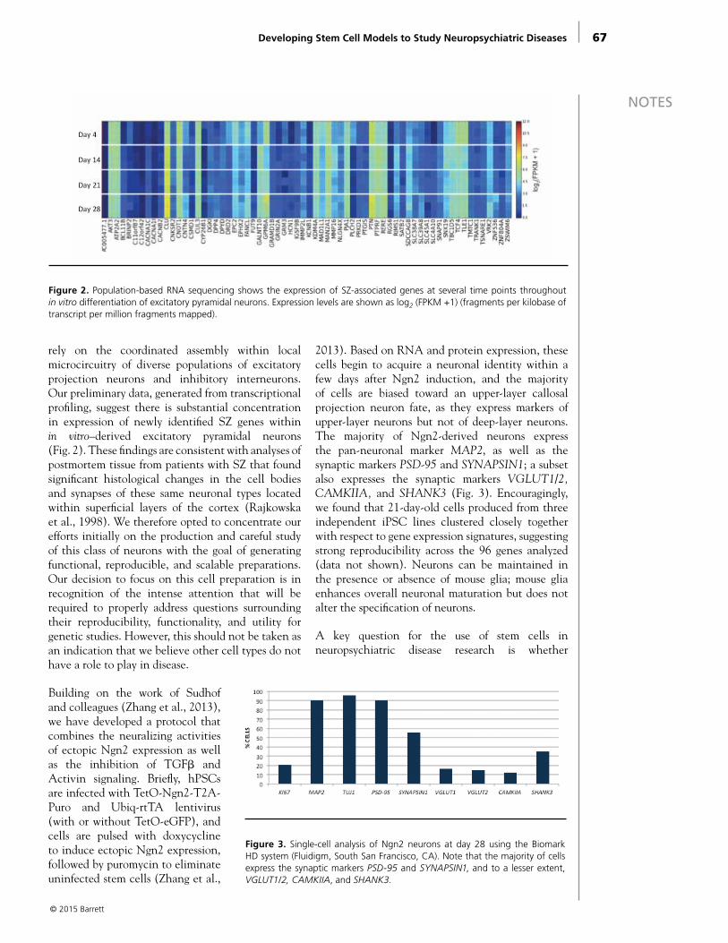
67
NOTES
rely on the coordinated assembly within local microcircuitry of diverse populations of excitatory projection neurons and inhibitory interneurons. Our preliminary data, generated from transcriptional profiling, suggest there is substantial concentration in expression of newly identified SZ genes within in vitro–derived excitatory pyramidal neurons (Fig. 2). These findings are consistent with analyses of postmortem tissue from patients with SZ that found significant histological changes in the cell bodies and synapses of these same neuronal types located within superficial layers of the cortex (Rajkowska et al., 1998). We therefore opted to concentrate our efforts initially on the production and careful study of this class of neurons with the goal of generating functional, reproducible, and scalable preparations. Our decision to focus on this cell preparation is in recognition of the intense attention that will be required to properly address questions surrounding their reproducibility, functionality, and utility for genetic studies. However, this should not be taken as an indication that we believe other cell types do not have a role to play in disease.
Building on the work of Sudhof and colleagues (Zhang et al., 2013), we have developed a protocol that combines the neuralizing activities of ectopic Ngn2 expression as well as the inhibition of TGFβ and Activin signaling. Briefly, hPSCs are infected with TetO-Ngn2-T2A-Puro and Ubiq-rtTA lentivirus (with or without TetO-eGFP), and cells are pulsed with doxycycline to induce ectopic Ngn2 expression, followed by puromycin to eliminate uninfected stem cells (Zhang et al.,
2013). Based on RNA and protein expression, these cells begin to acquire a neuronal identity within a few days after Ngn2 induction, and the majority of cells are biased toward an upper-layer callosal projection neuron fate, as they express markers of upper-layer neurons but not of deep-layer neurons. The majority of Ngn2-derived neurons express the pan-neuronal marker MAP2, as well as the synaptic markers PSD-95 and SYNAPSIN1; a subset also expresses the synaptic markers VGLUT1/2, CAMKIIA, and SHANK3 (Fig. 3). Encouragingly, we found that 21-day-old cells produced from three independent iPSC lines clustered closely together with respect to gene expression signatures, suggesting strong reproducibility across the 96 genes analyzed (data not shown). Neurons can be maintained in the presence or absence of mouse glia; mouse glia enhances overall neuronal maturation but does not alter the specification of neurons.
A key question for the use of stem cells in neuropsychiatric disease research is whether
Developing Stem Cell Models to Study Neuropsychiatric Diseases
© 2015 Barrett
Figure 2. Population-based RNA sequencing shows the expression of SZ-associated genes at several time points throughout in vitro differentiation of excitatory pyramidal neurons. Expression levels are shown as log2 (FPKM +1) (fragments per kilobase of transcript per million fragments mapped).
Figure 3. Single-cell analysis of Ngn2 neurons at day 28 using the Biomark HD system (Fluidigm, South San Francisco, CA). Note that the majority of cells express the synaptic markers PSD-95 and SYNAPSIN1, and to a lesser extent, VGLUT1/2, CAMKIIA, and SHANK3.
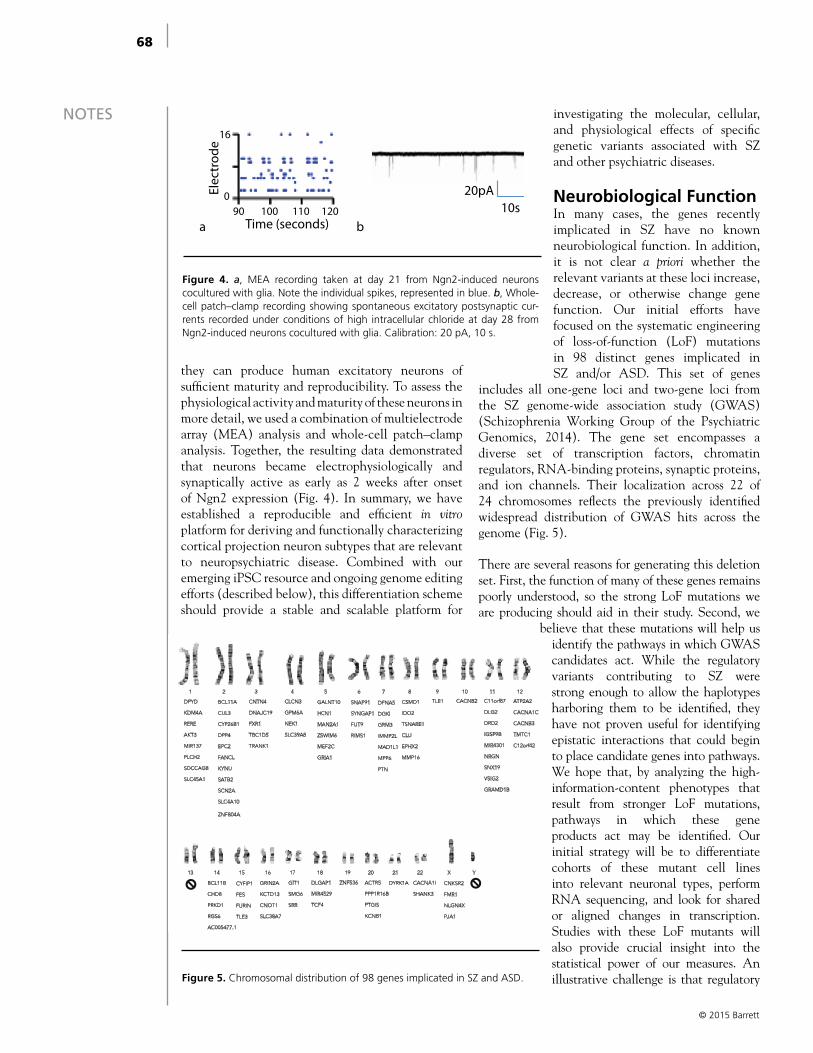
68
NOTES
they can produce human excitatory neurons of sufficient maturity and reproducibility. To assess the physiological activity and maturity of these neurons in more detail, we used a combination of multielectrode array (MEA) analysis and whole-cell patch–clamp analysis. Together, the resulting data demonstrated that neurons became electrophysiologically and synaptically active as early as 2 weeks after onset of Ngn2 expression (Fig. 4). In summary, we have established a reproducible and efficient in vitro platform for deriving and functionally characterizing cortical projection neuron subtypes that are relevant to neuropsychiatric disease. Combined with our emerging iPSC resource and ongoing genome editing efforts (described below), this differentiation scheme should provide a stable and scalable platform for
investigating the molecular, cellular, and physiological effects of specific genetic variants associated with SZ and other psychiatric diseases.
Neurobiological Function In many cases, the genes recently implicated in SZ have no known neurobiological function. In addition, it is not clear a priori whether the relevant variants at these loci increase, decrease, or otherwise change gene function. Our initial efforts have focused on the systematic engineering of loss-of-function (LoF) mutations in 98 distinct genes implicated in SZ and/or ASD. This set of genes
includes all one-gene loci and two-gene loci from the SZ genome-wide association study (GWAS) (Schizophrenia Working Group of the Psychiatric Genomics, 2014). The gene set encompasses a diverse set of transcription factors, chromatin regulators, RNA-binding proteins, synaptic proteins, and ion channels. Their localization across 22 of 24 chromosomes reflects the previously identified widespread distribution of GWAS hits across the genome (Fig. 5).
There are several reasons for generating this deletion set. First, the function of many of these genes remains poorly understood, so the strong LoF mutations we are producing should aid in their study. Second, we
believe that these mutations will help us identify the pathways in which GWAS candidates act. While the regulatory variants contributing to SZ were strong enough to allow the haplotypes harboring them to be identified, they have not proven useful for identifying epistatic interactions that could begin to place candidate genes into pathways. We hope that, by analyzing the high-information-content phenotypes that result from stronger LoF mutations, pathways in which these gene products act may be identified. Our initial strategy will be to differentiate cohorts of these mutant cell lines into relevant neuronal types, perform RNA sequencing, and look for shared or aligned changes in transcription. Studies with these LoF mutants will also provide crucial insight into the statistical power of our measures. An illustrative challenge is that regulatory
© 2015 Barrett
1 2 3 4 5 6 7 8 9 10 11 12
13 14 15 16 17 18 19 20 21 22 X Y
DPYD
KDM4A
RERE
AKT3
MIR137
PLCH2
SDCCAG8
SLC45A1
BCL11A
CUL3
CYP26B1
DPP4
EPC2
FANCL
KYNU
SATB2
SCN2A
SLC4A10 ZNF804A
CNTN4
DNAJC19
FXR1
TBC1D5
TRANK1
CLCN3
GPM6A
NEK1
SLC39A8
GALNT10
HCN1
MAN2A1
ZSWIM6
MEF2C
GRIA1
SNAP91
SYNGAP1
FUT9
RIMS1
DFNA5
DGKI
GRM3
IMMP2L
MAD1L1
MPP6
PTN
CSMD1
IDO2
TSNARE1
CLU
EPHX2
MMP16
TLE1 CACNB2 C11orf87
DLG2
DRD2
IGSF9B
MIR4301
NRGN
SNX19
VSIG2
GRAMD1B
ATP2A2
CACNA1C
CACNB3
TMTC1
C12orf42
BCL11B
CHD8
PRKD1
RGS6
AC005477.1
CYFIP1
FES
FURIN
TLE3
GRIN2A
KCTD13
CNOT1
SLC38A7
GIT1
SMG6
SRR
DLGAP1
MIR4529
TCF4
ZNF536 ACTR5
PPP1R16B
PTGIS
KCNB1
DYRK1A CACNA1I
SHANK3
CNKSR2
FMR1
NLGN4X
PJA1
Figure 5. Chromosomal distribution of 98 genes implicated in SZ and ASD.
a b 90 100 110 120
Time (seconds)
16
0Elec
trod
e
20pA10s
Figure 4. a, MEA recording taken at day 21 from Ngn2-induced neurons cocultured with glia. Note the individual spikes, represented in blue. b, Whole-cell patch–clamp recording showing spontaneous excitatory postsynaptic cur-rents recorded under conditions of high intracellular chloride at day 28 from Ngn2-induced neurons cocultured with glia. Calibration: 20 pA, 10 s.

69
NOTESvariants are predicted to have only modest effects on risk gene expression. By analyzing LoF mutations, we should be able to improve our ability to calculate the number of experimental replicates needed to reach significance at lower bounds of effect size. By measuring the impact of reduced as well as complete loss of gene product, we will be able to calibrate the sensitivity of our transcriptional and functional assays to detect perturbations of various magnitudes. Having calibrated our assays for neuronal function and transcription using hPSC-derived neurons with strong LoF mutants, we hope to ultimately produce brain cell types from hPSCs engineered with individual allelic variants or more-complex patient-derived iPSCs.
Engineering LoF MutationsWe are using CRISPR/Cas9–based genome engineering to generate hPSCs with heterozygous and homozygous LoF mutations. CRISPR/Cas9 technology has made genome engineering accessible for a host of in vitro and in vivo applications (Ran et al., 2013). The bacterial CRISPR/Cas9 system has been adapted to create double-stranded breaks (DSBs) in genomic DNA using sequence-specific small guide RNAs and the Cas9 enzyme. DSBs can be repaired by nonhomologous end-joining, resulting in insertions/deletions (indels) that disrupt a target gene, or by homology-directed repair, which copies a repair template into a target gene. Thus, it is possible to engineer LoF mutations, or insert desired DNA sequences into nearly any location in an organism’s genome.
To generate LoF mutations for the 98 selected SZ and ASD candidates, we aimed to generate indels in early constitutive exons of each gene. We identified early constitutive exons from the Illumina Human Body Map 2.0 and then used the Zhang Lab, MIT, CRISPR
design tool (http://crispr.mit.edu) to select the top two guide pairs for each gene. We next transfected the top guide pair, along with Cas9n-GFP, into the human embryonic stem-cell line 63 (“HUES63”), an XY line of normal karyotype. After purifying GFP+ cells using fluorescence-activated cell sorting, we isolated and expanded 96 individual clones per gene for cell-freezing and deep sequencing. We then used next-generation sequencing to determine the compliment of mutations generated for each gene targeting (Fig. 6). We plan to pair these resources with the differentiation paradigms described above in order to interrogate gene function in human brain cell types.
ConclusionsIn the long term, we will continue to evaluate the validity of a human stem cell platform for studies of neuropsychiatric diseases. While early work has shown promise, we must continue to balance our efforts between understanding this system and using it to further our understanding of biology. Myriad technical variations remain to be explored that could improve the reproducibility and utility of cells types manufactured from stem cells. We must stay abreast of advancements and learning in this area without letting the entirety of our efforts be consumed by them. Like any model system, there are important questions that are well suited to being addressed by stem cell technologies, and others that are not. For instance, the lack of meaningful neural circuitry we can build in vitro clearly limits our ability to draw higher-order conclusions that could connect cellular dysfunction with behavioral manifestations of disease. Despite these limitations, scalability and a human genome allow us to readily approach a number of important questions in neuropsychiatry with these cells. In the future, with meaningful assays in hand
Developing Stem Cell Models to Study Neuropsychiatric Diseases
© 2015 Barrett
0% 10% 20% 30% 40% 50% 60% 70% 80% 90% 100%
ZNF536
TLE1
TCF4
SYNGAP1
SRR
SHANK3
GRIN2A
CACNA1I
CACNA1C
% Clones WT
% Clones HET/MIXED (with FS)
% Clones Null (FS)
% Clones IN FRAME only
% Clones FAIL
Figure 6. Example of mutation efficiency for nine unique loci showing the percentages of wild-type (WT) clones (gray), clones that contain both WT sequences and frame-shift (FS) sequences (red), clones that contain only FS sequences (orange), clones that contain in-frame mutations only (blue), and clones that have ≥ 50% failed reads (black).
70
NOTES and a better understanding of the pathways that are negatively affected by predisposing genetic variants, we hope to be able to port human cell–based assays directly into drug discovery pipelines.
AcknowledgmentsThe primary data included here were produced by past and present lab members and collaborators at the Broad Institute, Harvard University, and MIT, including Ralda Nehme, Ema Zuccaro, Dia Ghosh, Danielle Manning, Jana Mitchell, Nolan Kamitaki, Zhanyan Fu, Chenchen Li, and John Sherwood. The MEA and whole-cell patch–clamp recordings were made in collaboration with Ralda Nehme and Zhanyan Fu. Technical support was provided by Mandy Herring, Nicole Dabkowski, Maggie Bara, and Carrie Ng. We thank our collaborators, including Paola Arlotta, Guoping Feng, Feng Zhang, and Steve McCarroll, as well as leadership at the Stanley Center, including Steve Hyman, Jennifer Moran, and Ed Scolnick. We thank our many colleagues at the Broad Institute, Harvard University, MIT, Novartis Neuroscience, and elsewhere who contributed to this work through helpful discussions and the sharing of resources and reagents. This work is supported by funding from the Stanley Center for Psychiatric Research at the Broad Institute.
References
Chambers SM, Fasano CA, Papapetrou EP, Tomishima M, Sadelain M, Studer L (2009) Highly efficient neural conversion of human ES and iPS cells by dual inhibition of SMAD signaling. Nat Biotechnol 27:275–280.
Drew LJ, Crabtree GW, Markx S, Stark KL, Chaverneff F, Xu B, Mukai J, Fenelon K, Hsu PK, Gogos JA, Karayiorgou M (2011) The 22q11.2 microdeletion: fifteen years of insights into the genetic and neural complexity of psychiatric disorders. Int J Dev Neurosci 29:259–281.
Ohgidani M, Kato TA, Setoyama D, Sagata N, Hashimoto R, Shigenobu K, Yoshida T, Hayakawa K, Shimokawa N, Miura D, Utsumi H, Kanba S (2014) Direct induction of ramified microglia-like cells from human monocytes: dynamic microglial dysfunction in Nasu–Hakola disease. Sci Rep 4:4957.
Purcell SM, Moran JL, Fromer M, Ruderfer D, Solovieff N, Roussos P, O’Dushlaine C, Chambert K, Bergen SE, Kähler A, Duncan L, Stahl E, Genovese G, Fernández E, Collins MO, Komiyama NH, Choudhary JS, Magnusson PK, Banks E, Shakir K, et al. (2014) A polygenic burden of rare disruptive mutations in schizophrenia. Nature 506:185–190.
Rajkowska G, Selemon LD, Goldman-Rakic PS (1998) Neuronal and glial somal size in the prefrontal cortex: a postmortem morphometric study of schizophrenia and Huntington disease. Arch Gen Psychiatry 55:215–224.
Ran FA, Hsu PD, Wright J, Agarwala V, Scott DA, Zhang F (2013) Genome engineering using the CRISPR-Cas9 system. Nat Protoc 8:2281–2308.
Schizophrenia Working Group of the Psychiatric Genomics Consortium (2014) Biological insights from 108 schizophrenia-associated genetic loci. Nature 511:421–427.
Shi Y, Kirwan P, Livesey FJ (2012) Directed differentiation of human pluripotent stem cells to cerebral cortex neurons and neural networks. Nat Protoc 7:1836–1846.
Woodard CM, Campos BA, Kuo SH, Nirenberg MJ, Nestor MW, Zimmer M, Mosharov EV, Sulzer D, Zhou H, Paull D, Clark L, Schadt EE, Sardi SP, Rubin L, Eggan K, Brock M, Lipnick S, Rao M, Chang S, Li A, et al. (2014) iPSC-derived dopamine neurons reveal differences between monozygotic twins discordant for Parkinson’s disease. Cell Rep 9:1173–1182.
Zhang Y, Pak C, Han Y, Ahlenius H, Zhang Z, Chanda S, Marro S, Patzke C, Acuna C, Covy J, Xu W, Yang N, Danko T, Chen L, Wernig M, Sudhof TC (2013) Rapid single-step induction of functional neurons from human pluripotent stem cells. Neuron 78:785–798.
© 2015 Barrett

© 2015 Brennand
Department of Psychiatry Icahn School of Medicine at Mount Sinai
New York, New York
Modeling Predisposition to Schizophrenia, a Genetically
Heterogeneous Neuropsychiatric Disorder, Using Induced
Pluripotent Stem CellsSeok-Man Ho, BSc, Erin Flaherty, BSc, and
Kristen J. Brennand, PhD

73
NOTES
© 2015 Brennand
IntroductionSchizophrenia (SZ) is a debilitating yet relatively common psychiatric disorder, affecting approximately 1% of the world population. Genetic and epigenetic variations underlie differences in clinical outcome and treatment responsiveness (Campbell et al., 2008; Akbarian, 2010; McClay et al., 2011; Ferentinos and Dikeos, 2012; Ota et al., 2012; Hasan et al., 2013). However, the explanation for the 41–65% discordance rate of SZ between monozygotic twins sharing identical genetic predisposition to disease remains unclear (Cardno and Gottesman, 2000).
Environmental stressors, such as cannabis use, maternal immune activation, and birth complications, may also contribute to SZ (Brown, 2011; Connor et al., 2012; Scherr et al., 2012; Torrey et al., 2012), and recent efforts have attempted to combine animal models of the disease with environmental stressors (Kannan et al., 2013). Although these studies provide important insights into biological mechanisms that underlie at least part of the pathology of SZ, it is still difficult to fully recapitulate the heterogeneity of the disease and address the mechanisms of this “human” condition. Studies based on human induced pluripotent stem cells (hiPSCs) have the potential to combine both environmental and genetic influences by using cells from patients with known genetic backgrounds (Brennand et al., 2014a).
Generation of Neuronal SubtypesDirected differentiationDirected differentiation is an in vitro strategy that mimics in vivo development by applying small molecules and/or morphogens that mimic the signaling involved in the patterning, specification, and commitment of defined cell types during embryonic development. Treatment of human embryonic stem cells (hESCs) with Noggin (a bone morphogenetic protein [BMP] inhibitor) and SB431542 (a transforming growth factor beta [TGF-β] inhibitor), so-called “dual SMAD inhibition,” directed 80% of hESCs into a population of neural stem and progenitor cells within 1 week, as assayed by PAX6 and HES5-eGFP (enhanced green fluorescent protein) reporter expression (Chambers et al., 2013). Dual SMAD inhibition is a remarkable method for rapidly differentiating neural populations from hiPSCs, thereby expediting the timeline and purity of neuronal differentiation protocols.
Dopaminergic neuronsDopamine (DA) neurons in the midbrain (mDA neurons) are associated with distinctive neurological
disorders, such as Parkinson’s disease (PD), SZ, and attention deficit hyperactivity disorder (ADHD) (Sillitoe and Vogel, 2008). Dual SMAD inhibition followed by sonic hedgehog (SHH), BDNF, FGF8, and ascorbic acid treatment generates tyrosine hydroxylase (TH)–positive neurons from hiPSCs within 1 month (Chambers et al., 2009). This adherent culture protocol bypasses the embryoid body (EB) stage between hiPSC and neural rosette, and produces three times more TH+ neurons (30%) than the conventional EB protocol (10%) (Boyer et al., 2012). Recent yields have been reported to be as high as 80% (Kriks et al., 2011). Overexpression of progerin (which facilitates cellular aging) in hiPSC-derived mDA neurons derived from PD patients and healthy controls reveals distinctive PD phenotypes, such as reduction of TH+ neurons, dendrite degeneration, and enlarged mitochondria specifically in “geriatric” PD hiPSC mDA neurons (Miller et al., 2013).
Glutamatergic neuronsGlutamatergic neurons in the cerebral cortex, generally represented by pyramidal neurons, are projection neurons that relay information to remote areas of the cerebral cortex and other regions of the brain. Aberrant neuronal connectivity and function of the glutamatergic neurons are believed to increase susceptibility to neuropsychiatric disorders such as autism spectrum disorder (ASD) and SZ (de Bartolomeis et al., 2014). Synergistically with dual SMAD inhibition, activation of retinoic acid signaling (to restrict dorsal forebrain development) specifies hESCs and hiPSCs into cortical stem cell and progenitor cells expressing FOXG1 and EMX1, which are markers for dorsalized forebrain (Shi et al., 2012). The cortical stem cell and progenitor cell population generates 70% class III β-tubulin–positive neurons by 40 d of differentiation; by 50 d, these neurons express VGLUT1, a glutamate synaptic vesicle marker with excitatory synaptic properties in electrophysiology (Shi et al., 2012). An alternative methodology, utilizing FGF2 and inhibitors of BMP, WNT/β-CATENIN, and TGF-β/ACTIVIN/NODAL pathways, also induces hiPSCs into neural progenitor cells with forebrain fate, which can be further differentiated into presynaptic (SYNAPSIN1+) and postsynaptic (PSD95+) excitatory cortical neurons (Mariani et al., 2012). When hESC-derived cortical progenitor cells and cortical pyramidal neurons were transplanted into the cortex of neonatal mice, engrafted cells sent axonal-like projections to multiple brain regions, where they established functional synapses and microcircuits with the host brain, suggesting the functional potential of
Modeling Predisposition to Schizophrenia, a Genetically Heterogeneous Neuropsychiatric Disorder, Using Induced Pluripotent Stem Cells
74
NOTES
© 2015 Brennand
cortical glutamatergic neurons directly differentiated from hiPSCs (Espuny-Camacho et al., 2013).
GABAergic neuronsIn the cerebral cortex, GABAergic interneurons are implicated in neuropsychiatric diseases such as epilepsy, seizure, ASD, and SZ, likely owing to their essential role in fine-tuning and integrating the neural network (Marin, 2012). Cortical GABAergic interneurons initially arise from medial ganglionic eminences (MGEs) of the developing telecephalon, and only subsequently do they migrate into the neocortex (Anderson et al., 2001). To generate MGE neural progenitor cells with potential GABAergic identity, combinatorial inhibition of dual SMAD (SB431542 and LDN-193189) and WNT (XAV939) can be combined with late activation of SHH and differentiated hiPSCs into 80% NKX2 .1-GFP–positive cells, a marker of ventral cells in the developing forebrain MGE (Maroof et al., 2013). If cocultured with mouse cortical neurons, these NKX2 .1-GFP neurons show physiological activity consistent with GABAergic interneurons, and they further differentiate into somatostatin (SST), parvalbumin (PV), and calbindin-positive GABAergic interneurons. In parallel, a similar set of chemical cocktails (dual SMAD inhibition, WNT inhibition [DKK1], and SHH signaling activation) (Nicholas et al., 2013) also gave rise to neurons expressing GABAergic markers. These neurons had functional synaptic properties consistent with GABAergic neurons and were capable of functionally integrating into the microcircuitry of the host brain. Recent data suggest that the addition of caudalizing signal by FGF8 enhances the yield of NKX2 .1-GFP+ cells more than WNT inhibition and SHH activation do alone (Kim et al., 2014).
Generation of hiPSC-derived neurons directed by morphogens and small molecules appears to faithfully reproduce the differentiation process into neuronal subtypes, similar to in vivo development, though a number of limitations of “directed differentiation” persist. First, the supply of recombinant growth factors may not be economically suitable for methodology such as massive high-throughput screening. Second, inefficient signaling activation by chemicals can restrain researchers from the precise combinational modulation required for proper differentiation. Third, the obstacle of spatially and temporally impure and heterogeneous neural subtype specification has not been overcome. Fourth, directed differentiation yields neurons that are immature relative to those in the human brain, with transcriptional profiles most resembling human fetal tissue (Mariani et al., 2012;
Nicholas et al., 2013; Brennand et al., 2015). Finally, directed differentiation protocols require an extended time course of neuronal differentiation—up to 3 months—leading to slow experiment turnaround. Recently, “neuronal induction” has been shown to be a viable alternative strategy that addresses many of these concerns.
Neuronal inductionPatient-derived somatic cells can now be rapidly and directly converted from differentiated cells into neurons. During neurogenesis, a series of proneuronal transcription factors orchestrates the global gene expression network required for cell-fate specification, driving the cellular transition from neural stem/progenitor cells to mature neurons. Expression of key neurogenic regulators is sufficient to induce donor fibroblasts into neurons (iNeurons). In 2009, the Wernig group demonstrated that three proneuronal transcription factors—Ascl1, Brn2, and Myt1l (BAM)—directly converted mouse fibroblasts into heterogeneous but functional neurons in just 20 d (Vierbuchen et al., 2010). In humans, combining NeuroD1 with these three BAM factors induced neurons from human fibroblasts, even though these iNeurons formed fully functional excitatory synapses only when cocultured with mouse primary cortical neurons (Pang et al., 2011). Expression of two micro RNAs (miR-9 and miR-124) with ASCL1, MYT1L, and NEUROD2 improved neuronal induction efficiency, yielding iNeurons with electrical synaptic properties independent of the primary neuron coculture (Yoo et al., 2011).
Induced glutamatergic neuronsExpression of neurogenin 2 (Ngn2), a dorsal telencephalic fate determinant, transdifferentiates cortical astroglial cells into glutamatergic neurons with functional synapses in vitro (Berninger et al., 2007; Heinrich et al., 2010) and hiPSCs into functional iNeurons (Zhang et al., 2013). When combined with selection for Ngn2 expression, more than 90% of cells express MAP2 (mitogen-activated protein 2), a dendritic marker, within 14 d, and, when cocultured with mouse cortical neurons, they elicit electrical characteristics of excitatory synaptic function within 21 d. Furthermore, Ngn2 iNeurons express the glutamatergic synaptic proteins VGLUT2, PSD95, and SYNAPSIN1 and successfully integrate when transplanted into a mouse brain.
Induced dopaminergic neuronsCombinatorial transduction of BAM factors, together with LMX1A and FOXA2 (fate determinants of mDA neurons), yields mDA-like cells that express
75
NOTES
© 2015 Brennand
TH but show poor functionality (Pfisterer et al., 2011). This may reflect the fact that BRN2 (one of the BAM factors) is enriched in pyramidal neurons (Dominguez et al., 2013). Accordingly, transduction of just Ascl1, Lmx1b, and Nurr1 in mouse astrocytes yielded TH+ neurons that secrete DA (Addis et al., 2011). Similar sets of transcription factors induced functional mDA neurons from both mouse and human fibroblasts, which expressed DA machinery components such as VMAT2, DAT, ALDH1A1, and CALBINDIN (Caiazzo et al., 2011; Kim et al., 2011a). Recently, transduction of ASCL1, LMX1A, and NURR1 (ALN) yielded populations of 60% pure DA neurons (TH+, class III β-tubulin+ double neurons) from hiPSCs within 14 d (Theka et al., 2013).
Induced GABAergic neuronsInduction into GABAergic neuronal fate has only just begun. Expression of Ascl1 together with Dlx2, a factor essential for GABAergic neuronal differentiation, is sufficient to transdifferentiate within 21 d mouse astroglial cells to synapse-forming neurons that are positive for GABAergic neuronal markers, including GAD67, calretinin, and vGAT (Heinrich et al., 2010). More recently, coexpression of miR-9 and miR-124, together with MYT1L and three transcription factors enriched in the developing striatum (BCL11B, also known as CTIP2, DLX1, and DLX2), induced human fibroblasts into a population analogous to striatal medium spiny neurons (Victor et al., 2014). These induced GABAergic neurons fire action potential trains with a long delay to initial spike, and if transplanted into the mouse brain, extend projections to the anatomical targets of medium spiny neurons.
One of the major concerns about “neuronal induction” is whether forced expression of neuronal transcription factors will overcome disease-specific deficits in neuronal patterning and/or maturation. An important proof of concept was the demonstration that iNeurons recapitulate the expected AMPA receptor–mediated excitation deficits when generated from mice with neuroligin-3 mutations, reminiscent of the neuronal phenotypes observed in primary neurons from these same mutant mice (Chanda et al., 2013). This finding strongly supports the utility of iNeurons for disease modeling. In contrast with directed differentiation, overexpression of transcription factor facilitates rapid conversion of somatic cells and iPSCs into a variety of functional neuronal subtypes in a dramatically shorter period of time. Furthermore, fate regulator–mediated induction orchestrates a more uniform conversion
process in donor cells, giving rise to relatively homogenous populations of neuronal subtypes. However, the necessity of coculture with other neurons or glial cells to enable synaptic maturation remains an important challenge when considering applying iNeurons to high-throughput screening for drug development.
Using hiPSCs to Link Genetics and Cellular Phenotypes of SchizophreniaSchizophrenia is ideal to study using hiPSC because it is a complex genetic disorder with no single causal mutation. Once neural progenitor cells or neurons are generated from patient hiPSCs, cellular phenotypes can be investigated to gain insight into the cellular mechanisms of SZ. Chiang et al. were the first group to derive and characterize hiPSCs from SZ patients alongside controls (Chiang et al., 2011). Follow-up studies by our group and others examined the neuronal phenotypes from neurons derived from patient hiPSCs. These neurons display decreased neurite outgrowth and synapse formation (Brennand et al., 2011; Robicsek et al., 2013; Wen et al., 2014) and impaired neurotransmitter release (Hook et al., 2014); in contrast, patient-derived neuronal progenitor cells exhibit defects in neuronal migration (Brennand et al., 2014b), deficits with adherens junctions (Yoon et al., 2014), and impaired mitochondrial function (Paulsen et al., 2011; Robicsek et al., 2013; Brennand et al., 2015).
Initial studies of hiPSC-derived neurons were limited by the availability of patient fibroblasts, and therefore, these findings represent a heterogeneous patient population in which little is known about the patients’ genetic or clinical background. Follow-up studies are beginning to take two distinct approaches. The first is to characterize the cellular phenotype of clinically homogeneous patient cohorts. This approach enables researchers to learn about disease mechanisms that are shared across patients who exhibit similar symptoms or have similar drug response profiles. This type of study may lead to finding commonly disrupted pathways that could be relevant across a large subset of SZ patients.
The second approach is to examine a cohort of patients all carrying the same genetic mutation, such as a copy number variant (CNV) (Yoon et al., 2014) or point mutation (Wen et al., 2014). This approach is similar to mouse models because it links cellular phenotypes to pathogenic risk variants. This approach was used to uncover a cellular phenotype of the 15q11.2 CNV; hiPSCs developed from a patient with a 15q11.2 microdeletion displayed
Modeling Predisposition to Schizophrenia, a Genetically Heterogeneous Neuropsychiatric Disorder, Using Induced Pluripotent Stem Cells
76
NOTES
© 2015 Brennand
deficits in adherens junctions and apical polarity (Yoon et al., 2014). This approach was also used to study a CNV at 7q11.23, where deletion results in Williams–Beuren syndrome, whereas duplication causes 7q-microduplication syndrome. Examination of patient-derived hiPSCs and the differentiated cell types showed dosage-dependent alterations in disease-relevant transcriptional circuits (Adamo et al., 2015). This group also investigated the role of a transcription factor, GTF2l, which is disrupted by the CNV, and found that manipulating GTF2l caused a significant proportion of the transcription dysregulation created by the entire CNV (Adamo et al., 2015).
Another useful tool for this second approach is genome editing technology, such as CRISPR/Cas9 (CRISPR signifies clustered regularly interspaced short palindromic repeats; Cas9 is a class of RNA-guided endonucleases). This technology provides a simple, efficient approach for introducing genetic mutations, correcting genetic mutations (Cong et al., 2013; Jiang et al., 2013), and even enhancing (Maeder et al., 2013) or repressing (Larson et al., 2013) gene expression (Hsu et al., 2014). For example, in order to confirm that the synaptic defects observed in two psychiatric patients resulted from the identified DISC1 frameshift mutation, Wen et al. produced isogenic hiPSC lines, both engineering the DISC1 mutation into a control hiPSC line and repairing the mutation in a DISC1 patient hiPSC line. In this way, they showed precisely that mutant DISC1 causes synaptic vesicle release deficits and dysregulates the expression of many genes related to synapses and psychiatric disorders in hiPSC-derived forebrain neurons (Wen et al., 2014).
CRISPR/Cas9 enables investigation into the casual relationship between genotype and neuronal phenotypes because isogenic control lines can be derived, leaving the rest of the patient’s genome unchanged, to ensure that a particular genetic variant is casual to the cellular phenotype (Martinez et al., 2015). Patients with the same variant but different genetic backgrounds can help researchers understand the contribution that additional risk alleles make to a cellular phenotype. Even the role of single genes within a CNV can be theoretically assessed in patient-derived hiPSCs by using CRISPR-Cas9 to replace or delete copies of single genes while maintaining the rest of the disrupted locus. Such studies could help determine the genetic elements and cell types that are most important for patients with SZ who carry a known neuropsychiatric risk allele, leading to more-targeted treatment of these patients.
Constraints of hiPSC-Based StudiesIn designing hiPSC-based studies, it is important to be aware that genetic mutations and epigenetic misremodeling can occur during the reprogramming process. First, both CNVs (Laurent et al., 2011; Liu et al., 2014; Lu et al., 2014) and somatic coding mutations (Gore et al., 2011) will change the donor DNA. Importantly, more CNVs are present in early-passage hiPSCs than in higher-passage hiPSCs, implying that most novel CNVs generated during the reprogramming process are lost before any neuronal differentiation would occur (Hussein et al., 2011). Across 22 hiPSC lines reprogrammed using five different methods, each contained an average of five protein-coding point mutations, though at least half of these reprogramming-associated mutations previously existed in fibroblast progenitors at low frequencies (Gore et al., 2011).
Second, at the epigenetic level, evidence now demonstrates that aberrant DNA methylation remodeling (Lister et al., 2011; Nazor et al., 2012; Ma et al., 2014) and an erosion of X chromosome inactivation (Mekhoubad et al., 2012; Nazor et al., 2012) can occur in hiPSCs. Consistent with this finding, evidence suggests that donor cell type can influence the epigenome and differentiation potential of hiPSCs (Bar-Nur et al., 2011; Kim et al., 2011b). These genetic and epigenetic effects contribute to the “intraindividual variation” observed in hiPSC-based studies that exists because each hiPSC line generated from a given person will show subtle differences in gene expression and propensity toward neural differentiation.
Conversely, “interindividual variation” represents biological differences between individuals and can be addressed by studying ever-larger cohorts of patients and controls, thereby better capturing the heterogeneity among individuals. Unpublished data by our group and others suggest that intrapatient variability is less than interpatient variability. Therefore, well-designed and controlled experiments are critical to ensuring that researchers can draw meaningful conclusions from hiPSC-based studies of psychiatric disorders. To this end, we recommend that at least three hiPSC lines be compared per individual, in order to reduce the likelihood that a rare genetic or epigenetic mutation might affect disease-specific hiPSC lines in a meaningfully different way than they affect control hiPSC lines.
When investigating the effect of a single disease-associated allele, whether in the context of a simple Mendelian disorder or a complex genetic disease, an
77
NOTES
© 2015 Brennand
alternative to increasing cohort size is to compare isogenic hiPSC lines. In fact, a burst of recent hiPSC-based studies has used isogenic controls to demonstrate the precise effects of a single gene on neural phenotypes or gene expression (Liu et al., 2012; Wen et al., 2014).
Perhaps most critically, although recent studies have reported the importance of heritable genetic factors to neuropsychiatric disease and modeled the correlation between these risk factors and disease phenotype, it is still challenging to unravel the causality of environmental risk factors such as stressful life events, social anxiety, and neurotrauma (Howes et al., 2004). These factors remain infeasible to recapitulate in existing cell-based systems in vitro. Moreover, the question remains as to how to link relatively simple cellular phenotypes from hiPSC-derived neurons with the complex behavioral phenotypes of neuropsychiatric patients, which encompass delusions, hallucinations, negative affect, and impaired cognition. One strategy will be to build increasing complexity into hiPSC-based models. Future models will necessarily incorporate neuronal circuits that comprise at least two distinct neuronal cell types, synapsed in a defined orientation, together with oligodendrocytes to provide myelination, and astrocytes and microglia to incorporate critical aspects of inflammation and synaptic pruning. Circuits will need to be stimulated repeatedly to establish plasticity and exposed to meaningful levels of stress hormones and other environmental factors. A second strategy will be to transplant each of these relevant human cell types (neurons, astrocytes, oligodendrocytes, and microglia) into mouse models of disease, yielding increasingly humanized platforms for study. The ultimate solution, of course, will be in pursuing all the strategies we have discussed in tandem: larger cohorts, isogenic controls, improved patterning, and maturation of a variety of human neural cell types, cultured either as artificial circuits or transplanted into mice. Although models, by definition, must always lack the intricacies of human disease, the goal of hiPSC scientists should always be to strive for ever-increasing complexity in their models.
Limitations Due to Clinical HeterogeneityThe sample size of hiPSC-based studies remains relatively very small compared with the standards of genome-wide association studies of complex genetic disorders. Although ultimately, methods will have to be developed to permit comparisons among thousands of patients, to date, technical constraints have
greatly limited hiPSC generation, neural progenitor cell differentiation, and cellular phenotyping (listed in order of increasing difficulty). Consequently, given the small sample size (typically one to four patients) of recent hiPSC-based studies, a major concern is whether the findings are representative of the larger patient population. Interpatient variability results from the heterogeneity among patients with SZ. As mentioned earlier, there are two strategies for hiPSC disease modeling of heterogeneous patient populations: (1) using a genetically homogenous patient cohort that share a single genetic lesion and characterizing the effect relative to isogenic lines generated through gene targeting technologies; and (2) selecting a patient cohort on the basis of a shared clinical phenotype and comparing them with individuals without the phenotype. The first strategy parallels traditional mouse-based studies of SZ that investigate the effects of rare loci, while the second takes full advantage of the ability of hiPSC-based studies to investigate complex genetic disorders without full knowledge of all the genes involved.
By modeling specific aspects of SZ rather than capturing the entire diversity of this disorder, researchers might be able to reduce the interpatient variability in hiPSC-based studies. In the short term, researchers can address this problem by selecting homogenous patient cohorts characterized by common genetic mutations, or by shared neurophysiological endophenotypes and/or pharmacological responses. Neurophysiological characterization of patients with SZ has identified abnormal responses to paired auditory stimuli (Geyer et al., 1990; Freedman et al., 1997) and defects in oculomotor control (Radant et al., 1997), though growing evidence suggests these endophenotypes may be heritable (Greenwood et al., 2012). Although strong evidence supports the pharmacogenetics of lithium-responsive bipolar disorder (McCarthy et al., 2010), new data support the heritability of antipsychotic resistance in SZ as well (Ota et al., 2012). Of course, additional factors ranging from epigenetic effects to circuit-based plasticity (derived from experience) may contribute to the heterogeneity of SZ. Nonetheless, though such risk factors are likely to be lost as part of the reprogramming process, we predict that some (even if not all) key mechanisms contributing to SZ can be studied using hiPSCs.
Multiple levels of etiology might contribute to SZ, increasing from biochemical to cellular to neuronal network and brain circuits. As the complexity of this “causal action” grows, it will become more difficult to resolve biological meaning through cell-based
Modeling Predisposition to Schizophrenia, a Genetically Heterogeneous Neuropsychiatric Disorder, Using Induced Pluripotent Stem Cells

78
NOTES
© 2015 Brennand
models (Kendler, 2013). Critics may also question the relevance of months-old in vitro–derived neurons in the study of SZ, a condition whose hallmark symptom of psychosis typically appears in late adolescence. For this reason, we posit that current hiPSC-based approaches may be most appropriately used in the study of SZ predisposition.
Future Considerations and ConclusionsFuture advancements in developing new therapeutics for SZ, or any other neurological disorder, will rely on an understanding of commonly disrupted pathways. These pathways can be uncovered through several methods:
• Careful integration of human genetic studies to identify disease-related variants;
• Studying mouse models to examine the function of a variant in a complex in vivo environment; and
• Investigating patient-derived hiPSC models to determine the casual role of a variant on a cellular phenotype and the contribution made by complex genetic interactions.
Incorporating this type of information from studies investigating CNVs in SZ has begun to implicate specific pathways, such as genes regulating NMDA receptors and ARC (activator recruited cofactor) complexes (Purcell et al., 2014; Szatkiewicz et al., 2014). This knowledge can inform the development of new therapeutics. However, the potential of hiPSCs does not end with mechanistic studies but rather continues into clinical trials where hiPSCs can be used as a complementary human in vitro component.
Before the full potential of hiPSCs can be realized, a large amount of work remains to be done. Optimized protocols for more-homogeneous populations of hiPSCs and pure populations of differentiated cells need to be fully developed and validated. Doing so will decrease the time and cost needed to develop patient-derived hiPSC models, allowing for more patient lines to be examined. Improved culturing techniques should be applied to form more complex neural circuitries that better resemble the human brain. This is currently an area of great interest in the context of the development of human cortical spheroids (Pasca et al., 2015). Finally, experiments investigating complex diseases should be designed to collect multilayer data, including the patient’s clinical information, genome, transcriptome, proteome, and
epigenome (Schadt et al., 2014). Gathering this type of multilayered information is now possible with the advancements being made in sequencing technology and bioinformatics. From this information, we can begin to build networks and identify the key drivers of complex genetic disorders such as SZ.
AcknowledgmentsParts of this paper are excerpted from other publications. The sections “Generation of Neuronal Subtypes” and “Constraints of hiPSC-Based Studies” are excerpted from Ho et al., 2015, From “directed differentiation” to “neuronal induction”: modeling neuropsychiatric disease. Biomark Insights 10(Suppl 1):31–41. The section “Limitations Due to Clinical Heterogeneity” is excerpted from Brennand et al., 2014a, Modeling heterogeneous patients with a clinical diagnosis of schizophrenia with induced pluripotent stem cells. Biol Psychiatry 75:936–944. Finally, the sections “Using hiPSCs to Link Genetics and Cellular Phenotypes of Schizophrenia” and “Future Considerations and Conclusions” are excerpted from Flaherty and Brennand, 2015 (manuscript in preparation).
ReferencesAdamo A, Atashpaz S, Germain PL, Zanella M,
D’Agostino G, Albertin V, Chenoweth J, Micale L, Fusco C, Unger C, Augello B, Palumbo O, Hamilton B, Carella M, Donti E, Pruneri G, Selicorni A, Biamino E, Prontera P, McKay R, et al. (2015) 7q11.23 dosage-dependent dysregulation in human pluripotent stem cells affects transcriptional programs in disease-relevant lineages. Nat Genet 47:132–141.
Addis RC, Hsu FC, Wright RL, Dichter MA, Coulter DA, Gearhart JD (2011) Efficient conversion of astrocytes to functional midbrain dopaminergic neurons using a single polycistronic vector. PLoS One 6:e28719.
Akbarian S (2010) The molecular pathology of schizophrenia—focus on histone and DNA modifications. Brain Res Bull 83:103–107.
Anderson SA, Marin O, Horn C, Jennings K, Rubenstein JL (2001) Distinct cortical migrations from the medial and lateral ganglionic eminences. Development 128:353–363.
Bar-Nur O, Russ HA, Efrat S, Benvenisty N (2011) Epigenetic memory and preferential lineage-specific differentiation in induced pluripotent stem cells derived from human pancreatic islet beta cells. Cell Stem Cell 9:17–23.
79
NOTES
Modeling Predisposition to Schizophrenia, a Genetically Heterogeneous Neuropsychiatric Disorder, Using Induced Pluripotent Stem Cells
© 2015 Brennand
Berninger B, Costa MR, Koch U, Schroeder T, Sutor B, Grothe B, Gotz M (2007) Functional properties of neurons derived from in vitro reprogrammed postnatal astroglia. J Neurosci 27:8654–8664.
Boyer LF, Campbell B, Larkin S, Mu Y, Gage FH (2012) Dopaminergic differentiation of human pluripotent cells. Curr Protoc Stem Cell Biol Chapter 1:Unit1H 6.
Brennand KJ, Simone A, Jou J, Gelboin-Burkhart C, Tran N, Sangar S, Li Y, Mu Y, Chen G, Yu D, McCarthy S, Sebat J, Gage FH (2011) Modelling schizophrenia using human induced pluripotent stem cells. Nature 473:221–225.
Brennand KJ, Landek-Salgado MA, Sawa A (2014a) Modeling heterogeneous patients with a clinical diagnosis of schizophrenia with induced pluripotent stem cells. Biol Psychiatry 75:936–944.
Brennand KJ, Silvas J, Kim Y, Tran N, Simone A, Ladran I, Beaumont K, Kim H-J, Mrksich M, Fang G, Zhang B, Yates JR 3rd, Gage FH (2014b) Phenotypic differences in hiPSC NPCs derived from patients with schizophrenia. Mol Psychiatry 20:361–368.
Brennand K, Savas JN, Kim Y, Tran N, Simone A, Hashimoto-Torii K, Beaumont KG, Kim HJ, Topol A, Ladran I, Abdelrahim M, Matikainen-Ankney B, Chao SH, Mrksich M, Rakic P, Fang G, Zhang B, Yates JR 3rd, Gage FH (2015) Phenotypic differences in hiPSC NPCs derived from patients with schizophrenia. Mol Psychiatry 20:361–368.
Brown AS (2011) The environment and susceptibility to schizophrenia. Prog Neurobiol 93:23–58.
Caiazzo M, Dell’Anno MT, Dvoretskova E, Lazarevic D, Taverna S, Leo D, Sotnikova TD, Menegon A, Roncaglia P, Colciago G, Russo G, Carninci P, Pezzoli G, Gainetdinov RR, Gustincich S, Dityatev A, Broccoli V (2011) Direct generation of functional dopaminergic neurons from mouse and human fibroblasts. Nature 476:224–227.
Campbell DB, Ebert PJ, Skelly T, Stroup TS, Lieberman J, Levitt P, Sullivan PF (2008) Ethnic stratification of the association of RGS4 variants with antipsychotic treatment response in schizophrenia. Biol Psychiatry 63:32–41.
Cardno AG, Gottesman II (2000) Twin studies of schizophrenia: from bow-and-arrow concordances to star wars Mx and functional genomics. Am J Med Genet 97:12–17.
Chambers SM, Fasano CA, Papapetrou EP, Tomishima M, Sadelain M, Studer L (2009) Highly efficient neural conversion of human ES and iPS cells by dual inhibition of SMAD signaling. Nat Biotechnol 27:275–280.
Chambers SM, Mica Y, Lee G, Studer L, Tomishima MJ (2013) Dual-SMAD Inhibition/WNT activation-based methods to induce neural crest and derivatives from human pluripotent stem cells. Methods Mol Biol 1307:329–343.
Chanda S, Marro S, Wernig M, Südhof TC (2013) Neurons generated by direct conversion of fibroblasts reproduce synaptic phenotype caused by autism-associated neuroligin-3 mutation. Proc Natl Acad Sci USA 110:16622–16627.
Chiang CH, Su Y, Wen Z, Yoritomo N, Ross CA, Margolis RL, Song H, Ming GL (2011) Integration-free induced pluripotent stem cells derived from schizophrenia patients with a DISC1 mutation. Mol Psychiatry 16:358–360.
Cong L, Ran FA, Cox D, Lin S, Barretto R, Habib N, Hsu PD, Wu X, Jiang W, Marraffini LA, Zhang F (2013) Multiplex genome engineering using CRISPR/Cas systems. Science 339:819–823.
Connor CM, Dincer A, Straubhaar J, Galler JR, Houston IB, Akbarian S (2012) Maternal immune activation alters behavior in adult offspring, with subtle changes in the cortical transcriptome and epigenome. Schizophr Res 140:175–184.
de Bartolomeis A, Latte G, Tomasetti C, Iasevoli F (2014) Glutamatergic postsynaptic density protein dysfunctions in synaptic plasticity and dendritic spines morphology: relevance to schizophrenia and other behavioral disorders pathophysiology, and implications for novel therapeutic approaches. Mol Neurobiol 49:484–511.
Dominguez MH, Ayoub AE, Rakic P (2013) POU-III transcription factors (Brn1, Brn2, and Oct6) influence neurogenesis, molecular identity, and migratory destination of upper-layer cells of the cerebral cortex. Cereb Cortex 23:2632–2643.
Espuny-Camacho I, Michelsen KA, Gall D, Linaro D, Hasche A, Bonnefont J, Bali C, Orduz D, Bilheu A, Herpoel A, Lambert N, Gaspard N, Peron S, Schiffmann SN, Giugliano M, Gaillard A, Vanderhaeghen P (2013) Pyramidal neurons derived from human pluripotent stem cells integrate efficiently into mouse brain circuits in vivo. Neuron 77:440–456.
80
NOTES
© 2015 Brennand
Ferentinos P, Dikeos D (2012) Genetic correlates of medical comorbidity associated with schizophrenia and treatment with antipsychotics. Curr Opin Psychiatry 25:381–390.
Freedman R, Coon H, Myles-Worsley M, Orr-Urtreger A, Olincy A, Davis A, Polymeropoulos M, Holik J, Hopkins J, Hoff M, Rosenthal J, Waldo MC, Reimherr F, Wender P, Yaw J, Young DA, Breese CR, Adams C, Patterson D, Adler LE, et al. (1997) Linkage of a neurophysiological deficit in schizophrenia to a chromosome 15 locus. Proc Natl Acad Sci USA 94:587–592.
Geyer MA, Swerdlow NR, Mansbach RS, Braff DL (1990) Startle response models of sensorimotor gating and habituation deficits in schizophrenia. Brain Res Bull 25:485–498.
Gore A, Li Z, Fung HL, Young JE, Agarwal S, Antosiewicz-Bourget J, Canto I, Giorgetti A, Israel MA, Kiskinis E, Lee JH, Loh YH, Manos PD, Montserrat N, Panopoulos AD, Ruiz S, Wilbert ML, Yu J, Kirkness EF, Izpisua Belmonte JC, et al. (2011) Somatic coding mutations in human induced pluripotent stem cells. Nature 471:63–67.
Greenwood TA, Light GA, Swerdlow NR, Radant AD, Braff DL (2012) Association analysis of 94 candidate genes and schizophrenia-related endophenotypes. PLoS One 7:e29630.
Hasan A, Mitchell A, Schneider A, Halene T, Akbarian S (2013) Epigenetic dysregulation in schizophrenia: molecular and clinical aspects of histone deacetylase inhibitors. Eur Arch Psychiatry Clin Neurosci 263:273–284.
Heinrich C, Blum R, Gascon S, Masserdotti G, Tripathi P, Sanchez R, Tiedt S, Schroeder T, Gotz M, Berninger B (2010) Directing astroglia from the cerebral cortex into subtype specific functional neurons. PLoS Biol 8:e1000373.
Ho SM, Topol A, Brennand KJ (2015) From “directed differentiation” to “neuronal induction”: modeling neuropsychiatric disease. Biomark Insights 10(Suppl 1):31–41.
Hook V, Brennand K, Kim Y, Toneff T, Funkelstein L, Ziegler M, Gage FH (2014) Human iPSC neurons display activity-dependent neurotransmitter secretion: aberrant catecholamine levels in schizophrenia neurons. Stem Cell Reports 3:531–538.
Howes OD, McDonald C, Cannon M, Arseneault L, Boydell J, Murray RM (2004) Pathways to schizophrenia: the impact of environmental factors. Int J Neuropsychopharmacol 7(Suppl 1):S7–S13.
Hsu PD, Lander ES, Zhang F (2014) Development and applications of CRISPR-Cas9 for genome engineering. Cell 157:1262–1278.
Hussein SM, Batada NN, Vuoristo S, Ching RW, Autio R, Narva E, Ng S, Sourour M, Hamalainen R, Olsson C, Lundin K, Mikkola M, Trokovic R, Peitz M, Brustle O, Bazett-Jones DP, Alitalo K, Lahesmaa R, Nagy A, Otonkoski T (2011) Copy number variation and selection during reprogramming to pluripotency. Nature 471:58–62.
Jiang W, Bikard D, Cox D, Zhang F, Marraffini LA (2013) RNA-guided editing of bacterial genomes using CRISPR-Cas systems. Nat Biotechnol 31:233–239.
Kannan G, Sawa A, Pletnikov MV (2013) Mouse models of gene–environment interactions in schizophrenia. Neurobiol Dis 57:5–11.
Kendler KS (2013) What psychiatric genetics has taught us about the nature of psychiatric illness and what is left to learn. Mol Psychiatry 18:1058–1066.
Kim J, Su SC, Wang H, Cheng AW, Cassady JP, Lodato MA, Lengner CJ, Chung CY, Dawlaty MM, Tsai LH, Jaenisch R (2011a) Functional integration of dopaminergic neurons directly converted from mouse fibroblasts. Cell Stem Cell 9:413–419.
Kim K, Zhao R, Doi A, Ng K, Unternaehrer J, Cahan P, Huo H, Loh YH, Aryee MJ, Lensch MW, Li H, Collins JJ, Feinberg AP, Daley GQ (2011b) Donor cell type can influence the epigenome and differentiation potential of human induced pluripotent stem cells. Nat Biotechnol 29:1117–1119.
Kim TG, Yao R, Monnell T, Cho JH, Vasudevan A, Koh A, Peeyush KT, Moon M, Datta D, Bolshakov VY, Kim KS, Chung S (2014) Efficient specification of interneurons from human pluripotent stem cells by dorsoventral and rostrocaudal modulation. Stem Cells 32:1789–1804.
Kriks S, Shim JW, Piao J, Ganat YM, Wakeman DR, Xie Z, Carrillo-Reid L, Auyeung G, Antonacci C, Buch A, Yang L, Beal MF, Surmeier DJ, Kordower JH, Tabar V, Studer L (2011) Dopamine neurons derived from human ES cells efficiently engraft in animal models of Parkinson’s disease. Nature 480:547–551.
Larson MH, Gilbert LA, Wang X, Lim WA, Weissman JS, Qi LS (2013) CRISPR interference (CRISPRi) for sequence-specific control of gene expression. Nat Protoc 8:2180–2196.

81
NOTES
Modeling Predisposition to Schizophrenia, a Genetically Heterogeneous Neuropsychiatric Disorder, Using Induced Pluripotent Stem Cells
© 2015 Brennand
Laurent LC, Ulitsky I, Slavin I, Tran H, Schork A, Morey R, Lynch C, Harness JV, Lee S, Barrero MJ, Ku S, Martynova M, Semechkin R, Galat V, Gottesfeld J, Izpisua Belmonte JC, Murry C, Keirstead HS, Park HS, Schmidt U, et al. (2011) Dynamic changes in the copy number of pluripotency and cell proliferation genes in human ESCs and iPSCs during reprogramming and time in culture. Cell Stem Cell 8:106–118.
Lister R, Pelizzola M, Kida YS, Hawkins RD, Nery JR, Hon G, Antosiewicz-Bourget J, O’Malley R, Castanon R, Klugman S, Downes M, Yu R, Stewart R, Ren B, Thomson JA, Evans RM, Ecker JR (2011) Hotspots of aberrant epigenomic reprogramming in human induced pluripotent stem cells. Nature 471:68–73.
Liu GH, Qu J, Suzuki K, Nivet E, Li M, Montserrat N, Yi F, Xu X, Ruiz S, Zhang W, Wagner U, Kim A, Ren B, Li Y, Goebl A, Kim J, Soligalla RD, Dubova I, Thompson J, Yates J 3rd, et al. (2012) Progressive degeneration of human neural stem cells caused by pathogenic LRRK2. Nature 491:603–607.
Liu P, Kaplan A, Yuan B, Hanna JH, Lupski JR, Reiner O (2014) Passage number is a major contributor to genomic structural variations in mouse iPSCs. Stem Cells 32:2657–2667.
Lu J, Li H, Hu M, Sasaki T, Baccei A, Gilbert DM, Liu JS, Collins JJ, Lerou PH (2014) The distribution of genomic variations in human iPSCs is related to replication-timing reorganization during reprogramming. Cell Rep 7:70–78.
Ma H, Morey R, O’Neil RC, He Y, Daughtry B, Schultz MD, Hariharan M, Nery JR, Castanon R, Sabatini K, Thiagarajan RD, Tachibana M, Kang E, Tippner-Hedges R, Ahmed R, Gutierrez NM, Van Dyken C, Polat A, Sugawara A, Sparman M, et al. (2014) Abnormalities in human pluripotent cells due to reprogramming mechanisms. Nature 511:177–183.
Maeder ML, Linder SJ, Cascio VM, Fu Y, Ho QH, Joung JK (2013) CRISPR RNA-guided activation of endogenous human genes. Nat Methods 10:977–979.
Mariani J, Simonini MV, Palejev D, Tomasini L, Coppola G, Szekely AM, Horvath TL, Vaccarino FM (2012) Modeling human cortical development in vitro using induced pluripotent stem cells. Proc Natl Acad Sci USA 109:12770–12775.
Marin O (2012) Interneuron dysfunction in psychiatric disorders. Nat Rev Neurosci 13:107–120.
Maroof AM, Keros S, Tyson JA, Ying SW, Ganat YM, Merkle FT, Liu B, Goulburn A, Stanley EG, Elefanty AG, Widmer HR, Eggan K, Goldstein PA, Anderson SA, Studer L (2013) Directed differentiation and functional maturation of cortical interneurons from human embryonic stem cells. Cell Stem Cell 12:559–572.
Martinez RA, Stein JL, Krostag AR, Nelson AM, Marken JS, Menon V, May RC, Yao Z, Kaykas A, Geschwind DH, Grimley JS (2015) Genome engineering of isogenic human ES cells to model autism disorders. Nucleic Acids Res 43:e65.
McCarthy MJ, Leckband SG, Kelsoe JR (2010) Pharmacogenetics of lithium response in bipolar disorder. Pharmacogenomics 11:1439–1465.
McClay JL, Adkins DE, Aberg K, Stroup S, Perkins DO, Vladimirov VI, Lieberman JA, Sullivan PF, van den Oord EJ (2011) Genome-wide pharmacogenomic analysis of response to treatment with antipsychotics. Mol Psychiatry 16:76–85.
Mekhoubad S, Bock C, de Boer AS, Kiskinis E, Meissner A, Eggan K (2012) Erosion of dosage compensation impacts human iPSC disease modeling. Cell Stem Cell 10:595–609.
Miller JD, Ganat YM, Kishinevsky S, Bowman RL, Liu B, Tu EY, Mandal PK, Vera E, Shim JW, Kriks S, Taldone T, Fusaki N, Tomishima MJ, Krainc D, Milner TA, Rossi DJ, Studer L (2013) Human iPSC-based modeling of late-onset disease via progerin-induced aging. Cell Stem Cell 13:691–705.
Nazor KL, Altun G, Lynch C, Tran H, Harness JV, Slavin I, Garitaonandia I, Müller FJ, Wang YC, Boscolo FS, Fakunle E, Dumevska B, Lee S, Park HS, Olee T, D'Lima DD, Semechkin R, Parast MM, Galat V, Laslett AL, et al. (2012) Recurrent variations in DNA methylation in human pluripotent stem cells and their differentiated derivatives. Cell Stem Cell 10:620–634.
Nicholas CR, Chen J, Tang Y, Southwell DG, Chalmers N, Vogt D, Arnold CM, Chen YJ, Stanley EG, Elefanty AG, Sasai Y, Alvarez-Buylla A, Rubenstein JL, Kriegstein AR (2013) Functional maturation of hPSC-derived forebrain interneurons requires an extended timeline and mimics human neural development. Cell Stem Cell 12:573–586.
82
NOTES
© 2015 Brennand
Ota VK, Spindola LN, Gadelha A, dos Santos Filho AF, Santoro ML, Christofolini DM, Bellucco FT, Ribeiro-dos-Santos AK, Santos S, Mari Jde J, Melaragno MI, Bressan RA, Smith Mde A, Belangero SI (2012) DRD1 rs4532 polymorphism: a potential pharmacogenomic marker for treatment response to antipsychotic drugs. Schizophr Res 142:206–208.
Pang ZP, Yang N, Vierbuchen T, Ostermeier A, Fuentes DR, Yang TQ, Citri A, Sebastiano V, Marro S, Sudhof TC, Wernig M (2011) Induction of human neuronal cells by defined transcription factors. Nature 476:220–223.
Pasca AM, Sloan SA, Clarke LE, Tian Y, Makinson CD, Huber N, Kim CH, Park JY, O’Rourke NA, Nguyen KD, Smith SJ, Huguenard JR, Geschwind DH, Barres BA, Pasca SP (2015) Functional cortical neurons and astrocytes from human pluripotent stem cells in 3D culture. Nat Methods 12:671–678.
Paulsen BD, Maciel RD, Galina A, da Silveira MS, Souza CD, Drummond H, Pozzato EN, Junior HS, Chicaybam L, Massuda R, Setti-Perdigao P, Bonamino M, Belmonte-de-Abreu PS, Castro NG, Brentani H, Rehen SK (2011) Altered oxygen metabolism associated to neurogenesis of induced pluripotent stem cells derived from a schizophrenic patient. Cell Transplant 21:1547–1559.
Pfisterer U, Kirkeby A, Torper O, Wood J, Nelander J, Dufour A, Bjorklund A, Lindvall O, Jakobsson J, Parmar M (2011) Direct conversion of human fibroblasts to dopaminergic neurons. Proc Natl Acad Sci USA 108:10343–10348.
Purcell SM, Moran JL, Fromer M, Ruderfer D, Solovieff N, Roussos P, O’Dushlaine C, Chambert K, Bergen SE, Kähler A, Duncan L, Stahl E, Genovese G, Fernández E, Collins MO, Komiyama NH, Choudhary JS, Magnusson PK, Banks E, Shakir K, et al. (2014) A polygenic burden of rare disruptive mutations in schizophrenia. Nature 506:185–190.
Radant AD, Claypoole K, Wingerson DK, Cowley DS, Roy-Byrne PP (1997) Relationships between neuropsychological and oculomotor measures in schizophrenia patients and normal controls. Biol Psychiatry 42:797–805.
Robicsek O, Karry R, Petit I, Salman-Kesner N, Muller FJ, Klein E, Aberdam D, Ben-Shachar D (2013) Abnormal neuronal differentiation and mitochondrial dysfunction in hair follicle-derived induced pluripotent stem cells of schizophrenia patients. Mol Psychiatry 18:1067–1076.
Schadt EE, Buchanan S, Brennand KJ, Merchant KM (2014) Evolving toward a human-cell based and multiscale approach to drug discovery for CNS disorders. Frontiers in Pharmacol 5:252.
Scherr M, Hamann M, Schwerthoffer D, Frobose T, Vukovich R, Pitschel-Walz G, Bauml J (2012) Environmental risk factors and their impact on the age of onset of schizophrenia: comparing familial to non-familial schizophrenia. Nord J Psychiatry 66:107–114.
Shi Y, Kirwan P, Smith J, Robinson HP, Livesey FJ (2012) Human cerebral cortex development from pluripotent stem cells to functional excitatory synapses. Nat Neurosci 15:477–486, S471.
Sillitoe RV, Vogel MW (2008) Desire, disease, and the origins of the dopaminergic system. Schizophr Bull 34:212–219.
Szatkiewicz JP, O’Dushlaine C, Chen G, Chambert K, Moran JL, Neale BM, Fromer M, Ruderfer D, Akterin S, Bergen SE, Kähler A, Magnusson PK, Kim Y, Crowley JJ, Rees E, Kirov G, O’Donovan MC, Owen MJ, Walters J, Scolnick E, et al. (2014) Copy number variation in schizophrenia in Sweden. Mol Psychiatry 19:762-773.
Theka I, Caiazzo M, Dvoretskova E, Leo D, Ungaro F, Curreli S, Manago F, Dell’Anno MT, Pezzoli G, Gainetdinov RR, Dityatev A, Broccoli V (2013) Rapid generation of functional dopaminergic neurons from human induced pluripotent stem cells through a single-step procedure using cell lineage transcription factors. Stem Cells Transl Med 2:473–479.
Torrey EF, Bartko JJ, Yolken RH (2012) Toxoplasma gondii and other risk factors for schizophrenia: an update. Schizophr Bull 38:642–647.
Victor MB, Richner M, Hermanstyne TO, Ransdell JL, Sobieski C, Deng PY, Klyachko VA, Nerbonne JM, Yoo AS (2014) Generation of human striatal neurons by microRNA-dependent direct conversion of fibroblasts. Neuron 84:311–323.
Vierbuchen T, Ostermeier A, Pang ZP, Kokubu Y, Sudhof TC, Wernig M (2010) Direct conversion of fibroblasts to functional neurons by defined factors. Nature 463:1035–1041.
Wen Z, Nguyen HN, Guo Z, Lalli MA, Wang X, Su Y, Kim NS, Yoon KJ, Shin J, Zhang C, Makri G, Nauen D, Yu H, Guzman E, Chiang CH, Yoritomo N, Kaibuchi K, Zou J, Christian KM, Cheng L, et al. (2014) Synaptic dysregulation in a human iPS cell model of mental disorders. Nature 515:414–418.

83
NOTES
Modeling Predisposition to Schizophrenia, a Genetically Heterogeneous Neuropsychiatric Disorder, Using Induced Pluripotent Stem Cells
© 2015 Brennand
Yoo AS, Sun AX, Li L, Shcheglovitov A, Portmann T, Li Y, Lee-Messer C, Dolmetsch RE, Tsien RW, Crabtree GR (2011) MicroRNA-mediated conversion of human fibroblasts to neurons. Nature 476:228–231.
Yoon KJ, Nguyen HN, Ursini G, Zhang F, Kim NS, Wen Z, Makri G, Nauen D, Shin JH, Park Y, Chung R, Pekle E, Zhang C, Towe M, Hussaini SM, Lee Y, Rujescu D, St Clair D, Kleinman JE, Hyde TM, et al. (2014) Modeling a genetic risk for schizophrenia in iPSCs and mice reveals neural stem cell deficits associated with adherens junctions and polarity. Cell Stem Cell 15:79-91.
Zhang Y, Pak C, Han Y, Ahlenius H, Zhang Z, Chanda S, Marro S, Patzke C, Acuna C, Covy J, Xu W, Yang N, Danko T, Chen L, Wernig M, Sudhof TC (2013) Rapid single-step induction of functional neurons from human pluripotent stem cells. Neuron 78:785–798.