short communication open access protein kinase a
TRANSCRIPT

SHORT COMMUNICATION Open Access
Protein kinase A antagonist inhibits b-cateninnuclear translocation, c-Myc and COX-2expression and tumor promotion in ApcMin/+ miceKristoffer W Brudvik1, Jan E Paulsen2, Einar M Aandahl1,3, Borghild Roald4 and Kjetil Taskén1*
Abstract
Background: The adenomatous polyposis coli (APC) protein is part of the destruction complex controllingproteosomal degradation of b-catenin and limiting its nuclear translocation, which is thought to play a gate-keeping role in colorectal cancer. The destruction complex is inhibited by Wnt-Frz and prostaglandin E2 (PGE2) - PI-3 kinase pathways. Recent reports show that PGE2-induced phosphorylation of b-catenin by protein kinase A (PKA)increases nuclear translocation indicating two mechanisms of action of PGE2 on b-catenin homeostasis.
Findings: Treatment of ApcMin/+ mice that spontaneously develop intestinal adenomas with a PKA antagonist (Rp-8-Br-cAMPS) selectively targeting only the latter pathway reduced tumor load, but not the number of adenomas.Immunohistochemical characterization of intestines from treated and control animals revealed that expression ofb-catenin, b-catenin nuclear translocation and expression of the b-catenin target genes c-Myc and COX-2 weresignificantly down-regulated upon Rp-8-Br-cAMPS treatment. Parallel experiments in a human colon cancer cell line(HCT116) revealed that Rp-8-Br-cAMPS blocked PGE2-induced b-catenin phosphorylation and c-Myc upregulation.
Conclusion: Based on our findings we suggest that PGE2 act through PKA to promote b-catenin nucleartranslocation and tumor development in ApcMin/+ mice in vivo, indicating that the direct regulatory effect of PKAon b-catenin nuclear translocation is operative in intestinal cancer.
Keywords: ApcMin/+/b-catenin, Colorectal cancer, COX-2, protein kinase A
FindingsThe adenomatous polyposis coli (APC) gene is thought toplay a gate-keeping role in the tumor formation and pro-gression and is the most commonly mutated gene in allcolorectal cancers. In humans, APC mutations can beacquired (spontaneous CRC) or inherited as in the auto-somal, familiar adenomatous polyposis (FAP), character-ized by the formation of multiple colonic adenomatouspolyps [1]. Inactivation of both APC alleles (APC-/-) isconsidered necessary for tumor formation. The APC pro-tein forms a destruction complex with Axin, glycogensynthase kinase 3b (GSK3b) and casein kinase 1 (CK1)which phosphorylates b-catenin at multiple sites [2], andtargets b-catenin for ubiquitination and to degradation
by the proteasome system [3]. A defective APC proteinleads to cytoplasmic accumulation and translocation ofb-catenin to the nucleus [4]. b-catenin, originally discov-ered as a cadherin-binding protein, has been shown tointeract with and function as a coactivator of T-cell fac-tor/lymphocyte enhancer factor (TCF/LEF) transcriptionfactors. Human transcription factor 4 (hTCF-4), a TCFfamily member that is expressed in human colonicepithelium and colon carcinoma cells, transactivates tran-scription only when associated with b-catenin [5]. Theresult is expression and production of mitogenic and sur-vival genes including c-Myc [6], cyclin D1 [7] andcyclooxygenase-2 (COX-2) [8].COX-2 levels are elevated in as many as 85% of human
CRCs and approximately 50% of colorectal adenomas [8].Studies have shown that COX inhibition by non-steroidalanti-inflammatory drugs (NSAIDS) or aspirin reduces therisk of CRC and may be beneficial in large population
* Correspondence: [email protected] for Molecular Medicine Norway, Nordic EMBL Partnership andBiotechnology Centre, University of Oslo, Oslo, NorwayFull list of author information is available at the end of the article
Brudvik et al. Molecular Cancer 2011, 10:149http://www.molecular-cancer.com/content/10/1/149
© 2011 Brudvik et al; licensee BioMed Central Ltd. This is an Open Access article distributed under the terms of the Creative CommonsAttribution License (http://creativecommons.org/licenses/by/2.0), which permits unrestricted use, distribution, and reproduction inany medium, provided the original work is properly cited.

groups at risk [9]. Selective COX-2 inhibitors are alsoassociated with a decline in the incidence of CRC andreduced mortality rate, although COX-2 inhibitors havebeen associated with serious cardiovascular events in thiscontext [10]. Prostaglandin E2 (PGE2) has been shown tobe an important mediator of COX-2 associated effects,and PGE2 levels are elevated in CRC biopsies comparedwith normal mucosa and even in patient blood samples[11]. Beside an anti-angiogenic effect [12], COX inhibi-tion promotes apoptosis and alters tumor growth [13].PGE2 and COX-2 over-expression also correlates withCRC risk and metastasis of CRC [14], making this path-way relevant also in follow-up after treatment of the pri-mary cancer. Furthermore, our observations show thatthe PGE2 produced also inhibits anti-tumor immunitythrough the EP2 prostanoid receptor - cAMP - proteinkinase A (PKA) - Csk pathway in effector T cells thatinhibit T cell activation [11].Both the Wnt-Frz and the PGE2-EP3 pathway acting
through phosphoinositide 3-kinase (PI3K) and proteinkinase B (PKB) negatively regulates the APC destructioncomplex that controls b-catenin proteosomal degrada-tion. COX inhibitors are thought to reverse the inhibitoryeffect of PGE2-EP3 receptor signaling on the APCdestruction complex promoting b-catenin degradationand reversing the mitogenic effects. However, homozy-gous deletion of the gene for the PGE2 receptor EP2 alsoreduced the number and size of colorectal polyps in apolyposis mouse model [15]. Furthermore, recent reportshave shown that PKA can phosphorylate b-catenin atSer552 [16] and Ser675 [16,17] and that the effect of b-catenin phosphorylation at the latter site is mediated bynon-canonical mechanism(s) that does not involve regu-lation of the formation of the destruction complex.While Taurin et al. show that Ser675 phosphorylationpromotes b-catenin interaction with the transcriptionalcoactivator CREB-binding protein in the nucleus anddoes not affect b-catenin stability and intracellular loca-tion [16], Hino et al. report that PKA phosphorylation ofthe same site stabilizes b-catenin and affects its intracel-lular localization [17]. These differences highlight thecomplexity of regulation of Wnt-b-catenin signaling andmay relate to the experimental conditions and systemexamined. Finally, PGE2 has been shown to control b-catenin homeostasis in zebrafish stem cells by signalingthrough both the EP3 receptor to the destruction com-plex and through the EP2 and EP4 receptors via cAMPto PKA affecting b-catenin stability [18]. Given theimportance of b-catenin as a trans-activator in CRC andthe interest in COX chemoprevention, the question ofwhether the PGE2-EP2/4-cAMP-PKA pathway is alsoactive in controlling b-catenin levels in CRC is highlyrelevant [19].
The ApcMin/+ mouse is a well-established model of FAPwith a germline mutation in one APC allele, thus increas-ing the probability of a double allele mutation and tumorformation. ApcMin/+ mice develop multiple adenomas inthe intestinal tract, mainly in the small intestine, at anearly age which can be blocked effectively by COX inhibi-tion through NSAIDS. Here, we asked whether perturba-tion of the EP2/4 but not the EP3 pathway by inhibition atthe level of PKA, could affect b-catenin levels and tumorformation. We show that treatment of ApcMin/+ mice witha PKA antagonist, Rp-8-Br-cAMPS, reduces tumor load,b-catenin levels and nuclear translocation as well asexpression of b-catenin target genes in ApcMin/+ micein vivo.
Differential effects of COX and PKA inhibition on tumorformation in ApcMin/+ miceTo more closely delineate the effect of PKA in the COX-2- PGE2 pathway active in colorectal cancer, we treatedApcMin/+ mice with the PKA antagonist Rp-8-Br-cAMPSfor 6 weeks using earlier established doses (see Additionalfile 1, Supplementary information) and compared theresult with that of treatment with the COX inhibitor indo-methacin, previously shown to inhibit tumor developmentin the ApcMin/+model [20]. Phosphate buffered saline(PBS) was used as vehicle control for the Rp-8-Br-cAMPS.Examination revealed that indomethacin reduced thenumber and area of tumors in the small intestine of theApcMin/+ mice compared to PBS (from 47 to 3 tumors permouse and from 0.44 mm2 to 0.10 mm2 tumor area; P <0.001; Figure 1A, B). The PKA antagonist Rp-8-Br-cAMPSdid not significantly reduce the number of adenomas (47versus 43 tumors; P = 0.368, Figure 1A, B), but reducedthe tumor area by 36% (from 0.44 mm2 to 0.28 mm2; P <0.001; Figure 1). Specifically, tumor load was reduced inthe distal part of the small intestine (Figure 1C). The dif-ferential effect of COX inhibitor and PKA antagonist ontumor numbers and tumor size indicated to us that themechanisms of action could be distinct and were exam-ined in more detail in the following.
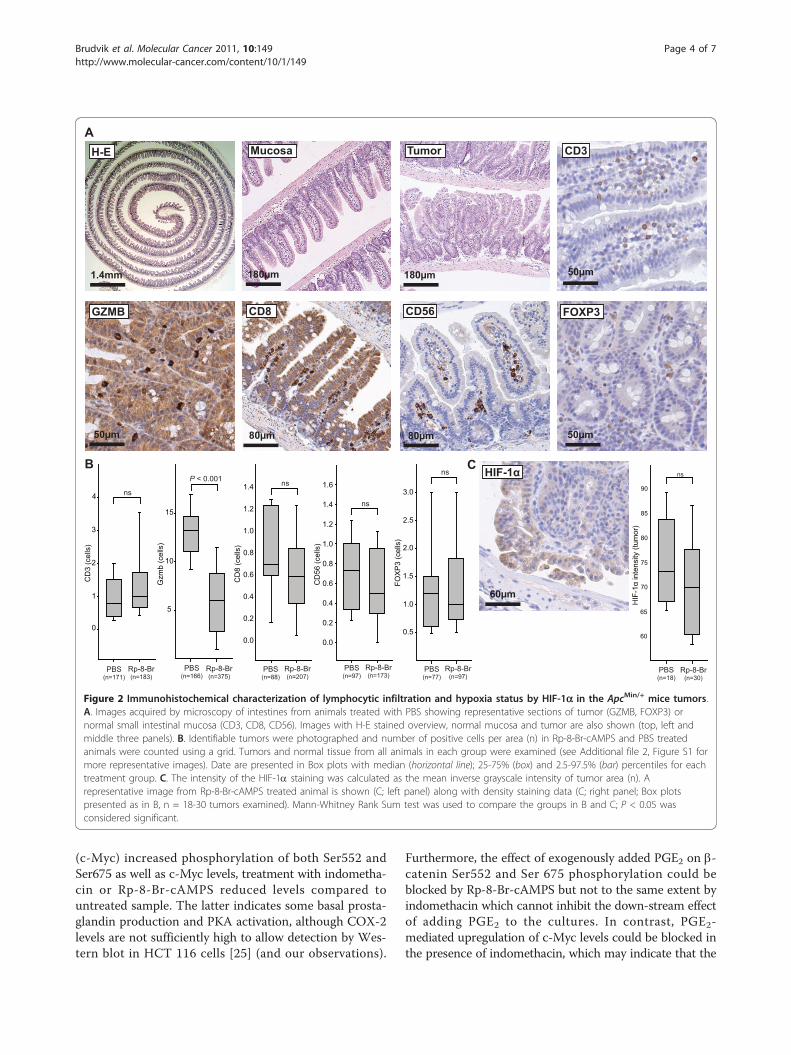
Inhibition of PKA does not affect lymphocytic tumorinfiltration or HIF-1a expression in ApcMin/+ mice tumorsLymphocytic tumor infiltration affects the course ofhuman CRC where type, density and location of immunecells are shown to have higher prognostic power than theclassical UICC-TNM staging [21]. Furthermore, thehypothesis of adaptive regulatory T cells (Treg) inhibitinganti-tumor immune responses has been subject to consid-erable interest [22]. Previously, we found that upon activa-tion, Tregs express COX-2 and suppress effector T cellsby PGE2 - cAMP dependent mechanisms that may be ofclinical relevance in patients with CRC [11]. However,
Brudvik et al. Molecular Cancer 2011, 10:149http://www.molecular-cancer.com/content/10/1/149
Page 2 of 7

immunohistochemical characterization of small intestinaltumors from PKA antagonist Rp-8-Br-cAMPS treated ani-mals did not reveal any significant changes in the numberof CD3+ T cells, CD8+ cytotoxic T cells, Foxp3+ Tregs orCD56+ natural killer (NK) cells (Figure 2A, B and Addi-tional file 2, Figure S1). In contrast, levels of granzyme B
(GZMB), a protein expressed in the cytotoxic T lympho-cytes (CD8+ T cells) and NK cells, were reduced in Rp-8-Br-cAMPS treated animals which may indicate moredegranulated cytotoxic cells post activation (Figure 2B).Our observations indicate that intestinal immuneresponses play a minor role in the development of theApcMin/+ mice tumor load, consistent with other observa-tions [23].PGE2 also affects angiogenesis and up-regulates vascular
endothelial growth factor receptor-1 (VEGFR-1) in ahuman colon cancer cell line [12] whereas indomethacininhibits the expression of VEGF and thereby angiogenesis[24]. To assess treatments effects on angiogenesis, weexamined levels of the hypoxia-inducible transcription fac-tor (HIF)-1a which regulates the expression of targetgenes important in angiogenesis by accumulation andtranslocation to the nucleus under hypoxic conditions.While apical regions of all tumors showed higher cytoplas-mic intensity and nuclear staining of HIF-1a, no differ-ences between treatment groups were observed (Figure 2Cand Additional file 2, Figure S1).
PKA antagonist treatment of ApcMin/+ mice decreases thelevels b-catenin signaling to the nucleus and of COX-2and c-Myc expression in ApcMin/+ mice tumorsWe next examined the effect of treatment on the activity ofthe PGE2- b-catenin pathway in tumor cells. As evidentfrom image analysis of immunohistochemically stained sec-tions, levels of b-catenin were significantly decreased intumors from the animals treated with the PKA antagonistRp-8-Br-cAMPS compared to tumors from the control-treated group (138 versus 117 median inverse grayscaleintensity units; P < 0.001, Figure 3A, B) Furthermore, themedian number of b-catenin positive nuclei were reducedfrom 20% in tumors in the control group to 10% in tumorsin the Rp-8-Br-cAMPS treated group (P = 0.024, Figure 3A,B and Additional file 3, Figure S2). In addition, expressionof the b-catenin/TCF/LEF transcription complex-regulatedgenes c-Myc and COX-2 were reduced in tumor cells upontreatment with the PKA antagonist Rp-8-Br-cAMPS as evi-dent from the median number of nuclei positive for c-Myc(reduced from 60% in control to 20%; P < 0.001, Figure 3A,B and Additional file 3, Figure S2) and from the cytoplas-mic expression levels of the COX-2 enzyme (reduced from136 in control to 106 median inverse grayscale intensityunits in the treated group; P < 0.001, Figure 3A, B andAdditional file 3, Figure S2).For further quantification of the observed effects on tumortissue and validation of the observed effects without dilu-tion into normal mucosa, we next looked at the regulationof b-catenin phosphorylation and c-Myc regulation in ahuman colonic cancer cell line, HCT 116 (Figure 4A and4B). While treatment of HCT 116 colon carcinoma cellswith PGE2 for 30 min (phosphorylated b-catenin) or 1 h
PBS (n=582)
1.4
1.2
1.0
0.8
0.6
0.4
0.2
Siz
e (m
m2 )
Indo(n=41)
0Rp-8-Br(n=293)
P < 0.001
P < 0.001
160
140
120
100
80
60
40
20
No.
of l
esio
ns
0PBS (n=9)
Indo(n=10)
Rp-8-Br(n=7)
P < 0.001
P = 0.368
0 5 10 15 20 25 30 35
Tum
or lo
ad (m
m2 )
0.0
0.5
1.0
1.5
2.0
2.5
3.0
3.5
Distance from ventricle (cm)
PBS Rp-8-Br
Size class (mm2)
<0,05
No.
of l
esio
ns
20
40
60
80
100
120
140
160
PBS Rp-8-Br
0,05-0,1
0,1-0,2
0,2-0,4
0,4-0,8
0,8-1,6
1,6-3,2
3,2-6,4
>6,4
0
Distance from ventricle (cm)0 5 10 15 20 25 30 35
No.
of le
sion
s
0
1
2
3
4
5
6PBS Rp-8-Br
B
A
C
PBS Rp-8-Br Indo
Figure 1 PKA antagonist reduces tumor load in ApcMin/+ mice.A. Images of developing adenomas representative of eachtreatment acquired by transillumination microscopy are shown.White bars = 500 μm. B. Number (B; left panel) and size (B; midpanel) of spontaneously occurring tumors and distribution of tumorsize in the treatment groups (B; right panel), tumor numbers/sizeclass). Box plots with median (horizontal line); 25-75% (box) and 2.5-97.5% (bar) percentiles are shown for each treatment group (n = 7-10 as indicated). All tumors from the same animals were pooledwhen comparing tumor size distribution between the groups (n =41-585) as indicated. Mann-Whitney Rank Sum test was used tocompare the groups (SigmaPlot 11.0 (CA, USA). C. Number (C; leftpanel) and size (right panel) of spontaneously occurring tumors inthe small intestine relative to the distance from the ventricle.
Brudvik et al. Molecular Cancer 2011, 10:149http://www.molecular-cancer.com/content/10/1/149
Page 3 of 7
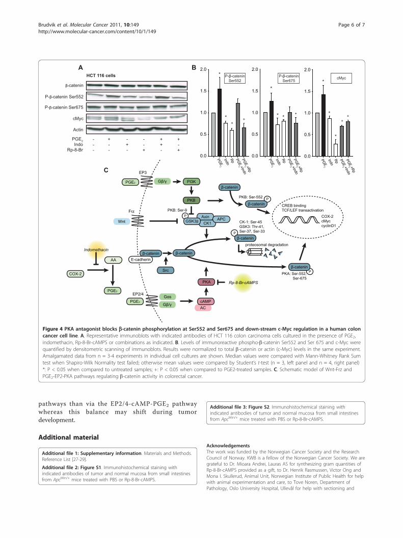
(c-Myc) increased phosphorylation of both Ser552 andSer675 as well as c-Myc levels, treatment with indometha-cin or Rp-8-Br-cAMPS reduced levels compared tountreated sample. The latter indicates some basal prosta-glandin production and PKA activation, although COX-2levels are not sufficiently high to allow detection by Wes-tern blot in HCT 116 cells [25] (and our observations).
Furthermore, the effect of exogenously added PGE2 on b-catenin Ser552 and Ser 675 phosphorylation could beblocked by Rp-8-Br-cAMPS but not to the same extent byindomethacin which cannot inhibit the down-stream effectof adding PGE2 to the cultures. In contrast, PGE2-mediated upregulation of c-Myc levels could be blocked inthe presence of indomethacin, which may indicate that the
CD3
GZMB FOXP3CD8 CD56
50μm
80μm50μm 50μm80μm
H-E Mucosa Tumor
PBS (n=166)
Gzm
b (c
ells
)
Rp-8-Br (n=375)
5
10
15
P < 0.001
PBS(n=171)
Rp-8-Br(n=183)
0
1
2
3
4
CD
3 (c
ells
)
ns
PBS(n=77)
Rp-8-Br(n=97)
FOX
P3
(cel
ls)
0.5
1.0
1.5
2.0
2.5
3.0
nsB
0.0
0.2
0.4
0.6
0.8
1.0
1.2
1.4
1.6
PBS(n=97)
Rp-8-Br(n=173)
CD
56 (c
ells
)
ns
0.0
0.2
0.4
0.6
0.8
1.0
1.2
1.4
PBS(n=88)
Rp-8-Br(n=207)
CD
8 (c
ells
)
ns
A
1.4mm 180μm 180μm
PBS(n=18)
Rp-8-Br(n=30)
60
65
70
75
80
85
HIF
-1α
inte
nsity
(tum
or)
90
nsHIF-1α
60μm
C
Figure 2 Immunohistochemical characterization of lymphocytic infiltration and hypoxia status by HIF-1a in the ApcMin/+ mice tumors.A. Images acquired by microscopy of intestines from animals treated with PBS showing representative sections of tumor (GZMB, FOXP3) ornormal small intestinal mucosa (CD3, CD8, CD56). Images with H-E stained overview, normal mucosa and tumor are also shown (top, left andmiddle three panels). B. Identifiable tumors were photographed and number of positive cells per area (n) in Rp-8-Br-cAMPS and PBS treatedanimals were counted using a grid. Tumors and normal tissue from all animals in each group were examined (see Additional file 2, Figure S1 formore representative images). Date are presented in Box plots with median (horizontal line); 25-75% (box) and 2.5-97.5% (bar) percentiles for eachtreatment group. C. The intensity of the HIF-1a staining was calculated as the mean inverse grayscale intensity of tumor area (n). Arepresentative image from Rp-8-Br-cAMPS treated animal is shown (C; left panel) along with density staining data (C; right panel; Box plotspresented as in B, n = 18-30 tumors examined). Mann-Whitney Rank Sum test was used to compare the groups in B and C; P < 0.05 wasconsidered significant.
Brudvik et al. Molecular Cancer 2011, 10:149http://www.molecular-cancer.com/content/10/1/149
Page 4 of 7

regulation at this later time point relies more on endogen-ously produced PGE2.Cytoplasmic b-catenin may be targeted to proteosomal
degradation through the destruction complex consistingof GSK3b, Axin, CK1 and APC (Figure 4C). However,in the presence of active Wnt signaling, b-catenin accu-mulates in the cytosol and translocates to the nucleus toact in a mitogenic fashion by transactivation of TCF/LEF leading to expression of target genes in a cell prolif-eration and survival program [5]. As is well established,the Wnt-Frz pathway inhibits the destruction complexat the level of GSK3b, leading to less proteosomaldegradation and more nuclear translocation and activa-tion of b-catenin [2]. Similarly, the up-regulation ofCOX-2 in colorectal cancer leads to production of PGE2which binds to the EP3 receptor leading to PI3K andPKB activation, phosphorylation and dissociation ofGSK3b and thereby inhibition of the destruction com-plex [26] (Figure 4C). In zebrafish stem cells, PGE2 act-ing through an EP2/4-cAMP-PKA pathway was recentlyshown to induce direct phosphorylation of b-catenin,thereby stimulating its translocation to the nucleus andmitogenic effect [18]. Here, we tested whether this sec-ond pathway was providing a mitogenic drive in
intestinal cancer. Using ApcMin/+ mice with a disturbedb-catenin degradation, we specifically inhibited thePGE2-cAMP pathway at the level of PKA by treatingmice with Rp-8-Br-cAMPS for 6 weeks (see, Figure 4Cfor point of action). We show that this not only reducestumor load but also specifically inhibits b-cateninnuclear translocation and the activation of b-catenin tar-get genes such as c-Myc and COX-2 which may indicatethat the direct regulatory effect of PKA on b-cateninnuclear translocation is also operative in intestinal can-cer cells. Furthermore, the fact that COX inhibitors mayblock the effect of PGE2 both in the b-catenin degrada-tion and b-catenin nuclear translocation pathways whileRp-8-Br-cAMPS only affects the latter may explain whyinhibitory effect of the PKA antagonist on tumor pro-motion is comparably weaker than that of indomethacin.Finally, our observation that COX inhibitor abolishestumor numbers whereas PKA antagonist reduces tumorload but not tumor numbers may indicate that the anti-tumorigenic and anti-proliferative effects are distinctand relate to different points of action in PGE2 signalpathways. It is interesting to speculate that stem cells incrypt foci that give origin to adenomas may be moresensitive to regulation via the Wnt-Frz and PGE2-EP3
20
40
60
80
0
PBS(n=48)
Rp-8-Br(n=39)
P = 0.024
β-ca
teni
n nu
clea
r sta
inin
g (%
cel
ls o
f tum
or)
Rp-8-Br(n=44)
CO
X-2
inte
nsity
(tum
or)
80
100
120
140
PBS(n=43)
160 P < 0.001
β-ca
teni
n in
tens
ity (t
umor
)
PBS(n=51)
Rp-8-Br(n=39)
100
120
140
160
P < 0.001A
0
20
40
60
80
PBS(n=18)
Rp-8-Br(n=37)
P < 0.001
c-M
yc n
ucle
ar s
tain
ing
(% c
ells
of t
umor
)
B
PB
SR
p-8-
Br-
cAM
PS
COX-2β-catenin c-Myc
60μm 30μm
30μm60μm
80μm
80μm
Figure 3 PKA antagonist treatment of ApcMin/+ mice decreased COX-2, b-catenin and c-Myc expression in small intestinal tumor tissue.A. The intensity of the b-catenin and COX-2 staining was calculated as the mean inverse grayscale intensity of tumor area (Image J 1.43usoftware package). Percent b-catenin and c-Myc positive nuclei were also assessed. Data are presented as Box plots with median (horizontal line);25-75% (box) and 2.5-97.5% (bar) percentiles shown for each treatment group. Median values compared with Mann-Whitney Rank Sum testwhen Shapiro-Wilk Normality test failed; otherwise mean values compared by Student’s t-test. N = no. of tumors examined (tumors from everyanimal in each treatment group were investigated). B. Micrographs of representative tumors from the PBS and Rp-8-Br-cAMPS (Rp-8-Br) treatedgroups stained with b-catenin, c-Myc and COX-2 antibodies.
Brudvik et al. Molecular Cancer 2011, 10:149http://www.molecular-cancer.com/content/10/1/149
Page 5 of 7
pathways than via the EP2/4-cAMP-PGE2 pathwaywhereas this balance may shift during tumordevelopment.
Additional material
Additional file 1: Supplementary information. Materials and Methods.Reference List [27-29].
Additional file 2: Figure S1. Immunohistochemical staining withindicated antibodies of tumor and normal mucosa from small intestinesfrom ApcMin/+ mice treated with PBS or Rp-8-Br-cAMPS.
Additional file 3: Figure S2. Immunohistochemical staining withindicated antibodies of tumor and normal mucosa from small intestinesfrom ApcMin/+ mice treated with PBS or Rp-8-Br-cAMPS.
AcknowledgementsThe work was funded by the Norwegian Cancer Society and the ResearchCouncil of Norway. KWB is a fellow of the Norwegian Cancer Society. We aregrateful to Dr. Mioara Andrei, Lauras AS for synthesizing gram quantities ofRp-8-Br-cAMPS provided as a gift, to Dr. Henrik Rasmussen, Victor Ong andMona I. Skullerud, Animal Unit, Norwegian Institute of Public Health for helpwith animal experimentation and care, to Tove Noren, Department ofPathology, Oslo University Hospital, Ullevål for help with sectioning and
P-β-catenin Ser675
P-β-catenin Ser552
β-catenin
PGE2Indo
Rp-8-Br
---
+--
-+-
--+
++-
+-+
HCT 116 cells
Actin
cMyc
PGE2
IndoRp PGE
2 +IndoPGE
2 +Rp0.0
0.5
1.0
1.5
2.0A B
PGE2
IndoRp PGE
2 +IndoPGE
2 +Rp
0.0
0.5
1.0
1.5
2.0P-β-catenin
Ser675P-β-catenin
Ser552
* + ** +
PGE2
IndoRp PGE
2 +IndoPGE
2 +Rp
0.0
0.5
1.0
1.5
2.0
cMyc
*
*+
+
*
P
proteosomal degradation
CK-1: Ser-45GSK3: Thr-41, Ser-37, Ser-33
PKA: Ser-552, Ser-675
CREB bindingTCF/LEF transactivation
COX-2cMyccyclinD1
P
PGE2
PI3K
PKB
AxinCK1
APC
ACcAMP
Wnt
PGE2
EP3
Gβ/γ
Gαs
Frz
Gβ/γ
β-catenin
GSK3β
β-catenin
β-catenin
EP2/4
PKA Rp-8-Br-cAMPS
COX-2
Indomethacin
AA
PGE2
E-cadherinβ-catenin
Src
C
PKB: Ser-9
PKB: Ser-552 Pβ-catenin
P
β-catenin
*
**
Figure 4 PKA antagonist blocks b-catenin phosphorylation at Ser552 and Ser675 and down-stream c-Myc regulation in a human coloncancer cell line. A. Representative immunoblots with indicated antibodies of HCT 116 colon carcinoma cells cultured in the presence of PGE2,indomethacin, Rp-8-Br-cAMPS or combinations as indicated. B. Levels of immunoreactive phospho-b-catenin Ser552 and Ser 675 and c-Myc werequantified by densitometric scanning of immunoblots. Results were normalized to total b-catenin or actin (c-Myc) levels in the same experiment.Amalgamated data from n = 3-4 experiments in individual cell cultures are shown. Median values were compared with Mann-Whitney Rank Sumtest when Shapiro-Wilk Normality test failed; otherwise mean values were compared by Student’s t-test (n = 3, left panel and n = 4, right panel)*: P < 0.05 when compared to untreated samples; +: P < 0.05 when compared to PGE2-treated samples. C. Schematic model of Wnt-Frz andPGE2-EP2-PKA pathways regulating b-catenin activity in colorectal cancer.
Brudvik et al. Molecular Cancer 2011, 10:149http://www.molecular-cancer.com/content/10/1/149
Page 6 of 7

immunohistochemistry, and to Jorun Solheim and Gladys Tjørhom,Biotechnology Centre of Oslo for help with Western blot analysis.
Author details1Centre for Molecular Medicine Norway, Nordic EMBL Partnership andBiotechnology Centre, University of Oslo, Oslo, Norway. 2Norwegian Schoolof Veterinary Science, Oslo, Norway. 3Department of Transplantation Surgery,Oslo University Hospital Rikshospitalet, Oslo, Norway. 4Department ofPathology, Oslo University Hospital Ullevål, Oslo, Norway.
Authors’ contributionsKWB, EMA and KT designed the experiments; KWB performed animal andWB experiments; KWB and JEP characterized intestinal lesions; KWB and BRperformed and analyzed IHC images; KWB and KT wrote the manuscriptwith comments from all authors; all authors read and approved the finalversion of the manuscript.
Competing interestsThe authors declare that they have no competing interests.
Received: 7 June 2011 Accepted: 15 December 2011Published: 15 December 2011
References1. Half E, Bercovich D, Rozen P: Familial adenomatous polyposis. Orphanet J
Rare Dis 2009, 4:22.2. Rubinfeld B, Albert I, Porfiri E, Fiol C, Munemitsu S, Polakis P: Binding of
GSK3beta to the APC-beta-catenin complex and regulation of complexassembly. Science 1996, 272:1023-1026.
3. Aberle H, Bauer A, Stappert J, Kispert A, Kemler R: beta-catenin is a targetfor the ubiquitin-proteasome pathway. EMBO J 1997, 16:3797-3804.
4. Städeli R, Hoffmans R, Basler K: Transcription under the Control of NuclearArm/[beta]-Catenin. Current Biology 2006, 16:R378-R385.
5. Korinek V, Barker N, Morin PJ, van Wichen D, de Weger R, Kinzler KW,Vogelstein B, Clevers H: Constitutive Transcriptional Activation by a b-Catenin-Tcf Complex in APC-/- Colon Carcinoma. Science 1997,275:1784-1787.
6. He TC, Sparks AB, Rago C, Hermeking H, Zawel L, da Costa LT, Morin PJ,Vogelstein B, Kinzler KW: Identification of c-MYC as a Target of the APCPathway. Science 1998, 281:1509-1512.
7. Tetsu O, McCormick F: [beta]-Catenin regulates expression of cyclin D1 incolon carcinoma cells. Nature 1999, 398:422-426.
8. Eberhart CE, Coffey RJ, Radhika A, Giardiello FM, Ferrenbach S, DuBois RN:Up-regulation of cyclooxygenase 2 gene expression in human colorectaladenomas and adenocarcinomas. Gastroenterology 1994, 107:1183-1188.
9. Kraus S, Arber N: Cancer: Do aspirin and other NSAIDs protect againstcolorectal cancer? Nat Rev Gastroenterol Hepatol 2011, 8:125-126.
10. Psaty BM, Potter JD: Risks and Benefits of Celecoxib to Prevent RecurrentAdenomas. New England Journal of Medicine 2006, 355:950-952.
11. Yaqub S, Henjum K, Mahic M, Jahnsen FL, Aandahl EM, Bjornbeth BA,Tasken K: Regulatory T cells in colorectal cancer patients suppress anti-tumor immune activity in a COX-2 dependent manner. Cancer ImmunolImmunother 2008, 57:813-821.
12. Fujino H, Toyomura K, Chen Xb, Regan JW, Murayama T: Prostaglandin E2regulates cellular migration via induction of vascular endothelial growthfactor receptor-1 in HCA-7 human colon cancer cells. BiochemicalPharmacology 2011, 81:379-387.
13. Sheng H, Shao J, Washington MK, DuBois RN: Prostaglandin E2 increasesgrowth and motility of colorectal carcinoma cells. J Biol Chem 2001,276:18075-18081.
14. Tsujii M, Kawano S, DuBois RN: Cyclooxygenase-2 expression in humancolon cancer cells increases metastatic potential. Proc Natl Acad Sci USA1997, 94:3336-3340.
15. Sonoshita M, Takaku K, Sasaki N, Sugimoto Y, Ushikubi F, Narumiya S,Oshima M, Taketo MM: Acceleration of intestinal polyposis throughprostaglandin receptor EP2 in Apc[Delta]716 knockout mice. Nat Med2001, 7:1048-1051.
16. Taurin S, Sandbo N, Qin Y, Browning D, Dulin NO: Phosphorylation ofbeta-catenin by cyclic AMP-dependent protein kinase. J Biol Chem 2006,281:9971-9976.
17. Hino S, Tanji C, Nakayama KI, Kikuchi A: Phosphorylation of beta-cateninby cyclic AMP-dependent protein kinase stabilizes beta-catenin throughinhibition of its ubiquitination. Mol Cell Biol 2005, 25:9063-9072.
18. Goessling W, North TE, Loewer S, Lord AM, Lee S, Stoick-Cooper CL,Weidinger G, Puder M, Daley GQ, Moon RT, et al: Genetic interaction ofPGE2 and Wnt signaling regulates developmental specification of stemcells and regeneration. Cell 2009, 136:1136-1147.
19. Buchanan FG, DuBois RN: Connecting COX-2 and Wnt in cancer. CancerCell 2006, 9:6-8.
20. Chiu CH, McEntee MF, Whelan J: Discordant effect of aspirin andindomethacin on intestinal tumor burden inApcMin/+ mice.Prostaglandins, Leukotrienes and Essential Fatty Acids 2000, 62:269-275.
21. Galon J, Costes A, Sanchez-Cabo F, Kirilovsky A, Mlecnik B, Lagorce-Pages C,Tosolini M, Camus M, Berger A, Wind P, et al: Type, density, and locationof immune cells within human colorectal tumors predict clinicaloutcome. Science 2006, 313:1960-1964.
22. Wang RF: Immune suppression by tumor-specific CD4+ regulatory T-cellsin cancer. Semin Cancer Biol 2006, 16:73-79.
23. Kettunen HL, Kettunen AS, Rautonen NE: Intestinal immune responses inwild-type and Apcmin/+ mouse, a model for colon cancer. Cancer Res2003, 63:5136-5142.
24. Wang HM, Zhang GY: Indomethacin suppresses growth of colon cancervia inhibition of angiogenesis in vivo. World J Gastroenterol 2005,11:340-343.
25. Agarwal B, Swaroop P, Protiva P, Raj SV, Shirin H, Holt PR: Cox-2 is neededbut not sufficient for apoptosis induced by Cox-2 selective inhibitors incolon cancer cells. Apoptosis 2003, 8:649-654.
26. Castellone MD, Teramoto H, Williams BO, Druey KM, Gutkind JS:Prostaglandin E2 promotes colon cancer cell growth through a Gs-axin-beta-catenin signaling axis. Science 2005, 310:1504-1510.
27. Andrei M, Bjornstad V, Langli G, Romming C, Klaveness J, Tasken K,Undheim K: Stereoselective preparation of (RP)-8-hetaryladenosine-3[prime or minute],5[prime or minute]-cyclic phosphorothioic acids. OrgBiomol Chem 2007, 5:2070-2080.
28. Nayjib B, Zeddou M, Drion P, Boniver J, Tasken K, Rahmouni S,Moutschen M: In vivo administration of a PKA type I inhibitor (Rp-8-Br-cAMPS) restores T-cell responses in retrovirus-infected mice. OpenImmunol J 2008, 20-24.
29. Koch T, Petro A, Darrabie M, Opara E: Effects of Esomeprazole Magnesiumon Nonsteroidal Anti-Inflammatory Drug Gastropathy. Digestive Diseasesand Sciences 2005, 50:86-93.
30. Paulsen JE, Steffensen IL, Andreassen A, Vikse R, Alexander J: Neonatalexposure to the food mutagen 2-amino-1-methyl-6-phenylimidazo[4,5-b]pyridine via breast milk or directly induces intestinal tumors in multipleintestinal neoplasia mice. Carcinogenesis 1999, 20:1277-1282.
doi:10.1186/1476-4598-10-149Cite this article as: Brudvik et al.: Protein kinase A antagonist inhibits b-catenin nuclear translocation, c-Myc and COX-2 expression and tumorpromotion in ApcMin/+ mice. Molecular Cancer 2011 10:149.
Submit your next manuscript to BioMed Centraland take full advantage of:
• Convenient online submission
• Thorough peer review
• No space constraints or color figure charges
• Immediate publication on acceptance
• Inclusion in PubMed, CAS, Scopus and Google Scholar
• Research which is freely available for redistribution
Submit your manuscript at www.biomedcentral.com/submit
Brudvik et al. Molecular Cancer 2011, 10:149http://www.molecular-cancer.com/content/10/1/149
Page 7 of 7