sheddingofcollagenxvii/bp180inskindependsonboth … ·...
TRANSCRIPT

Shedding of Collagen XVII/BP180 in Skin Depends on BothADAM10 and ADAM9*□S
Received for publication, June 15, 2009 Published, JBC Papers in Press, July 1, 2009, DOI 10.1074/jbc.M109.034090
Claus-Werner Franzke‡§, Leena Bruckner-Tuderman§¶, and Carl P. Blobel‡1
From the ‡Arthritis and Tissue Degeneration Program, Hospital for Special Surgery, New York, New York 10021, the §Departmentof Dermatology, University of Freiburg, 79104 Freiburg, Germany, and the ¶Freiburg Institute of Advanced Studies, School of LifeSciences, D-79085 Freiburg, Germany
Collagen XVII is a transmembrane collagen and the majorautoantigen of the autoimmune skin blistering disease bullouspemphigoid. Collagen XVII is proteolytically released from themembrane, and the pathogenic epitope harbors the cleavage sitefor its ectodomain shedding, suggesting that proteolysis has animportant role in regulating the function of collagen XVII inskin homeostasis. Previous studies identified ADAMs 9, 10, and17 as candidate collagen XVII sheddases and suggested thatADAM17 is amajor sheddase.Herewe show thatADAM17onlyindirectly affects collagenXVII shedding and thatADAMs9 and10 are the most prominent collagen XVII sheddases in primarykeratinocytes because (a) collagen XVII shedding was notstimulated by phorbol esters, known activators of ADAM17, (b)constitutive and calcium influx-stimulated shedding was sensi-tive to the ADAM10-selective inhibitor GI254023X and wasstrongly reduced in Adam10�/� cells, (c) there was a 55%decrease in constitutive collagen XVII ectodomain sheddingfromAdam9�/� keratinocytes, and (d) H2O2 enhancedADAM9expression and stimulated collagen XVII shedding in skin andkeratinocytes of wild type mice but not of Adam9�/� mice. Weconclude that ADAM9 and ADAM10 can both contribute tocollagen XVII shedding in skin with an enhanced relative con-tribution of ADAM9 in the presence of reactive oxygen species.These results provide critical new insights into the identity andregulation of themajor sheddases for collagenXVII in keratino-cytes and skin and have implications for the treatment of blis-tering diseases of the skin.
Collagen XVII (also called BP180 or BPAG2) is a hemides-mosomal adhesion component in the skin and mucosa andbelongs to the emerging group of collagenous transmembraneproteins (1). This type II oriented transmembrane protein isinvolved in the molecular pathology of human skin diseases.Mutations in the COL17A1 gene are associated with junctional
epidermolysis bullosa, a genetic skin blistering disease (2).Patientswith bullous pemphigoid and related autoimmune bul-lous dermatoses have tissue-bound and circulating autoanti-bodies targeting collagen XVII (3). Structural and functionalchanges of collagen XVII play an important role in these dis-eases, although the molecular pathology is not yet fully under-stood. The collagen XVII consists of three 180-kDa �1 (XVII)chains, each with an intracellular N-terminal domain, a shorttransmembrane stretch, and a flexible extracellular C-terminalectodomain with collagenous (Col)2 subdomains that are inter-rupted by short non-collagenous (NC) sequences. The humanand murine collagen XVII molecules differ in size and in thenumber of the Col and NC domains. Human collagen XVIIconsists of 1497 amino acid residues with 15 Col and 16 NCdomains, whereas the murine form, which is 86% identical (4),consists of 1433 amino acid residues with 13 Col and 14 NCdomains. In humans the extracellular linker domain NC16Abetween the plasma membrane and the Col15 domain is func-tionally important because it is believed to play a role in bothectodomain shedding and in the proper folding of the triplehelical structure of collagen XVII (5–7).Our previous studies revealed two forms of collagen XVII,
the 180-kDa membrane-anchored form and the soluble 120-kDa form. The latter represents the extracellular collagenousectodomain, which is released by cleavage by membrane-an-chored metalloproteinases of the a disintegrin and metallopro-teinase (ADAM) family (8). The shed ectodomain of collagenXVII is very stable in vivo and in vitro. In wound scratch assays,both addition of the purified soluble ectodomain or overexpres-sion of ADAMs suppressed cell motility (8), indicating that theectodomain has a role in regulating keratinocyte-matrix inter-actions. In the context of the known functions of collagen XVIIas an adhesion molecule, its shedding could therefore regulateits functions in keratinocyte migration, differentiation, andproliferation.ADAMs are also involved in the release of several other type
I or type II transmembrane proteins and are considered to becritical regulators of epidermal growth factor receptor signal-ing, tumor necrosis factor � release, and Notch signaling toname a few examples (9, 10). Previously ADAM9, ADAM10,and ADAM17 had been identified as potential sheddases for
* This work was supported in part by National Institutes of Health GrantGM64750 (to C. P. B.). This work was also supported by scholarships fromthe German Research Foundation, Deutsche Forschungsgemeinschaft(DFG) (Grant FR 2524/1), the German Network Epidermolysis bullosa, andthe Emerald Foundation (to C.-W. F.) and by the Excellence Initiative of theGerman Research Foundation (Freiburg Institute for Advanced StudiesSchool of Life Sciences) and DFG Grant Br 1475/9-1 (to L. B.-T.).
□S The on-line version of this article (available at http://www.jbc.org) containssupplemental Fig. 1.
1 To whom correspondence should be addressed: Arthritis and Tissue Degen-eration Program, Caspary Research Bldg., Rm. 426, Hospital for Special Sur-gery, 535 East 70th St., New York, NY 10021. Tel.: 212-606-1429; Fax: 212-774-2560; E-mail: [email protected].
2 The abbreviations used are: Col, collagenous; NC, non-collagenous; ADAM, adisintegrin and metalloproteinase; AP, alkaline phosphatase; IM, ionomy-cin; mEF, mouse embryonic fibroblast; E, embryonic day; GI, GI254023X;PMA, phorbol 12-myristate 13-acetate.
THE JOURNAL OF BIOLOGICAL CHEMISTRY VOL. 284, NO. 35, pp. 23386 –23396, August 28, 2009© 2009 by The American Society for Biochemistry and Molecular Biology, Inc. Printed in the U.S.A.
23386 JOURNAL OF BIOLOGICAL CHEMISTRY VOLUME 284 • NUMBER 35 • AUGUST 28, 2009
by guest on July 12, 2018http://w
ww
.jbc.org/D
ownloaded from
collagen XVII in keratinocytes by overexpression in cell-basedassays (8). Moreover Adam17�/� keratinocytes had 50%diminished collagen XVII shedding, which was interpreted tosuggest that ADAM17 represents an important, if not themajor, physiological collagen XVII sheddase (8). The majorgoal of the current study was to further explore the contribu-tion of ADAM17 and other candidate sheddases to the releaseof collagen XVII from primary keratinocytes and mouse skin.The identification of the major collagen XVII sheddases andtheir regulation is critical for understanding the role of collagenXVII shedding in the pathogenesis of skin diseases.
EXPERIMENTAL PROCEDURES
Animals—Adam8�/� (11), Adam9�/� (12), Adam15�/�
(13), Adam17�/� mice (14), and wild type littermate controlsweremaintained in an accredited animal facility at the Hospitalfor Special Surgery according to the guidelines of the AmericanVeterinary Association, and all experiments were approved bythe Hospital for Special Surgery Institutional Animal Care andUse Committee.Cell Lines and Reagents—TheAdam17�/� fibroblast cell line
(E2 cells) and the Adam10�/� fibroblast cell line were derivedfrom E13.5 and E9.5 embryos, respectively (15, 16). Immortal-ized fibroblasts derived from Adam9�/�, Adam12�/�, andwild type E13.5 embryos were generated by transfecting pri-mary mouse embryonic fibroblast (mEF) cells from the appro-priate mouse lines (17) with a vector carrying the SV40 large Tantigen. Wild type mEFs express ADAMs 9, 10, 12, 15, 17, and19 (17). All cell lines were grown inDulbecco’s modified Eagle’smedium supplementedwith 5% fetal calf serum and antibiotics.The ionophores monensin, valinomycin, and nystatin werepurchased from Sigma-Aldrich. Chloride ionophore 1 waspurchased from Fluka, and ionomycin was obtained from Cal-biochem. Batimastat (BB94) was kindly provided by Dr. D.Becherer (GlaxoSmithKline, Research Triangle Park, NC), andmarimastat was provided by Dr. Ouathek Ouerfelli (MemorialSloan-Kettering Cancer Center). The hydroxamate inhibitorsGI254023X (GI) andGW280264X are described elsewhere (18)andwere also provided byDr. Becherer. The following proteaseinhibitors were used: 4-(2-aminoethyl)benzolsulfonylfluoridehydrochloride (Roche Applied Science) and 1,10-ortho-phe-nanthroline (Sigma-Aldrich). All other reagents were obtainedfrom Sigma-Aldrich unless otherwise indicated.Expression Vectors—The expression vector for alkaline phos-
phatase (AP)-tagged Kit ligand 2 has been described previously(19). The full-length cDNA for human collagen XVII was gen-erated as described previously (20) and cloned into theNotI siteof pcDNA3 (Invitrogen). To produce theC-terminalAP-taggedmurine truncated collagen XVII construct, we generated a PCRfragment spanning nucleotides 294–2777 (amino acids 1–828)of murine collagen XVII, including the largest collagenous sub-domainCol13 (GenBankTMaccessionnumberNM007732) andcloned it into pAPtag5 (Genhunter, Nashville, TN; see Fig. 1Bfor a diagram of the AP-tagged collagen XVII fragment). Foramplification of the fragment we used the forward primer 3�-cgcgggctagccaccatggatgtgaccaagaaaagc-5� and reverse primer3�-cgcggaagcttctccgggcacagtgattgttga-5� with PfuTurbo DNApolymerase (Stratagene) and the IntegratedMolecular Analysis
of Genomes and their Expression (IMAGE) ConsortiummousecDNA clone 40086691 (Open Biosystems) as template. Theproper insertion and the sequence of murine AP-collagen XVIIconstruct (Col13-AP) was verified by DNA sequencing.mRNA Expression Analysis—For real time PCR, total RNA
from primary wild type and Adam17�/� keratinocyte cultureswas extracted using an RNeasy Mini kit (Qiagen), and 1 �g oftotal RNA was reverse transcribed using a First Strand cDNASynthesis kit (Fermentas). Relative quantification of geneexpression was performed by real time PCR using iQ SYBRGreen Supermix on the iCycler iQ thermal cycler (Bio-Rad)following themanufacturer’s protocols. Primer sequences wereas follows: mouse glyceraldehyde-3-phosphate dehydrogenase:sense primer, 5�-tggagaaacctgccaagtatg-3�; antisense primer,5�-gttgaagtcgcaggagacaac-3�; mouse ADAM9: sense primer,5�-tgaccatcccaacgtacaga-3�; antisense primer, 5�-ttccaaaactgg-cattctcc-3�; mouse ADAM10: sense primer, 5�-tctccggaatccgt-aacatc-3�; antisense primer, 5�-tccaggaacttctccacacc-3�; mouseADAM17: sense primer, 5�-cagcagcactccataaggaaa-3�; anti-sense primer, 5�-tttgtaaaagcgttcggta-3�; and mouse collagenXVII: sense primer, 5�-ctggattaggcaaggctgag-3�; antisenseprimer, 5�-cttgactccccatgtcacct-3�. Relative expression wasnormalized for levels of glyceraldehyde-3-phosphate dehydro-genase. The generation of amplification products of the correctsize was confirmed using agarose gel electrophoresis.Cell Culture, Transfection, and AP Shedding Assay—Immor-
talized fibroblast cell lines and COS-7 cells were seeded on12-well plates at 75% confluency and transfected with 1.5 �g ofDNA/well with the indicated plasmids using Lipofectamine2000 (Invitrogen) according to the manufacturer’s recommen-dations, and 50 �g/ml ascorbic acid was added to the media toallow for hydroxylation of collagen and proper triple helix for-mation. FreshOpti-MEM(Invitrogen)mediumwith orwithoutthe indicated reagents was added the next day after transfectionand incubated for the designated time period. The AP activityin 100�l of supernatant and 10�l of cell lysatewasmeasured bycolorimetry as described previously (17, 21, 22). The normal-ized percentage of shedding was calculated as ratio of superna-tant AP activity divided by total AP activity (supernatant APactivity � cell lysate AP activity). All experiments wererepeated at least three times with similar results.For verification of the expression of the collagen XVII-AP
fusion protein and its shedding in murine fibroblasts, themedia and cell lysates were processed separately after theindicated times as described earlier (23) and analyzed byWestern blot with murine collagen XVII NC14A rabbit anti-serum (MO-NC14A (8)).Keratinocyte and Epidermis Sheet Preparation and
Cultivation—Keratinocytes were isolated from the skin ofADAM-deficient mice and their wild type littermates essen-tially as described previously (24, 25). Briefly the skin waswashed with 70% ethanol and phosphate-buffered saline, andepidermis and dermis were detached by overnight digestionwith 5 units/ml dispase II (Roche Applied Science) at 4 °C. Theepidermis was mechanically separated from the dermis andincubated with 0.25% trypsin (w/v) and 2mM EDTA for 30 minat 37 °Cwith vigorous shaking. After stopping the reactionwithphosphate-buffered saline containing 10% fetal calf serum, the
Collagen XVII Is Shed by ADAMs 9 and 10
AUGUST 28, 2009 • VOLUME 284 • NUMBER 35 JOURNAL OF BIOLOGICAL CHEMISTRY 23387
by guest on July 12, 2018http://w
ww
.jbc.org/D
ownloaded from
keratinocyte suspensionwas passed through a 70-�msieve, and105 cells/cm2 were plated in defined serum-free keratinocytemedium supplementedwith 100 pM cholera toxin, 100 units/mlpenicillin, 100 �g/ml streptomycin, and 0.25 �g/ml amphoter-
icin B (all from Invitrogen) on 6-well plates coated with gelatin(2.5 mg/ml; Sigma-Aldrich). The cells were maintained indefined serum-free keratinocytemedium at 37 °C, 5%CO2, and95% humidity, and medium was replaced every 48 h. Subcon-
Collagen XVII Is Shed by ADAMs 9 and 10
23388 JOURNAL OF BIOLOGICAL CHEMISTRY VOLUME 284 • NUMBER 35 • AUGUST 28, 2009
by guest on July 12, 2018http://w
ww
.jbc.org/D
ownloaded from

fluent cells derived from passages 2–5 were used for the exper-iments, and all cells were cultured with medium containing 50�g/ml ascorbate for 48 h to allow full prolyl and lysyl hydroxy-lation of newly synthesized collagens.To isolate epidermis sheets from ADAM9-deficient mice
and their wild type littermates, the skin was removed fromeuthanized 2–3-day-old pups, flattened on a Petri dish, and cutinto epidermis sheets of equal size using an 8-mm biopsypunch. After 1-h digestion with dispase II at 37 °C, the epider-mis was detached from the dermis with tweezers and trans-ferred to a 12-well plate with serum-free keratinocyte growthmedium. After a maximum of 2-h incubation at 37 °C, the epi-dermis sheets were used for the experiments outlined under“Results.”For ectodomain shedding analysis by immunoblotting, cell
lysates and media were processed separately as described pre-viously (23). Briefly the cells were washed twice with phos-phate-buffered saline and lysed on ice for 30 min in lysis buffer(1% Nonidet P-40, 0.1 M NaCl, and 25 mM Tris-HCl, pH 7.4)containing 1 mM Pefabloc (Merck), 2 mM EDTA, 10 mM 1,10-ortho-phenanthroline, and 10 �l/ml protease inhibitor mixtureset III (Calbiochem). Then the cell lysate was collected with arubber cell scraper, centrifuged for 30min at 13,000� g at 4 °C,and then used immediately or stored at �80 °C. Total proteincontent was determined using the microtiter BCATM ProteinAssay kit (Pierce), and 30 �g of total protein/sample was usedfor SDS-PAGE. The medium was collected on ice, 1 mM Pefab-loc and 2 mM EDTA were added immediately, and cell debriswere then removed by centrifugation. Proteins were precipi-tated with chloroform-methanol and centrifuged, and the pel-lets were dissolved in Laemmli sample loading buffer contain-ing 5 mM dithiothreitol and heated at 95 °C for 5 min.Preparation of Mouse Tissue Lysates—Mouse tissues were
dissected immediately after euthanasia (performed accordingto the guidelines of the American Veterinary Association; allanimal experiments were approved by the Hospital for SpecialSurgery Internal Animal Care and Use Committee). Wehomogenized 0.5 g of each tissue (lung, liver, skeletal muscle,and skin) in 600 �l of lysis buffer consisting of 0.1 M Tris-HCl,pH 6.8, 1 M urea, 1% Nonidet P-40, 10 mM EDTA, 1 mM 4-(2-aminoethyl)benzolsulfonylfluoride hydrochloride, and pro-teinase inhibitormixture (26) using a Polytron homogenizer for3 min on ice (Kinematica, Littau, Switzerland). All lysates werecentrifuged at 15,000 � g for 30 min to remove debris, and the
supernatants were used for microtiter detergent-compatiblecolorimetric protein detection using the BCA Protein Assay kit(Pierce). Samples of equal protein content (30 �g) were mixedwith 5-fold concentrated Laemmli buffer containing 50 mM di-thiothreitol, heated at 95 °C for 5 min, and then analyzed byWestern blotting (see below).Immunofluorescence Microscopy and Western Blot Analysis—
For immunofluorescence microscopy, cryosections of mouseskin were fixed in ice-cold acetone for 10 min, washed in Tris-buffered saline, and blocked with 10% normal goat serum inTris-buffered saline for 30min at room temperature. The poly-clonal goat anti-ADAM9antiserum (R&DSystems)was used asprimary antibody, and fluorescein isothiocyanate-labeled Affi-niPure donkey anti-goat IgG (Jackson ImmunoResearch Labo-ratories, West Grove, PA) was used as secondary antibody.Mounting medium supplemented with 4�,6-diamidino-2-phe-nylindole was purchased from Vector Laboratories (Burl-ingame, CA).For immunoblotting, the proteins were separated by electro-
phoresis on 7 or 10% SDS-polyacrylamide gels as indicated.Immunoblotting was performed with rabbit polyclonal anti-serum against the human collagen XVII NC16A domain(NC16A (27) and human collagen XVII cytodomain (Endo-2(8)), murine collagen XVII NC14A domain (MO-NC14A (8)),murine ADAM17 cytoplasmic domain (28), and murineADAM9 cytoplasmic domain (29). Anti-ADAM10 cytoplasmicdomain antibodies were from R&D Systems. Immunoblot sig-nals of collagen XVII ectodomain in the media of at least threeindependent experiments were analyzed and quantified withQuantity One software (Bio-Rad). All values are expressed asmeans � S.D. Two populations of data were statistically ana-lyzed using the unpaired two-tailed t test and considered signif-icantly different at p values smaller than 0.05.
RESULTS
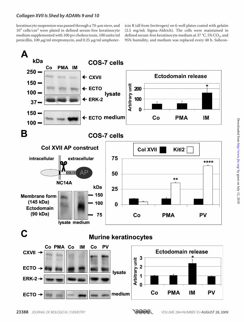
Collagen XVII Shedding Is Stimulated by Short Term Treat-ment with Ionomycin but Not PMA or Pervanadate—ADAM10and ADAM17 have different responses to treatment of cellswith phorbol esters or calcium ionophores in cell-based assays.Low concentrations of PMA (20 ng/ml) predominantly activateADAM17, whereas the calcium ionophore ionomycin stimu-lates both ADAMs 10 and 17 (30, 31). Because we had shownpreviously that ADAMs 9, 10, and 17 have the potential to shedcollagen XVII and that these ADAMs were constitutively
FIGURE 1. Collagen XVII shedding is stimulated by ionomycin but not by PMA or pervanadate. A, COS-7 cells transfected with full-length human collagenXVII were treated with DMSO as vehicle control (Co) or with 20 ng/ml PMA or 2.5 �M IM for 25 min. Representative immunoblots of the 180-kDa full-lengthhuman collagen XVII (CXVII) and the 120-kDa shed ectodomain (ECTO) from lysates and media probed with the polyclonal rabbit anti-human NC16A antibodyare shown. ERK-2 was used as lysate loading control. Collagen XVII shedding was significantly stimulated by ionomycin but not PMA. The graph summarizes thedensitometric analysis of collagen XVII ectodomain shedding in three independent experiments (mean � S.D.; *, p � 0.05). B, COS-7 cells were eithertransfected with a cDNA vector coding for an AP-tagged murine collagen (Col) XVII construct or an AP-tagged construct for Kit ligand 2 (Kitl2; Ref. 19), which wasused as a representative substrate of ADAM17. The C-terminally truncated collagen XVII construct consists of its cytoplasmic domain, transmembrane domain,and the extracellular domains NC14A, Col13, and NC13, which were fused in-frame with a C-terminal AP tag. A Western blot probed with the MO-NC14Amonoclonal antibodies on the left shows the 145-kDa membrane-bound form in the cell lysate and the shed 90-kDa form in the supernatant. Twenty-four hoursafter transfection the cells were washed, and fresh medium with DMSO vehicle control (Co), 20 ng/ml PMA, or 100 �M pervanadate (PV) was added. After 60 min,100 �l of culture media and 10 �l of cell lysate were used for the AP assay. Treatment with either PMA or pervanadate failed to stimulate collagen XVII sheddingin contrast to the significant stimulation of shedding of the ADAM17 substrate Kit ligand 2. Data are represented as mean � S.D. (n � 3; **, p � 0.01; ****, p �0.0001). C, murine keratinocytes were treated with DMSO vehicle as control (Co), 20 ng/ml PMA, or 100 �M pervanadate (PV) for 60 min or with 1 �M IM for 20min, and the cell lysates and the concentrated supernatants were subjected to Western blot analysis with the polyclonal rabbit anti-mouse MO-NC14Aantibody with ERK-2 levels serving as loading control. The graph summarizes the analysis of immunoblots of three independent experiments (one represent-ative blot is shown) as the ratio of released ectodomain from treated cells versus control cells (mean � S.D.). As in transfected COS-7 cells, shedding of endog-enously expressed collagen XVII from murine keratinocytes was stimulated by ionomycin (*, p � 0.05) but not by PMA or pervanadate. Error bars indicate S.D.
Collagen XVII Is Shed by ADAMs 9 and 10
AUGUST 28, 2009 • VOLUME 284 • NUMBER 35 JOURNAL OF BIOLOGICAL CHEMISTRY 23389
by guest on July 12, 2018http://w
ww
.jbc.org/D
ownloaded from

expressed in COS-7 cells (32), we analyzed the shedding ofhuman full-length collagen XVII in response to PMA, pervana-date, and ionomycin in transiently transfected COS-7 cells.Weobserved no stimulation of shedding by 20 ng/ml PMA (Fig. 1A)
or 100 �M pervanadate for 1 h (not shown), whereas treatmentwith 2.5 �M ionomycin strongly increased the release of thesoluble collagen XVII ectodomain into the culture supernatant(Fig. 1A). For further analysis, a C-terminally truncatedmurinecollagen XVII-alkaline phosphatase fusion protein (Col13-AP)was generated, and its expression and shedding were validatedin transfectedCOS-7 cells.When shedding of the collagenXVIIectodomain-AP fusion protein was assessed by Western blotanalysis, the 145-kDa transmembrane formwas detected in thecell lysates, and the 90-kDa soluble form was detected in theculture supernatant (Fig. 1B). Stimulation with 20 ng/ml PMAor 100 �M pervanadate for 1 h did not increase the AP activityreleased into the supernatant, although both stimuli stronglyenhanced shedding of the ADAM17 substrate Kit ligand 2 (19),which was used in parallel as a positive control in transientlytransfected COS-7 cells to show that ADAM17 was activatedunder these conditions (Fig. 1B). Moreover shedding of endog-enous collagen XVII from murine keratinocytes was also notstimulated by 20 ng/ml PMA or 100 �M pervanadate for 1 h(Fig. 1C). However, treatment of murine keratinocytes with1 �M ionomycin for 20 min (Fig. 1C) resulted in strong stimula-tion of collagen XVII shedding as did stimulation with a 25 �M
concentration of the calmodulin inhibitor trifluoroperazine(data not shown). Furthermore collagen XVII shedding frommurine keratinocytes was only stimulated by incubation withionomycin but not with ionophores for sodium (monensin; 20�M), potassium (valinomycin; 1 �M), monovalent cations (nys-tatin; 20 �M), or chloride ions (chloride ionophore 1; 1 �M)(data not shown). The lack of stimulation by 1-h treatmentwithPMA or pervanadate in COS-7 cells and murine keratinocytes(see above) as well as inHaCaT keratinocytes and immortalizedmEFs (data not shown) was not consistent with the hypothesisthat ADAM17 is a major sheddase for collagen XVII as stimu-lation of shedding by these agents is a characteristic feature ofADAM17 (30, 31).
FIGURE 2. Constitutive and ionomycin-stimulated collagen XVII shed-ding is strongly inhibited by the ADAM10-selective GI254023X. A, inhibi-tion of constitutive collagen XVII shedding from murine primary keratino-cytes by increasing concentrations of the hydroxamate inhibitors GI andGW280264X (GW). Confluent murine keratinocytes were incubated withDMSO vehicle control or with 0.5, 1, and 2 �M inhibitor for 3 h in culturemedium. Western blots of the cell lysates were probed for full-length collagenXVII (CXVII) and the ectodomain (ECTO) with a mixture of MO-NC14A andEndo-2 antibodies with ERK-2 serving as loading control. The releasedectodomain (ECTO) in cell supernatants was visualized using the MO-NC14Aantibody. Each immunoblot is representative of three independent experi-ments, the results of which are summarized as mean � S.D. in the graph in thelower panel (##, p � 0.01; ###, p � 0.001). Collagen XVII shedding is stronglyinhibited by 1 �M GI, a concentration that is selective for ADAM10 overADAM17 (34, 35). B, the constitutive and stimulated shedding of murine AP-tagged collagen XVII in transfected COS-7 cells was analyzed by AP assay. Thecells were incubated for 25 min without (Co) or with 2.5 �M IM and/or 1 �M GIor the non-selective hydroxamate BB94 (BB). Data represent the mean � S.D.(n � 3). Both constitutive and stimulated shedding (**, p � 0.01) was effi-ciently inhibited by 1 �M GI (##, p � 0.01; ###, p � 0.001). C, inhibition ofionomycin-stimulated shedding of collagen XVII from murine keratinocyteswith GI. Western blot analysis of cell lysates probed with both MO-NC14A andthe Endo-2 antibody with ERK2 used as loading control and Western blots ofconcentrated supernatants probed with MO-NC14A after treatment with1 �M ionomycin for 20 min revealed strong inhibition of shedding in thepresence of 1 �M GI. The graph in the panel on the right summarizes thedensitometric analysis of immunoblots of the released collagen XVII ectodo-main from three different experiments (mean � S.D.; **, p � 0.01; #, p � 0.05).
FIGURE 3. Loss-of-function experiments demonstrate that ADAM10 isrequired for ionomycin-stimulated shedding of collagen XVII in mouseembryonic fibroblasts. Immortalized wild type (WT) and ADAM (AD)-defi-cient mouse embryonic fibroblasts were transfected with a cDNA vector cod-ing for AP-tagged murine collagen XVII. Twenty-four hours after transfectionthe cells were washed, and fresh medium with DMSO vehicle as control (Co),1 �M batimastat (BB), 2.5 �M IM, or 2.5 �M ionomycin plus 1 �M batimastat(IM/BB) was added. After 30 min, 100 �l of culture media and 10 �l of celllysate were used for the AP assay. The constitutive shedding activity of eachcell line is set to 100% to be able to compare the relative increases in sheddingupon stimulation with IM. Collagen XVII shedding was stimulated by ionomy-cin in Adam9�/�, -12�/�, -17�/�, and wild type murine immortalized embry-onal fibroblasts but not in Adam10�/� cells. Constitutive and stimulatedshedding was strongly sensitive to batimastat. Data represent the mean �S.D. (n � 4; *, p � 0.05; **, p � 0.01; ***, p � 0.001; #, p � 0.05; ##, p � 0.01).
Collagen XVII Is Shed by ADAMs 9 and 10
23390 JOURNAL OF BIOLOGICAL CHEMISTRY VOLUME 284 • NUMBER 35 • AUGUST 28, 2009
by guest on July 12, 2018http://w
ww
.jbc.org/D
ownloaded from

Constitutive and Ionomycin-stim-ulated Shedding of Collagen XVII IsStrongly Inhibited by the ADAM10-selective Hydroxamate GI254023X—Because ADAM10 is activated bythe calcium ionophore ionomycin(IM) or the calmodulin inhibitor tri-fluoroperazine but not by PMA (30,31, 33, 34) we further corroboratedthe involvement of ADAM10 in col-lagen XVII shedding by testing theinhibition of constitutive collagenXVII shedding in murine keratino-cytes byGI, a hydroxamate inhibitorwith selectivity for ADAM10 versusADAM17 at 1 �M (34, 35). More-over we used GW280264X, whichhas nearly identical inhibitory activ-ity for ADAM17 andADAM10 (18).As shown in Fig. 2A, both inhibitorsblocked constitutive shedding ofcollagen XVII at 1 �M by about 80%.In addition, ionomycin-stimulatedshedding of collagen XVII was alsoinhibited by 1 �M GI or the non-selective hydroxamate batimastat(BB94) in COS-7 cells transfectedwith murine collagen XVII-AP(Col13-AP) (Fig. 2B) and by 1 �M
GI in primary murine keratino-cytes (Fig. 2C) consistent with amajor contribution of ADAM10 toconstitutive and IM-stimulatedshedding of collagen XVII.Loss of Function Experiments with
ADAM-deficient Fibroblasts Cor-roborate That ADAM10 Is Requiredfor Ionomycin-stimulated Sheddingof Collagen XVII—To evaluate thecontribution of various ADAMpro-teases to collagen XVII shedding incell-based assays, we transientlyexpressedmurine collagenXVII-AP(Col13-AP) in immortalized mEFsfrom wild type mice or mice lackingone of the three potential collagenXVII sheddases (ADAM9, ADAM10,or ADAM17). As controls, we alsoincluded Adam12�/� mEFs in theanalysis because we had seen highercollagen XVII shedding in COS-7and HaCaT cells transfected withADAM12 (data not shown) andbecause ADAM12 was recentlyreported to play a potentially inhib-itory role in healing of chronicwounds (36). Analysis of the APactivity released into the superna-
Collagen XVII Is Shed by ADAMs 9 and 10
AUGUST 28, 2009 • VOLUME 284 • NUMBER 35 JOURNAL OF BIOLOGICAL CHEMISTRY 23391
by guest on July 12, 2018http://w
ww
.jbc.org/D
ownloaded from

tant of ADAM-deficient cells expressing collagen XVIIshowed a comparable ionomycin-stimulated shedding inwild type cells and in Adam9�/�, -12�/�, or -17�/�cells,whereas Adam10�/� cells showed no significant ionomycin-stimulated shedding (Fig. 3A).CollagenXVII Shedding Is StronglyDiminished inAdam9�/�
Keratinocytes—The experiments using mouse embryonicfibroblasts described above allowed a side by side compari-son of the response to activators of shedding in cells isolatedfrom Adam10�/� mice and mice lacking other ADAMs.Because Adam10�/� mice die at E9.5 and thus too early toallow preparation of primary keratinocytes, the use of mEFcells from Adam10�/� mice thus represented the only cur-rently feasible approach to perform loss-of-function experi-ments for ADAM10 in cell-based assays. However, it isimportant to note that it is difficult to compare levels ofconstitutive shedding in immortalized mEF cells becauseimmortalization with SV40 large T antigen is likely to affectthe activation state of these cell lines compared with primarymEF cells.To analyze the effects of deletion of ADAM9 on constitutive
shedding of endogenous collagen XVII, we prepared primarykeratinocytes fromAdam8�/�, -9�/�, or -15�/�mice and theirwild type littermate controls. To avoid interindividual varia-tion, at least four different individuals of each ADAM-deficientmouse strain and their wild type littermates were included inthe analysis as described under “Experimental Procedures.” Pri-mary keratinocytes at passages 2–5 were cultured in definedserum-free keratinocyte medium until confluent, then washedtwice with phosphate-buffered saline, and incubated for 5 h infresh medium. The media and the cell lysates were analyzed byimmunoblotting for full-length collagen XVII or the ectodo-main, and shedding was quantified by densitometry. Theseexperiments confirmed a strong reduction in collagen XVIIshedding in Adam9�/� cells (46.0 � 11.3%; n � 5) comparedwith wild type controls, whereas no significant change in shed-ding was observed in keratinocytes from Adam8�/� orAdam15�/� mice (Fig. 4A). However, similar to our results inAdam9�/� fibroblasts, the ionomycin-stimulated shedding ofcollagen XVII was not affected in Adam9�/� keratinocytescompared with controls, arguing against a role for ADAM9 inionomycin-stimulated shedding (Fig. 4B).ADAM9 and ADAM10 Cooperate in the Constitutive Shed-
ding of Collagen XVII—To determine whether ADAM9 andADAM10 cooperate in the constitutive shedding of collagen
XVII, we incubated Adam9�/� keratinocytes with theADAM10-selective inhibitor GI254023X to assess the contri-bution of ADAM10 to constitutive shedding in keratinocytes inthe absence of ADAM9. We detected significant inhibition ofcollagen XVII shedding by 0.5 �M GI254023X in Adam9�/�
keratinocytes, suggesting that ADAMs 9 and 10 cooperate inthe constitutive shedding of collagen XVII (Fig. 4C). In agree-ment with this finding, constitutive shedding of collagen XVIIfrom Adam10�/� mEF cells can also be further reduced bybatimastat (see Fig. 3A), demonstrating that part of the remain-ing constitutive activity is also due to a metalloproteinase, pre-sumably ADAM9.Knock-out of ADAM17 in Keratinocytes Does Not Alter
Expression or Maturation of ADAMs 9 and 10—To clarifythe reason for the 50% diminished collagen XVII sheddingin Adam17�/� keratinocytes, we analyzed the expression ofADAMs 9 and 10 at the mRNA and protein levels inAdam17�/� keratinocytes. Quantitative real time reverse tran-scription-PCR analysis of keratinocyte mRNA did not uncoversignificant differences in the expression of ADAM9, ADAM10,or collagen XVII (supplemental Fig. 1A).Immunoblot analysis of ADAM9 and ADAM10 also did not
uncover differences in their expression or maturation inAdam17�/� keratinocytes as determined by the appearance ofthe fastermigratingmature form of either ADAM (supplemen-tal Fig. 1B). Thus the cause for the reduced collagen XVII shed-ding in Adam17�/� keratinocytes remains to be determined.This result is in agreement with previous studies that failed todetect differences in the expression of ADAMs 9 and 10 inprimary embryonic Adam17�/� fibroblasts (17).Collagen XVII Shedding Is Stimulated by Hydrogen Peroxide
in Wild Type Cells but Not in Adam9�/� Keratinocytes—Re-cent investigations have shown that oxidative stress inducesADAM9 expression in several human prostate cancer cell lines(37, 38). Therefore we transfectedwild typemEFswith collagenXVII-AP and analyzed its shedding in semiconfluent (about70%) cultures after 5.5 h of stimulation with increasing concen-trations of H2O2 (100–500 �M). We observed a dose-depend-ent increase of collagen XVII ectodomain shedding with a 1.8-fold induction at 500 �M H2O2 (Fig. 5A). When collagenXVII-AP shedding was analyzed in Adam9�/� mEFs underidentical conditions, the addition of H2O2 did not stimulatecollagenXVII shedding. To assess the involvement ofADAM10in hydrogen peroxide-stimulated shedding of collagenXVII, wealso treated Adam10�/� mEFs with increasing concentrations
FIGURE 4. Constitutive collagen XVII shedding is strongly diminished in Adam9�/� keratinocytes and can be further reduced by the ADAM10-selective GI254023X, whereas ionomycin-stimulated shedding is not affected in the absence of ADAM9. A, shedding in primary murine keratinocyteswas analyzed as described under “Experimental Procedures.” Keratinocytes derived from at least four different mice for each Adam�/� deficient strain and theirwild type littermate controls were used for each experiment. All experiments were performed in duplicate, and cell lysates and concentrated media wereprobed for full-length collagen XVII (CXVII) and/or the ectodomain (ECTO) by Western blot. The percentage of ectodomain release was calculated by densito-metric quantification. Shedding of collagen XVII was strongly diminished in Adam9�/� keratinocytes (46.0 � 11.3%; n � 5; *, p � 0.05) but not in keratinocytesfrom Adam8�/� (95.5 � 14.9%; n � 4) or Adam15�/� mice (86.5 � 19.1%; n � 4). Representative immunoblots of membrane-bound collagen XVII in the lysateand the ectodomain in the medium are shown. B, analysis of the ionomycin stimulation (1 �M; 20 min) of Adam9�/� keratinocytes (AD9�/�) revealedcomparable responses to IM and sensitivity to batimastat (BB; #, p � 0.05; ##, p � 0.01) as in wild type keratinocytes (WT). The graph represents the densito-metric analysis of collagen XVII ectodomain immunoblot signals from three experiments (mean � S.D.; *, p � 0.05; **, p � 0.01). The DMSO vehicle controlactivities (Co) are normalized to 100% to be able to compare activated shedding between different experiments. C, confluent cultures of Adam9�/� keratino-cytes were washed, and fresh medium with DMSO vehicle as control (Co) or 0.5 �M GI was added and incubated for 3 h. The cell lysates and concentrated mediawere probed for full-length collagen XVII and/or the ectodomain by Western blot, and the inhibition of shedding was determined by densitometric comparisonof the blots of supernatants from control- or GI-treated keratinocytes from three experiments, each with keratinocytes from different mice (mean � S.D.; #, p �0.05). Collagen XVII shedding in Adam9�/� keratinocytes was further reduced by the GI, which is selective for ADAM10 over ADAM17 at 0.5 �M, indicating thatADAM9 and ADAM10 are both involved in the constitutive shedding of collagen XVII.
Collagen XVII Is Shed by ADAMs 9 and 10
23392 JOURNAL OF BIOLOGICAL CHEMISTRY VOLUME 284 • NUMBER 35 • AUGUST 28, 2009
by guest on July 12, 2018http://w
ww
.jbc.org/D
ownloaded from

of H2O2 (100–500 �M). These cells had a dose-dependentincrease of collagen XVII shedding similar to that in wild typemEFs (Fig. 5A).To test the relevance of these findings in the context of ex
vivo skin explant cultures, we treated epidermis sheets derivedfromAdam9�/�mice and theirwild type littermateswith 100–500 �M H2O2 for 5.5 h at 37 °C. Western blot analysis with thepolyclonal rabbit anti-collagen XVII antiserum MO-NC14A,anti-ADAM9 cytotail antibodies, or polyclonal rabbit antibod-ies against ERK-2 used as a loading control showed a dose-de-pendent increase in the ectodomain of collagen XVII in wildtype skin with a concomitant increase in ADAM9 expression.However, no increase in collagen XVII shedding was observedin epidermis sheets from Adam9�/� mice (Fig. 5B).
To corroborate that H2O2 also stimulated collagen XVIIshedding in primary keratinocytes, we treated these cells with300�MH2O2 for 8.5 h. The viability of the keratinocytes treatedunder these conditions was verified by trypan blue staining andmicroscopy, which revealed no differences compared withuntreated controls (data not shown).H2O2 stimulation resultedin an �2-fold increase in collagen XVII ectodomain release inwild type keratinocytes but not in keratinocytes lackingADAM9 (Fig. 5C).To assess whether hydrogen peroxide also affects the expres-
sion of ADAM9 in vivo, we treated the shaved back skin ofAdam9�/� mice and their wild type littermates with two dosesof 100 �l of 30% H2O2 within 24 h. Six hours after the secondapplication, the animals were sacrificed, and a biopsy of thetreated skin and a control area in untreated back skin of thesame animal were extracted and analyzed by immunoblottingfor both collagen XVII forms, ADAM9, and ERK-2 as loadingcontrol. Skin samples from all three H2O2-treated wild typemice showed up-regulated expression of ADAM9protein and aconcomitant increase in collagen XVII shedding (a representa-tive blot is shown in Fig. 5D). In contrast, no differences in theshed forms of collagen XVII were seen inAdam9�/� mice (n�3; Fig. 5D). Finally we found no up-regulation of ADAM10expression after H2O2 treatment (Fig. 5D).
FIGURE 5. Collagen XVII shedding is stimulated by H2O2 in cells and skinfrom wild type but not Adam9�/� mice. A, Adam9�/�, Adam10�/�, and wildtype (WT) mEFs were transfected with murine AP-tagged collagen XVII cDNAand treated with increasing concentrations of H2O2 for 5.5 h. AP assaysrevealed a dose-dependent increase in collagen XVII shedding in Adam10�/�
and wild type but not in Adam9�/� cells. Data are shown as mean � S.D.
(n � 3; **, p � 0.01). B, epidermis sheets were prepared from 3-day-old wildtype or Adam9�/� pups as described under “Experimental Procedures” andincubated with 0, 250, and 500 �M H2O2 for 5.5 h. Lysates of epidermis sheetsfrom three different experiments, each using tissue from a different animal,were analyzed by Western blotting for the presence of full-length collagenXVII (CXVII) and the collagen XVII ectodomain (ECTO), ADAM9 (AD9), andERK-2 as loading control. Densitometric analysis of immunoblots for the col-lagen XVII ectodomain and for ADAM9 content revealed a dose-dependentincrease of the collagen XVII ectodomain concomitant with increased ADAM9expression in epidermis sheets from wild type but not from Adam9�/� mice(data represent the mean � S.D.; n � 3; *, p � 0.05). C, wild type andAdam9�/� keratinocytes were treated with 300 �M H2O2 with or without 3 �M
marimastat (MM) for 8.5 h. Cell lysates and methanol-chloroform-precipitatedmedia of three different experiments were analyzed by Western blotting forthe presence of full-length collagen XVII (CXVII; in lysates) or ectodomain(ECTO; in medium) and ERK-2 as lysate loading control. Densitometric analysisof immunoblots for collagen XVII ectodomain revealed a nearly 2-foldincrease in H2O2-treated wild type but not Adam9�/� keratinocytes, and theH2O2-stimulated shedding was sensitive to marimastat (data represent themean � S.D.; n � 3; **, p � 0.01; #, p � 0.05; ##, p � 0.01). D, in vivo applicationof H2O2 on shaved skin on the back of wild type mice (details under “Experi-mental Procedures”) increased the level of the collagen XVII ectodomain aswell as of ADAM9 (AD9) in skin lysates. These effects were not seen in skinlysates of Adam9�/� animals. Representative immunoblots of three inde-pendent experiments are shown. Co, control; 9KO, ADAM9 knock-out.
Collagen XVII Is Shed by ADAMs 9 and 10
AUGUST 28, 2009 • VOLUME 284 • NUMBER 35 JOURNAL OF BIOLOGICAL CHEMISTRY 23393
by guest on July 12, 2018http://w
ww
.jbc.org/D
ownloaded from

ADAM9 Expression in Mouse Skin—The results presentedabove show that both ADAM9 andADAM10 can participate incollagen XVII shedding. The ionomycin-stimulated sheddingof collagen XVII depends on ADAM10, whereas increasedshedding following stimulation by H2O2 depends on ADAM9.To analyze the expression pattern of ADAM9 in the skin, weused a polyclonal anti-ADAM9 ectodomain antibody for stain-ing of wild type mouse skin. ADAM9-positive immunostainingwas detected in the keratinocytes in all layers of the interfollicu-lar epidermis and in the hair follicles (Fig. 6a). Some solitarycells preferentially localized near the basalmembrane exhibitedvery high ADAM9 expression (Fig. 6, d and f–h; indicated bywhite arrowheads). There was only weak, if any, positive immu-nostaining of dermal cells (Fig. 6, a and d). Immunostaining oflongitudinal back sections of anagen stage hair follicles of9-day-old mice revealed ADAM9 staining in the outer rootsheath (Fig. 6, c). No ADAM9 signal was seen in Adam9�/�
skin, confirming the specificity of the antibody (Fig. 6, b and e).
DISCUSSION
Collagen XVII is a hemidesmosomal membrane protein,which is important for the anchorage of the epidermis to the
underlying basement membrane (1, 39–41). Our previousstudies revealed two forms of collagenXVII, the 180-kDamem-brane-anchored form and the soluble 120-kDa form, whichrepresents almost the entire extracellular collagenous ectodo-main. Here we used ADAM-deficient embryonic fibroblastsand primary keratinocytes as well as mouse skin samples toidentify the major physiological collagen XVII sheddases.Our results demonstrate that both ADAMs 9 and 10 are
prominent collagen XVII sheddases that both contribute to itsconstitutive shedding. The evidence pointing towardADAM10as a major collagen XVII sheddase is based on several findings.First, collagen XVII shedding is not stimulated by short termtreatment with phorbol esters, arguing against the involvementof ADAM17, a sheddase for many cell surface molecules whoseactivity in cell-based assays can be strongly stimulated by phor-bol esters (30). It should be noted that the previously publishedPMA induction of collagenXVII shedding resulted from longerstimulations (6–24 h) at 5-fold higher PMA concentrations (8).Under these conditions, various cellular processes, includingbiosynthesis and transport through the secretory pathway, arestrongly induced, and it has been shown that the expression ofseveral ADAMs is up-regulated (42). Second, the strong simu-lation of collagen XVII shedding by calcium influx is consistentwith a contribution of ADAM10 as this ADAM is known torespond to ionomycin treatment in cell-based assays (30, 33)and keratinocytes (43, 44). Third, constitutive and calciuminflux-stimulated shedding of collagen XVII is highly sensitiveto low concentrations of the ADAM10-selective inhibitorGI254023X (18, 34, 35). Fourth, calcium influx-stimulatedshedding of collagen XVII was normal in Adam9�/�,Adam12�/�, or Adam17�/� mEFs but was abrogated inAdam10�/� mEFs.The mechanism underlying the calcium influx-dependent
stimulation of ADAM10 activity remains to be determined.Although Nagano et al. (33) have reported that ionomycininduces ADAM10 maturation and its accumulation at the cellsurface, Horiuchi et al. (30) have reported that stimulation ofADAM10 is independent of prodomain removal or relocaliza-tion. Recently treatment of human keratinocytes with proin-flammatory cytokines, especially IL1-�, has been reported toincrease ADAM10 activity due to its maturation by prodomainremoval (44). The previously reported finding that IL1-� treat-ment leads to pronounced increases in collagen XVII sheddingin human keratinocytes (8) therefore most likely reflected acti-vation of ADAM10. Up-regulation of IL1-� in keratinocytes isinvolved in UV-induced skin blistering and acute wound clo-sure (45, 46) and is known to contribute to the pathogenesis ofinflammatory epithelial skin diseases such as psoriasis (47).Moreover significant up-regulation ofADAM10 expression hasbeen observed in human oral squamous cell carcinoma andprostatic adenocarcinomas (48, 49).In addition to identifying ADAM10 as a major collagen XVII
sheddase, we also found evidence for a significant role ofADAM9 in constitutive shedding of collagen XVII. InAdam9�/� skin and keratinocytes in vitro, shedding of collagenXVII was strongly decreased. This decrease was not caused byindirect effects on ADAM10 because neither expression ofADAM10 nor calcium influx-stimulated shedding of collagen
FIGURE 6. ADAM9 is strongly expressed in the epidermis and in the outerroot sheaths of hair follicles. Immunofluorescence staining of wild type(a, c, d, and f– h) and Adam9�/� skin sections (b and e) with an anti-ADAM9ectodomain antibody is shown. Green, FITC staining of ADAM9; blue, 4�,6-diamidino-2-phenylindole cell nucleus counterstaining. ADAM9 expression islocalized to the epidermal keratinocytes and interfollicular keratinocytes ofhair follicles (a). This staining pattern for ADAM9 was not seen in Adam9�/�
skin where only minor unspecific staining in the surface stratum corneum andin hair was observed (b). Immunostaining of longitudinal skin sections of ana-gen stage hair follicles showed ADAM9 staining in keratinocytes of the outerroot sheath (c). ADAM9 immunoreactivity in wild type epidermis (d and f– h)and murine Adam9�/� epidermis (e) is shown. Wild type epidermis (d and g)and hair follicles (f and h) are shown at higher magnification. E, epidermis;D, dermis. ADAM9 reactivity is distributed all over the epidermis and the outerroot sheath of the hair follicle. Both contain solitary cells with high ADAM9expression that are most likely basal keratinocytes near the basement mem-brane (d and f– h; indicated by white arrowheads). Scale bar, 100 �m.
Collagen XVII Is Shed by ADAMs 9 and 10
23394 JOURNAL OF BIOLOGICAL CHEMISTRY VOLUME 284 • NUMBER 35 • AUGUST 28, 2009
by guest on July 12, 2018http://w
ww
.jbc.org/D
ownloaded from

XVII were detectably affected inAdam9�/� keratinocytes. Theinvolvement of ADAM9 was further corroborated by its strongexpression in normal mouse epidermis and hair follicles. Con-sequently because overexpressedADAM9 can process collagenXVII in cell-based assays, we hypothesize that the high levels ofADAM9 in keratinocytes and in skin result in a significant con-tribution to the constitutive but not calcium influx-stimulatedshedding of collagen XVII in these cells and tissues.A recent study demonstrated that oxidative stress induces
ADAM9expression in humanprostate cancer cell lines (37, 38).When we used H2O2 stimulation as a tool to induce ADAM9expression in cultured cells as well as in isolated epidermissheets and inmouse skin in vivo, we observed a dose-dependentstimulation ofADAM9expression and collagenXVII shedding.Importantly no H2O2-dependent increase in collagen XVIIshedding was seen usingAdam9�/� cells, epidermis, or skin. Incontrast, H2O2 stimulation of collagen XVII shedding wasintact in Adam10�/� mEFs, and no up-regulation of ADAM10expression was observed in H2O2-treated skin, further corrob-orating that this effect does not involve ADAM10.In a previous study, we observed a 50% reduction in shedding
of collagen XVII in Adam17�/� keratinocytes (8). However,this effect is likely to be indirect because the properties of themain collagen sheddases do not match those of ADAM17 withrespect to short term PMA stimulation or the response to anADAM10-selective inhibitor, which does not inhibit ADAM17at the concentration used here. Reverse transcription-PCR andimmunoblotting ruled out differences in expression level of col-lagenXVII aswell as expression and activation ofADAMs9 and10 in wild type versus Adam17�/� keratinocytes (see supple-mental Fig. 1), so the mechanism responsible for the reducedcollagen XVII shedding in Adam17�/� keratinocytes remainsto be determined.Taken together, our results suggest that both ADAMs 9 and
10 contribute to constitutive shedding of collagen XVII in ke-ratinocytes and in adult skin.Moreover the activity ofADAM10can be up-regulated by calcium influx and potentially also otherstimuli, whereasADAM9-dependent collagenXVII shedding isincreased in the presence of reactive oxygen species throughtranscriptional up-regulation of ADAM9 expression (50).Because reactive oxygen species are generated through insultsto the skin, such as by infiltrating activated leukocytes duringinflammation, by exposure to UV irradiation, or in cutaneousneoplasias, we propose that such insults affect the relative con-tribution of ADAMs 9 and 10 to collagen XVII shedding inhealthy and diseased skin (51). Any increase in shedding of col-lagen XVII is predicted to alter keratinocyte motility, prolifer-ation, and differentiation. However, it should be noted that theoverall consequences of increased expression of ADAMs 9 and10 in skin will likely be determined by the sum of all processingevents of the substrates cleaved by these enzymes. In addition,the non-catalytic domains of these ADAMs, such as their dis-integrin and cysteine-rich domain, might also affect cell-cell orcell-matrix interactions in skin independently of their catalyticproperties. Future studies will be aimed at analyzing collagenXVII shedding in the skin of conditional ADAM10 knock-outmice asmice lackingADAM10die too early during embryogen-esis to study skin development (16).Moreover it will be of inter-
est to test how inactivation of the cleavage site of collagen XVIIin mice affects its function in skin.
Acknowledgments—We gratefully acknowledge the generous gift ofhuman full-length collagen XVII cDNA from L. Borradori, Depart-ment of Dermatology, University Hospital, Geneva, Switzerland. Weare grateful to Joshua Namm and Elin Mogollon for excellent techni-cal assistance.
REFERENCES1. Franzke, C. W., Tasanen, K., Schumann, H., and Bruckner-Tuderman, L.
(2003)Matrix Biol. 22, 299–3092. Aumailley, M., Has, C., Tunggal, L., and Bruckner-Tuderman, L. (2006)
Expert Rev. Mol. Med. 8, 1–213. Yancey, K. B. (2005) J. Clin. Investig. 115, 825–8284. Li, K., Tamai, K., Tan, E. M., and Uitto, J. (1993) J. Biol. Chem. 268,
8825–88345. Areida, S. K., Reinhardt, D. P., Muller, P. K., Fietzek, P. P., Kowitz, J.,
Marinkovich, M. P., and Notbohm, H. (2001) J. Biol. Chem. 276,1594–1601
6. McLaughlin, S. H., and Bulleid, N. J. (1998)Matrix Biol. 16, 369–3777. Myllyharju, J., and Kivirikko, K. I. (2001) Ann. Med. 33, 7–218. Franzke, C. W., Tasanen, K., Schacke, H., Zhou, Z., Tryggvason, K.,
Mauch, C., Zigrino, P., Sunnarborg, S., Lee, D. C., Fahrenholz, F., andBruckner-Tuderman, L. (2002) EMBO J. 21, 5026–5035
9. Blobel, C. P. (2005) Nat. Rev. Mol. Cell Biol. 6, 32–4310. Murphy, G. (2008) Nat. Rev. Cancer 8, 929–94111. Kelly, K., Hutchinson, G., Nebenius-Oosthuizen, D., Smith, A. J., Bartsch,
J. W., Horiuchi, K., Rittger, A., Manova, K., Docherty, A. J., and Blobel,C. P. (2005) Dev. Dyn. 232, 221–231
12. Weskamp, G., Cai, H., Brodie, T. A., Higashyama, S., Manova, K., Ludwig,T., and Blobel, C. P. (2002)Mol. Cell. Biol. 22, 1537–1544
13. Horiuchi, K.,Weskamp, G., Lum, L., Hammes, H. P., Cai, H., Brodie, T. A.,Ludwig, T., Chiusaroli, R., Baron, R., Preissner, K. T., Manova, K., andBlobel, C. P. (2003)Mol. Cell. Biol. 23, 5614–5624
14. Peschon, J. J., Slack, J. L., Reddy, P., Stocking, K. L., Sunnarborg, S.W., Lee,D. C., Russell, W. E., Castner, B. J., Johnson, R. S., Fitzner, J. N., Boyce,R. W., Nelson, N., Kozlosky, C. J., Wolfson, M. F., Rauch, C. T., Cerretti,D. P., Paxton, R. J., March, C. J., and Black, R. A. (1998) Science 282,1281–1284
15. Reddy, P., Slack, J. L., Davis, R., Cerretti, D. P., Kozlosky, C. J., Blanton,R. A., Shows, D., Peschon, J. J., and Black, R. A. (2000) J. Biol. Chem. 275,14608–14614
16. Hartmann, D., de Strooper, B., Serneels, L., Craessaerts, K., Herreman, A.,Annaert, W., Umans, L., Lubke, T., Lena Illert, A., von Figura, K., andSaftig, P. (2002) Hum. Mol. Genet. 11, 2615–2624
17. Sahin, U., Weskamp, G., Kelly, K., Zhou, H.M., Higashiyama, S., Peschon,J., Hartmann, D., Saftig, P., and Blobel, C. P. (2004) J. Cell Biol. 164,769–779
18. Ludwig, A., Hundhausen, C., Lambert, M. H., Broadway, N., Andrews,R. C., Bickett, D. M., Leesnitzer, M. A., and Becherer, J. D. (2005) Comb.Chem. High Throughput Screen. 8, 161–171
19. Kawaguchi, N., Horiuchi, K., Becherer, J. D., Toyama, Y., Besmer, P., andBlobel, C. P. (2007) J. Cell Sci. 120, 943–952
20. Borradori, L., Koch, P. J., Niessen, C. M., Erkeland, S., van Leusden, M. R.,and Sonnenberg, A. (1997) J. Cell Biol. 136, 1333–1347
21. Sahin, U.,Weskamp,G., Zheng, Y., Chesneau, V., Horiuchi, K., and Blobel,C. P. (2006) in Epidermal Growth Factor: Methods and Protocols (Patel,T. B., and Bertics, P. J., eds) pp. 99–113, Humana Press Inc., Totowa, NJ
22. Zheng, Y., Schlondorff, J., and Blobel, C. P. (2002) J. Biol. Chem. 277,42463–42470
23. Schacke, H., Schumann, H., Hammami-Hauasli, N., Raghunath, M., andBruckner-Tuderman, L. (1998) J. Biol. Chem. 273, 25937–25943
24. Caldelari, R., Suter, M. M., Baumann, D., De Bruin, A., and Muller, E.(2000) J. Invest. Dermatol. 114, 1064–1065
25. Echtermeyer, F., Streit, M., Wilcox-Adelman, S., Saoncella, S., Denhez, F.,
Collagen XVII Is Shed by ADAMs 9 and 10
AUGUST 28, 2009 • VOLUME 284 • NUMBER 35 JOURNAL OF BIOLOGICAL CHEMISTRY 23395
by guest on July 12, 2018http://w
ww
.jbc.org/D
ownloaded from

Detmar, M., and Goetinck, P. (2001) J. Clin. Investig. 107, R9–R1426. Bruckner-Tuderman, L., Schnyder, U.W.,Winterhalter, K.H., and Bruck-
ner, P. (1987) Eur. J. Biochem. 165, 607–61127. Schumann, H., Baetge, J., Tasanen, K., Wojnarowska, F., Schacke, H., Zil-
likens, D., and Bruckner-Tuderman, L. (2000)Am. J. Pathol. 156, 685–69528. Schlondorff, J., and Blobel, C. P. (1999) J. Cell Sci. 112, 3603–361729. Weskamp, G., Kratzschmar, J., Reid, M. S., and Blobel, C. P. (1996) J. Cell
Biol. 132, 717–72630. Horiuchi, K., LeGall, S., Schulte,M., Yamaguchi, T., Reiss, K.,Murphy, G.,
Toyama, Y., Hartmann, D., Saftig, P., and Blobel, C. P. (2007) Mol. Biol.Cell 18, 176–188
31. LeGall, S.M., Bobe, P., Reiss, K., Horiuchi, K., Niu, X.D., Lundell, D., Gibb,D. R., Conrad, D., Saftig, P., and Blobel, C. P. (2009) Mol. Biol. Cell 20,1785–1794
32. Franzke, C. W., Tasanen, K., Borradori, L., Huotari, V., and Bruckner-Tuderman, L. (2004) J. Biol. Chem. 279, 24521–24529
33. Nagano, O.,Murakami, D., Hartmann, D., De Strooper, B., Saftig, P., Iwat-subo, T., Nakajima,M., Shinohara,M., and Saya,H. (2004) J. Cell Biol. 165,893–902
34. Weskamp, G., Ford, J. W., Sturgill, J., Martin, S., Docherty, A. J., Swende-man, S., Broadway, N., Hartmann, D., Saftig, P., Umland, S., Sehara-Fuji-sawa, A., Black, R. A., Ludwig, A., Becherer, J. D., Conrad, D. H., andBlobel, C. P. (2006) Nat. Immunol. 7, 1293–1298
35. Hundhausen, C., Misztela, D., Berkhout, T. A., Broadway, N., Saftig, P.,Reiss, K., Hartmann, D., Fahrenholz, F., Postina, R., Matthews, V., Kallen,K. J., Rose-John, S., and Ludwig, A. (2003) Blood 102, 1186–1195
36. Harsha, A., Stojadinovic, O., Brem, H., Sehara-Fujisawa, A., Wewer, U.,Loomis, C. A., Blobel, C. P., and Tomic-Canic, M. (2008) J. Mol. Med. 86,961–969
37. Sung, S. Y., Kubo, H., Shigemura, K., Arnold, R. S., Logani, S., Wang, R.,
Konaka, H., Nakagawa, M., Mousses, S., Amin, M., Anderson, C., John-stone, P., Petros, J. A.,Marshall, F. F., Zhau, H. E., and Chung, L.W. (2006)Cancer Res. 66, 9519–9526
38. Shigemura, K., Sung, S. Y., Kubo,H., Arnold, R. S., Fujisawa,M., Gotoh, A.,Zhau, H. E., and Chung, L. W. (2007) Prostate 67, 722–731
39. Franzke, C. W., Bruckner, P., and Bruckner-Tuderman, L. (2005) J. Biol.Chem. 280, 4005–4008
40. Tasanen, K., Tunggal, L., Chometon, G., Bruckner-Tuderman, L., andAumailley, M. (2004) Am. J. Pathol. 164, 2027–2038
41. Van den Bergh, F., and Giudice, G. J. (2003) Adv. Dermatol. 19, 37–7142. Worley, J. R., Baugh, M. D., Hughes, D. A., Edwards, D. R., Hogan, A.,
Sampson, M. J., and Gavrilovic, J. (2003) J. Biol. Chem. 278, 51340–5134643. Scholz, F., Schulte, A., Adamski, F., Hundhausen, C., Mittag, J., Schwarz,
A., Kruse, M. L., Proksch, E., and Ludwig, A. (2007) J. Invest. Dermatol.127, 1444–1455
44. Maretzky, T., Scholz, F., Koten, B., Proksch, E., Saftig, P., and Reiss, K.(2008) J. Invest. Dermatol. 128, 1737–1746
45. Myers, S. R., Leigh, I.M., andNavsaria, H. (2007)WoundRepair Regen. 15,693–701
46. Kondo, S., Sauder, D. N., Kono, T., Galley, K. A., and McKenzie, R. C.(1994) Exp. Dermatol. 3, 29–39
47. Raychaudhuri, S. P., Jiang,W. Y., Farber, E.M., Schall, T. J., Ruff,M. R., andPert, C. B. (1999) Acta Derm. Venereol. 79, 9–11
48. Ko, S. Y., Lin, S. C., Wong, Y. K., Liu, C. J., Chang, K. W., and Liu, T. Y.(2007) Cancer Lett. 245, 33–43
49. Karan, D., Lin, F. C., Bryan, M., Ringel, J., Moniaux, N., Lin, M. F., andBatra, S. K. (2003) Int. J. Oncol. 23, 1365–1371
50. Guaiquil, V., Swendeman, S., Yoshida, T., Chavala, S., Campochiaro, P. A.,and Blobel, C. P. (2009)Mol. Cell. Biol. 29, 2694–2703
51. Bickers, D. R., and Athar, M. (2006) J. Invest. Dermatol. 126, 2565–2575
Collagen XVII Is Shed by ADAMs 9 and 10
23396 JOURNAL OF BIOLOGICAL CHEMISTRY VOLUME 284 • NUMBER 35 • AUGUST 28, 2009
by guest on July 12, 2018http://w
ww
.jbc.org/D
ownloaded from

Claus-Werner Franzke, Leena Bruckner-Tuderman and Carl P. BlobelADAM9
Shedding of Collagen XVII/BP180 in Skin Depends on Both ADAM10 and
doi: 10.1074/jbc.M109.034090 originally published online July 1, 20092009, 284:23386-23396.J. Biol. Chem.
10.1074/jbc.M109.034090Access the most updated version of this article at doi:
Alerts:
When a correction for this article is posted•
When this article is cited•
to choose from all of JBC's e-mail alertsClick here
Supplemental material:
http://www.jbc.org/content/suppl/2009/07/01/M109.034090.DC1
http://www.jbc.org/content/284/35/23386.full.html#ref-list-1
This article cites 50 references, 23 of which can be accessed free at
by guest on July 12, 2018http://w
ww
.jbc.org/D
ownloaded from