shapes from recent projects at embl-hh complexes … · complexes and assemblies domain and...
TRANSCRIPT

Rigid body refinement (basics)
D.Svergun, EMBL-Hamburg
Shapes from recent projects at EMBLShapes from recent projects at EMBL--HHHHDomain and quaternary structureComplexes and assemblies
Structural transitionsFlexible/transient systems
Jørgensen et al JBC (2008)
Bernado et al JMB (2008)
Glucoamylase
Src kinase
Boczkowska et alStructure (2008)
Filament nucleation complex Arp2/3
She et al, Mol Cell (2008)
Dcp1/Dcp2 complex
Xu et al JACS (2008)
Cytochrome c-adrenodoxin
Németh-Pongráczet al NAR (2007)
(NC)-dUTPase
Hillig et al JMB (2008)
Fab-dye interactions
Vestergaard et al PLoS Biol (2007)
Insulin fibrillation
In most cases, high resolution models
are drawn inside the shapes

Using SAXS with MX/NMR: Using SAXS with MX/NMR: ‘‘hybridhybrid’’ modellingmodelling
Model building where high resolution portions are positioned to fit the low resolution SAXS data
The use of high resolution models in SASThe use of high resolution models in SAS
Structure of subunits available Rigid body model of
the complex
Theoretical model or completecrystal structure available
Validation in solution
Incomplete structure available Addition of missing
loops/domains
Structure of domains and multiple curves available Model of the domain
structure

Isolution(s) Isolvent (s) Iparticle(s)
♦ To obtain scattering from the particles, solvent scattering must be subtracted to yield effective density distribution Δρ = <ρ(r) - ρs>, where ρs is the scattering density of the solvent
How to compute SAS from atomic modelHow to compute SAS from atomic model
♦ Further, the bound solvent density may differ from that of the bulk
Scattering from a macromolecule in solution Scattering from a macromolecule in solution
♦ Aa(s): atomic scattering in vacuum
♦ As(s): scattering from the excluded volume
♦Ab(s): scattering from the hydration shell
ΩΩ− 2
bbssa2 )(A+ )(A)(A=)A(=I(s) ssss δρρ
CRYSOL (X-rays): Svergun et al. (1995). J. Appl. Cryst. 28, 768 CRYSON (neutrons): Svergun et al. (1998) P.N.A.S. USA, 95, 2267

If the intensity of each contribution is represented using spherical harmonics
the average is performed analytically:
This approach permits to further use rapid algorithms for rigid body refinement
The use of multipole expansionThe use of multipole expansion
ΩΩ− 2
bsa2 )B(+ )E()(A=)A(=I(s) ssss δρρ
∑∑= −=
+−=L
l
l
lmlmlmlm sBsEsAsI
0
20
2 )()()(2)( δρρπ
2
0
2 )(2)( sAsI lm
l
lml∑∑−=
∞
=
= π
CRYSOLCRYSOL andand CRYSONCRYSON:: XX--ray and ray and neutron scattering from macromoleculesneutron scattering from macromolecules
The programs:The programs:
either fit the experimental data by varying the density either fit the experimental data by varying the density of the hydration layer of the hydration layer δρδρ (affects the third term) and (affects the third term) and the total excluded volume (affects the second term) the total excluded volume (affects the second term) or predict the scattering from the atomic structure or predict the scattering from the atomic structure using default parameters (theoretical excluded volume using default parameters (theoretical excluded volume and bound solvent density of 1.1 g/cmand bound solvent density of 1.1 g/cm3 3 ))provide output files (scattering amplitudes) for rigid provide output files (scattering amplitudes) for rigid body refinement routinesbody refinement routinescompute particle envelope function F(compute particle envelope function F(ωω))
∑∑= −=
+−=L
l
l
lmlmlmlm sBsEsAsI
0
20
2 )()()(2)( δρρπ

Scattering components (lysozyme)Scattering components (lysozyme)
1)1) AtomicAtomic2)2) ShapeShape3)3) BorderBorder4)4) DifferenceDifference
s, nm-10 1 2 3 4
lg I, relative
-1
0
1
2
3
Experimental dataFit with shellFit without shell
Lysozyme
Hexokinase
EPT
PPase
Effect of the hydration shell, XEffect of the hydration shell, X--raysrays

Denser shell or floppy chains:X-rays versus neutrons
-2
0
2
4
6
8
10
12
protein density floppy side chains denser solvent layer solvent density
SAXS SANS in H2O SANS in D2O
Scattering length density, 1010cm-2
♦ For X-rays: both lead to similar effect (particle appears larger)
♦ Floppy chains would in all cases increase the apparent particle size
♦ Neutrons in H2O (shell): particle would appear nearly unchanged
♦ Neutrons in D2O (shell): particle would appear smaller than the atomic model
0 2 4
-3
-2
-1
0
1
s, nm-1
lg I, relative
Neutrons, D2O
Neutrons, H2O
X-rays
X-rays versus neutrons: experiment
0 1 2 3
-2
-1
Neutrons, D2ONeutrons, H2OX-rays
s, nm-1
lg I, relative
Lysozyme: appears larger for X-rays Thioredoxine reductase : CRYSOLand smaller for neutrons in D2O and CRYSON fits with denser shell

Other approaches/programs IOther approaches/programs I
The The ‘‘cube methodcube method’’ ((LuzzatiLuzzati et al, 1972; et al, 1972; FedorovFedorov and and Pavlov, 1983; Pavlov, 1983; MMüüllerller, 1983) ensures uniform filling of the , 1983) ensures uniform filling of the excluded volume. Could/should/must be superior over excluded volume. Could/should/must be superior over the effective atomic factors method at higher angles. the effective atomic factors method at higher angles.
CRYDAM CRYDAM (still unpublished)(still unpublished)
s, nm-10 5 10
lg I, relative
1
2
X-ray data, lysozymeFit by CRYSOLFit by CRYDAM
♦ Represents hydration shell by dummy water atoms
♦ Computes X-ray and neutron scattering profiles
♦ Handles proteins, carbohydrates, nucleic acids and their complexes
♦ Is applicable for wide angle scattering range
Malfois, M. & Svergun, D.I. (2001), to be submitted
Other approaches/programs IIOther approaches/programs II
J. J. BardhanBardhan, S. Park and L. , S. Park and L. MakowskiMakowski (2009) (2009) SoftWAXSSoftWAXS: a : a computational tool for computational tool for modelingmodeling widewide--angle Xangle X--ray solution scattering ray solution scattering from from biomoleculesbiomolecules J. J. ApplAppl. . CrystCryst.. 4242, 932, 932--943943A program to compute WAXSA program to compute WAXS
SchneidmanSchneidman--DuhovnyDuhovny D, D, HammelHammel M, M, SaliSali A. (2010) A. (2010) FoXSFoXS: a web : a web server for rapid computation and fitting of SAXS profiles. Nucleserver for rapid computation and fitting of SAXS profiles. Nucleic ic Acids Res. Acids Res. 3838 Suppl:W540Suppl:W540--4. 4. DebyeDebye--like computations, Web serverlike computations, Web server
GrishaevGrishaev A, A, GuoGuo L, Irving T, L, Irving T, BaxBax A. (2010) Improved Fitting of A. (2010) Improved Fitting of Solution XSolution X--ray Scattering Data to Macromolecular Structures and ray Scattering Data to Macromolecular Structures and Structural Ensembles by Explicit Water Modeling. Structural Ensembles by Explicit Water Modeling. J Am J Am ChemChem Soc. Soc. 132, 132, 1548415484--6.6.They really do fit the data to the modelsThey really do fit the data to the models……

DARA, a database for rapid DARA, a database for rapid characterization of proteinscharacterization of proteins
http://dara.embl-hamburg.de/
About 15000 atomic models of biologically active molecules are generated from the entries in Protein Data Bank and the scattering patterns computed by CRYSOL
Rapidly identifies proteins with similar shape (from low resolution data) and neighbors in structural organization (from higher resolution data)
Sokolova, A.V., Volkov, V.V. & Svergun, D.I. (2003) J. Appl. Crystallogr. 36, 865-868
Validation of high resolution modelsValidation of high resolution models
Crystallographic packing forces are comparable with the intersubunit interactions. The solution structures of multisubunit macromolecules could be significantly different from those in the crystal
Packing forces in the crystal restrict the allosteric transition in aspartate transcarbamylase
Svergun, D.I., Barberato, C., Koch, M.H.J., Fetler, L. & Vachette, P. (1997). Proteins, 27, 110-117

Validation of high resolution modelsValidation of high resolution models
Nicastro, G., Habeck, M., Masino, L., Svergun, D.I. & Pastore, A. (2006) J. Biomol. NMR, 36, 267.
NMR models of the Josephin domain of ataxin-3: red curve and chain: 1yzb, Nicastro et al. (2005) PNAS USA 102, 10493; blue curve and chain: 2aga, Mao et al. (2005) PNAS USA 102, 12700.
s, A-10.0 0.2 0.4 0.6 0.8
lg I, relative
0
1
2
SAXS experimentFit by 1yzbFit by 2aga
o
Domain Closure in 3-Phosphoglycerate KinaseClosure of the two domains of PGK upon substrate binding is essential for the enzyme function. Numerous crystal structures do not yield conclusive answer, which conditions
are required for the closure
Varga, A., Flachner, B., Konarev, P., Gráczer, E., Szabó, J., Svergun, D., Závodszky, P. & Vas, M. (2006) FEBS Lett. 580, 2698-2706.
A SAXS fingerprint of open/closed conformation (human PGK)
Pig PGK Bs PGK Pig PGK Tm
PGK Tb
PGK Ligands/ Parameters Substr.
free MgADP binary
MgATP binary
3-PG binary
atern1 atern2 atern1 atern2
No 2.746 4.332 3.524 3.158 3.664 4.767 9.135 9.560 3-PG 2.678 5.329 3.297 1.958 3.655 4.234 6.052 6.125 MgATP 3.855 2.848 2.409 3.389 7.827 7.766 3.179 3.910 MgADP 1.486 3.235 1.627 1.140 1.780 2.463 5.151 6.193 MgATP*3-PG 6.140 6.044 4.656 5.307 5.146 4.805 2.247 1.611 MgADP*3-PG 2.303 3.522 2.795 2.049 2.712 2.810 2.018 2.922 Rg (theor), A 24.25 24.34 24.02 23.97 24.24 24.16 23.26 22.64
SAXS proves that binding of both substrates induces the closure
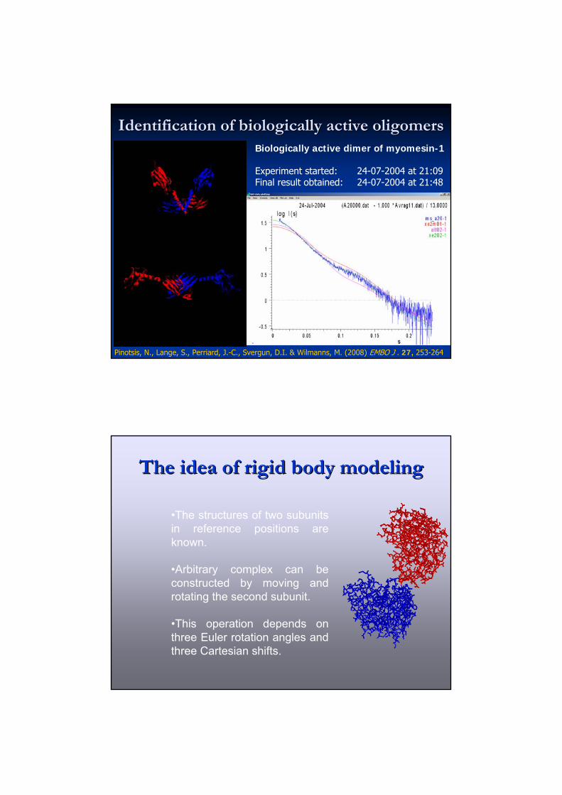
Identification of biologically active oligomers Identification of biologically active oligomers Biologically active dimer of myomesin-1
Experiment started: 24-07-2004 at 21:09 Final result obtained: 24-07-2004 at 21:48
Pinotsis, N., Lange, S., Perriard, J.-C., Svergun, D.I. & Wilmanns, M. (2008) EMBO J . 27, 253-264
•The structures of two subunits in reference positions are known.
•Arbitrary complex can be constructed by moving and rotating the second subunit.
•This operation depends on three Euler rotation angles and three Cartesian shifts.
The idea of rigid body modelingThe idea of rigid body modeling

•The structures of two subunits in reference positions are known.
•Arbitrary complex can be constructed by moving and rotating the second subunit.
•This operation depends on three Euler rotation angles and three Cartesian shifts.
The idea of rigid body modelingThe idea of rigid body modeling
Equation for rigid body modelingEquation for rigid body modeling
Using spherical harmonics, the amplitude(s) of arbitrarily Using spherical harmonics, the amplitude(s) of arbitrarily rotated and displaced subunit(s) are analytically expressed rotated and displaced subunit(s) are analytically expressed viaviathe initial amplitude and the six positional parameters: the initial amplitude and the six positional parameters: CClmlm(s) = (s) = CClmlm(B(Blmlm, , αα, , ββ, , γγ, x, y, z)., x, y, z).
The scattering from the complex is then rapidly calculated asThe scattering from the complex is then rapidly calculated as
Rotation: α, β, γ
A
Shift: x, y, z
C B
( ) [ ]∑∑∞
−
++=0
*2 )()(Re4)()(l
llmlmBA sCsAsIsIsI π
Svergun, D.I. (1991). J. Appl. Cryst. 24, 485-492

Constraints for rigid body modellingConstraints for rigid body modelling
InterconnectivityInterconnectivityAbsence of steric clashesAbsence of steric clashesSymmetrySymmetryIntersubunit contacts Intersubunit contacts (from chemical shifts by (from chemical shifts by NMR or mutagenesis) NMR or mutagenesis) Distances between Distances between residues (FRET or residues (FRET or mutagenesis) mutagenesis) Relative orientation of Relative orientation of subunits (RDC by NMR)subunits (RDC by NMR)Scattering data from Scattering data from subcomplexessubcomplexes
Petoukhov & Svergun Petoukhov & Svergun (2005) (2005) Biophys J.Biophys J. 8989, 1237; , 1237; (2006) (2006) Eur. Biophys. JEur. Biophys. J. . 3535, , 567.567.
Interactive and local refinementInteractive and local refinement
♦ ASSA (SUN/SGI/DEC)Kozin & Svergun (2000). J. Appl. Cryst. 33, 775-777
♦ MASSHA (Win9x/NT/2000)
Konarev, Petoukhov & Svergun (2001).J. Appl. Cryst. 34, 527-532
EPSPS

Manual refinement: quaternary structure of the Manual refinement: quaternary structure of the dimeric dimeric αα--crystallin domaincrystallin domain
Feil, I.K., Malfois, M., Hendle, J., van der Zandt, H. & Svergun, D.I.(2001) J. Biol. Chem. 276, 12024-12029
s, nm-10.5 1.0 1.5 2.0
lg I, relative
8
9
10
11
Global rigid body modelling (SASREF)Global rigid body modelling (SASREF)Fits (multiple XFits (multiple X--ray and neutron) scattering curve(s) from partial ray and neutron) scattering curve(s) from partial constructs or contrast variation using simulated annealing constructs or contrast variation using simulated annealing Requires models of subunits, builds interconnected models withouRequires models of subunits, builds interconnected models without t steric clashes steric clashes Uses constraints: symmetry, distance (FRET or mutagenesis) Uses constraints: symmetry, distance (FRET or mutagenesis) relative orientation (RDC from NMR), if applicablerelative orientation (RDC from NMR), if applicable
Petoukhov & Svergun (2005) Petoukhov & Svergun (2005) Biophys J.Biophys J. 8989, 1237;, 1237;(2006) (2006) Eur. Biophys. JEur. Biophys. J. . 3535, 567., 567.

A global refinement run with distance constraints A global refinement run with distance constraints
ProgramSASREF
Single curve fitting with
distance constraints:
C to N termini contacts
A tyrosine kinase MET (118 kDa) consisting of five domains
Gherardi, E., Sandin, S., Petoukhov, M.V., Finch, J., Youles, M.E., Ofverstedt, L.G., Miguel, R.N., Blundell, T.L., Vande Woude, G.F., Skoglund, U. & Svergun, D.I. (2006) PNAS USA, 103, 4046.
Quaternary structure of tetanus toxin
Qazi, O., Bolgiano, B., Crane, D., Svergun, D.I., Konarev, P.V., Yao, Z.P., Robinson, C.V., Brown, K.A. & Fairweather N. (2007) J Mol Biol. 365, 123–134.
Ab initio and rigid body analysis of the dimeric H(C) domain using the structure of the monomer in the crystal (1FV2) and accounting that the mutant Cys869Ala remains always monomeric yield a unique model of the dimer
Monomeric fraction
Dimeric fraction
Polydispersefractions
100 : 0
0 : 100
64 : 36
43 : 57
21 : 79
14 : 86
Mon:Dim
Receptor binding H(C) domainreveals concentraton-dependent oligomerization

Solution structure of eucaryotic release factor RF1
Vestergaard, B., Sanyal, S., Roessle, M., Mora, L., Buckingham, R. H., Kastrup, J. S., Gajhede, M., Svergun, D. I. & Ehrenberg, M. (2005) Mol. Cell, 20, 929–938.
s0.0 0.1 0.2 0.3
lg I, relative
0
1
2
(1)(2)(3)(4)(5)(6)(7)
A
s0.0 0.1 0.2 0.3
lg I, relative
1
2
3(1)(2)(3)(4)(5)(6)(7)
B
• Cryo-EM: extended; spanss the distance between the ribosomal decoding and peptidyl transferase centers •Crystal: compact, does not span this distance
Rigid body modelling of the Xpot ternary Rigid body modelling of the Xpot ternary complexcomplex
Atomic and homology Atomic and homology modelsmodels
Eleven X-ray and neutron curves
Distance restrains from tRNA Distance restrains from tRNA footprinting (Arts et al. footprinting (Arts et al.
(1998) (1998) EMBO J. EMBO J. 17,17, 7430)7430)
Fukuhara et al. (in preparation)

Addition of missing fragmentsAddition of missing fragments
Flexible loops or domains Flexible loops or domains are often not resolved in are often not resolved in high resolution models or high resolution models or genetically removed to genetically removed to facilitate crystallizationfacilitate crystallizationTentative configuration of Tentative configuration of such fragments are such fragments are reconstructed by fixing the reconstructed by fixing the known portion and adding known portion and adding the missing parts to fit the the missing parts to fit the scattering from the fullscattering from the full--length macromolecule.length macromolecule.
Building nativeBuilding native--like folds of missing fragmentslike folds of missing fragments
Primary sequence
Secondary structure
Excluded volume
Shell radius, nm
0.2 0.4 0.6 0.8 1.0
Number of neighbours
0
1
2
3
4
5
6
Neighbors distribution
Knowledge-based potentials
Bond angles & dihedrals distribution
Using DRUsing DR--type models and proteintype models and protein--specific penalty functions specific penalty functions
Petoukhov, M.V., Eady, N.A.J., Brown, K.A. & Svergun, D.I. (2002) Biophys. J. 83, 3113

BUNCH combines rigid body and BUNCH combines rigid body and ab initioab initiomodelling to find the positions and orientations modelling to find the positions and orientations of rigid domains and probable conformations of of rigid domains and probable conformations of flexible linkers represented as flexible linkers represented as ““dummy residuesdummy residues””chainschains
Multiple experimental scattering data sets from Multiple experimental scattering data sets from partial constructs (e.g. deletion mutants) can be partial constructs (e.g. deletion mutants) can be fitted simultaneously with the data of the fullfitted simultaneously with the data of the full--length protein.length protein.
BUNCH accounts for symmetry, allows one to fix BUNCH accounts for symmetry, allows one to fix some domains and to restrain the model by some domains and to restrain the model by contacts between specific residuescontacts between specific residues
Petoukhov, M. V. & Svergun, D. I. (2005). Biophys. J. 89, 1237-1250
Addition of missing fragments: BUNCHAddition of missing fragments: BUNCH
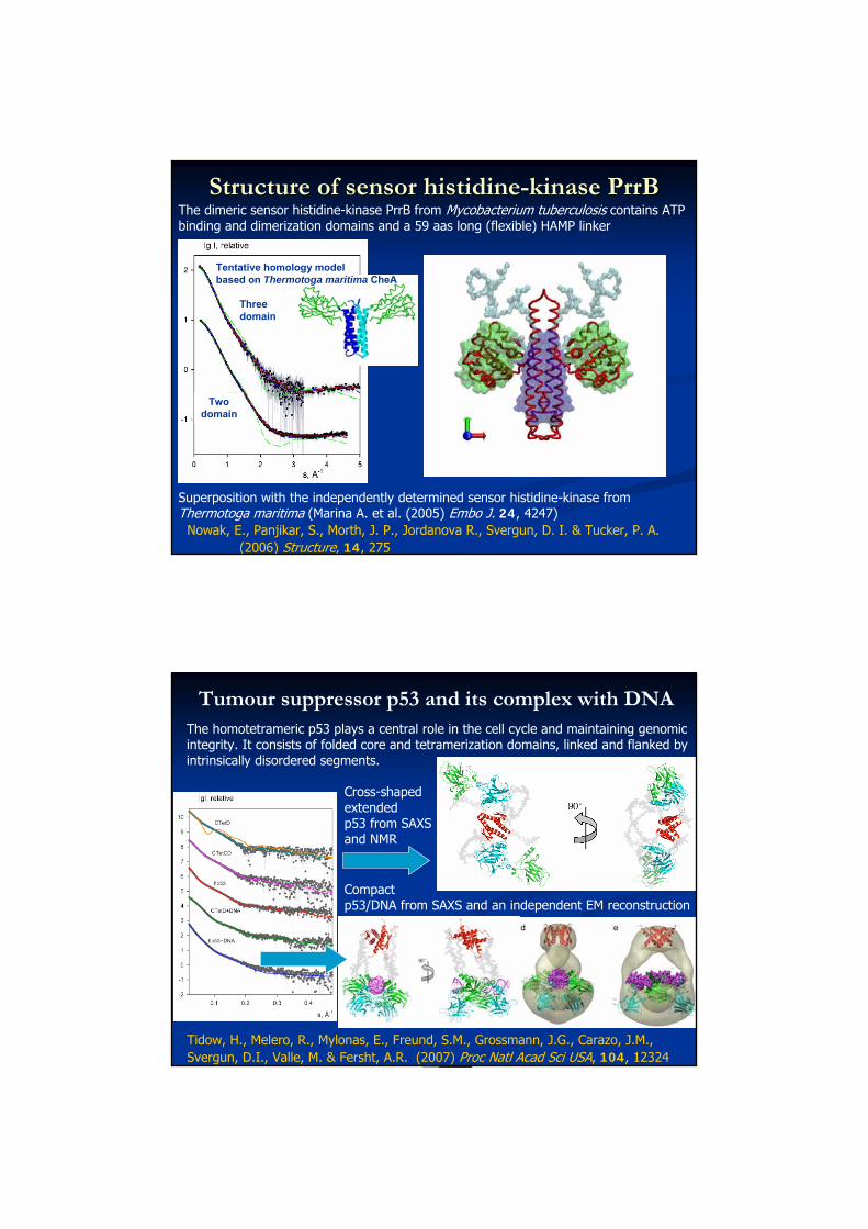
Structure of sensor histidineStructure of sensor histidine--kinase PrrBkinase PrrBThe dimeric sensor histidine-kinase PrrB from Mycobacterium tuberculosis contains ATP binding and dimerization domains and a 59 aas long (flexible) HAMP linker
Nowak, E., Panjikar, S., Morth, J. P., Jordanova R., Svergun, D. I. & Tucker, P. A. (2006) Structure, 14, 275
PrrB model after rigid body refinement and addition of HAMP linker
Tentative homology model based on Thermotoga maritima CheA
Threedomain
Twodomain
Structure of sensor histidineStructure of sensor histidine--kinase PrrBkinase PrrB
Superposition with the independently determined sensor histidine-kinase from Thermotoga maritima (Marina A. et al. (2005) Embo J. 24, 4247)
Nowak, E., Panjikar, S., Morth, J. P., Jordanova R., Svergun, D. I. & Tucker, P. A. (2006) Structure, 14, 275
PrrB model after rigid body refinement and addition of HAMP linker
Tentative homology model based on Thermotoga maritima CheA
Threedomain
Twodomain
The dimeric sensor histidine-kinase PrrB from Mycobacterium tuberculosis contains ATP binding and dimerization domains and a 59 aas long (flexible) HAMP linker
Tumour suppressor p53 and its complex with DNAThe homotetrameric p53 plays a central role in the cell cycle and maintaining genomic integrity. It consists of folded core and tetramerization domains, linked and flanked by intrinsically disordered segments.
Tidow, H., Melero, R., Mylonas, E., Freund, S.M., Grossmann, J.G., Carazo, J.M., Svergun, D.I., Valle, M. & Fersht, A.R. (2007) Proc Natl Acad Sci USA, 104, 12324
Compact p53/DNA from SAXS and an independent EM reconstruction
Cross-shapedextended p53 from SAXS and NMR

Some words of caution
Or Always remember about ambiguity!
Information content in SAS: simple explanationInformation content in SAS: simple explanation
A solution scattering curve from a particle with maximum size D can be represented by
its values taken at discrete points (Shannon channels)
sk = kπ/ D
In a typical SAS experiment, Ns ≈ 5-15
0.00 0.05 0.10 0.15 0.20
100
101
102
I(s)
s, A-1
0 2 4 6 8 10 12
Ns
C. E. Shannon & W. Weaver (1949). The mathematical theory of
communication. University of Illinois Press, Urbana.
∑∞
=⎥⎦
⎤⎢⎣
⎡++
−−−
=1 )(
)(sin)(
)(sin)()(k k
k
k
kkk ssD
ssDssDssDsIssI

Shape determination: M≈ 103 variables (e.g. 0 or 1 bead assignments in DAMMIN
Rigid body methods: M≈ 101 variables (positional and rotational parameters of the subunits)
From the informational point of view, rigid body modeling should provide unique or at least much less ambiguous models than shape determination
Simple explanations do not work in SASSimple explanations do not work in SAS
NO WAY
As all the problems are non-linear, the number of Shannon channels does not give you exact number of parameters, which is possible to extract from the scattering data (depending on accuracy, this number varies between zero and infinity).
Further, uniqueness of reconstruction depends largely on the complexity of the function f(x) to be minimized
Ambiguity of rigid body analysisAmbiguity of rigid body analysis
Petoukhov, M.V. & Svergun, D. I. (2006) Eur. Biophys. J. 35, 567-576
A synthetic example: two different orientations of A synthetic example: two different orientations of tRNA in a dimeric complex with aspartyltRNA in a dimeric complex with aspartyl--tRNA tRNA synthetase obtained by rigid body modelling and synthetase obtained by rigid body modelling and compatible with Xcompatible with X--ray and contrast variation neutron ray and contrast variation neutron scattering data scattering data
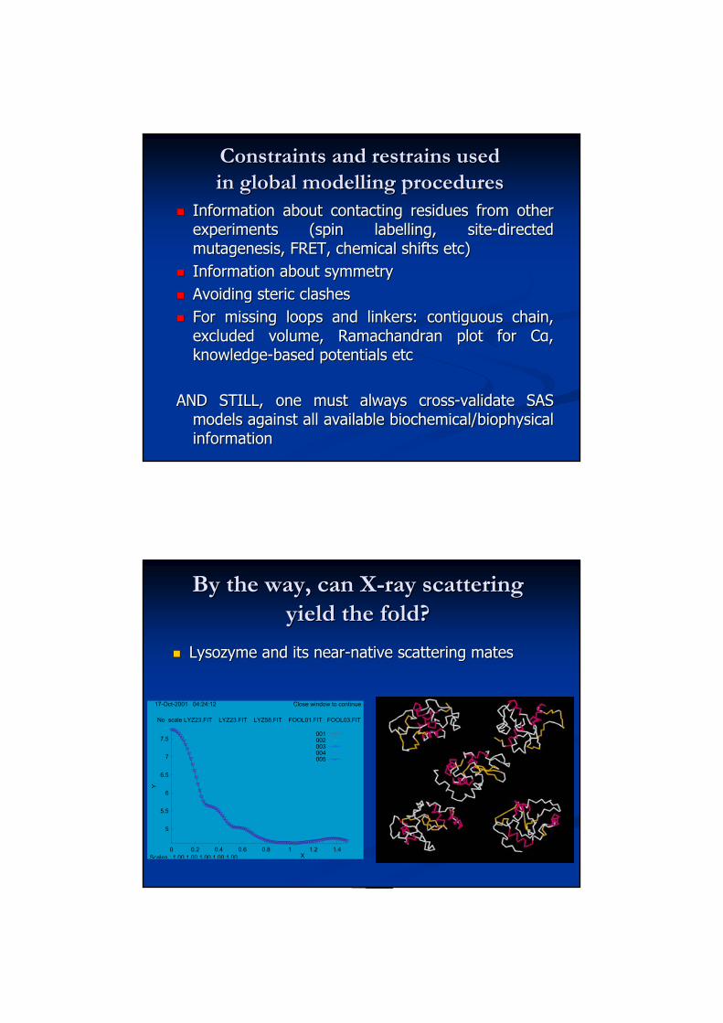
Constraints and restrains used Constraints and restrains used in global modelling proceduresin global modelling procedures
Information about contacting residues from other Information about contacting residues from other experiments (spin labelling, siteexperiments (spin labelling, site--directed directed mutagenesis, FRET, chemical shifts etc)mutagenesis, FRET, chemical shifts etc)Information about symmetryInformation about symmetryAvoiding steric clashesAvoiding steric clashesFor missing loops and linkers: contiguous chain, For missing loops and linkers: contiguous chain, excluded volume, Ramachandran plot for Cexcluded volume, Ramachandran plot for Cαα, , knowledgeknowledge--based potentials etcbased potentials etc
AND STILL, one must always crossAND STILL, one must always cross--validate SAS validate SAS models against all available biochemical/biophysical models against all available biochemical/biophysical information information
By the way, can XBy the way, can X--ray scattering ray scattering yield the fold?yield the fold?
Lysozyme and its nearLysozyme and its near--native scattering matesnative scattering mates
5
5.5
6
6.5
7
7.5
0 0.2 0.4 0.6 0.8 1 1.2 1.4
Y
X
No scale LYZ23.FIT LYZ23.FIT LYZ58.FIT FOOL01.FIT FOOL03.FIT
17-Oct-2001 04:24:12 Close window to continue
Scales : 1.00 1.00 1.00 1.00 1.00
001002003004005

Recent ‘hybrid’ projects at EMBL-HHDomain and quaternary structureComplexes and assemblies
Structural transitionsFlexible macromolecules
Shiozawa et al JBC (2009)
Pex5.Pex14.PTS1 complex
Cheng et al Genes Dev (2009)
eRF3/eFR1 interactions
Rochel et alNSMB (2011)
Nuclear receptors
Albesa-Jové et alJMB (2010)
Toxin B
Calmodulin
Bertini et al JACS (2010)
Complement factor H
Morgan et al NSMB (2011)
Fagan et al Mol. Microbiol (2009)
S-layer proteins
Giehm et alPNAS USA (2011)
α-synuclein oligomers
Recent reviews on solution SAXS
Structural characterization of proteins and complexes using small-angle X-ray solution scatteringHaydyn D.T. Mertens, Dmitri I. SvergunJournal of Structural Biology (2010) 172, 128–141
X-ray solution scattering (SAXS) combined with crystallography and computation: defining accurate macromolecular structures, conformations and assemblies in solution Christopher Putnam, Michal Hammel, Greg Hura and John Tainer. Quarterly Review of Biophysics (2007), 40, 191-285.
Analysis of X-ray and neutron scattering from biomacromolecular solutions. Maxim V. Petoukhov and Dmitri I. Svergun Current Opinion in Structural Biology (2007), 17, 562-571.
EMBL SAS URLsATSAS Home page:http://www.embl-hamburg.de/biosaxs/software.html
ATSAS online:http://www.embl-hamburg.de/biosaxs/atsas-online/
A forum on ATSAS programs:www.saxier.org/forum
A textbook on SAS:Feigin, L.A. & Svergun, D.I. (1987) Structure analysis by small-angle X-ray and neutron scattering. New York: Plenum Presshttp://www.embl-hamburg.de/biosaxs/reprints/feigin_svergun_1987.pdf
EMBO questionnaire:http://www.conference-service.com/glc11-04/welcome.cgi
And now, let us awake for the And now, let us awake for the handshands--on practical on practical