seslhd procedure cover sheet · 2015-12-17 · spare inner cannula (where dual lumen tracheostomy...
TRANSCRIPT

SESLHD PROCEDURE
COVER SHEET
COMPLIANCE WITH THIS DOCUMENT IS MANDATORY Feedback about this document can be sent to [email protected]
NAME OF DOCUMENT
Tracheostomy Clinical Management Procedures for Adults Inpatients
TYPE OF DOCUMENT
Procedure
DOCUMENT NUMBER
SESLHDPR/298
DATE OF PUBLICATION
November 2014
RISK RATING
High
LEVEL OF EVIDENCE
Not available (reflective of ACI Clinical Practice Guideline)
REVIEW DATE
November 2016
FORMER REFERENCE(S)
SESLHNPD/98 Tracheostomy Clinical Management Policy and Procedures for Adults Inpatients
EXECUTIVE SPONSOR or
EXECUTIVE CLINICAL SPONSOR
Dr Gregory Keogh
Director Surgery, Perioperative and Anaesthetics
AUTHOR
Mary Dunford (SGH), Paula Gunner (POWH)
Clinical Nurse Consultants
POSITION RESPONSIBLE FOR THE DOCUMENT
Mary Dunford
Clinical Nurse Consultant
Respiratory
KEY TERMS Trachea, Tracheostomy, Tracheostomy Tube, Stoma, Cannula, Suction
SUMMARY
This document outlines procedures for the tracheostomy management for adult inpatients including (but not restricted to): Tracheostomy Emergency, Changing a Tracheostomy Tube, Removal of Tracheostomy Tube, Suction, Oral Hygiene, Decannulation and Humidification

SESLHD PROCEDURE
Tracheostomy Clinical Management Procedures for Adults Inpatients
PR 298
Revision 2 Trim No. T14/5267 Date: November 2014 Page 1 of 54
THIS DISTRICT DOCUMENT BECOMES UNCONTROLLED WHEN PRINTED OR DOWNLOADED UNLESS REGISTERED BY LOCAL DOCUMENT CONTROL PROCEDURES
1. PROCEDURE STATEMENT SESLHD is committed to ensuring best quality and safety outcomes for patients by implementing best practice by relevant health related government bodies. The recently published Clinical Practice Guideline ‘NSW Agency for Clinical Innovation (ACI): Care of Adult Patients in Acute Care Facilities with a Tracheostomy’ have set best practice recommendations for health organisations in this specific clinical procedure. Considering the above Clinical Practice Guideline and existing clinical practice across SESLHD, this procedure aims to provide clinicians a comprehensive understanding, instruction and best practice evidence-based recommendations for the use of Tracheostomy Tube in adult patients.
2. BACKGROUND
This procedure aims to implement best practice recommendations set by ACI’s Clinical Practice Guideline in the use of Tracheostomy Tube inserted in a tracheal stoma in adult patients and provides further instruction on clinical practice management of adult patients on:
Tracheostomy Emergency
Changing a Tracheostomy tube
Removal of a Tracheostomy tube
Suction
Oral Hygiene
Decannulation
Humidification
Please note that compliance to this clinical procedure is mandatory.
2.1 DEFINITIONS
Trachea: The anatomical structure used for breathing
Tracheostomy: An artificial opening in the trachea, which may be permanent or temporary
Tracheostomy tube: A tube placed through a tracheostomy to provide an airway and to remove secretions from the lungs
Stoma: An opening, either natural or surgically created, which connects a portion of the body cavity to the outside environment
Cannula: A tube that can be inserted into the body, often for the delivery or removal of fluid or air
Suction: The use of devices to clear airways of materials that would impede breathing or cause infections
Aseptic non-touch technique: Prevents microorganisms on hands from being introduced into a susceptible site

SESLHD PROCEDURE
Tracheostomy Clinical Management Procedures for Adults Inpatients
PR 298
Revision 2 Trim No. T14/5267 Date: November 2014 Page 2 of 54
THIS DISTRICT DOCUMENT BECOMES UNCONTROLLED WHEN PRINTED OR DOWNLOADED UNLESS REGISTERED BY LOCAL DOCUMENT CONTROL PROCEDURES
3. RESPONSIBILITIES
3.1 Clinical Staff will:
Safely manage patients within practice limitations, attend education sessions and maintain competency in tracheostomy management and tracheostomy emergency response, including:
Implementing this Clinical Procedure in line with ‘NSW Agency for Clinical
Innovation (ACI): Care of Adult Patients in Acute Care Facilities with a
Tracheostomy’
3.2 Line Managers will:
Line Managers will (within their clinical area) ensure nursing staff caring for the patient with a tracheostomy have the appropriate skills and experience in tracheostomy management and clinical response to a tracheostomy emergency
Line Managers will ensure education resources and clinical protocols are readily available in the clinical environment
3.3 District Managers/ Service Managers will:
Ensure provision of clinical education in tracheostomy management and response to a tracheostomy emergency, is available to clinical staff within SESLHD facilities
3.4 Medical Management will:
Be responsible for medical orientation and ongoing medical education
4. PROCEDURE
4.1 Competency of Clinical Staff in Tracheostomy Management & Tracheostomy Emergencies
All clinical staff providing direct care must be trained and assessed in this tracheostomy management procedure, including the clinical response to a tracheostomy emergency
Patients with a tracheostomy must be cared for in a clinical environment where staff are competent in the clinical management of tracheostomy and the clinical response to a tracheostomy emergency
All clinical staff providing direct care must be familiar with this tracheostomy management procedure, including the clinical emergency response to airway emergencies
All RNs and ENs caring for a patient with a tracheostomy must be educated in tracheostomy management by a designated assessor (nurse educator, clinical nurse consultant or senior physiotherapist with the appropriate clinical expertise). Provision of education is required to include all facets of tracheostomy care, including airway

SESLHD PROCEDURE
Tracheostomy Clinical Management Procedures for Adults Inpatients
PR 298
Revision 2 Trim No. T14/5267 Date: November 2014 Page 3 of 54
THIS DISTRICT DOCUMENT BECOMES UNCONTROLLED WHEN PRINTED OR DOWNLOADED UNLESS REGISTERED BY LOCAL DOCUMENT CONTROL PROCEDURES
emergencies within practice limitations
Senior clinicians responding to patients that require airway and/or breathing assistance with an artificial airway must be provided with ongoing education and training to manage difficult airway situations and undertake ‘difficult airway drills’
4.2 Tracheostomy Plan and Clinical Communication
All patients with a tracheostomy must have a documented plan for tracheostomy assessment, management and review
The Tracheostomy Plan should be reviewed by the Tracheostomy Review Team (TRT) or primary care team. Any changes made to the tracheostomy management plan need to be brought to the notice of the TRT or primary care team. If changes are made by the TRT such changes similarly need to be brought to the notice of the nurse caring for the patient
Patients with a long-term tracheostomy require an appropriate clinical management plan customised to the level of self-care provided and any additional assistance indicated
4.3 Transfer of Care and Clinical Handover
Note: The following section has been developed in line with SESLHD procedure
‘Clinical Handover: Implementation of ISBAR and Key Standard Principles’ SESLHDPR/303 and NSW Ministry of Health Policy Directive ‘Clinical Handover: Key Standard Principles’ PD2009_060.
The unit transferring a patient with a tracheostomy must notify the ward and relevant clinical support personnel eg. specialist CNE/CNC, ICU Liaison, to facilitate clinical support and education of staff and the patient
The transferring RN must handover clinical information using a handover checklist/ Tracheostomy Management and Observation chart including tube size and type, insertion date and method of insertion; method anchored; humidification, secretions and suction requirements
Written communication and verbal bedside, clinical handover regarding potential risks, relevant respiratory history including baseline respiratory rate, work of breathing, chest sounds, tube patency, cough/swallow reflex, oxygenation and oxygen administered must also be handed over
Specific information re management and nursing care required by the receiving area is to be provided during the transfer of care. The clinical handover process must include a visual check to ensure that the tracheostomy tube is patent, aligned and secure

SESLHD PROCEDURE
Tracheostomy Clinical Management Procedures for Adults Inpatients
PR 298
Revision 2 Trim No. T14/5267 Date: November 2014 Page 4 of 54
THIS DISTRICT DOCUMENT BECOMES UNCONTROLLED WHEN PRINTED OR DOWNLOADED UNLESS REGISTERED BY LOCAL DOCUMENT CONTROL PROCEDURES
4.4 Documentation
All tracheostomy interventions including assessments and care provided should be documented on the Tracheostomy Management and Observation chart .
Variances or abnormal findings and management of variances should also be documented in the progress notes
4.5 Observations
Vital signs, respiratory rate, respiratory pattern (including auscultation), oxygen saturations, heart rate, blood pressure and temperature level of consciousness, to be monitored in critical care areas at frequency dictated by clinical condition and on the wards at a frequency not less than every 6 hours. Consider continuous pulse oximetry for patients with a new tracheostomy and/or any respiratory compromise
Patients who require continuous pulse oximetry should be cared for in a suitable clinical environment where staff can continually observe the patient
Monitor sputum and record amount, colour and consistency on Tracheostomy Management and Observation chart.

SESLHD PROCEDURE
Tracheostomy Clinical Management Procedures for Adults Inpatients
PR 298
Revision 2 Trim No. T14/5267 Date: November 2014 Page 5 of 54
THIS DISTRICT DOCUMENT BECOMES UNCONTROLLED WHEN PRINTED OR DOWNLOADED UNLESS REGISTERED BY LOCAL DOCUMENT CONTROL PROCEDURES

SESLHD PROCEDURE
Tracheostomy Clinical Management Procedures for Adults Inpatients
PR 298
Revision 2 Trim No. T14/5267 Date: November 2014 Page 6 of 54
THIS DISTRICT DOCUMENT BECOMES UNCONTROLLED WHEN PRINTED OR DOWNLOADED UNLESS REGISTERED BY LOCAL DOCUMENT CONTROL PROCEDURES
Minimum Frequency Tracheostomy Checks and Care (Document Assessment and Care Provided on Tracheostomy Management and Observation Chart)
1-2 Hourly 2- 4 Hourly 6 Hourly Once per Shift Daily
Assess adequacy of humidification
Inner cannula remove, check for secretion build up; clean and replace
Document:
Airway - skin colour, air entry - bilateral at axilla, expired air felt from tracheostomy tube
Breathing - bilateral chest movement, and depth of respirations
Check and restock Emergency bedside equipment
Trache tapes changed (more frequently if soiled)
Assess need for suction. Document amount, viscosity and colour of secretions
Normal saline nebulisers 4 hourly/prn, (More frequently for patients with thick secretions)
Vital Signs: Respiratory rate Oxygen saturation Heart rate BP Temperature
Cuff pressure measurement
Assess systemic hydration (fluid balance)
For adjustable flange tracheostomy tubes – observe and document the position of the flange to the tube at the skin following each suction to detect tube migration
Check Heat Moisture Exchanger (HME)
Clean stoma site
Change heat/moisture exchanger (HME) NB more frequently if soiled
Assess position and alignment of the tracheostomy tube
Mouth care Stoma site - observe for bleeding in new stomas; note crusting, signs of infection, smell, discharge
SESLHD PROCEDURE
Tracheostomy Clinical Management Procedures for Adults Inpatients
PR 298
Revision 2 Trim No. T14/5267 Date: November 2014 Page 7 of 54
THIS DISTRICT DOCUMENT BECOMES UNCONTROLLED WHEN PRINTED OR DOWNLOADED UNLESS REGISTERED BY LOCAL DOCUMENT CONTROL PROCEDURES
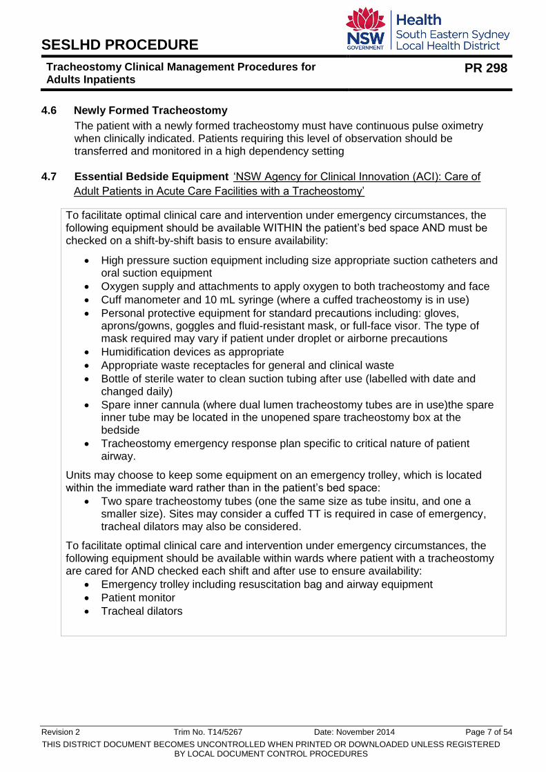
4.6 Newly Formed Tracheostomy
The patient with a newly formed tracheostomy must have continuous pulse oximetry when clinically indicated. Patients requiring this level of observation should be transferred and monitored in a high dependency setting
4.7 Essential Bedside Equipment ‘NSW Agency for Clinical Innovation (ACI): Care of
Adult Patients in Acute Care Facilities with a Tracheostomy’
To facilitate optimal clinical care and intervention under emergency circumstances, the following equipment should be available WITHIN the patient’s bed space AND must be checked on a shift-by-shift basis to ensure availability:
High pressure suction equipment including size appropriate suction catheters and oral suction equipment
Oxygen supply and attachments to apply oxygen to both tracheostomy and face
Cuff manometer and 10 mL syringe (where a cuffed tracheostomy is in use)
Personal protective equipment for standard precautions including: gloves, aprons/gowns, goggles and fluid-resistant mask, or full-face visor. The type of mask required may vary if patient under droplet or airborne precautions
Humidification devices as appropriate
Appropriate waste receptacles for general and clinical waste
Bottle of sterile water to clean suction tubing after use (labelled with date and changed daily)
Spare inner cannula (where dual lumen tracheostomy tubes are in use)the spare inner tube may be located in the unopened spare tracheostomy box at the bedside
Tracheostomy emergency response plan specific to critical nature of patient airway.
Units may choose to keep some equipment on an emergency trolley, which is located within the immediate ward rather than in the patient’s bed space:
Two spare tracheostomy tubes (one the same size as tube insitu, and one a smaller size). Sites may consider a cuffed TT is required in case of emergency, tracheal dilators may also be considered.
To facilitate optimal clinical care and intervention under emergency circumstances, the following equipment should be available within wards where patient with a tracheostomy are cared for AND checked each shift and after use to ensure availability:
Emergency trolley including resuscitation bag and airway equipment
Patient monitor
Tracheal dilators

SESLHD PROCEDURE
Tracheostomy Clinical Management Procedures for Adults Inpatients
PR 298
Revision 2 Trim No. T14/5267 Date: November 2014 Page 8 of 54
THIS DISTRICT DOCUMENT BECOMES UNCONTROLLED WHEN PRINTED OR DOWNLOADED UNLESS REGISTERED BY LOCAL DOCUMENT CONTROL PROCEDURES
4.8 Tracheostomy Emergency Procedure
In line with ‘NSW Agency for Clinical Innovation (ACI): Care of Adult Patients in Acute Care Facilities with a Tracheostomy’, the following are evidence-based recommendations for the tracheostomy emergency procedure.
Recommendations Grade of
Recommendation
All hospitals are to have documented action plans that identify how tracheostomy emergencies are to be managed. These action plans must include
Action/s to be taken especially those of clinicians who are caring for the patient and are not part of emergency teams
Key personnel at all times of the day
Emergencies include but are not limited to - displaced or dislodged tracheostomy - blocking or blocked tracheostomy - airway haemorrhage - cuff leak or rupture
And be available at point of care.
Consensus
Elective early tracheostomy tube change, defined as within 72 hours of the formation of the tracheal stoma, may be hazardous and should be avoided, particularly in patients with a history of difficult intubation.
Consensus
Emergency management must be included in education programs to ensure that clinicians who care for patients with tracheostomies are able to provide care in the event of an emergency.
Consensus
Hospitals should include tracheostomy emergency scenarios within education on difficult airway drill.
Consensus
All wards where patients are cared for with a tracheostomy must have emergency airway equipment available in the event of an emergency.
Consensus
The appropriate level of emergency call should be activated if any of the signs and symptoms of respiratory distress are present and are unable to be resolved quickly (SESLHD PACE2 call).
Consensus
A competent clinician must stay with patient so that interventions can be commenced.
Consensus
A blocked or displaced TT should only be replaced by an experienced clinician.
Consensus
When a patient experiences a complication, this must be documented in the patient notes and the patient’s primary care team and tracheostomy review team, where in place, notified.
Consensus
Once a complication or emergency has resolved, the TT plan should be reviewed in relation to factors which may have contributed to the emergency. This may include but is NOT limited to the following:
Type of TT
Consensus

SESLHD PROCEDURE
Tracheostomy Clinical Management Procedures for Adults Inpatients
PR 298
Revision 2 Trim No. T14/5267 Date: November 2014 Page 9 of 54
THIS DISTRICT DOCUMENT BECOMES UNCONTROLLED WHEN PRINTED OR DOWNLOADED UNLESS REGISTERED BY LOCAL DOCUMENT CONTROL PROCEDURES
Recommendations Grade of
Recommendation
Whether the TT stabilisation method is appropriate
The patient’s current level of supervision and visibility on ward
Use of chemical or physical restraints where appropriate
Humidification method and hydration status.
Tracheostomy Emergency
Immediate Action
Respiratory Distress Signs of Respiratory Distress Include:
Increased work of breathing ie. patient acutely distressed/restless, tachypnoea, stridor, accessory muscle use, diaphoretic, cyanotic
Decreased/gurgling breath sounds
High inspiratory airway pressures/low tidal volumes if mechanically ventilated
O2 desaturation
Stay with patient and provide 100% high flow O2 via tracheostomy
and/or face mask and manually ventilate if indicated (it may be
necessary to deflate trachea cuff)
Check O2 source and connection, cuff inflation, humidifier
Check /change the inner cannula if in situ
Call for assistance – emergency number (as per site procedure)
Dislodged tracheostomy tube in situ with suspected obstruction:
Pass suction catheter and apply suction (change inner cannula if
present, using non-fenestrated cannula if possible), if
tracheostomy tube obstructed then let down cuff (if present and
inflated)
Observe tube patency, secretions and patient response to
suctioning
If the patient becomes less distressed, airflow is present,
unobstructed and oxygenation is satisfactory, then undertake a full
clinical assessment to establish the cause of respiratory distress
If no airflow around/through tracheostomy tube then insert tracheal
dilators around tube into stoma, remove tube, insert bougie or
suction catheter and maintain oropharyngeal airway to achieve
oxygenation
Laryngectomy patients: concentrate all measures on clearing
stoma/trachea, the sole airway access
Potential Causes
Airway partially/completely obstructed due to blockage
Tracheostomy dislodgement
Persistent cuff leak
Faulty O2 source or ventilation device
Ineffective humidification

SESLHD PROCEDURE
Tracheostomy Clinical Management Procedures for Adults Inpatients
PR 298
Revision 2 Trim No. T14/5267 Date: November 2014 Page 10 of 54
THIS DISTRICT DOCUMENT BECOMES UNCONTROLLED WHEN PRINTED OR DOWNLOADED UNLESS REGISTERED BY LOCAL DOCUMENT CONTROL PROCEDURES
No breath sounds
Unable to pass suction catheter or inner cannula
Consider non-tracheostomy related causes for distress
Patient Distressed with Tube Obstructed Dislodged Or Cuff Leaking
100% high flow O2 via face mask and manually ventilate if indicated (it may be necessary to deflate the cuff
If no tracheostomy tube in place then clean stoma, open and support stoma with forceps, insert new tube, inflate cuff if present, re-oxygenate and assess air entry, work of breathing and clinical status
If tracheostomy tube in place then prepare for rapid tracheostomy tube exchange/placement (provide brief explanation to patient)
Assemble and check equipment
Position patient supine with head of bed elevated slightly (ensure no clinical contraindication)
Consider the need for sedation – this will be indicated based on individual patient assessment and the senior medical officer orders
Remove pillow and extend neck (ensure no clinical contra-indication)
Suction oropharynx If tracheostomy tube in place:
<72 hours (early change) clean stoma, loosen ties, hold tube in place, insert bougie into tracheostomy tube, assistant deflate cuff, remove tracheostomy tube over bougie while ensuring bougie is held in situ, immediately slide new tracheostomy tube over bougie into the trachea, hold in place, remove bougie, inflate cuff, reoxygenate and assess air entry, work of breathing and clinical status
>72 hours (formed stoma) clean stoma, loosen ties, hold tube in place, support open stoma with forceps, assistance deflates cuff, remove tracheostomy tube immediately slide new tracheostomy tube into the trachea soma, hold in place, inflate cuff, reoxygenate and assess air entry, work of breathing and clinical status
Correct tube placement is confirmed by checking air flow, chest auscultation, improved SpO2 and if available ETCO2

SESLHD PROCEDURE
Tracheostomy Clinical Management Procedures for Adults Inpatients
PR 298
Revision 2 Trim No. T14/5267 Date: November 2014 Page 11 of 54
THIS DISTRICT DOCUMENT BECOMES UNCONTROLLED WHEN PRINTED OR DOWNLOADED UNLESS REGISTERED BY LOCAL DOCUMENT CONTROL PROCEDURES
Successful
Secure tracheostomy tube
Review O2 and ventilation
Reposition patient
Provide education and further reassure the patient and family
Unsuccessful
Maintain oxygenation
Manually ventilate if required
Prepare for intubation or LMA insertion
Intubate or place LMA
Only use stoma if laryngectomy patient
4.9 Signs of Respiratory Distress
Difficult, laboured or noisy breathing - In complete tracheostomy tube occlusion, there are no breath sounds heard - however in partial obstruction air entry is diminished and often noisy.
Use of accessory muscles - A sign of airway obstruction. In complete airway obstruction patients often develop a see-saw pattern of breathing in which inspiration is concurrent with outward movement of the abdomen and inward movement of the chest wall and vice-versa.
No or Limited expired air from the tracheostomy tube. Reduced chest movement or reduced air entry upon auscultation - All indicate a lack of air movement into and out of the respiratory tract.
Pale/cyanosed skin colour - Central cyanosis is a sign of late airway obstruction.
Anxiety / Agitation - The patient will become anxious and agitated as they struggle to breathe and become hypoxic.
Increased pulse/respiratory rate - Increased respiratory and pulse rate are signs of illness and an indicator that the patient may suddenly deteriorate.
Clammy / diaphoretic skin - Associated with an increased work of breathing from an occluded airway and stimulation of the sympathetic nervous system causing vasoconstriction
Stridor – Is caused by an obstruction above or at the level of the larynx
5. EMERGENCY ALGORITHM
The following algorithm has been designed to guide clinicians when dealing with situations outside of the critical care setting. Each situation should be assessed on an individual basis and correctly managed. As with any emergency, emergency protocols should be initiated immediately.

SESLHD PROCEDURE
Tracheostomy Clinical Management Procedures for Adults Inpatients
PR 298
Revision 2 Trim No. T14/5267 Date: November 2014 Page 12 of 54
THIS DISTRICT DOCUMENT BECOMES UNCONTROLLED WHEN PRINTED OR DOWNLOADED UNLESS REGISTERED BY LOCAL DOCUMENT CONTROL PROCEDURES
5.1 Humidification
Patient with Tracheostomy
Patient in Respiratory Distress Patient in respiratory arrest
Activate Arrest Call
ACTIVATE PACE TIER 2 /CODE BLUE TEAM
INITIATE BASIC LIFE
SUPPORT
Assess and Clear Airway - (tracheostomy)
Obstruction algorithm
Accidental decannulation algorithm
YES NO
Tube displacement algorithm
Haemorrhage algorithm
Remove inner cannula
Suction tracheostomy
Oxygen to tracheostomy
NO YES
Is condition Immediately Life Threatening?
Airway Patent / Breathing Present
Airway Compromised
Remains in respiratory distress post suction and oxygen
Education Note: A tracheostomy bypasses the normal mechanisms for humidification. Failure to provide adequate humidification contributes to tube blockage and subsequent airway obstruction. Adequate systemic hydration via oral, nasogastric and/or intravenous is required to ensure secretions remain easy to suction or expectorate. (Burgess 1999, Clark 1995, St Georges Healthcare NHS trust, Harkin 1998)

SESLHD PROCEDURE
Tracheostomy Clinical Management Procedures for Adults Inpatients
PR 298
Revision 2 Trim No. T14/5267 Date: November 2014 Page 13 of 54
THIS DISTRICT DOCUMENT BECOMES UNCONTROLLED WHEN PRINTED OR DOWNLOADED UNLESS REGISTERED BY LOCAL DOCUMENT CONTROL PROCEDURES
5.1.1 Symptoms of insufficient humidification include:
Shortness of breath and decreased oxygen saturations (indicative of mucus plugging or micro atelectasis)
Increased, unproductive cough
Change in mucous colour (clear to pale), amount or increased viscosity (ie. a change from thin to thick, sticky consistency)
Increased temperature (indicative of infection and impaired secretion removal)
Blood-streaked mucous
Noisy laboured respirations.
5.1.2 Care of the Heat Moisture Exchange (HME) Includes:
Educate patient to remove the HME before coughing.
5.1.3 Policy Alert:
An HME must be used for patients with a tracheostomy tube with an inner cannula (inpatients). Patients with single lumen adjustable flange tracheostomy tube or foam cuff tracheostomy tube must have continuous warm humidification with connector directly attached to the tracheostomy tube
Monitor the patient for signs of adequate humidification every 1-2 hours (see symptoms above)
Check disposable HME two hourly and change daily or whenever soiled
Continuous warm humidification via water bath and heated base should be considered where a HME is not providing effective humidification
Patients with sputum plugging or blood stained sputum require warm humidification (contact CNC Respiratory/ENT or equivalent person (educator) if unfamiliar with equipment
Nebulisation with normal saline may be prescribed by the primary care team if indicated and should be considered in all patients using a HME
Condensation in the circuit must be avoided as it is a medium for bacterial growth.
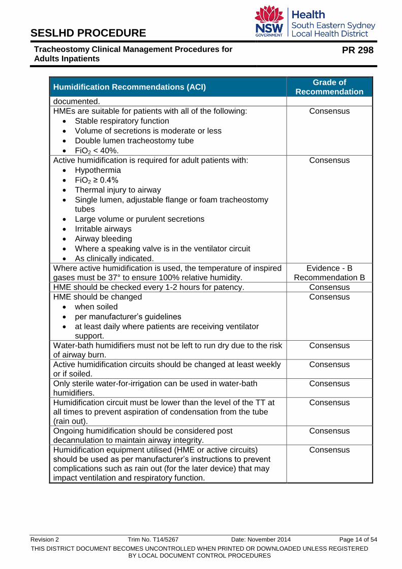
5.1.4 Humidification Recommendations
In line with ‘NSW Agency for Clinical Innovation (ACI): Care of Adult Patients in Acute Care Facilities with a Tracheostomy’, the following are evidence-based recommendations for humidification
Humidification Recommendations (ACI) Grade of
Recommendation
Inspired gases must be humidified to maintain effective mucociliary function and gas exchange and prevent complications.
Consensus
Patient’s systemic hydration must be assessed and maintained to reduce the viscosity of sputum and prevent complications.
Consensus
The choice of humidification method should be made on an individual patient basis, be assessed at least daily and
Consensus
SESLHD PROCEDURE
Tracheostomy Clinical Management Procedures for Adults Inpatients
PR 298
Revision 2 Trim No. T14/5267 Date: November 2014 Page 14 of 54
THIS DISTRICT DOCUMENT BECOMES UNCONTROLLED WHEN PRINTED OR DOWNLOADED UNLESS REGISTERED BY LOCAL DOCUMENT CONTROL PROCEDURES
Humidification Recommendations (ACI) Grade of
Recommendation
documented.
HMEs are suitable for patients with all of the following:
Stable respiratory function
Volume of secretions is moderate or less
Double lumen tracheostomy tube
FiO2 < 40%.
Consensus
Active humidification is required for adult patients with:
Hypothermia
FiO2 ≥ 0.4%
Thermal injury to airway
Single lumen, adjustable flange or foam tracheostomy tubes
Large volume or purulent secretions
Irritable airways
Airway bleeding
Where a speaking valve is in the ventilator circuit
As clinically indicated.
Consensus
Where active humidification is used, the temperature of inspired gases must be 37° to ensure 100% relative humidity.
Evidence - B Recommendation B
HME should be checked every 1-2 hours for patency. Consensus
HME should be changed
when soiled
per manufacturer’s guidelines
at least daily where patients are receiving ventilator support.
Consensus
Water-bath humidifiers must not be left to run dry due to the risk of airway burn.
Consensus
Active humidification circuits should be changed at least weekly or if soiled.
Consensus
Only sterile water-for-irrigation can be used in water-bath humidifiers.
Consensus
Humidification circuit must be lower than the level of the TT at all times to prevent aspiration of condensation from the tube (rain out).
Consensus
Ongoing humidification should be considered post decannulation to maintain airway integrity.
Consensus
Humidification equipment utilised (HME or active circuits) should be used as per manufacturer’s instructions to prevent complications such as rain out (for the later device) that may impact ventilation and respiratory function.
Consensus

SESLHD PROCEDURE
Tracheostomy Clinical Management Procedures for Adults Inpatients
PR 298
Revision 2 Trim No. T14/5267 Date: November 2014 Page 15 of 54
THIS DISTRICT DOCUMENT BECOMES UNCONTROLLED WHEN PRINTED OR DOWNLOADED UNLESS REGISTERED BY LOCAL DOCUMENT CONTROL PROCEDURES
5.1.5 Provision of Humidification
5.1.6 Inner Cannula Maintenance
Practice Limitations: Registered Nurses who are deemed competent by a designated assessor Enrolled Nurses who have achieved “Extended Practice Skill” in Tracheostomy Maintenance of an Inner Cannula and been deemed competent by a designated assessor
5.1.7 Policy Alert
All ward patients with a tracheostomy tube must have an inner cannula insitu. With the exception of adjustable flange tracheostomy tubes which do not have an inner cannula
Procedure: Provision of Humidification Key Notes
For all patients with loose or no evidence of
secretions or in longer term patients use an HME
Replace HME daily or more frequently if
contaminated by secretions. Patients on BIPAP
may require HME changes more frequently
To moisten inspired gases by trapping and re-breathing humidity; to prevent inhalation of particulate matter To maintain effectiveness and reduce infection risk
For patients with thick/dry secretions, ensure 4
hourly prescription of normal saline nebulisers
Administer 5mls of normal saline via nebuliser
every 4 hours and PRN to provide fully saturated
air with fine mist of moisture
Increase frequency of nebulised normal saline if
sputum thickness/tenacity impairs suctioning
Assess systemic hydration daily – inform medical
staff of inadequate fluid intake (especially if
patient is NBM)
To loosen and thin secretions, to
prevent atelectasis and sputum
thickening
To reduce unnecessary
interventions and to assess
whether present level of
humidification adequate
To highlight potential issues and
instigate early intervention if
required

SESLHD PROCEDURE
Tracheostomy Clinical Management Procedures for Adults Inpatients
PR 298
Revision 2 Trim No. T14/5267 Date: November 2014 Page 16 of 54
THIS DISTRICT DOCUMENT BECOMES UNCONTROLLED WHEN PRINTED OR DOWNLOADED UNLESS REGISTERED BY LOCAL DOCUMENT CONTROL PROCEDURES
The inner cannula must be removed 2-4 hourly to check for patency and secretion build up. It must be immediately replaced with a clean inner cannula. More frequent checks will depend on viscosity and volume of secretions
All patients with a tracheostomy will require 4 hourly normal saline nebulisers
This procedure is a clean procedure which requires hand hygiene before and after donning appropriate PPE eg. gloves, apron, full-face visor
5.1.8 Equipment
Clean gloves
Facial protection and plastic apron
Clean dry replacement inner cannula
Sterile pipe cleaners (may be required) In line with ‘NSW Agency for Clinical Innovation (ACI): Care of Adult Patients in Acute Care Facilities with a Tracheostomy’, the following are evidence-based recommendations for Inner-Cannula
Inner Cannula Recommendations (ACI) Grade of
Recommendation
The inner cannula must be checked for patency, cleaned and replaced 2-4 hourly. More frequent checks will depend on the volume and viscosity of secretions.
Consensus
The inner cannula should be cleaned and dried according to manufacturer’s guidelines and stored in a clean dry container.
Consensus
Under most circumstances the inner cannula can be cleaned with sterile water with a tracheostomy cleaning brush or a pipe cleaner (with the end turned over). Where secretions are tenacious, alternative solutions can be used; however, the tube should not be soaked for more than 15 minutes.
Consensus
This procedure is a clean procedure which requires hand hygiene before and after donning appropriate PPE eg. gloves, apron, full-face visor.
Consensus
It is inappropriate to clean or rinse the inner cannula at hand basins used for hand washing because of the risk of contaminating the basin with organisms or contamination of the inner cannula.
Consensus
When placing a clean inner cannula into a TT tube it should be rinsed with sterile water immediately prior to insertion.
Consensus
Procedure: Changing an Inner Cannula Key Notes
1 Perform hand hygiene before and after donning appropriate PPE eg. gloves, apron, full-face visor
To reduce the risk of cross infection
2 Hyper-oxygenate or ask patient to take 5 deep breaths
To prevent hypoxia

SESLHD PROCEDURE
Tracheostomy Clinical Management Procedures for Adults Inpatients
PR 298
Revision 2 Trim No. T14/5267 Date: November 2014 Page 17 of 54
THIS DISTRICT DOCUMENT BECOMES UNCONTROLLED WHEN PRINTED OR DOWNLOADED UNLESS REGISTERED BY LOCAL DOCUMENT CONTROL PROCEDURES
3 Position patient with neck slightly extended To provide patient comfort and ease procedure
4 Remove oxygen, remove inner cannula and insert clean spare inner cannula, replace oxygen
To maintain the airway, prevent early build-up of secretions in outer tube and to maintain oxygenation
5 Observe for excessive crusting inside cannula
Crusting should not occur if the tube is kept clean with the provision of adequate humidification and suction. More frequent checks will depend on viscosity and volume of secretions
6 Clean the inner cannula and flush with sterile saline or water prior to reinsertion (may require the use of a cleaning brush or pipe cleaners to remove any dried or tenacious secretions) Agitate inner cannula to remove all visible secretions prior to re-inserting in airway If using a spare inner cannula store in a clean, dry identified container
Ensure end of pipe cleaner is folded over to prevent any exposed metal scratching or damaging the inner tube
7 Document procedure and findings on Tracheostomy Management and Observation chart
To facilitate communication and evaluation
6. CHECKING CUFF PRESSURE
Practice Limitations: Registered Nurses who are deemed competent by a designated assessor Enrolled Nurses who have achieved “Extended Practice Skill” in Tracheostomy checking Cuff Pressure and been deemed competent by a designated assessor
6.1 Education Note
Tracheostomy tubes may be cuffed or un-cuffed. The cuff seals the space around the tracheostomy tube and prevents aspiration of oral or gastric secretions. A cuff pressure maintained at 20-30 cm H2O prevents tracheal wall necrosis. A leaking cuff may indicate that the cuff is over inflated and/or the tube is dislodged.
6.2 Policy Alert:
Where a cuffed tracheostomy tube is in situ the cuff pressure must be checked at least once a shift using a manometer and maintained at 20-30cm H20
Cuff deflation is not recommended (unless part of the formal weaning process) as it may increase the risk of aspiration

SESLHD PROCEDURE
Tracheostomy Clinical Management Procedures for Adults Inpatients
PR 298
Revision 2 Trim No. T14/5267 Date: November 2014 Page 18 of 54
THIS DISTRICT DOCUMENT BECOMES UNCONTROLLED WHEN PRINTED OR DOWNLOADED UNLESS REGISTERED BY LOCAL DOCUMENT CONTROL PROCEDURES
If cuff pressure cannot be maintained eg. spontaneously reducing or a leak is suspected (indicated by audible gurgling) escalate to senior medical/nursing staff for assistance. The patient with a leaking cuff is at increased risk of aspiration and the tracheostomy tube may need to be changed.
In line with ‘NSW Agency for Clinical Innovation (ACI): Care of Adult Patients in Acute Care Facilities with a Tracheostomy’, the following are evidence-based recommendations for Cuff pressure
Cuff Pressure Recommendations (ACI) Grade of
Recommendation
When using a cuffed tracheostomy tube, the intra-cuff pressure should be high enough to achieve a closed respiratory system and be between 20-30cm H2O to
Maintain a closed respiratory system to facilitate mechanical ventilation and
Prevent tracheal mucosal necrosis and minimize micro-aspiration.
Evidence - C Recommendation
C
Intra-cuff pressure should be measured directly using a cuff manometer, optimised and documented
At least once every 8 hours and when clinically indicated
Immediately post intubation
Immediately on receiving patient from another clinical area
After significant patient movement
Where there are any concerns about air leak from the respiratory system such as when the patient vocalizes or a ventilator alarms.
Evidence - B Recommendation
B
Where there is a persistent cuff leak, the nurse must notify the TRT or ENT team to review.
Consensus
Preferably patients should have their own cuff manometer. However, where a cuff manometer is used among multiple patients, it MUST be cleaned between patients using the usual disinfection practices and according to manufacturer’s instructions.
Consensus
Infectious patients MUST have their own cuff manometer. Consensus
Cuff deflation is to be managed by experienced clinicians and may include members of the ENT team or TRT (eg. speech pathologist, specialist nurses, physiotherapists or medical professional).
Consensus
Where patients are transported by air, tracheostomy cuffs will require specific care to prevent over-distension and high intra-cuff pressures.
Consensus

SESLHD PROCEDURE
Tracheostomy Clinical Management Procedures for Adults Inpatients
PR 298
Revision 2 Trim No. T14/5267 Date: November 2014 Page 19 of 54
THIS DISTRICT DOCUMENT BECOMES UNCONTROLLED WHEN PRINTED OR DOWNLOADED UNLESS REGISTERED BY LOCAL DOCUMENT CONTROL PROCEDURES
Procedure: Checking Cuff Pressure
Key Points
1
Inform the patient of procedure
2
Securely connect pressure gauge to pilot balloon of the manometer
If not secure - pressure will be inaccurate
3
Determine if pressure is above or below the optimum range
Optimum is any level in the green range of the manometer
4
Above range - press the release button on the side of the pressure gauge until it returns to green
An over-inflated cuff may cause tracheal necrosis, fistulas, dilation or stenosis. 20-30cm H2O is the acceptable pressure range
5
Below range - inflate balloon one depression at a time until the needle enters the optimal range 20-30cm H2O
Deflated or partially inflated cuff increases risk of aspiration and may compromise respiratory status
6
Disconnect the gauge when in the optimal range. If cuff will not inflate or continues to lose pressure inform ENT/ICU Registrar, CNC Respiratory/ENT or CNC Surgery
The one way valve will ensure that air will remain in cuff
7
If the cuff pressure is reading lower than the acceptable range, inflate the cuff and reassess within the hour to detect/confirm a cuff leak
7. SUCTIONING A TRACHEOSTOMY TUBE
Practice Limitations: Registered Nurses who are deemed competent by a designated assessor Enrolled Nurses who have achieved “Extended Practice Skill” in Tracheostomy Suctioning and been deemed competent by a designated assessor
Indications:
Contraindications:
Potential Complications:
Persistent coughing
Platelets < 20 Tracheal trauma
Respiratory distress
Acute pulmonary oedema
Suctioning induced hypoxemia
Audible gurgling or Acute respiratory Hypertension

SESLHD PROCEDURE
Tracheostomy Clinical Management Procedures for Adults Inpatients
PR 298
Revision 2 Trim No. T14/5267 Date: November 2014 Page 20 of 54
THIS DISTRICT DOCUMENT BECOMES UNCONTROLLED WHEN PRINTED OR DOWNLOADED UNLESS REGISTERED BY LOCAL DOCUMENT CONTROL PROCEDURES
visible secretions
haemorrhage
Decreasing oxygen saturation
Cardiac arrhythmias
Increased peak inspiratory pressures for mechanically ventilated patients
Raised intracranial pressure
Increased or decreased respiratory rate
Infection
Laryngospasm
Haemorrhage
7.1 Policy Alert:
Suction frequency should be based on patient assessment and clinical need. Patients should be regularly assessed for work of breathing, oxygen saturations and chest auscultation
Light suction to the end of the tracheostomy tube is preferable if the patient is awake and has an effective cough
Deep suction (catheter fully advanced to the carina and then withdrawn 1cm before applying suction), must be used with caution as it may cause trauma to the trachea and increase intracranial pressure
Suction should only be applied during catheter withdrawal and should be applied continuously as opposed to intermittently
Suctioning should take no longer than 10 seconds. Longer periods of suction are associated with increased risk of hypoxaemia and trauma
Non-sterile gloves should be used when using a closed suction system
Suction using an open system must use aseptic non-touch technique.
In line with ‘NSW Agency for Clinical Innovation (ACI): Care of Adult Patients in Acute Care Facilities with a Tracheostomy’, the following are evidence-based recommendations for suctioning a tracheostomy tube
Recommendations Grade of
Recommendation
Due to the potential for adverse effects and significant patient discomfort, suctioning a tracheostomy tube should be performed on the basis of clinical need and not be carried out on a routine basis.
Evidence - B Recommendation
B

SESLHD PROCEDURE
Tracheostomy Clinical Management Procedures for Adults Inpatients
PR 298
Revision 2 Trim No. T14/5267 Date: November 2014 Page 21 of 54
THIS DISTRICT DOCUMENT BECOMES UNCONTROLLED WHEN PRINTED OR DOWNLOADED UNLESS REGISTERED BY LOCAL DOCUMENT CONTROL PROCEDURES
Recommendations Grade of
Recommendation
For ventilated patients, assessment of the patient to identify the need to suction a tracheostomy tube should be continuous with chest auscultation performed every two hours or more frequently as indicated by clinical signs (see table 8).
Consensus
For non-ventilated patients, assessment of the patient to identify the need to suction should be based on observation and clinical assessment.
Consensus
The size of the suction catheter should occlude no more than 50% of the internal diameter of the artificial airway to avoid greater negative pressure in the airway [35].
Evidence - C Recommendation
C
Closed suction catheter systems should be used as the system of choice for patients with high FiO2 or PEEP or at risk of lung derecruitment) [39].
Evidence - B Recommendation
C
During a suction procedure, the patient must be assessed for clinical stability and tolerance.
Consensus
The effectiveness of the suction procedure should be evaluated using clinical indicators.
Consensus
Suction pressure should be set at 100-150mmHg for adults [35, 39].
Evidence - C Recommendation
C
To prevent the occurrence of adverse events, bolus instillation of normal saline should not be used routinely during suctioning [35, 39].
Evidence - B Recommendation
B
Where a fenestrated tracheostomy tube is insitu, a non-fenestrated inner cannula must be inserted prior to suction.
Consensus
The upper airway should be suctioned periodically to remove oral secretions and to minimise stasis of pooled secretions about the tracheostomy cuff with subsequent potential for aspiration to lower airways.
Evidence - C Recommendation
C
7.2 Equipment:
High pressure wall suction
Oxygen as required
Clean examination gloves and disposable apron/gown
Eye/Facial protection/shield
Water to wash through tubing after suctioning
Yankauer sucker
Suction catheter of appropriate size not exceeding 50 % of the internal diameter of the tracheostomy tube.

SESLHD PROCEDURE
Tracheostomy Clinical Management Procedures for Adults Inpatients
PR 298
Revision 2 Trim No. T14/5267 Date: November 2014 Page 22 of 54
THIS DISTRICT DOCUMENT BECOMES UNCONTROLLED WHEN PRINTED OR DOWNLOADED UNLESS REGISTERED BY LOCAL DOCUMENT CONTROL PROCEDURES
Tracheostomy Tube Size Suction Catheter Size (fg)
Mini- tracheostomy 8-10
Shiley 6 Shiley 8 Shiley 10
10 or 12 12 14
Portex 7 Portex 8 Portex 9
10 12 10 or 12
Procedure: Suction Technique Key Points
1. Assess patient clinically to determine necessity for suction
(see indications above)
2. Perform hand hygiene Wear eye protection and apron/gown throughout the procedure
To reduce the risk of cross infection
3. Time suctioning to occur before patient has eaten/drunk
Decrease the risk of aspiration
4. Explain procedure to the patient To obtain consent, cooperation and confidence
5. Provide reassurance to the patient Suctioning can produce increased levels of anxiety due to alternation of inspiration during procedure
6. Position patient in semi-fowler’s if clinically appropriate
Facilitate clearance
7. Hyper-oxygenate the patient for 1 minute before suctioning Or ask the patient to take 5 deep breaths NB COPD patients should be assessed for the need to hyper-oxygenate as they may only require a 20% increase of current oxygen concentration
To maintain arterial oxygenation and reduce risk of hypoxia and arrhythmias. Patients with COPD have an altered CO2 response mechanism and should not routinely be given 100% O2
8. Insert a non-fenestrated inner cannula if the patient has a fenestrated tracheostomy tube
This prevents the suction catheter from damaging the mucosa by passing through the fenestrations
9. Select appropriate sized catheter. Do not use a catheter more than half of the internal diameter of the tracheostomy tube.
If sputum is tenacious it is recommended to increase the size of the suction catheter to no more than ½ the internal diameter of the trache tube.
10. Turn on the suction at the source and attach a sterile catheter. Check there is a good seal.
Ensure equipment is working correctly

SESLHD PROCEDURE
Tracheostomy Clinical Management Procedures for Adults Inpatients
PR 298
Revision 2 Trim No. T14/5267 Date: November 2014 Page 23 of 54
THIS DISTRICT DOCUMENT BECOMES UNCONTROLLED WHEN PRINTED OR DOWNLOADED UNLESS REGISTERED BY LOCAL DOCUMENT CONTROL PROCEDURES
Procedure: Suction Technique Key Points
11. Put a clean disposable glove onto the dominant hand At this point avoid touching anything other than the suction catheter
This reduces the risk of cross infection and ensures the technique is as clean as possible
12. Introduce the catheter with the suction port uncovered Suction pressure should be set at 100-150mmHg pg31 ACI Light Suction: Insert catheter to just past the inner most tip of the tracheostomy tube (15cm), encourage patient to cough and apply suction Deep Suction: Insert catheter until resistance is felt (at the carina) or until the patient coughs. Remove 1cm and apply suction The catheter tip should go no further than the patient’s carina where the cough reflex is stimulated This is approximately at 0.5-1.0 cm beyond the tip of the tracheostomy tube
The catheter is inserted with the suction off to reduce the risk of trauma. Damage and infection of the respiratory mucosa can occur if the practitioner is not gentle Light suction is preferable if the patient is awake with an effective cough and assessment is done to ensure all sputum and secretions have been removed Higher negative pressure can cause mucosal trauma, hypoxaemia and atelectasis. Deep suction must be used with caution as it may cause tracheal trauma and increase intracranial pressure
13. Gently withdraw catheter without rotating the catheter and with continuous suction until completely removed from the tracheostomy tube. The entire process should not exceed 10 seconds
Continuous suctioning is the most effective technique of removing secretions. Withdrawal of the catheter without rotation reduces the risk of trauma Prolonged suction will result in hypoxia
14. The same suction catheter may be used up to 3 times prior to disregarding of catheter (providing it is not blocked with secretions or become unsterile)
15. Immediately reapply the patient’s oxygen or ask the patient to take 5 deep breaths Ask the patient to huff in order to assess whether further suctioning is required
To reduce the risk of further hypoxia and restore their arterial PaO2 immediately When the patient huffs listen

SESLHD PROCEDURE
Tracheostomy Clinical Management Procedures for Adults Inpatients
PR 298
Revision 2 Trim No. T14/5267 Date: November 2014 Page 24 of 54
THIS DISTRICT DOCUMENT BECOMES UNCONTROLLED WHEN PRINTED OR DOWNLOADED UNLESS REGISTERED BY LOCAL DOCUMENT CONTROL PROCEDURES
Procedure: Suction Technique Key Points
for harsh or gurgled breath sounds which indicates need for further suctioning
16. Repeat the process until the patient is breathing comfortably and the secretions have been successfully removed NB maximum of three passes unless emergency ie. tube blocking If repeating allow patient at least 5 breaths between catheter passes Dispose of catheter by wrapping catheter around hand and turning glove inside out over dirty catheter Clear suction tubing with tap or bottled water Perform hand hygiene
To reduce the risk of infection and trauma and to ensure that secretions are removed and the patients breathing becomes more comfortable ie. maximise removal with minimal attempts Cleaning the tubing to minimise risk of infection and prevent the circuit from blocking.
17. Reattach heat moisture exchanger Return oxygen flow to preoxygenated level
If the patient is comfortable and there are no signs of respiratory distress, return oxygen to flow level prior to procedure to prevent oxygen toxicity.
18. Record secretion volume, consistency and colour on tracheostomy chart If secretions are thick and tenacious increase humidification and consider more frequent use of nebulised saline
7.3 Oral Hygiene
In line with ‘NSW Agency for Clinical Innovation (ACI): Care of Adult Patients in Acute Care Facilities with a Tracheostomy’, the following are evidence-based recommendations for oral hygiene during a tracheostomy
Oral Hygiene Recommendations (ACI) Grade of
Recommendation
A comprehensive oral hygiene program will:
Reduce the incidence of nosocomial pneumonia (GRADE: B)
Improve oral health
Improve patient comfort and appetite.
Pneumonia – Grade B
Consensus

SESLHD PROCEDURE
Tracheostomy Clinical Management Procedures for Adults Inpatients
PR 298
Revision 2 Trim No. T14/5267 Date: November 2014 Page 25 of 54
THIS DISTRICT DOCUMENT BECOMES UNCONTROLLED WHEN PRINTED OR DOWNLOADED UNLESS REGISTERED BY LOCAL DOCUMENT CONTROL PROCEDURES
Oral Hygiene Recommendations (ACI) Grade of
Recommendation
A comprehensive oral hygiene program includes:
Daily assessment of the oral cavity using an oral assessment tool to evaluate oral health and plan appropriate oral care
Cleaning and moistening of all structures within the oral cavity
Evaluation of the patient’s ability to complete their own oral care
Escalation of care for patients with poor oral health including dentistry assessment if required.
Consensus
The oral cavity should be cleaned at least twice daily to: reduce colonisation with nosocomial organisms; and promote oral health and immunity. This cleaning should include:
Checking the cuff pressure (if applicable) ensuring the pressure is 20-30cmH2O to prevent microaspiration
Brushing of the teeth, gums, tongue and hard palate with a soft-toothbrush to remove and prevent plaque development (GRADE: B). Toothpaste is not necessary, however, if one is to be used a small amount of low-foaming or anti-bacterial toothpaste is appropriate. Excess use of toothpaste will dry the mouth
Rinsing or irrigation with small amount of clean water to remove toothpaste and debris (GRADE: Consensus).
Consensus
To maintain a moist oral cavity the mouth should be rinsed or moistened at regular intervals. Clean or sterile water can be applied using swabs or a dental syringe. However, for patients with poor oral health, regular sodium bicarbonate or chlorhexidine based mouth rinse may be necessary. Excess fluid should be aspirated using a sucker.
Consensus
7.4 Securing a Tracheostomy – Tape Change and Dressing
Practice Limitations: Registered Nurses who are deemed competent by a designated assessor Enrolled Nurses who have achieved “Extended Practice Skill” in Tracheostomy Tape Change and Dressing and been deemed competent by a designated assessor.
7.4.1 Policy Alert:
Securing and positioning of the tracheostomy tube must prevent dislodgement and maintain alignment, particularly during patient repositioning and suctioning

SESLHD PROCEDURE
Tracheostomy Clinical Management Procedures for Adults Inpatients
PR 298
Revision 2 Trim No. T14/5267 Date: November 2014 Page 26 of 54
THIS DISTRICT DOCUMENT BECOMES UNCONTROLLED WHEN PRINTED OR DOWNLOADED UNLESS REGISTERED BY LOCAL DOCUMENT CONTROL PROCEDURES
Educate the patient to not manipulate the tape and/or tracheostomy tube to reduce the risk of tracheostomy dislodgment or misalignment
Cloth tape is the preferred method to secure tubes (rather than Velcro) for the newly formed (<72 hours) tracheostomy
Cloth tape should be changed daily or whenever soiled/moist and always fastened using a double knot
Tension of tapes should be tested by putting small finger between the tape and the patient’s neck (with the patient’s neck in a neutral position)
Tracheostomy tube must not move more than 1cm in any direction from the midline
If the tube is an adjustable flanged tube: o Flange position marked and document it in the notes each time the tube is
accessed
Two people are required when changing tapes - one to hold the tube, the other to perform the procedure. One should be a registered nurse
Any patient at risk of upper airway obstruction, caused by accidental decannulation, must have the tracheostomy tube secured with cloth tape (not Velcro) and tied securely
Following neck surgery pressure on any part of the neck must be avoided. In these cases the tracheostomy tube should be sutured to the skin
Dressing Frequency: First 24 hours post insertion - Avoid changing the tracheostomy dressing (to reduce the risk of bleeding) o Next 24-48 hours - every shift or PRN o Post 48 hours – Dressing may not be required. Stoma site needs to be cleaned
every 8 hours or more frequently if evidence of discharge or secretions
Cotton balls or material that has not been pre-cut by manufacturers must NEVER be used to clean around the stoma due to the potential for inhalation of loose fibres.
In line with ‘NSW Agency for Clinical Innovation (ACI): Care of Adult Patients in Acute Care Facilities with a Tracheostomy’, the following are evidence-based recommendations for positioning and maintaining the tube for a tracheostomy
Tube Position Maintenance Recommendations (ACI) Grade of
Recommendation
To minimise damage to the tracheal wall by the distal end of the tracheostomy tube, the tube has to be maintained in a central position, avoiding angling and contact between tracheal mucosa and tube. Traction as well as unnecessary movement of the tube should be avoided.
Consensus
Two clinicians must always be present to change the method of securing the tracheostomy tube. One clinician changes the tapes while the other holds the tracheostomy in position.
Consensus
Of the two clinicians changing the tracheostomy tube securement, at least one clinician must be experienced in tracheostomy care.
Consensus
Due to the risks of TT dislodgment, the tracheostomy tube tapes MUST not be changed for 24 hrs after insertion or as
Consensus

SESLHD PROCEDURE
Tracheostomy Clinical Management Procedures for Adults Inpatients
PR 298
Revision 2 Trim No. T14/5267 Date: November 2014 Page 27 of 54
THIS DISTRICT DOCUMENT BECOMES UNCONTROLLED WHEN PRINTED OR DOWNLOADED UNLESS REGISTERED BY LOCAL DOCUMENT CONTROL PROCEDURES
Tube Position Maintenance Recommendations (ACI) Grade of
Recommendation
specified by the team.
The method of stabilisation should be consistent within units to promote staff proficiency in safe and effective tracheostomy care.
Consensus
The most appropriate method of stabilisation should be used based on the:
Patients diagnosis
Patient’s level of consciousness, orientation, understanding, memory and cooperation
Age of tracheostomy stoma or maturity of cutaneotracheal tract
Skin condition and
Level of difficulty in achieving an airway if the tracheostomy tube was to become dislodged.
Consensus
Careful consideration should be given to the method chosen for securing the tracheostomy tube. A combination of techniques may be required by some patients
Sutures may be appropriate where there is Oedema formation secondary to interruption of
venous and lymph drainage Increased intra-cranial hypertension as venous flow
from the head may be impaired by ties around the patient’s neck
Complete loss of the airway if the tracheostomy was to be displaced
Patients who have undergone micro vascular reconstruction (flap) to the head/neck area
Cotton tapes secured with a double knot are appropriate for newly formed tracheostomy stomas (< one week old) as these are less likely to become loose
Manufactured tapes using Velcro should only be used for TT > 7 days old and patients unlikely to self-extubate
Shoulder epaulettes (created using elastoplasts and white tape) may be of use where there are concerns regarding the skin or blood flow of the neck.
Consensus
Tracheostomy tapes should be changed at least once daily (except within the first 24 hrs) and under the following circumstances:
Soiled or wet
Excess movement (> 1cm in any direction) of the tracheostomy tube
Restriction of blood flow
Where tapes are too tight (unable to insert one digit between tapes and skin).
Consensus

SESLHD PROCEDURE
Tracheostomy Clinical Management Procedures for Adults Inpatients
PR 298
Revision 2 Trim No. T14/5267 Date: November 2014 Page 28 of 54
THIS DISTRICT DOCUMENT BECOMES UNCONTROLLED WHEN PRINTED OR DOWNLOADED UNLESS REGISTERED BY LOCAL DOCUMENT CONTROL PROCEDURES
Tube Position Maintenance Recommendations (ACI) Grade of
Recommendation
Where sutures are used, these should be reviewed daily. Sutures used to close surgical incisions should be removed by Day 7-10.
Consensus
Assessment of the neck should be completed and documented at least daily with abnormalities reported to the treating team. Assessment includes:
Visual inspection of all skin
Evaluation of tracheostomy stoma healing.
Consensus
Where closed suction devices are being used, the suction tubing should be removed (non-ventilated patients only) or supported so that there is no lateral drag on the tracheostomy tube.
Consensus
If the patient is on a ventilator, the tubing should be supported by a ventilator arm that maintains the tracheostomy tube in a central position with no lateral drag.
Consensus
Where an adjustable flange tracheostomy tube is used, the position of the flange relative to the tube must be
Marked permanently
Inspected at least each shift and
Documented to identify tube migration.
Consensus
Prevention of dislodgement or displacement when the patient is moved including
Position changes in a bed, theatre table or chair
From bed to chair and the reverse
Standing from a chair
Walking.
Under the circumstances listed above, an experienced clinician must complete a risk assessment and decide if a designated tube holder is required. However a designated clinician must hold the TT when:
When the patient is on mechanical ventilation
Newly inserted TT (< 7days)
Where reinsertion of the tube OR oral intubation is difficult if the tube were to become dislodged.
Consensus
7.4.2 Equipment
Normal saline Sachet (sodium bicarbonate if skin is ulcerated, red or swollen)
Dressing Pack
Gauze squares
Clean gloves
Face shield and eye protection
Plastic apron
Cloth tape (13mm wide, 2 x 1 metre lengths)
Tracheostomy dressing (Lyofoam or gauze keyhole)

SESLHD PROCEDURE
Tracheostomy Clinical Management Procedures for Adults Inpatients
PR 298
Revision 2 Trim No. T14/5267 Date: November 2014 Page 29 of 54
THIS DISTRICT DOCUMENT BECOMES UNCONTROLLED WHEN PRINTED OR DOWNLOADED UNLESS REGISTERED BY LOCAL DOCUMENT CONTROL PROCEDURES
Split gauze dressing (required for initial 48 hours or until no ooze from stoma).
Procedure: Tracheostomy Tape Change and Dressing
Key Points
1.
Remove soiled dressing and tapes
It is essential that two people are present when tapes are changed to avoid dislodgment of tube
Wet tapes predispose to growth of bacteria such as pseudomonas
2.
Inspect stoma site for signs of infection, swelling, bleeding, maceration or excoriation. If any of these signs are present or if the patient is at increased risk of infection sterile dressing technique must be used
3.
Initial 24-48 hours:
Clean with normal saline or sodium bicarbonate if skin is ulcerated, red or swollen
Apply a single split dressing under the flange
After 48 hours:
Clean site 8 hourly or PRN with warm tap water and a clean soft cloth
Bicarbonate soothes the skin
Dressing only required for the initial 48 hours or until no ooze. Review skin integrity around stoma.
4.
Assistant holds tracheostomy flange during procedure
5.
Fasten tapes to each flange leaving one end longer on each side
6.
Take long end of tape on one side of neck to short end of tape on other side. Fasten using a double knot Repeat. Test tension of tapes by putting two fingers between the tape and the patient’s neck (with the patient’s neck in a neutral position)
Do not tie over the carotid artery
Unequal traction will cause pressure on one side and may cause tissue damage Pressure over the carotid artery may compromise cerebral perfusion

SESLHD PROCEDURE
Tracheostomy Clinical Management Procedures for Adults Inpatients
PR 298
Revision 2 Trim No. T14/5267 Date: November 2014 Page 30 of 54
THIS DISTRICT DOCUMENT BECOMES UNCONTROLLED WHEN PRINTED OR DOWNLOADED UNLESS REGISTERED BY LOCAL DOCUMENT CONTROL PROCEDURES
8. COMMUNICATION
The presence of a tracheostomy may result in a patient being unable to communicate verbally. Where verbal communication is not possible, patients should undergo assessment by a speech pathologist. There are a variety of verbal and non-verbal systems that are available. The success of the chosen communication system will be dependent on a number of factors including the patients’ clinical condition (eg. respiratory function, level of alertness, cognitive status), ENT anatomy, physical dexterity, co-morbidities and environmental factors (eg. staffing limitations and skills). Patient preferences, language and cultural background may influence the communication system that is chosen.
8.1 Non-verbal Communication can be enhanced by the following aids:
Pen and paper or whiteboard and marker
Generic communication board
Strategies to maximise communication eg. Encourage the patient to exaggerate lip movement and use short complete sentences to facilitate lip reading
In line with ‘NSW Agency for Clinical Innovation (ACI): Care of Adult Patients in Acute Care Facilities with a Tracheostomy’, the following are evidence-based recommendations for improving communication with patients during a tracheostomy
Recommendations Grade of
Recommendation
Pre-operative communication assessment is recommended for all patients when speech is likely to be temporarily lost/impaired to improve psychological and communication success post surgery. Assessment should include the choice of appropriate augmentative communication system(s).
Consensus
All conscious patients without speech should have access to alternative communication systems (eg. pen/paper, whiteboard, communication board) at all times to supplement mouthing and gesture.
Consensus
Where simple alternative communication methods are not effective, the patient is experiencing significant distress with their communication or no speech is expected for an extended period, patients should be referred for a communication assessment by a Speech Pathologist or similar person with skills in AAC devices.
Consensus
Where an effective communication system has been Consensus
Education Note Verbal communication is mechanically impossible for patients with an inflated tracheostomy cuff unless a specialised tracheostomy tube is used. An inflated cuff blocks air from the lungs, passing the vocal cords and producing voice.

SESLHD PROCEDURE
Tracheostomy Clinical Management Procedures for Adults Inpatients
PR 298
Revision 2 Trim No. T14/5267 Date: November 2014 Page 31 of 54
THIS DISTRICT DOCUMENT BECOMES UNCONTROLLED WHEN PRINTED OR DOWNLOADED UNLESS REGISTERED BY LOCAL DOCUMENT CONTROL PROCEDURES
established, consistent use should be encouraged.
A variety of communication methods should be available during communication assessment to ensure individual patient needs are met including voice output devices, picture boards, and electrolarynges.
Consensus
Consultation by specialised communication services should be considered when an effective communication system has not been able to be established, for patients, and in particular, for long-term patients without speech.
Consensus
The effectiveness of communication methods needs to be evaluated on an ongoing basis, in accordance with patient’s preferences and clinical status.
Consensus
Communication assessment should be considered by all members of multidisciplinary team with engagement of the patient and caregivers.
Consensus
Communication assessment should occur as soon as clinically indicated.
Consensus
Speech should be used where possible. However, when it is not possible, alternative communication strategies should be trialled.
Consensus
8.2 Voice production may be achieved by the following:
Cuff Deflation – Deflation of the cuff facilitates voice, by allowing air to pass into the upper airway on expiration. Phonation may be achieved as air is directed up into the larynx. The strength of the voice is usually weak as some air passes out of the open tracheostomy and by the resistance created by the tracheostomy tube and the deflated cuff.
Fenestrated Tracheostomy Tube – A fenestrated tracheostomy tube may allow additional air to pass into the upper airway on expiration and improve voice production. It is recommended that a fenestrated inner cannula is used with a fenestrated outer cannula.
Intermittent Finger Occlusion – Intermittent occlusion of the tracheostomy tube with a gloved finger.
Downsizing of Tracheostomy Tube – Use of a smaller tracheostomy tube, which increases passage of air between the tube and the tracheal walls on exhalation, may facilitate improved voice production. Decisions regarding tube downsizing should be a multidisciplinary decision.
Specialised Tracheostomy Tube/Use of subglottic suction aid In patients who are unable to tolerate cuff deflation there are a variety of specialised tracheostomy tubes available to achieve speech. Speaking tracheostomy tubes work by directing the flow of air, above the level of the tracheostomy cuff, to allow

SESLHD PROCEDURE
Tracheostomy Clinical Management Procedures for Adults Inpatients
PR 298
Revision 2 Trim No. T14/5267 Date: November 2014 Page 32 of 54
THIS DISTRICT DOCUMENT BECOMES UNCONTROLLED WHEN PRINTED OR DOWNLOADED UNLESS REGISTERED BY LOCAL DOCUMENT CONTROL PROCEDURES
phonation without cuff deflation. These include use of “talking tracheostomy tubes” (e.g. Portex Blue Line Trach-talkTM), use of a subglottic suction tube aid to facilitate speech, or the use of specialised “speaking inner cannulae” tubes (eg. BlomTM tracheostomy tube). These tracheostomies should be used for short periods initially and the tolerance and comfort of each patient monitored closely.
8.3 Policy Alert
The Speech Pathologist, in consultation with the Multidisciplinary team, will provide information and advice on achieving the most appropriate communication system for the patient
In line with ‘NSW Agency for Clinical Innovation (ACI): Care of Adult Patients in Acute Care Facilities with a Tracheostomy’, the following are evidence-based recommendations for managing cuff deflation.
Recommendations Grade of
Recommendation
Cuff deflation in a non-ventilated patient should be assessed/conducted with an understanding of the potential risks of aspiration. Further management and planning for cuff deflation for speech should be managed within a team approach considering appropriate parameters/contingency plans that allow for changes to the patient’s clinical condition.
Consensus
If intermittent finger occlusion is recommended, education on the importance on hand hygiene is needed to patient, carer or staff. A HME speaking valve may be considered as a way to reduce infection risk.
Consensus
Using a fenestrated tracheostomy (or inner-cannula), downsizing a tracheostomy tube, or inserting a cuffless tracheostomy tube may be considered as methods to facilitate/improve speech with less respiratory effort.
Consensus
Recommendations Grade of
Recommendation
Specialised tracheostomy tubes with speaking inner cannulae, may be considered in patients who are ventilator dependent and unable to tolerate cuff deflation to achieve speech with agreement of patient’s primary clinical team.
Recommendation D
Subglottic suction aid tracheostomy with air redirection may be considered in patients who are ventilator dependent and unable to tolerate cuff deflation to achieve verbal speech.
Recommendation D
8.4 One Way Speaking Valve

SESLHD PROCEDURE
Tracheostomy Clinical Management Procedures for Adults Inpatients
PR 298
Revision 2 Trim No. T14/5267 Date: November 2014 Page 33 of 54
THIS DISTRICT DOCUMENT BECOMES UNCONTROLLED WHEN PRINTED OR DOWNLOADED UNLESS REGISTERED BY LOCAL DOCUMENT CONTROL PROCEDURES
8.4.1 Contraindications include:
Inability to tolerate cuff deflation
Severe tracheal/laryngeal stenosis
Airway obstruction
End stage pulmonary disease
Unstable pulmonary status
Anarthria
Laryngectomy
Cognitive dysfunction.
8.4.2 Policy Alert
Patients requiring a speaking valve should be referred to Speech Pathology
Speech Pathology will liaise with the medical/nursing team and ENT
Speaking valves should be used with a fenestrated outer and inner cannula
Baseline respiratory rate and oxygen saturation must be monitored and documented prior to the procedure
The speaking valve must be removed at night or when the patient is asleep
A trial period using the one way speaking valve must be conducted.
8.4.3 Remove the speaking valve if any of the following occur:
Respiratory difficulty (eg. respiratory rate increase)
Decrease in SpO2
The patient becomes fatigued/wants to sleep
Patient request
Inadequate clearance of secretions
Deterioration in chest/medical status.
Action Key Points
Monitor SpO2 and respiratory rate prior and during trial
To obtain the patient’s baseline status
Explain the procedure and gain consent. Check for patient comprehension
To gain cooperation and reduce patient anxiety which can influence the success of the voice production
Encourage patient to cough and clear secretions. Suction as required – both above and below cuff
To reduce risk of aspiration of saliva and remove secretions
Deflate tracheostomy tube cuff (See procedure on Weaning and Decannulation
To ensure the patient can breathe (ie. exhale) when speaking valve placed
Education Note One Way Speaking Valve: this device contains a diaphragm which remains during the inhalation phase and closes on forced exhalation (e.g. ‘neck breathing’ on inhalation and upper airway when exhaling, coughing and speaking.

SESLHD PROCEDURE
Tracheostomy Clinical Management Procedures for Adults Inpatients
PR 298
Revision 2 Trim No. T14/5267 Date: November 2014 Page 34 of 54
THIS DISTRICT DOCUMENT BECOMES UNCONTROLLED WHEN PRINTED OR DOWNLOADED UNLESS REGISTERED BY LOCAL DOCUMENT CONTROL PROCEDURES
Action Key Points
BEFORE deflating cuff)
Encourage the patient to cough. Suction only as required – below cuff as indicated
To remove secretions and aspirated saliva
Once suctioning is not indicated, remove non-fenestrated inner cannula and replace with fenestrated inner cannula
Insert fenestrated inner cannula to promote improved air flow for voice
Place speaking valve firmly on the hub at the end of the tracheostomy tube
Once the speaking valve is insitu, instruct the patient to breathe in (via the tracheostomy tube) and blow out through the mouth
Reassure the patient that this may feel different, as they may not be used to breathing through their upper airway
Begin trial attempts at phonation by asking the patient to count from 1 to 5
Automatic speech such as counting is often easier for the patient than spontaneous speech
If the patient’s voice sounds “wet” or “gurgly” ask them to cough and clear secretions
This will be usually coughed out of the mouth when speaking valve on
During the trial period Monitor and document vital signs Observe for complications
Liaise with the other team members regarding the length of time the speaking valve remains insitu, and the tracheostomy decannulation weaning plan
Remove the speaking valve at the end of the trial period
In line with ‘NSW Agency for Clinical Innovation (ACI): Care of Adult Patients in Acute Care Facilities with a Tracheostomy’, the following are evidence-based recommendations for one-way valves
Recommendations Grade of
Recommendation
A one-way valve should be considered in an acute and chronic non-ventilated population to promote phonation. Manufacturers’ guidelines need to be adhered to. There are risks of baroutraumas, respiratory arrest and death if the valve is placed on an inflated cuff. Additional external humidification may be considered in patients with a one way speaking valve insitu.
Recommendation D

SESLHD PROCEDURE
Tracheostomy Clinical Management Procedures for Adults Inpatients
PR 298
Revision 2 Trim No. T14/5267 Date: November 2014 Page 35 of 54
THIS DISTRICT DOCUMENT BECOMES UNCONTROLLED WHEN PRINTED OR DOWNLOADED UNLESS REGISTERED BY LOCAL DOCUMENT CONTROL PROCEDURES
9. MANAGEMENT OF SWALLOWING AND ORAL INTAKE
Dysphagia may not improve immediately post removal of the tracheostomy tube, as the patients clinical condition or co-morbidities may also result in dysphagia.
When the cough reflex is impaired or absent, aspiration of saliva, food, drink or gastric secretions can occur without overt signs of aspiration. The risk of aspiration is increased by prolonged tracheal intubation which can desensitize the airway.
Clinical consequences of aspiration include transient hypoxemia, chemical pneumonitis, pulmonary infection or obstruction.
9.1 Policy Alert
All patients who are at risk of dysphagia or aspiration, (eg. Stroke, Head and Neck or COPD), must be referred to Speech Pathology prior to commencing oral intake or cuff deflation.
For all other patients with a tracheostomy, a referral to Speech Pathology is highly recommended prior to commencing oral intake or cuff deflation. Speech Pathology will document drinking and feeding instructions in the progress notes, compliance with instructions is mandatory (ie. use of one way speaking valve or capping of tracheostomy during feeding or drinking).
9.2 Risk Factors for Dysphagia and Aspiration include:
Decreased level of consciousness
Neurological impairment, including cognitive impairment
Dependence for feeding
Dependence for oral hygiene
Poor oral hygiene
Prior history of aspiration pneumonia
History of Head and Neck Cancer
Respiratory compromise.
Education Note: The clinical condition of a patient with a tracheostomy or in some cases the tracheostomy tube itself can affect swallowing function and result in dysphagia.

SESLHD PROCEDURE
Tracheostomy Clinical Management Procedures for Adults Inpatients
PR 298
Revision 2 Trim No. T14/5267 Date: November 2014 Page 36 of 54
THIS DISTRICT DOCUMENT BECOMES UNCONTROLLED WHEN PRINTED OR DOWNLOADED UNLESS REGISTERED BY LOCAL DOCUMENT CONTROL PROCEDURES
In line with ‘NSW Agency for Clinical Innovation (ACI): Care of Adult Patients in Acute Care Facilities with a Tracheostomy’, the following are evidence-based recommendations for managing swallowing and oral intake
Recommendations Grade of
Recommendation
Consideration should be given to the patient’s underlying clinical status as this will be a major factor that will impact on swallowing function.
Consensus
The presence of a tracheostomy tube should therefore not preclude an assessment of swallowing.
Evidence - C Recommendation B
Patients should undergo a swallowing assessment by a speech pathologist if any swallowing dysfunction is observed or if requested by the treating team.
Consensus
9.3 Swallowing Assessment
Where dysphagia is suspected or identified a speech pathologist should assess the patient’s swallowing function. Swallowing assessments may use a combination of clinical and instrumental assessment techniques. Generally it is recommended that the patient undergo a swallowing assessment with the cuff deflated however there are situations where it may be recommended that the patient undergo a swallowing assessment with the cuff inflated. Historically blue food dye assessments, for example the Modified Evan’s Blue Dye Test (MEBDT), have been used to detect the presence of aspiration at the bedside in patients with tracheostomy however they are no longer recommended as they have been shown to be an unreliable assessment. In the first instance a bedside (clinical) assessment will be undertaken and if deemed necessary an instrumental assessment of swallowing using videofluoroscopy termed a Modified Barium Swallow (MBS) or Videofluoroscopic Swallow Study (VFSS) may be recommended. A fibreoptic endoscopic evaluation of swallow (FEES) is an alternative objective assessment that may be recommended particularly following ENT surgery, in the ICU setting or where a VFSS is unable to be performed. A FEES appears to be a far more sensitive assessment than a clinical examination alone and provides an objective opportunity to observe how well the patient is able to tolerate saliva.
In line with ‘NSW Agency for Clinical Innovation (ACI): Care of Adult Patients in Acute Care Facilities with a Tracheostomy’, the following are evidence-based recommendations for assessing swallowing.
Recommendations Grade of
Recommendation
Swallowing assessment by a speech pathologist is required in adult patients to determine whether changes to tracheostomy tubes (cuff up/down, tube occlusion, use of
Consensus

SESLHD PROCEDURE
Tracheostomy Clinical Management Procedures for Adults Inpatients
PR 298
Revision 2 Trim No. T14/5267 Date: November 2014 Page 37 of 54
THIS DISTRICT DOCUMENT BECOMES UNCONTROLLED WHEN PRINTED OR DOWNLOADED UNLESS REGISTERED BY LOCAL DOCUMENT CONTROL PROCEDURES
Recommendations Grade of
Recommendation
speaking valve) change patient’s swallowing status. Cuff deflation should be assessed/conducted with an understanding of the potential risks of aspiration. Further management and planning for cuff deflation should be managed within a team approach considering appropriate parameters/contingency plans that allow for changes to the patient’s clinical condition.
An objective assessment using VFSS or FEES may be required if a clinical assessment is not sufficient to determine whether cuff up/down, tube occlusion, use of speaking valve, can improve swallow function in adult patients
Recommendation C
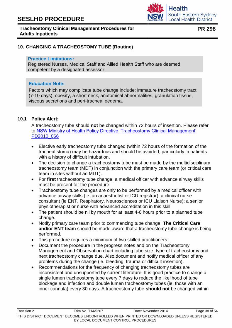
SESLHD PROCEDURE
Tracheostomy Clinical Management Procedures for Adults Inpatients
PR 298
Revision 2 Trim No. T14/5267 Date: November 2014 Page 38 of 54
THIS DISTRICT DOCUMENT BECOMES UNCONTROLLED WHEN PRINTED OR DOWNLOADED UNLESS REGISTERED BY LOCAL DOCUMENT CONTROL PROCEDURES
10. CHANGING A TRACHEOSTOMY TUBE (Routine)
Practice Limitations: Registered Nurses, Medical Staff and Allied Health Staff who are deemed competent by a designated assessor.
10.1 Policy Alert:
A tracheostomy tube should not be changed within 72 hours of insertion. Please refer to NSW Ministry of Health Policy Directive ‘Tracheostomy Clinical Management’ PD2010_066
Elective early tracheostomy tube changed (within 72 hours of the formation of the tracheal stoma) may be hazardous and should be avoided, particularly in patients with a history of difficult intubation.
The decision to change a tracheostomy tube must be made by the multidisciplinary tracheostomy team (MDT) in conjunction with the primary care team (or critical care team in sites without an MDT).
For first tracheostomy tube change, a medical officer with advance airway skills must be present for the procedure.
Tracheostomy tube changes are only to be performed by a medical officer with advance airway skills (ie. an anaesthetist or ICU registrar); a clinical nurse consultant (ie ENT, Respiratory, Neurosciences or ICU Liaison Nurse); a senior physiotherapist or nurse with advanced accreditation in this skill.
The patient should be nil by mouth for at least 4-6 hours prior to a planned tube change.
Notify primary care team prior to commencing tube change. The Critical Care and/or ENT team should be made aware that a tracheostomy tube change is being performed.
This procedure requires a minimum of two skilled practitioners.
Document the procedure in the progress notes and on the Tracheostomy Management and Observation chart including tube size, type of tracheostomy and next tracheostomy change due. Also document and notify medical officer of any problems during the change (ie. bleeding, trauma or difficult insertion).
Recommendations for the frequency of changing tracheostomy tubes are inconsistent and unsupported by current literature. It is good practice to change a single lumen tracheostomy tube every 7 days to reduce the likelihood of tube blockage and infection and double lumen tracheostomy tubes (ie. those with an inner cannula) every 30 days. A tracheostomy tube should not be changed within
Education Note:
Factors which may complicate tube change include: immature tracheostomy tract (7-10 days), obesity, a short neck, anatomical abnormalities, granulation tissue, viscous secretions and peri-tracheal oedema.

SESLHD PROCEDURE
Tracheostomy Clinical Management Procedures for Adults Inpatients
PR 298
Revision 2 Trim No. T14/5267 Date: November 2014 Page 39 of 54
THIS DISTRICT DOCUMENT BECOMES UNCONTROLLED WHEN PRINTED OR DOWNLOADED UNLESS REGISTERED BY LOCAL DOCUMENT CONTROL PROCEDURES
72 hours of insertion. Please refer to NSW Ministry of Health Policy Directive ‘Tracheostomy Clinical Management’ PD2010_066
In line with ‘NSW Agency for Clinical Innovation (ACI): Care of Adult Patients in Acute Care Facilities with a Tracheostomy’, the following are evidence-based recommendations for changing a tracheostomy tube.
Recommendations Grade of
Recommendation
Ideally the TT should not be changed for the first week. Consensus
Elective early tracheostomy tube changed within 72 hours of the formation of the tracheal stoma may be hazardous and should be avoided, particularly in patients with a history of difficult intubations.
Consensus
The decision to change a tracheostomy must be made by the multidisciplinary team experienced in tracheostomy care in conjunction with the primary care team.
Consensus
Where the TT change is required within the first seven days, or when there is a risk of difficult recannulation, a senior medical officer with advanced airway skills MUST be present.
Consensus
Tracheostomy tube changes are only to be performed by or under the direct supervision of a medical officer, clinical nurse consultant, physiotherapist or nurse with advanced accreditation in this skill.
Consensus
A TT change should be at least a two person procedure, one of whom has proven experience in tracheostomy tube changes.
Consensus
Where the TT change is a planned procedure, the patient should be assessed to identify the risk of aspiration of gastric contents. This may require fasting for 4-6 hours prior to change of TT, or aspiration of the NG tube.
Consensus
The procedure is undertaken in a safe environment with
Two clinicians present; with at least one of these being assessed as competent at the procedure
Monitoring of the patient including pulse, respiratory rate and work of breathing, continuous SpO2 monitoring
Emergency equipment available at the bedside.
Consensus
The procedure must be documented in the patient notes, tracheostomy management and observation chart and should include:
Size and type of tracheostomy tube
Any complications/problems arising during procedure (abnormal bleeding, trauma or difficult insertion)
The condition of the tracheostomy stoma and the surrounding skin (ie. over-granulation, wound breakdown)
When the next tube change is required.
Consensus

SESLHD PROCEDURE
Tracheostomy Clinical Management Procedures for Adults Inpatients
PR 298
Revision 2 Trim No. T14/5267 Date: November 2014 Page 40 of 54
THIS DISTRICT DOCUMENT BECOMES UNCONTROLLED WHEN PRINTED OR DOWNLOADED UNLESS REGISTERED BY LOCAL DOCUMENT CONTROL PROCEDURES
Recommendations Grade of
Recommendation
Any complications must be reported to the medical officer and clearly documented in the patient notes.
Consensus
Patients should be monitored post tube change as clinically indicated
Consensus
10.2 Equipment required
Dressing pack
Suction catheter (with Y connection removed)
Correct size tracheostomy tube and one size smaller,
Tracheostomy tube holder/tracheostomy tape
Manometer if tube is cuffed
Sterile water-soluble lubricant
Sterile normal saline
Pre-cut slim line key hole dressing (ie drain sponge)
Sterile or clean gloves, apron and protective eye wear/face visor
Tracheal dilators and suction
Functioning suction unit and appropriate sized suction catheters
Stethoscope
Resuscitation equipment.
Procedure: Changing a Tracheostomy Tube Key Points
1. Aspirate naso-gastric tube if present Nil by mouth for 4-6 hours prior to tube change
To reduce the risk of aspiration while airway is unprotected
2. Two skilled clinicians should perform the procedure. Both must wear eye protection, gloves and apron/gown
To ensure a swift and safe procedure
3. Explain the procedure and rationale to the patient The Critical Care and/or ENT team should be made aware that a tracheostomy tube change is being performed
The patient should give their verbal consent to the procedure, unless unable or an emergency procedure Ensuring the availability of a medical officer with advanced airway skills is safe practice should problems arise
4. Perform the 5 moments of hand hygiene and prepare dressing trolley
To reduce the risk of cross infection
5. Position patient in semi-recumbent position, extending the neck, remove any obstructive clothing
Extending the neck will make the removal and insertion of the tube easier To ensure adequate view of patients neck

SESLHD PROCEDURE
Tracheostomy Clinical Management Procedures for Adults Inpatients
PR 298
Revision 2 Trim No. T14/5267 Date: November 2014 Page 41 of 54
THIS DISTRICT DOCUMENT BECOMES UNCONTROLLED WHEN PRINTED OR DOWNLOADED UNLESS REGISTERED BY LOCAL DOCUMENT CONTROL PROCEDURES
Procedure: Changing a Tracheostomy Tube Key Points
6. If the patient is dependent on oxygen, hyper-oxygenate the patient with 100% oxygen and monitor oxygen saturations Assess COPD patients for need to hyper-oxygenation as they may only require a 20% increase of their oxygen concentration
During tube change the patient will be at risk of hypoxia Patients with COPD have an altered CO2 response mechanism and should not routinely be given 100% O2
7. If the new tracheostomy tube is cuffed, check the cuff by inflating it with air, & observe cuff for leakage. Deflate cuff fully. Ensure a smaller tracheostomy tube is to hand
To check for air leaks and spontaneous deflation In case the same size tube cannot be inserted easily
8. Check obturator can be removed To become familiar with removing obturator prior to insertion
9. Lubricate the tube sparingly with a water soluble lubricant. Place on sterile surface
To facilitate insertion, and maintain sterility
10. Remove old dressing and tapes, observe site and clean around stoma site
To clean skin of debris and potential skin contaminants. To enable removal of the tracheotomy tube
11. Suction patient if required. Suction oro-pharyngeal secretions. Using the synchronised cuff deflation and suction technique deflate the cuff with a10ml syringe
Pooled secretions above the cuff may enter the lungs when the cuff is deflated. A fully deflated cuff reduces the risk of trauma on removal of the old tube
12. Remove old tube in a firm upwards and downwards motion on expiration. Observe the stoma site
To cause minimal trauma and reduce the risk of coughing To identify signs of stoma infection and granulation tissue
13. With clean gloves insert the new tracheostomy tube with the obturator in place as the patient exhales
The obturator guides the tracheostomy tube along the contour of the trachea. Relaxation of the neck muscles makes insertion easier
14. Remove the obturator immediately The patient will be unable to breathe with the obturator blocking the lumen
15. Inflate the cuff using the minimal occlusion technique and check with a cuff pressure gauge
An inflated cuff reduces the risk of aspiration. Correct

SESLHD PROCEDURE
Tracheostomy Clinical Management Procedures for Adults Inpatients
PR 298
Revision 2 Trim No. T14/5267 Date: November 2014 Page 42 of 54
THIS DISTRICT DOCUMENT BECOMES UNCONTROLLED WHEN PRINTED OR DOWNLOADED UNLESS REGISTERED BY LOCAL DOCUMENT CONTROL PROCEDURES
Procedure: Changing a Tracheostomy Tube Key Points
inflation reduces the risk of tracheal wall damage
16. Insert inner cannula if using a two piece system To prevent secretions collecting on the inside of the outer cannula
17. Observe the patient for respiratory distress. Feel for respiration via the tube, where able, ask the patient to breathe deeply and observe for chest movement
The flow of air is felt via the tracheostomy lumen if the tube is correctly positioned NB If tube insertion fails or patient becomes compromised and cyanosed initiate Emergency Procedures
18. Listen for equal air entry To ensure bilateral inflation of the lungs
19. Clean stoma site if required, renew dressing and secure tracheostomy tube with trache tapes
To reduce the risk of dislodgement, maintain patient comfort and reduce the risk of infection
20. If the patient is comfortable and there are no signs of respiratory distress, return oxygen to levels prior to procedure and observe respiratory rate and oxygen saturation
21. Record tube change on Tracheostomy Care Chart and progress notes, document time, date, size, type of tube and any complications
To ensure effective communication
22. There may be a small amount of bleeding post tube change. If there is excessive bleeding, ensure cuff is inflated and call for immediate medical assistance
Trauma can occur to stoma site during a tube change. An inflated cuff will protect the patients airway whilst controlling the bleeding
10.3 Weaning/Decannulation Process
In line with ‘NSW Agency for Clinical Innovation (ACI): Care of Adult Patients in Acute Care Facilities with a Tracheostomy’, the following are evidence-based recommendations for undertaking the weaning/decannulation process.
Recommendations Grade of
Recommendation
The decision to commence weaning and proceed to decannulation is a collaborative decision involving the treating medical team and TRT. This must be documented in patient notes prior to beginning the weaning process.
Consensus

SESLHD PROCEDURE
Tracheostomy Clinical Management Procedures for Adults Inpatients
PR 298
Revision 2 Trim No. T14/5267 Date: November 2014 Page 43 of 54
THIS DISTRICT DOCUMENT BECOMES UNCONTROLLED WHEN PRINTED OR DOWNLOADED UNLESS REGISTERED BY LOCAL DOCUMENT CONTROL PROCEDURES
Recommendations Grade of
Recommendation
The weaning/decannulation plan should be documented. Following this, it should be reviewed at least daily and updated as required.
Consensus
In the presence of a cuffed TT, a successful cuff deflation is a minimal requirement for decannulation.
Consensus
An absent swallow in isolation is not a contraindication for an initial cuff deflation trial. Where there is no swallow present, it is recommended that a Speech Pathologist is consulted.
Consensus
Where airway patency is insufficient a medical/ENT assessment of the upper airway is recommended.
Consensus
Before placing a cap or speaking valve, the cuff needs to be completely deflated and airway patency established.
Consensus
Emergency equipment must be available and checked prior to changes in TT management related to weaning/decannulation.
Consensus
A medical officer with advanced airway management skills MUST be present when the TT is changed for the first time or is removed.
Consensus
In head and neck patients, the decision to decannulate is made by, or in consultation with, the ENT team.
Consensus
In general, the decision to decannulate is based on clinical criteria indicating the patient’s readiness including:
Clinically stable
Resolution of need for TT
Able to manage own oral secretions or a secretion management plan (eg. alternate suction route, pharmacological agents, positional management)
No signs of deteriorating lung function
Strong effective cough
Minimal oxygen requirements
Airway patency.
Evidence – C Recommendation C
The patient should be NBM for a minimum of 4 hours prior to TT removal OR if a nasogastric tube is present, this should be aspirated to empty the stomach.
Consensus
An airtight dressing must be applied to the stoma after the tracheostomy tube is removed. This dressing should be changed
At least DAILY
If odorous or contaminated by secretions
If no longer airtight.
Consensus
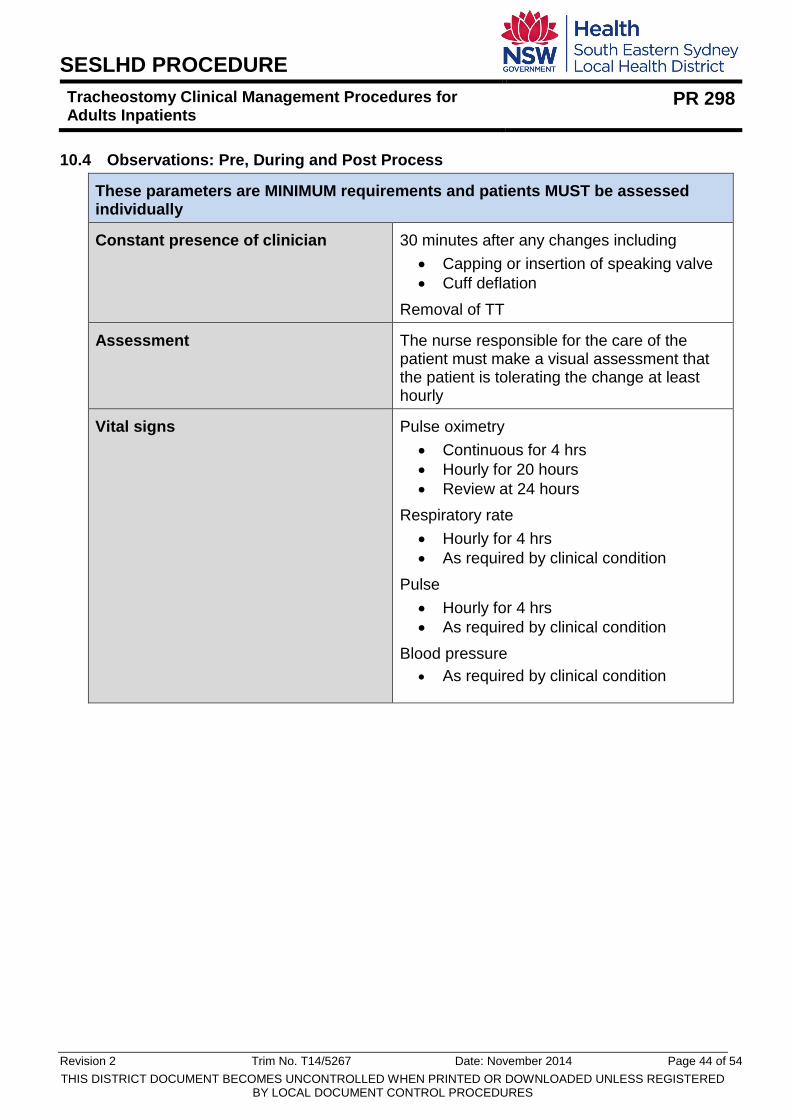
SESLHD PROCEDURE
Tracheostomy Clinical Management Procedures for Adults Inpatients
PR 298
Revision 2 Trim No. T14/5267 Date: November 2014 Page 44 of 54
THIS DISTRICT DOCUMENT BECOMES UNCONTROLLED WHEN PRINTED OR DOWNLOADED UNLESS REGISTERED BY LOCAL DOCUMENT CONTROL PROCEDURES
10.4 Observations: Pre, During and Post Process
These parameters are MINIMUM requirements and patients MUST be assessed individually
Constant presence of clinician 30 minutes after any changes including
Capping or insertion of speaking valve
Cuff deflation
Removal of TT
Assessment The nurse responsible for the care of the patient must make a visual assessment that the patient is tolerating the change at least hourly
Vital signs Pulse oximetry
Continuous for 4 hrs
Hourly for 20 hours
Review at 24 hours
Respiratory rate
Hourly for 4 hrs
As required by clinical condition
Pulse
Hourly for 4 hrs
As required by clinical condition
Blood pressure
As required by clinical condition

SESLHD PROCEDURE
Tracheostomy Clinical Management Procedures for Adults Inpatients
PR 298
Revision 2 Trim No. T14/5267 Date: November 2014 Page 45 of 54
THIS DISTRICT DOCUMENT BECOMES UNCONTROLLED WHEN PRINTED OR DOWNLOADED UNLESS REGISTERED BY LOCAL DOCUMENT CONTROL PROCEDURES
11. CUFF DEFLATION, OCCLUSION AND TUBE REMOVAL
Practice Limitations:
Registered Nurses, Speech Pathologists and Physiotherapists who are
deemed competent by a designated assessor.
Enrolled Nurses who have achieved “Extended Practice Skill” in Tracheostomy
Decannulation and been deemed competent by a designated assessor.
11.1 Stage 1: Cuff Deflation
11.1.1 Policy Alert:
Cuff deflation is only recommended as part of the weaning process or in an emergency due to occlusion, as it may increase the risk of aspiration and hypoxia
If cuff deflation is unsuccessful the cuff must be reinflated using the cuff manometer
This is a two person procedure
Weaning is a progressive, three stage process. The patient must successfully achieve cuff deflation and tube occlusion (for at least 24-48 hour period), before the tracheostomy tube can be removed
OR
An alternative to assess suitability for decannulation, when not appropriate or safe to occlude the tracheostomy, is for the patient to tolerate continuous cuff deflation for a 24-48 hour period without need for tracheal suctioning.
Education Note: The patient must meet the following clinical criteria prior to the weaning process:
Clinical indications for tracheostomy are resolved, nil deterioration within last 24hrs
Stable chest status – nil deterioration within last 24hrs per physio and medical team
Absence of respiratory/chest infection
Oxygen requirement – self ventilating with or without oxygen (as per medical order)
Minimal suctioning requirements
Adequate airway protective mechanisms – ie. ability to cough and clear airway secretions via tracheostomy or by mouth
No granulation tissue, oedema around tracheostomy site
Patent upper airway eg. absence of oedema, no obstructions
Agreement by all members of the MDT.

SESLHD PROCEDURE
Tracheostomy Clinical Management Procedures for Adults Inpatients
PR 298
Revision 2 Trim No. T14/5267 Date: November 2014 Page 46 of 54
THIS DISTRICT DOCUMENT BECOMES UNCONTROLLED WHEN PRINTED OR DOWNLOADED UNLESS REGISTERED BY LOCAL DOCUMENT CONTROL PROCEDURES
Procedure: Cuff Deflation Rationale
1. Encourage the patient to cough and clear secretions
To reduce the amount of secretions in the airway
2. Person 1 If the patient has a Suction-aid tracheostomy tube with an ‘above cuff suction port’, suction above the cuff before deflating the cuff
To reduce the amount of oro-pharyngeal saliva that may be aspirated when the cuff is deflated
3. Person 1 If the patient requires a tracheal suction prior to cuff deflation, pass a sterile catheter into the tracheostomy tube, approximately 0.5 cm longer than the tracheostomy tube tip. This should not be conducted unless indicated
To minimise the risk of cross infection and trauma
4. Person 2 Advise the patient that they may cough after cuff deflation and this is normal. Deflate the cuff steadily, removing all air from the balloon. Person 1 may need to suction post cuff deflation if indicated
To prepare the patient for the sensation they may feel and what may happen The suction post deflation may be indicated to assist the patient if they have had saliva previously pooling above the tracheostomy cuff
Indicators of Successful Cuff Deflation
SpO2 maintainted at or above 90% or within 5%of baseline
Respiratory rate within breaths/minute above the pre-procedure baseline
Blood pressure (BP) and heart rate within 10% of patient’s baseline
No increase in shortness of breath
No uncontrollable coughing post deflation (after the first 5 minutes)
No increase in suctioning requirements or secretions.
11.2 Stage 2: Tube Occlusion
11.2.1 Policy Alert:
Successful cuff deflation must be achieved prior to tracheostomy tube occlusion
Tube occlusion is based on individual patient assessment and is a MDT decision
Ensure cuff is completely deflated before the tube is occluded
When occluding the tube for the first time, stay and observe the patient for a minimum of 15 minutes post occlusion

SESLHD PROCEDURE
Tracheostomy Clinical Management Procedures for Adults Inpatients
PR 298
Revision 2 Trim No. T14/5267 Date: November 2014 Page 47 of 54
THIS DISTRICT DOCUMENT BECOMES UNCONTROLLED WHEN PRINTED OR DOWNLOADED UNLESS REGISTERED BY LOCAL DOCUMENT CONTROL PROCEDURES
Continuously monitor patient’s oxygen saturation
If occluding tracheostomy as a method to assess suitability for decannulation, occlusion must be tolerated for a minimum of 24-48 hours without the need to remove the tracheostomy cap for suctioning or respiratory purposes, before tracheostomy tube removal is considered OR
An alternative method to assess suitability for decannulation (when not appropriate or safe to occlude the tracheostomy) is for the patient to tolerate continuous cuff deflation for 24-48 hours without need for tracheal suctioning.
Observe for: Action
Increase in heart rate
Decrease in oxygen saturation
Signs of respiratory distress
Irritation, confusion or agitation
Cyanosis
Remove cap and activate appropriate
management including escalating for
assistance if necessary
11.3 Stage 3: Removal of Tracheostomy Tube
11.3.1 Policy Alert
This is a two person procedure. At least one person must be a clinician who has reached competency in the procedure.
Weaning is a progressive, ongoing process. The patient must successfully achieve cuff deflation and tube occlusion, before the tracheostomy tube can be removed.
Tracheostomy MDT consultation should inform the decision to remove a tracheostomy tube Medical orders needs to be documented in patient medical record prior to tube removal.
In a ward area tracheostomy tube removal should only be attempted before 12 midday Monday to Thursday.
Stay with the patient for a minimum of 15 mins post tube removal or until the patient is stable, whichever is the longer.
11.3.2 Equipment
Emergency airway resuscitation equipment immediately available
Clean gloves
Disposable apron
Protective eyewear
Disposable drawsheet
Practice Limitations: Registered Nurses, Physiotherapists who are deemed competent by a designated assessor

SESLHD PROCEDURE
Tracheostomy Clinical Management Procedures for Adults Inpatients
PR 298
Revision 2 Trim No. T14/5267 Date: November 2014 Page 48 of 54
THIS DISTRICT DOCUMENT BECOMES UNCONTROLLED WHEN PRINTED OR DOWNLOADED UNLESS REGISTERED BY LOCAL DOCUMENT CONTROL PROCEDURES
Yankauer sucker
10ml syringe if cuffed tube
Large steristrip
Sleek tape
O2 if required via face mask or nasal prongs.
Note: Prior to removal the patient must have an effective cough, patent airway and gag reflex
11.3.3 Management Post Removal
Encourage patient to breathe via mouth and nose
Explain to patient that they can now talk
Remind patient to manually press over dressing to adequately occlude stoma when speaking or coughing
Request patient to try to vocalise and try a strong cough
Daily dressing for 3-5 days until wound discharge decreased, and then PRN until complete wound closure
The stoma may be left exposed when clean and dry and there is no evidence of air leak.
Procedure: Removal of a Tracheostomy Tube
1.
Sit patient up at 60 degree angle and pre oxygenate
2.
Don PPE and perform 5 moments of hand hygiene
3.
Thoroughly suction oropharynx with Yankauer sucker
4.
Suction Tracheostomy tube
5.
The role of the assistant is to undo the ties deflate cuff with syringe and support the tube
6.
Remove tube, with consistent firm but gentle digital pressure
7.
If required, give O2 via facial mask
8.
Observe for signs of respiratory distress and airway obstruction
9.
Clean stoma with normal saline
10.
Apply airtight dressing to the stoma (as per MDT preference)
11.
Check tube for blood or damaged cuff

SESLHD PROCEDURE
Tracheostomy Clinical Management Procedures for Adults Inpatients
PR 298
Revision 2 Trim No. T14/5267 Date: November 2014 Page 49 of 54
THIS DISTRICT DOCUMENT BECOMES UNCONTROLLED WHEN PRINTED OR DOWNLOADED UNLESS REGISTERED BY LOCAL DOCUMENT CONTROL PROCEDURES
12. DISCHARGE OF A PATIENT WITH A TRACHEOSTOMY
In line with ‘NSW Agency for Clinical Innovation (ACI): Care of Adult Patients in Acute Care Facilities with a Tracheostomy’, the following are evidence-based recommendations for discharging a patient with a tracheostomy.
12.1 Patient and Carer required competency and skills
The following competencies and skills are required from the patient and carer for a successful patient discharge:
The caregiver is able to:
Explain reasons for and purposes of the tracheostomy
Describe the type and size of the current tracheostomy tube
Describe and demonstrate TT stabilisation
Demonstrate safe and effective suctioning of the tracheostomy tube including: o Size of suction catheter/apparatus o Insertion depth of catheter o Length of suction procedure o Use of suction equipment o Assessment of outcomes of procedure o Normal and abnormal sputum, especially tenacity and infection
Demonstrate safe and effective care of the tracheostomy stoma and the skin of the neck including: o Cleaning of stoma o Application of dressing products, if applicable o Listing signs of infection and poor skin integrity
Provide effective humidification of inspired gases including: o Explanation of the purposes of humidification o Demonstration of attachment of humidification devices including passive
humidification attachment o When, why and how to use nebulised saline
Identifies and justifies essential equipment to be with person at all times
Demonstrates safe and effective care of all equipment including: o Correct use, cleaning and maintenance o Identification of contact person for malfunctioning equipment
Demonstrates safe and effective care where home ventilation will be used, including: o Correctly assembling ventilator equipment and circuit components
Recommendation Grade of
Recommendation
A caregiver for the patient with a tracheostomy is identified early in the patient’s hospital admission and their education program should commence as soon as possible to facilitate discharge. Adult patients will be responsible for their own care, however, an individual’s dexterity and cognitive abilities need to be taken into consideration.
Consensus

SESLHD PROCEDURE
Tracheostomy Clinical Management Procedures for Adults Inpatients
PR 298
Revision 2 Trim No. T14/5267 Date: November 2014 Page 50 of 54
THIS DISTRICT DOCUMENT BECOMES UNCONTROLLED WHEN PRINTED OR DOWNLOADED UNLESS REGISTERED BY LOCAL DOCUMENT CONTROL PROCEDURES
o Correctly switching ventilator on and off o Correctly attaching/removing ventilator to/from tracheostomy o Demonstrate competence with inflating/deflating cuff and changing inner cannula
(e.g. Between fenestrated and non-fenestrated) as needed to correctly attach/remove ventilator, as prescribed by home ventilation specialist
o Demonstrate awareness of ventilator alarms and how to respond to them o Demonstrate awareness of possible problems requiring immediate intervention
that may arise during ventilation via tracheostomy, and appropriate knowledge of how to respond to these problems
o Knowledge of how and when to clean equipment, change disposable items and perform basic machine maintenance such as changing filters Note: This competency only applies to patients on home ventilation
Identifies, articulates and demonstrates the appropriate action [on an airway mannequin] for emergencies including: o Dislodged tube o Blocked airway o Acute respiratory distress
Demonstrates the following clinical skills on airway mannequin and patient: o Changing of tracheostomy tube o Suctioning o Changing of tapes o Application of oxygen
Describes and demonstrates effective infection prevention principles including: o Hand hygiene o Cleaning and storage of reusable equipment
Where to get further equipment consumables.
13. DOCUMENTATION
Not required
14. AUDIT
Auditing undertaken at a site level
15. REFERENCES
NSW Ministry of Health Policy Directive ‘Tracheostomy Clinical Management (Adult) NSW Health Policy Directive 2010_066
ACI - Care of Adult Patients in Acute Care - Clinical Practice Guideline
Blackwood B and Webb C (1998) Closed tracheal suction systems and infection control in the intensive care unit. Journal of Hospital Infection 39:315-321
Bond P, Grant F, Coltart L, Elder F (2003) Best Practice in the care of patients with a tracheostomy. Nursing Times 99(30): 24-25

SESLHD PROCEDURE
Tracheostomy Clinical Management Procedures for Adults Inpatients
PR 298
Revision 2 Trim No. T14/5267 Date: November 2014 Page 51 of 54
THIS DISTRICT DOCUMENT BECOMES UNCONTROLLED WHEN PRINTED OR DOWNLOADED UNLESS REGISTERED BY LOCAL DOCUMENT CONTROL PROCEDURES
Buglass, E. (1999) Tracheostomy care: tracheal suctioning and humidification. British Journal of Nursing, 8(8): 500-4 1A3
Burns, SM., Spilman, M., Wilmoth, D., Carpenter, R., Turrentine, B., Wiley, B., Marshall, M., Martens, S., Burns, JE. & Truwit, JD. (1998) Are frequent inner cannula changes necessary?: A pilot study. Heart and Lung, 27(1): 58-62 1A2
Clark L (1995) A critical event in tracheostomy care. British Journal of Nursing 4(12): 676, 678-681
Clinical Safety and Governance Branch, NSW Department of Health (2009) Reducing Incidents Involving Tracheostomy Tube Care. Safety Notice 018/09.
Crimlinsk, JT, Horn, M., Wilson, DJ and Marino, B. (1996) Artificial airways: A survey of cuff management practices. Heart & Lung 25(3): 225-35 1D1
Day T (2000) Tracheal suctioning: when, why and how? Nursing Times insert 96 (20) 13-15
Day T, Wainwright SP, Wilson-Barnett J (2001) An evaluation of teaching to improve the practice of endotracheal suctioning in intensive care units. Journal of Critical Nursing 10: 682-696
Day T., Farnell S., Waiwright S., Wilson-Barnett J. (20O2) Trachael suctioning: an exploration of nurses’ knowledge and competence in acute and high dependency ward areas. Journal of Advanced Nursing, 39 (1), 35-45.
Demir, F, Dramali, A (2005) Requirement for 100% oxygen before and after closed suction. Journal of advanced Nursing 51 (3) pp 245-251.
Dixon, L. (1998-20O2) Tracheostomy: Postoperative Recovery. Perspectivesinnursing.org1D1
Dixon, L. & Wasson, D. (1998) Comparing use and cost effectiveness of trachesostomy tube securing devices. MEDSURG Nursing 7(5): 270-4 1A2
Docherty B, Bench S (20O2) Tracheostomy management for patients in general ward settings. Professional Nurse 18: 100-104
Druding, M. (1997) Reexamining the practice of normal saline instillation prior to suctioning. Medsurg Nursing. August, 6,4, pp. 209-212.
Elpern, EH., Scott, MG., Petro, L. & Lreis, MG. (1994) Pulmonary aspiration in mechanically ventilated patients with tracheostomies. Chest 105(2): 563-66

SESLHD PROCEDURE
Tracheostomy Clinical Management Procedures for Adults Inpatients
PR 298
Revision 2 Trim No. T14/5267 Date: November 2014 Page 52 of 54
THIS DISTRICT DOCUMENT BECOMES UNCONTROLLED WHEN PRINTED OR DOWNLOADED UNLESS REGISTERED BY LOCAL DOCUMENT CONTROL PROCEDURES
1D1Griggs, A. (1998) Tracheostomy: suctioning and humidification. Nursing Standard 13(2): 49-53 & 55-6. 1D1
Galvin W, Cusano A (1998) Making a clean sweep. Using a closed tracheal system. Nursing 98 (June) 50-51
Goodwin JE. & Heffner, JE. (1991) Special critical care considerations in tracheostomy management. Clinical in Chest Medicine 12: 573-583
Griggs A (1998) Tracheostomy: suctioning and humidification. Nursing Standard 13(2): 49-53 & 55-56
Hackeling, T., Traina, R., Ma, O.J. & Shockley, W. (1998) Emergency care of patients with tracheostomies: a 7-year review. American Journal of Emergency Nursing 16(7): 681-5 1A2
Harkin, H. (1998) Tracheostomy management. Nursing Times 94(21): 56-8 1D1
Hettige R., Arora A., Ifeacho S., Araula A. (2008) Improving Tracheostomy Management though design, implementation and prospective audit of a care bundle: How do we do it? Clinical Otolaryngology 33 (5): 488-491
Higgins,D.M.& Maclean JCF(1997) Dysphagia in the patient with tracheostomy: six cases of inappropriate cuff deflation or removal Heart & Lung The Journal of Acute and critical Care 26(3):215-20
Hooper M (1996) Nursing care of the patient with a tracheostomy. Nursing Standard 10 (34): 40-43
Joanna Briggs Institute for Evidence Based Practice (20O2) Changing the tapes. Acute Care Practice Manual D16-1 1C
Joanna Briggs Institute (20O2) Routine care for patients with tracheostomy tubes. Acute Care Practice Manual, D13-1 1C
Joanna Briggs Institute (20O2) Tracheal suction - literature review. Acute Care Practice Manual, D14:3-5 1A3
Joanna Briggs Institute for Evidence Based Nursing and Midwifery (20O2) Tracheostomy tube care - literature review. Clinical Practice Manuals. 1A3
Joanna Briggs Institue (20O2) Routine care for patients with tracheostomy tubes. Acute Care Practice Manual, D13-1
SESLHD PROCEDURE
Tracheostomy Clinical Management Procedures for Adults Inpatients
PR 298
Revision 2 Trim No. T14/5267 Date: November 2014 Page 53 of 54
THIS DISTRICT DOCUMENT BECOMES UNCONTROLLED WHEN PRINTED OR DOWNLOADED UNLESS REGISTERED BY LOCAL DOCUMENT CONTROL PROCEDURES
Kerr, ME., Rudy, EB, Brucia, J. & Stone, KS. (1993) Head injured adults: recommendations for endotracheal suctioning. Journal of Neuroscience Nursing 25(2):86-91 1A3
Ladyshewsky, A. & Gousseau, A. (1996) Successulf tracheal weaning. Canadian Nurse 92 (2): 35-8
Langmore, SE. (1998) Predictors of Aspiration Pneumonia: How important is dysphagia? Dysphagia 13(2) 69-81 1A2
Langmore , SE., Scott, MG., Petro, L. & Lreis, MG. (1994) Pulmonary aspiration in mechanically ventilated patients with tracheostomies. Chest 105 (2): 563-66
Laws-Chapman C. (1998) Tracheostomy tube management. Care of the Critically Ill. 14(5):insert 4p 1D125(2):86-91 1A3
McEleny, M. (1998) Endotracheal suction. Professional Nurse. 13, 6, pp. 373-376.
Mirza, S. & Cameron, DS. (2001) The tracheostomy tube change: a review of techniques. Hospital Medicine 62(3): 158-3 1A3
Murray, K. Brozowski, L (1998) Swallowing in patients with a tracheotomies. AACN Clinical Issues. Advanced Practice in Acute Critical Care. 9 (3) pp 416-426.
NSW Department of Health Clinical Safety, Quality and Governance Branch (2009). Safety Notice 018/09: Reducing Incidents Involving Tracheostomy Tube Care.
Pannunzio, TG. (1996) Aspiration of oral feeding in patients with tracheostomies. American Association of Critical Care Nurses Clinical Issues: Advanced Practice in Acute Critical Care 1A3
Russell, C (2005) High-dependency nursing. Providing the nurse with a guide to tracheostomy care and management. British Journal of Nursing. 14 (8) pp 428-433.
Schreiber, D. (2001) Tracheostomy care at home: a how to guide. RN 64(7): 43-6 1D1
Serra, A. (2000) Tracheostomy care. Nursing Standard 14(42): 44-55. 1D1
Sey, SJ. & Gay, SL. (1997) Problem in tracheostomy patient care: recognizing the patient with a displaced tracheostomy tube. ORL-Head and Neck Nursing 15(2):10-1 1D1

SESLHD PROCEDURE
Tracheostomy Clinical Management Procedures for Adults Inpatients
PR 298
Revision 2 Trim No. T14/5267 Date: November 2014 Page 54 of 54
THIS DISTRICT DOCUMENT BECOMES UNCONTROLLED WHEN PRINTED OR DOWNLOADED UNLESS REGISTERED BY LOCAL DOCUMENT CONTROL PROCEDURES
Shaker, R., Mailbrath, M., Ren, J., Campbell, B., Toothill, R. & Hogan, W. (1995) Deglutitive Aspiration in patients with tracheostomy: Effect of tracheostomy on duration of vocal cord closure. Gastroenterology 108(5): 1357-60 1A2
Sims Portex Limited (1996 & 2000) Portex Adjustable Flange Tracheostomy Tube and Portex Blue Line Ultra Tracheostomy Tube Sims Portex Product Information St George's Healthcare NHS Trust (2000) Guidelines for the Care of Patients with Tracheostomy Tubes 1D1
St Georges Healthcare NHS Trust (2000) Guidelines for the care of patients with tracheostomy tubes.
Tamburri, L.M. (2000) Care of the patient with a tracheostomy. Orthopedic Nursing 19(2): 49-58 1D1 4 Hooper, M. (1996) Nursing care of the patient with a tracheostomy. Nursing Standard 10(34): 40-3 1D1
Theaker, C. (1999) How to guides: Pitfalls in tracheostomy care. Care of the Critically Ill 15(5): insert 4p 1D1
Thompson –Ward, E., Boots, R., Frisby, J., Bassett, L. & Timm, M. (1999) Evaluation suitability for tracheostomy decannulation: a critical evaluation of two management protocols. Journal of Medical Speech-Language Pathology 7(4): 273-81
Wilson, EB & Malley, N (1990)Discharge planning a patient with a new tracheostomy Critical Care Nurse 10 (7):73-8.
Zgvotsky, K and D’Amelio, L. (1995) Bedside percutaneous tracheostomy: Implications for critical care nursing. Critical Care Nurse, October, pp. 37-43.
8. REVISION AND APPROVAL HISTORY
Date Revision No. Author and Approval
February 2014 2 Converted to Procedure by Perioperative, Surgery and Anaesthetics Clinical Stream Nurse Manager
March 2014 2 Reviewed and aligned with ‘NSW Agency for Clinical Innovation (ACI): Care of Adult Patients in Acute Care Facilities with a Tracheostomy’ by Mary Dunford, Respiratory CNC, Paula Gunner CNC Neck & Surgery and District Policy Officer
November 2014 2 Endorsed by SESLHD Clinical and Quality Council