sense of sight cameras operate like the human eye. the human eye has approximately 576 mp
TRANSCRIPT

Sense of Sight Cameras operate like the human eye. The human eye has approximately
576 MP.
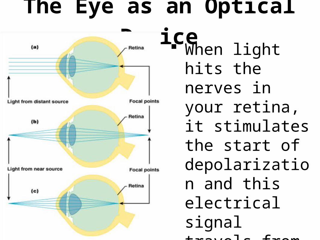
The Eye as an Optical Device When light hits the
nerves in your retina, it stimulates the start of depolarization and this electrical signal travels from the optic nerve thalamus occipital lobe
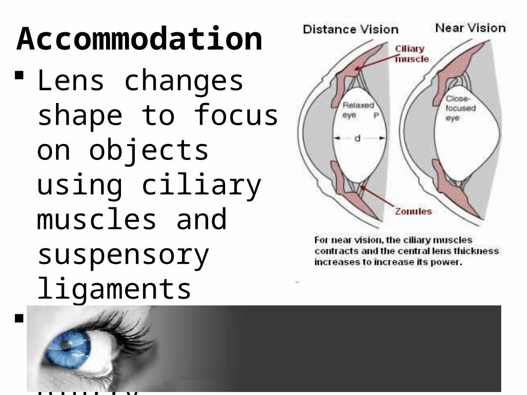
Accommodation Lens changes shape to
focus on objects using ciliary muscles and suspensory ligaments
Otherwise, image appears blurry
The eye is in the orbit of the cranium for protection.
Within the orbit are 6 extrinsic eye muscles, which move the eye.
There are 4 cranial nerves: Optic (II), Occulomotor (III), Trochlear (IV), and Abducens (VI) which innervate the eye.
THE EYE
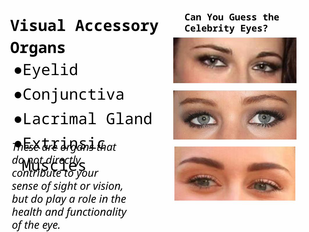
Visual Accessory Organs●Eyelid
●Conjunctiva
●Lacrimal Gland
●Extrinsic Muscles
Can You Guess the Celebrity Eyes?
These are organs that do not directly contribute to your sense of sight or vision, but do play a role in the health and functionality of the eye.

Eyelid Thin skin that covers
and protects the eye.
Skin will not protect you from intense radiation, that’s why we use special goggles in a tanning bed!
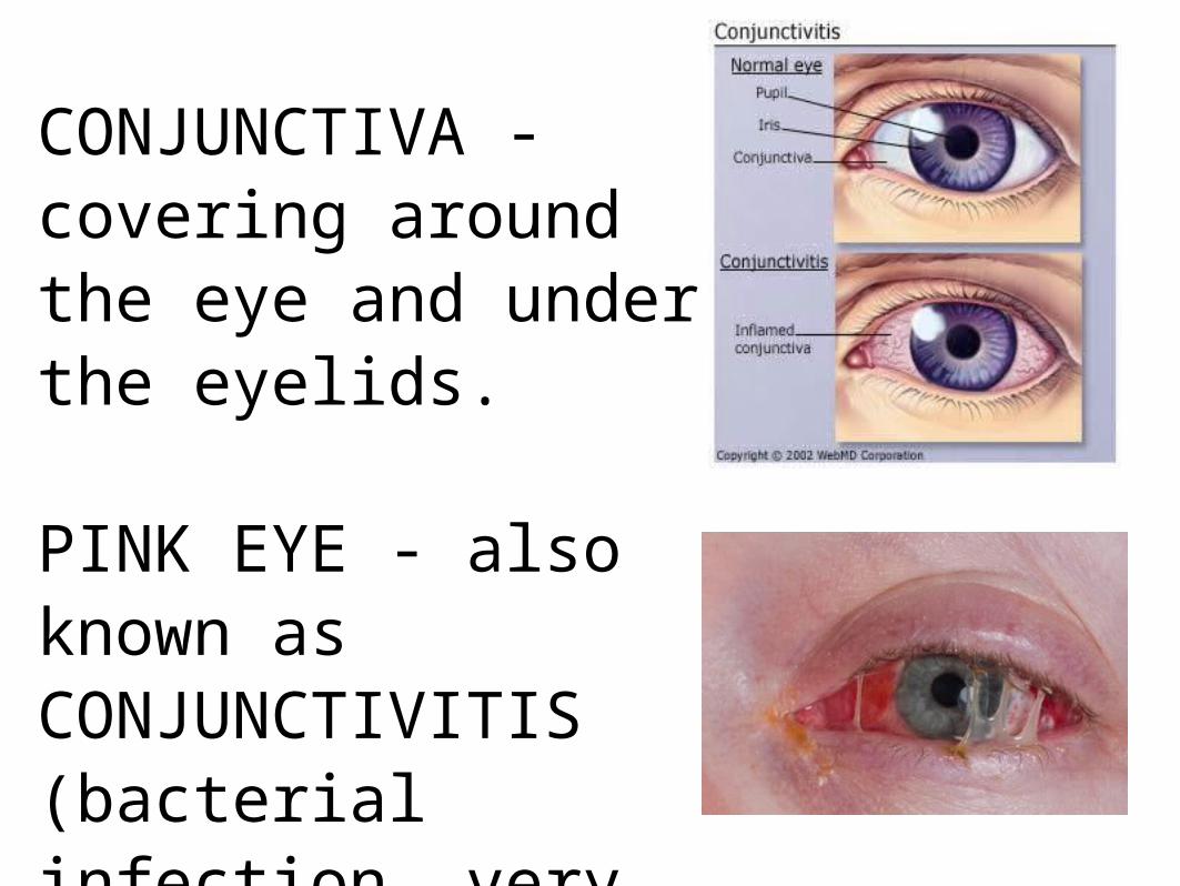
CONJUNCTIVA - covering around the eye and under the eyelids. PINK EYE - also known as CONJUNCTIVITIS (bacterial infection, very contagious)

LACRIMAL GLANDS – located on the superior lateral eyelid and produce tears, which drain into the nasal cavity via the LACRIMAL (tear) DUCT.The function of tears is to moisten and lubricate the eye surface and contain enzymes to kill bacteria

Extrinsic Eye Muscles Moves the eyeball
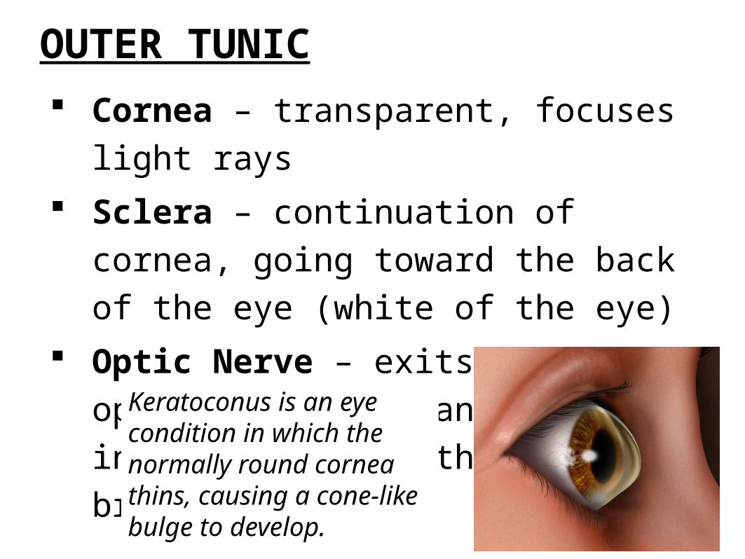
OUTER TUNIC
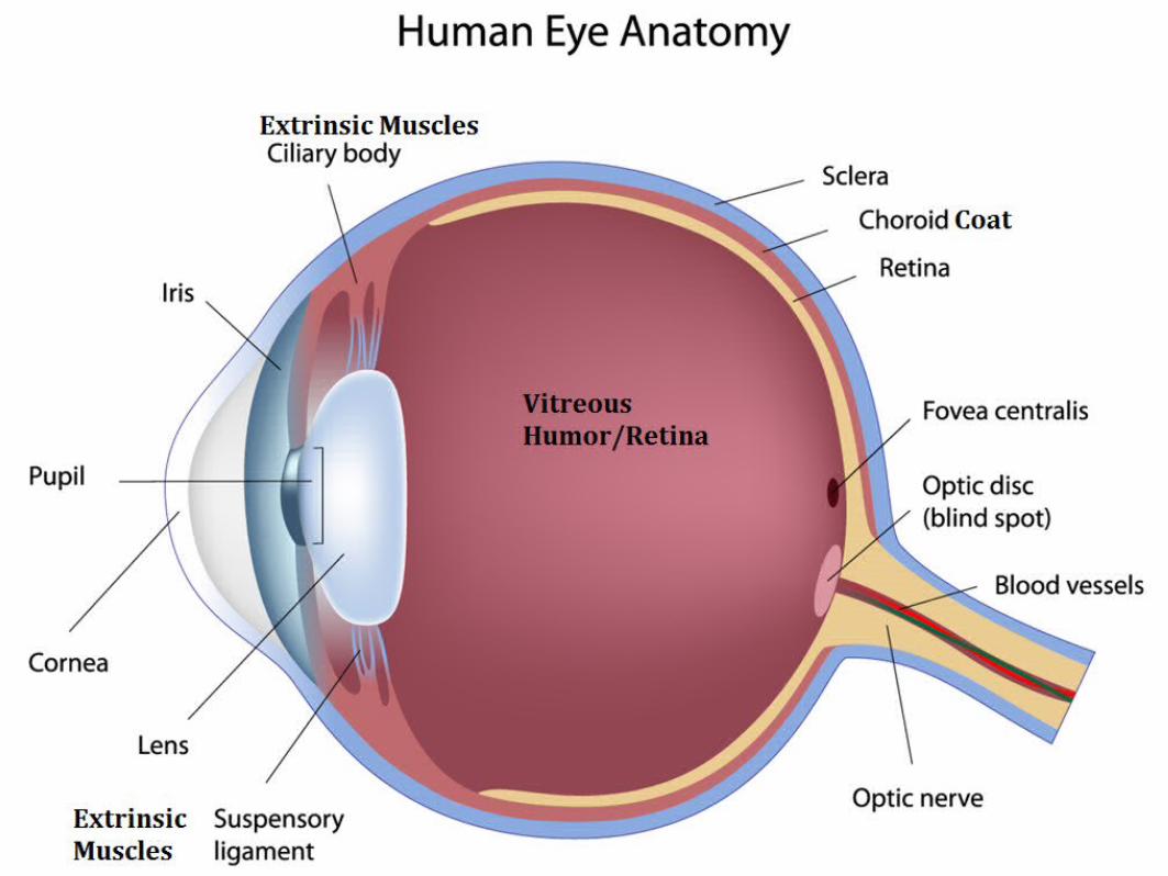
Cornea – transparent, focuses light rays Sclera – continuation of cornea, going toward the
back of the eye (white of the eye) Optic Nerve – exits at the optic disk and transmits
visual information from the eye to the brain.
Keratoconus is an eye condition in which the normally round cornea thins, causing a cone-like bulge to develop.

MIDDLE TUNIC Choroid Coat – contains
blood vessels Lens – focuses light Iris – colored portion of
the eye Aqueous humor – liquid
surrounding the cornea and iris
Pupil – opening for light to enter

INNER TUNIC Retina - visual receptor cells Optic Disc – where nerve fibers leave the eye,
creating the blind spot Vitreous Humor – gel-like fluid surrounding lens
and retina Fovea Centralis – area of retina filled with
cones for sharp central vision (reading, driving)
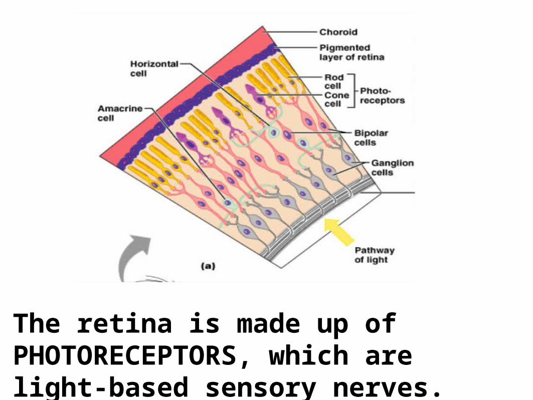
The retina is made up of PHOTORECEPTORS, which are light-based sensory nerves.
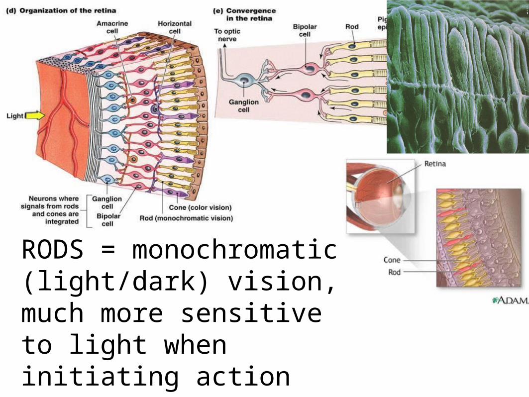
RODS = monochromatic (light/dark) vision, much more sensitive to light when initiating action potentials vs. CONES = color vision

1. Light is focused primarily by the cornea — the clear front surface of the eye, which acts like a camera lens.
2. The iris of the eye functions like the diaphragm of a camera, controlling the amount of light reaching the back of the eye by automatically adjusting the size of the pupil (aperture).
3. The eye's lens is located directly behind the pupil and further focuses light. Through a process called accommodation, this lens helps the eye automatically focus on near and approaching objects, like an autofocus camera lens.
4. Light focused by the cornea and lens (and limited by the iris and pupil) then reaches the retina — the light-sensitive inner lining of the back of the eye. The retina acts like an electronic image sensor of a digital camera, converting optical images into electronic signals. The optic nerve then transmits these signals to the visual cortex located in the occipital lobe after the signal gets relayed from the thalamus.

When you are looking at someone you love, your pupils dilate, and they do the same when you are looking at someone you hate.
FIGHT OR FLIGHT! more light entersWhat neurotransmitter? Evolutionary advantage!
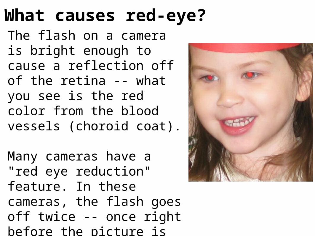
What causes red-eye?The flash on a camera is bright enough to cause a reflection off of the retina -- what you see is the red color from the blood vessels (choroid coat).
Many cameras have a "red eye reduction" feature. In these cameras, the flash goes off twice -- once right before the picture is taken, and then again to actually take the picture. The first flash causes people's pupils to contract, reducing "red eye."