semireplication-competent vesicular stomatitis virus as a novel platform for oncolytic virotherapy
TRANSCRIPT
ORIGINAL ARTICLE
Semireplication-competent vesicular stomatitis virusas a novel platform for oncolytic virotherapy
Alexander Muik & Catherine Dold & Yvonne Geiß &
Andreas Volk & Marina Werbizki & Ursula Dietrich &
Dorothee von Laer
Received: 26 July 2011 /Revised: 3 January 2012 /Accepted: 16 January 2012 /Published online: 28 January 2012# The Author(s) 2012. This article is published with open access at Springerlink.com
Abstract Among oncolytic viruses, the vesicular stomatitisvirus (VSV) is especially potent and a highly promisingagent for the treatment of cancer. But, even though effectiveagainst multiple tumor entities in preclinical animal models,replication-competent VSV exhibits inherent neurovirulence,which has so far hindered clinical development. To overcomethis limitation, replication-defective VSV vectors for cancergene therapy have been tested and proven to be safe. Howev-er, gene delivery was inefficient and only minor antitumorefficacy was observed. Here, we present semireplication-competent vector systems for VSV (srVSV), composed oftwo trans-complementing, propagation-deficient VSV vec-tors. The de novo generated deletion mutants of the twoVSV polymerase proteins P (phosphoprotein) and L (largecatalytic subunit), VSVΔP and VSVΔL respectively, wereused mutually or in combination with VSVΔG vectors. ThesesrVSV systems copropagated in vitro and in vivo withoutrecombinatory reversion to replication-competent virus. ThesrVSV systems were highly lytic for human glioblastoma celllines, spheroids, and subcutaneous xenografts. Especially thecombination of VSVΔG/VSVΔL vectors was as potent aswild-type VSV (VSV-WT) in vitro and induced long-termtumor regression in vivo without any associated adverse
effects. In contrast, 90% of VSV-WT-treated animals suc-cumbed to neurological disease shortly after tumor clearance.Most importantly, evenwhen injected into the brain, VSVΔG/VSVΔL did not show any neurotoxicity. In conclusion,srVSV is a promising platform for virotherapeutic approachesand also for VSV-based vector vaccines, combining improvedsafety with an increased coding capacity for therapeutictransgenes, potentially allowing for multipronged approaches.
Keywords Vesicular stomatitis virus . Oncolytic virus .
Virotherapy .Malignant glioma
Introduction
The use of viruses as targeted cancer therapeutics has shownsignificant promise in the last few years. Especially thevesicular stomatitis virus (VSV), a relatively new player inthe oncolytic virotherapy field, has proven to be effectiveagainst a variety of tumor entities such as malignant glioma[1, 2], hepatocellular carcinoma [3, 4], prostate cancer [5, 6],and ovarian carcinoma [7]. However, to date, the inherentneurotoxicity of VSV has hindered clinical developmentsince intracerebral administration causes fatal encephalitisin rodents and nonhuman primates [8, 9]. Thus, replication-competent VSV is associated with an increased risk ofsystemic dissemination and potentially severe pathology ifit enters the CNS. Therefore, attenuated virus variants andpropagation-deficient viral vectors were generated. Unfor-tunately, the reduced toxicity of attenuated replication-competent VSV is invariably accompanied with some re-duction of replicative and oncolytic activity [10, 11], whereasthe major limitation of propagation-deficient viral vectors hasbeen the inefficient transduction rate of cancer cells in vivo[2, 12].
Electronic supplementary material The online version of this article(doi:10.1007/s00109-012-0863-6) contains supplementary material,which is available to authorized users.
A. Muik :Y. Geiß :A. Volk :M. Werbizki :U. DietrichGeorg-Speyer-Haus,60596 Frankfurt am Main, Germany
C. Dold :D. von Laer (*)Institute for Virology, Innsbruck Medical University,Fritz-Pregl-Str. 3,A-6020 Innsbruck, Austriae-mail: [email protected]
J Mol Med (2012) 90:959–970DOI 10.1007/s00109-012-0863-6
A new strategy to potentially enhance safety of replication-competent VSV while increasing the capacity for therapeu-tic transgenes is the use of a semireplication-competentvector system similar to those described for retrovirusesand adenoviruses [13, 14]. Here, we successfully developeda semireplication-competent vector system for VSV(srVSV), which is based on two trans-complementingpropagation-deficient VSV vectors. The genes essential forviral replication are divided onto two separate packageablevector genomes, so that infectious progeny can only beproduced in double-infected host cells. Importantly, theVSV RNA genome does not undergo genetic reassortmentor recombination, making it unlikely that the binary systemreverts into a replication-competent recombinant VSV [15].In this study, we used the propagation-deficient, eGFP-expressing VSV*ΔG-vector [16], which lacks the G gene,in combination with de novo synthesized and rescued de-letion mutants VSVΔP-DsRed and VSVΔL-DsRed, lack-ing the genes P and L, respectively, that encode thecomponents of the viral polymerase complex. Accordingly,three different srVSV combinations were feasible:VSV*ΔG/VSVΔP-DsRed (srVSV(ΔG/ΔP)), VSV*ΔG/VSVΔL-DsRed (srVSV(ΔG/ΔL)), and VSVΔP-DsRed/VSVΔL-DsRed (srVSV(ΔP/ΔL)). All srVSV systemsallowed for in vitro reciprocal complementation thus lead-ing to copropagation associated with clear antitumor poten-cy against human glioblastoma cell lines. In addition, themost potent vector combination, srVSV(ΔG/ΔL), was test-ed in a preclinical subcutaneous (s.c.) glioblastoma mousemodel and proved to be only slightly attenuated comparedto wild-type VSV (VSV-WT). Tumors regressed in bothcohorts, but in contrast to the srVSV-treated group, 90% ofVSV-WT-treated animals succumbed to viral neurotoxicity.Most importantly, neither srVSV treatment of tumor-bearing animals nor direct intracranial administration inhealthy mice was associated with any sign of neurotoxicity.Eventually, all srVSV systems proved to be safe as wehave not been able to detect any sign of recombinatoryreversion to the wild-type strain.
Materials and methods
Cell culture
BHK-21 baby hamster kidney and U-87 MG human glioblas-toma cells were obtained from the American Type CultureCollection (Manassas, VA). G62 human glioblastoma cellswere kindly provided by M. Westphal (University HospitalEppendorf, Hamburg, Germany). HEK 293-NPeGFPL (clone206) stably expressing VSV-N, P, and L protein were a giftfromA. Pattnaik (University of Nebraska, Lincoln, USA) [17].All cells were kept in a humidified atmosphere containing 5%
CO2 at 37°C. BHK-21, U-87 MG, G62, and 293-NPeGFPLcells were maintained in DMEM (Gibco) supplemented with10% FBS (Perbio Science). 293-NPeGFPL cells were keptunder G418 selection.
Viruses
The propagation-incompetent VSV*ΔG vector, coding foreGFP as reporter, as well as the particularly strong type Iinterferon (IFN) inducing VSV*MQ, a replication-competent VSV with multimutated matrix protein (VSV-M), have been described previously [16, 18]. The deletionmutants VSVΔP-DsRed and VSVΔL-DsRed were generat-ed de novo: To exchange the VSV-P gene for DsRed, the N-P intergenic region (IGR) and a part of the VSV-N gene aswell as the P-M IGR and a part of the M gene were PCRamplified from pVSV-XN2 using the primers 5′-CGATCTCGAGGTATACATCTCTTACTACAGCAGG-3 ′/5′-CAGTGAATTCGATATCTGTTAGTTTTTTTCATATGTAGC-3′ (N-P IGR) and 5′-CGATGCGGCCGCACTATGAAAAAAAGTAACAGATATCACG-3′/5′-CAGTCCGCGGACGCGTAAACAGATCGATCTCTG-3′ (P-M IGR)with unique restriction sites (shown in bold). In parallel,DsRed was subcloned from pDsRed-Express-N1 (Clontech)into the multiple cloning site (MCS) of the pBluescript-IIcloning vector (Stratagene) with BamHI/NotI. Subsequently,PCR products were digested with XhoI/EcoRI (N-P IGR)and NotI/SacII (P-M IGR) and sequentially cloned in frontand behind the DsRed gene. Finally, the DsRed cassette wasexcised with BstZ17i/MluI and inserted into the BstZ17i/MluI site of pVSV-XN2, replacing VSV-P to yieldpVSVΔP-DsRed. A similar cloning strategy was appliedto generate pVSVΔL-DsRed: The G-L IGR and a part ofthe VSV-G gene as well as the L-HDV ribozyme regionwere PCR amplified from pVSV-XN2 using the primers 5′-CAGTGGTACCCTAAAATACTTTGAGACCAG-3′/5′-C G ATGGATCCGAT T G C T G T TA G T T T T T TTCATAAAAATTAAAAACTC-3′ (G-L IGR) and 5′-CAGTGCGGCCGCAAAATCATGAGGAGACTCCAAACTTTAAG-3 ′/5 ′-CGATGAGCTCGCACTAGTATCGAGGTCTCGATC-3′ (L-HDV ribozyme) withunique restriction sites (shown in bold). PCR products weredigested with KpnI/BamHI (G-L IGR) and NotI/SacI(L-HDV ribozyme) and cloned in front and behind theDsRed gene in the pBluescript-II-DsRed vector. Finally,DsRed was excised with NheI/SpeI and inserted into theNheI/SpeI site of pVSV-XN2, yielding pVSVΔL-DsRed.Novel recombinant viruses (Fig. 1a) were rescued as describedpreviously [19]. To produce infectious virions, VSV*ΔG-vec-tors were propagated on BHK-21 cells transiently expressingVSV-G [20]. VSVΔP-DsRed and VSVΔL-DsRed were am-plified on 293-NPeGFPL cells. Vector titers were determinedas 50% tissue culture infective dose (TCID50) using the
960 J Mol Med (2012) 90:959–970
Spearman–Kärber method [21]. VSVΔP-DsRed andVSVΔL-DsRed titration was performed on 293-NPeGFPLcells, VSV-WT and VSV*MQ were titrated on BHK-21 cells,and VSV*ΔG titration was performed on BHK-GP [20].
Quantitative PCR-based multicycle growth curve analysis
BHK-21 cells were infected in 6-well plates (106 cells/well)with a multiplicity of infection (MOI) of 0.05 of each
individual vector of the three potential srVSV vector sys-tems or VSV-WT as positive control. Filtered (0.45 μm)supernatants were collected at the indicated time points,and RNA was extracted from 50 μl supernatant using theRNeasy Mini Kit (Qiagen). RNA was reverse transcribedusing the High Capacity RNA-to-cDNA Kit (Applied Bio-systems). Vector propagation was monitored via real-timeRT-PCR to determine the total VSV genomic RNA (gRNA)amount in supernatants [19]. Known plasmid amounts wereused to determine the standard curve for real-time RNAquantification. Two independent qPCR primer and probesets were used, spanning the N-P and the M-G IGR of theVSV genome (see Supplementary Fig. S1c, d). Real-timePCR was carried out with the TaqMan® Gene ExpressionMaster Mix (Applied Biosystems) using a LightCycler®480 Real-Time PCR System (Roche). For both appliedreal-time PCRs, the detection limit was 102 gRNA/ml.
In vitro cytotoxicity assay
Human glioblastoma cells were plated in 96-well plates at104 cells/well in 100 μl medium. Cells were cultured asmonolayer or multicellular tumor spheroids. For spheroidcultures, 96-well plates were precoated with 75 μl 1% agarnoble (Difco). Cultures were infected with the respectiveviral system (srVSV or VSV-WT) at an MOI of 0.2 ortreated with phosphate-buffered saline (PBS) the followingday. Cell viability was assayed in dodecaplicates in n03independent experiments at the indicated time points post-infection using the cell proliferation agent WST-1 (Roche).Results are expressed as percentage of viable cells comparedto PBS-treated controls.
Animal studies
For antitumor efficacy testing, 6-week old NOD/SCID mice(Jackson Laboratories) were anesthetized with isoflurane and106 G62 human glioblastoma cells were subcutaneouslyinjected into the left and right flanks. Tumor growth wasmonitored with a caliper. At a tumor volume of 0.1 cm3, micewere treated intratumorally with two doses of either 2.8×10⁵
TCID50 srVSV(ΔG/ΔL) or 2.8×10⁵ TCID50 VSV-WT andPBS as controls. Bilateral tumors were treated alike. Whentumor size exceeded 0.8 cm3, mice were sacrificed. In addition,two mice were sacrificed at 3 days post-srVSV treatment ands.c. tumors were prepared for immunofluorescence analysis.
For neurotoxicity analysis, 6-week old CD1 Swiss mice(Charles River) were anesthetized by intraperitoneal injectionof ketamine/xylazine (100 and 10 mg/kg of body weight,respectively). 102, 103, and 104 TCID50 srVSV(ΔG/ΔL), aswell as 1.4×101 and 1.4×10⁴ TCID50 VSV-WT or PBS werestereotactically injected into the right frontal lobe of micebrains (1.5 mm lateral, 2 mm rostral to the bregma at 2 mm
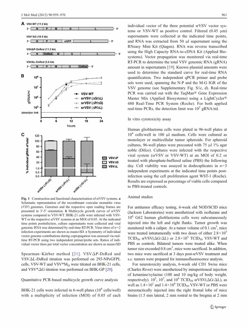
Fig. 1 Construction and functional characterization of srVSV systems. aSchematic representation of the recombinant vesicular stomatitis virus(VSV) genomes. Genomes and the respective open reading frames arepresented in 3′-5′ orientation. b Multicycle growth curves of srVSVsystems compared to VSV-WT. BHK-21 cells were infected with VSV-WTor the respective srVSV systems at an MOI of 0.05. At the indicatedtime points postinfection, culture supernatants were collected and viralgenomic RNAwas determined by real-time RT-PCR. Virus titers of n02infection experiments are shown as mean±SD. c Symmetry of individualvector genome contributions during copropagation was assessed via real-time RT-PCR using two independent primer/probe sets. Ratios of indi-vidual vector titers per total vector concentration are shown as mean±SD
J Mol Med (2012) 90:959–970 961
depth). Animals were monitored for signs of neurologicalimpairment. Two mice of the 104 TCID50 srVSV(ΔG/ΔL)-treated group were sacrificed at 3 days postinjection (dpi), andbrains were prepared for immunofluorescence analysis. Thebrains were sectioned (40 μm) on a Leica VT1000S vibratome(Leica, Bensheim, Germany). Nuclear counterstaining wasperformed with TO-PRO-3 iodide (Invitrogen). Sections wereanalyzed by confocal laser scanning microscopy using aNikon C1S1 microscope (Nikon, Düsseldorf, Germany). Allprocedures were approved by the governmental board for thecare of animal subjects (Regierungspräsidium Darmstadt,Germany).
Stimulation and IFN-α detection
Murine bone marrow (BM)-derived plasmacytoid dendriticcells (pDCs) were generated as previously described [22]. Inbrief, BM cells were flushed from femur and tibia with RPMIsupplemented with 10% FBS (Perbio Science). Erythrocyteswere lysed, cells were washed, and single-cell suspensionswere cultivated for 8 days in medium supplemented with100 ng/ml Flt3-L (R&D Systems). As determined by FACSanalysis, Flt3-L cultures consisted of ≈20% CD11c+B220+
pDCs (data not shown). For IFN stimulation experiments,2×106 Flt3-L-stimulated BM-pDC bulk culture cells wereseeded per 24 well. Cultures were infected with either srVSV,VSV*ΔG, VSVΔL-DsRed, VSV-WT, or VSV*MQ (each n02) at an MOI of 2. Supernatants were collected at 24 h post-infection (hpi) and analyzed for IFN-α via ELISA (PBLBiomedical Laboratories).
Statistical analysis
For comparison of individual time points or columns, statisti-cal difference was determined using unpaired t test. Micesurvival curves were plotted as Kaplan–Meier analysis, andstatistical significance between treatment groups was com-pared using the log-rank test.
Results
Novel recombinant viruses were cloned based on the pVSV-XN2 plasmid background and rescued as described previ-ously [19]. A schematic representation of the VSV vectorgenomes is shown in Fig. 1a, and their identity was con-firmed by gene-specific RT-PCR (Supplementary Fig. S1a,b). Both deletion mutants, VSVΔP-DsRed and VSVΔL-DsRed, were unable to propagate and did not generateprogeny virions in cell cultures not providing the respectivedeleted viral gene in trans, as real-time RT-PCR (Supple-mentary Fig. S1c, d) of supernatants were negative for VSVgRNA (data not shown).
srVSV(ΔG/ΔL) is the most potent srVSV system in terms ofvector propagation In order to assess the replication com-petence of the three potential srVSV systems, BHK-21 cellswere infected with an MOI of 0.05 of each individual vectoror VSV-WTas control to generate multicycle growth curves.Vector propagation was monitored on the gRNA level viareal-time RT-PCR [19]. In VSV-WT-infected cultures,gRNA associated with secreted progeny virions was firstdetectable at 6 hpi, reaching a plateau around 12–18 hpiwith maximum titers of more than 8×10⁸ gRNA/ml (8.77×10⁸±9.28×10⁷ gRNA/ml, see Fig. 1b). In comparison, allsrVSV vector systems showed an earlier onset of replicationwith first gRNA detectable at 3 hpi and srVSV(ΔP/ΔL)being the most potent in the initial phase with titers of 5.33×10⁴±3.05×10³ gRNA/ml 3 hpi. Both, the srVSV(ΔP/ΔG)and the srVSV(ΔG/ΔL) system lagged behind with titersbeing about tenfold reduced 3–6 hpi. Consistently, srVSV(ΔP/ΔL) was also the first to reach its plateau at 10–12 hpiwith a maximum of 8.44×10⁷±3.63×10⁶ gRNA/ml beforeits titer slowly started to regress. In contrast, both srVSV(ΔP/ΔG) and srVSV(ΔG/ΔL) ended up with a more robustreplication, reaching titers of 1.19×10⁸±1.63×10⁶ gRNA/ml for srVSV(ΔP/ΔG) and 7.60×10⁸±4.47×10⁷ gRNA/mlfor srVSV(ΔG/ΔL) at 24 hpi. Thus, the binary system usingVSV*ΔG and VSVΔL-DsRed was the most potent srVSVsystem in terms of vector dissemination even reaching max-imum gRNA titers comparable to VSV-WT.
In parallel, srVSV functional titers of supernatants col-lected at 24 hpi were determined as TCID50 per milliliter, asdouble-infected cells are a prerequisite to initiate copropa-gation. Correspondingly, the TCID50 of srVSV systemswere 180- (srVSV(ΔG/ΔL)) to 2,000-fold (srVSV(ΔP/ΔL)) lower than their gRNA titers, primarily reflecting thechance of coinfection, and to a considerably lesser extentreflecting the difference between genome and functionaltiters, as VSV-WT gRNA titers were only 6-fold highercompared to the respective TCID50. However, consistentwith the maximum obtained VSV gRNA per milliliter con-centrations during the multicycle growth curve, srVSV(ΔG/ΔL) displayed the highest TCID50 per milliliter of 4.22×10⁶, whereas titers for srVSV(ΔP/ΔG) were approx. 20-foldand for srVSV(ΔP/ΔL) around 100-fold lower (Table 1).
srVSV systems are characterized by asymmetric copropaga-tion As srVSV systems are composed of two vectors with
Table 1 Functionaltiters Viral system TCID50/ml
VSV-WT 1.58×10⁸
srVSV(ΔG/ΔL) 4.22×10⁶
srVSV(ΔP/ΔG) 2.37×10⁵
srVSV(ΔP/ΔL) 4.22×10⁴
962 J Mol Med (2012) 90:959–970
different properties such as gene composition and genomesize, the mode of copropagation during the multicycle growthcurve was analyzed via two independent qPCRs with ampli-cons spanning the N-P or M-G IGR of the VSV genome(Supplementary Fig. S1c, d). Combining the obtained qPCRdata, single-vector titers were calculated as ratio of the indi-vidual vector gRNA per milliliter per total vector gRNA permilliliter for all time points of the multicycle growth curve. Asratios proved to be consistent for each srVSV system through-out the whole observation period of 24 h, time-independentmeans and standard deviations were calculated. Indeed, theassessment revealed that vector copropagation was not due tosymmetric replication of both vector genomes, but couldrather be characterized as an asymmetric process (Fig. 1c).Each srVSV system could be defined by a distinct preferenceof one vector over the other. In case of srVSV(ΔP/ΔG), theVSVΔP vector accounts for 65.49±2.16% and VSVΔG for34.51±2.16% of the total titer. Even more pronounced is theasymmetry in favor of the VSVΔL vector with a share of65.99±6.78% (ΔP/ΔL) and 99.55±14.03% (ΔG/ΔL) of thetotal progeny generated.
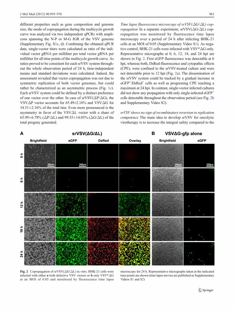
Time lapse fluorescence microscopy of srVSV(ΔG/ΔL) cop-ropagation In a separate experiment, srVSV(ΔG/ΔL) cop-ropagation was monitored by fluorescence time lapsemicroscopy over a period of 24 h after infecting BHK-21cells at an MOI of 0.05 (Supplementary Video S1). As nega-tive control, BHK-21 cells were infected with VSV*ΔG only.Representative micrographs at 0, 6, 12, 18, and 24 hpi areshown in Fig. 2. First eGFP fluorescence was detectable at 6hpi, whereas both, DsRed-fluorescence and cytopathic effects(CPE), were confined to the srVSV-treated culture and werenot detectable prior to 12 hpi (Fig. 2a). The dissemination ofthe srVSV system could be tracked by a gradual increase ineGFP+/DsRed+ cells as well as progressing CPE reaching amaximum at 24 hpi. In contrast, single-vector infected culturesdid not show any propagation with only single-infected eGFP+
cells detectable throughout the observation period (see Fig. 2band Supplementary Video S2).
srVSV shows no sign of recombinatory reversion to replicationcompetence The main idea to develop srVSV for oncolyticvirotherapy is to increase the integral safety compared to the
Fig. 2 Copropagation of srVSV(ΔG/ΔL) in vitro. BHK-21 cells wereinfected with either a both defective VSV vectors or b only VSV*ΔGat an MOI of 0.05 and monitored by fluorescence time lapse
microscopy for 24 h. Representative micrographs taken at the indicatedtime points are shown (time lapsemovies are published as SupplementaryVideos S1 and S2)
J Mol Med (2012) 90:959–970 963
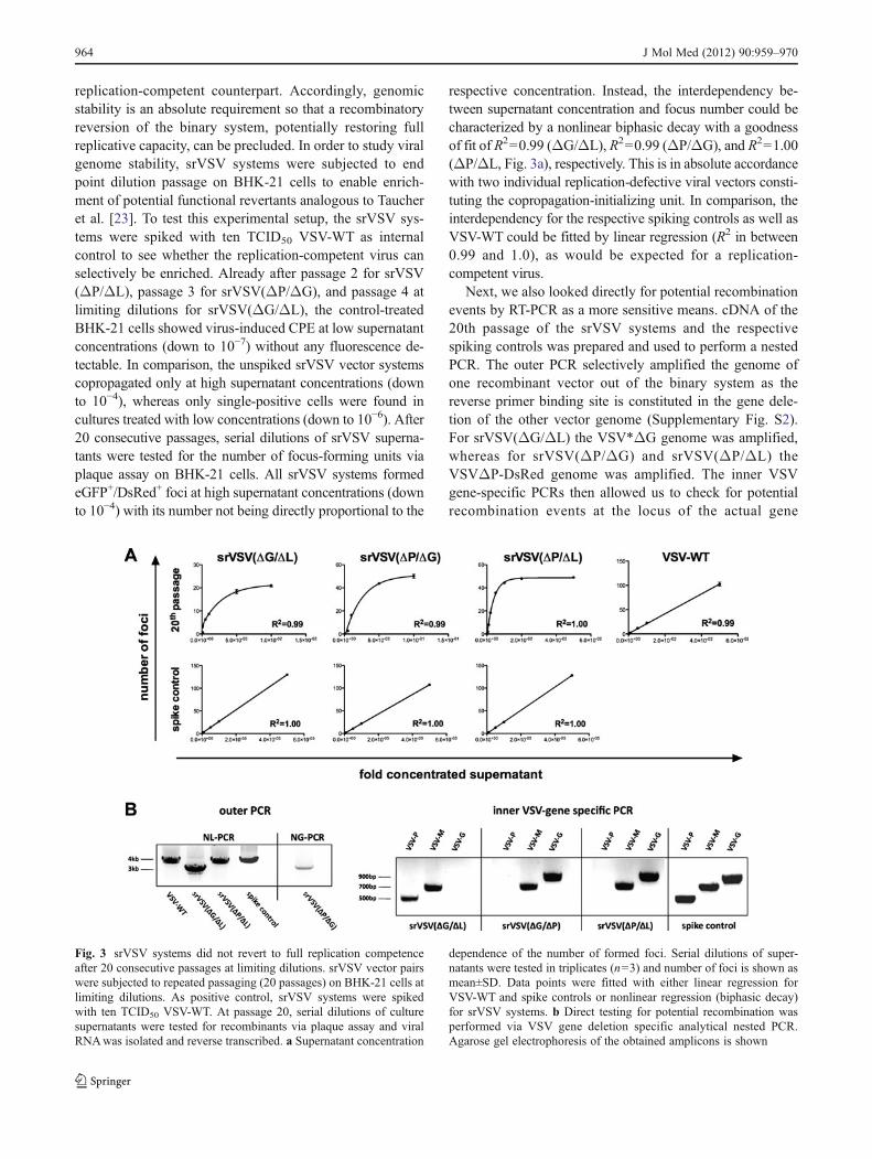
replication-competent counterpart. Accordingly, genomicstability is an absolute requirement so that a recombinatoryreversion of the binary system, potentially restoring fullreplicative capacity, can be precluded. In order to study viralgenome stability, srVSV systems were subjected to endpoint dilution passage on BHK-21 cells to enable enrich-ment of potential functional revertants analogous to Taucheret al. [23]. To test this experimental setup, the srVSV sys-tems were spiked with ten TCID50 VSV-WT as internalcontrol to see whether the replication-competent virus canselectively be enriched. Already after passage 2 for srVSV(ΔP/ΔL), passage 3 for srVSV(ΔP/ΔG), and passage 4 atlimiting dilutions for srVSV(ΔG/ΔL), the control-treatedBHK-21 cells showed virus-induced CPE at low supernatantconcentrations (down to 10−7) without any fluorescence de-tectable. In comparison, the unspiked srVSV vector systemscopropagated only at high supernatant concentrations (downto 10−4), whereas only single-positive cells were found incultures treated with low concentrations (down to 10−6). After20 consecutive passages, serial dilutions of srVSV superna-tants were tested for the number of focus-forming units viaplaque assay on BHK-21 cells. All srVSV systems formedeGFP+/DsRed+ foci at high supernatant concentrations (downto 10−4) with its number not being directly proportional to the
respective concentration. Instead, the interdependency be-tween supernatant concentration and focus number could becharacterized by a nonlinear biphasic decay with a goodnessof fit of R200.99 (ΔG/ΔL), R200.99 (ΔP/ΔG), and R201.00(ΔP/ΔL, Fig. 3a), respectively. This is in absolute accordancewith two individual replication-defective viral vectors consti-tuting the copropagation-initializing unit. In comparison, theinterdependency for the respective spiking controls as well asVSV-WT could be fitted by linear regression (R2 in between0.99 and 1.0), as would be expected for a replication-competent virus.
Next, we also looked directly for potential recombinationevents by RT-PCR as a more sensitive means. cDNA of the20th passage of the srVSV systems and the respectivespiking controls was prepared and used to perform a nestedPCR. The outer PCR selectively amplified the genome ofone recombinant vector out of the binary system as thereverse primer binding site is constituted in the gene dele-tion of the other vector genome (Supplementary Fig. S2).For srVSV(ΔG/ΔL) the VSV*ΔG genome was amplified,whereas for srVSV(ΔP/ΔG) and srVSV(ΔP/ΔL) theVSVΔP-DsRed genome was amplified. The inner VSVgene-specific PCRs then allowed us to check for potentialrecombination events at the locus of the actual gene
Fig. 3 srVSV systems did not revert to full replication competenceafter 20 consecutive passages at limiting dilutions. srVSV vector pairswere subjected to repeated passaging (20 passages) on BHK-21 cells atlimiting dilutions. As positive control, srVSV systems were spikedwith ten TCID50 VSV-WT. At passage 20, serial dilutions of culturesupernatants were tested for recombinants via plaque assay and viralRNAwas isolated and reverse transcribed. a Supernatant concentration
dependence of the number of formed foci. Serial dilutions of super-natants were tested in triplicates (n03) and number of foci is shown asmean±SD. Data points were fitted with either linear regression forVSV-WT and spike controls or nonlinear regression (biphasic decay)for srVSV systems. b Direct testing for potential recombination wasperformed via VSV gene deletion specific analytical nested PCR.Agarose gel electrophoresis of the obtained amplicons is shown
964 J Mol Med (2012) 90:959–970
deletion. Consistent with the phenotypic analysis (Fig. 3a),we have not been able to detect any recombination event forthe srVSV systems. The VSV-G (for srVSV(ΔG/ΔL)) andVSV-P gene (for srVSV(ΔP/ΔG) and srVSV(ΔP/ΔL))were not detectable, whereas the according amplicons weredetected for the spiking control (see Fig. 3b). These datawere corroborated by sequence analysis of the outer PCRamplicons, which clearly evidenced presence of the respectivefluorescence marker gene (data not shown). Thus, in both, thephenotypic and genotypic analysis, recombination among thevector genomes was not detectablewhile the respective spikingcontrols were positive, the latter validating the applicability ofthe applied assays.
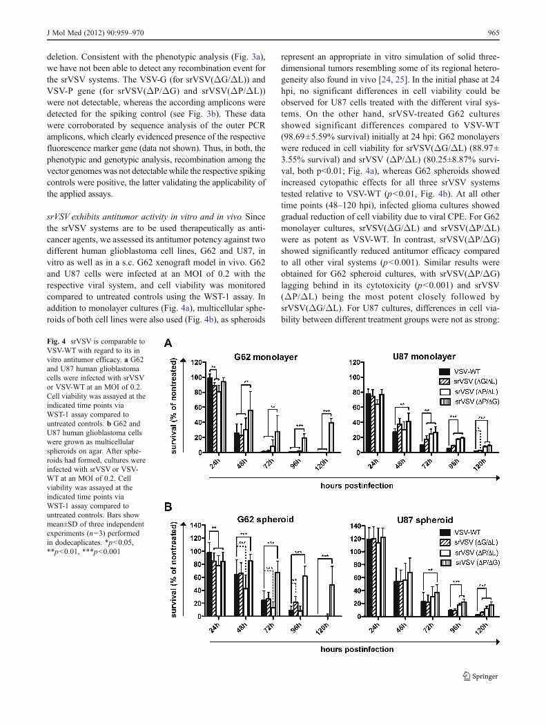
srVSV exhibits antitumor activity in vitro and in vivo Sincethe srVSV systems are to be used therapeutically as anti-cancer agents, we assessed its antitumor potency against twodifferent human glioblastoma cell lines, G62 and U87, invitro as well as in a s.c. G62 xenograft model in vivo. G62and U87 cells were infected at an MOI of 0.2 with therespective viral system, and cell viability was monitoredcompared to untreated controls using the WST-1 assay. Inaddition to monolayer cultures (Fig. 4a), multicellular sphe-roids of both cell lines were also used (Fig. 4b), as spheroids
represent an appropriate in vitro simulation of solid three-dimensional tumors resembling some of its regional hetero-geneity also found in vivo [24, 25]. In the initial phase at 24hpi, no significant differences in cell viability could beobserved for U87 cells treated with the different viral sys-tems. On the other hand, srVSV-treated G62 culturesshowed significant differences compared to VSV-WT(98.69±5.59% survival) initially at 24 hpi: G62 monolayerswere reduced in cell viability for srVSV(ΔG/ΔL) (88.97±3.55% survival) and srVSV (ΔP/ΔL) (80.25±8.87% survi-val, both p<0.01; Fig. 4a), whereas G62 spheroids showedincreased cytopathic effects for all three srVSV systemstested relative to VSV-WT (p<0.01, Fig. 4b). At all othertime points (48–120 hpi), infected glioma cultures showedgradual reduction of cell viability due to viral CPE. For G62monolayer cultures, srVSV(ΔG/ΔL) and srVSV(ΔP/ΔL)were as potent as VSV-WT. In contrast, srVSV(ΔP/ΔG)showed significantly reduced antitumor efficacy comparedto all other viral systems (p<0.001). Similar results wereobtained for G62 spheroid cultures, with srVSV(ΔP/ΔG)lagging behind in its cytotoxicity (p<0.001) and srVSV(ΔP/ΔL) being the most potent closely followed bysrVSV(ΔG/ΔL). For U87 cultures, differences in cell via-bility between different treatment groups were not as strong:
Fig. 4 srVSV is comparable toVSV-WT with regard to its invitro antitumor efficacy. a G62and U87 human glioblastomacells were infected with srVSVor VSV-WT at an MOI of 0.2.Cell viability was assayed at theindicated time points viaWST-1 assay compared tountreated controls. b G62 andU87 human glioblastoma cellswere grown as multicellularspheroids on agar. After sphe-roids had formed, cultures wereinfected with srVSV or VSV-WT at an MOI of 0.2. Cellviability was assayed at theindicated time points viaWST-1 assay compared tountreated controls. Bars showmean±SD of three independentexperiments (n03) performedin dodecaplicates. *p<0.05,**p<0.01, ***p<0.001
J Mol Med (2012) 90:959–970 965
srVSV(ΔP/ΔG)-treated cultures were clearly the most via-ble (ranging from p<0.01 to p<0.001), whereas the srVSV(ΔG/ΔL) system performed best with regard to its antitu-mor effect. However, all srVSV-treated groups showedattenuated antitumor efficacy compared to VSV-WT-treatedcultures.
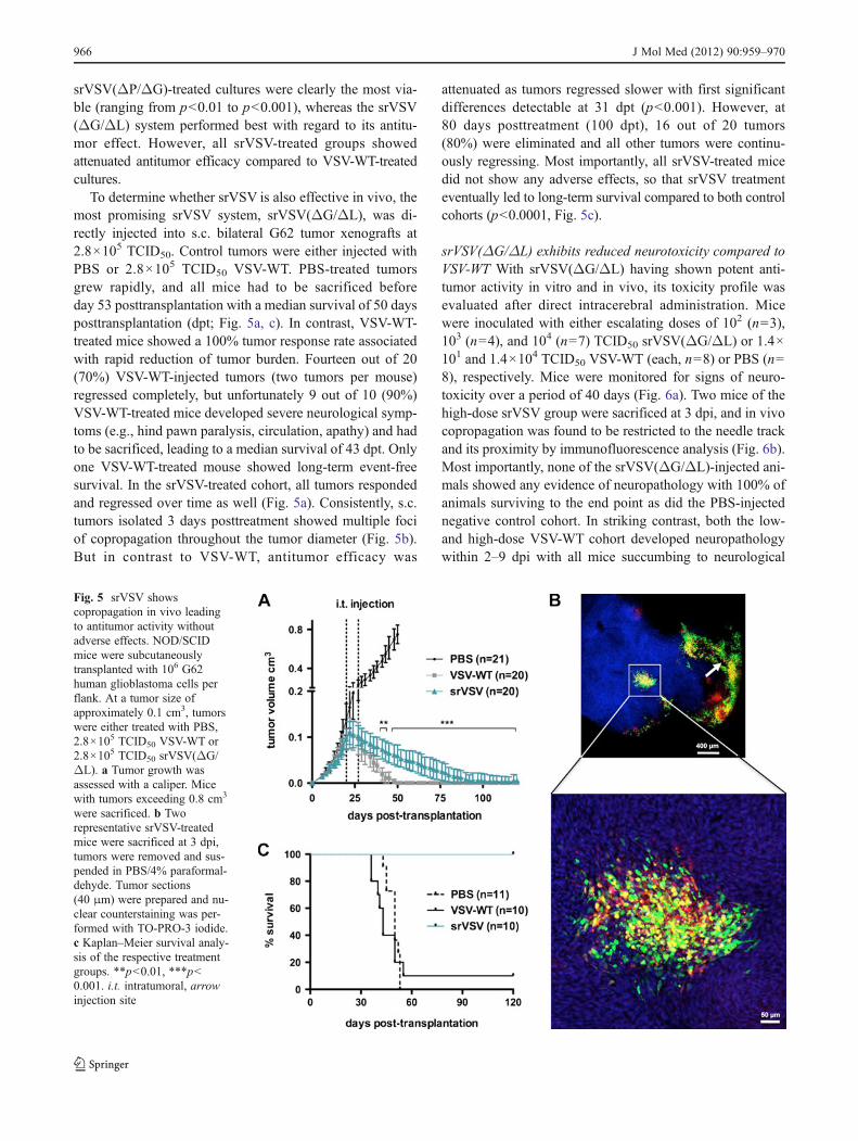
To determine whether srVSV is also effective in vivo, themost promising srVSV system, srVSV(ΔG/ΔL), was di-rectly injected into s.c. bilateral G62 tumor xenografts at2.8×105 TCID50. Control tumors were either injected withPBS or 2.8×10⁵ TCID50 VSV-WT. PBS-treated tumorsgrew rapidly, and all mice had to be sacrificed beforeday 53 posttransplantation with a median survival of 50 daysposttransplantation (dpt; Fig. 5a, c). In contrast, VSV-WT-treated mice showed a 100% tumor response rate associatedwith rapid reduction of tumor burden. Fourteen out of 20(70%) VSV-WT-injected tumors (two tumors per mouse)regressed completely, but unfortunately 9 out of 10 (90%)VSV-WT-treated mice developed severe neurological symp-toms (e.g., hind pawn paralysis, circulation, apathy) and hadto be sacrificed, leading to a median survival of 43 dpt. Onlyone VSV-WT-treated mouse showed long-term event-freesurvival. In the srVSV-treated cohort, all tumors respondedand regressed over time as well (Fig. 5a). Consistently, s.c.tumors isolated 3 days posttreatment showed multiple fociof copropagation throughout the tumor diameter (Fig. 5b).But in contrast to VSV-WT, antitumor efficacy was
attenuated as tumors regressed slower with first significantdifferences detectable at 31 dpt (p<0.001). However, at80 days posttreatment (100 dpt), 16 out of 20 tumors(80%) were eliminated and all other tumors were continu-ously regressing. Most importantly, all srVSV-treated micedid not show any adverse effects, so that srVSV treatmenteventually led to long-term survival compared to both controlcohorts (p<0.0001, Fig. 5c).
srVSV(ΔG/ΔL) exhibits reduced neurotoxicity compared toVSV-WT With srVSV(ΔG/ΔL) having shown potent anti-tumor activity in vitro and in vivo, its toxicity profile wasevaluated after direct intracerebral administration. Micewere inoculated with either escalating doses of 102 (n03),103 (n04), and 104 (n07) TCID50 srVSV(ΔG/ΔL) or 1.4×101 and 1.4×10⁴ TCID50 VSV-WT (each, n08) or PBS (n08), respectively. Mice were monitored for signs of neuro-toxicity over a period of 40 days (Fig. 6a). Two mice of thehigh-dose srVSV group were sacrificed at 3 dpi, and in vivocopropagation was found to be restricted to the needle trackand its proximity by immunofluorescence analysis (Fig. 6b).Most importantly, none of the srVSV(ΔG/ΔL)-injected ani-mals showed any evidence of neuropathology with 100% ofanimals surviving to the end point as did the PBS-injectednegative control cohort. In striking contrast, both the low-and high-dose VSV-WT cohort developed neuropathologywithin 2–9 dpi with all mice succumbing to neurological
Fig. 5 srVSV showscopropagation in vivo leadingto antitumor activity withoutadverse effects. NOD/SCIDmice were subcutaneouslytransplanted with 106 G62human glioblastoma cells perflank. At a tumor size ofapproximately 0.1 cm3, tumorswere either treated with PBS,2.8×105 TCID50 VSV-WT or2.8×105 TCID50 srVSV(ΔG/ΔL). a Tumor growth wasassessed with a caliper. Micewith tumors exceeding 0.8 cm3
were sacrificed. b Tworepresentative srVSV-treatedmice were sacrificed at 3 dpi,tumors were removed and sus-pended in PBS/4% paraformal-dehyde. Tumor sections(40 μm) were prepared and nu-clear counterstaining was per-formed with TO-PRO-3 iodide.c Kaplan–Meier survival analy-sis of the respective treatmentgroups. **p<0.01, ***p<0.001. i.t. intratumoral, arrowinjection site
966 J Mol Med (2012) 90:959–970
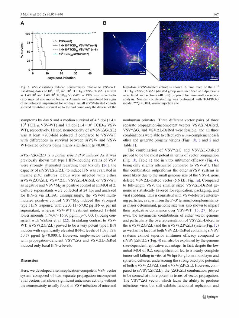
symptoms by day 9 and a median survival of 4.5 dpi (1.4×10⁴ TCID50 VSV-WT) and 7.5 dpi (1.4×101 TCID50 VSV-WT), respectively. Hence, neurotoxicity of srVSV(ΔG/ΔL)was at least >700-fold reduced if compared to VSV-WTwith differences in survival between srVSV- and VSV-WT-treated cohorts being highly significant (p<0.001).
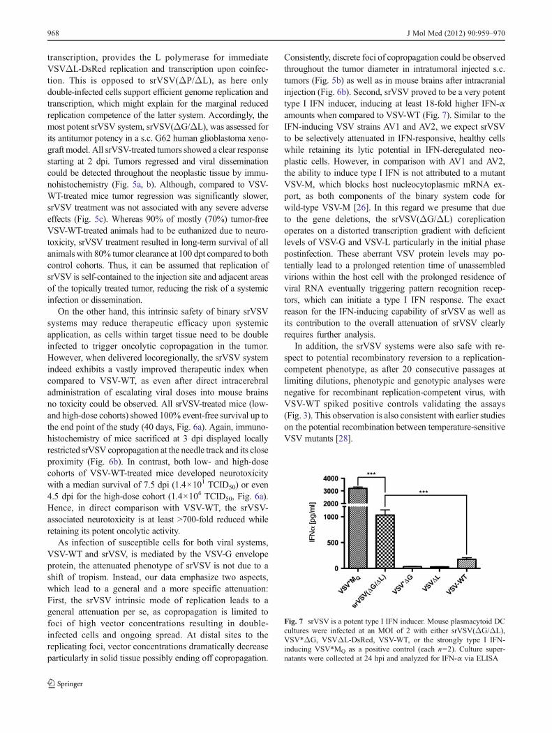
srVSV(ΔG/ΔL) is a potent type I IFN inducer As it waspreviously shown that type I IFN-inducing strains of VSVwere strongly attenuated regarding their toxicity [26], thecapacity of srVSV(ΔG/ΔL) to induce IFN was evaluated inmurine pDC cultures. pDCs were infected with eithersrVSV(ΔG/ΔL), VSV*ΔG, VSVΔL-DsRed, or VSV-WTas negative and VSV*MQ as positive control at an MOI of 2.Culture supernatants were collected at 24 hpi and analyzedfor IFN-α via ELISA. Unsurprisingly, the VSV-M multi-mutated positive control VSV*MQ induced the strongesttype I IFN response, with 3,200.11±57.02 pg IFN-α per mlsupernatant, whereas VSV-WT treatment induced 18-foldlower amounts (174.47±16.70 pg/ml; p<0.0001), being con-sistent with Waibler et al. [22]. In striking contrast to VSV-WT, srVSV(ΔG/ΔL) proved to be a very potent type I IFNinducer with significantly elevated IFN-α levels of 1,035.52±50.57 pg/ml (p<0.0001). However, single-vector treatmentwith propagation-deficient VSV*ΔG and VSVΔL-DsRedinduced only basal IFN-α levels.
Discussion
Here, we developed a semireplication-competent VSV vectorsystem composed of two separate propagation-incompetentviral vectors that shows significant anticancer activity withoutthe neurotoxicity usually found in VSV infection of mice and
nonhuman primates. Three different vector pairs of threeseparate propagation-incompetent vectors VSVΔP-DsRed,VSV*ΔG, and VSVΔL-DsRed were feasible, and all threecombinations were able to effectively trans-complement eachother and generate progeny virions (Figs. 1b, c and 2 andTable 1).
The combination of VSV*ΔG and VSVΔL-DsRedproved to be the most potent in terms of vector propagation(Fig. 1b, Table 1) and in vitro antitumor efficacy (Fig. 4),being only slightly attenuated compared to VSV-WT. Thatthis combination outperforms the other srVSV systems ismost likely due to the small genome size of the VSV-L genedeleted VSVΔL-DsRed vector (5.6 kB, Fig. 1a). Comparedto full-length VSV, the smaller sized VSVΔL-DsRed ge-nome is statistically favored for replication, packaging, andviral shedding. This is consistent with VSV-defective interfer-ing particles, as apart from the 5′–3′ terminal complementarityas major determinant, genome size was also shown to impacttheir replicative dominance over VSV-WT [15, 27]. More-over, the asymmetric contributions of either vector genomeand particularly the overrepresentation of VSVΔL-DsRed inthe srVSV(ΔG/ΔL) and the srVSV(ΔP/ΔL) system (Fig. 1c)as well as the fact that both VSVΔL-DsRed containing srVSVsystems exhibit superior antitumor efficacy compared tosrVSV(ΔP/ΔG) (Fig. 4) can also be explained by the genomesize-dependent replicative advantage. In fact, despite the lowinitial MOI of 0.2, coamplification led to a nearly completetumor cell killing in vitro at 96 hpi for glioma monolayer andspheroid cultures, underscoring the strong oncolytic potentialof both srVSV(ΔG/ΔL) and srVSV(ΔP/ΔL). However, com-pared to srVSV(ΔP/ΔL), the (ΔG/ΔL) combination provedto be somewhat more potent in terms of vector propagation.The VSV*ΔG vector, which lacks the ability to produceinfectious virus but still exhibits functional replication and
Fig. 6 srVSV exhibits reduced neurotoxicity relative to VSV-WT.Escalating doses of 102, 103, and 104 TCID50 srVSV(ΔG/ΔL) as wellas 1.4×101 and 1.4×10⁴ TCID50 VSV-WT or PBS were stereotacti-cally injected into mouse brains. a Animals were monitored for signsof neurological impairment for 40 days. As all srVSV-treated cohortsshowed event-free survival up to the end point, only the data set of the
high-dose srVSV-treated cohort is shown. b Two mice of the 104
TCID50 srVSV(ΔG/ΔL)-treated group were sacrificed at 3 dpi, brainswere fixed and sections (40 μm) prepared for immunofluorescenceanalysis. Nuclear counterstaining was performed with TO-PRO-3iodide. ***p<0.001, arrow injection site
J Mol Med (2012) 90:959–970 967
transcription, provides the L polymerase for immediateVSVΔL-DsRed replication and transcription upon coinfec-tion. This is opposed to srVSV(ΔP/ΔL), as here onlydouble-infected cells support efficient genome replication andtranscription, which might explain for the marginal reducedreplication competence of the latter system. Accordingly, themost potent srVSV system, srVSV(ΔG/ΔL), was assessed forits antitumor potency in a s.c. G62 human glioblastoma xeno-graft model. All srVSV-treated tumors showed a clear responsestarting at 2 dpi. Tumors regressed and viral disseminationcould be detected throughout the neoplastic tissue by immu-nohistochemistry (Fig. 5a, b). Although, compared to VSV-WT-treated mice tumor regression was significantly slower,srVSV treatment was not associated with any severe adverseeffects (Fig. 5c). Whereas 90% of mostly (70%) tumor-freeVSV-WT-treated animals had to be euthanized due to neuro-toxicity, srVSV treatment resulted in long-term survival of allanimals with 80% tumor clearance at 100 dpt compared to bothcontrol cohorts. Thus, it can be assumed that replication ofsrVSV is self-contained to the injection site and adjacent areasof the topically treated tumor, reducing the risk of a systemicinfection or dissemination.
On the other hand, this intrinsic safety of binary srVSVsystems may reduce therapeutic efficacy upon systemicapplication, as cells within target tissue need to be doubleinfected to trigger oncolytic copropagation in the tumor.However, when delivered locoregionally, the srVSV systemindeed exhibits a vastly improved therapeutic index whencompared to VSV-WT, as even after direct intracerebraladministration of escalating viral doses into mouse brainsno toxicity could be observed. All srVSV-treated mice (low-and high-dose cohorts) showed 100% event-free survival up tothe end point of the study (40 days, Fig. 6a). Again, immuno-histochemistry of mice sacrificed at 3 dpi displayed locallyrestricted srVSV copropagation at the needle track and its closeproximity (Fig. 6b). In contrast, both low- and high-dosecohorts of VSV-WT-treated mice developed neurotoxicitywith a median survival of 7.5 dpi (1.4×101 TCID50) or even4.5 dpi for the high-dose cohort (1.4×10⁴ TCID50, Fig. 6a).Hence, in direct comparison with VSV-WT, the srVSV-associated neurotoxicity is at least >700-fold reduced whileretaining its potent oncolytic activity.
As infection of susceptible cells for both viral systems,VSV-WT and srVSV, is mediated by the VSV-G envelopeprotein, the attenuated phenotype of srVSV is not due to ashift of tropism. Instead, our data emphasize two aspects,which lead to a general and a more specific attenuation:First, the srVSV intrinsic mode of replication leads to ageneral attenuation per se, as copropagation is limited tofoci of high vector concentrations resulting in double-infected cells and ongoing spread. At distal sites to thereplicating foci, vector concentrations dramatically decreaseparticularly in solid tissue possibly ending off copropagation.
Consistently, discrete foci of copropagation could be observedthroughout the tumor diameter in intratumoral injected s.c.tumors (Fig. 5b) as well as in mouse brains after intracranialinjection (Fig. 6b). Second, srVSV proved to be a very potenttype I IFN inducer, inducing at least 18-fold higher IFN-αamounts when compared to VSV-WT (Fig. 7). Similar to theIFN-inducing VSV strains AV1 and AV2, we expect srVSVto be selectively attenuated in IFN-responsive, healthy cellswhile retaining its lytic potential in IFN-deregulated neo-plastic cells. However, in comparison with AV1 and AV2,the ability to induce type I IFN is not attributed to a mutantVSV-M, which blocks host nucleocytoplasmic mRNA ex-port, as both components of the binary system code forwild-type VSV-M [26]. In this regard we presume that dueto the gene deletions, the srVSV(ΔG/ΔL) coreplicationoperates on a distorted transcription gradient with deficientlevels of VSV-G and VSV-L particularly in the initial phasepostinfection. These aberrant VSV protein levels may po-tentially lead to a prolonged retention time of unassembledvirions within the host cell with the prolonged residence ofviral RNA eventually triggering pattern recognition recep-tors, which can initiate a type I IFN response. The exactreason for the IFN-inducing capability of srVSV as well asits contribution to the overall attenuation of srVSV clearlyrequires further analysis.
In addition, the srVSV systems were also safe with re-spect to potential recombinatory reversion to a replication-competent phenotype, as after 20 consecutive passages atlimiting dilutions, phenotypic and genotypic analyses werenegative for recombinant replication-competent virus, withVSV-WT spiked positive controls validating the assays(Fig. 3). This observation is also consistent with earlier studieson the potential recombination between temperature-sensitiveVSV mutants [28].
Fig. 7 srVSV is a potent type I IFN inducer. Mouse plasmacytoid DCcultures were infected at an MOI of 2 with either srVSV(ΔG/ΔL),VSV*ΔG, VSVΔL-DsRed, VSV-WT, or the strongly type I IFN-inducing VSV*MQ as a positive control (each n02). Culture super-natants were collected at 24 hpi and analyzed for IFN-α via ELISA
968 J Mol Med (2012) 90:959–970
In summary, relative to VSV-WT, srVSV systems present apromising platform for virotherapeutic approaches, as they aregenetically stable and exhibit considerably reduced neurotox-icity while retaining their antitumor potency. Furthermore,srVSV systems offer a strongly increased coding capacity sothat both viral vectors can be “armed” to express therapeutictransgenes allowing for multipronged approaches, combiningtheir inherent oncolytic effect with a tumor microenvironmentmodulating suicide and/or immunostimulatory “payload” toboost antitumor potency. Eventually, with respect to bothbiosafety and coding capacity, srVSV systems may not onlyprove valuable for oncolytic virotherapy but also represent anattractive vector vaccine platform.
Acknowledgments This work was supported by grants from theWilhelm-Sander-Foundation and the Schering foundation Deutsche For-schungsgemeinschaft (Graduate College 1172).We thank StefanMommaand Anna Kraft for CLSM assistance.
Disclosure of potential conflict of interests The authors declare noconflict of interests related to this study.
Open Access This article is distributed under the terms of the Crea-tive Commons Attribution License which permits any use, distribution,and reproduction in any medium, provided the original author(s) andthe source are credited.
References
1. Alain T, Lun X, Martineau Y, Sean P, Pulendran B, Petroulakis E,Zemp FJ, Lemay CG, Roy D, Bell JC et al (2010) Vesicularstomatitis virus oncolysis is potentiated by impairing mTORC1-dependent type I IFN production. Proc Natl Acad Sci USA107:1576–1581
2. Wollmann G, Rogulin V, Simon I, Rose JK, van den Pol AN (2010)Some attenuated variants of vesicular stomatitis virus show enhancedoncolytic activity against human glioblastoma cells relative to normalbrain cells. J Virol 84:1563–1573
3. Shinozaki K, Ebert O, Woo SL (2005) Eradication of advancedhepatocellular carcinoma in rats via repeated hepatic arterial infu-sions of recombinant VSV. Hepatology 41:196–203
4. Ebert O, Shinozaki K, Huang TG, Savontaus MJ, Garcia-Sastre A,Woo SL (2003) Oncolytic vesicular stomatitis virus for treatmentof orthotopic hepatocellular carcinoma in immune-competent rats.Cancer Res 63:3605–3611
5. Ahmed M, Cramer SD, Lyles DS (2004) Sensitivity of prostatetumors to wild type and M protein mutant vesicular stomatitisviruses. Virology 330:34–49
6. Moussavi M, Fazli L, Tearle H, Guo Y, Cox M, Bell J, Ong C, JiaW, Rennie PS (2010) Oncolysis of prostate cancers induced byvesicular stomatitis virus in PTEN knockout mice. Cancer Res70:1367–1376
7. Capo-chichi CD, Yeasky TM, Heiber JF, Wang Y, Barber GN, XuXX (2010) Explicit targeting of transformed cells by VSV inovarian epithelial tumor-bearing Wv mouse models. GynecolOncol 116:269–275
8. Huneycutt BS, Bi Z, Aoki CJ, Reiss CS (1993) Central neuro-pathogenesis of vesicular stomatitis virus infection of immunode-ficient mice. J Virol 67:6698–6706
9. Johnson JE, Nasar F, Coleman JW, Price RE, Javadian A, DraperK, Lee M, Reilly PA, Clarke DK, Hendry RM et al (2007) Neuro-virulence properties of recombinant vesicular stomatitis virus vec-tors in non-human primates. Virology 360:36–49
10. Clarke DK, Nasar F, Lee M, Johnson JE, Wright K, Calderon P,Guo M, Natuk R, Cooper D, Hendry RM et al (2007) Synergisticattenuation of vesicular stomatitis virus by combination of specificG gene truncations and N gene translocations. J Virol 81:2056–2064
11. Kelly EJ, Nace R, Barber GN, Russell SJ (2010) Attenuation ofvesicular stomatitis virus encephalitis through microRNA targeting. JVirol 84:1550–1562
12. Duntsch CD, Zhou Q, Jayakar HR, Weimar JD, Robertson JH,Pfeffer LM, Wang L, Xiang Z, Whitt MA (2004) Recombinantvesicular stomatitis virus vectors as oncolytic agents in the treatmentof high-grade gliomas in an organotypic brain tissue slice-gliomacoculture model. J Neurosurg 100:1049–1059
13. Alemany R, Lai S, Lou YC, Jan HY, Fang X, Zhang WW (1999)Complementary adenoviral vectors for oncolysis. Cancer GeneTher 6:21–25
14. Trajcevski S, Solly SK, Frisen C, Trenado A, Cosset FL, KlatzmannD (2005) Characterization of a semi-replicative gene delivery systemallowing propagation of complementary defective retroviral vectors.J Gene Med 7:276–287
15. Wagner RR, Rose JK (1996) Rhabdoviridae: the viruses andtheir replication. In: Whitley BNF RJ, Knipe DM, Howley PM(eds) Fields virology, 3rd edn. Lippincott-Raven Publishers,Philadelphia
16. Hanika A, Larisch B, Steinmann E, Schwegmann-Wessels C,Herrler G, Zimmer G (2005) Use of influenza C virus glycoproteinHEF for generation of vesicular stomatitis virus pseudotypes. JGen Virol 86:1455–1465
17. Panda D, Dinh PX, Beura LK, Pattnaik AK (2010) Inductionof interferon and interferon signaling pathways by replicationof defective interfering particle RNA in cells constitutively express-ing vesicular stomatitis virus replication proteins. J Virol 84:4826–4831
18. Hoffmann M, Wu YJ, Gerber M, Berger-Rentsch M, Heimrich B,Schwemmle M, Zimmer G (2010) Fusion-active glycoprotein Gmediates the cytotoxicity of vesicular stomatitis virus M mutantslacking host shut-off activity. J Gen Virol 91:2782–2793
19. Ebert O, Shinozaki K, Kournioti C, Park MS, Garcia-Sastre A,Woo SL (2004) Syncytia induction enhances the oncolytic poten-tial of vesicular stomatitis virus in virotherapy for cancer. CancerRes 64:3265–3270
20. Muik A, Kneiske I, Werbizki M, Wilflingseder D, Giroglou T,Ebert O, Kraft A, Dietrich U, Zimmer G, Momma S et al (2011)Pseudotyping vesicular stomatitis virus with lymphocytic chorio-meningitis virus glycoproteins enhances infectivity for glioma cellsand minimizes neurotropism. J Virol 85:5679–5684
21. Kärber G (1931) 50% end-point calculation. Arch Exp PatholPharmak 162:480–483
22. Waibler Z, Detje CN, Bell JC, Kalinke U (2007) Matrix proteinmediated shutdown of host cell metabolism limits vesicular sto-matitis virus-induced interferon-alpha responses to plasmacytoiddendritic cells. Immunobiology 212:887–894
23. Taucher C, Berger A, Mandl CW (2010) A trans-complementingrecombination trap demonstrates a low propensity of flaviviruses forintermolecular recombination. J Virol 84:599–611
24. Sutherland RM (1988) Cell and environment interactions in tumormicroregions: the multicell spheroid model. Science 240:177–184
25. Tamaki M, McDonald W, Amberger VR, Moore E, Del Maestro RF(1997) Implantation of C6 astrocytoma spheroid into collagen type I
J Mol Med (2012) 90:959–970 969
gels: invasive, proliferative, and enzymatic characterizations.J Neurosurg 87:602–609
26. Stojdl DF, Lichty BD, tenOever BR, Paterson JM, Power AT,Knowles S, Marius R, Reynard J, Poliquin L, Atkins H et al(2003) VSV strains with defects in their ability to shutdown innateimmunity are potent systemic anti-cancer agents. Cancer Cell4:263–275
27. Von Laer DM, Mack D, Kruppa J (1988) Delayed formation ofdefective interfering particles in vesicular stomatitis virus-infectedcells: kinetic studies of viral protein and RNA synthesis duringautointerference. J Virol 62:1323–1329
28. Wong PK, Holloway AF, Cormack DV (1971) A search forrecombination between temperature-sensitive mutants of vesicularstomatitis virus. J Gen Virol 13:477–479
970 J Mol Med (2012) 90:959–970