seminars in fetal & neonatal medicine · fetal heart defects: ... the use of transvaginal...
TRANSCRIPT
at SciVerse ScienceDirect
Seminars in Fetal & Neonatal Medicine 18 (2013) 251e260
Contents lists available
Seminars in Fetal & Neonatal Medicine
journal homepage: www.elsevier .com/locate/s iny
Fetal heart defects: Potential and pitfalls of first-trimester detection
Asma Khalil a, Kypros H. Nicolaides a,b,*
aDepartment of Fetal Medicine, St George’s Hospital, University of London, UKbDepartment of Fetal Medicine, King’s College Hospital, London, UK
Keywords:Congenital heart defectsDuctus venosusEchocardiographyFetalFirst trimesterNuchal translucencyTricuspid regurgitation
* Corresponding author. Address: Harris BirthrighMedicine, King’s College Hospital, Denmark Hill, Lond203 2998256; fax: þ44 (0) 207 7339534.
E-mail address: [email protected] (K.H. N
1744-165X/$ e see front matter � 2013 Published byhttp://dx.doi.org/10.1016/j.siny.2013.05.004
s u m m a r y
Congenital heart defects (CHDs) are the leading cause of infant mortality due to birth defects. In the last15 years, with the shift in screening for aneuploidies to the first trimester, extensive research hasconcentrated on early screening and detection of CHDs. Early detailed assessment of the fetal heartrequires a high level of expertise in early anomaly scanning and fetal echocardiography. However, thedetection of major CHDs at 11e13 weeks is influenced by their association with easily detectablemarkers, such as the nuchal translucency, ductus venosus blood flow and tricuspid regurgitation, and apolicy decision as to the objectives of this scan and the allocation of resources necessary to achieve them.The use of transvaginal ultrasound and newer techniques are likely to improve the detection rate.However, the limitations of fetal echocardiography in the first trimester must be borne in mind, andfollow-up at mid-gestational echocardiography is prudent in some cases.
� 2013 Published by Elsevier Ltd.
1. Introduction
Congenital heart defects (CHDs) account for one-third of allcongenital anomalies and are the leading cause of infant mortalitydue to birth defects.1 They are commonly associated with fetalaneuploidy and genetic syndromes. In the last 30 years extensivestudies have reported the prenatal diagnosis of cardiac defectsduring the second trimester of pregnancy.2 However, in the last 15years, with the shift in screening for aneuploidies to the firsttrimester, extensive research has concentrated on early screeningand detection of CHDs.3e8 Although the primary aims of the earlyultrasound scan, which takes place at 11e13 weeks of gestation, aredating of the pregnancy, detection of multiple pregnancies andscreening for aneuploidies there is increasing emphasis on the earlydetection of major defects. The advantages of early detection ofmajor fetal defects include the possibility of scheduling additionalassessment well before the limits for legal termination, the optionfor an earlier and safer pregnancy termination, and, in cases with anormal scan, earlier reassurance that amajor defect is unlikely to bepresent.9,10
This article reviews the detection of major cardiac defects dur-ing the first trimester of pregnancy including description of the
t Research Centre for Fetalon SE5 9RS, UK. Tel.: þ44 (0)
icolaides).
Elsevier Ltd.
markers which could help identify the high-risk group requiringspecialist fetal echocardiography and the techniques which couldimprove the detection of these defects.
2. Detection rate of congenital cardiac defects in the firsttrimester
The results of screening studies providing data on the preva-lence of cardiac abnormalities and the proportion detected in thefirst-trimester scan are summarised in Table 1.3e8,11e30 In most ofthese studies, all abnormalities were classified by the authors asbeing major. Most studies included only euploid fetuses but fourincluded fetuses with aneuploidies. The combined data on specificgroups of cardiac abnormalities and their early detection in euploidfetuses from 14 studies that provided such details are presented inTable 2. The early detection rate for the most common cardiac ab-normalities varied from around 51% for hypoplastic left heart to 16%for coarctation of the aorta, 18% for tetralogy of Fallot and trans-position of the great arteries.3e8,11e28
The largest study, involving 44 859 singleton pregnancies un-dergoing a first-trimester ultrasound scan as part of routinescreening for aneuploidies, reported that the detection rate ofmajor CHDs was 34%.3 The study reported that this scan led to thediagnosis of around half of the cases of double outlet right ventricle,hypoplastic left heart and transposition of the great arteries,around one-third of the cases of atrio-ventriculal septal defect,coarctation of the aorta, tetralogy of Fallot and pulmonary atresia,

Table 1Screening studies reporting on the effectiveness of the first-trimester scan in the diagnosis of major fetal cardiac abnormalities.
Study Total Scanroute
GA(weeks)
Minordefectsexcluded
Prevalence Aneuploidies Early detection Increased NT
Cut-off Prevalence
Hernádi and Töröcsik11 3991 TA, TV 11e14 2 1 (0.02%) e e e Not statedD’Ottavio et al.4 4078 TV 13e14 2 12 (0.29%) e 3 (25.0%) e Not statedBilardo et al.13 1690 TA, TV 10e14 e 4 (0.23%) e e 3.0 mm 2 (50.0%)Hafner et al.14 4233 TA 10e14 5 14 (0.33%) e 1 (7.1%) 2.5 mm 4 (28.6%)Hyett et al.12 29 154 TA 10e14 7 43 (0.15%) e 1 (2.3%) 95th centile 25 (58.1%)Schwarzler et al.19 4523 TA 10e14 2 9 (0.20%) e e 2.5 mm 1 (11.1%)Mavrides et al.17,a 7339 TA 10e14 2 24 (0.33%) e 4 (16.7%) 2.5 mm 4 (16.7%)Michailidis and Economides18 6650 TA, TV 10e14 2 9 (0.14%) e 2 (22.2%) 95th centile 2 (22.2%)Orvos et al.20 4309 TV 10e13 7 32 (0.74%) e e 3.0 mm 16 (53.3%)b
Taipale et al.5 4789 TV 10e16c 7 18 (0.38%) e 1 (5.6%) 3.0 mm 4 (22.2%)Chen et al.6 1609 TA, TV 12e14 5 7 (0.44%) 4 (57.1%) 4 (57.1%) e Not statedBahado-Singh et al.21 8167 TA 10e14 15 6 (0.07%) e e 2.5 mm 3 (50.0%)Bruns et al.22 3664 ? 11e14 11 9 (0.25%) e e 95th centile 2 (22.2%)Becker and Wegner23 3094 TA, TV 11e14 e 11 (0.36%) e 6 (54.5%) 2.5 mm 6 (54.5%)Cedergren and Selbing7 2708 TA 11e14 6 3 (0.11%) e e e Not statedDane et al.16 1290 TA 11e14 e 1 (0.08%) e e e Not statedWestin et al.24,d 16 260 TA 12e14 e 29 (0.18%) e e 3.0 mm 2 (6.9%)Muller et al.25 4144 TA 10e14 e 13 (0.31%) e e 99th centile 2 (15.4%)Chen et al.15 7642 TA 10e14 13 19 (0.25%) 10 (52.6%) 7 (36.8%) e Not statedOztekin et al.26 1805 TA 11e14 1 2 (0.11%) e e 95th centile 0 (0.0%)Hildebrand et al.27 21 189 ? 11e14 e 62 (0.29%) e 0 e Not statedSyngelaki et al.3 44 859 TA, TVe 11e13 e 106 (0.24%) e 36 (34%) 95th centile 30 (28.3%)Volpe et al.8 4445 TA, TV 11e14 11 28 (0.63%) 10 (35.7%) 23 (82.1%) 95th centile 14 (50%)Grande et al.28 13 723 TA, TV 11e14 80 44 (0.32%) 312 (2.2%) 25 (56.8%) 97.5th centile 16 (36.4%)
GA, gestational age; TA, transabdominal; TV, transvaginal; NT, nuchal translucency., In some of the studies, the authors included minor defects (atrial or ventricular septaldefect) and functional abnormalities (tricuspid or aortic regurgitation) and in this analysis we have excluded these abnormalities.Adapted from Syngelaki et al.11
a Includes the data published by Carvalho et al.29 (not shown) and 25% of data of Schwarzler et al.19b NT available in 30 of the 32 fetuses with cardiac defects.c 10% of the population were above 14 weeks.d Includes all data published by Westin et al.30e TA mainly, only TV if inadequate views.
A. Khalil, K.H. Nicolaides / Seminars in Fetal & Neonatal Medicine 18 (2013) 251e260252
but none of the cases of ventricular septal defect, Ebstein anomaly,aortic or pulmonary stenosis, tricuspid atresia or cardiac tumours.3
A recent review of the published series with more than 1000cases from 1993 to 2008, which included data from 36237 preg-nancies generated by eight centres, suggests that the overalldetection rate of major congenital anomalies at 11e13 weeks is 29%(95% confidence interval: 25e33).27 The pooled detection rate ofcardiac defects was 17% (10e25%). The authors suggested that the
Table 2Studies providing details on the early diagnosis of specific cardiac abnormalities in eupl
Cardiac abnormality Screening studya
1 2 3 4 5 6 7
Coarctation of the aorta 0/1 0/3 0/1Tetralogy of Fallot 1/2 0/1 0/2 0/1 0Hypoplastic left heart 1/2 0/2 1/3Transposition of the great arteries 0/2 0/1Atrioventricular septal defect 0/3 0/7Pulmonary stenosis 0/1 0/1 0/1Aortic stenosis 1/3Tricuspid atresia 0/1Ebstein’s anomalyDouble outlet right ventricle 0/2Anomalous pulmonary venous return 0/1Mitral atresiaInterrupted aortic archPulmonary atresiaDouble inlet left ventricleCommon truncus arteriosusVentricular septal defect 1/2 0/7 1/1 0/4Total 1/3 3/12 0/4 1/25 1/4 0/7 0
Adapted from Syngelaki et al.11a 1, Hernandi and Torocsic11; 2, D’Ottavio et al.4; 3, Bilardo et al.13; 4, Taipale et al.5; 5,
et al.17; 10, Michailidis and Economides18; 11, Hyett et al.12; 12, Syngelaki et al.3; 13, Vo
detection rate could be improved if the ultrasound assessment atthe first trimester follows well-delineated protocols.31
3. Approach to ultrasound examination of the heart in thefirst trimester
The basic principles are the same as ultrasound examination ofthe heart in the second or third trimester but colour flow mapping
oid fetuses.
Total
8 9 10 11 12 13 14
0/1 2/5 0/2 0/8 4/15 0/1 6/37 (16.2%)/1 0/3 0/2 0/9 3/10 2/3 6/34 (17.6%)
0/1 1/2 1/3 0/6 5/10 2/2 10/10 21/41 (51.2%)0/3 0/3 0/1 0/8 2/5 2/2 2/8 6/33 (18.2%)
0/2 3/9 8/9 4/5 15/35 (42.9%)0/1 0/4 0/1 0/5 1/1 1/15 (6.7%)
0/2 0/1 2/3 3/9 (33.3%)0/2 1/1 1/1 2/5 (40.0%)
0/1 0/2 0/5 0/8 (0.0%)4/7 4/9 (44.4%)
0/1 0/2 (0.0%)0/1 1/1 1/2 (50.0%)0/1 1/1 1/2 (50.0%)0/1 1/3 1/4 (25.0%)
0/1 0/1 (0.0%)0/1 0/1 (0.0%)
0/8 0/1 0/2 0/7 0/16 5/8 7/56 (12.5%)/1 0/15 3/23 1/10 0/48 23/87 25/32 16/23 74/294 (25.1%)
Chen et al.6; 6, Cedergren and Selbing7; 7, Dane et al.16; 8, Chen et al.15; 9, Mavrideslpe et al.8; 14, Grande et al.28.

A. Khalil, K.H. Nicolaides / Seminars in Fetal & Neonatal Medicine 18 (2013) 251e260 253
has a more important role in the first trimester. A systematicapproach should be used which includes assessment of the fetalposition and orientation, examination of the four-chamber view toassess heart size, position, chamber sizes and the crux, assessmentof the tricuspid valve and slow sweep upwards towards the headfrom the four-chamber plane in order to identify the great arteries(Figs. 1 and 2).32
3.1. Assessment of the fetal position and orientation
The position of the abdominal aorta and inferior vena cava at thelevel of the diaphragmmay be clear enough to determine the atrialsitus. The stomach and cardiac apex can always be identified.
3.2. Examination of the four-chamber view
This should be assessed in both apical and transverse views.Colour flowmapping should delineate the flow into both ventriclesand gives an indication of the ventricular size.
3.3. Assessment of the tricuspid valve
The presence or absence of tricuspid regurgitation (TR) isdetermined by pulsed-wave Doppler during fetal quiescence. The
Fig. 1. (a) Ultrasound image at 12 weeks demonstrating a normal four-chamber view with edemonstrating a normal four-chamber view with forward flow and equal filling of both vent(X sign). (d) Colour Doppler demonstrating forward flow and equal size of the aortic arch andLA, left atrium; RA, right atrium. Adapted from Persico et al.35
presence of TR is best detected by colour flowmapping. If TR is seenon colour, a sample volume of 2.0e3.0 mm is positioned above thetricuspid valve in an apical four-chamber view such that the angleto the direction of flow is <20�. The colour Doppler will demon-strate the direction of the regurgitation jet, which may vary itsdirection within the right atrium. Tricuspid regurgitation is diag-nosed if it was found during at least half of the systole and with avelocity of >80 cm/s, since aortic or pulmonary arterial blood flowat this gestation can produce a maximum velocity of 50 cm/s(Fig. 3). Examples of CHD associated with tricuspid regurgitationare atrioventricular septal defect, Ebstein’s anomaly, and pulmo-nary atresia with intact ventricular septum.
3.4. Slow sweep upwards towards the head from the four-chamberplane
The left outflow appears first in the heart with concordant ven-triculo arterial connections and continues as the aorta, initiallydirected towards the right shoulder. At a slightly higher level, thepulmonaryarteryarises anteriorly fromthe right ventricleandpassesalmost directly posteriorly, in continuity with the arterial duct.Slightly higher still, the aortic arch is seen close to the right side of thearterial duct as the two converge to meet the descending aorta.Colour flowmapping is useful in delineating the great arteries.
qual ventricles and normal offsetting of the atrio-ventricular valves. (b) Colour Dopplerricles. (c) Colour Doppler demonstrating crossing of the aorta and the pulmonary arterythe ductus arteriosus at their confluence (V sign). LV, left ventricle; RV, right ventricle;
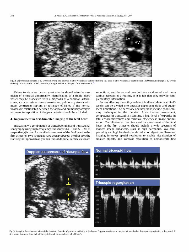
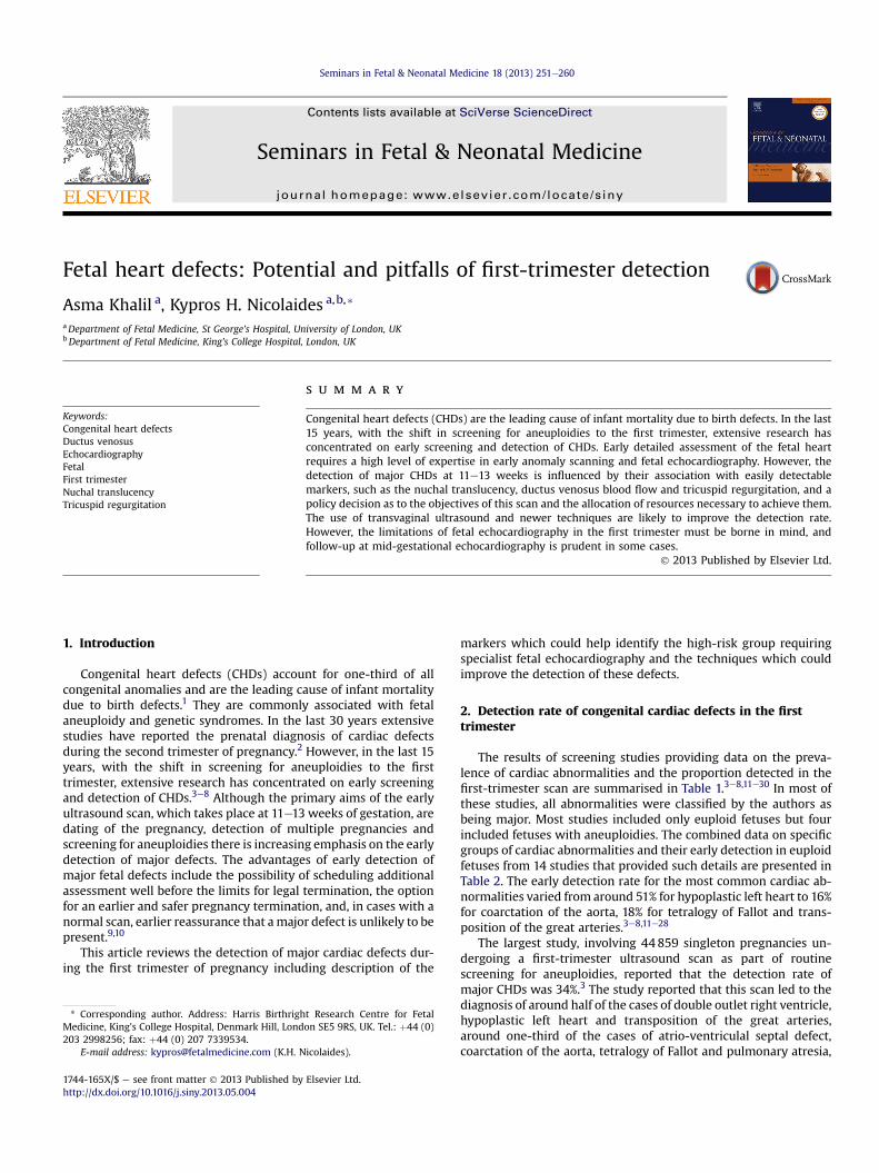
Fig. 2. (a) Ultrasound image at 12 weeks showing the absence of atrio-ventricular valves offsetting in a case of atrio-ventricular septal defect. (b) Ultrasound image at 12 weeksshowing disproportion. LV, left ventricle; RV, right ventricle. Adapted from Persico et al.35
Fi
A. Khalil, K.H. Nicolaides / Seminars in Fetal & Neonatal Medicine 18 (2013) 251e260254
Failure to visualise the two great arteries should raise the sus-picion of a cardiac abnormality. Identification of a single bloodvessel may be associated with a diagnosis of a common arterialtrunk, aortic atresia or severe coarctation, pulmonary atresia withintact ventricular septum or tetralogy of Fallot. If the normal‘crossover’ relationship between the aorta and pulmonary artery isnot seen, transposition of the great arteries should be excluded.
4. Improvement in first-trimester imaging of the fetal heart
Increasingly, a combination of transabdominal and transvaginalsonography using high-frequency transducers (4e8 and 5e9 MHz,respectively) is used for detailed assessment of the fetal heart in thefirst trimester. Two strategies have been proposed; the first uses thetransvaginal approach only when transabdominal cardiac views are
ig. 3. An apical four-chamber view of the heart at 12 weeks of gestation, with the pulsed wavt is found during at least half of the systole and with a velocity of >80 cm/s.
suboptimal, and the second uses both transabdominal and trans-vaginal accesses as a routine, as it is felt that they provide com-plementary information.
Factors affecting the ability to detect fetal heart defects at 11e13weeks can be divided into operator-dependent skills and equip-ment limitations. The necessary operator skills include good scan-ning technique in the detailed first-trimester assessment,competence in transvaginal scanning, a high level of expertise infetal echocardiography, and technical efficiency in image optimi-zation. The ultrasound machine used for assessment of the fetalheart in the first trimester should include a wide spectrum ofmodern image enhancers, such as high harmonics, low com-pounding and high levels of speckle reduction algorithm. Harmonicimaging improves spatial resolution to enable visualization ofsmaller objects, and contrast resolution to demonstrate fine
e Doppler positioned across the tricuspid valve. Tricuspid regurgitation is diagnosed if
0
50
100
150
> M-<95th >95th- 3.5 3.5-4.4 4.5-5.4 >5.5
Nuchal translucency thickness (mm)
Prev
alen
ce o
f maj
or c
ardi
ac d
efec
ts (p
er 1
000) 126.7
64.4
35.2
18.28.7
2,064 2,365 654 202 221
<Median
1,415
4.9
N=
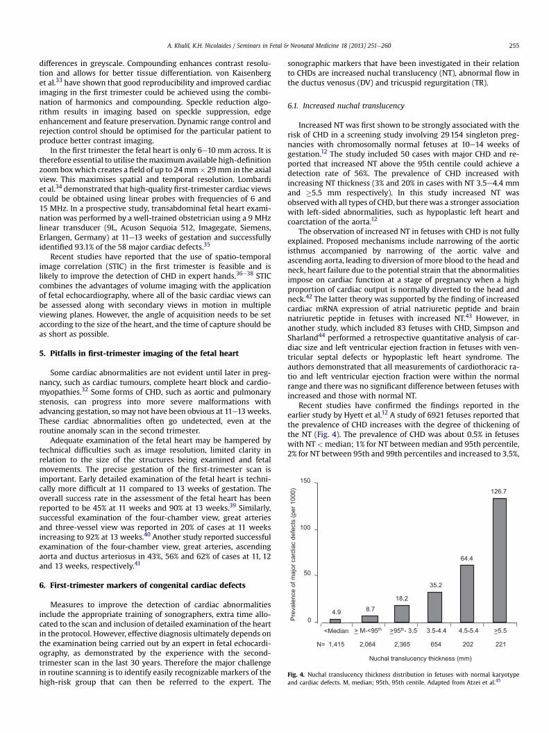
Fig. 4. Nuchal translucency thickness distribution in fetuses with normal karyotypeand cardiac defects. M, median; 95th, 95th centile. Adapted from Atzei et al.45
A. Khalil, K.H. Nicolaides / Seminars in Fetal & Neonatal Medicine 18 (2013) 251e260 255
differences in greyscale. Compounding enhances contrast resolu-tion and allows for better tissue differentiation. von Kaisenberget al.33 have shown that good reproducibility and improved cardiacimaging in the first trimester could be achieved using the combi-nation of harmonics and compounding. Speckle reduction algo-rithm results in imaging based on speckle suppression, edgeenhancement and feature preservation. Dynamic range control andrejection control should be optimised for the particular patient toproduce better contrast imaging.
In the first trimester the fetal heart is only 6e10 mm across. It istherefore essential to utilise themaximumavailable high-definitionzoom boxwhich creates a field of up to 24mm� 29mm in the axialview. This maximises spatial and temporal resolution. Lombardiet al.34 demonstrated that high-quality first-trimester cardiac viewscould be obtained using linear probes with frequencies of 6 and15 MHz. In a prospective study, transabdominal fetal heart exami-nation was performed by a well-trained obstetrician using a 9 MHzlinear transducer (9L, Acuson Sequoia 512, Imagegate, Siemens,Erlangen, Germany) at 11e13 weeks of gestation and successfullyidentified 93.1% of the 58 major cardiac defects.35
Recent studies have reported that the use of spatio-temporalimage correlation (STIC) in the first trimester is feasible and islikely to improve the detection of CHD in expert hands.36e38 STICcombines the advantages of volume imaging with the applicationof fetal echocardiography, where all of the basic cardiac views canbe assessed along with secondary views in motion in multipleviewing planes. However, the angle of acquisition needs to be setaccording to the size of the heart, and the time of capture should beas short as possible.
5. Pitfalls in first-trimester imaging of the fetal heart
Some cardiac abnormalities are not evident until later in preg-nancy, such as cardiac tumours, complete heart block and cardio-myopathies.32 Some forms of CHD, such as aortic and pulmonarystenosis, can progress into more severe malformations withadvancing gestation, so may not have been obvious at 11e13 weeks.These cardiac abnormalities often go undetected, even at theroutine anomaly scan in the second trimester.
Adequate examination of the fetal heart may be hampered bytechnical difficulties such as image resolution, limited clarity inrelation to the size of the structures being examined and fetalmovements. The precise gestation of the first-trimester scan isimportant. Early detailed examination of the fetal heart is techni-cally more difficult at 11 compared to 13 weeks of gestation. Theoverall success rate in the assessment of the fetal heart has beenreported to be 45% at 11 weeks and 90% at 13 weeks.39 Similarly,successful examination of the four-chamber view, great arteriesand three-vessel view was reported in 20% of cases at 11 weeksincreasing to 92% at 13 weeks.40 Another study reported successfulexamination of the four-chamber view, great arteries, ascendingaorta and ductus arteriosus in 43%, 56% and 62% of cases at 11, 12and 13 weeks, respectively.41
6. First-trimester markers of congenital cardiac defects
Measures to improve the detection of cardiac abnormalitiesinclude the appropriate training of sonographers, extra time allo-cated to the scan and inclusion of detailed examination of the heartin the protocol. However, effective diagnosis ultimately depends onthe examination being carried out by an expert in fetal echocardi-ography, as demonstrated by the experience with the second-trimester scan in the last 30 years. Therefore the major challengein routine scanning is to identify easily recognizable markers of thehigh-risk group that can then be referred to the expert. The
sonographic markers that have been investigated in their relationto CHDs are increased nuchal translucency (NT), abnormal flow inthe ductus venosus (DV) and tricuspid regurgitation (TR).
6.1. Increased nuchal translucency
Increased NT was first shown to be strongly associated with therisk of CHD in a screening study involving 29154 singleton preg-nancies with chromosomally normal fetuses at 10e14 weeks ofgestation.12 The study included 50 cases with major CHD and re-ported that increased NT above the 95th centile could achieve adetection rate of 56%. The prevalence of CHD increased withincreasing NT thickness (3% and 20% in cases with NT 3.5e4.4 mmand �5.5 mm respectively). In this study increased NT wasobservedwith all types of CHD, but therewas a stronger associationwith left-sided abnormalities, such as hypoplastic left heart andcoarctation of the aorta.12
The observation of increased NT in fetuses with CHD is not fullyexplained. Proposed mechanisms include narrowing of the aorticisthmus accompanied by narrowing of the aortic valve andascending aorta, leading to diversion of more blood to the head andneck, heart failure due to the potential strain that the abnormalitiesimpose on cardiac function at a stage of pregnancy when a highproportion of cardiac output is normally diverted to the head andneck.42 The latter theory was supported by the finding of increasedcardiac mRNA expression of atrial natriuretic peptide and brainnatriuretic peptide in fetuses with increased NT.43 However, inanother study, which included 83 fetuses with CHD, Simpson andSharland44 performed a retrospective quantitative analysis of car-diac size and left ventricular ejection fraction in fetuses with ven-tricular septal defects or hypoplastic left heart syndrome. Theauthors demonstrated that all measurements of cardiothoracic ra-tio and left ventricular ejection fraction were within the normalrange and there was no significant difference between fetuses withincreased and those with normal NT.
Recent studies have confirmed the findings reported in theearlier study by Hyett et al.12 A study of 6921 fetuses reported thatthe prevalence of CHD increases with the degree of thickening ofthe NT (Fig. 4). The prevalence of CHD was about 0.5% in fetuseswith NT <median; 1% for NT between median and 95th percentile,2% for NT between 95th and 99th percentiles and increased to 3.5%,

A. Khalil, K.H. Nicolaides / Seminars in Fetal & Neonatal Medicine 18 (2013) 251e260256
6.5% and 12.5% for NT of 3.5e4.4 mm, 4.535.4 mm, and �5.5 mm,respectively. The study also reported that there was no obviousdifference in the distribution of NT in the different types of cardiacdefects.45 In the study by Syngelaki et al.3 involving 44 859singleton pregnancies including 85 with major CHDs, the incidenceof increased NT was 35.3% in the cases with CHD. The NT wasincreased in 64.3% of the 28 fetuses with cardiac abnormalitiesdiagnosed at 11e13 weeks compared with 15.4% of the 78 diag-nosed in the second trimester or postnatally. Table 1 shows the dataon NT screening performance in the studies reporting onthe effectiveness of the first-trimester scan in the diagnosis ofmajor CHD.
A pooled analysis of data from major fetal echocardiographycentres concluded that finding NT �3.5 mm may lead to an earlierdiagnosis of all major types of CHDs.46 Earlier studies havedemonstrated a significant association between increased NT andleft heart lesions and septal defects.12,21 However, the multicentrestudy, based on 637 CHDs, did not replicate this finding.46 In ameta-analysis, involving 58492 pregnancies which aimed to assessthe screening performance of NT for major CHD, the detection ratewas poor (31% for a false-positive rate of 1%).47 However, thestudies varied in their definition of what constituted increased NT(Table 1).
The NT cut-off recommended to use for referral for specialistfetal echocardiography varies according to the local set-up andfacilities, in particular the access to specialist fetal echocardiogra-phy service. In the study by Atzei et al.45 we have demonstratedthat the prevalence of major CHD in fetuses with NT above the 99thcentile (>30 per 1000) is substantially higher than in patients witha family history of cardiac defects and diabetes mellitus (about 20per 1000), which are commonly used as indications for fetalechocardiography.
Fetal NT >3.5 mm is found in about 1% of pregnancies. The riskof major chromosomal abnormalities in such fetuses is very highand increases from about 20% for NT of 4.0 mm to 33% for NT of5.0 mm and 60% for NT of �5.5 mm.48 Consequently, our proposedmanagement would be first to offer the parents the option of fetalkaryotyping by chorionic villus sampling. The prevalence of majorfetal defects or fetal death in the chromosomally normal group
Fig. 5. Mid-sagittal view of the fetal trunk demonstrating, with colour flowmapping, the umshowing a positive waveform and reversed a-wave.
increases with NT thickness from about 10% for NTof 4.0mm to 20%for NT of 5.0 mm and 50% for NT of �5.5 mm.18,49 The next stepwould be to carry out a detailed scan, including fetal echocardi-ography at 14e16 weeks of gestation in the fetuses with normalkaryotype with increased NT.45
In fetuses with NT between the 95th and 99th centiles, theprevalence of cardiac defects is about 2%, which is similar to thatfound in patients with a family history of cardiac defects and dia-betes mellitus. The extent to which specialist fetal echocardiogra-phy should be offered to these pregnancies, which constitute about4% of the total population, depends on the availability of suchservices.
6.2. Abnormal ductus venosus blood flow
Abnormal DV flow was initially reported in the second and thirdtrimesters in association with cardiac dysfunction associated withstructural heart defects, post-tachycardia cardiomyopathy and end-stage fetal hypoxia or increased right ventricular afterload.50,51 Inhearts with markedly impaired diastolic function, atrial contractionoccurs against increased impedance to forward flow, resulting in atransient flow reversal in the ductus venosus, which constitutes thenegative a-wave. However, DV flow reversal beyond the firsttrimesterhasbeennotedmainly in situationswhere therewereothermanifestations of cardiac dysfunction, such as fetal hydrops.
In the assessment of ductus venosus a right ventral mid-sagittalview of the fetal trunk is obtained and colour flow mapping is usedto demonstrate the umbilical vein, ductus venosus and fetal heart. Asmall pulsed Doppler sample (0.5e1.0 mm) is used to avoidcontamination from the adjacent veins and it is placed in theyellowish aliasing area which is the portion immediately above theumbilical sinus. The insonation angle should be <30�, the filtershould be set at a low frequency (50e70 Hz) to allow visualization ofthe whole waveform and the sweep speed should be high (2e3 cm/s) so that the waveforms are widely spread. Impedance to flow isassessed by measuring the pulsatility index or by qualitative clas-sification of the a-wave into positive, negative or reversed (Fig. 5).
The first study reporting on the association between abnormalDV flow and CHDs in the first trimester demonstrated reversed or
bilical vein, ductus venosus and fetal heart and ductus venosus flow velocity waveforms
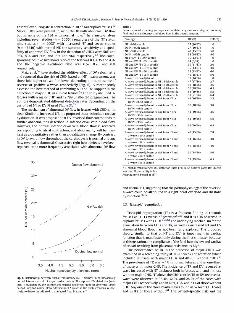
Table 3Performance of screening for major cardiac defects by various strategies combiningfetal nuchal translucency and blood flow in the ductus venosus.
Strategy DR (%) FPR (%)
NT >99th centile 27 (10/37) 1.0DV PI >99th centile 27 (10/37) 1.0NT >95th centile 40 (15/37) 5.0DV PI >95th centile 38 (14/37) 5.0NT or DV PI >99th centile 35 (13/37) 1.9NT and DV PI >99th centile 24 (9/37) 1.0NT and DV PI >98th centile 30 (11/37) 2.0NT and DV PI >97th centile 35 (13/37) 3.0NT and DV PI >96th centile 35 (13/37) 4.0NT and DV PI >95th centile 40 (15/37) 5.0A-wave reversed/absent 39 (14/36) 1.8A-wave reversed/absent or NT >99th centile 47 (17/36) 2.7A-wave reversed/absent or NT >98th centile 50 (18/36) 3.6A-wave reversed/absent or NT >97th centile 50 (18/36) 4.5A-wave reversed/absent or NT >96th centile 53 (19/36) 5.5A-wave reversed/absent or NT >95th centile 58 (21/36) 6.4A-wave reversed/absent or risk from NT or
DV PI >99th centile44 (16/36) 2.8
A-wave reversed/absent or risk from NT orDV PI >98th centile
50 (18/36) 3.8
A-wave reversed/absent or risk from NT orDV PI >97th centile
53 (19/36) 4.6
A-wave reversed/absent or risk from NT orDV PI >96th centile
53 (19/36) 5.5
A-wave reversed/absent or risk from NT orDV PI >95th centile
56 (20/36) 6.5
A-wave reversed/absent or risk from NT anda-wave >99th centile
42 (15/36) 2.8
A-wave reversed/absent or risk from NT anda-wave >98th centile
44 (16/36) 3.8
A-wave reversed/absent or risk from NT anda-wave >97th centile
50 (18/36) 4.9
A. Khalil, K.H. Nicolaides / Seminars in Fetal & Neonatal Medicine 18 (2013) 251e260 257
absent flow during atrial contraction in 10 of 140 euploid fetuses.52
Major CHDs were present in six of the 10 with abnormal DV flowbut in none of the 134 with normal flow.52 In a meta-analysisincluding seven studies (n ¼ 50354) regardless of the NT status,nine studies (n ¼ 2908) with increased NT and seven studies(n ¼ 47610) with normal NT, the summary sensitivity and speci-ficity of abnormal DV flow in the detection of CHDs were 50% and93%, 83% and 80%, and 19% and 96% respectively.53 The corre-sponding positive likelihood ratio of the test was 8.1, 4.35 and 4.97and the negative likelihood ratio was 0.52, 0.20 and 0.8,respectively.
Maiz et al.54 have studied the additive effect of DV velocimetryand reported that the risk of CHD, based on NT measurement, wasthree-fold higher or two-fold lower depending on the presence ofreverse or positive a-wave, respectively (Fig. 6). A recent studyassessed the best method of combining NT and DV Doppler in thedetection of major CHD in euploid fetuses.55 The study included 37fetuses with a major CHD and 12799 unaffected pregnancies. Theauthors demonstrated different detection rates depending on thecut-offs of NT or DV PI used (Table 3).55
The mechanism of abnormal DV flow in fetuses with CHD is notclear. Similar to increased NT, the proposed theories include cardiacdysfunction. It was proposed that DV reversed flow corresponds tosimilar abnormalities described in inferior caval vein blood flow.However, the normal inferior caval vein blood flow is reversed,corresponding to atrial contraction, and abnormality will be man-ifest as a quantitative rather than a qualitative change. By contrast,in DV forward flow throughout the cardiac cycle is normal and anyflow reversal is abnormal. Obstructive right heart defects have beenreported to be more frequently associated with abnormal DV flow
0
5
10
15
20
25
30
35
3.5 4.0 4.5 5.0 5.5 6.0 6.5
Ductus flow normal
Ductus flow abnormal
A priori risk
Ris
k fo
r car
diac
def
ects
(%)
Nuchal translucencty thickness (mm)
Fig. 6. Relationship between nuchal translucency (NT) thickness in chromosomallynormal fetuses and risk of major cardiac defects. The a-priori NT-related risk (solidline) is multiplied by the positive and negative likelihood ratios for abnormal (upperdashed line) and normal (lower dashed line) A-waves in the ductus venosus, respec-tively, to derive the adjusted risk. Adapted from Maiz et al.54
A-wave reversed/absent or risk from NT anda-wave >96th centile
50 (18/36) 5.6
A-wave reversed/absent or risk from NT anda-wave >95th centile
53 (19/36) 6.5
NT, nuchal translucency; DR, detection rate; FPR, false-positive rate; DV, ductusvenosus; PI, pulsatility index.Adapted from Borrell et al.55
and normal NT, suggesting that the pathophysiology of the reverseda-wave could be attributed to a right heart overload and diastolicdysfunction.56e58
6.3. Tricuspid regurgitation
Tricuspid regurgitation (TR) is a frequent finding in trisomicfetuses at 11e13 weeks of gestation59,60 and it is also observed ineuploid fetuses with CHDs.8,35,61 The underlyingmechanism for theassociation between CHD and TR, as well as increased NT and DVabnormal blood flow, has not been fully explored. The proposedtheory, similar to that of NT and DV, is impairment in cardiacfunction that is manifested only during the first trimester because,at this gestation, the compliance of the fetal heart is lowand cardiacafterload resulting from placental resistance is high.
The performance of TR in the detection of major CHDs wasexamined in a screening study at 11e13 weeks of gestation whichincluded 85 cases with major CHDs and 40905 without CHDs.59
The prevalence of TR was w1% in normal fetuses and in one-thirdof those with major CHD. The incidence of TR and DV reversed a-wave increased with NT thickness both in fetuses with and in thosewithout major CHD. NT above the 95th centile, TR or DV reversed a-wave were observed in 35.3%, 32.9%, and 28.2% of the cases withmajor CHD, respectively, and in 4.8%,1.3%, and 2.1% of thosewithoutCHD. Any one of the three markers was found in 57.6% of CHD casesand in 8% of those without.61 The patient-specific risk and the

Delta nuchal translucency (mm)
Ris
k of
maj
or c
ardi
ac d
efec
t (%
)
0 2.0 4.0 6.0-2.0
0
20
40
60
80
100A
B
C
D
Fig. 7. Patient-specific risk for major cardiac defects according to fetal nuchal trans-lucency thickness (interrupted line) and nuchal translucency combined with the re-sults of blood flow in the ductus venosus and across the tricuspid valve (continuouslines) in a fetus with crownerump length of 65 mm. (A) Tricuspid regurgitation andreversed A-wave in the ductus venosus. (B) Tricuspid regurgitation and normal flow inthe ductus venosus. (C) Normal flow across the tricuspid valve and reversed A-wave inthe ductus venosus. (D) Normal flow across the tricuspid valve and in the ductusvenosus. Adapted from Pereira et al.61
A. Khalil, K.H. Nicolaides / Seminars in Fetal & Neonatal Medicine 18 (2013) 251e260258
performance of screening for major CHD using NT, DV Doppler andTR are shown in Fig. 7 and Table 4. The screening performance formajor CHD using NT alonewas improved by the addition of DV flowand further improved by the addition of TR. For fixed false-positiverates of 1%, 3%, and 5%, the detection rates of major CHD by acombination of NT, DV flow, and TR were 36.5%, 48.2%, and 54.1%,respectively.
The association between TR and CHD was also observed inanother prospective study in which transabdominal fetal heart
Table 4Performance of screening for major cardiac defects by fetal nuchal translucency andblood flow across the tricuspid valve and in the ductus venosus.
Screening test Major cardiac defect
Present(n ¼ 85)
Absent(n ¼ 40905)
NT above the 99th centile 18 (21.2%) 290 (0.7%)NT between the 95th and 99th centile 12 (14.1%) 1666 (4.1%)NT above the 95th centile 30 (35.3%) 1956 (4.8%)Reversed a-wave in ductus venosus 24 (28.2%) 856 (2.1%)Reversed DV a-wave or NT above
the 99th centile33 (38.8%) 1118 (2.7%)
Reversed DV a-wave or NT abovethe 95th centile
40 (47.1%) 2732 (6.7%)
TR 28 (32.9%) 516 (1.3%)Either TR or NT above the 99th centile 35 (41.2%) 792 (1.9%)Either TR or NT above the 95th centile 43 (50.6%) 2405 (5.9%)Either TR or reversed DV A-wave (Doppler) 41 (48.2%) 1309 (3.2%)Either Doppler or NT above the 99th centile 44 (51.8%) 1669 (4.1%)Either Doppler or NT above the 95th centile 49 (57.6%) 3265 (8.0%)
NT, nuchal translucency; DV, ductus venosus; TR, tricuspid regurgitation.Adapted from Pereira et al.61
examination was performed by a well-trained obstetrician using ahigh-frequency linear transducer in 886 cases. TR was reported in16 (61.5%) euploid fetuses with CHD and in 62 (9.2%) of 670 ineuploid fetuses with normal heart.35 Similar results have been re-ported in first-trimester study in 4445 pregnancies.8 There was ahigher prevalence of TR in fetuses with CHD compared with normalfetuses (33% vs 1.7%). In the same study the corresponding valuesfor NTabove the 95th centile and abnormal DV flowwere 38%, 5.6%,22% and 3.1%, respectively.
Two approaches have been proposed for the use of the algo-rithm combining NT, DV Doppler and TR to estimate the patient-specific risk for major CHD.61 The first one is to define the riskcut-off that selects the patients requiring referral for specialistfetal echocardiography. The risk increases exponentially with NTthickness from 1 per 1000 in those with NT at or below the 95thcentile to 7 per 1000 for NT between the 95th and 99th centileand 58 per 1000 for NT above the 99th centile. The risk is furtherincreased if there is DV reversed a-wave, TR, or both and isdecreased if flow in the DV and across the tricuspid valve isnormal. The second approach is to define as high risk all caseswith TR, DV reversed a-wave, or both, which constitute w3% of thepopulation and contain 48% of those with major cardiac defects. Ifcases with nuchal translucency above the 99th centile are alsoincluded, the screen-positive rate would increase to w4% and theestimated detection rate would be 52%. If there are available re-sources for performing fetal echocardiography in 8% of the pop-ulation, then the NT cut-off for defining the high-risk group couldbe reduced to the 95th percentile with an increase in the esti-mated detection rate to 58%.
6.4. Maternal serum markers and congenital cardiac defects
A caseecontrol study of 68 cases of isolated fetal CHDs and 340normal controls at 11e13 weeks of gestation reported lowermaternal serum placental growth factor (PLGF) levels in CHD (0.80vs 1.00 multiple of median).62 This decrease in PLGF was observedin conotruncal and valve defects but not in left heart defects. Thedecrease in serum PLGF was not related to impaired placentalperfusion.62
A caseecontrol study of 306 cases of fetal CHDs and 1224 no-CHD controls reported abnormal second-trimester serum a-feto-protein (AFP), human chorionic gonadotrophin (hCG) and uncon-jugated estriol (uE3) in the CHD group.63 Cases with critical CHDswere more than twice as likely to have AFP multiple of the median(MoM) �95th centile and/or an hCG and/or uE3 MoM �5thcentile.63
The value of first- and second-trimester maternal serumbiochemical markers in screening for fetal CHDs remains to bedetermined.
7. Conclusion
First-trimester detection of CHD is feasible, but early detailedassessment of the fetal heart requires a high level of expertise inearly anomaly scanning and fetal echocardiography. However,the detection of major CHDs at 11e13 weeks is influenced bytheir association with easily detectable markers and a policydecision as to the objectives of this scan and the allocation ofresources necessary to achieve them. The use of transvaginalultrasound and newer techniques are likely to improve thedetection rate. However, the limitations of fetal echocardiogra-phy in the first trimester must be borne in mind, and resort tofollow-up mid-gestational echocardiography should always beconsidered.

Practice points
� The detection rate of CHD at the first trimester is low
and varies according to the experience of the centre and
the population studied.
� The detection rate varies according to the type of the
cardiac abnormality, e.g. from around 51% for hypo-
plastic left heart to 18% for tetralogy of Fallot and
transposition of the great arteries.
� The detection of major CHDs at 11e13 weeks could be
improved if we use easily detectable markers for
screening for CHD, e.g. nuchal translucency.
� The detection rate could be improved if the ultrasound
assessment at the first trimester follows structured
protocols.
� The detection rate of CHD could be improved by the use
of transvaginal ultrasound and newer techniques.
� The limitations of fetal echocardiography in the first
trimester must be borne in mind, and resort to follow-
up mid-gestational echocardiography should always
be considered.
Research directions
� The development of algorithms for the screening for
CHD in the first trimester, using a combination of
maternal and pregnancy characteristics, nuchal trans-
lucency, ductus venosus Doppler and tricuspid
regurgitation.
� Prospective assessment of the routine implementation
of tools, such as transvaginal ultrasound and STIC, for
improving the detection rate of CHD.
A. Khalil, K.H. Nicolaides / Seminars in Fetal & Neonatal Medicine 18 (2013) 251e260 259
Conflict of interest statement
None.
Funding sources
The study was supported by a grant from The Fetal MedicineFoundation (UK Charity No: 1037116).
References
1. Mensah GA, Brown DW. An overview of cardiovascular disease burden in theUnited States. Health Affairs 2007;26:38e48.
2. Garne E, Dolk H, Loane M, Boyd PA. EUROCAT website data on prenataldetection rates of congenital anomalies. J Med Screen 2010;17:97e8.
*3. Syngelaki A, Chelemen T, Dagklis T, Allan L, Nicolaides KH. Challenges in thediagnosis of fetal non-chromosomal abnormalities at 11e13 weeks. PrenatDiagn 2011;31:90e102.
4. D’Ottavio G, Mandruzzato G, Meir YJ, Rustico MA, Fischer-Tamaro L,Conoscenti G. Comparison of first trimester and second trimester screeningfor fetal anomalies. Ann NY Acad Sci 1998;847:200e9.
5. Taipale P, Ammala M, Salonen R, Hilesmaa V. Two-stage ultrasonography inscreening for fetal anomalies at 13e14 and 18e22 weeks of gestation. ActaObstet Gynecol Scand 2004;83:1141e6.
6. Chen M, Lam YH, Lee CP, Tang MH. Ultrasound screening of fetal structuralabnormalities at 12 to 14 weeks in Hong Kong. Prenat Diagn 2004;24:92e7.
7. Cedergren M, Selbing A. Detection of fetal structural abnormalities by an 11e14-week ultrasound dating scan in an unselected Swedish population. ActaObstet Gynecol Scand 2006;85:912e5.
8. Volpe P, Ubaldo P, Volpe N, et al. Fetal cardiac evaluation at 11e14 weeks byexperienced obstetricians in a low-risk population. Prenat Diagn 2011;31:1054e61.
9. Kornman LH, Wortelboer MJM, Beekhuis JR, Morssink LP, Mantingh A.Women’s opinions and the implications of first-versus second-trimesterscreening for fetal Down’s syndrome. Prenat Diagn 1997;17:1011e8.
10. Department of Health. Abortion Statistics, England andWales: 2008. StatisticalBulletin 2009/1. London: DoH; 2009.
11. Hernádi L, Töröcsik M. Screening for fetal anomalies in the 12th week ofpregnancy by transvaginal sonography in an unselected population. PrenatDiagn 1997;17:753e9.
*12. Hyett J, Perdu M, Sharland G, Snijders R, Nicolaides KH. Using fetal nuchaltranslucency to screen for major congenital cardiac defects at 10e14 weeks ofgestation: population based cohort study. BMJ 1999;318:81e5.
13. Bilardo CM, Pajkrt E, de Graaf I, Mol BW, Bleker OP. Outcome of fetuses withenlarged nuchal translucency and normal karyotype. Ultrasound ObstetGynecol 1998;11:401e6.
14. Hafner E, Schuchter K, Liebhart E, Philipp K. Results of routine fetal nuchaltranslucency measurement at weeks 10e13 in 4233 unselected pregnantwomen. Prenat Diagn 1998;18:29e34.
15. Chen M, Lee CP, Lam YHH, et al. Comparison of nuchal and detailedmorphology ultrasound examinations in early pregnancy for fetal structuralabnormality screening: a randomized controlled trial. Ultrasound ObstetGynecol 2008;31:136e46.
16. Dane B, Dane C, Sivri D, Kiray M, Cetin A, Yayla M. Ultrasound screening forfetal major abnormalities at 11e14 weeks. Acta Obstet Gynecol Scand 2007;86:666e70.
17. Mavrides E, Cobian-Sanchez F, Tekay A, et al. Limitations of using firsttrimester nuchal translucency measurement in routine screening for majorcongenital heart defects. Ultrasound Obstet Gynecol 2001;17:106e10.
18. Michailidis GD, Economides DL. Nuchal translucency measurement andpregnancy outcome in karyotypically normal fetuses. Ultrasound ObstetGynecol 2001;17:102e5.
19. Schwarzler P, Carvalho JS, Senat MV, Masroor T, Campbell S, Ville Y.Screening for fetal aneuploidies and fetal cardiac abnormalities by nuchaltranslucency thickness measurement at 10e14 weeks of gestation as part ofroutine antenatal care in an unselected population. Br J Obstet Gynaecol1999;106:1029e34.
20. Orvos H, Wyadaka K, Kozinszky YZ, Katona M, Pal A, Szabo J. Increased nuchaltranslucency and congenital heart defects in euploid fetuses. The Szegedexperience. Eur J Obstet Gynecol Reprod Biol 2002;101:124e8.
21. Bahado-Singh RO, Wapner R, Thom E, et al. Elevated first-trimester nuchaltranslucency increases the risk of congenital heart defects. Am J Obstet Gynecol2005;192:1357e61.
22. Bruns RF, Moron AF, Murta CGV, Goncalves LFA, Zamith MM. The role ofnuchal translucency in the screening of congenital heart defects. Arq BrasCardiol 2006;87:272e9.
23. Becker R, Wagner RD. Detailed screening for fetal anomalies and cardiac de-fects at the 11e13 weeks scan. Ultrasound Obstet Gynecol 2006;27:613e8.
24. Westin M, Saltvedt S, Almstro H, Grunewald C, Valentin L. By how much doesincreased nuchal translucency increase the risk of adverse pregnancy outcomein chromosomally normal fetuses? A study of 16 260 fetuses derived from anunselected pregnant population. Ultrasound Obstet Gynecol 2007;29:150e8.
25. Muller MA, Clur SA, Timmerman E, Bilardo CM. Nuchal translucency mea-surement and congenital heart defects: models association in low-risk preg-nancies. Prenat Diagn 2007;27:164e9.
26. Oztekin O, Oztekin D, Týnar S, Adýbelli Z. Ultrasonographic diagnosis of fetalstructural abnormalities in prenatal screening at 11e14 weeks. Diagn IntervRadiol 2009;15:221e5.
27. Hildebrand E, Selbing A, Blomberg M. Comparison of first and secondtrimester ultrasound screening for fetal anomalies in the southeast region ofSweden. Acta Obstet Gynecol Scand 2010;89:1412e9.
28. Grande M, Arigita M, Borobio V, Jimenez JM, Fernandez S, Borrell A. First-trimester detection of structural abnormalities and the role of aneuploidymarkers. Ultrasound Obstet Gynecol 2012;39:157e63.
29. Carvalho JS, Mavrides E, Shinebourne AE, Campbell S, Thilaganathan B.Improving the effectiveness of routine prenatal screening for major congenitalheart defects. Heart 2002;88:387e91.
30. Westin M, Saltvedt S, Bergman G, Almstro H, Grunewald C, Valentin L. Ismeasurement of nuchal translucency thickness a useful screening tool forheart defects? A study of 16,383 fetuses. Ultrasound Obstet Gynecol 2006;27:632e9.
31. Borrell A, Robinson JN, Santolaya-Forgas J. Clinical value of the 11- to 13 þ 6-week sonogram for detection of congenital malformations: a review. Am JPerinatol 2011;28:117e24.
32. Allan L, Cook A, Huggon I. First trimester fetal heart scanning. In: Allan L,Cook A, Huggon I, editors. Fetal echocardiography e a practical guide. Cam-bridge: Cambridge University Press; 2009. p. 190e202.
33. von Kaisenberg CS, Kuhling-von Kaisenberg H, Fritzer E, Schemm S, Meinhold-Heerlein I, Jonat W. Fetal transabdominal anatomy scanning using standardviews at 11 to 14 weeks’ gestation. Am J Obstet Gynecol 2005;192:535e42.
34. Lombardi CM, Bellotti M, Fesslova V, Cappellini A. Fetal echocardiography atthe time of the nuchal translucency scan. Ultrasound Obstet Gynecol 2007;29:249e57.
35. Persico N, Moratalla J, Lombardi CM, Zidere V, Allan L, Nicolaides KH. Fetalechocardiography at 11e13 weeks by transabdominal high-frequency ultra-sound. Ultrasound Obstet Gynecol 2011;37:296e301.
36. Bennasar M, Martínez JM, Olivella A, et al. Feasibility and accuracy of fetalechocardiography using four-dimensional spatiotemporal image correlationtechnology before 16 weeks’ gestation. Ultrasound Obstet Gynecol 2009;33:645e51.

A. Khalil, K.H. Nicolaides / Seminars in Fetal & Neonatal Medicine 18 (2013) 251e260260
37. Vinals F, Ascenzo R, Naveas R, Huggon I, Giuliano A. Fetal echocardiography at11þ0 to 13 þ 6 weeks using four-dimensional spatiotemporal image correla-tion telemedicine via an Internet link: a pilot study. Ultrasound Obstet Gynecol2008;31:633e8.
38. Turan S, Turan OM, TY-Torredes K, Harman CR, Baschat AA. Standardization ofthe first-trimester fetal cardiac examination using spatiotemporal imagecorrelation with tomographic ultrasound and color Doppler imaging. Ultra-sound Obstet Gynecol 2009;33:652e6.
39. Smrcek JM, Berg C, Geipel A, et al. Detection rate of early fetal echocardiog-raphy and in utero development of congenital heart defects. J Ultrasound Med2006;25:187e96.
40. Haak MC, Twisk JWR, Van Vught MG. How successful is fetal echocardiogra-phy examination in the first trimester of pregnancy? Ultrasound ObstetGynecol 2002;20:9e13.
41. Vimpelli T, Huhtala H, Acharya G. Fetal echocardiography during routine first-trimester screening: a feasibility study in an unselected population. PrenatDiagn 2006;26:475e82.
42. Hyett J, Moscoso G, Papapanagiotou G, Perdu M, Nicolaides KH. Abnormalitiesof the heart and great arteries in chromosomally normal fetuses withincreased nuchal translucency thickness at 11e13 weeks of gestation. Ultra-sound Obstet Gynecol 1996;7:245e50.
43. Hyett JA, Brizot ML, Von Kaisenberg CS, McKie AT, Farzaneh F, Nicolaides KH.Cardiac gene expression of atrial natriuretic peptide and brain natriureticpeptide in trisomic fetuses. Obstet Gynecol 1996;87:506e610.
44. Simpson JM, Sharland GK. Nuchal translucency and congenital heart defects:heart failure or not? Ultrasound Obstet Gynecol 2000;16:30e6.
45. Atzei A, Gajewska K, Huggon IC, Allan L, Nicolaides KH. Relationship betweennuchal translucency thickness and prevalence of major cardiac defects in fe-tuses with normal karyotype. Ultrasound Obstet Gynecol 2005;26:154e7.
*46. Makrydimas G, Sotiriadis A, Hugon IC, et al. Nuchal translucency and fetalcardiac defects: a pooled analysis of major fetal echocardiography centers. AmJ Obstet Gynecol 2005;192:89e95.
*47. Makrydimas G, Sotiriadis A, Ioannidis JP. Screening performance of first-trimester nuchal translucency for major cardiac defects: a meta-analysis. Am JObstet Gynecol 2003;189:1330e5.
48. Snijders RJ, Noble P, Sebire NJ, Souka AP, Nicolaides KH. UK multicentre projecton assessment of risk of trisomy 21 by maternal age and fetal nuchal-translucency thickness at 10e14 weeks of gestation. Fetal Medicine Founda-tion First Trimester Screening Group. Lancet 1998;352:343e6.
49. Souka AP, Krampl E, Bakalis S, Heath V, Nicolaides KH. Outcome of pregnancyin chromosomally normal fetuses with increased nuchal translucency in thefirst trimester. Ultrasound Obstet Gynecol 2001;18:9e17.
50. Kiserud T, Eik-Nes SH, Hellevik LR, Blaas H-G. Ductus venosus blood velocitychanges in fetal cardiac diseases. J Matern Fetal Invest 1993;3:15e20.
51. Kiserud T, Eik-Nes SH, Blaas H-G, Hellevik LR, Simensen B. Ductus venosusblood velocity and the umbilical circulation in the seriously growth-retardedfetus. Ultrasound Obstet Gynecol 1994;4:109e14.
52. Matias A, Huggon I, Areias JC, Montenegro N, Nicolaides KH. Cardiac defects inchromosomally normal fetuses with abnormal ductus venosus blood flow at10e14 weeks. Ultrasound Obstet Gynecol 1999;14:307e10.
*53. Papatheodorou SI, Evangelou E, Makrydimas G, Ioannidis JP. First-trimesterductus venosus screening for cardiac defects: a meta-analysis. Br J ObstetGynaecol 2011;118:1438e45.
*54. Maiz N, Plasencia W, Dagklis T, Faros E, Nicolaides K. Ductus venosus Dopplerin fetuses with cardiac defects and increased nuchal translucency thickness.Ultrasound Obstet Gynecol 2008;31:256e60.
*55. Borrell A, Grande M, Bennasar M, et al. First trimester detection of cardiacdefects with the use of ductus venosus blood flow. Ultrasound Obstet Gynecol2012 Nov. http://dx.doi.org/10.1002/uog.12349. [Epub ahead of print].
56. Martínez JM, Comas M, Borrell A, et al. Abnormal first-trimester ductusvenosus blood flow: a marker of cardiac defects in fetuses with normal kar-yotype and nuchal translucency. Ultrasound Obstet Gynecol 2010;35:267e72.
57. Berg C, Kremer C, Geipel A, Kohl T, Germer U, Gembruch U. Ductus venosusblood flow alterations in fetuses with obstructive lesions of the right heart.Ultrasound Obstet Gynecol 2006;28:137e42.
58. Gardiner HM, Belmar C, Tulzer G, et al. Morphologic and functional predictorsof eventual circulation in the fetus with pulmonary atresia or critical pul-monary stenosis with intact septum. J Am Coll Cardiol 2008;51:1299e308.
59. Huggon IC, DeFigueiredo DB, Allan LD. Tricuspid regurgitation in the diagnosisof chromosomal anomalies in the fetus at 11e14 weeks of gestation. Heart2003;89:1071e3.
60. Kagan KO, Valencia C, Livanos P, Wright D, Nicolaides KH. Tricuspid regurgi-tation in screening for trisomies 21, 18 and 13 and Turner syndrome at 11þ0e13þ6 weeks of gestataion. Ultrasound Obstet Gynecol 2009;33:18e22.
*61. Pereira S, Ganapathy R, Syngelaki A, Maiz N, Nicolaides KH. Contribution offetal tricuspid regurgitation in first-trimester screening for major cardiac de-fects. Obstet Gynecol 2011;117:1384e91.
62. Llurba E, Syngelaki A, Sánchez O, Carreras E, Cabero L, Nicolaides K. Maternalserum placental growth factor at 11e13 weeks’ gestation and fetal cardiacdefects. Ultrasound Obstet Gynecol 2012. http://dx.doi.org/10.1002/uog.12346.[Epub ahead of print].
63. Jelliffe-Pawlowski L, Baer R, Moon-Grady AJ, Currier RJ. Second trimesterserum predictors of congenital heart defects in pregnancies without chro-mosomal or neural tube defects. Prenat Diagn 2011;31:466e72.