selective inhibition of jak1 and jak2 is efficacious in rodent
TRANSCRIPT

of January 31, 2018.This information is current as
Preclinical Characterization of INCB028050Efficacious in Rodent Models of Arthritis: Selective Inhibition of JAK1 and JAK2 Is
Friedman and Kris VaddiGreg Hollis, Robert C. Newton, Brian Metcalf, Steven M. Stacey Shepard, James D. Rodgers, Swamy Yeleswaram,Xiaoming Wen, Jack Shi, Ryan McGee, Patrick J. Haley, Covington, Beth Thomas, Paul Collier, Margaret F. Favata,Timothy C. Burn, Yanlong Li, Jun Li, Maryanne B. Jordan S. Fridman, Peggy A. Scherle, Robert Collins,
ol.0902819http://www.jimmunol.org/content/early/2010/04/02/jimmun
published online 2 April 2010J Immunol
MaterialSupplementary
9.DC1http://www.jimmunol.org/content/suppl/2010/04/02/jimmunol.090281
average*
4 weeks from acceptance to publicationSpeedy Publication! •
Every submission reviewed by practicing scientistsNo Triage! •
from submission to initial decisionRapid Reviews! 30 days* •
?The JIWhy
Subscriptionhttp://jimmunol.org/subscription
is online at: The Journal of ImmunologyInformation about subscribing to
Permissionshttp://www.aai.org/About/Publications/JI/copyright.htmlSubmit copyright permission requests at:
Email Alertshttp://jimmunol.org/alertsReceive free email-alerts when new articles cite this article. Sign up at:
Print ISSN: 0022-1767 Online ISSN: 1550-6606. All rights reserved.1451 Rockville Pike, Suite 650, Rockville, MD 20852The American Association of Immunologists, Inc.,
is published twice each month byThe Journal of Immunology
by guest on January 31, 2018http://w
ww
.jimm
unol.org/D
ownloaded from
by guest on January 31, 2018
http://ww
w.jim
munol.org/
Dow
nloaded from

The Journal of Immunology
Selective Inhibition of JAK1 and JAK2 Is Efficacious inRodent Models of Arthritis: Preclinical Characterizationof INCB028050
Jordan S. Fridman,1 Peggy A. Scherle,1 Robert Collins,1 Timothy C. Burn,
Yanlong Li, Jun Li, Maryanne B. Covington, Beth Thomas, Paul Collier,
Margaret F. Favata, Xiaoming Wen, Jack Shi, Ryan McGee, Patrick J. Haley,
Stacey Shepard, James D. Rodgers, Swamy Yeleswaram, Greg Hollis, Robert C. Newton,
Brian Metcalf, Steven M. Friedman, and Kris Vaddi
Inhibiting signal transduction induced by inflammatory cytokines offers a new approach for the treatment of autoimmune diseases
such as rheumatoid arthritis. Kinase inhibitors have shown promising oral disease-modifying antirheumatic drug potential with
efficacy similar to anti-TNF biologics. Direct and indirect inhibition of the JAKs, with small molecule inhibitors like CP-690,550
and INCB018424 orneutralizingAbs, such as the anti-IL6 receptorAb tocilizumab, have demonstrated rapid and sustained improve-
ment in clinical measures of disease, consistent with their respective preclinical experiments. Therefore, it is of interest to identify
optimized JAK inhibitors with unique profiles to maximize therapeutic opportunities. INCB028050 is a selective orally bioavailable
JAK1/JAK2 inhibitor with nanomolar potency against JAK1 (5.9 nM) and JAK2 (5.7 nM). INCB028050 inhibits intracellular sig-
naling ofmultiple proinflammatory cytokines including IL-6 and IL-23 at concentrations,50 nM. Significant efficacy, as assessed by
improvements in clinical, histologic and radiographic signs of disease, was achieved in the rat adjuvant arthritis model with doses of
INCB028050 providing partial and/or periodic inhibition of JAK1/JAK2 and no inhibition of JAK3. Diminution of inflammatory
Th1 and Th17 associated cytokine mRNA levels was observed in the draining lymph nodes of treated rats. INCB028050 was also
effective in multiple murine models of arthritis, with no evidence of suppression of humoral immunity or adverse hematologic
effects. These data suggest that fractional inhibition of JAK1 and JAK2 is sufficient for significant activity in autoimmune disease
models. Clinical evaluation of INCB028050 in RA is ongoing. The Journal of Immunology, 2010, 184: 000–000.
The pathogenic role of inflammatory cytokines in rheu-matoid arthritis (RA) is well established (1). Multiplestrategies have been pursued to affect their function using
molecularly targeted biotherapeutics, such as those neutralizingTNF-a. More recently, Ab-mediated antagonism of IL-6 withtocilizumab has proven effective and bolsters the hypothesis thatmitigating the presence and/or activity of cytokines can be effica-cious (2). Preliminary clinical data with Abs targeted against IL-15or IL-17 in RA are consistent with this concept (3, 4). Nonetheless,because of the complex etiology of RA, the potential redundancy ofILs, the inconvenience and risk of the dosing route, and limitingtoxicities of current therapeutics, an unmet medical need clearlypersists.JAKs associate with various cytokine receptors and translate
signals triggered by cytokine binding into intracellular responses
(5). Cytokines that signal through JAKs include those associatedwith both adaptive and innate immune responses as well as in-flammation (6). Multiple inflammatory cytokines signal throughreceptors that recruit two of the four JAK family members (JAK1,JAK2, JAK3, and Tyk2) into a signaling complex, and it has beenproposed that inhibiting either or both JAKs in the signalingcomplex would effectively inhibit cytokine signaling (7). Theo-retically, it is not necessary to inhibit all four JAK kinases to affectsignaling from those cytokines associated with the pathogenesis ofRA. In fact, doing somay be detrimental because of the fundamentalroles that JAKs play in the immune system. JAK3 is expressedprimarily in lymphocytes where it associates with essentially onecytokine receptor subunit, the common g-subunit (gc), and conveyssignals from six known cytokines, including IL-2 and IL-7 (8).Interestingly, mutations in gc, JAK3, or the IL-7 receptor (IL-7R)account for the majority of cases of SCID in humans. For thetreatment of chronic inflammatory diseases, in which immunesuppression is undesirable, we hypothesized that an attractive al-ternative to pan-JAK inhibition would be selective inhibition ofJAK1 and JAK2. Conceptually, this strategy can antagonize theeffects of inflammatory cytokines such as IL-6 while minimizingimmune suppression.The realization of therapeutic benefit from JAK inhibition in RA
has now been demonstrated with the use of CP-690,550, a pan-JAKinhibitor originally believed to be an immune suppressive JAK3 in-hibitor for use in organ transplant recipients (9). However, it is nowclear that in addition to its effects on JAK3, this compound inhibitsJAK1 and JAK2 at similar concentrations (10, 11). Moreover, toci-lizumab has exhibited impressive clinical activity by antagonizing
Drug Discovery, Incyte Corporation, Wilmington, DE 19880
1J.S.F., P.A.S., and R.C. contributed equally to this work.
Received for publication August 26, 2009. Accepted for publication February 28,2010.
Address correspondence and reprint requests to Dr. Jordan S. Fridman, Incyte Cor-poration, Experimental Station, E400, Route 141 and Henry Clay Road, Wilmington,DE 19880. E-mail address: [email protected]
The online version of this article contains supplemental material.
Abbreviations used in this paper: CAIA, collagen Ab-induced arthritis; CIA, collagen-induced arthritis; Ct, cycle threshold; DMARD, disease-modifying antirheumatic drug;DTH, delayed-type hypersensitivity; micro-CT, microcomputed tomography; RA,rheumatoid arthritis; rAIA, rat adjuvant-induced arthritis; WBA, whole blood assay.
Copyright� 2010 by The American Association of Immunologists, Inc. 0022-1767/10/$16.00
www.jimmunol.org/cgi/doi/10.4049/jimmunol.0902819
Published April 2, 2010, doi:10.4049/jimmunol.0902819 by guest on January 31, 2018
http://ww
w.jim
munol.org/
Dow
nloaded from
IL-6 signaling, a JAK1/2-dependent process (2, 12). Therefore, weproposed to test the therapeutic hypothesis that selective inhibition ofJAK1/2 could be efficacious using a number of cellular and animalmodels relevant to RA and other autoimmune disorders. INCB0-28050, a potent and selective inhibitor of JAK1/2, is shown to inhibitsignaling from pathogenic cytokines such as IL-6 and IL-23. In-hibition of JAK signaling prevents the production of chemokines andcytokines associated with uncontrolled RA (13). Finally, in threecomplementary rodentmodels of arthritis, we show that selective andfractional inhibition of JAK1/2 is both sufficient for efficacy andwell tolerated.
Materials and MethodsBiochemical assays
Enzyme assays were performed using a homogeneous time-resolvedfluorescence assay with recombinant epitope tagged kinase domains (JAK1,837-1142; JAK2, 828-1132; JAK3, 718-1124; Tyk2, 873-1187) or full-length enzyme (cMET and Chk2) and peptide substrate. Each enzymereaction was performed with or without test compound (11-point dilution),JAK, cMET, or Chk2 enzyme, 500 nM (100 nM for Chk2) peptide, ATP (atthe Km specific for each kinase or 1 mM), and 2.0% DMSO in assay buffer.The calculated IC50 value is the compound concentration required forinhibition of 50% of the fluorescent signal. Additional kinase assays wereperformed at Cerep (Redmond, WA) using standard conditions at 200 nM.Enzymes tested included: Abl, Akt1, AurA, AurB, CDC2, CDK2, CDK4,CHK2, c-kit, EGFR, EphB4, ERK1, ERK2, FLT-1, HER2, IGF1R, IKKa,IKKb, JNK1, Lck, MEK1, p38a, p70S6K, PKA, PKCa, Src, and ZAP70.
Cellular assays
Human PBMCs were isolated by leukapheresis followed by Ficoll-Hypaquecentrifugation. For the determination of IL-6–induced MCP-1 production,PBMCs were plated at 3.33 105 cells per well in RPMI 1640 + 10% FCS inthe presence or absence of various concentrations of INCB028050. Fol-lowing preincubation with compound for 10 min at room temperature, cellswere stimulated by adding 10 ng/ml human recombinant IL-6 to each well.Cells were incubated for 48 h at 37˚C, 5%CO2. Supernatants were harvestedand analyzed by ELISA for levels of human MCP-1. The ability ofINCB028050 to inhibit IL-6–induced secretion of MCP-1 is reported as theconcentration required for 50% inhibition (IC50). Proliferation of Ba/F3-TEL-JAK3 cells (a gift fromRoss Levine,Memorial Sloan-KetteringCancerCenter, New York, NY) was performed over 3 d using Cell-Titer Glo(Promega, Madison, WI) following standard assay conditions.
For the determination of IL-23–induced IL-17 and IL-22, PBMCs weremaintained in RPMI 1640 medium supplemented with 10% FBS, 2 mML-glutamine, 100 mg/ml streptomycin, and 100 U/ml penicillin. T cells wereactivated by culturing with anti-CD3 and anti-CD28 Abs. After 2 d, the cellswere washed and recultured with IL-23 (100 ng/ml), IL-2 (10 ng/ml) andvarious concentrations of INCB028050. Cells were incubated for an addi-tional 4 d at 37˚C, then supernatants were collected, and secretion of IL-17and IL-22 were measured by ELISA. The ability of INCB028050 to inhibitIL-23–induced secretion of IL-17 and IL-22 is reported as the concentrationrequired for 50% inhibition (IC50).
Phospho-STAT3 analysis
Isolated cells. For analysis of phospho-STAT3 in human PBMCs or PHA-stimulated T cells, cell extracts were prepared after 10215 min preincubationwith different concentrations of INCB028050 and stimulation of cells for15 min with IL-6 (100 ng/ml), IL-12 (20 ng/ml), or IL-23 (100 ng/ml). Theextracts were then analyzed for phosphorylated STAT3 by using a phospho-STAT3 specific ELISA.
Whole blood. Blood drawn from rats was collected into heparinized tubesand then aliquoted into microfuge tubes (0.3 ml per sample). In stimu-lation experiments, INCB028050 at various concentrations was added for10 min prior to stimulation with human IL-6 (100 ng/ml) for 15 min at 37˚C. RBCs were lysed using hypotonic conditions. WBCs were thenquickly pelleted and lysed to make total cellular extracts. The extractswere analyzed for phosphorylated STAT3 by using a phospho-STAT3–specific ELISA. Blood from animals that were dosed with INCB028050was drawn at various times after INCB028050 administration and pro-cessed as described above.
In vivo experiments
Animals were housed in a barrier facility accredited by the Association forAssessment and Accreditation of Laboratory Animal Care International. All
of the procedures were conducted in accordancewith the U.S. Public HealthService Policy on Humane Care and Use of Laboratory Animals and withIncyte Animal Care and Use Committee guidelines. Animals were fedstandard rodent chow and provided with water ad libitum.
Pharmacokinetics. Female rats (n = 6 per gender per group) were givena dose of 10 mg/kg INCB028050 suspended in 0.5% methylcellulose andgiven by oral gavage at 10 ml/kg. The first three rats were bled at 0 (pre-dose), 2, 8, and 24 h, and the second three rats were bled 1, 4, and 12 hafter dosing. EDTA was used as the anticoagulant, and samples werecentrifuged to obtain plasma. An analytical method for the quantificationof INCB028050 has been developed and used to analyze samples fromtoxicology studies. The method combines a protein precipitation extractionwith 10% methanol in acetonitrile and LC/MS/MS analysis. The methodhas demonstrated a linear assay range 1–5000 nM using 0.1 ml of studysamples. Data were processed using Analyst 1.3.1 (Applied Biosystems,Foster City, CA). A standard curve was determined from peak area ratioversus concentration using a weighted linear regression (1/x2).
Rat adjuvant-induced arthritis. Adjuvant-induced arthritis was elicited inrats according to established methods. Lewis rats (150–200 g, female;Charles River Laboratories, Wilmington, MA) are injected at the base ofthe tail with 100 ml of an emulsion of CFA (10 mg/ml Mycobacteriumbutyricum in incomplete Freund’s adjuvant). Rats exhibited signs ofinflammation within 2 wk of the injection of CFA. Each rat paw wasscored following visual observation using a rating of 0–3, (0 = normal;1 = redness and minimal swelling of digits; 2 = moderate swelling of thedigits and/or paw; 3 = severe swelling of digits and/or paw). Individualanimal paw scores are combined and recorded as a sum of all four pawsand groups means of these totals are reported. Percent inhibition inclinical score/severity is calculated using the following formula:
ðVehicle2TreatedÞ=Vehicle3 100:
In addition, a plethysmometer (Stoelting, Wood Dale, IL) was used tomeasure paw volumes taken at baseline and study termination. At thetermination of the experiment, paws were removed from euthanized ratsfor histologic analyses. Treatment was initiated when significant signs ofdisease were noted, and groups of animals were sorted so that mean scoreswould be equivalent—usually occurring 2 wk after adjuvant injection.Graphs reflect endpoints collected only immediately prior to and aftertherapy was initiated (treatment day 0). Groups consisted of six animals,and statistical differences between treatment and vehicle controls wereassessed using two-tailed Student t tests or ANOVA with a Dunnett’s testwhen appropriate.
Collagen-induced arthritis. DBA/1j mice (4–5-wk old males) were pur-chased from The Jackson Laboratory (Bar Harbor, ME). The model wasestablished as described with minor modifications (14). Mice are im-munized intradermally with 100 ml bovine type II collagen solution(Chondrex, Redmond, WA; IMB11) in CFA (Chondrex; 7001) in the baseof the tail. Twenty-one days later, mice are reimmunized with 50 mlcollagen solution in IFA (Condrex; 7002). Mouse paws and ankles weremonitored for clinical signs of disease, scored on a scale from 0–3 (0 =normal; 1 = slight redness; 2 = moderate redness and swelling; 3 =moderate/severe redness and swelling). In the experiments performed inthis study, treatment began when all animals had at least one affectedpaw and groups randomized to contain similar mean scores. Each groupcontained six animals. Anti-type II collagen Ab titers were determinedusing the Rheumera ELISA platform (no. 3000; Astarte Biologics,Redmond, WA) following the manufacturer’s instructions (n = 4 pergroup). Serum samples were diluted 1:100,000 and frozen prior toanalysis. Two-tailed Student t tests were used to compare individualtreatment groups to controls.
Anti-collagen Ab-induced arthritis. BALB/c mice (7–8-wk-old, female)were purchased from Charles River Laboratories. The model was initiatedas described with minor modifications (15). Mice were injected with 200ml arthogenic anti-collagen Ab (ArthroMAB; Millipore, Bedford, MA).Two days later, mice were injected i.p. with LPS (Escherichia coli-derived,25 mg) and treatment was initiated the following day (n = 5 per group).Scoring of mice was similar to that described above in the collagen-induced arthritis model. Differences in clinical scores at study termination(last day shown) were analyzed for significance using a Student two-sidedt test. Hematalogic parameters were measured using a Bayer Advia120(Leverkusen, Germany). Two-tailed Student t tests were used to compareindividual treatment groups to controls.
Delayed-type hypersensitivity. Experiments were performed as describedpreviously (16). Dosing was initiated the evening preceding immunechallenge and continued through termination (n = 8 per group). Two-tailedStudent t tests were used to compare individual treatment groups.
2 INHIBITION OF JAK1 AND JAK2 IN RA
by guest on January 31, 2018http://w
ww
.jimm
unol.org/D
ownloaded from

Histology and microcomputed tomography imaging. Ankles from rats andmice were fixed in 10% neutral buffered formalin and sent to BolderBioPATH (Boulder, Colorado) for analysis. Following incubation in 5%formic acid decalcifier (2–3 d for mice, 5–7 d for rats), tissues were pro-cessed for paraffin embedding and then stained with H&E (rats) or tolui-dine blue (mice). All tissues from all animals were examinedmicroscopically by a board certified veterinary pathologist. Rat joints weregiven scores of 0–7 for inflammation and bone resorption. Measurementswere taken from the dorsal skin surface (in flexion angle) to ventral skinsurface (across the tarsal joints) to semiquantitate the inflammatory edema.Corrections were made for any artifactual tissue separation. Scoring ofmice was on a scale of 0–5 for inflammation, pannus, cartilage damage,and bone resporption. A four-joint mean score was determined for eachparameter as were summed parameters for each animal, and these valueswere used to determine group means. Statistical analysis of histopathologicparameters were done by comparing group data using the Student t testwith significance set at 5%.
Rat samples for microcomputed tomography (micro-CT) imaging werefixed in 10% neutral buffered formalin and sent to MIR Preclinical Services(Ann Arbor, MI). Micro-CT images were obtained of each sample usinga GE RS-100 (General Electric, Waukesha, WI) small animal micro-CTscanner. After each acquisition, the images were isosurface rendered, andimages of each sample were generated using EVS Beam and MicroView2.1.2 software (General Electric).
Real-time PCR. Transcript levels of cytokineswere evaluated using real-timePCR. Primers and probes were synthesized and purified by Biosearch Tech-nologies (Novato, CA)with the 18S rRNAprobe/primers being obtained fromApplied Biosystems. Rat IFNg transcript levels were analyzed using primers59-AACAGTAAAGCAAAAAAGGATGCA-39 and 59-TGCTGGATCTGT-GGGTTGTTC-39 and probe FAM-TCATGAGCATCGCCAAGTTCGAGG-BHQ. Rat IL12A was detected with primers 59-GGACCTGCCAAGTGT-CTTAACC-39 and 59-CTCTGGCCGTCCTCACCAT-39 and probe FAM-TCCCAAAACCTGCTGAAGACCACGG-BHQ. The primers 59-TTCCAT-CCATGTGCCTGATG-39 and 59-GATGAGTACCGCTGCCTTCAC-39were used to detect rat IL17 along with the probe FAM-TTGCTGCTACT-GAACCTGGAGGC-BHQ. Primers and probes for analysis of rat IL21transcript levels were 59-GGCAATGTGAGCACGAAGCT-39, 59-TTTCC-AGTGTTTGATGGCTTGA-39 and FAM-TTGCCTGTTTTCAGAAAGC-CAAA-BHQ. Rat IL22 transcript levels were assessed with primers 59-TCCCCCAGTCAGACAGATTCC-39 and 59-GAGGTGAATGCTGAGCT-TGGT-39 and probe FAM-ACATGCAGGAGGTGGTGCCTTT-BHQ.cDNA synthesis was performed with the Advantage RT-PCR kit (Clontech
Laboratories, Palo Alto, CA) according to the manufacturer’s instructionsusing random hexamers and DNase I-treated total RNA. Real-time PCRanalysis was performed using the ABI Prism 7900HT (Applied Biosystems).Relative expression levels of the various transcripts were determined usingstandardmethodology.A survey of all sampleswas performedwith each assayto identify samples with the lowest cycle threshold (Ct). Samples with thelowest Ct values were used to generate a standard curve for each transcriptusing a serial dilution of cDNA.Reactions performed in the absence of reversetranscriptase were used to confirm the lack of genomic DNA contamination.Relative abundance was determined by comparing the Ct values for each re-action with this standard curve. Input levels of cDNAwere normalized to 18 SrRNA levels and relative abundance levels calculated based on the mediannaive control. All expression measurements were performed in duplicate.
ResultsBiochemical and cellular activity of INCB028050
To characterize the biochemical potency and selectivity ofINCB028050 within the JAK family of kinases, we assessed theability of this compound to inhibit the enzymatic activity of thefour JAK family members. Because INCB028050 is an ATPcompetitive kinase inhibitor, we were able to characterize therelative selectivity between JAKs (Table I) in a biologically rele-vant manner by performing these assays at ATP concentrationsapproximating thosewithin cells. INCB028050was a potent inhibitorof JAK1and JAK2with IC50 values of 5.9 and 5.7 nM, respectively. Inan attempt to mitigate the potentially immunosuppressive effects ofJAK3 inhibition, INCB028050 was designed using previously es-tablished structure-activity relationships to be JAK3 sparing (IC50 ∼560 nM), as demonstrated by ∼100-fold selectivity over the enzy-matic IC50 values for JAK1 and JAK2. INCB028050 also demon-strated moderate (∼10-fold) selectivity against Tyk2 (IC50 = 53 nM)and marked selectivity over the unrelated c-Met (IC50. 10,000 nM)and Chk2 (IC50 . 1,000 nM) kinases. Moreover, when tested ata concentration ∼100-fold the IC50 of JAK1 and JAK2 against a di-verse panel of 28 kinases (see Materials and Methods for list), nosignificant inhibition was observed (17). Two structurally relatedcompounds (INCB027753 and INCB029843) were found to haveJAK1/2 potencies exceeding 200 nM (the highest concentrationtested; Supplemental Table I) andwere used to support the conclusionthat the cellular activities of INCB028050 described below were dueto JAK1/2 inhibition.In cell-based assays, INCB028050 proved to be a potent inhibitor
of JAK signaling and function. In PBMCs, INCB028050 inhibitedIL-6–stimulated phosphorylation of the canonical substrate STAT3(pSTAT3) and subsequent production of the chemokineMCP-1with
FIGURE 1. Cellular activity of INCB028050.
PBMCs were stimulated with IL-6 in the presence of
increasing concentrations of INCB028050. INCB0-
28050 potently inhibited phosphorylation of STAT3
(A) and subsequent production of MCP-1 (B), an in-
flammatory chemokine with similarly impressive po-
tency. INCB028050 also affected IL-23 induced
STAT3 phosphorylation (C, black squares) and the
ensuing production of the pathogenic cytokines IL-17
(blue triangles) and IL-22 (green triangles) in naive
T cells. IC50s for all T cell endpoints were∼20–50 nM.
In whole blood from rats (D), the addition of
INCB028050 inhibited IL-6 stimulation of STAT3
phosphorylation in a dose-dependent manner with an
IC50 of 128 nM. All experiments were conducted at
least twice, and error bars represent SE.
Table I. Enzyme potency of INCB028050 (nM 6 SD)
JAK1 JAK2 JAK3 Tyk2 cMET Chk2
5.9 6 0.9n = 4
5.7 6 1.7n = 6
.400n = 2
53n = 2
.10,000a
n = 1.1,000a
n = 1
aHighest concentration tested.
The Journal of Immunology 3
by guest on January 31, 2018http://w
ww
.jimm
unol.org/D
ownloaded from

IC50 values of 44 nM and 40 nM, respectively (Fig. 1A, 1B). Inisolated naive T-cells, INCB028050 also inhibited pSTAT3 stimu-lated by IL-23 (IC50 = 20 nM; Fig. 1C). Importantly, this inhibitionprevented the production of two pathogenic cytokines (IL-17 andIL-22) produced by Th17 cells—a subtype of helper T cells withdemonstrable inflammatory and pathogenic properties—with anIC50 value of ∼50 nM. In stark contrast, the structurally similar butineffective JAK1/2 inhibitors INCB027753 and INCB029843 hadno significant effect in any of these assays systems when tested atconcentrations up to 10 mM (Supplemental Table I).
Pharmacodynamic and pharmacokinetic characterization ofINCB028050
Because plasma protein binding of kinase inhibitors can sometimesbe significant and may therefore impact their in vivo effectiveness,we developed a whole blood assay (WBA) to better characterizewhat might be considered pharmacodynamically active plasmalevels of INCB028050. Recombinant IL-6 was added to ratwhole blood following preincubation with INCB028050 at variousconcentrations. The effects of selective JAK1/2 inhibition on IL-6signalingweredeterminedby analyzing the extent of phosphorylationof STAT3 by ELISA. INCB028050 inhibited IL-6–stimulated phos-phorylation of STAT3 in whole blood with an IC50 of 128 nM (Fig.1D). Comparedwith results obtained inPBMCs (IC50 = 44nM), thesedata suggest that constituents in blood (e.g., plasma proteins) se-quester nearly two thirds of the otherwise active drug.Because of the essential role JAKs play in a normal physiology,
we were interested to determine whether modest or intermittentinhibitionofJAK1/2wouldbeeffective.ByintegratingtheWBAdatawith the favorable rat pharmcokinetics (Fig. 2), it is possible tocalculate the approximate extent and duration of JAK1/2 inhibitionin these experiments, recognizing there are limitations to such ex-trapolations. Making use of this modeling, an oral dose of 10 mg/kgis expected to inhibit JAK1/2 signaling (by$50%) in rats for ∼8 h.To determine whether inhibition of JAK3 was likely to occur at thedose levels of INCB028050 used in these studies, we determinedthe effect of INCB028050 on the proliferation of the cell lineshown elsewhere to depend on JAK3 activity—Ba/F3-TEL-JAK3cells (18). We did so because some cytokines can signal throughinteractions between JAK1 and JAK3, and thismay be dependent oncontext. INCB028050, at concentrations up to 10 mM, had no effecton the proliferation of Ba/F3-TEL-JAK3 cells (SupplementalFig. 1). Because a dose of 10 mg/kg does not approach 10 mM, even
at its peak (Fig. 2), the doses used in this study are not expected toinhibit JAK3 signaling.
In vivo characterization of INCB028050 in a rat model ofarthritis
To assess the potential therapeutic utility of systemic JAK1/2 in-hibition for the treatment of autoimmune arthridities, we usedmultiple rodent models of arthritis. Although related data have beenpublished on a distinct compound (19), we evaluated the effects ofa novel JAK3-sparing inhibitor in animals with established dis-ease, a more strenuous measure of activity. Furthermore, we as-sessed the effects of INCB028050 on both cellular and humoralimmunity. Initially, we used the rat adjuvant-induced model ofarthritis (rAIA) beginning treatment in the therapeutic mode—thatis, when rodents have established disease (see Materials andMethods). This model results in T cell-dependent inflammatoryarthritis. A number of JAK-activating cytokines have been im-plicated in the pathobiology of this model, including IL-6 andmultiple cytokines associated with Th17 cells (20). Although theIL-17 produced by pathogenic T cells does not necessarily signalthrough JAKs, the differentiation to and maintenance of IL-17–producing Th17 cells requires IL-6 and IL-23, both of which useJAK1 and/or JAK2 (21, 22).We assessed the efficacy following daily oral administration of
INCB028050 at doses of 1, 3, or 10 mg/kg based on its pharma-cokinetic profile in this species. Disease severity was assessed pe-riodically, scoring clinical signs of disease (see Materials andMethods for details). These doses were based on the PK/PDrelationship established with the IL-6 WBA, with the goal of in-hibiting JAK1/2 signaling by no more than 50% for half of theday. Relative to vehicle-treated animals, increasing doses ofINCB028050 inhibited disease scores by 24% (p, 0.05), 57% (p,0.01), and 81% (p, 0.01), respectively (Fig. 3A). Moreover, whilethe observed changes in clinical scores at study termination wereimpressive, suppression of clinical disease by INCB028050 wasobserved as early as 2 d after initiation of treatment. On the last dayof treatment, paw volumes were measured by plethysmography asan objective endpoint. INCB028050 treatment, compared withvehicle, inhibited the increase in hind paw volumes during the 2 wkof treatment by 50% at a dose of 1 mg/kg and.95% at doses of 3 or10 mg/kg (Fig. 3B). Because baseline paw volume measurementsare taken on treatment day 0—in animals with significant signs ofdisease—it is possible to have .100% inhibition in animalsshowing marked improvement in swelling.
Blinded histologic analysis of the paws was performed on vehicle-or INCB028050-treated rats, and two specific parameters wereevaluated—joint inflammation and ankle width (dorsal to ventral).Joint inflammation—a composite score of immune infiltrate, edema,and periarticular tissue appearance—was inhibited by 27% (p .0.05), 64%(p,0.05), and82%(p,0.05) at dosesof 1, 3, and10mg/kg, respectively, compared with vehicle controls (Fig. 3C). This wasconsistent with respective decrements in ankle width of 19% (p .0.05), 62% (p , 0.05), and 77% (p , 0.05) compared with vehiclecontrols (0%) and historical naive animals (100%; Fig. 3C).We next evaluated the disease-modifying potential of INCB0-
28050 by examining histologic evidence of bone resorption (Fig.3C). Bone resorption was significant in vehicle-treated animals andcoincided with severe joint destruction. Treatment of diseased ani-malswith INCB028050 reduced bone resorption by 15% (p. 0.05),61% (p, 0.05), and 67% (p, 0.05)with increasing dose level (1, 3,and 10 mg/kg). Radiographic analysis was conducted on a separatecohort of animals with similar clinical signs of disease treated withvehicle or INCB028050 (Fig. 3D). Compared with naive healthyrats, ankles from vehicle-treated mice exhibited massive joint
FIGURE 2. Oral pharmacokinetics of INCB028050 in rats. Female rats
were dosed orally with 10 mg/kg INCB028050 in methocellulose (0.5%),
and anticoagulated blood samples were harvested prior to and 1, 2, 4, 8, 12,
and 24 h after dosing. Six animals were dosed, using three animals for each
time point. Blood samples were centrifuged and individual plasma samples
were analyzed for compound content using LC/MS/MS with average con-
centration shown on the graph. Error bars represent SE. For reference,
a hashed horizontal linewas inserted at thewhole blood IC50 for inhibition of
IL-6–stimulated STAT3 phosphorylation.
4 INHIBITION OF JAK1 AND JAK2 IN RA
by guest on January 31, 2018http://w
ww
.jimm
unol.org/D
ownloaded from
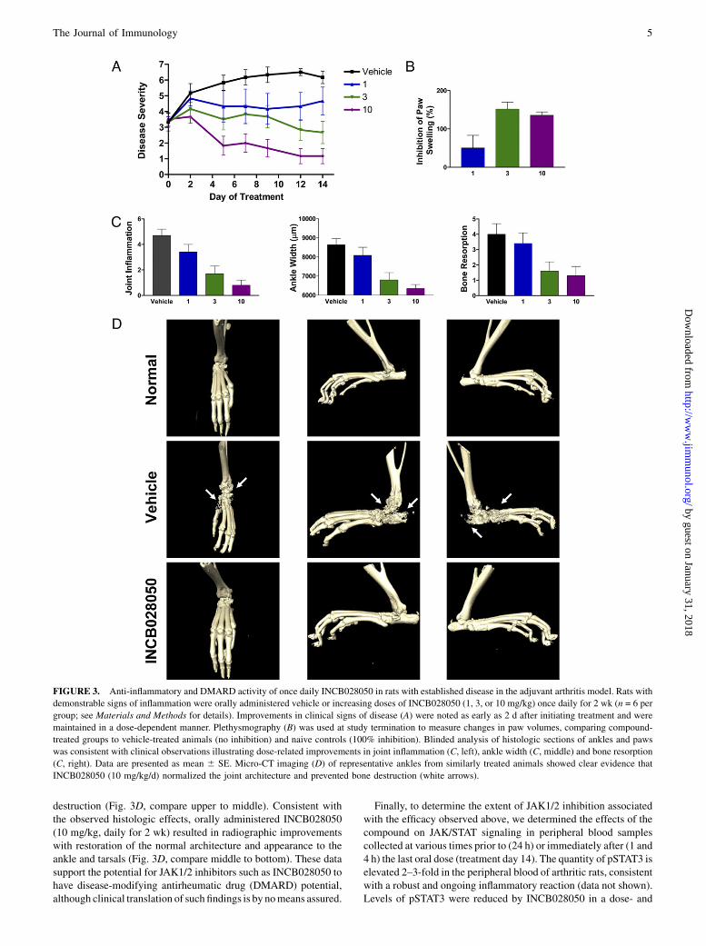
destruction (Fig. 3D, compare upper to middle). Consistent withthe observed histologic effects, orally administered INCB028050(10 mg/kg, daily for 2 wk) resulted in radiographic improvementswith restoration of the normal architecture and appearance to theankle and tarsals (Fig. 3D, compare middle to bottom). These datasupport the potential for JAK1/2 inhibitors such as INCB028050 tohave disease-modifying antirheumatic drug (DMARD) potential,although clinical translation of such findings is by nomeans assured.
Finally, to determine the extent of JAK1/2 inhibition associatedwith the efficacy observed above, we determined the effects of thecompound on JAK/STAT signaling in peripheral blood samplescollected at various times prior to (24 h) or immediately after (1 and4 h) the last oral dose (treatment day 14). The quantity of pSTAT3 iselevated 2–3-fold in the peripheral blood of arthritic rats, consistentwith a robust and ongoing inflammatory reaction (data not shown).Levels of pSTAT3 were reduced by INCB028050 in a dose- and
FIGURE 3. Anti-inflammatory and DMARD activity of once daily INCB028050 in rats with established disease in the adjuvant arthritis model. Rats with
demonstrable signs of inflammation were orally administered vehicle or increasing doses of INCB028050 (1, 3, or 10 mg/kg) once daily for 2 wk (n = 6 per
group; see Materials and Methods for details). Improvements in clinical signs of disease (A) were noted as early as 2 d after initiating treatment and were
maintained in a dose-dependent manner. Plethysmography (B) was used at study termination to measure changes in paw volumes, comparing compound-
treated groups to vehicle-treated animals (no inhibition) and naive controls (100% inhibition). Blinded analysis of histologic sections of ankles and paws
was consistent with clinical observations illustrating dose-related improvements in joint inflammation (C, left), ankle width (C, middle) and bone resorption
(C, right). Data are presented as mean 6 SE. Micro-CT imaging (D) of representative ankles from similarly treated animals showed clear evidence that
INCB028050 (10 mg/kg/d) normalized the joint architecture and prevented bone destruction (white arrows).
The Journal of Immunology 5
by guest on January 31, 2018http://w
ww
.jimm
unol.org/D
ownloaded from

time-dependent manner (Table II). Interestingly, efficacy was ach-ieved in this rAIA experiment at doses that inhibit pSTAT3 levels by,15% 4 h after an oral dose (3 mg/kg). Moreover, even the maxi-mally efficacious dose of 10 mg/kg resulted in no detectablepSTAT3 inhibition 24 h after administration. These data show thatINCB028050 possesses significant activity in this aggressive pre-clinical model of arthritis in the absence of complete and continuouspathway inhibition.In similar experiments, we explored the efficacy of continuously
infused INCB028050 to better determine the average amount ofJAK inhibition required for efficacy at steady-state in the rAIAmodel. To do so, we treated arthritic cohorts of rats using miniatureosmotic pumps implanted on treatment day 0 when the meanclinical score was greater than 3 (∼2 wk after adjuvant injection).Importantly, surgical implantation of pumps has no significantimpact on the progression of the disease model (R. Collins and J.S. Fridman, unpublished data). Implanted pumps were set up todeliver vehicle or 0.18, 0.6, or 1.8 mg/kg/day of INCB028050.These doses resulted in mean plasma levels of 13, 53, and 181 nM,respectively. Based on the potency of INCB028050 in inhibitingpSTAT3 levels in IL-6–stimulated rat whole blood (IC50 ∼ 130nM; Fig. 1D), these plasma levels correlate with 14%, 36% and62% inhibition of JAK/STAT signaling. However, these values areextrapolated and might not accurately reflect variables such asorgan site-specific compound and inhibition levels. Administrationof INCB028050 at these levels was efficacious as indicated byinhibition in clinical signs of disease of 25% (p . 0.05), 50%(p , 0.05), and 90% (p , 0.01; Table III) at 0.18, 0.6, or 1.8 mg/kg, respectively. Similar to oral dosing, the improvement in clin-ical signs of disease following continuous administration co-incided with a reduction in paw swelling, as determined byplethysmography, by 63%, 96%, or $100% with increasing doselevels. A dose-dependent improvement in histologic evidence ofdisease, measured as inflammation and bone resorption scores,was also noted (Table III; p , 0.05 for middle and high doses; seeMaterials and Methods for details) in addition to improvements inradiologic endpoints (not shown).Molecular analysis of draining lymph nodes from a satellite group
of similarly diseased animals (treated orally as described above, 10mg/kg once daily) was conducted to determine the potential con-tribution of Th1- and Th17-associated cytokines in this model.Whereas the former has been well established, the latter is a morenascent finding and, as mentioned previously, preliminary clinicaldata with IL-17 Abs implies that antagonizing this signalingpathway may be advantageous. Both Th1- and Th17-associated
cytokines were found to be elevated during the inflammatory phaseof the rAIA model (day 14, Table IV). Observed increases ranged
from an ∼6-fold increase in expression of the Th1 cytokines IL-12and IFNg to a .100-fold increase in expression of the Th17 cy-
tokines IL-17 and IL-22. Treatment with INCB028050 reduced theexpression of these pathogenic cytokines by 55 to ∼ 80%. Thesedata are consistent with the in vitro cellular data showing that se-
lective JAK1/2 inhibition mitigates signaling from multiple in-flammatory cytokines as well as the production of inflammatory
factors in response to cytokine stimulation (Fig. 1).
Effects of INCB028050 in murine models of humoral andcellular autoimmunity
The therapeutic potential of INCB028050 was also explored inmouse models of arthritis, which are largely driven by humoralmechanisms. The collagen-induced arthritis (CIA) model has beenextensively used in the evaluation of novel therapeutics and reca-pitulates many of the clinical and histologic features of humanRA. This complex model requires humoral and cellular immuneresponses fora robust inflammatory response.Anumberofcytokineshave been implicated as major regulators in mouse CIA, includingIL-6, IL-23, IL-17, TNF-a, and IL-1b (23–27). Indeed, circulatingplasma levels of IL-6 correlate with disease severity in the CIAmodel, and genetic or biologic antagonism of IL-6 markedly sup-presses or eliminates disease incidence and severity, at least in part,by suppressing a robust humoral response (23, 27). Similar ex-periments have demonstrated an important role for Th17 cells andassociated cytokines (e.g., IL-17, IL-21, and IL-23) (20, 24–26).In the murine CIA model, we assessed the effectiveness of
INCB028050 in animals with established disease (i.e., therapeuticmode; Fig. 4A). Doses of 1, 3, or 10 mg/kg were administered twicedaily for 15 d in animals with demonstrable signs of inflammation.This dosing schedule was used because the t1/2 of INCB028050in mice is approximately one third that observed in rats (data notshown). Improvements in clinical signs of disease were noted asearly as 4 d after initiation of dosing, and a dose-dependent re-duction of clinical scores compared with vehicle controls—19%(p = 0.55), 67% (p, 0.0001, and 61% (p, 0.0001), respectively—was seen at study termination. This finding was not surprising basedon the potent ability of INCB028050 to inhibit IL-6 and IL-23signaling and function (Fig. 1), both of which are essential in thedevelopment of disease in the CIA model. However, because of thesupportive effects of some JAK-activating cytokines on B cellproliferation and function, it was possible that the observed efficacyof INCB028050 in this model was due to suppression of a humoralresponse to collagen. To assess this experimentally, we determinedthe levels of anti-collagen Abs in serum samples from a separatecohort of control and treated mice. INCB028050 treatment admin-istered at the doses used in this study in a therapeutic model did notsuppress the production of anti-collagen Abs in these experiments(Fig. 4B; p = 0.4), indicating that the effects are not due to directsuppression of Ab responses. However, because INCB028050 wasadministered in therapeutic mode in these studies, we cannot ruleout the possibility that earlier administration (preventative mode)
Table II. Suppression of phosphorylated STAT3 in the peripheral bloodof rAIA animals treated orally with INCB028050 (n = 3)
Dose (mg/kg)% Inhibition at
1 h% Inhibition at
4 h% Inhibition at
24 h
1 55 6 6 0.1 6 37 03 100 13 6 37 010 100 84 6 18 0
Table III. Sustained modest inhibition of JAK1/2 is efficacious in the rAIA model (n = 6, 6 SD)
Dose (mg/kg/d)aINCB028050 PlasmaConcentration (nM)
pSTAT3(% Inhibition)b
Clinical Score(% Inhibition)
Paw Swelling(% Inhibition)
HistologicImprovement (%)
0.18 13 6 2 14 25 6 42 63 6 15 29 6 150.6 53 6 8 36 50 6 34 96 6 36 40 6 81.8 181 6 19 62 90 6 18 $100 60 6 10
aBased on a body weight of 200 g.bExtrapolated from Fig. 1D.
6 INHIBITION OF JAK1 AND JAK2 IN RA
by guest on January 31, 2018http://w
ww
.jimm
unol.org/D
ownloaded from
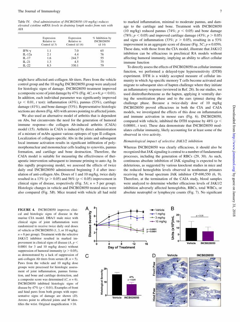
might have affected anti-collagen Ab titers. Paws from the vehiclecontrol group and the 10 mg/kg INCB028050 group were analyzedfor histologic signs of damage. INCB028050 treatment improveda composite score of joint damage by 47% (Fig. 4C; n= 6; p, 0.01).In addition, each individual parameter was significantly improved(p , 0.01, t test): inflammation (43%), pannus (53%), cartilagedamage (41%), and bone damage (53%). Representative histologicsections are shown (Fig. 4D) with arrows pointing to affected joints.We also used an alternative model of arthritis that is dependent
on Abs, but circumvents the need for the generation of humoralimmune response—the collagen Ab-induced arthritis (CAIA)model (15). Arthritis in CAIA is induced by direct administrationof a mixture of mAbs against various epitopes of type II collagen.Localization of collagen-specific Abs in the joints and subsequentlocal immune activation results in significant infiltration of poly-morphonuclear and mononuclear cells leading to synovitis, pannusformation, and cartilage and bone destruction. Therefore, theCAIA model is suitable for measuring the effectiveness of ther-apeutic intervention subsequent to immune priming to auto-Ag. Inthis rapidly progressing model, we assessed the effects of twicedaily oral INCB028050 administered beginning 3 d after inoc-ulation of anti-collagen Abs. Doses of 1 and 10 mg/kg, twice dailyresulted in a 13% (p . 0.05) and 56% (p , 0.05) improvement inclinical signs of disease, respectively (Fig. 5A; n = 5 per group).Histologic changes in vehicle and INCB028050 treated mice werealso compared (Fig. 5B). Mice treated with vehicle all had mild
to marked inflammation, minimal to moderate pannus, and dam-age to the cartilage and bone. Treatment with INCB028050(10 mg/kg) reduced pannus (74%; p , 0.05) and bone damage(78%; p , 0.05) and improved cartilage damage (43%; p . 0.05)and signs of inflammation (33%; p . 0.05), resulting in a 53%improvement in an aggregate score of disease (Fig. 5C; p = 0.059).These data, with those from the CIA model, illustrate that JAK1/2inhibition can be efficacious in preclinical RA models withoutaffecting humoral immunity, implying an ability to affect cellularimmune functionTo directly assess the effects of INCB028050 on cellular immune
function, we performed a delayed-type hypersensitivity (DTH)experiment. DTH is a widely accepted measure of cellular im-munity in which Ag-specific memory T cells become activated andmigrate to subsequent sites of hapten challenge where they initiatean inflammatory response (reviewed in Ref. 28). In our studies, weused dinitroflurobenzene as the hapten, applying it ventrally dur-ing the sensitization phase and to the ears (pinna) during thechallenge phase. Because a twice-daily dose of 10 mg/kgINCB028050 proved efficacious in both the CIA and CAIAmodels, we investigated the effects of this dose on inflammationand immune activation in mouse ears (Fig. 6). INCB028050,compared with vehicle, inhibited the DTH response by 48% (p ,0.00001, t test). These data demonstrate that INCB028050 mod-ulates cellular immunity, likely accounting for at least some of theobserved in vivo activity.
Hematological impact of selective JAK1/2 inhibition
Whereas INCB028050 was clearly efficacious, it should also berecognized that JAK signaling is central to a number of fundamentalprocesses, including the generation of RBCs (29, 30). As such,continuous absolute inhibition of JAK signaling is expected to bedeleterious, as suggested by various knockout studies in mice andthe reduced hemoglobin levels observed in nonhuman primatesreceiving the broad spectrum JAK inhibitor CP-690,550 (6, 9).Therefore, at the termination of the CAIA study, blood sampleswere analyzed to determine whether efficacious levels of JAK1/2inhibition adversely affected hemoglobin, RBCs, total WBCs, orabsolute neutrophil or lymphocyte counts (Fig. 7). No significant
FIGURE 4. INCB028050 improves clini-
cal and histologic signs of disease in the
murine CIA model. DBA/1 male mice with
clinical signs of joint inflammation were
randomized to receive twice daily oral doses
of vehicle or INCB028050 (1, 3, or 10 mg/kg;
n = 8 per group). Treatment with the selective
JAK1/2 inhibitor resulted in marked im-
provement in clinical signs of disease (A, p ,0.0001 for 3 and 10 mg/kg doses) without
suppression of humoral immunity (p . 0.05),
as demonstrated by a lack of suppression of
anti-collagen Ab titers from serum (B, n = 5).
Paws from the vehicle and 10 mg/kg dose
groups were processed for histologic assess-
ment of joint inflammation, pannus forma-
tion, and bone and cartilage destruction, and
a composite score was determined (C, n = 6).
INCB028050 inhibited histologic signs of
disease by 47% (p, 0.01). Examples of front
and hind paws from both groups with repre-
sentative signs of damage are shown (D).
Arrows point to affected joints and W iden-
tifies the wrist. Original magnification 316.
Table IV. Oral administration of INCB028050 (10 mg/kg) reduceselevated cytokine mRNA levels in draining lymph nodes from rats withAIA
ExpressionRelative toControl (d 3)
ExpressionRelative to
Control (d 14)
% Inhibition byINCB028050
(d 14)
IFN-g 1.1 7.0 65IL-12a 2.1 4.4 76IL-17 1.6 244.7 55IL-21 1.3 4.5 75IL-22 8.3 330.1 79
The Journal of Immunology 7
by guest on January 31, 2018http://w
ww
.jimm
unol.org/D
ownloaded from

differences were noted in any parameter, which is consistent withthe hypothesis that periodic incomplete inhibition of JAK1 andJAK2 can be efficacious in the absence of suppressing normal bonemarrow function (p . 0.05, t test). Recognizing that any effect onhemoglobin and/or RBCs can be underestimated owing to the du-ration of treatment, we also analyzed reticulocytes, which aretemporally more responsive, and found a similar lack of effect (p.0.05, t test), consistent with a lack of myelosuppression at theseotherwise efficacious and well-tolerated doses.
DiscussionINCB028050 is a potent, selective, orally bioavailable inhibitor ofJAK1 and JAK2 with demonstrable selectivity against JAK3 anda broad panel of unrelated kinases. In cell-based assays relevant toautoimmune diseases such as RA and psoriasis, INCB028050 in-hibited JAK signaling and function initiated by two clinically vali-dated therapeutic targets (IL-6 and IL-23) at concentrations#60 nMand did not significantly affect JAK3-dependent proliferationat concentrations up to 10 mM (Supplemental Fig. 1). Structurallysimilar compounds lacking JAK1/2 inhibitory activity were inactivein these cellular cytokine assays (Supplemental Table I) consistentwith a central role for JAK1/2 in these models systems and the se-lectivity of INCB028050. However, we recognize that the absence of
observable off-target activity with INCB028050 does not excludethe possibility that this compound impacts an untested enzyme, re-ceptor, channel or transporter.In vivo, INCB028050 was efficacious in the rAIA model when
administered orally or by continuous infusion. In this aggressive,T cell-driven arthritis model periodic and/or fractional inhibitionof JAK1/2 signaling by INCB028050 appears sufficient to achieveremarkable efficacy inanimalswithactive signsofdisease regardlessof what endpoint is used, including radiographic improvementsassociatedwithDMARDactivity.Thisconclusionisbasedontheoralefficacy of INCB028050 (Fig. 3) and the associated pharmacody-namic data (pSTAT3 inhibition; Table II), which takes into accountany potential accumulation of drug during the experiment as well asany potentially active metabolites of INCB028050 produced duringits elimination, although no data for either exist at this time (datanot shown). Similarly, continuous infusion of INCB028050 is effi-cacious at steady-state plasma drug levels below the whole bloodassay IC50 for JAK1/2 signaling (Table III; Fig. 1D). Plasma levelsachieved by either route of administration were well below thoseexpected to inhibit JAK3 based both on enzyme and cellular potency(Table I; Supplemental Fig. 1) supporting the hypothesis that JAK1/2 inhibition is sufficient for efficacy in this destructive preclinicalmodel of arthritis. Moreover, these efficacy data were at least asimpressive as those associated with selective inhibition of IL-17,using a receptor IgG1Fc fusion protein, consistentwith the ability ofINCB028050 to affect signaling from multiple inflammatory
FIGURE 6. Suppression of delayed-type hypersensitivity by INCB0-
28050. Animals were sensitized on the abdomen and subsequently chal-
lenged on the pinna with dinitrofluorobenzene (n = 8 per group).
INCB028050 (10 mg/kg, by mouth, twice per day) was administered just
prior to and during the challenge phase. Ear measurements (A) were made
and swelling is shown in units (1 U = 0.0001 inch). Treatment with
INCB028050 resulted in a 48% suppression in ear swelling compared with
vehicle controls (p , 0.00001, t test).
FIGURE 5. INCB028050 is efficacious and well tolerated independently of effects on humoral immunity. Oral doses of INCB028050 (1 or 10 mg/kg, twice per
day) proved effective at improving clinical (A; 10 mg/kg, 56% inhibition, p , 0.05) and histologic (B, C, 10 mg/kg) signs of disease in mice inoculated with
amixture of arthrogenicmAbs against collagen (n= 5 per group). Arrows point to areas of significant inflammation and/or bone destruction (B). Vehicle sections are
representative of the entire group, and sections from INCB028050-treated mice represent the majority of the treatment group. Original magnification 316.
FIGURE 7. Lack of effect on hematologic parameters. Terminal blood
samples from the mouse CAIA experiment were analyzed for changes in
hematologic parameters (n = 5 per group for treated mice and n = 2 for
naives). No significant effects were observed (p . 0.05, t test).
8 INHIBITION OF JAK1 AND JAK2 IN RA
by guest on January 31, 2018http://w
ww
.jimm
unol.org/D
ownloaded from

cytokines that signal through JAK1 and/or JAK2 (31). However,because a direct comparison of these modalities was not performed,quantitative comparisons are difficult.In the murine CIA model, dependent on both B and T cells, oral
INCB028050 was also efficacious. Interestingly, although IL-6 isreported to be important in both murine CIA and in RA patients, theeffectiveness of INCB028050 in this setting may be due, at least inpart, to itsability toaffect IL-6signaling tosuchadegree that it is anti-inflammatory without being immune suppressive, as assessed byquashing an established humoral responses. This hypothesis is basedon reports that anti-collagen Ab titers were suppressed in both theIL-6 knockout mice and those treated with anti-IL-6R Abs (23, 27).This implies that some degree of IL-6 signaling is central tomountingand/or maintaining a robust humoral response and that completeablation of IL-6 signaling, whether through genetic recombination orAb sequestration, significantly antagonizes humoral immunity. Incontrast, INCB028050 did not suppress an ongoing humoral re-sponse to collagen, but was efficacious when administered in thetherapeutic mode at doses affecting but not eradicating IL-6 sig-naling. Conceivably, the ability of INCB028050 to affect the IL-17/23 axis may have also contributed to the impressive responses in theabsence of humoral suppression (Fig.1; Table IV). Mice with ge-netic disruption of IL-23p19, but not IL-12p35, are resistant to CIA,and this correlates with reduced production of IL-17, presumablyfrom Th17 cells (25). In agreement with this finding, mice lackingIL-17 are also resistant to CIA but, similar to the IL-6 knockoutmice, have a suppressed humoral response to Ag (26). Nonetheless,neutralization of IL-17 in the therapeutic mode also suppresses theincidence and severity of disease, but does not affect anti-collagenAb titers (24). The differences in effects on humoral responses likelyreflect the ability of IL-17 to contribute to a humoral response duringthe immune education phase, although this effect is not necessaryfor IL-17 antagonism to be effective. More exhaustive studies withJAK inhibitors during the various phases of humoral immunity willbe required to definitively describe the effects of these kinases andINCB028050 on this biology. However, the ability of INCB028050to inhibit a DTH response clearly demonstrates its capacity tomodulate cellular immunity in a hypersensitivity setting and sug-gests that at least part of the observed efficacy with INCB028050 isdue to this immune modulating capability.The ability of INCB028050 to affect joint inflammation in-
dependently from suppressing an established humoral responsewasalso addressed in the CAIA model, in which joint inflammation isinduced by systemic administration of a mixture of anti-collagen IIAbs (15). This model induces a polymorphonuclear and mono-nuclear cell infiltrate and a robust inflammatory response, result-ing in pannus formation and joint destruction. Interestingly,Kagari et al. (32) showed that IL-6 knockout mice were suscep-tible to CAIA, which is consistent with a dominant role for IL-6 insupporting a humoral response to autoantigen. In this model,INCB028050 suppressed both clinical and histologic signs ofdisease consistent with its ability to affect JAK activating patho-genic cytokines including, but not limited to, IL-6.Because genetic data indicate that complete ablation of JAK1 or
JAK2 in mice is lethal, and because decades of research definea fundamental role for these kinases in the signaling pathways fornumerous important cytokines, it is unlikely that unremitting in-hibition of their activity would be tolerated (reviewed in Ref. 6). Inthis study, we have demonstrated that a selective JAK1/2 inhibitor(INCB028050) is efficacious in three rodent models of arthritiswithout complete or constant inhibition of JAK signaling. Effec-tive treatment paradigms in these models have been well toleratedas exemplified by the lack of effects of INCB028050 on a numberof hematologic parameters and body weight (Fig. 3E and data not
shown). Moreover, a related compound, INCB018424, has beenwell tolerated in RA and cancer patients suggesting that thismechanism is a viable therapeutic option for patients sufferingfrom RA or other diseases in which JAK signaling is dysregulated(33, 34). These results warrant further clinical evaluation of se-lective JAK1/2 inhibitors such as INCB028050
DisclosuresAll authors have been or are currently employees of the Incyte Corporation
and own shares and/or participate in the company stock option plan.
References1. McInnes, I. B., and F. Y. Liew. 2005. Cytokine networks—towards new therapies
for rheumatoid arthritis. Nat. Clin. Pract. Rheumatol. 1: 31–39.2. Smolen, J. S., A. Beaulieu, A. Rubbert-Roth, C. Ramos-Remus, J. Rovensky,
E. Alecock, T. Woodworth, and R. Alten; OPTION Investigators. 2008. Effect ofinterleukin-6 receptor inhibition with tocilizumab in patients with rheumatoidarthritis (OPTION study): a double-blind, placebo-controlled, randomised trial.Lancet 371: 987–997.
3. Baslund, B., N. Tvede, B. Danneskiold-Samsoe, P. Larsson, G. Panayi,J. Petersen, L. J. Petersen, F. J. Beurskens, J. Schuurman, J. G. van de Winkel,et al. 2005. Targeting interleukin-15 in patients with rheumatoid arthritis:a proof-of-concept study. Arthritis Rheum. 52: 2686–2692.
4. Sloan-Lancaster, J., M. C. Genovese, S. A. Roberson, and F. Van den Bosch.2009. Safety, tolerability, and evidence of efficacy of intravenous ly2439821 inpatients with rheumatoid arthritis receiving background oral DMARDs. Ann.Rheum. Dis. 68: 125.
5. Ghoreschi, K., A. Laurence, and J. J. O’Shea. 2009. Janus kinases in immunecell signaling. Immunol. Rev. 228: 273–287.
6. Pesu, M., A. Laurence, N. Kishore, S. H. Zwillich, G. Chan, and J. J. O’Shea.2008. Therapeutic targeting of Janus kinases. Immunol. Rev. 223: 132–142.
7. Ihle, J. N. 1995. The Janus protein tyrosine kinases in hematopoietic cytokinesignaling. Semin. Immunol. 7: 247–254.
8. O’Shea, J. J., M. Pesu, D. C. Borie, and P. S. Changelian. 2004. A new modalityfor immunosuppression: targeting the JAK/STAT pathway. Nat. Rev. DrugDiscov. 3: 555–564.
9. Changelian, P. S., M. E. Flanagan, D. J. Ball, C. R. Kent, K. S. Magnuson,W. H. Martin, B. J. Rizzuti, P. S. Sawyer, B. D. Perry, W. H. Brissette, et al. 2003.Prevention of organ allograft rejection by a specific Janus kinase 3 inhibitor. Sci-ence 302: 875–878.
10. Williams, N. K., R. S. Bamert, O. Patel, C. Wang, P. M. Walden, A. F. Wilks,E. Fantino, J. Rossjohn, and I. S. Lucet. 2009. Dissecting specificity in the Januskinases: the structures of JAK-specific inhibitors complexed to the JAK1 andJAK2 protein tyrosine kinase domains. J. Mol. Biol. 387: 219–232.
11. Jiang, J. K., K. Ghoreschi, F. Deflorian, Z. Chen, M. Perreira, M. Pesu, J. Smith,D. T. Nguyen, E. H. Liu, W. Leister, et al. 2008. Examining the chirality, con-formation and selective kinase inhibition of 3-((3R,4R)-4-methyl-3-(methyl(7H-pyrrolo[2,3-d]pyrimidin-4-yl)amino)piperidin-1-yl)-3-oxopropanenitrile (CP-690,550). J. Med. Chem. 51: 8012–8018.
12. Guschin, D., N. Rogers, J. Briscoe, B. Witthuhn, D. Watling, F. Horn,S. Pellegrini, K. Yasukawa, P. Heinrich, G. R. Stark, et al. 1995. A major role forthe protein tyrosine kinase JAK1 in the JAK/STAT signal transduction pathwayin response to interleukin-6. EMBO J. 14: 1421–1429.
13. Ellingsen, T., A. Buus, and K. Stengaard-Pedersen. 2001. Plasma monocytechemoattractant protein 1 is a marker for joint inflammation in rheumatoid ar-thritis. J. Rheumatol. 28: 41–46.
14. Brand, D. D., K. A. Latham, and E. F. Rosloniec. 2007. Collagen-induced ar-thritis. Nat. Protoc. 2: 1269–1275.
15. Khachigian, L. M. 2006. Collagen antibody-induced arthritis. Nat. Protoc. 1:2512–2516.
16. Brodmerkel, C. M., R. Huber, M. Covington, S. Diamond, L. Hall, R. Collins,L. Leffet, K. Gallagher, P. Feldman, P. Collier, et al. 2005. Discovery andpharmacological characterization of a novel rodent-active CCR2 antagonist,INCB3344. J. Immunol. 175: 5370–5378.
17. Fridman, J., P. Scherle,R.Collins,T.Burn,Y.Li, J.Li,M.Covington,B.Thomas,M.Favata, J. Shi, et al. 2008. Preclinical characterization of INCB028050, JAK1/JAK2selective clinical candidate. Arthritis Rheum. 58: S296.
18. Lacronique, V., A. Boureux, R. Monni, S. Dumon, M. Mauchauffe, P. Mayeux,F. Gouilleux, R. Berger, S. Gisselbrecht, J. Ghysdael, and O. A. Bernard. 2000.Transforming properties of chimeric TEL-JAK proteins in Ba/F3 cells. Blood 95:2076–2083.
19. Milici, A. J., E. M. Kudlacz, L. Audoly, S. Zwillich, and P. Changelian. 2008.Cartilage preservation by inhibition of Janus kinase 3 in two rodent models ofrheumatoid arthritis. Arthritis Res. Ther. 10: R14.
20. Young, D. A., M. Hegen, H. L. Ma, M. J. Whitters, L. M. Albert, L. Lowe,M. Senices, P. W. Wu, B. Sibley, Y. Leathurby, et al. 2007. Blockade of theinterleukin-21/interleukin-21 receptor pathway ameliorates disease in animalmodels of rheumatoid arthritis. Arthritis Rheum. 56: 1152–1163.
21. Diveu, C., M. J. McGeachy, and D. J. Cua. 2008. Cytokines that regulate au-toimmunity. Curr. Opin. Immunol. 20: 663–668.
22. Parham, C., M. Chirica, J. Timans, E. Vaisberg, M. Travis, J. Cheung, S. Pflanz,R. Zhang, K. P. Singh, F. Vega, et al. 2002. A receptor for the heterodimeric
The Journal of Immunology 9
by guest on January 31, 2018http://w
ww
.jimm
unol.org/D
ownloaded from

cytokine IL-23 is composed of IL-12Rbeta1 and a novel cytokine receptorsubunit, IL-23R. J. Immunol. 168: 5699–5708.
23. Alonzi, T., E. Fattori, D. Lazzaro, P. Costa, L. Probert, G. Kollias, F. DeBenedetti, V. Poli, and G. Ciliberto. 1998. Interleukin 6 is required for the de-velopment of collagen-induced arthritis. J. Exp. Med. 187: 461–468.
24. Lubberts, E., M. I. Koenders, B. Oppers-Walgreen, L. van den Bersselaar,C. J. Coenen-de Roo, L. A. Joosten, and W. B. van den Berg. 2004. Treatmentwith a neutralizing anti-murine interleukin-17 antibody after the onset of collagen-induced arthritis reduces joint inflammation, cartilage destruction, and bone ero-sion. Arthritis Rheum. 50: 650–659.
25. Murphy, C. A., C. L. Langrish, Y. Chen, W. Blumenschein, T. McClanahan,R. A. Kastelein, J. D. Sedgwick, and D. J. Cua. 2003. Divergent pro- and an-tiinflammatory roles for IL-23 and IL-12 in joint autoimmune inflammation.J. Exp. Med. 198: 1951–1957.
26. Nakae,S.,A.Nambu,K.Sudo, andY. Iwakura. 2003.Suppressionof immune inductionof collagen-induced arthritis in IL-17-deficient mice. J. Immunol. 171: 6173–6177.
27. Takagi, N., M. Mihara, Y. Moriya, N. Nishimoto, K. Yoshizaki, T. Kishimoto,Y. Takeda, and Y. Ohsugi. 1998. Blockage of interleukin-6 receptor amelioratesjoint disease in murine collagen-induced arthritis. Arthritis Rheum. 41: 2117–2121.
28. Price, K. 2008. Cellular immune response in delayed-type hypersensitivity tests.In Immunotoxicology Strategies for Pharmaceutical Saftey Assessment. D. J.Herzyk and J. L. Bussiere, ed. John Wiley & Sons, Hoboken, NJ, p. 87.
29. Neubauer, H., A. Cumano, M. Muller, H. Wu, U. Huffstadt, and K. Pfeffer. 1998.Jak2 deficiency defines an essential developmental checkpoint in definitive he-matopoiesis. Cell 93: 397–409.
30. Parganas, E., D. Wang, D. Stravopodis, D. J. Topham, J. C. Marine, S. Teglund,E. F. Vanin, S. Bodner, O. R. Colamonici, J. M. van Deursen, et al. 1998. Jak2is essential for signaling through a variety of cytokine receptors. Cell 93: 385–395.
31. Bush, K. A., K. M. Farmer, J. S. Walker, and B. W. Kirkham. 2002. Reduction ofjoint inflammation and bone erosion in rat adjuvant arthritis by treatment withinterleukin-17 receptor IgG1 Fc fusion protein. Arthritis Rheum. 46: 802–805.
32. Kagari, T., H. Doi, and T. Shimozato. 2002. The importance of IL-1 beta andTNF-alpha, and the noninvolvement of IL-6, in the development of monoclonalantibody-induced arthritis. J. Immunol. 169: 1459–1466.
33. Williams, W., P. Scherle, J. Shi, R. Newton, E. McKeever, J. Fridman, T. Burn,K. Vaddi, R. Levy, and L. Moreland. 2008. A randomized placebo-controlledstudy of INCB018424, a selective janus kinase1&2 (JAK1&2) inhibitor inrheumatoid arthritis (RA). Arthritis Rheum. 58: S431.
34. Verstovsek, S., H. Kantarjian, A. Pardanani, D. Thomas, J. Cortes, R. Mesa, J.Redman, C. M. Staschen, J. Fridman, K. Vaddi, and A. Tefferi. 2007.INCB018424, an oral, selective JAK2 inhibitor, shows significant clinical ac-tivity in a phase i/ii study in patients with primary myelofibrosis (PMF) and postpolycythemia ver/essential thrombocythemia myelofibrosis (Post-PV/ET MF). InASH Annual Meeting 110: 171a.
10 INHIBITION OF JAK1 AND JAK2 IN RA
by guest on January 31, 2018http://w
ww
.jimm
unol.org/D
ownloaded from