selective enhancing effect of early mitotic inhibitor 1 (emi1
TRANSCRIPT
Selective Enhancing Effect of Early Mitotic Inhibitor 1 (Emi1)Depletion on the Sensitivity of Doxorubicin or X-rayTreatment in Human Cancer Cells*
Received for publication, December 18, 2012, and in revised form, April 17, 2013 Published, JBC Papers in Press, May 3, 2013, DOI 10.1074/jbc.M112.446351
Natsumi Shimizu‡1,2, Nakako Izumi Nakajima§1, Takaaki Tsunematsu‡, Ikuko Ogawa¶, Hidehiko Kawai�,Ryoichi Hirayama§, Akira Fujimori§, Akiko Yamada**, Ryuichi Okayasu§, Naozumi Ishimaru**, Takashi Takata‡,and Yasusei Kudo‡**3
From the ‡Department of Oral and Maxillofacial Pathobiology, Graduate School of Biomedical & Health Sciences, HiroshimaUniversity, Hiroshima 734-8553, Japan, the §Research Center for Charged Particle Therapy and International Open LaboratoryParticle Therapy Molecular Target Research Unit, National Institute of Radiological Sciences, Chiba 263-8555, Japan, the ¶Center ofOral Clinical Examination, Hiroshima University Hospital, Hiroshima 734-8553, Japan, the �Department of Molecular Radiobiology,Research Institute for Radiation Biology and Medicine, Hiroshima University, Hiroshima 734-8553, Japan, and the **Department ofOral Molecular Pathology, Institute of Health Biosciences, The University of Tokushima Graduate School, Tokushima770-8504, Japan
Background: To improve the effectiveness of chemo- and radiotherapy only in cancer tissue is important for avoiding sideeffects.Results: Emi1 depletion enhanced the sensitivity of anticancer reagents and X-ray irradiation in cancer cells.Conclusion: Emi1 siRNA would be a useful new modality for enhancing the effect of chemo- and radiotherapy in varioustumors.Significance: This work provides new insights regarding synergistic effect of Emi1 knockdown in combination therapies.
Chemotherapy and radiation in addition to surgery hasproven useful in a number of different cancer types, but theeffectiveness in normal tissue cannot be avoided in these thera-pies. To improve the effectiveness of these therapies selectivelyin cancer tissue is important for avoiding side effects. Earlymitotic inhibitor 1 (Emi1) is known to have the function toinhibit anaphase-promoting complex/cyclosome ubiquitinligase complex, which ubiquitylates the cell cycle-related pro-teins. It recently has been shown that Emi1 knockdownpreventstransition from S to G2 phase by down-regulating geminin viaanaphase-promoting complex/cyclosome activation. At pres-ent, anticancer drugs for targeting DNA synthesis to interferewith rapidly dividing cells commonly are used. As Emi1 deple-tion interfereswith completionofDNAsynthesis in cancer cells,we thought that Emi1 knockdownmight enhance the sensitivityfor anticancer agents. Here, we confirmed that Emi1 siRNAinduced polyploidy for preventing transition from S toG2 phasein several cancer cell lines. Then, we treated Emi1 depleted cellswith doxorubicin. Interestingly, increased apoptotic cells were
observed after doxorubicin treatment in Emi1 siRNA-treatedcancer cells. In addition, Emi1 depletion enhanced the sensitiv-ity of x-ray irradiation in cancer cells. Importantly, synergisticeffect of Emi1 knockdown in these combination therapies wasnot observed in normal cells. These results suggest that Emi1siRNA can be a useful tool for enhancing of sensitivity of cancercells to anticancer reagents and radiation.
Approximately 12.7 million cancers were diagnosed and 7.6million people died of cancerworldwide in 2008 (1). Thismakescancer the leading cause of death in the developed world andthe second leading cause of death in the developing world (1).Cancer can be treated by various means such as surgery, chem-otherapy, radiation therapy, immunotherapy, and monoclonalantibody therapy. The choice of therapy depends upon thelocation and grade of the tumor and the stage of the disease, aswell as the physical condition of the patient. Complete inacti-vation of the cancer without damage to the rest of the body isthe goal of treatment. Sometimes this can be accomplished bysurgery, but the propensity of cancers to invade adjacent tissuesor to spread to distant sites by microscopic metastasis oftenlimits its effectiveness. Chemotherapy alone or in combinationwith other treatments such as surgery or radiation has provenuseful in a number of different cancer types, but the effective-ness of chemotherapy is often limited by the toxicity to non-targeted tissues in the body. The majority of chemotherapeuticdrugs can be divided into alkylating agents, antimetabolites,anthracyclines, plant alkaloids, topoisomerase inhibitors, andother anticancer agents (2, 3). All of these drugs affect cell divi-sion or DNA synthesis and act by killing cells that divide rap-idly, one of themain properties ofmost cancer cells. Anticancer
* This work was supported in part by Grants-in-aid for Scientific Researchfrom the Ministry of Education, Culture, Sports, Science and Technology(23689074 and 25670778 (to Y. K.), 21249088 (to T. Takata), and 23390301and 24249067 (to R. O.)), Grant-in-aid for Cancer Research from the Minis-try of Education, Culture, Sports, Science and Technology (19-9, H23-A-43;to R. O.), and a Research Fellowship for Young Scientists from the JapanSociety for the Promotion of Science (23-6562; to T. Tsunematsu).
1 Both authors contributed equally to this work.2 Present address: Div. of Biological Science, Graduate School of Biology-Ori-
ented Science and Technology, Kinki University, Wakayama 649-6493,Japan.
3 To whom correspondence should be addressed: Dept. of Oral MolecularPathology, Institute of Health Biosciences, The University of TokushimaGraduate School, Tokushima, 3-18-15 Kuramoto, Tokushima 770-8504,Japan. Fax: 81-88-633-7328; E-mail: [email protected].
THE JOURNAL OF BIOLOGICAL CHEMISTRY VOL. 288, NO. 24, pp. 17238 –17252, June 14, 2013© 2013 by The American Society for Biochemistry and Molecular Biology, Inc. Published in the U.S.A.
17238 JOURNAL OF BIOLOGICAL CHEMISTRY VOLUME 288 • NUMBER 24 • JUNE 14, 2013
by guest on April 2, 2018
http://ww
w.jbc.org/
Dow
nloaded from

agents also harm cells that divide rapidly under normal circum-stances: cells in the bone marrow, digestive tracts, and hair fol-licles. These effects result in the common side effects of chem-otherapy, includingmyelosuppression,mucositis, and alopecia.Therefore, newer anticancer drugs, includingmonoclonal anti-bodies and the new tyrosine kinase inhibitors, e.g. imatinibmesylate (Gleevec orGlivec), which directly targets amolecularabnormality in certain types of cancer (chronic myelogenousleukemia, gastrointestinal stromal tumors), have been used (4).Targeted therapy is a type of medication that blocks the growthof cancer cells by interfering with specific targeted moleculesneeded for carcinogenesis and tumor growth, rather than bysimply interfering with rapidly dividing cells. Targeted cancertherapies thus are expected to be more effective than conven-tional treatments and less harmful to normal cells.Many oncol-ogists believe that targeted therapies are the chemotherapy ofthe future. At present, however, many traditional anticancerdrugs for targeting DNA synthesis to interfere with rapidlydividing cells are commonly used. Radiation therapy is themedical application of ionizing radiation to suppressing tumorgrowth. Ionizing radiation works by damaging DNA to controltumor cell growth/division, but the effect of radiation in normaltissues cannot be avoided in these therapies.The cell cycle is regulated by cell cycle regulation factors and
many of these are degraded via ubiquitylation (5). Abnormalityof ubiquitylation in degradation of proteins induces various dis-eases such as cancer (6–8). It is known that SCF (Skp1-Cullin-F-box) and anaphase-promoting complex/cyclosome (APC/C),4 ubiquitin ligases, are involved in ubiquitylation of cell cycleregulating factors (9, 10). In particular, APC/C is associatedwith the degradation of proteins in the M-G1 phase and plays arole in the regulation of spindle checkpoint and processionfrom the M to G1 phase. APC/C is composed of several dozensubunits, and its activity is regulated by co-activators Cdc20 orCdh1 and phosphorylation of constitutive subunits (9). Activityof APC/CCdc20 increases from the prophase to prometaphase,and decrease in the anaphase by Cdc20 degradation (9). How-ever, activity of APC/CCdh1 is maintained from the anaphase toG1/S phase, after which the activity is inhibited by Emi1 (11).Emi1 was identified as a factor inhibiting the function of APC/CCdh1 and is degraded by SCF�Trcp at early M phase (12–15). Itrecently has been reported that an abnormally high expressionof Emi1 protein can be observed in various cancers (14, 16, 17).Moreover, inhibition of Emi1 interferes with progression to Mphase by degradation of geminin, which is necessary for thecompletion of DNA synthesis (18, 19). Emi1-depleted cellsshow polyploidy and large nuclei because these cells cannotcomplete DNA synthesis (18, 19). These results suggest thatEmi1-depleted cells remain in S phase. As Emi1 depletion inter-feres with completion of DNA synthesis in cancer cells (18, 19),we speculated that inhibition of Emi1 in cancer cells mightenhance the sensitivity of anticancer agents. Moreover, cellslacking Emi1 undergo DNA damage, likely explained by repli-cation stress (20). Therefore, we examined the combined effect
of Emi1 knockdown and x-rays. In this study, we also examinedthe combined effects by one of the major anticancer agents,doxorubicin, and Emi1 depletion in various tumor cells.
EXPERIMENTAL PROCEDURES
Reagents and Antibodies—Doxorubicin hydrochloride,camptothecin, etoposide, taxol (paclitaxel), and cobalt chloride(CoCl2) were obtained from Sigma. Commercial antibodieswere from the following companies: anti-Emi1 and anti-Cul1antibodies (Zymed Laboratories Inc.); anti-Aurora-A antibodyand anti-fypoxia-inducible factor-1� (HIF-1�) (TransductionLaboratories); anti-cyclin A and anti-cyclin B antibodies (SantaCruz Biotechnology); anti-Cdh1 and anti-Cdc20 antibodies,MBL; anti-E2F1 antibody (Cell Signaling Technology); andanti-�-actin antibody (Sigma). Anti-geminin polyclonal anti-body was gift from Dr. Nishitani (University of Hyogo), andanti-TPX2 monoclonal antibody was a gift from Dr. Hans-Jür-gen Heidebrecht (University of Kiel). For detection of theimmunocomplex, the ECL Western blotting detection system(Amersham Biosciences) was used.Tissue Samples—Sixty tissue samples of human head and
neck squamous cell carcinomawere retrieved from the SurgicalPathology Registry of Hiroshima University Hospital, afterapproval by the Ethical Committee of Hiroshima UniversityHospital. Sixty head and neck squamous cell carcinoma caseswere surgically resected before radiochemotherapy. Clinicalinformation was gathered from surgical records of the patients.The histological grade of tumor was classified according to thecriteria of the Japan Society for Head andNeck Cancer. Tissueswere fixed in 10% buffered formalin and embedded in paraffin.Cell Lines andCultureConditions—Six head and neck cancer
cell lines (HSC2, HSC3, HSC4, Ca9–22, Ho-1-N-1, and Ho-1-U-1), glioma cell line (T98G), and normal human lung fibro-blasts (HFLIII) were provided by the Japanese Collectionof Research Bioresources Cell Bank. HOC119, HOC313,HOC621, HOC719-PE, HOC719-NE, TSU, OM-1, and ZAcells have been described previously (21). Breast cancer cell line(MCF-7), colon cancer cell line (RKO and HCT116), osteosar-coma cell line (SaOS-2), and human breast epithelial cell line(MCF-10A) were purchased from ATCC. HaCaT cell line wasobtained from N. E. Fusenig (German Cancer Research Cen-ter). The p53-null HCT116 cell line was obtained from B.Vogelstein (The Johns Hopkins University). Cells were main-tained in RPMI 1640 medium or Dulbecco’s modified Eagle’smedium (Nissui Pharmaceutical Co., Tokyo, Japan) supple-mented with 10% heat-inactivated FBS (Invitrogen) and 100units/ml penicillin-streptomycin (Invitrogen) under conditionsof 5% CO2 in air at 37 °C. Upon receiving the cell lines, theywere immediately cultured and expanded to prepare frozenampule stocks. Cells were passaged for no more than 2 to 3months before establishing new culture from early-passage fro-zen ampules.RT-PCR—Using the RNeasy mini kit (Qiagen, Hilden, Ger-
many), total RNA from cultures of confluent cells and tissueswas isolated. These isolateswere quantified and their puritywasevaluated by spectrophotometer. The cDNA was synthesizedfrom 1 �g of total RNA according to ReverTra Dash (ToyoboBiochemicals, Tokyo, Japan). We used the following primers:
4 The abbreviations used are: APC/C, anaphase-promoting complex/cyclo-some; DSB, double-stranded break; PI, propidium iodide; EdU, 5-ethynyl-2�-deoxyuridine; NHDF, neonatal normal human dermal fibroblast.
Selective Effects of Emi1 Depletion in Cancer
JUNE 14, 2013 • VOLUME 288 • NUMBER 24 JOURNAL OF BIOLOGICAL CHEMISTRY 17239
by guest on April 2, 2018
http://ww
w.jbc.org/
Dow
nloaded from
human Emi1, 5�-tctttcgaaggggactcaga-3� (forward) and 5�-tct-ggttgaagcatgaggtg-3� (reverse); human GAPDH, 5�-tccac-caccctgttgctgta-3� (forward) and 5�-accacagtccatgccatcac-3�(reverse). Aliquots of total cDNAwere amplifiedwith 1.25 unitsof rTaq-DNA polymerase (Qiagen), and this amplification wasdone in a thermal cycler (MyCyler, Bio-Rad) for 30 cycles afterinitial 30 s of denaturation at 94 °C, annealing for 30 s at 60 °C,and extension for 1min at 72 °C in all primers used. The ampli-fication reaction products were resolved on 1.2% agarose/TAEgels (Nacalai Tesque), electrophoresed at 100 mV, and thenfinally visualized by using ethidium bromide.Immunohistochemistry—Tumor tissues were fixed in 10%
formalin, embedded in paraffin, and cut 4�m thick. For immu-nohistochemical staining, tissue sections were deparaffinizedin xylene and rehydrated in descending grades of ethanol.Endogenous peroxidase activity was blocked with methanolcontaining 0.3%H2O2 for 30min.Antigen retrieval was done bythe microwaving using a citrate phosphate buffer (pH 6.0), andthen the sections were incubated with the primary antibody at4 °C overnight. Immunohistochemical staining was carried outby a polyclonal anti-Emi1 antibody (Zymed Laboratories, Inc.).For detection of the reaction after incubation with secondaryantibodies, we used diaminobenzidine (DAKO, Glostrup, Den-mark). The sections were counterstained by hematoxylin anddehydrated in ascending grades of ethanol, and the slides weremounted. For evaluation of staining intensity, we graded �(weak/focal immunopositivity), �� (strong/focal immunopo-sitivity and weak/diffuse immunopositivity),��� (strong/dif-fuse immunopositivity). Then, the expression of Emi1 wasgraded as high (�30% of tumor cells showing �� or ���intensity) or low (no staining or�30%of tumor cells showing�intensity).Western Blot Analysis—Western blotting was carried out as
described previously (15). The protein concentrations weremeasured by Bradford protein assay (Bio-Rad). Twenty �g ofprotein was subjected to 10% polyacrylamide gel electrophore-sis, followed by electroblotting onto a nitrocellulose filter. Fordetection of the immunocomplex, the ECL Western blottingdetection system (Amersham Biosciences) was used.Cell Proliferation—For proliferation assay, 5000 cells were
plated onto a 24-well plate, and the cells were allowed to growand expand. The cells were then trypsinized and counted at 0, 2,4, and 6 days by using cell counter (Coulter Z1, Beckman-Coulter). Doubling time was calculated by rate of cellproliferation.Mitotic Index—Cells were seeded on glass coverslips. Forty
eight hours after incubation, cells were fixed, permeabilized,and stained with pSer-10 histone H3 (Millipore) and DAPI.pSer-10 histone H3-positive and condensed chromatin cellswere counted as mitotic cells. More than 400 cells were scoredper condition.RNA Interference—Logarithmically growing cells were
seeded at a density of 105 cells/6-cm dish and transfected witholigonucleotides at 24 h after replating by usingOligofectamine(Invitrogen). Forty-eight hours after transfection, lysates wereprepared and analyzed by SDS-PAGE and immunoblotting.The siRNA is a 19-bp duplex oligoribonucleotide with a sensestrand corresponding to nucleotides 1446–1464 of the human
Emi1mRNA sequence; 5�-GAUUGUGAUCUCUUAUUAA-3�(Dharmacon, Chicago, IL). Human cyclin A siRNA (sc-29282)and human E2F1 siRNA (sc-61861) were obtained from SantaCruz Biotechnology. The negative control (siTrio negative con-trol siRNA) was obtained fromCosmo Bio, Inc. (Tokyo, Japan).Immunofluorescence—Cells grown on cover slips were fixed
in 4% paraformaldehyde for 10 min at room temperature,rinsed three timeswith ice-cold PBS, and then permeabilized in0.1% Triton X-100 in PBS for 15 min at room temperature.After rinsing three times with PBS, coverslips were incubatedwith DAPI staining. Immunostaining of cell preparations wasrecorded using an epifluorescence Zeiss Axioplan 2 (Zeiss, Inc.,Thorwood, NY) microscope attached to a charge-coupleddevice camera.For staining with �H2AX, cells were fixed in 4% paraformal-
dehyde for 10 min. Cells were then permeabilized for 2 min in0.2% Triton X-100 (Sigma) in PBS and washed twice in PBS.Antibodies were diluted with 4% w/v BSA in PBS. Cells wereincubated withmouse anti-�H2AX antibody (Millipore) for 1 hat 37 °C, washed three times in PBS, and incubated with FITC-conjugated rabbit anti-mouse IgG antibody (Sigma) for 1 h atroom temperature. Slides were incubated in PBS containingDAPI for 5 min to stain the DNA and mounted usingVectashield (Vector Laboratories). Images were captured usingan Olympus DP70 fluorescence microscope. Cell scoring wascarried out blindly with �200 cells/sample (� two individualexperiments).Flow Cytometry—Cell cycle distribution was determined by
DNA content analysis after propidium iodide (PI) staining(Sigma). Cells were cultured as described above, fixed in 70%ethanol, and stored at 4 °C before analysis. Flow cytometricdetermination of DNA content was performed on a FACSCali-bur (Becton-Dickinson, San Jose, CA) flow cytometer. For eachsample, 20,000 events were stored.For flow cytometric analysis of �H2AX, cells were
trypsinized, washed with PBS, and fixed for 10 min in 4% (w/v)paraformaldehyde and post-fixed in 70% ethanol for at least 2 hat �20 °C. After two washes with PBS, cells were incubated for10min on ice in hybridization buffer (PBS containing 0.1% BSAand 0.1%TritonX-100). After centrifugation, cells were hybrid-ized with mouse anti-�H2AX antibody (Millipore) diluted at aratio of 1:400 in hybridization buffer and incubated for 2 h inthe dark at room temperature. Cells were washed with hybrid-ization buffer and incubated with FITC-conjugated rabbit anti-mouse IgG antibody (Sigma) for 1 h at room temperature. Cellswere then rinsed twice with PBS containing 0.1% Triton X-100and stained with PI solution (PBS containing 5 �g/ml PI, 100�g/ml RNase A) for 30 min before flow cytometry.5-Ethynyl-2�-deoxyuridine (EdU) Labeling—For the labeling
of DNA synthesis, cells were cultured with 10 �M EdU for 1 h.EdU-incorporated cells were detected by using the Click-it�EdU imaging kit (Invitrogen). EdU staining was conducted fol-lowing the manufacturer’s instructions. Briefly, cells were fixedwith paraformaldehyde for 15min, washed once with PBS, per-meabilized with 0.2% Triton X-100 buffer for 10 min, washedonce, treated with the click-reaction mixture for 30 min,washed once, and resuspended in buffer before staining ofDNAcontent with PI solution.
Selective Effects of Emi1 Depletion in Cancer
17240 JOURNAL OF BIOLOGICAL CHEMISTRY VOLUME 288 • NUMBER 24 • JUNE 14, 2013
by guest on April 2, 2018
http://ww
w.jbc.org/
Dow
nloaded from

Annexin V and PI Dual-staining Assay—After doxorubicintreatment, the cells were then stained with phycoerythrin-con-jugated annexin V and 7-aminoactinomycin D, using the phy-coerythrin-annexinV apoptosis detection kit (BDPharmingen)according to the manufacturer’s instructions. Apoptotic cellswere identified by dual-staining with recombinant phycoeryth-rin-conjugated with annexin V and 7-aminoactinomycin D.Data acquisition and analysis were done in a FACS Calibur(Becton-Dickinson) flow cytometer using CellQuest software.Radiation Exposure and Colony Formation Assay—Sensitiv-
ity to radiation was evaluated by colony formation assays. Cellswere irradiated with x-rays (1.2 gray/min) generated by aTITAN x-ray irradiator (Shimadzu, Japan) at room tempera-ture. After irradiation, cells were trypsinized and plated onto100-mm culture dishes at appropriate cell density. After incu-bated for 10–14 days, cells were rinsed with PBS, fixed with100% ethanol, and stained by crystal violet. Colonies consistingof �50 cells were scored as survivor.Statistical Analysis—Statistical analyses were performed
using STATVIEW (Abacus Concepts, Piscataway, NJ), and aFidher protected least significant difference multiple-compar-ison test was used for all of the analyses in this study. Overalldifferences between the groups were determined by one-wayanalysis of variance, and Fisher protected least significant dif-ference multiple comparison test was applied when the resultsof one-way analysis of variance were significant (p � 0.05).
RESULTS
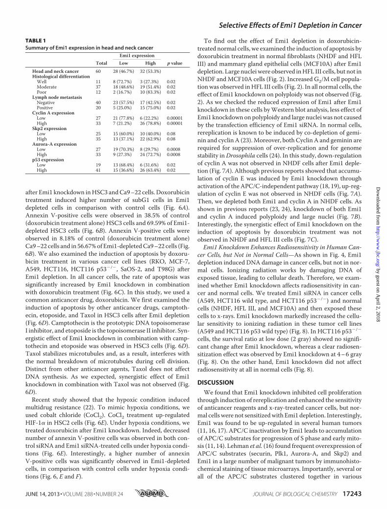
High Expression of Emi1 Is Frequently Observed in CancerTissues—Recent reports show that high expression of Emi1protein can be observed in various cancers (14, 16, 17).We alsofound Emi1 mRNA overexpression in head and neck cancercells (Fig. 1A). Moreover, we investigated Emi1 expression invarious tumor tissues by using the Oncomine database. Ascompared with normal counterparts, Emi1 expression was sig-nificantly increased in many types of tumors, such as head andneck, breast, prostate, cervix, pancreas, and brain tumors (Fig.1B). Then, we examined the expression of Emi1 protein in headand neck cancer cases by immunohistochemistry. Emi1 expres-sion was graded as high (over 30% of tumor cells showing ��or��� intensity) or low (no staining or less than 30%of tumorcells showing� intensity). Indeed,most caseswith high expres-sion of Emi1 showed strong and diffuse immunopositivity andmost cases with low expression of Emi1 showed no or weakimmunopositivity. Expression level of Emi1 in cancer cells wasmuch higher than that in non-neoplastic cells (Fig. 1C). HighEmi1 expressionwas observed in 53.3%of head andneck cancercases and was well correlated with histological differentiationand lymph node metastasis (Table 1). Moreover, we comparedthe expression of Emi1 with p53 and APC/C substrates, cyclinA, Skp2, and Aurora-A in cancer tissues (Fig. 1D and Table 1).Interestingly, Emi1 expression was well correlated with p53expression and APC/C substrates (Table 1).AsEmi1 itself is an E2F target gene, Emi1mRNAoverexpres-
sion might be caused by activated E2F in cancer cells (20).Indeed, Emi1 expression was well correlated with the E2F1 andE2F1 target gene, cyclin E in cancer cell lines, and normal cells(Fig. 1E). However, Emi1 expression was not correlated with
doubling time and mitotic index (Fig. 1E). Accordingly, E2F1knockdown reduced Emi1 expression in cancer cells (Fig. 1F).Emi1 Knockdown Induces Polyploidy in Cancer Cells—As
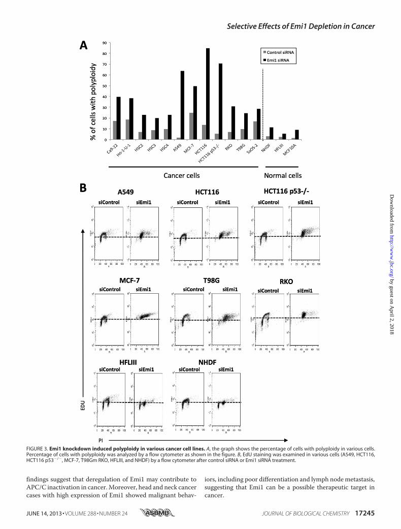
shown in Fig. 1, Emi1 was frequently overexpressed in varioustypes of cancer. Next, we examined the effect of Emi1 knock-down by using siRNA in various types of cancer cell lines. Pre-vious reports showed that Emi1 depletion induced polyploidyand large nuclei because cells cannot complete DNA synthesis(18, 19). Similar to previous reports, we confirmed the effect ofEmi1 knockdown on polyploidy and large nuclei in various can-cer cell lines, including head and neck cancer (Ca9–22, Ho-1-U-1, HSC2, HSC3, and HSC4), lung cancer (A549), breast can-cer (MCF-7), colon cancer (HCT116, HCT116 p53�/�, andRKO), glioma (T98G), and osteosarcoma (SaOS-2) (Fig. 2).Larger nuclei and increased number of cells with polyploidywere observed in all cancer cells after Emi1 siRNA treatment.Moreover, the efficiency of Emi1 knockdown was independentof p53 mutation status (Fig. 2). As shown in Fig. 3A, higherpercentage of cells with polyploidywas observed in cancer cells,in comparison with that in normal cells. Indeed, Emi1siRNA-treated cancer cells but not normal cells showed posi-tive EDU (Fig. 3B), indicating that Emi1 depletion inducedpolyploidy through rereplication in cancer cells.It has been shown that cells lacking Emi1 undergo DNA
damage, likely explained by replication stress upon deregulatedcyclin E- and A-associated kinase activities (20). Here, we exam-ined the nuclear foci containing phosphorylated histone H2AX(�-H2AX), amarker ofDNAdamage foci in Emi1 depleted cancercells (A549, HCT116, and HCT116 p53�/�) and normal cells(HFLIII and neonatal normal human dermal fibroblast (NHDF)).Increased�-H2AXfoci andDNAcontentwereobserved incancercells, but not in normal cells (Fig. 4A). In particular, frequency ofhigh levels of �H2AX foci (more than 10 foci per cell) in cancercells was significantly higher than that in normal cells (Fig. 4B).Most cells did not show annexin V after Emi1 siRNA treatment(Fig. 4C). As the expression of phosphohistone H3 was notobserved in Emi1 siRNA-treated cells (Fig. 4D), the elevated levelof �H2AXwas not caused by arresting atM phase or induction ofapoptosis. These findings show that replication stress caused byEmi1 knockdownmight lead to DNA damage.Next, we examined the detailed mechanism of Emi1 knock-
down in Ca9–22 and Ho-1-U-1 cells. Emi1 is known as aninhibitor of APC/CCdc20 and APC/CCdh1 (12–15) and inhibi-tion of Emi1 interferes with progression to the M phase by theAPC/CCdh1-dependent proteolysis of geminin protein (18, 19).Indeed, expression of Emi1 and APC/CCdh1 substrates such asAurora-A and gemininwas down-regulated by Emi1 siRNA treat-ment in both cells (Fig. 5A). Moreover, Emi1 knockdown signifi-cantly suppressed proliferation (p � 0.05) (Fig. 5B). Then, weexamined theco-depletionofEmi1andCdh1orCdc20 inCa9–22and Ho-1-U-1 cells. Cdh1 depletion corrected Emi1 knockdown-inducedpolyploidy to a certain extent (Fig. 5C).As expected, Emi1knockdown induced down-regulation of APC/CCdh1 substratessuch as Cdc20, cyclin A, cyclin B, Aurora-A, geminin, and TPX2(Fig. 5D). Cdh1 depletion up-regulated these APC/CCdh1 sub-strates (Fig. 5D). Thus, as shown in previous reports (18, 19), Emi1depletion induced rereplication because of geminin down-regula-tion via APCCdh1-dependent proteolysis.
Selective Effects of Emi1 Depletion in Cancer
JUNE 14, 2013 • VOLUME 288 • NUMBER 24 JOURNAL OF BIOLOGICAL CHEMISTRY 17241
by guest on April 2, 2018
http://ww
w.jbc.org/
Dow
nloaded from

Emi1 Knockdown Enhances the Induction of Apoptosis byDoxorubicin in Human Cancer Cells, but Not in Normal Cells—Many anticancer agents used in cancer therapy are intended to
control DNA synthesis. Our observations made us hypothesizethat Emi1 depletion in cancer cells might enhance the induc-tion of apoptosis by anticancer agents. We treated doxorubicin
FIGURE 1. Emi1 overexpression in head and neck cancer. A, Emi1 mRNA expression was examined in 14 head and neck cancer cell lines, 2 normal cells, and20 head and neck cancer tissues by RT-PCR. GAPDH was used as a control. B, mRNA level of Emi1 in different human tumors. All data were provided by theOncomine database. Data from Refs. 43–50 were reanalyzed to show expression level of Emi1 in normal brain, glioblastoma, oligodendroglioma, anaplasticoligodendroglioma, astrocytoma, head and neck cancer, breast cancer, pancreatic ductal adenocarcinoma, cervical cancer, transitional cell carcinoma, andadult male germ cell tumor (43–50). N, normal tissues; T, tumor tissues. C, immunohistochemical expression of Emi1 in head and neck cancer. D, correlationbetween Emi1 expression and APC substrates, cyclin A, and Skp2 expression. We examined the expression of Emi1, cyclin A, and Skp2 in head and neck cancercases. A representative head and neck cancer case is shown. E, high expression of Emi1 is regulated by E2F1 in cancer cells. Expression of Emi1, E2F1, and cyclinE was examined by Western blot analysis in cancer cell lines (HSC2, HSC3, HSC4, Ca9 –22, Ho-1-N-1, Ho-1-U-1, HeLa, A549, HCT116, HCT116 p53�/�, T98G,SaOS-2, MCF-7, and RKO) and normal cells (HFL III and NHDF). �-Actin expression was used as a loading control. The graph shows the mitotic index (%) anddoubling time (h) in each cell. F, E2F1 siRNA was transfected into HCT116 and HSC2 cells. After 48 h of E2F1 siRNA treatment, cells were collected. Emi1 and E2F1expression was examined by Western blot analysis. �-Actin expression was used as a loading control.
Selective Effects of Emi1 Depletion in Cancer
17242 JOURNAL OF BIOLOGICAL CHEMISTRY VOLUME 288 • NUMBER 24 • JUNE 14, 2013
by guest on April 2, 2018
http://ww
w.jbc.org/
Dow
nloaded from
after Emi1 knockdown inHSC3 andCa9–22 cells. Doxorubicintreatment induced higher number of subG1 cells in Emi1depleted cells in comparison with control cells (Fig. 6A).Annexin V-positive cells were observed in 38.5% of control(doxorubicin treatment alone) HSC3 cells and 69.59% of Emi1-depleted HSC3 cells (Fig. 6B). Annexin V-positive cells wereobserved in 8.18% of control (doxorubicin treatment alone)Ca9–22 cells and in 56.67%of Emi1-depletedCa9–22 cells (Fig.6B). We also examined the induction of apoptosis by doxoru-bicin treatment in various cancer cell lines (RKO, MCF-7,A549, HCT116, HCT116 p53�/�, SaOS-2, and T98G) afterEmi1 depletion. In all cancer cells, the rate of apoptosis wassignificantly increased by Emi1 knockdown in combinationwith doxorubicin treatment (Fig. 6C). In this study, we used acommon anticancer drug, doxorubicin. We first examined theinduction of apoptosis by other anticancer drugs, camptoth-ecin, etoposide, and Taxol in HSC3 cells after Emi1 depletion(Fig. 6D). Camptothecin is the prototypic DNA topoisomeraseI inhibitor, and etoposide is the topoisomerase II inhibitor. Syn-ergistic effect of Emi1 knockdown in combination with camp-tothecin and etoposide was observed in HSC3 cells (Fig. 6D).Taxol stabilizes microtubules and, as a result, interferes withthe normal breakdown of microtubules during cell division.Distinct from other anticancer agents, Taxol does not affectDNA synthesis. As we expected, synergistic effect of Emi1knockdown in combination with Taxol was not observed (Fig.6D).Recent study showed that the hypoxic condition induced
multidrug resistance (22). To mimic hypoxia conditions, weused cobalt chloride (CoCl2). CoCl2 treatment up-regulatedHIF-1� in HSC2 cells (Fig. 6E). Under hypoxia conditions, wetreated doxorubicin after Emi1 knockdown. Indeed, decreasednumber of annexin V-positive cells was observed in both con-trol siRNA and Emi1 siRNA-treated cells under hypoxia condi-tions (Fig. 6E). Interestingly, a higher number of annexinV-positive cells was significantly observed in Emi1-depletedcells, in comparison with control cells under hypoxia condi-tions (Fig. 6, E and F).
To find out the effect of Emi1 depletion in doxorubicin-treated normal cells, we examined the induction of apoptosis bydoxorubicin treatment in normal fibroblasts (NHDF and HFLIII) and mammary gland epithelial cells (MCF10A) after Emi1depletion. Large nucleiwere observed inHFL III cells, but not inNHDF andMCF10A cells (Fig. 2). Increased G2/M cell popula-tionwas observed inHFL III cells (Fig. 2). In all normal cells, theeffect of Emi1 knockdown on polyploidy was not observed (Fig.2). As we checked the reduced expression of Emi1 after Emi1knockdown in these cells byWestern blot analysis, less effect ofEmi1 knockdownonpolyploidy and large nuclei was not causedby the transfection efficiency of Emi1 siRNA. In normal cells,rereplication is known to be induced by co-depletion of gemi-nin and cyclin A (23).Moreover, both Cyclin A and geminin arerequired for suppression of over-replication and for genomestability inDrosophila cells (24). In this study, down-regulationof cyclin A was not observed in NHDF cells after Emi1 deple-tion (Fig. 7A). Although previous reports showed that accumu-lation of cyclin E was induced by Emi1 knockdown throughactivation of theAPC/C-independent pathway (18, 19), up-reg-ulation of cyclin E was not observed in NHDF cells (Fig. 7A).Then, we depleted both Emi1 and cyclin A in NHDF cells. Asshown in previous reports (23, 24), knockdown of both Emi1and cyclin A induced polyploidy and large nuclei (Fig. 7B).Interestingly, the synergistic effect of Emi1 knockdown on theinduction of apoptosis by doxorubicin treatment was notobserved in NHDF and HFL III cells (Fig. 7C).Emi1 Knockdown Enhances Radiosensitivity in Human Can-
cer Cells, but Not in Normal Cells—As shown in Fig. 4, Emi1depletion induced DNA damage in cancer cells, but not in nor-mal cells. Ionizing radiation works by damaging DNA ofexposed tissue, leading to cellular death. Therefore, we exam-ined whether Emi1 knockdown affects radiosensitivity in can-cer and normal cells. We treated Emi1 siRNA in cancer cells(A549, HCT116 wild type, and HCT116 p53�/�) and normalcells (NHDF, HFL III, and MCF10A) and then exposed thesecells to x-rays. Emi1 knockdown markedly increased the cellu-lar sensitivity to ionizing radiation in these tumor cell lines(A549 and HCT116 p53 wild type) (Fig. 8). In HCT116 p53�/�
cells, the survival ratio at low dose (2 gray) showed no signifi-cant change after Emi1 knockdown, whereas a clear radiosen-sitization effect was observed by Emi1 knockdown at 4–6 gray(Fig. 8). On the other hand, Emi1 knockdown did not affectradiosensitivity at all in normal cells (Fig. 8).
DISCUSSION
We found that Emi1 knockdown inhibited cell proliferationthrough induction of rereplication and enhanced the sensitivityof anticancer reagents and x-ray-treated cancer cells, but nor-mal cells were not sensitizedwith Emi1 depletion. Interestingly,Emi1 was found to be up-regulated in several human tumors(11, 16, 17). APC/C inactivation by Emi1 leads to accumulationof APC/C substrates for progression of S phase and early mito-sis (11, 14). Lehman et al. (16) found frequent overexpression ofAPC/C substrates (securin, Plk1, Aurora-A, and Skp2) andEmi1 in a large number of malignant tumors by immunohisto-chemical staining of tissue microarrays. Importantly, several orall of the APC/C substrates clustered together in various
TABLE 1Summary of Emi1 expression in head and neck cancer
TotalEmi1 expression
p valueLow High
Head and neck cancer 60 28 (46.7%) 32 (53.3%)Histological differentiationWell 11 8 (72.7%) 3 (27.3%) 0.02Moderate 37 18 (48.6%) 19 (51.4%) 0.02Poor 12 2 (16.7%) 10 (83.3%) 0.02
Lymph node metastasisNegative 40 23 (57.5%) 17 (42.5%) 0.02Positive 20 5 (25.0%) 15 (75.0%) 0.02
Cyclin A expressionLow 27 21 (77.8%) 6 (22.2%) 0.00001High 33 7 (21.2%) 26 (78.8%) 0.00001
Skp2 expressionLow 25 15 (60.0%) 10 (40.0%) 0.08High 35 13 (37.1%) 22 (62.9%) 0.08
Aurora-A expressionLow 27 19 (70.3%) 8 (29.7%) 0.0008High 33 9 (27.3%) 24 (72.7%) 0.0008
p53 expressionLow 19 13 (68.4%) 6 (31.6%) 0.02High 41 15 (36.6%) 26 (63.4%) 0.02
Selective Effects of Emi1 Depletion in Cancer
JUNE 14, 2013 • VOLUME 288 • NUMBER 24 JOURNAL OF BIOLOGICAL CHEMISTRY 17243
by guest on April 2, 2018
http://ww
w.jbc.org/
Dow
nloaded from

tumors with elevated Emi1 protein (16). Moreover, Emi1 over-expression leads to unscheduled cell proliferation, tetraploidy,and chromosomal instability in p53-deficient cells (25). Nota-bly, Emi1 is the target of E2F transcription factors (11), poten-tially linking the frequent deregulation of the E2F pathway toAPC/Cderegulation. This observation is supported by our find-
ings that (i) Emi1 expression was well correlated with E2F1expression in cancer cells (Fig. 1E), and (ii) E2F1 knockdowndown-regulated Emi1 expression (Fig. 1F). We also confirmedthat Emi1 is frequently overexpressed in various cancers,including head and neck cancers, and that Emi1 expression iswell correlated with Aurora-A and Skp2 expression. These
FIGURE 2. Emi1 knockdown in various cancer cell lines. Emi1 siRNA was transfected into various cancer cell lines, including head and neck cancer (Ca9 –22,Ho-1-U-1, HSC2, HSC3, and HSC4), lung cancer (A549), breast cancer (MCF-7), colon cancer (RKO, HCT116, and HCT116 p53�/�), glioma (T98G), and osteosar-coma (SaOS-2). Em1i siRNA was also transfected into normal cells, including NHDF (dermal fibroblast), HFL III (fibroblasts), and MCF10A (mammary glandepithelial cells). The status of p53 was shown in each cell line. Cells were stained with DAPI to visualize the nuclei. Cell cycle distribution was determined by DNAcontent analysis after PI staining using a flow cytometer. Scale bar, 100 �m. IF, immunofluorescence.
Selective Effects of Emi1 Depletion in Cancer
17244 JOURNAL OF BIOLOGICAL CHEMISTRY VOLUME 288 • NUMBER 24 • JUNE 14, 2013
by guest on April 2, 2018
http://ww
w.jbc.org/
Dow
nloaded from
findings suggest that deregulation of Emi1 may contribute toAPC/C inactivation in cancer.Moreover, head and neck cancercases with high expression of Emi1 showed malignant behav-
iors, including poor differentiation and lymph nodemetastasis,suggesting that Emi1 can be a possible therapeutic target incancer.
FIGURE 3. Emi1 knockdown induced polyploidy in various cancer cell lines. A, the graph shows the percentage of cells with polyploidy in various cells.Percentage of cells with polyploidy was analyzed by a flow cytometer as shown in the figure. B, EdU staining was examined in various cells (A549, HCT116,HCT116 p53�/�, MCF-7, T98Gm RKO, HFLIII, and NHDF) by a flow cytometer after control siRNA or Emi1 siRNA treatment.
Selective Effects of Emi1 Depletion in Cancer
JUNE 14, 2013 • VOLUME 288 • NUMBER 24 JOURNAL OF BIOLOGICAL CHEMISTRY 17245
by guest on April 2, 2018
http://ww
w.jbc.org/
Dow
nloaded from

DNA replication depends on the APC/C-mediated degrada-tion of geminin, which releases the inhibition of Cdt1 incorpo-ration into prereplicative complexes and permits the properassembly of these complex components (26, 27). Origin firingcan only occur after the APC/C is inactivated and CDKsbecome active. To strictly inhibit the assembly of prereplicativecomplexes during S to G2 phase transition, APC/C is inacti-
vated by several mechanisms: binding to its inhibitor Emi1,CDK-mediated phosphorylation of Cdh1 (which blocks its abil-ity to activate APC/C), and degradation of both Cdh1 (28) andits major ubiquitin C, namely UbcH10 (29). Indeed, Emi1depletion induced rereplication with large nuclei and poly-ploidy in various cancer cells (Fig. 2), and Cdh1 rescued thedown-regulation of geminin and rereplication caused by Emi1
FIGURE 4. DNA damage in Emi1 depleted cancer cells. A, Emi1 knockdown induce �H2AX foci in tumor cells. Tumor cells (A549, HCT116, and HCT116 p53�/�)and normal cells (HFLIII and NHDF) were transfected with the indicated siRNA. After 48 h the transfection, cells were fixed and stained with anti-�H2AXantibodies. The levels of H2AX phosphorylation were detected using by the flow cytometer. Bivariate distributions representing expression of �H2AX versusDNA content of tumor and normal cells are shown. The levels of H2AX phosphorylation were also detected using by the fluorescence microscope. Represent-ative images of �H2AX immunostaining (green) and nuclear staining (blue) in tumor or normal cells are shown. B, images on the left show typical images of cellspresenting low (0�10 foci/cell) or high levels of �H2AX foci or showing pan-nuclear staining �H2AX. The right graph indicates percentages of cells with highlevel (�10 foci/cell) of �H2AX foci. Results presented are the means of two independent experiments � S.D. C, apoptosis was examined by annexin V stainingusing a flow cytometer in various cells (A549, HCT116, HCT116 p53�/�, HFLIII, and NHDF) after control siRNA or Emi1 siRNA treatment. D, expression ofphosphohistone H3 (pHH3) was examined by the fluorescence microscope in A549 and HFLIII cells after control siRNA or Emi1 siRNA treatment. Cells were alsostained with DAPI to visualize the nuclei.
Selective Effects of Emi1 Depletion in Cancer
17246 JOURNAL OF BIOLOGICAL CHEMISTRY VOLUME 288 • NUMBER 24 • JUNE 14, 2013
by guest on April 2, 2018
http://ww
w.jbc.org/
Dow
nloaded from

depletion (Fig. 5C). These findings are consistent with previousfindings showing that (i) rereplication caused by Emi1 deple-tion is due to decreased geminin via APC/C-mediated proteol-ysis (18, 19), and (ii) geminin depletion causes rereplication incancer cells (30). In addition, Emi1 depletion induces DNAdamage, likely explained by replication stress upon deregulatedcyclin E- and A-associated kinase activities (20). We also foundthat Emi1 knockdown induced the frequency of high levels of�H2AX foci (�10 foci per cell) in cancer cells (Fig. 4B). �H2AXfoci are known to be an early response marker for double-stranded breaks (DSBs) (31).Moreover, premature terminationof DNA replication can lead to fork collapse and consequentDSBs, eventually triggering robust activation of the DNA dam-age checkpoint response (32). Therefore, Emi1 depletion islikely to induce DSBs via replication stress. Interestingly, fre-quency of high levels of �H2AX foci by Emi1 depletion in can-cer cells was higher than that in normal cells (Fig. 4B). Chemo-therapy and/or radiation in addition to surgery have provenuseful in a number of different cancer types. At present manyanticancer drugs for targeting DNA synthesis to interfere withrapidly dividing cells are commonly used. Ionizing radiation
works by damaging DNA of exposed tissue leading to cellulardeath. Therefore, the finding that Emi1 depletion inducedrereplication andDNA damagemade us hypothesize that Emi1knockdown might enhance the sensitivity of anticancer drugsand ionizing radiation. Interestingly, in the present study, wedemonstrated that Emi1 knockdown by using siRNA enhancedthe sensitivity of anticancer reagents treatment and x-rays in can-cer cells.Althoughwe found synergistic effect ofEmi1knockdownin combination with doxorubicin, camptothecin, and etoposide(Fig. 6D),we areplanning to examine the synergistic effect of Emi1knockdown in combination with other anticancer drugs thatinterfere with DNA synthesis. Recent study showed that thehypoxic condition induced multidrug resistance (22). Interest-ingly, Emi1 siRNA enhanced the sensitivity of anticancer drugsunder hypoxia condition (Fig. 6, E and F), indicating that Emi1depletion can be a useful for clinical application.It is likely that the radioresistance is strong in the S phase
because the DNA repair enzymes that allow radiation-induceddamage to appear in DNA are at full activity during S phase andless so at other stages (33). The G2/M phase of the cell cycle iswhen cells are most sensitive to radiation (33). Indeed, pacli-
FIGURE 5. DNA damage and polyploidy by Emi1 knockdown via activation of APC/CCdh1. A, Emi1 siRNA was transfected into HSC3 or Ca9 –22 cells. After48 h of Emi1 siRNA transfection, cells were collected. The indicated proteins were examined by Western blotting. B, Emi1 siRNA inhibits cell growth in head andneck cancer cells. Cell growth of Ho-1-U-1 cells after Emi1 siRNA transfection. After 48 h of transfection, cells were plated in 24-well plates. Cells were countedby Cell Counter at 0, 2, 4, and 6 days. We assayed three times. C, Emi1 siRNA induces polyploidy through activation of APC/C. Co-depletion of Cdh1 or Cdc20 withEmi1 by siRNA in Ca9 –22 and Ho-1-U-1 cells. These cells were transfected with the indicated siRNAs. Cell cycle distribution was determined by DNA contentanalysis after PI staining using a flow cytometer. D, co-depletion of Cdh1 or Cdc20 with Emi1 by siRNA in Ca9 –22 and Ho-1-U-1 cells. The indicated proteinsincluding APC/CCdh1 substrates in siRNA-treated cells were examined by Western blotting.
Selective Effects of Emi1 Depletion in Cancer
JUNE 14, 2013 • VOLUME 288 • NUMBER 24 JOURNAL OF BIOLOGICAL CHEMISTRY 17247
by guest on April 2, 2018
http://ww
w.jbc.org/
Dow
nloaded from

Selective Effects of Emi1 Depletion in Cancer
17248 JOURNAL OF BIOLOGICAL CHEMISTRY VOLUME 288 • NUMBER 24 • JUNE 14, 2013
by guest on April 2, 2018
http://ww
w.jbc.org/
Dow
nloaded from

taxel, which inhibits the formation of mitotic spindles andarrests the cells in G2/M phase, enhances radiation efficacy oncell killing and suppression of growth (34–36). Moreover, inhi-bition of Aurora kinase enhances the sensitivity of radiation(37–40). Thus, some drugs for inducing cell cycle arrest at Mphase are used for enhancing the sensitivity of radiation.Although Emi1-depleted cells were arrested at S phase by
rereplication decreased survival cells was observed after x-rayexposure. As Emi1 depletion leads to DSBs via replicationstress, DSBs induced by Emi1 depletion may enhance the sen-sitivity of radiation in cancer cells.Emi1 knockdown suppressed cancer cell growthwith contin-
uous DNA synthesis, but normal cells did not seem to beaffected by the knockdown. Therefore, the synergistic effect of
FIGURE 6. Synergistic anticancer effect of Emi1 knockdown in combination with doxorubucin in cancer cells. A, after 48 h of Emi1 siRNA transfection, cellswere treated with doxorubicin (0.5 �g/ml) for 12 h. Cells were fixed in 70% ethanol. Cell cycle distribution was determined by DNA content analysis after PIstaining using a flow cytometer. For each sample, 20,000 events were stored. Percentage of the sub-G1 population is indicated. Representative data are shown.We performed three independent experiments. B, flow cytometric analysis of annexin V and PI staining in control and Emi1 siRNA-treated HSC3 or Ca9 –22 cellsafter treatment with doxorubicin (0.5 �g/ml) for 12 h. We performed three independent experiments. C, synergistic anticancer effect of Emi1 knockdown incombination with doxorubucin (DOXY) in various cancer cells. Graph shows the average percentage of apoptotic cells in various cancer cells after treatmentwith doxorubicin (0.5 �g/ml) for 12 h. In this study, various cancer cell lines including lung cancer (A549), breast cancer (MCF-7), colon cancer (RKO, HCT116, andHCT116 p53�/�), glioma (T98G), and osteosarcoma (SaOS-2) were used. We performed three independent experiments. *, p � 0.05. D, synergistic anticancereffect of Emi1 knockdown in combination with camptothecin and etoposide but not with Taxol in HSC3 cells. The graph shows the average percentage ofapoptotic cells in HSC3 cells after treatment with camtothecin (2 �M), etoposide (1 �M), or Taxol (1 nM) for 12–24 h. We performed two independentexperiments. *, p � 0.05. E, hypoxic condition induced drug resistance. CoCl2 (100 �M) was treated for 6 h in HSC2 cells after 24 h of control or Emi1 siRNAtransfection. Cells were collected and expression of HIF-1� and Emi1 was examined by Western blot analysis. �-Actin expression was used as a loading control.After 6 h of CoCl2 treatment, doxorubicin (DOXY; 0.5 �g/ml) was treated for 12 h in control or Emi1-depleted cells. Graph shows the average percentage ofapoptotic cells (annexin V-positive cells) in HSC2 cells after treatment with doxorubicin for 12 h under normal or hypoxia condition (CoCl2 treatment). Weperformed two independent experiments. *, p � 0.05. F, synergistic anticancer effect of Emi1 knockdown in combination with doxorubicin in HSC3 and HSC4cells under hypoxia conditions. The graph shows the average percentage of apoptotic cells (annexin V-positive cells) in HSC3 and HSC4 cells after treatmentwith doxorubicin for 12 h under normal or hypoxia conditions (CoCl2 treatment). We performed two independent experiments. *, p � 0.05.
FIGURE 7. Synergistic anticancer effect of Emi1 knockdown in combination with doxorubucin in normal cells. A, Emi1 or control siRNA was transfectedinto NHDF cells, and cells were collected after 48 h. The indicated proteins in siRNA-treated NHDF cells were examined by Western blotting. B, Emi1 siRNAand/or cyclin A siRNA were transfected into NHDF cells. The left panel shows the expression of Emi1 and cyclin A examined by Western blot analysis after 48 hof siRNA transfection. �-Actin expression was used as a loading control. The right panel shows DAPI staining and percentage of cells with polyploidy after Emi1siRNA and/or cyclin A siRNA transfection. Cells were stained with DAPI to visualize the nuclei, and percentage of cells with polyploidy was determined by DNAcontent analysis after PI staining using a flow cytometer. *, p � 0.05. C, flow cytometric analysis of annexin V and PI staining in control and Emi1 siRNA treatedNHDF or HFL III cells after treatment with doxorubucin (DOXY; 0.5 �g/ml) for 12 h. We performed three independent experiments.
Selective Effects of Emi1 Depletion in Cancer
JUNE 14, 2013 • VOLUME 288 • NUMBER 24 JOURNAL OF BIOLOGICAL CHEMISTRY 17249
by guest on April 2, 2018
http://ww
w.jbc.org/
Dow
nloaded from

Emi1 knockdown in combination with doxorubicin treatmentand radiation was not observed in normal cells. These findingsare consistent with previous finding that none of the effects aredetected either in normal cells by geminin depletion (23). Innormal cells, both geminin and cyclin A suppress rereplicationand genomic instability (23, 24). In this study, down-regulationof cyclin A was not observed in normal fibroblasts after Emi1depletion (Fig. 7A), whereas down-regulation of cyclin A wasobserved in cancer cells (Fig. 5D). As shown in Fig. 1, overex-pression of Emi1 andAPC/C substrates, including cyclinA, wasobserved in cancer cells. Activated APC/C by Emi1 depletionmay degrade overexpressed APC/C substrates at S-G2 phase incancer cells but not in normal cells. In normal cells, inhibitorymechanism of degradation of APC/C substrates may exist atS-G2 in addition to proper phase for their protein degradation.Indeed, depletion of both Emi1 and cyclinA induced polyploidyand large nuclei (Fig. 7B). Therefore, cyclin A may suppressrereplication innormal cells.Thisobservation is supportedbypre-vious finding that treatment with geminin siRNA and cyclin AsiRNA induced DNA rereplication in normal cells in similar tocancer cells induced by geminin siRNA alone (23). Previousreports showedthataccumulationofcyclinEwas inducedbyEmi1knockdown through the APC/C-independent pathway (18, 19).As cyclin E is not a substrate ofAPC/C, it is still unclearwhy cyclinE is up-regulated by Emi1 depletion. Interestingly, up-regulationof cyclin E was not observed in normal cells (Fig. 7A), suggestingthat induction of cyclin Emay be involved in rereplication only incancer cells. These findings suggest that Emi1may play an impor-tant role in initiatingDNAreplication incancer cells,whereasnor-
mal cells may possess additional safeguards such as cyclin A forpreventing DNA rereplication. Importantly, no effect of Emi1knockdown and no synergistic effect of Emi1 knockdown in com-binationwith doxorubicin treatment and radiation in normal cellsare useful findings for clinical application.The therapeutic utility of siRNAs is thought to be limited by
the requirement for complex formulations to deliver them totissues. Recently, however, it has been identified single-strandsiRNAs for silencing gene expression in animals (41). Indeed,single-strand siRNAs achieved potencies and selectivity forinhibiting mutant huntingtin in Huntington disease modelmice (42). It is anticipated that technologies of siRNA deliverywill be developed for efficient gene silencing in clinical applica-tion.Moreover, we will look for the small compound or peptidefor inhibiting Emi1 function in addition to siRNA. In conclu-sion, we suggest that inhibition of Emi1 function could be auseful for enhancing sensitivity of cancer cells to chemothera-peutic drugs and ionizing radiation.
Acknowledgments—We thank Dr. Michele Pagano (New York Univer-sity) for helpful discussions, Michie Akaishizawa (National Institute ofRadiological Sciences) for expert technical assistance, andDr. RiekoAra-kaki (Tokushima University) and Dr. Keiji Tanimoto (Hiroshima Uni-versity) for technical advice. We also thank Dr. Nishitani (University ofHyogo), Dr. Heidebrecht (University of Kiel), Dr. Fusenig (German Can-cer Research Center), andDr. Vogelstein (The Johns Hopkins University)for providing materials. This work was carried out at the Joint Usage/Research Center (RIRBM), HiroshimaUniversity.
FIGURE 8. Synergistic anticancer effect of Emi1 knockdown in combination with radiation. The effects of Emi1 knockdown on cell survival of A549, HCT116wild type, and HCT116 p53�/�, HFLIII, NHDF, and MCF10A cells. Cells were transfected with Emi1 siRNA or negative control siRNA. After 48 h, cells were exposedto 0 – 6 gray x-rays. Cell survival was determined by a colony formation assay. Data shown are mean and S.E. of three independent experiments.
Selective Effects of Emi1 Depletion in Cancer
17250 JOURNAL OF BIOLOGICAL CHEMISTRY VOLUME 288 • NUMBER 24 • JUNE 14, 2013
by guest on April 2, 2018
http://ww
w.jbc.org/
Dow
nloaded from
REFERENCES1. Jemal, A., Bray, F., Center, M. M., Ferlay, J., Ward, E., and Forman, D.
(2011) Global cancer statistics. CA-Cancer J. Clin. 61, 69–902. Takimoto, C. H., and Calvo, E. (2008) Principles of Oncologic Pharmaco-
therapy in Cancer Management: A Multidisciplinary Approach (Pazdur,R., Wagman, L. D., Camphausen, K. A., Hoskins W. J., eds) 11th Ed. pp.1–9, Cmp United Business Media, Manhasset, NY
3. Pommier, Y., Leteurtre, F., Fesen, M. R., Fujimori, A., Bertrand, R., Solary,E., Kohlhagen, G., and Kohn, K. W. (1994) Cellular determinants of sen-sitivity and resistance to DNA topoisomerase inhibitors. Cancer Invest.12, 530–542
4. Dancey, J. E., Bedard, P. L., Onetto, N., and Hudson, T. J. (2012) Thegenetic basis for cancer treatment decisions. Cell 148, 409–420
5. Reed, S. (2003) Ratchets and clocks: the cell cycle, ubiquitylation and pro-tein turnover. Nat. Rev. Mol. Cell Biol. 4, 855–864
6. Pagano, M., and Benmaamar, R. (2003) When protein destruction runsamok, malignancy is on the loose. Cancer Cell 4, 251–256
7. Yamasaki, L., and Pagano, M. (2004) Cell cycle, proteolysis and cancer.Curr. Opin. Cell Biol. 16, 623–628
8. Nakayama, K. I., and Nakayama, K. (2006) Ubiquitin ligases: cell-cyclecontrol and cancer. Nat. Rev. Cancer 6, 369–381
9. Peters, J. M. (2006) The anaphase promoting complex/cyclosome: a ma-chine designed to destroy. Nat. Rev. Mol. Cell Biol. 7, 644–656
10. Cardozo, T., and Pagano,M. (2004) The SCF ubiquitin ligase: insights intoa molecular machine. Nat. Rev. Mol. Cell Biol. 5, 739–751
11. Hsu, J. Y., Reimann, J. D., Sørensen, C. S., Lukas, J., and Jackson, P. K.(2002) E2F-dependent accumulation of hEmi1 regulates S phase entry byinhibiting APCCdh1. Nat. Cell Biol. 4, 358–366
12. Reimann, J. D., Gardner, B. E., Margottin-Goguet, F., and Jackson, P. K.(2001) Emi1 regulates the anaphase-promoting complex by a differentmechanism than Mad2 proteins. Genes Dev. 15, 3278–3285
13. Reimann, J. D., Freed, E., Hsu, J. Y., Kramer, E. R., Peters, J.M., and Jackson,P. K. (2001) Emi1 is a mitotic regulator that interacts with Cdc20 andinhibits the anaphase promoting complex. Cell 105, 645–655
14. Margottin-Goguet, F., Hsu, J. Y., Loktev, A., Hsieh, H.M., Reimann, J. D., andJackson, P. K. (2003) Prophase destruction of Emi1 by the SCF�TrCP/Slimb
ubiquitin ligase activates the anaphase promoting complex to allow progres-sion beyond prometaphase.Dev. Cell 4, 813–826
15. Guardavaccaro, D., Kudo, Y., Boulaire, J., Barchi, M., Busino, L., Donzelli,M., Margottin-Goguet, F., Jackson, P. K., Yamasaki, L., and Pagano, M.(2003) Control of meiotic and mitotic progression by the F box protein�-Trcp1 in vivo. Dev. Cell 4, 799–812
16. Lehman, N. L., Tibshirani, R., Hsu, J. Y., Natkunam, Y., Harris, B. T.,West,R. B., Masek, M. A., Montgomery, K., van de Rijn, M., and Jackson, P. K.(2007) Oncogenic regulators and substrates of the anaphase promotingcomplex/cyclosome are frequently overexpressed in malignant tumors.Am. J. Pathol. 170, 1793–1805
17. Gütgemann, I., Lehman, N. L., Jackson, P. K., and Longacre, T. A. (2008)Emi1 protein accumulation implicatesmisregulation of the anaphase pro-moting complex/cyclosome pathway in ovarian clear cell carcinoma.Mod. Pathol. 21, 445–454
18. Machida, Y. J., and Dutta, A. (2007) The APC/C inhibitor, Emi1, is essen-tial for prevention of rereplication. Genes Dev. 21, 184–194
19. Di Fiore, B., and Pines, J. (2007) Emi1 is needed to couple DNA replicationwith mitosis but does not regulate activation of the mitotic APC/C. J. CellBiol. 177, 425–437
20. Verschuren, E. W., Ban, K. H., Masek, M. A., Lehman, N. L., and Jackson,P. K. (2007) Loss of Emi1-dependent anaphase-promoting complex/cy-closome inhibition deregulates E2F target expression and elicits DNAdamage- induced senescence.Mol. Cell. Biol. 27, 7955–7965
21. Yokoyama, K., Kamata, N., Fujimoto, R., Tsutsumi, S., Tomonari, M.,Taki, M., Hosokawa, H., andNagayama,M. (2003) Increased invasion andmatrix metalloproteinase-2 expression by Snail-induced mesenchymaltransition in squamous cell carcinomas. Int. J. Oncol. 22, 891–898
22. Onozuka, H., Tsuchihara, K., and Esumi, H. (2011) Hypoglycemic/hy-poxic condition in vitro mimicking the tumor microenvironment mark-edly reduced the efficacy of anticancer drugs. Cancer Sci. 102, 975–982
23. Zhu,W., and Depamphilis, M. L. (2009) Selective killing of cancer cells bysuppression of geminin activity. Cancer Res. 69, 4870–4877
24. Mihaylov, I. S., Kondo, T., Jones, L., Ryzhikov, S., Tanaka, J., Zheng, J.,Higa, L. A., Minamino, N., Cooley, L., and Zhang, H. (2002) Control ofDNA replication and chromosome ploidy by geminin and cyclin A. Mol.Cell. Biol. 22, 1868–1880
25. Lehman, N. L., Verschuren, E. W., Hsu, J. Y., Cherry, A. M., and Jackson,P. K. (2006) Overexpression of the anaphase promoting complex/cyclo-some inhibitor Emi1 leads to tetraploidy and genomic instability of p53-deficient cells. Cell Cycle 5, 1569–1573
26. Wohlschlegel, J. A., Dwyer, B. T., Dhar, S. K., Cvetic, C., Walter, J. C., andDutta, A. (2000) Inhibition of eukaryotic DNA replication by gemininbinding to Cdt1. Science 290, 2309–2312
27. Tada, S., Li, A., Maiorano, D., Méchali, M., and Blow, J. J. (2001) Repres-sion of origin assembly in metaphase depends on inhibition of RLF-B/Cdt1 by geminin. Nat. Cell Biol. 3, 107–113
28. Listovsky, T., Oren, Y. S., Yudkovsky, Y., Mahbubani, H.M.,Weiss, A.M.,Lebendiker, M., and Brandeis, M. (2004) Mammalian Cdh1/Fzr mediatesits own degradation. EMBO J. 23, 1619–1626
29. Rape, M., and Kirschner, M. W. (2004) Autonomous regulation of theanaphase-promoting complex couples mitosis to S-phase entry. Nature432, 588–595
30. Melixetian, M., Ballabeni, A., Masiero, L., Gasparini, P., Zamponi, R., Bar-tek, J., Lukas, J., andHelin, K. (2004) Loss ofGeminin induces rereplicationin the presence of functional p53. J. Cell Biol. 165, 473–482
31. Rogakou, E. P., Pilch, D. R., Orr, A. H., Ivanova, V. S., and Bonner, W. M.(1998) DNA double-stranded breaks induce histone H2AX phosphoryla-tion on serine 139. J. Biol. Chem. 273, 5858–5868
32. Stiff, T., Walker, S. A., Cerosaletti, K., Goodarzi, A. A., Petermann, E.,Concannon, P., O’Driscoll, M., and Jeggo, P. A. (2006) ATR-dependentphosphorylation and activation of ATM in response to UV treatment orreplication fork stalling. EMBO J. 25, 5775–5782
33. Terashima, T., and Tolmach, L. J. (1963) X-ray sensitivity and DNA syn-thesis in synchronous populations of HeLa cells. Science 140, 490–492
34. Tishler, R. B., Geard, C. R., Hall, E. J., and Schiff, P. B. (1992) Taxol sensi-tizes human astrocytoma cells to radiation. Cancer Res. 52, 3495–3497
35. Milas, L., Hunter, N. R., Mason, K. A., Kurdoglu, B., and Peters, L. J. (1994)Enhancement of tumor radioresponse of a murine mammary carcinomaby paclitaxel. Cancer Res. 54, 3506–3510
36. Zhang, A. L., Russell, P. J., Knittel, T., and Milross, C. (2007) Paclitaxelenhanced radiation sensitization for the suppression of human prostatecancer tumor growth via a p53 independent pathway. Prostate 67,1630–1640
37. Guan, Z., Wang, X. R., Zhu, X. F., Huang, X. F., Xu, J., Wang, L. H., Wan,X. B., Long, Z. J., Liu, J. N., Feng, G. K., Huang,W., Zeng, Y. X., Chen, F. J.,and Liu, Q. (2007) Aurora-A, a negative prognostic marker, increasesmigration and decreases radiosensitivity in cancer cells. Cancer Res. 67,10436–10444
38. Tao, Y., Zhang, P., Frascogna, V., Lecluse, Y., Auperin, A., Bourhis, J., andDeutsch, E. (2007) Enhancement of radiation response by inhibition ofAurora-A kinase using siRNA or a selective Aurora kinase inhibitorPHA680632 in p53-deficient cancer cells. Br. J. Cancer 97, 1664–1672
39. Moretti, L., Niermann, K., Schleicher, S., Giacalone, N. J., Varki, V., Kim,K. W., Kopsombut, P., Jung, D. K., and Lu, B. (2011) MLN8054, a smallmolecule inhibitor of aurora kinase a, sensitizes androgen-resistant pros-tate cancer to radiation. Int. J. Radiat. Oncol. Biol. Phys. 80, 1189–1197
40. Niermann, K. J., Moretti, L., Giacalone, N. J., Sun, Y., Schleicher, S. M.,Kopsombut, P., Mitchell, L. R., Kim, K. W., and Lu, B. (2011) Enhancedradiosensitivity of androgen-resistant prostate cancer: AZD1152-medi-ated Aurora kinase B inhibition. Radiation Res. 175, 444–451
41. Lima,W. F., Prakash, T. P., Murray, H.M., Kinberger, G. A., Li,W., Chap-pell, A. E., Li, CS, Murray, S. F., Gaus, H., Seth, P. P., Swayze, E. E., andCrooke, S. T. (2012) Single-stranded siRNAs activate RNAi in animals.Cell 150, 883–894
42. Yu, D., Pendergraff, H., Liu, J., Kordasiewicz, H. B., Cleveland, D. W.,Swayze, E. E., Lima, W. F., Crooke, S. T., Prakash, T. P., and Corey, D. R.(2012) Single-stranded RNAs use RNAi to potently and allele-selectivelyinhibit mutant Huntingtin expression. Cell 150, 895–908
Selective Effects of Emi1 Depletion in Cancer
JUNE 14, 2013 • VOLUME 288 • NUMBER 24 JOURNAL OF BIOLOGICAL CHEMISTRY 17251
by guest on April 2, 2018
http://ww
w.jbc.org/
Dow
nloaded from

43. Buchholz, M., Braun, M., Heidenblut, A., Kestler, H. A., Klöppel, G.,Schmiegel,W., Hahn, S. A., Lüttges, J., and Gress, T.M. (2005) Transcrip-tome analysis of microdissected pancreatic intraepithelial neoplastic le-sions. Oncogene 24, 6626–6636
44. Dyrskjøt, L., Kruhøffer, M., Thykjaer, T., Marcussen, N., Jensen, J. L.,Møller, K., and Ørntoft, T. F. (2004) Gene expression in the urinary blad-der: a common carcinoma in situ gene expression signature exists disre-garding histopathological classification. Cancer Res. 64, 4040–4048
45. French, P. J., Swagemakers, S.M., Nagel, J. H., Kouwenhoven,M. C., Brou-wer, E., van der Spek, P., Luider, T.M., Kros, J. M., van den Bent, M. J., andSillevis Smitt, P. A. (2005) Gene expression profiles associated with treat-ment response in oligodendrogliomas. Cancer Res. 65, 11335–11344
46. Ginos, M. A., Page, G. P., Michalowicz, B. S., Patel, K. J., Volker, S. E.,Pambuccian, S. E., Ondrey, F. G., Adams, G. L., and Gaffney, P. M. (2004)Identification of a gene expression signature associated with recurrentdisease in squamous cell carcinoma of the head and neck. Cancer Res. 64,55–63
47. Korkola, J. E., Houldsworth, J., Chadalavada, R. S., Olshen, A. B., Dobrzyn-
ski, D., Reuter, V. E., Bosl, G. J., and Chaganti, R. S. (2006) Down-regula-tion of stem cell genes, including those in a 200-kb gene cluster at12p13.31, is associated with in vivo differentiation of human male germcell tumors. Cancer Res. 66, 820–827
48. Pyeon, D., Newton, M. A., Lambert, P. F., den Boon, J. A., Sengupta, S.,Marsit, C. J.,Woodworth, C. D., Connor, J. P., Haugen, T. H., Smith, E.M.,Kelsey, K. T., Turek, L. P., andAhlquist, P. (2007) Fundamental differencesin cell cycle deregulation in human papillomavirus-positive and humanpapillomavirus-negative head/neck and cervical cancers. Cancer Res. 67,4605–4619
49. Richardson, A. L., Wang, Z. C., De Nicolo, A., Lu, X., Brown, M., Miron,A., Liao, X., Iglehart, J. D., Livingston, D. M., and Ganesan, S. (2006) Xchromosomal abnormalities in basal-like human breast cancer. CancerCell 9, 121–132
50. Sun, L., Hui, A. M., Su, Q., Vortmeyer, A., Kotliarov, Y., Pastorino, S.,Passaniti, A., Menon, J., Walling, J., Bailey, R., Rosenblum, M., Mikkelsen,T., and Fine, H. A. (2006) Neuronal and glioma-derived stem cell factorinduces angiogenesis within the brain. Cancer Cell 9, 287–300
Selective Effects of Emi1 Depletion in Cancer
17252 JOURNAL OF BIOLOGICAL CHEMISTRY VOLUME 288 • NUMBER 24 • JUNE 14, 2013
by guest on April 2, 2018
http://ww
w.jbc.org/
Dow
nloaded from

Naozumi Ishimaru, Takashi Takata and Yasusei KudoHidehiko Kawai, Ryoichi Hirayama, Akira Fujimori, Akiko Yamada, Ryuichi Okayasu,
Natsumi Shimizu, Nakako Izumi Nakajima, Takaaki Tsunematsu, Ikuko Ogawa,Sensitivity of Doxorubicin or X-ray Treatment in Human Cancer Cells
Selective Enhancing Effect of Early Mitotic Inhibitor 1 (Emi1) Depletion on the
doi: 10.1074/jbc.M112.446351 originally published online May 3, 20132013, 288:17238-17252.J. Biol. Chem.
10.1074/jbc.M112.446351Access the most updated version of this article at doi:
Alerts:
When a correction for this article is posted•
When this article is cited•
to choose from all of JBC's e-mail alertsClick here
http://www.jbc.org/content/288/24/17238.full.html#ref-list-1
This article cites 49 references, 18 of which can be accessed free at
by guest on April 2, 2018
http://ww
w.jbc.org/
Dow
nloaded from