section b: protein structureyang xu, college of life sciences section b protein structure b2 protein...
TRANSCRIPT

Section B: Protein Structure Yang Xu, College of Life Sciences
Section BProtein Structure
B2 Protein structure
B3 Protein Analysis
Section B: Protein Structure Yang Xu, College of Life Sciences
B2 Protein structure
• Primary structure
• Secondary structure
• Tertiary structure
• Quaternary structure
• Domains and motifs

Section B: Protein Structure Yang Xu, College of Life Sciences
Primary structure• Definition:
– the sequence of amino acids from the N to the C terminus is the primary structure of the polypeptide.
• Amide bond (peptide bond ) • Dipeptide• Polypeptide• Sizes for single polypeptide chains:
– within the range 100-1500 amino acids, though longer and shorter ones exist.

Section B: Protein Structure Yang Xu, College of Life Sciences
Secondary structure• Definition: Since the formation of the
hydrogen bonds (C=O•••N-H) between peptide bond units of the polypeptide backbone , polypeptides fold into several regular structures, include:
-helix: The polypeptide backbone forms a right-handed helix with 3.6 amino acid residues per turn so that each peptide N-H group is hydrogen bonded to the C=O group of the peptide bond three residues away.
3.613 3.010 4.416

Section B: Protein Structure Yang Xu, College of Life Sciences
Secondary structure -pleated sheet (-sheet): is formed by hydrogen bonding of
the peptide bond N - H and C=O groups to the complementary groups of another section of the polypeptide chain, include parallel -Sheets and antiparallel -Sheets.

Section B: Protein Structure Yang Xu, College of Life Sciences
Tertiary structure
• Definition:
The different sections of -helix, -sheet, and other minor secondary structures and connecting loops fold in three dimensions, it is the tertiary structure of the polypeptide.
• The bond forces:
Various types of non-covalent forces between side chains hold the tertiary structure together: van der Waals forces, hydrogen bonds, salt bridges.

Section B: Protein Structure Yang Xu, College of Life Sciences
Tertiary structure • In addition, covalent di-sulfide bonds can form between
two cysteine residues which may be far apart in the primary structure but close together in the folded tertiary structure.
Section B: Protein Structure Yang Xu, College of Life Sciences
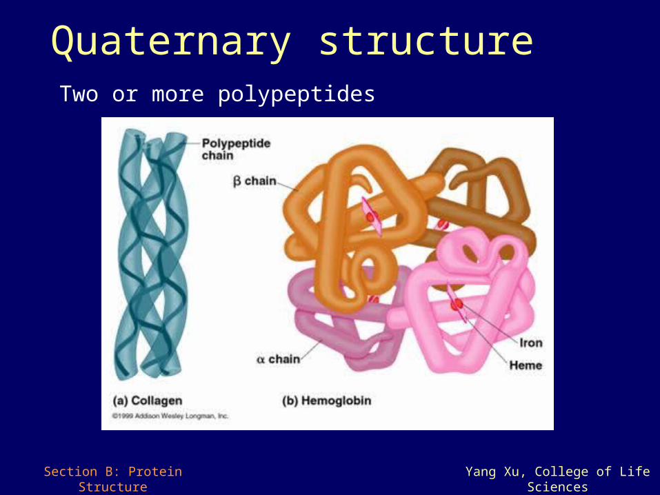
Quaternary structure Two or more polypeptides
Section B: Protein Structure Yang Xu, College of Life Sciences
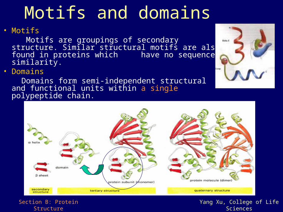
Motifs and domains• Motifs Motifs are groupings of secondary structure. Similar
structural motifs are also found in proteins which have no sequence similarity.
• Domains Domains form semi-independent structural and
functional units within a single polypeptide chain.
Section B: Protein Structure Yang Xu, College of Life Sciences
B3 Protein Analysis
• Protein purification– Size – Charge– Affinity– Two dimensional electrophoresis
• Protein sequencing
Section B: Protein Structure Yang Xu, College of Life Sciences
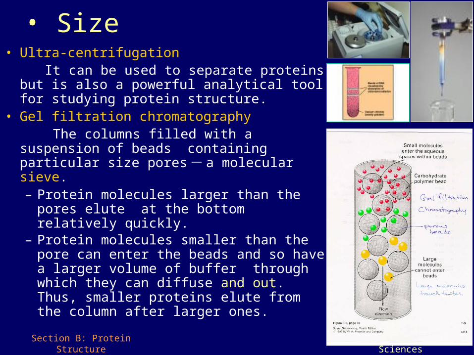
• Size• Ultra-centrifugation It can be used to separate proteins but is also a
powerful analytical tool for studying protein structure.
• Gel filtration chromatography The columns filled with a suspension of beads
containing particular size pores - a molecular sieve. – Protein molecules larger than the pores elute
at the bottom relatively quickly. – Protein molecules smaller than the pore can
enter the beads and so have a larger volume of buffer through which they can diffuse and out. Thus, smaller proteins elute from the column after larger ones.

Section B: Protein Structure Yang Xu, College of Life Sciences
• Charge
• Electrophoresis: protein mixture is applied to a gel and an electric field across the gel. Depending on their net charge, proteins will travel at different rates towards the anode or cathode and can be recovered from the gel after separation.
• Ion-exchange chromatography: ions that are electrostatically bound to the ion exchanger packed in a column are reversibly replaced with charged proteins from solution. Then, a salt gradient of increasing ionic strength is passed through the column and the bound proteins elute separately.
protein DNA & RNA
Section B: Protein Structure Yang Xu, College of Life Sciences
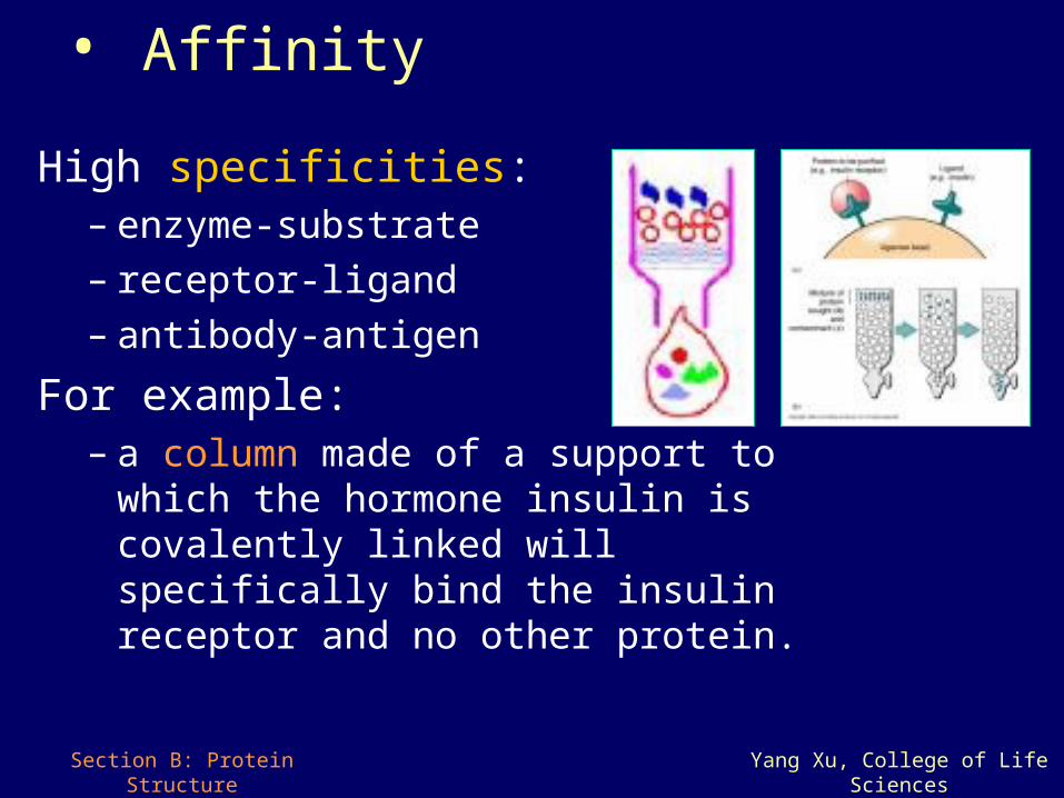
• Affinity
High specificities:– enzyme-substrate– receptor-ligand– antibody-antigen
For example:– a column made of a support to which the
hormone insulin is covalently linked will specifically bind the insulin receptor and no other protein.
Section B: Protein Structure Yang Xu, College of Life Sciences
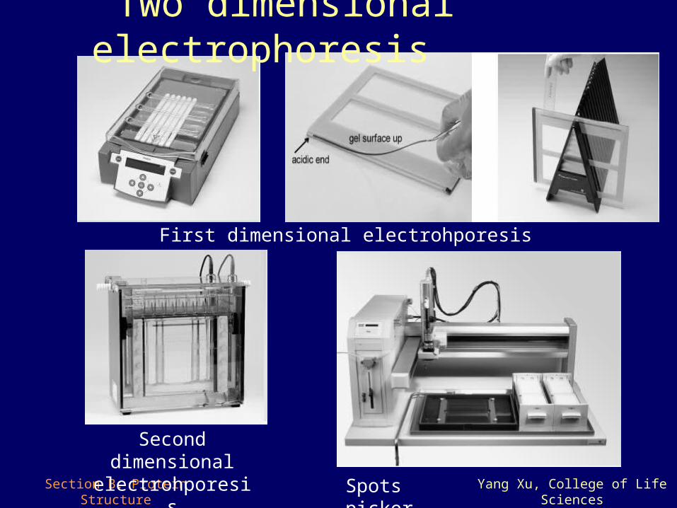
First dimensional electrohporesis
Second dimensional electrohporesis
Spots picker
Two dimensional electrophoresis
Section B: Protein Structure Yang Xu, College of Life Sciences
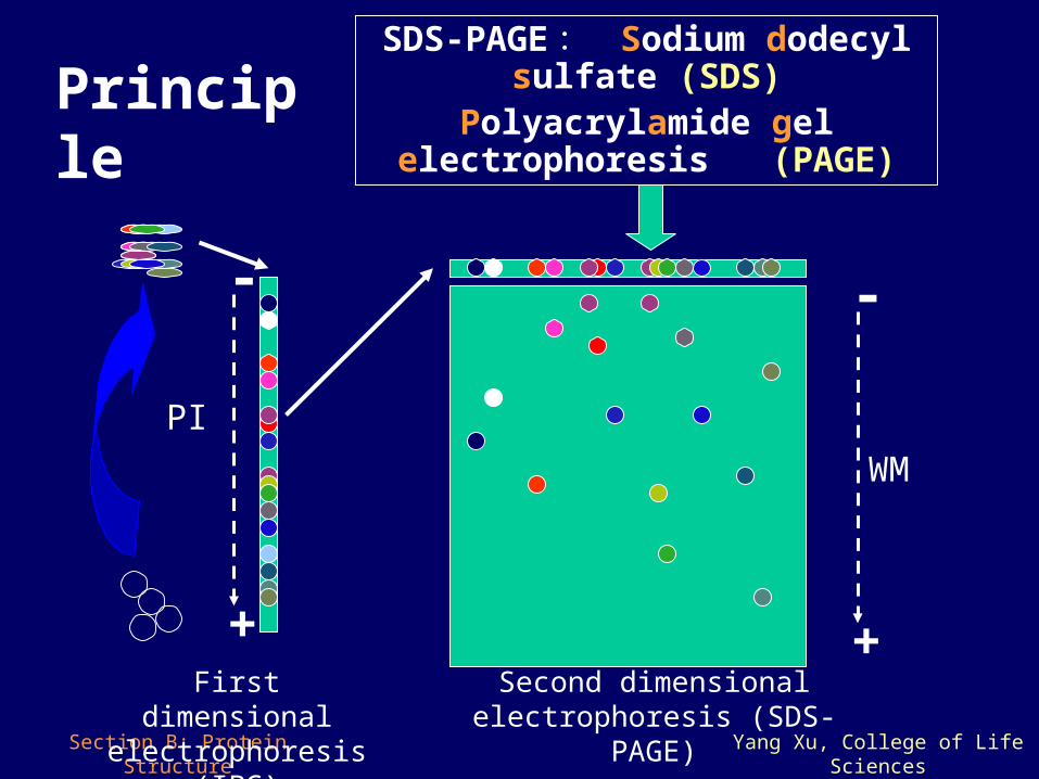
Principle
-
+
PI
-
+
WM
First dimensional electrophoresis (IPG)
Second dimensional electrophoresis (SDS-PAGE)
SDS-PAGE : Sodium dodecyl sulfate (SDS)
Polyacrylamide gel electrophoresis (PAGE)
Section B: Protein Structure Yang Xu, College of Life Sciences
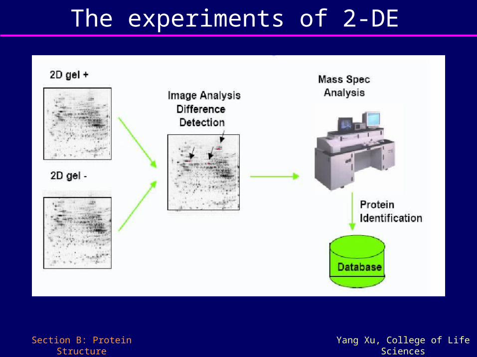
The experiments of 2-DE
Section B: Protein Structure Yang Xu, College of Life Sciences
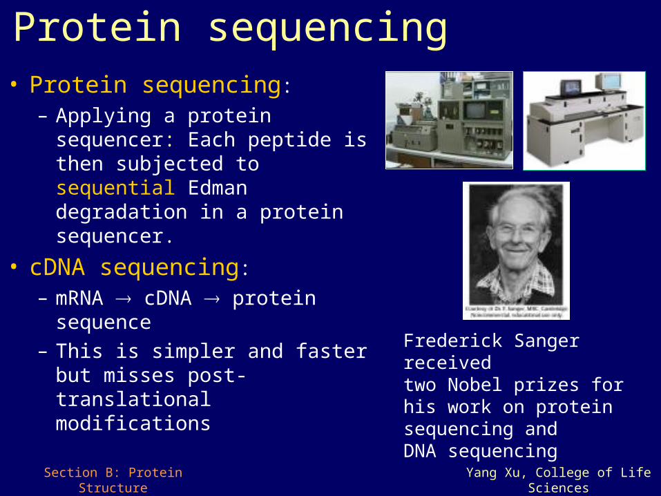
Protein sequencing• Protein sequencing:
– Applying a protein sequencer: Each peptide is then subjected to sequential Edman degradation in a protein sequencer.
• cDNA sequencing:
– mRNA cDNA protein sequence
– This is simpler and faster but misses post-translational modifications
Frederick Sanger received two Nobel prizes for his work on protein sequencing and DNA sequencing

Section B: Protein Structure Yang Xu, College of Life Sciences
That’s all for Section B