seamless, faster navigation for an interactive web experience
TRANSCRIPT

16
G L O B A L M E E T I N G S O F I N T E R E S T
The 12th International Implant Meeting
September 24-26, 2009Verona, Italy
For additional information and registration, pleasecontact Mrs. Angela Negri at +39-0444-913410 oremail her at: [email protected]
European Association For Osseointegration(EAO) 18th Annual Scientific Meeting
September 30 – October 3, 2009Monaco
BIOMET 3i Corporate ForumOctober 1, 2009 5:00-7:00PM
Prosthetic & Surgical Considerations in thePursuit of Optimal Treatment OutcomesModerator: Prof. Michael Matejka
Faculty: Prof. Dr. Markus Hürzeler, Dr. Otto Zuhr
For more information and to register, please visitwww.eao.org
NYU International Implantology Week:Current Concepts In American Dentistry:Advances In Implantology And OralRehabilitation – A Five Day Course
August 3-7, 2009New York UniversityNew York, NY
Faculty: Dr. Dennis Tarnow, Dr. Stephen Chu, Dr. Ziv Mazor, Dr. Christian Stappert, Dr. HaroldBaumgarten
For more information or registration, pleasecontact Ms. Barbara De Wildeman at +34-93-445-81-28 or email at: [email protected]
The 9th BIOMET 3i Iberic Symposium
January 14-16, 2010Palacio de CongresosMadrid, Spain
For more information or registration, pleasecontact Mrs. Olga Blanco at +34-93-470-59-50 oremail at: [email protected]
Australasian Osseointegration Society 7thBiennial Congress
November 4-7, 2009Gold Coast Convention and Exhibition CentreGold Coast, Queensland, Australia
Faculty: Dr. Richard Lazzara, Dr. Ueli Grunder
For further information, please contact theconference managers:Phone: +61 7 3858 5525Fax: +61 7 3858 5499Email: [email protected]: www.aosconference.com.au
Certain, Encode, Gold-Tite, Navigator, OsseoGuard, OSSEOTITE, PREVAIL, Provide, QuickSeat, RegenerOss and ZiReal are registered trademarks and JIRD, Journal of Implant and Reconstructive Dentistry, and NanoTite are trademarks of BIOMET 3i LLC. Endobon is a registered trademark of BIOMET Deutschland GmbH. VICRYL is a registered trademark of Ethicon, Inc. BIOMET is a registered trademark and BIOMET 3i and design are trademarks of BIOMET, Inc. ©2009 BIOMET 3i LLC. All rights reserved.
REV A 08/09
Asia PacificEurope
Seamless, Faster Navigation For An Interactive Web Experience… The Essence of BIOMET 3i’s Re-designed Global Website
BIOMET 3i’s new corporate global website provides users with a moreefficient, informative web experience.
Visit BIOMET 3i at www.biomet3i.com for:
• More Interactivity
• Easier Navigation, Less Time Spent Searching
• A New Modern Look With Authentic Content
• Multilingual Sections/Global Accessibility
• A Seamless Transition From The Website To The InteractiveProduct Catalog To Initiate Dental Product Orders Effortlessly
Seamless, Faster Navigation For An Interactive Web Experience… The Essence of BIOMET 3i’s Re-designed Global Website
O N L I N E A C C E S S
1
Volume 5 / Issue 3 / Fall 2009
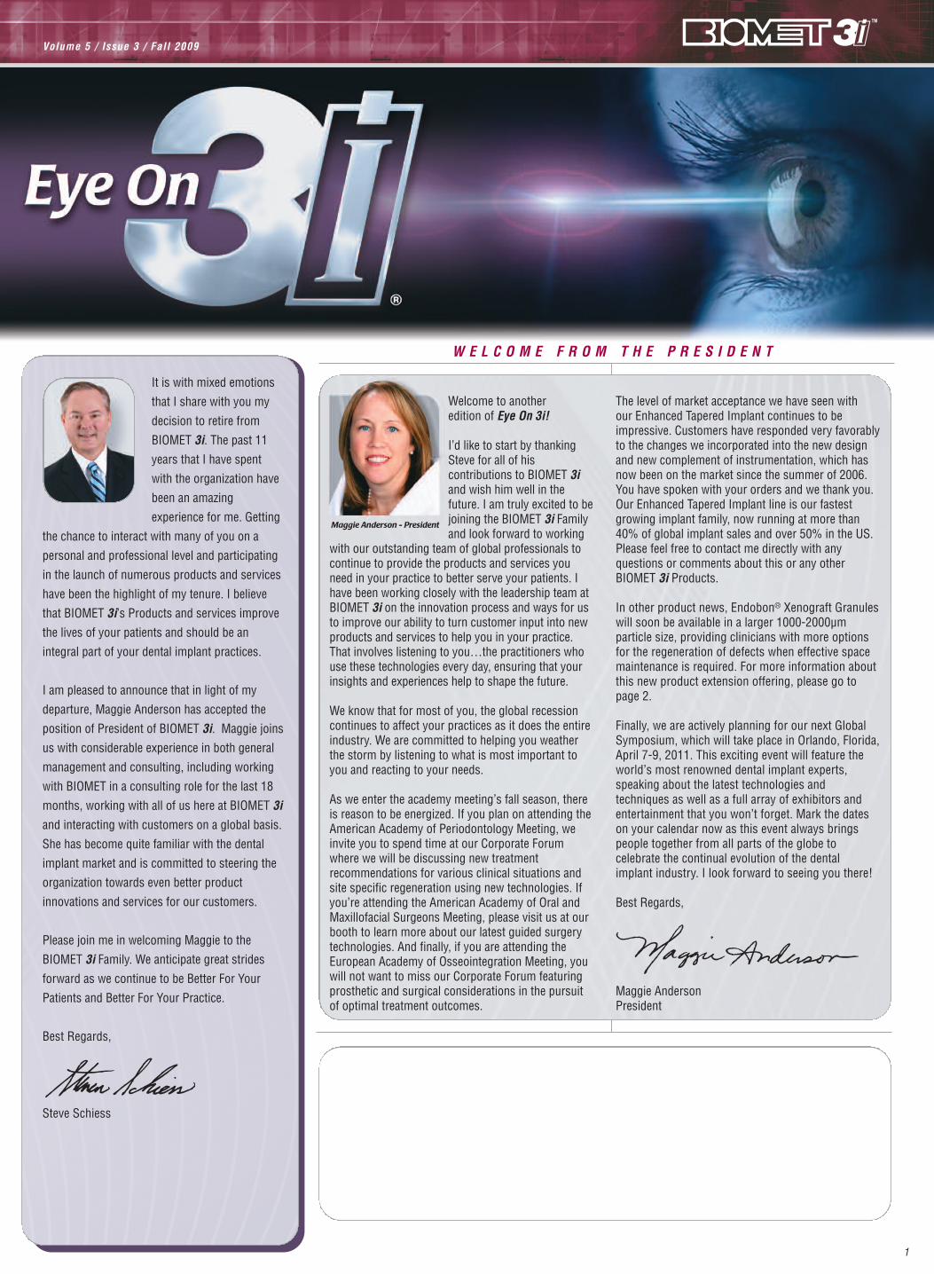
W E L C O M E F R O M T H E P R E S I D E N T
Welcome to another edition of Eye On 3i!
I’d like to start by thankingSteve for all of hiscontributions to BIOMET 3iand wish him well in thefuture. I am truly excited to bejoining the BIOMET 3i Familyand look forward to working
with our outstanding team of global professionals tocontinue to provide the products and services youneed in your practice to better serve your patients. Ihave been working closely with the leadership team atBIOMET 3i on the innovation process and ways for usto improve our ability to turn customer input into newproducts and services to help you in your practice.That involves listening to you…the practitioners whouse these technologies every day, ensuring that yourinsights and experiences help to shape the future.
We know that for most of you, the global recessioncontinues to affect your practices as it does the entireindustry. We are committed to helping you weatherthe storm by listening to what is most important toyou and reacting to your needs.
As we enter the academy meeting’s fall season, thereis reason to be energized. If you plan on attending theAmerican Academy of Periodontology Meeting, weinvite you to spend time at our Corporate Forumwhere we will be discussing new treatmentrecommendations for various clinical situations andsite specific regeneration using new technologies. Ifyou’re attending the American Academy of Oral andMaxillofacial Surgeons Meeting, please visit us at ourbooth to learn more about our latest guided surgerytechnologies. And finally, if you are attending theEuropean Academy of Osseointegration Meeting, youwill not want to miss our Corporate Forum featuringprosthetic and surgical considerations in the pursuitof optimal treatment outcomes.
The level of market acceptance we have seen with our Enhanced Tapered Implant continues to beimpressive. Customers have responded very favorablyto the changes we incorporated into the new designand new complement of instrumentation, which hasnow been on the market since the summer of 2006.You have spoken with your orders and we thank you.Our Enhanced Tapered Implant line is our fastestgrowing implant family, now running at more than40% of global implant sales and over 50% in the US.Please feel free to contact me directly with anyquestions or comments about this or any otherBIOMET 3i Products.
In other product news, Endobon® Xenograft Granuleswill soon be available in a larger 1000-2000µmparticle size, providing clinicians with more optionsfor the regeneration of defects when effective spacemaintenance is required. For more information aboutthis new product extension offering, please go topage 2.
Finally, we are actively planning for our next GlobalSymposium, which will take place in Orlando, Florida,April 7-9, 2011. This exciting event will feature theworld’s most renowned dental implant experts,speaking about the latest technologies andtechniques as well as a full array of exhibitors andentertainment that you won’t forget. Mark the dateson your calendar now as this event always bringspeople together from all parts of the globe tocelebrate the continual evolution of the dental implant industry. I look forward to seeing you there!
Best Regards,
Maggie AndersonPresident
Maggie Anderson - President
It is with mixed emotions
that I share with you my
decision to retire from
BIOMET 3i. The past 11
years that I have spent
with the organization have
been an amazing
experience for me. Getting
the chance to interact with many of you on a
personal and professional level and participating
in the launch of numerous products and services
have been the highlight of my tenure. I believe
that BIOMET 3i’s Products and services improve
the lives of your patients and should be an
integral part of your dental implant practices.
I am pleased to announce that in light of my
departure, Maggie Anderson has accepted the
position of President of BIOMET 3i. Maggie joins
us with considerable experience in both general
management and consulting, including working
with BIOMET in a consulting role for the last 18
months, working with all of us here at BIOMET 3i
and interacting with customers on a global basis.
She has become quite familiar with the dental
implant market and is committed to steering the
organization towards even better product
innovations and services for our customers.
Please join me in welcoming Maggie to the
BIOMET 3i Family. We anticipate great strides
forward as we continue to be Better For Your
Patients and Better For Your Practice.
Best Regards,
Steve Schiess

2
P R O D U C T I N N O V A T I O N S
Please Note: Not all products are available outside the U.S. Please contact your local BIOMET 3i Sales Representative for availability.
Endobon Xenograft Granules will soon beavailable in a 1000-2000µm particle sizeproviding the clinician with more optionsfor the regeneration of defects wheneffective space maintenance is required.
A bovine-derived, hydroxyapatite, graftingmaterial, Endobon Xenograft Granules arefully deproteinated by a two-step, hightemperature manufacturing process forsafety from bacteria, viruses and prions.The osseoconductive properties enablebone to grow directly on the surface andthrough the entire graft. EndobonXenograft Granules can be mixed withother suitable materials (utilized as a graftextender) that have a faster rate ofturnover, if desired.
Endobon Xenograft Granules have:
• Excellent handling characteristics foreasy transfer to the defect site
• Interconnecting micro andmacropores for bony integration,which leads to graft stability andvascular ingrowth
• Two available particle size rangesideal for dental applications: 500-1000µm and 1000-2000µm
With more than 10 years of combined usein oral maxillofacial and orthopedicprocedures, Endobon Xenograft Granulesare indicated for use in a variety of dentaland/or oral surgical procedures including:
• Filling defects after resection,cystectomy, apicoectomy or otherdefects in the alveolar ridge or wall
• Peri-implant defects
• Alveolar ridge augmentation includingaesthetic contouring defects
• Aesthetic contouring defects
• Extraction socket grafting
*The Endobon Xenograft Granules largerparticle size will be available fall 2009.
Endobon Xenograft Granules are manufactured by
BIOMET Orthopaedics Switzerland GmbH.
Providing Clinicians With More Regenerative Options - Introducing A Larger Particle Size For Endobon®
Xenograft Granules*
Endobon Xenograft Granules
Table Of Contents Page
Welcome From The President 1
Product Innovations 2Providing Clinicians With More Regenerative Options - Introducing A Larger Particle Size For Endobon® Xenograft Granules
Say Goodbye To Impression Copings –Introducing The Encode® Impression System
Introducing The BIOMET 3i Torque IndicatingRatchet Wrenches
Point Of View 5Dr. Marcus Dagnelid Shares His Experience With The Encode Impression System
Technology In Motion 6Regeneration Of An Atrophic Partially Edentulous Maxilla: A Case Presentation
Technical Tips 8A Technique To Avoid Tissue Impingement When Using Certain® Provide® Abutments
The Use Of Dedicated Screws For The Encode Zirconia Abutment
Literature Review 10Immediate Provisionalization Of NanoTite™ Implants In Support Of Single-Tooth And Unilateral Restorations: One-Year Interim Report Of A Prospective, Multicenter Study
A Prospective, Multicenter, Randomized-Controlled Five-Year Study of Hybrid And Fully-Etched Implants For The Incidence Of Peri-Implantitis
Sign-up For A Free Subscription To e-JIRD™ Today!
Patient News 12Accelerating Healing With The Use Of NanoTite Implants
Global News 12BIOMET 3i Establishes Direct Operations In Japan And South Korea
Professional Development Corner 13Supporting The Professional Development Of Clinicians
Corporate News 13BIOMET 3i Hosts Charity Build-A-Bike Event
Global Meetings Of Interest 14North America Europe Asia Pacific
Online Access 16

3
P R O D U C T I N N O V A T I O N S
Please Note: Not all products are available outside the U.S. Please contact your local BIOMET 3i Sales Representative for availability.
The Encode Impression System offers a simple way to impress implants and increase productivity in your practice. No extra
components or instrumentation are needed.
The Encode Impression System:
• Working above the gingiva is quicker and helps to increase productivity
• No new tools, multiple parts or pieces are required making impressions simpler and more cost effective
• Results in an aesthetic patient specific abutment available in titanium, gold-colored titanium nitride and zirconia
Try the Encode Impression System and see how simple it is to increase productivity in your practice.
Say Goodbye To Impression Copings –Introducing The Encode® Impression System
Encode Healing Abutment

4
P R O D U C T I N N O V A T I O N S
Please Note: Not all products are available outside the U.S. Please contact your local BIOMET 3i Sales Representative for availability.
Introducing The BIOMET 3i Torque Indicating Ratchet Wrenches*
Restorative Torque Indicating Ratchet Wrench
The new Torque Indicating Ratchet Wrenches from BIOMET 3i
are sleek and easy to use providing a variety of unique user
friendly benefits:
• Convenient - ratchet wrench and torque indicator are in
one device
• Functional - consists of an ISO Latch, which can be used
with most drivers on the market and provides a torque
reading of 0-35Ncm for the Restorative Wrench and
50-90Ncm for the Surgical Wrench
• Minimal Pieces - consists of two pieces for minimal
inventory and ease of disassembly for autoclaving
*The Restorative Torque Indicating Ratchet Wrench will be
available Fall 2009, the Surgical Torque Indicating Ratchet
Wrench will be available early 2010.
For more information, please contact your local BIOMET 3i
Sales Representative.

5
Please Note: Not all products are available outside the U.S. Please contact your local BIOMET 3i Sales Representative for availability.
Marcus Dagnelid, DDS Shares His Experience With The Encode® Impression System
Q. How has this simple Encode Impression Procedure benefittedyour practice?
A. The time savings is really important. Not only does the EncodeImpression Procedure save me valuable chairtime, but thelaboratory work is in many ways simplified. Freeing my dentalassistants from the need to manage an inventory of differentimpression copings is another important benefit and it is great notto have to make any new capital investments.
Q. As a prosthodontist specializing in implant reconstruction, howhas the Encode System benefitted your patients? How is thistechnology better for your practice?
A. Optimally, abutments should be tailored to each and every patientand clinical situation. The Encode System allows me to choosefrom a variety of restorative materials and create a detailed designof individualized abutments. The new zirconia option has alsomeant aesthetic results and greater patient satisfaction.
As a clinician, I appreciate anything that improves my ability tocommunicate with my dental technicians. Also, the time that I saveby using the system allows me to put more effort into optimizingthe prosthetic results before delivery.
Q. Has the use of this simple impression system expanded thenumber of patients to whom you can offer implants?
A. Clinical realities are often different from what you would like themto be. Implant patients differ in age as well as physical and mentalhealth. Also, although the goal is always to place implants in a waythat will optimize the prosthetic result, a patient’s anatomysometimes forces compromises in placement. Differences inimplant angulation can sometimes make it challenging to manageimpression copings. In such difficult situations, the EncodeSystem’s simpler and more reliable impression technique makes it easier for me to treat patients with limited opening or basic gagging reflexes.
Q. In which clinical situations do you choose to use the EncodeSystem?
A. I place Encode Healing Abutments routinely, regardless of the loadingprotocol. I have used the Encode System for both single units and forpartially edentulous situations because that gives me great flexibilityin creating individualized abutments when finalizing my cases.
Q. Where do you find this technology most advantageous to yourpractice?
A. We are part of a large clinic, so every year we treat many patients–both clinic clients and also referrals. Using the Encode System
allows us to use fewer components and save time at each visit. Weappreciate being able to minimize soft-tissue trauma by eliminatingthe use of impression copings. And tailoring the abutments toindividualize the margins and emergence profiles can provide betterfunctional and aesthetic results. Our patient satisfaction is the truetest of our success.
Another important consideration for using fewer components isthat it minimizes errors for us and the GPs who are referringpatients to us. That makes the GPs feel more comfortable aboutoffering implant treatment to more patients.
Q. How has digital dentistry advanced the profession?A. The recent advances in this technology have had an incredible
impact on prosthetic dentistry. In my clinical practice, the relevantsteps in the treatment process are more efficient and precise, andthe quality of our work is higher.
Using Computed Tomography and sophisticated software lets meplan the placement of implants so that the prosthetic outcome canbe optimized. Placing the implants with the help of CT generatedsurgical guides has enabled us to help patients whom might nothave been treated before. These new technologies have taken ourability to deliver functional and aesthetic solutions to an entirelydifferent level. We can now accomplish all this while minimizing theoverall patient treatment time. The impact on our ability to functionas a treatment team is enhanced.
Q. What direction would you like to see BIOMET 3i go with digital dentistry?
A. I am privileged to be working closely with BIOMET 3i on producttesting and evaluation. For the Encode System, the next step will beto further streamline the process of transforming models intofinished abutments. By using the new 3Shape Scanner in thelaboratory to design Encode Lab-Designed Abutments, the need toship articulated models will be eliminated, thus saving both timeand money. Working together with restorative clinicians, dentaltechnicians will gain even more control over the abutment design.
In the future, I hope to take impressions at the healing abutmentlevel in my edentulous cases. That would allow me to deliver afinished CAM StructSURE® Framework without disrupting the softtissue while saving both time and money for me and my patients.
Marcus Dagnelid, DDS (SWEDEN), received his dental degree from theUniversity of Göteborg, Sweden. After graduation he joined the esteemedTeam Dagnelid with Prof. Ingvar Ericsson, Dr. Pelle Peterson, Dr. ChristerDagnelid and Dr. Carl Johan Ivanoff, in Mölndal, Sweden. Recently TeamDagnelid launched a new conference center, the Swedish Academy forAdvanced Clinical Dentistry (SAACD). The goal of the center is to create aunique platform for interactive dental education. Dr. Dagnelid lecturesextensively on guided surgery, prosthetics and implant dentistry with afocus on aesthetics. He is currently attending a postgraduate education inprosthodontics at the University of Malmö, Sweden.
P O I N T O F V I E W

T E C H N O L O G Y
6
Fig. 1
Fig. 3
Fig. 2
Fig. 4
Fig. 5 Fig. 6
Fig. 7 Fig. 8
Fig. 9 Fig. 10
Please Note: Not all products are available outside the U.S. Please contact your local BIOMET 3i Sales Representative for availability.
Regeneration Of An Atrophic Partially E
Patients with periodontally compromised dentitionsor missing teeth often present with less than optimal clinical conditions for implant treatment.Advancements in regenerative materials provideclinicians with a variety of choices for successfully
performing Guided Bone Regeneration (GBR) procedures.Regenerative procedures may be performed in combination with, orprior to, implant therapy to replace the missing hard and soft tissuesfor optimal restoration of function and aesthetics.
The clinical case presentation to follow demonstrates the placement ofa combination of xenograft and allograft regenerative materials, and aresorbable collagen membrane to regenerate an atrophic partiallyedentulous maxilla. In a staged approach, regeneration of the anteriormaxilla followed with subsequent implant therapy to replace thepatient’s missing teeth. The patient desired a fixed restoration toaddress his primary concern of aesthetics.
A 34-year-old male patient presented with an unaesthetic removablepartial denture that replaced missing teeth Nos. 9 and 10 [21 and 22](Fig. 1). Clinical and radiographic findings revealed a large bony defectand loss of the soft tissue (Figs. 2 and 3). A cone beam CT Scan wastaken and the data was converted to SimPlant Software to assess andvirtually treatment plan the clinical situation. Dehiscences on the facialaspect of the alveolus of tooth positions 9 and 10 [21 and 22] werenoted relative to the planned implant positions (Fig. 4). The insufficientlabial/palatal ridge width as noted on the cross sectional images of theCT Scan (Figs. 5 and 6) necessitated regeneration of the site prior toimplant placement. The treatment plan accepted by the patient includeda staged approach to treatment starting with bone grafting, followed byplacement of dental implants, in a two-stage protocol.
GBR and Provisionalization Following administration of local anesthesia, a wide, deep vestibularincision was made on the labial aspect to expose the edentulous ridge.This incision design was chosen to avoid the grafted site, obtainprimary closure and prevent membrane exposure during healing. Asplit-thickness mucoperiosteal flap was reflected 1-2mm apical to thedefect, followed by a full thickness mucoperiosteal flap. Intrasulcularincisions were made around the adjacent teeth to release the flap. Thedefect noted on the CT Scan was confirmed clinically (Fig. 7). Sincethere was insufficient bone volume for immediate implant placement,a decision was made to graft the sites. The periosteum wascompletely debrided and five tenting screws were placed to maintainthe space. An OsseoGuard® Resorbable Collagen Membrane wastrimmed without hydration and placed into the edentulous site overthe tenting screws. Using an explorer, the membrane was pierced inthe area of the two superior-most tenting screws. The membrane wasremoved and a tissue punch was used to create holes in themembrane. The site was decorticated with a round carbide bur (#2).The membrane was fixated with two screws. A mixture of Endobon®
Xenograft Granules and RegenerOss® Allograft Putty was placedunder the membrane until the defect area was filled. The xenograftgranules were chosen for their osseoconductive, space-maintainingproperties; these were combined with the osseoinductive properties of
Robert A. del Castillo, DMD

Y I N M O T I O N
7
Robert A. del Castillo, DMD, received his dental degree and hisCertificate in Periodontics from Tufts University, School of DentalMedicine. He is an Adjunct Professor, Department of Periodontics atTufts University School of Dental Medicine and a guest lecturer atMaryland University Dental School. Dr. del Castillo lectures bothnationally and internationally and has published on regenerative andimplant therapy. Dr. del Castillo maintains a private practice, limited toperiodontics with strong emphasis on implant and regenerativetherapies, in Miami Lakes, Florida.
For more information regarding the RegenerOss® Portfolio of Regenerative Products, please speak with your local BIOMET 3i Representative or visit the BIOMET 3i Website at www.biomet3i.com
Fig. 18
Fig. 11 Fig. 12
Fig. 14
Fig. 15 Fig. 16
Fig. 17
Fig. 13
Fig. 19 Fig. 20
Edentulous Maxilla: A Case Presentation
the allograft material. The membrane was then fixated with the twoinferior-most tenting screws to contain the graft. The soft-tissue flapwas then closed over the grafted site. VICRYL® Rapide 5.0 Sutures(Ethicon, Inc.) were used to secure the periosteum (on the undersideof the flap) to the connective tissue at the base of the vestibule. Theouter aspect of the flap was secured with PDS II (polydioxanone) 6.0Sutures (Ethicon, Inc.) (Figure 9). An Essix retainer was chosen as aprovisional restoration to prevent pressure on the grafted site (Fig.10). The patient was released with post-operative medications andoral hygiene instructions. Periodic post-operative visits progressedwith uneventful healing. A surgical guide was fabricated using thedata obtained from the diagnostic cast.
Implant Placement Seven months post-regeneration of the edentulous site, the patientwas seen for evaluation. The Essix retainer was removed and revealed excellent soft-tissue healing over the grafted site (Fig. 11). A midcrestal incision was made and a full thickness mucoperiostealflap was reflected revealing excellent regeneration of the ridge. Thefixation screws were removed and the surgical guide was placed (Fig. 12). A 2mm diameter trephine bur was advanced through theholes in the guide in the planned position of the implants to obtainhistologic cores. Preparation of the osteotomies continued (Fig. 13)following the manufacturer’s instructions for placement of aNanoTite™ Tapered Certain® PREVAIL® Implant into tooth site No. 9[21] and an OSSEOTITE® Certain 3.25mm Implant into tooth site No.10 [22]. Cover screws were placed into the internal interfaces of theimplants and the soft-tissue flaps were secured with PDS II 6.0Sutures (Fig.14). A periapical radiograph was taken. The Essixretainer was reinserted (Fig. 15). The patient was released with post-operative instructions and medications, and will be seen forreentry in approximately six months.
HistologyThe histology report of the bone in the trephined sections revealedthat dense cortical type bone (B) was present. New trabeculae wereevident (arrows) at one pole of the bony tissue (Fig. 16). In thehistologic section shown in Figure 17, dense cortical bone (B) wasnoted, along with marrow spaces containing blood vessels (v), bonefragments (f) and particulate debris (d). Viable osteocytes filled thebony matrix. At one pole of the trephine (Fig. 18), Endobon® Particles(arrows) were present. The particles are present in the soft tissue andsurround the new trabecular bone (b).
A cone beam CT Scan taken five months post-implant placementconfirmed bone regeneration on the labial aspect of the alveolus(Figs. 19 and 20).

8
T E C H N I C A L T I P S
Please Note: Not all products are available outside the U.S. Please contact your local BIOMET 3i Sales Representative for availability.
A Technique To Avoid Tissue Impingement When Using Certain® Provide® AbutmentsBy Frederick Solomon, DMD, FAGD
The Provide Abutment is designed to be utilized when restorativeclinicians desire a simple implant restorative procedure. Thistechnical tip demonstrates an easy way to deliver and impress theProvide Abutment while avoiding tissue impingement (Fig 1).
Measure intra-orally to ensure there is adequate inter-occlusalspace for fabricating a restoration using the Provide Abutment.Select the proper abutment diameter and height. Snap thematching color Provide Impression Coping onto the ProvideAbutment. Using a diamond bur, open up a vent hole at the top ofthe impression coping to accommodate the Gold-Tite® Screw(IUNIHG) and Large Hex Driver Tip (RASH3N) (Fig 2).
Verify that any plastic debris is removed from the inside of theProvide Abutment to ensure that the screw will seat completely(Fig 3).
Insert the Large Hex Driver Tip into the Gold-Tite Screw and placethese through the impression coping/ abutment assembly (Fig 4and 5). Place the entire assembly into the Certain Implant andverify complete seating (Fig 6).
Using a torque device attached to the Large Hex Driver Tip, tightenthe screw to 20Ncm. Remove the driver tip and block the venthole opening with wax or a temporary filling material. Syringe amedium to heavy body impression material around the ProvideImpression Coping, load the impression tray and seat in themouth. Allow the impression material to set per themanufacturer’s instructions.
Remove the impression from the mouth and place the ProvideSnap Cap on the abutment. Send the case to the laboratory forfabrication of the restoration.
Figure 1 Figure 3
Figure 6
Figure 2
Figure 5Figure 4
Clinical Images Courtesy Of Frederick Solomon, DMD, FAGD, Private Practice In Melrose, MA.

9
T E C H N I C A L T I P S
Please Note: Not all products are available outside the U.S. Please contact your local BIOMET 3i Sales Representative for availability.
The Use Of Dedicated Screws For The Encode® Zirconia Abutment
Encode Zirconia Abutments are packaged with an Encode Zirconia Gold-Tite® Screw (ICZGS) and Encode Zirconia Try-in Screw (ICZTIS). These screwsare designed to be used with these abutments.
The following steps should be considered when placing Encode Zirconia Abutments:
Incorrect
1. The Encode Zirconia Abutment has no retention fingers as is found withother Certain® Abutments (Fig. 1a). To keep the abutment in place andto aide in the handling of the product, place and remove the Encode Zirconia Abutment together with either of the dedicated screws(ICZGS or ICZTIS) and a Large Hex Driver with a narrow shank (Fig. 1b).
3. When delivering the abutment, begin to thread the screw into the analog/implant to prevent the abutment from falling out (Fig. 3a). Avoid any lateral movement of the abutment or driver when placing and removing the Encode Zirconia Abutment. Lateral movement of the abutment or driver/driver tip may damage the abutment.
Note: Use of the Contra Angle Torque Driver (CATD0) may inadvertentlycause lateral forces against the abutment, increasing the chance offracture.
2. Use only the ICZGS or ICZTIS screws (Fig. 2a). Not using the appropriate screw and driver can make handling of the Encode ZirconiaAbutment difficult and increase the risk of product breakage (Fig. 2b).
4. Be sure to insert and remove the abutment vertically (Fig. 4a). Seat thedriver/driver tip completely into the hex of the screw before hand tightening. Position the hex of the abutment into the analog/implantand hand tighten the abutment (Fig. 4b).
5. Confirm proper seating radiographically (Fig. 5a). Seat the driver/drivertip completely into the hex of the screw before applying torque. Thetorque driver tip must be parallel with the access hole during torqueapplication to prevent possible fracture or chipping of the post walls.Torque the abutment screw to 20Ncm using a 0.48” Large Hex DriverTip and a torque device (Fig. 5b). The Restorative Torque Indicator (pictured) is recommended for use with Zirconia Abutments.
SEATEDNOT
SEATED
Incorrect
Fig. 1a Fig. 1b
Fig. 2a Fig. 2b
Fig. 3a Fig. 3b
Fig. 4a Fig. 4b
Fig. 5a
Fig. 5b
EncodeZirconiaAbutment
CertainAbutment

10
L I T E R A T U R E R E V I E W
Pär-Olov Östman, DDS, PhD, MD
Markijan Hupalo, MDSc
Robert del Castillo, DMD
Robert Emery, DDS
Roberto Cocchetto, MD, DDS
Giampaolo Vincenzi, MD, DDS
Barry Wagenberg, DMD
Bruno Vanassche, MD, DDS
Andreas Valentin, DDS, PhD, MSC
Gerard Clausen, BDSc, LDS, MDSc, FRACDS
Paul Hogan, BDS, MDSc, FRACDS
Ronnie Goené, DMD
Christopher Evans, BDSc Hons, MDSc
Tiziano Testori, DDS, MD
Clin Implant Dent Relat Res. 2009 May 7 (8pp.)
[Epub ahead of print]
AbstractBackground: Clinical studies reporting immediate loading of
endosseous implants for edentulous cases and for fixed partial
restorations have been well documented with satisfactory survival
rates. Implants with a recently developed, nanometer-scale surface
topography (NanoTite, BIOMET 3i, Palm Beach Gardens, FL, USA),
created by discrete crystalline depositions (DCD) of calcium phosphate
nano-crystals onto a dual acid-etched (DAE) surface, show enhanced
early fixation in preclinical studies when compared with DAE-surfaced
implants. These outcomes suggest DCD-surfaced implants may be
advantageous for immediate loading approaches.
Objective: The aim of this prospective, multicenter, observational study
is to report clinical outcomes for DCD-surfaced implants placed in
immediate functional support of single- and multi-unit restorations
according to an immediate loading protocol.
Materials and Methods: One hundred eighty-five patients enrolled at 15
international study centers received a total of 335 implants supporting
216 immediate provisionalizations consisting of 128 single-tooth
restorations and 88 fixed restorations. Of the 335 implants, 77% are
located in posterior and 23% in anterior regions with 55.5% of the total
in mandibles and 44.5% in maxillae. Patients were evaluated for
implant mobility, gingival health, symptomatology and radiographic
outcomes.
Results: At the time of this 1-year interim report, a total of 17 failures
have been observed in 11 patients, yielding a cumulative survival rate
of 94.9%.
Conclusion: Relative to other prospective, multicenter studies of
immediately loaded implants with various surface enhancements,
NanoTite Implants perform comparatively well when immediately
provisionalized with single-tooth and fixed restorations.
Immediate Provisionalization Of NanoTite™ Implants In Support Of Single-Tooth And Unilateral Restorations: One-Year Interim Report Of A Prospective, Multicenter Study

11
L I T E R A T U R E R E V I E W
A Prospective, Multicenter, Randomized-Controlled Five-Year Study Of Hybrid And Fully-Etched Implants For The Incidence Of Peri-Implantitis
Lars Zetterqvist, DDS
Sylvan Feldman, DDS
Bruce Rotter, DMD, MS
Giampaolo Vincenzi, MD, DDS
Jan Wennström, DDS
Andrea Chierico, DDS
Renée Stach, DDS
James Kenealy, PhD
Submitted for publication J Perio.
AbstractThe incidence of peri-implantitis has been reported to be as high as
14% and because peri-implantitis can cause progressive bone loss and
is difficult to treat, it often leads to implant failure. Implants with a
roughened collar surface are perceived to be at a higher risk for peri-
implantitis and other mucosal complications. In 1996 the OSSEOTITE®
Implant (BIOMET 3i) was commercially introduced with a “hybrid”
design having a dual acid-etched surface (DAE, OSSEOTITE) extending
the length of the implant from the apex to approximately the third
thread, where a machined surface is present up to the seating surface.
Considerations for potential benefits of extending the DAE surface to
the seating surface led to this prospective randomized-controlled study
designed to assess the risk and incidence of peri-implantitis for fully-
DAE-surfaced implants.
Study implants, fully- DAE-surfaced “test” implants and hybrid-DAE
“control” implants, were placed in a single-stage approach with the
seating surface level with the crestal margin of alveolar bone.
Transmucosal abutments were placed and after two months of healing,
implants were provisionalized with one of each implant type supporting
each prosthesis to ensure that all conditions were consistent between
groups. Final restorations were placed at six months and patients were
followed for five years at annual intervals. Follow-up evaluations
included Sulcus Bleeding Index scores (SBI), probing for suppuration,
assessments for mobility, and periapical radiographs to identify
radiolucencies and crestal bone levels.
One-hundred twelve patients were enrolled and 165 test and 139
control implants were placed supporting 127 prostheses. No
substantial differences in mucosal health outcomes between test and
control groups were observed throughout the 5-year follow-up. For
both groups the bleeding-on-probing scores were no different. There
was one case of peri-implantitis reported over the five years of
observation and this was for a hybrid implant. The condition was
resolved following surgical intervention. Radiographic analyses of
crestal bone regression demonstrate that the mean change from
baseline (provisionalization) is less for test implants in comparison to
control implants (P<.01). The results of this 5-year study do not reflect
increased risk in soft tissue outcomes and peri-implantitis for fully-
DAE-surfaced implants.
Have you heard the buzz about the Journal of Implant and Reconstructive
Dentistry™ (JIRD), a BIOMET 3i Publication? If not, take a moment to visit the
journal online at www.JIRD-online.com and sign-up for e-JIRD, a free
electronic subscription to the journal. The electronic subscription includes the
inaugural issue as well as all upcoming issues. e-JIRD has expanded content
from the printed version as well as interactive elements such as treatment
videos and interviews with leading clinicians in the field of dental implant and
reconstructive dentistry.
Look for the next issue to be published in the Fall of 2009. All e-JIRD
subscribers will be notified via email once the issue is online.
For more information on JIRD, contact your local BIOMET 3i Sales
Representative today.
Sign-up For A Free Subscription To e-JIRD™ Today!

12
BIOMET 3i Establishes Direct Operations In Japan And South Korea
Many of us have patients who, despite everyone's efforts, have hadfailures in maintaining implant-supported fixed dentition. Jo presentedin my practice having lost four out of five implants placed over thepreceding 19 years. Because of that history, she had been encouragedto avoid attempting further implant reconstruction. But unfortunately,her xerostomia (due to Sjögrens Syndrome) made it difficult for her tofunction with a removable partial denture. Instead, she relied upon herremaining teeth for mastication until her left maxillary canine fractured– compromising the long fixed partial denture that it had supported.
At that point, Jo decided she wanted a new implant-supportedrestoration. Comprehensive evaluation indicated that she would need asinus lift/graft in the maxillary left (her right sinus having already beengrafted 14 years previously). We developed a treatment plan that wouldallow us to completely restore her dentition in just over one year.However, Jo had something else in mind: she wanted to be enjoyingher new teeth in time for her 80th birthday celebration and that wasapproaching in a little more than four months.
We chose to place NanoTite Implants due to the enhanced surfacetopography of these implants. After Jo was fully informed about andconsented to an accelerated treatment plan, the fractured canine wasextracted, a sinus lift/graft was performed and five NanoTite Certain®
Implants were placed. Three weeks later, the provisional prosthesissupported by the maxillary right lateral incisor and canine fractured,
complicating the situation. But it proved possible to extract the twohopeless teeth and place an additional implant in the lateral incisor sitein a one-stage protocol. Two months later, the remaining implants wereuncovered, and the definitive restoration was delivered two days beforeJo's big birthday.
I was fortunate to attend the celebration, where Jo happily chewedcelery sticks and enjoyed her birthday cake in the company of friendsand relatives. She was all smiles throughout this special occasion.
BIOMET 3i is pleased to announce that it has established direct operations to serve the Japanese and
Korean markets.
In 1987, BIOMET 3i’s Founders, Dr. Richard Lazzara and engineer Keith Beaty began operations in
the United States focused on offering unique technologies, outstanding customer service and
clinician driven peer-to-peer therapeutic support. This tradition continued in both Japan and Korea
several years later as distribution partners were selected that maintained this approach. The growing
importance of these markets in the global dental implant market, together with BIOMET 3i’s desire to
focus more on the Asian market in general, led to the decision to establish direct operations in these
two regions. This new chapter in BIOMET 3i’s History allows the company to interface directly with
customers and will help the organization better understand the needs and preferences of clinicians
and dental technicians in these regions.
Stephen Wheeler, DDS, is a board-certified oral and maxillofacial surgeonwho received his postgraduate and residency training from the University ofSouthern California, School of Dentistry. Since the early 1980s, he has beenactively involved in various aspects of implant dentistry. He practices inEncinitas, California.
G L O B A L N E W S
P A T I E N T N E W S
Accelerating Healing With The Use Of NanoTite™ ImplantsBy Stephen Wheeler, DDS
Dr. Stephen Wheeler with Jo at her 80th birthday party.

P R O F E S S I O N A L D E V E L O P M E N T C O R N E R
BIOMET 3i is committed to furthering the professional development of
clinicians through consistent investment in dental implant education.
This support includes a variety of education and training programs,
academic scholarships, educational grants and active committee
participation.
In collaboration with
the American
Academy of
Periodontology
Foundation (AAPF),
BIOMET 3i supports
an annual $50,000
fellowship for an
outstanding post-
periodontal candidate.
The Richard J. Lazzara Fellowship in Advanced Implant Surgery is a
12-month award that is intended to help facilitate contemporary
research and clinical experiences for the Fellow.
The AAPF recently awarded the 2009 Richard J. Lazzara Fellowship in
Advanced Implant Surgery to Jonathon A. Waasdorp, DMD, a resident
in the periodontal program at the University of Maryland Dental School,
Baltimore College of Dental Surgery. D. Walter Cohen, DDS, Chairman
of the Foundation’s Lazzara Fellowship Selection Committee believes
that by fostering the development of future academics and researchers,
the Fellowship is helping to stem the pending loss of periodontal
educators as a large number of them approach retirement.
To learn more about the AAPF’s Richard J. Lazzara Fellowship in
Advanced Implant Surgery Fellowship, go to www.perio.org or contact
your BIOMET 3i Representative.
Supporting The Professional Development Of Clinicians
BIOMET 3i hosted the 1st annual Neil Cronin Memorial Build-A-Bike event on Friday,
June 19, 2009 as part of the 2009 North American Sales Meeting held at the Hyatt
Regency Tamaya Resort & Spa in Santa Ana Pueblo, New Mexico.
The event was held in remembrance of Neil Cronin, a 32 year old BIOMET 3i Sales
Representative, who passed away suddenly on February 25, 2009 of a brain aneurysm.
Neil’s energetic and positive nature is remembered by all who interacted with him. To
honor Neil’s memory, 169 BIOMET 3i Team Members participated in a charity Build-A-
Bike Event for children who participate in the E. Rene’ Clark Boys and Girls Club, which
serves the Albuquerque and Rio Rancho area. During the three and a half hour
program, the BIOMET 3i Team Members built 20 bicycles that were then presented to
the children who were in attendance at the event.
BIOMET 3i Hosts Charity Build-A-Bike Event
Dr. Richard J. Lazzara,Co-founder of BIOMET 3i
Neil Cronin12/5/1977 – 2/25/2009
C O R P O R A T E N E W S
13

14
American Academy Of Periodontology (AAP) 95th Annual Meeting
September 12-15, 2009Boston Convention and Exhibition CenterBoston, MA
BIOMET 3i Corporate ForumSeptember 12, 2009Room 204B
Site Specific Regeneration Using NewTechnologiesDr. John Lupovici1:00-1:45PM
New Treatment Protocols For ChallengingClinical Situations: Management Of InfectedSites, Immediate Molar Replacement AndFailed ImplantsDr. Alan Meltzer2:00-2:45PM
For more information, please visit: www.perio.org
New Solutions For Enhanced Aesthetic Restorations
September 30, 2009Hyatt Regency, Philadelphia Philadelphia, PA
Faculty: Dr. Victor Martel
October 7, 2009 Westchester Marriott Tarrytown, NY
Faculty: Dr. George Priest
December 3, 2009Los Angeles Airport Marriott Hotel Los Angeles, CA
Faculty: Dr. Jon Ruel
For more information and to register, please visiteducational opportunities at www.biomet3i.com orcall Professional Education: inside the U.S. 800-717-4143; outside the U.S. +1-561-776-6700
Advanced Aesthetic Provisionalization:Restorative “Hands-On” Learning UsingNew Technologies
October 3, 2009The Institute For Implant And Reconstructive SurgeryPalm Beach Gardens, FL
Faculty: Dr. George Priest
January 30, 2010The Integra InstituteMetairie, LA
Faculty: Dr. George Priest
For more information and to register, please visiteducational opportunities at www.biomet3i.com orcall Professional Education: inside the U.S. 800-717-4143; outside the U.S. +1-561-776-6700
The Third Annual Aiden Morris Memorial Symposium
October 21, 2009The Westin Michigan Avenue ChicagoChicago, IL
Faculty: Dr. Alan Meltzer, Dr. Harold Baumgarten,Dr. Curtis Jansen, Dr. Lee Walker, Ms. Anita Daniels
For more information and to register, please visiteducational opportunities at www.biomet3i.com orcall Professional Education: inside the U.S. 800-717-4143; outside the U.S. +1-561-776-6700
Overcoming Economic Challenges
October 24-25, 2009The Fairmont Washington, DC
November 7-8, 2009The FairmontSan Francisco, CA
Faculty: Ms. Cynthia Bollinger, Dr. Paul Korb, Dr.Jay Malmquist, Dr. Donald Steinberg, Dr. ClarkTaylor, Dr. Lee Walker, Ms. Kathi Carlson, Ms. Nicole Dorsey
For more information, please visit: www.idia.org
Practical And Predictable Solutions ForSuccessful Soft Tissue Aesthetics AroundImplants - A Hands-On Program
November 20-21, 2009 (Friday - Saturday)New York University College Of Dentistry, Linhart Continuing Dental Education ProgramIn Conjunction With BIOMET 3iNYU College Of DentistryNew York, NY
Faculty: Dr. Michael Sonick, Dr. Christian Stappert
For more information, please visit:www.nyu.edu/dental/ce
The “Surgical” Implant Coordinator:Enhancing The Success Of Your ImplantPractice
January 22-23, 2010Sheraton Fisherman's Wharf HotelSan Francisco, CA
Faculty: Ms. Cynthia Bollinger, Dr. Robert Blackwell
For more information and to register, please visiteducational opportunities at www.biomet3i.com orcall Professional Education: inside the U.S. 800-717-4143; outside the U.S. +1-561-776-6700
Yankee Dental Congress
January 27-31, 2010Boston Convention and Exhibition CenterBoston, MA
Esthetics and Implant Innovations
Faculty: Dr. Dennis Tarnow
For more information, please visit:www.yankeedental.com
North America
G L O B A L M E E T I N G

15
Implant Surgery: Fundamentals To Details
February 15-20, 2010 (Monday – Saturday)The Anspach EffortPalm Beach Gardens, FL
Faculty: Dr. Robert London
For more information, please contact: The LondonInstitute 206-683-0655
Expanding The Role Of The “Surgical” Implant Coordinator: Taking Your ImplantPractice To The Next Level
February 19-20, 2010 Manhattan Beach MarriottManhattan Beach, CA
Faculty: Ms. Cynthia Bollinger, Ms. Heather Collins
For more information and to register, please visiteducational opportunities at www.biomet3i.com orcall Professional Education: inside the U.S. 800-717-4143; outside the U.S. +1-561-776-6700
American Prosthodontic Society (APS)82nd Annual Meeting
February 27, 2010Westin River NorthChicago, IL
Faculty: Dr. Dean Vafiadis
For more information, please visit:www.prostho.org/
Implant Therapy: A Systematic ApproachProviding The Clinician With A PredictableAnd Systematic Approach ThatDemonstrates Successful Treatments InVarious Situations
September 18, 2009McGill University, Montreal, Québec
Faculty: Dr. Samer Abi Nader
For more information contact Susan Young at(514) 398-7203 ext 0315 orhttp://www.mcgill.ca/dentistry/conted
The Effects Of CAD/CAM And CT GuidedTechnology With Patient Care
September 25, 2009Le Westin, Montreal, Québec
Faculty: Dr. Paulino Castellon
For more information visithttp://cardp.ca/english/montreal-09
Restoring The Multiple Unit Implant Case
October 1, 2009Montreal, Québec
Faculty: Dr. Robert Blackwell
For more information contact BIOMET 3i Canadaat 514.956.9843 ext 2221
AAOMS / CAOMS
October 14-17, 2009Toronto, Ontario
For more information visit www.caoms.com
Ontario Society Of Periodontists
October 17, 2009Toronto, Ontario
Faculty: Dr. Jonathan Schofield
Contact Information www.osp.on.ca
Prosthodontic Review
December 4, 2009Toronto, Ontario
Faculty: Dr. Izchak Barzilay, Dr. Effrat Habsha, Dr. John Zarb
For more information contact Danielle Kontos [email protected] or visitwww.buildyoursmile.com
Résultats esthétiques et prévisibles pour lescas complexes en implantologie : De laplanification à la restauration
February 20, 2010University of Montreal, Montreal, Quebec
Faculty: Dr. Denis Gosselin, Dr. Jean-FrancoisAubin, Dr. Louis Drouin, Dr. Gaetan Noreau, Dr.Louis De Koninck
For more information visit: http://www.fdc-umontreal.ca
G S O F I N T E R E S T